150 questions, one per key fact, picked by working through every practice question on the full question page (learning questions left out; the partly-CP1 "Borderline" questions counted at low weight) and both 2026 CP1 histology TA reviews (Harlem and Great Falls), and keeping the facts that answer the most of them, either directly or by crossing out the impossible choices. Histology is mostly recognition, so the key facts carry what each structure looks like and how to tell it from its look-alikes. After you answer, the Key fact box states the whole fact, What it unlocks shows how the fact is usually tested, what it lets you eliminate and links to the straight-recall questions it answers, and Slides shows where the lectures and labs teach it. Questions it helps you answer, at the bottom, holds the full practice and TA questions you might not expect it to answer (vignettes and micrographs that never name it, answers reached by elimination, traps, questions asked from the other direction), answerable in place, each with how it follows from the main question.
150 key questions737 of 747 practice questions unlocked120 of 120 TA review questions unlocked5 sections150 with slides
The heart: wall layers, Purkinje fibers, valves and cardiac skeleton
32 questions · unlock 141 of 142
Heart wall layers and pericardium
1unlocks 3TA ×11 not obvious
A shard of glass enters the anterior chest and passes into the right ventricular cavity. In what order does it cross the coverings and wall of the heart?
WhyThe fibrous pericardium is outermost; its inner face is lined by the parietal serous layer, then comes the pericardial cavity, then the visceral serous layer (= epicardium) on the heart, then myocardium and last the endocardium facing the lumen. Any sequence that puts visceral before parietal, or endocardium before myocardium, is wrong.
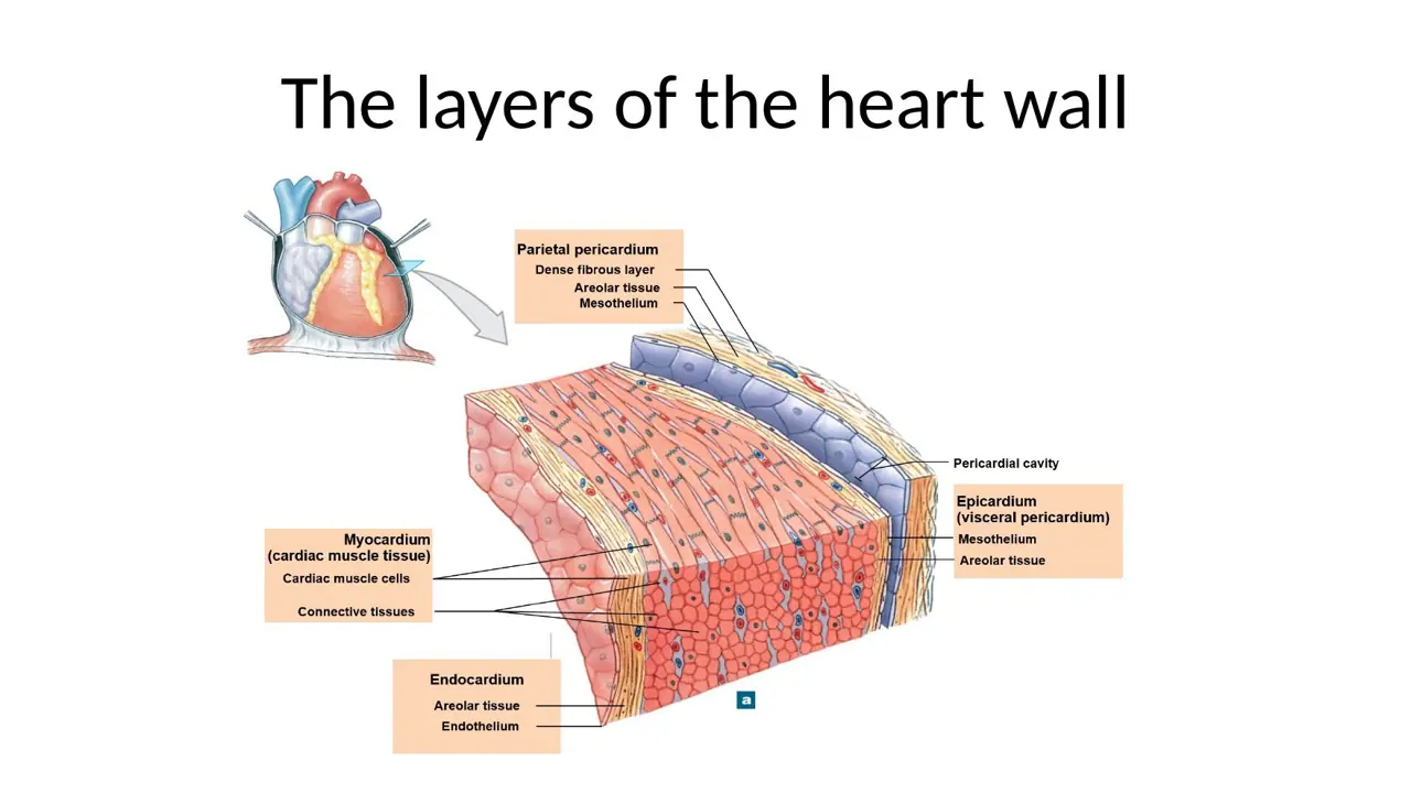
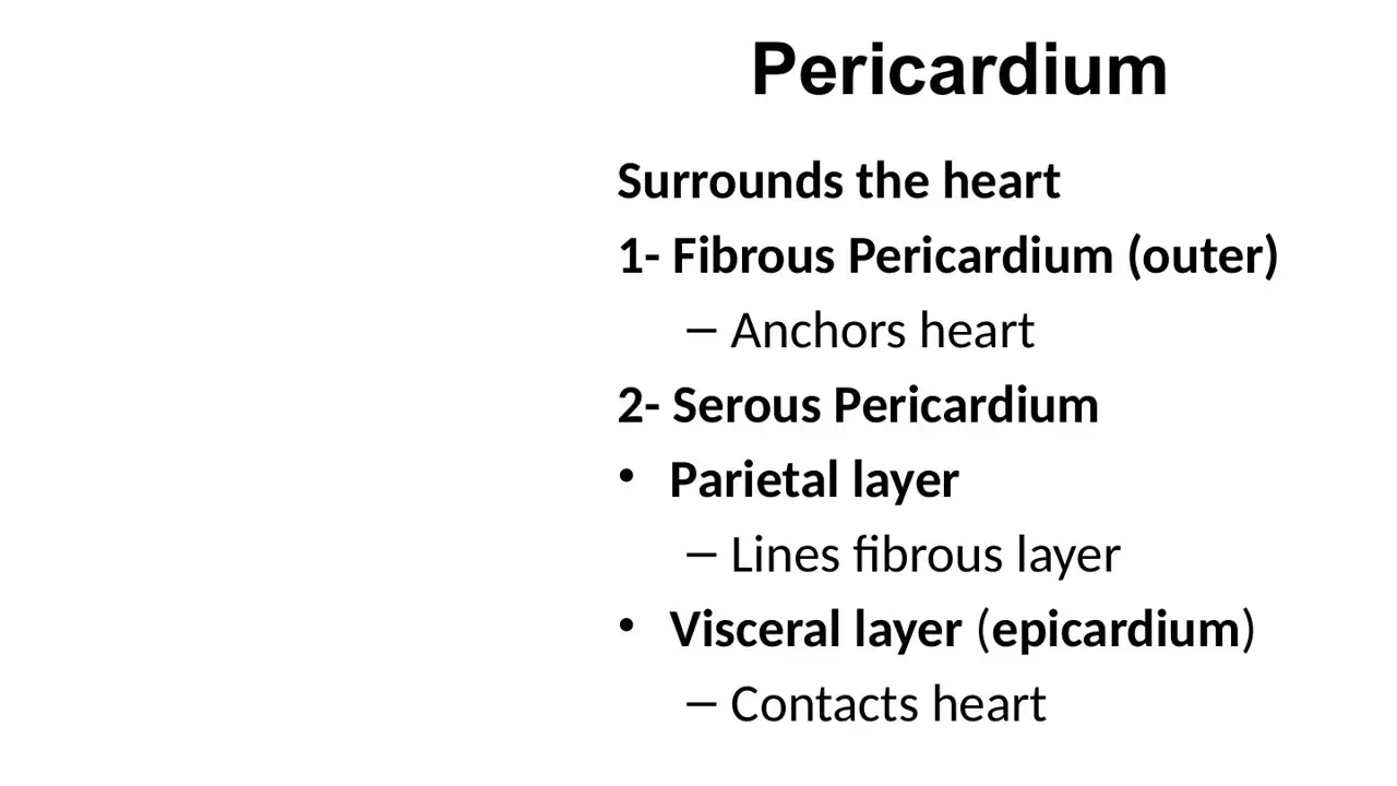
Key factOutside → inside: fibrous pericardium (tough, anchors the heart) → parietal serous pericardium (mesothelium lining the fibrous sac) → pericardial cavity (thin film of serous fluid) → visceral serous pericardium = epicardium (mesothelium) → subepicardial loose CT and fat (coronary vessels) → myocardium → endocardium (faces the chamber lumen). Lumen outward (heart wall only): endocardium → myocardium → epicardium.
q90: the printed source key put adipose tissue most superficial (1) and fibrous pericardium 2. The page key note and the lecture (F0718:22: epicardium = mesothelium over loose CT with fat) put the epicardial fat deep to the serous pericardium: fibrous → serous → adipose → myocardium → endocardium. Follow the key note.
What it unlocks: 4 questions
The 1 question you might not expect it to answer is in the dropdown at the bottom.
How it shows up
Ordering drag-and-drop: inner → outer (endocardium, myocardium, visceral pericardium/epicardium, parietal pericardium); TA version from the chamber lumen toward the pericardial cavity
Penetrating-trauma vignette: order the knife or glass passes from the fibrous pericardium to the endocardium
Superficial → deep ordering that adds "adipose tissue": epicardial fat sits under the serous (visceral) mesothelium and above the myocardium
What it lets you eliminate
Visceral pericardium and epicardium are the same layer: never parietal after visceral going inward
Endocardium is always the last (innermost) layer; myocardium is always in the middle
Straight-recall questions it answers (3)
TAH14 Starting from the chamber lumen and moving outward toward the pericardial cavity, place in order: epicardium, endocardium, myocardium. → Endocardium → myocardium → epicardium
Key check: Key order (superficial→deep): Adipose tissue 1, Fibrous pericardium 2, Serous pericardium 3, Myocardium 4, Endocardium 5. 'Adipose tissue' as most superficial fits only the mediastinal/pericardial fat outside the fibrous pericardium (as drawn on the TA's slide-22 figure). Epicardial (subepicardial) fat, which carries the coronary vessels, lies deep to the visceral serous pericardium, i.e. between serous pericardium and myocardium (Fibrous → Serous → Adipose → Myocardium → Endocardium). — Confirmed on re-check; correct answer: Fibrous pericardium (1) -> Serous pericardium (2) -> Adipose tissue (3) -> Myocardium (4) -> Endocardium (5)
Key check: The key puts adipose tissue outermost, but the epicardial fat carrying the coronary vessels lies deep to the visceral serous pericardium, just above the myocardium. Answer used: Fibrous pericardium 1; Serous pericardium 2; Adipose tissue 3; Myocardium 4; Endocardium 5
WhyFrom outside in lie the fibrous pericardium, the serous pericardium (parietal layer, cavity, visceral mesothelium), subepicardial adipose tissue, myocardium, then endocardium lining the chambers.
How it follows from the main questionThe fat that belongs to the heart wall is epicardial: under the serous (visceral) mesothelium, above muscle → fibrous → serous → adipose → myocardium → endocardium. Putting adipose first (outermost) is the trap.
Fluid that accumulates in the pericardial cavity lies directly between which two layers?
A. Myocardium and endocardium
B. Parietal serous pericardium and visceral serous pericardium (epicardium)
C. Fibrous pericardium and parietal pleura
D. Epicardium and myocardium
E. Fibrous pericardium and parietal serous pericardium
AnswerB. Parietal serous pericardium and visceral serous pericardium (epicardium)
WhyThe pericardial cavity is the space between the parietal serous layer (lining the fibrous sac) and the visceral serous layer, which is the mesothelial surface of the epicardium. The fibrous pericardium lies outside the parietal layer, so fluid or blood in the cavity never touches it.
Key factPericardial cavity = space between the parietal and visceral layers of the serous pericardium; holds a thin film of lubricating fluid (made by the mesothelium) that reduces friction.
Heart side = visceral serous pericardium = epicardium → blood or fluid in the cavity is in direct contact with the epicardium
Outer side = parietal serous layer lining the fibrous pericardium (the fibrous layer itself does not touch the fluid)
Myocardium and endocardium never face the cavity; the pleura belongs to the lungs
What it unlocks: 4 questions
No surprises here: every question it answers asks the fact straight out.
How it shows up
Autopsy follow-up: "400 mL of blood in the pericardial cavity is in direct contact with..." → epicardium (distractors endocardium, fibrous pericardium, myocardium, parietal pleura)
"The pericardial space is lined by which layers?" → visceral and parietal pericardium
Short recall: "Blood in the pericardial cavity would be in contact with which of the following?"
What it lets you eliminate
Fibrous pericardium: outside the parietal serous layer, so not in contact
Lecture: pericardial cavity lies between parietal and visceral layers and holds pericardial fluidH12A Circulatory I lecture (Dr Bruno) · slide 15Balloon-and-fist diagram of visceral pericardium, pericardial cavity and parietal pericardiumCirculatory lab 2026 · slide 8Lecture: fibrous pericardium (outer) and serous pericardium, parietal and visceral (epicardium)H12A Circulatory I lecture (Dr Bruno) · slide 14
3unlocks 41 not obvious
Two pericardial disorders both restrict ventricular filling. Which statement is correct?
A. Rupture of an infarct drains blood into the pleural cavity, causing tamponade
B. Constrictive pericarditis is primarily a disease of the endocardium
C. Constrictive pericarditis results from thinning of the fibrous pericardium
D. In cardiac tamponade, blood or fluid in the pericardial cavity compresses the heart
E. In cardiac tamponade, blood collects between the endocardium and the myocardium
AnswerD. In cardiac tamponade, blood or fluid in the pericardial cavity compresses the heart
WhyTamponade = blood/fluid in the pericardial cavity (e.g., hemopericardium from a ruptured infarct) that the inelastic fibrous pericardium cannot accommodate, so the heart is compressed and cannot fill. Constrictive pericarditis is a disease of the pericardium, which becomes thick, fibrotic and calcified, not thin.
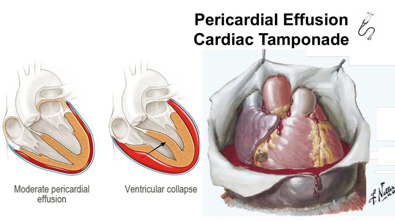
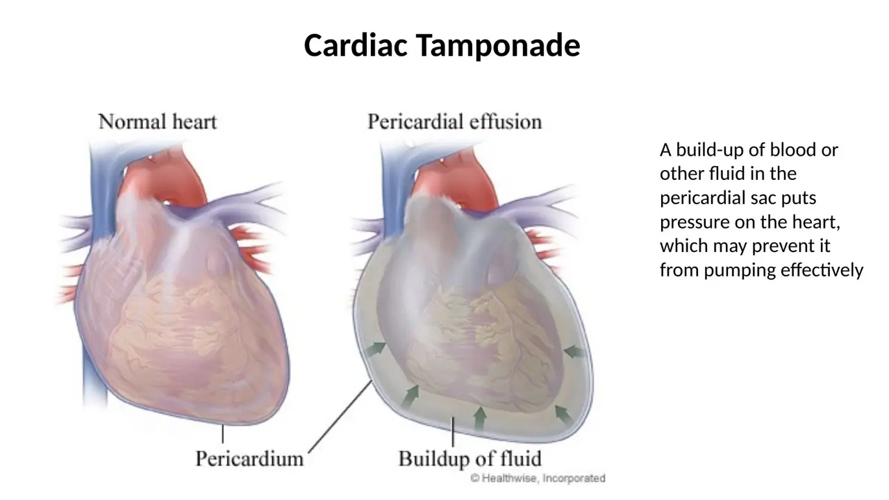
Key factCardiac tamponade: blood or fluid builds up in the pericardial cavity (e.g., hemopericardium when a softened infarct ruptures, usually within ~3 weeks of an MI) → rigid fibrous pericardium cannot stretch → heart compressed → ↓ ventricular filling → hypotension, tachycardia, shock (pulsus paradoxus). Constrictive pericarditis: inflammation of the pericardium (e.g., spread of tuberculosis) heals with thickening, fibrosis and calcification that obliterates the cavity and encases the heart → restricted filling → right-sided failure signs (hepatomegaly, ascites, leg edema) and a friction rub.
The Lippincott explanation for q1/q32 says blood fills the "peritoneal cavity (hemopericardium)"; that is a typo for the pericardial cavity.
What it unlocks: 4 questions
The 1 question you might not expect it to answer is in the dropdown at the bottom.
How it shows up
Classic vignette (repeated 3 times): 2 weeks after a massive MI, BP 80/40, pulse 100, death; autopsy shows a left ventricular rupture → cause of death = cardiac tamponade (distractors dissecting aortic aneurysm, pulmonary edema, pulmonary thromboembolism, septic shock)
2026 lab: cardiac tamponade, fluid or blood in the pericardial sac compressing the heartCirculatory lab 2026 · slide 11Lecture: pericardial effusion progressing to cardiac tamponade with ventricular collapseH12A Circulatory I lecture (Dr Bruno) · slide 172026 lab: constrictive pericarditis, inflamed pericardium encasing the heartCirculatory lab 2026 · slide 13
Questions it helps you answer (1)
Practice Q1Vignette never names it
Two weeks after suffering a massive heart attack, a 54-year-old man presents to the emergency room complaining of sharp pain on the left side of his chest. On physical examination, the patient is apprehensive and sweating. His blood pressure is 80/40 mm Hg, and the pulse rate is 100 per minute. The patient expires shortly after admission. At autopsy, the left ventricle shows a rupture in the myocardial wall. What was the most likely cause of the patient’s death?
(A) Cardiac tamponade
(B) Dissecting aortic aneurysm
(C) Pulmonary edema
(D) Pulmonary thromboembolism
(E) Septic shock
AnswerA. Cardiac tamponade
WhyRupture of a softened infarct floods the pericardial cavity with blood (hemopericardium), compressing the heart and blocking ventricular filling, which is cardiac tamponade.
Explanation from the source
Rupture of a myocardial infarct causes blood to fill the peritoneal cavity (hemopericardium). This medical catastrophe restricts the motion of the heart and is referred to as cardiac tamponade. When it happens, it typically occurs within the first 3 weeks following myocardial infarction. Pulsus paradoxus (>10 mm Hg drop in arterial blood pressure with inspiration) is commonly observed in patients with hemopericardium. Although aortic dissections (choice B) can break through to the pericardium, they do not rupture the heart wall. None of the other cardiovascular diseases are associated with rupture of the heart wall and cardiac tamponade. Keywords: Hemopericardium, cardiac tamponade
How it follows from the main questionRuptured ventricle bleeds into the pericardial cavity (hemopericardium) → the stiff sac compresses the heart → no filling, hypotension, tachycardia = cardiac tamponade. Not aortic dissection, pulmonary edema, embolism or sepsis. Also asked as Q32, Q89.
A low-power H&E section through the atrioventricular junction shows (1) a pale outer zone of adipose tissue holding the coronary sinus and a branch of the circumflex artery, (2) a thick, dense, dark-pink mass of interlacing muscle, and (3) a thin lining facing the ventricular lumen from which the mitral valve projects. Zone (1) is the:
A. Epicardium
B. Myocardium
C. Fibrous skeleton
D. Subendocardium
E. Endocardium
AnswerA. Epicardium
WhyThe pale, fat-rich outer zone carrying the coronary vessels is the epicardium; the thick dark muscle mass is the myocardium, and the thin luminal lining that gives rise to the valve is the endocardium. The fibrous skeleton is dense CT at the valve base, not an outer fatty layer.
Key factLow-power heart wall, lumen outward:
Endocardium = thin pale lining on the chamber (lumen) side; the valves are folds of it
Epicardium = pale outer layer of loose CT and fat holding the coronary arteries, coronary sinus and veins, covered by mesothelium
2026 lab right-ventricle section: arrow at the pale outer surface = epicardium; asterisk in the thick muscle = myocardium; arrowhead at the luminal surface = endocardium.
q2: one older review keyed "Endo, Myo and Pericardium"; the heart-wall layer is the epicardium (visceral pericardium), as in Dr Bruno's key.
What it unlocks: 13 questions
No surprises here: every question it answers asks the fact straight out.
How it shows up
"Identify the 3 layers of the heart" on a gross slice or low-power section (free response, labels A/B/C in any order)
"Which annotation indicates the epicardium? arrowhead / asterisk / arrow" (2026 lab and TA)
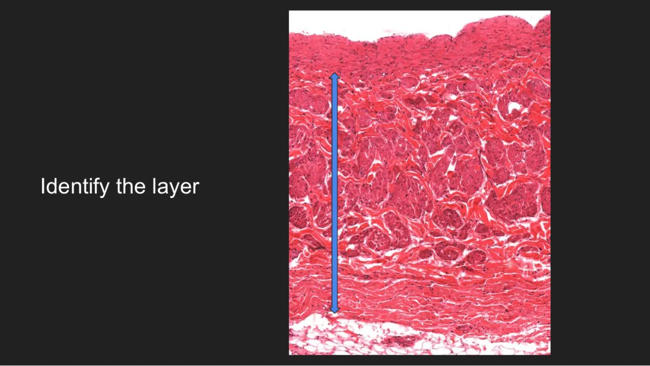
"Identify what the blue label is covering" (myocardium) and Lippincott autopsy "Identify the indicated layer" (thick middle layer = myocardium)
TA: "Identify A and C", then "Identify A, B and C again" on the AV-junction section; "What layer do the heart valves belong to?" → endocardium
What it lets you eliminate
A zone with fat or large vessels is never endocardium or subendocardium
The bulk of the wall is always myocardium
The surface facing the chamber, where valves attach, is endocardium
Straight-recall questions it answers (13)
TAG30 Identify A and C (low-power section through the atrioventricular junction: A on the outer surface, C in the thick ventricular wall). → A = epicardium; C = myocardium
TAG31 Identify A, B and C again (same low-power heart section). → A = endocardium; B = myocardium; C = epicardium
TAG32 What is the Arrow indicating? → B. Epicardium
TA slide: labelled micrographs of epicardium (fat, vessel), myocardium and endocardiumHarlem TA review 2026 · slide 372026 lab diagram: parietal pericardium, pericardial cavity, epicardium, myocardium, endocardiumCirculatory lab 2026 · slide 3
Epicardium
5unlocks 1TA ×2
A layer of the heart wall is also called the visceral pericardium. Which feature is found in this layer but NOT in the endocardium?
A. A simple squamous surface layer
B. Purkinje fibers in its deepest part
C. Mesothelium over loose connective tissue with adipose tissue, coronary vessels and autonomic nerves
D. Scattered smooth muscle cells beneath an endothelium
E. Intercalated discs
AnswerC. Mesothelium over loose connective tissue with adipose tissue, coronary vessels and autonomic nerves
WhyThe epicardium (visceral pericardium) is mesothelium on loose CT with fat, coronary vessels and nerves. Both layers have a simple squamous surface, so A does not discriminate; Purkinje fibers and subendothelial smooth muscle belong to the endocardium; intercalated discs belong to the myocardium.
Key factEpicardium (outer layer) = visceral layer of serous pericardium:
Fibrous pericardium and Purkinje fibers are not lined by mesothelium
Used together with
Myocardium (cardiac muscle) on H&E; Endocardium: three sublayers; Valve histology: endocardial fold, avascular dense CT
Straight-recall questions it answers (3)
TAH2 During examination of a cardiac histology specimen, a student identifies the outermost layer of the heart wall. Which of the following best describes the tissue compositi… → D. Simple squamous mesothelium with underlying loose connective tissue containing fat, blood vessels, and nerves
TAH13 Match each cardiac structure with its most characteristic histologic feature: (A) endocardium, (B) myocardium, (C) epicardium, (D) heart valve. Features: 1. mesothelium a… → 1 → epicardium; 2 → myocardium; 3 → endocardium; 4 → heart valve
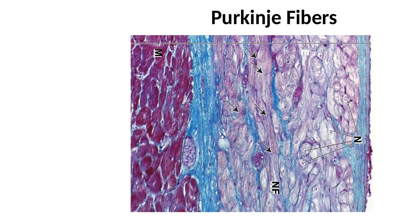
Lecture: epicardium = visceral pericardium, mesothelium over loose CT with fat, vessels, nervesH12A Circulatory I lecture (Dr Bruno) · slide 22Junqueira 11-4: epicardium with mesothelium, CT, nerve (N) and fat (F) over myocardium (M)Junqueira’s Basic Histology, fig 11-4TA summary of endocardium, epicardium, myocardium and Purkinje fiber featuresGreat Falls TA review 2026 · slide 84
6unlocks 12TA ×2
A section of atrial wall shows, from the free surface inward: a single row of flattened cells, a zone of loose connective tissue containing an oval bundle of wavy fibers and a few adipocytes, then cross-cut striated muscle bundles. The surface cells plus the loose connective tissue zone are the:
A. Endocardium
B. Subendocardium
C. Visceral pleura
D. Epicardium
E. Fibrous pericardium
AnswerD. Epicardium
WhyFlat mesothelium over loose CT with a nerve and fat, resting directly on myocardium, is the epicardium. The endocardium faces a chamber with no fat or nerves beneath it; the fibrous pericardium is dense CT not attached to the muscle; pleura covers the lung.
Key factEpicardium micrograph (lab labels Mes / CT / N / F / Ep / M):
Mes = thin wavy row of flat mesothelial cells on the free surface (faces the pericardial cavity)
CT = pale loose connective tissue; N = autonomic nerve; F = fat (adipocytes = empty white rings; can be abundant, with large coronary vessels, M/A/F labels)
M = cardiac muscle (myocardium) directly below, with intercalated discs and gap junctions
Rule: fat, nerves or a big vessel under the surface layer → epicardium, not endocardium.
TAG35: the TA answer slide reads "Myocardium - B, Epicardium - A", but on the image A sits in the muscle bundles (M) and B in the connective tissue beside the Ep bracket, so A = myocardium and B = epicardium. Learn the layer by its features, not the TA letter key.
What it unlocks: 14 questions
No surprises here: every question it answers asks the fact straight out.
How it shows up
"Which layer of the heart is shown?" / "Identify the overall tissue" / "The figure mostly represents" (epicardium; often labelled M = mesothelium, A = adipose, F = fibro-elastic)
Lab colour labels on the atrial epicardium image: green = mesothelium, red = epicardium, purple = myocardium; "What is B, D, C, A?" → nerve, fat, epicardium, mesothelium (serous pericardium)
Matching descriptions to letters: mesothelium makes lubricant fluid; CT with fat, vessels, nerves; layer with gap-junction junctional complexes = myocardium
TA "Identify A & B" (myocardium, epicardium, adipose tissue, DICT); Lippincott "Identify the indicated zone" (epicardium vs fibrous pericardium, visceral pleura)
What it lets you eliminate
Visceral pleura: lung, not heart; fibrous pericardium / DICT: dense, no adipocytes, not on muscle
"Adipose tissue" is a component; the layer name is epicardium
Used together with
Nerve vs vessel in the epicardium; Gap junctions spread the impulse
Straight-recall questions it answers (14)
TAG34 Identify the overall tissue (heart micrograph). → A. Epicardium
TAG35 Identify A and B (heart wall micrograph). Choices: myocardium, epicardium, adipose tissue, DICT. → A = myocardium; B = epicardium
2026 lab: atrial epicardium labelled Mes, CT, N (autonomic nerve), F (fat), Ep and MCirculatory lab 2026 · slide 102026 lab: epicardium with coronary artery branch in fat; M = mesothelium, A = adiposeCirculatory lab 2026 · slide 9Lecture: epicardium micrograph labelled Mes, CT, N, F and Ep over myocardium (M)H12A Circulatory I lecture (Dr Bruno) · slide 21
7unlocks 3
In epicardial connective tissue, a circled structure is a solid oval bundle of pale, wavy fibers with many elongated nuclei; it has no lumen and no red blood cells. Beside it is a vessel with a thick muscular wall around a small round lumen. The circled structure is a(n):
A. Lymphatic vessel
B. Arteriole
C. Nerve
D. Venule
E. Cardiac muscle bundle
AnswerC. Nerve
WhyA solid bundle of wavy fibers with elongated (Schwann cell) nuclei and no lumen is an autonomic nerve. The arteriole is the thick-walled vessel beside it; venules and lymphatics both have a lumen, and cardiac muscle is striated and lies below in the myocardium.
Key factThe epicardium carries the heart's nerves and vessels:
Nerve = solid oval bundle, pale wavy fibers, elongated nuclei, no lumen, no RBCs (sympathetic, parasympathetic and visceral afferent fibers)
Cardiac muscle = striated fibers below, in the myocardium
What it unlocks: 3 questions
No surprises here: every question it answers asks the fact straight out.
How it shows up
Lippincott myxoma vignette: "The epicardium ... identify the structure indicated by the circle" (arteriole, cardiac muscle, connective tissue, nerve, venule)
Lab "Blue?" arrow on the bundle labelled N → autonomic nerve
Labelling the epicardium image: B = nerve, D = fat
What it lets you eliminate
Anything with a lumen or red cells is a vessel, not a nerve
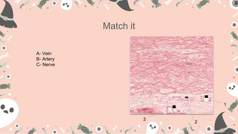
2026 lab: atrial epicardium labelled Mes, CT, N (autonomic nerve), F (fat), Ep and MCirculatory lab 2026 · slide 10Neurovascular bundle: muscular artery, thin-walled vein and solid nerve with no lumenCourse lecture — neurovascular bundle: muscular artery (small round lumen, thick wall, wavy internal elastic lamina), vein (large irregular lumen, thin wall) and nerve (solid, no lumen)
8unlocks 31 not obvious
During coronary artery bypass surgery, the main coronary arteries are found and grafted in which layer of the heart wall?
A. Fibrous pericardium
B. Epicardium
C. Endocardium
D. Endomysium
E. Myocardium
AnswerB. Epicardium
WhyThe main coronary arteries and cardiac veins travel in the subepicardial fat of the epicardium; only smaller branches penetrate the myocardium. The endomysium carries capillaries, not the main vessels, and the fibrous pericardium is the outer sac, not the heart wall.
Key factLeft and right coronary arteries arise from the aortic sinuses (elastic recoil of the aorta drives blood into them between contractions) and run with the cardiac veins and nerves in the epicardium (subepicardial loose CT and fat); their branches then penetrate and supply the myocardium. A thick-walled muscular vessel in epicardial fat = branch of a coronary artery (not a cardiac vein, the coronary sinus or the internal thoracic artery).
What it unlocks: 3 questions
The 1 question you might not expect it to answer is in the dropdown at the bottom.
How it shows up
Dr Bruno lecture quiz Q2: "Where are the main blood vessels which supply the heart located?" (epicardium, pericardium, myocardium, endocardium, endomysium)
"Which layer of the heart contains the coronary arteries?"
Lippincott myxoma vignette: "the structure within the circle represents a branch of which blood vessel?" → coronary artery
What it lets you eliminate
Endomysium: capillaries only; pericardium: the sac, not the wall
Cardiac vein / coronary sinus: thin-walled venous channels, not a muscular artery
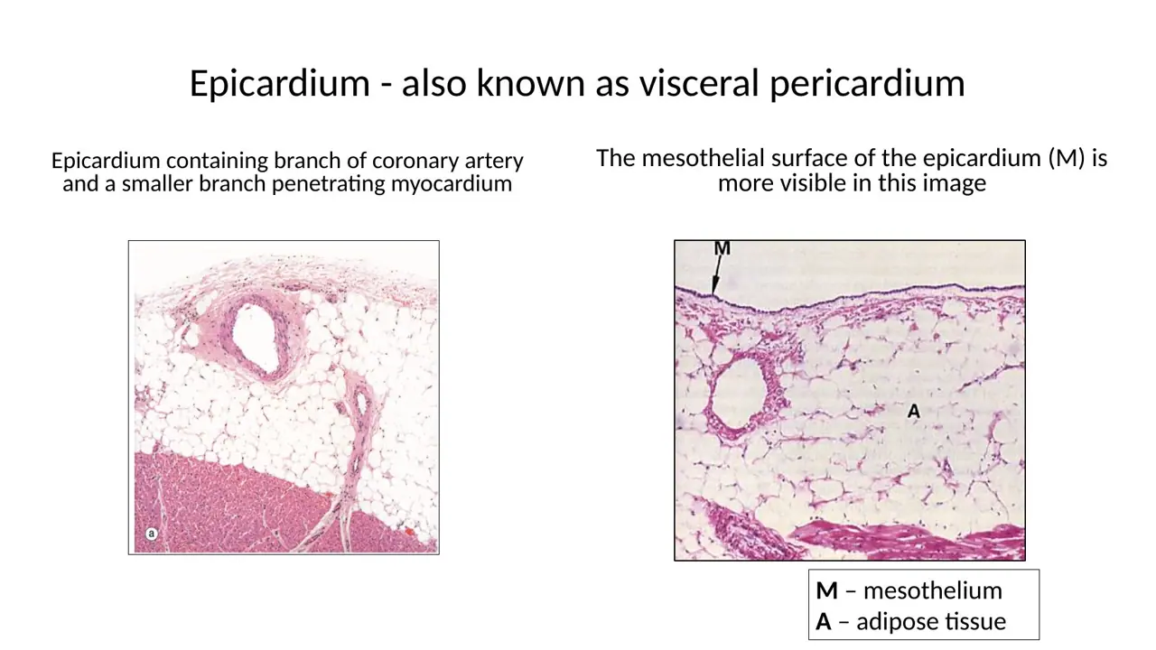
2026 lab: epicardium with coronary artery branch in fat; M = mesothelium, A = adiposeCirculatory lab 2026 · slide 9Lecture: left and right coronary arteries arise at the aortic sinusesH12A Circulatory I lecture (Dr Bruno) · slide 59
Questions it helps you answer (1)
Practice Q36Vignette never names it
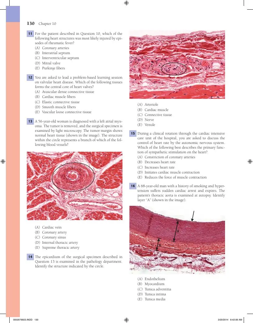
A 56-year-old woman is diagnosed with a left atrial myxoma. The tumor is removed, and the surgical specimen is examined by light microscopy. The tumor margin shows normal heart tissue (shown in the image). The structure within the circle represents a branch of which of the following blood vessels?
(A) Cardiac vein
(B) Coronary artery
(C) Coronary sinus
(D) Internal thoracic artery
(E) Supreme thoracic artery
Answer(B) Coronary artery
WhyThe thick-walled vessel with concentric smooth muscle in the heart wall is a branch of the coronary arteries, which arise from the aortic sinuses and supply the myocardium.
Explanation from the source
The answer is B: Coronary artery. The photomicrograph shows an arteriole and an accompanying venule embedded in adipose connective tissue. The heart receives arterial blood supply from the right and left coronary arteries— branches off the initial part of the ascending aorta. The coronary arteries give rise to numerous branches that travel within the adipose tissue of the epicardium near the surface of the heart. Cardiac veins (choice A) accompany branches of the coronary arteries. These veins drain to the coronary sinus (choice C) on the posterior wall of the heart, which, in turn, empties into the right atrium. None of the other blood vessels supply the heart. Keywords: Arteries, coronary arteries
How it follows from the main questionThick muscular wall, small round lumen = artery branch; arteries in epicardial fat are branches of the coronary arteries. Cardiac veins and the coronary sinus are thin-walled; thoracic arteries never enter the wall.
Slide (1)Circulatory lab 2026 · slide 9
Myocardium and intercalated discs
9unlocks 112 not obvious
A high-power H&E field shows branching striated fibers, each cell with one (occasionally two) large oval central nucleus surrounded by a pale zone, dark transverse step-like lines where cells meet end to end, and many capillaries between fibers. This field is:
A. Subendocardium with Purkinje fibers
B. Epicardium
C. Skeletal muscle
D. Smooth muscle of a vessel media
E. Myocardium
AnswerE. Myocardium
WhyBranching striated fibers with 1–2 central nuclei, a perinuclear clear zone and intercalated discs = cardiac muscle = myocardium. Skeletal muscle has many peripheral nuclei and no discs; Purkinje fibers are much larger and paler; smooth muscle is not striated.
Key factCardiac muscle (myocardium) on LM:
branching, cross-striated fibers
one or two oval central nuclei with a pale perinuclear zone; golden-brown lipofuscin granules at the nuclear poles = residues of lysosomal digestion ("wear and tear" pigment)
intercalated discs = dark transverse, step-like lines at cell-to-cell junctions (not node of Ranvier, perimysium, endomysium or lipofuscin)
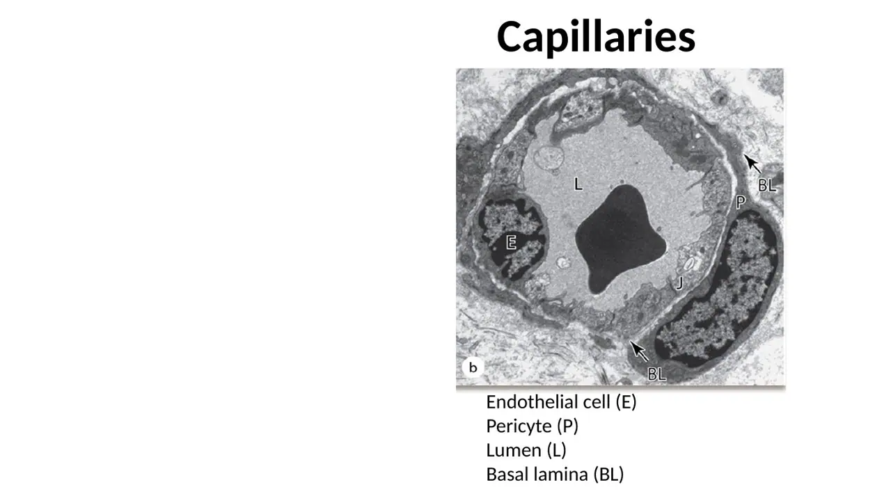
thin endomysium rich in capillaries (capillary = one endothelial cell around a lumen about one RBC wide, ± pericyte)
Myocardium = the muscular middle layer of the wall: thickest layer, concentric/spiral sheets, thicker in ventricles.
What it unlocks: 11 questions
The 2 questions you might not expect it to answer are in the dropdown at the bottom.
How it shows up
"Identify (this layer)" on a field of striated fibers: epicardium / endocardium / myocardium (sometimes with subendocardium and skeletal muscle as options)
2026 lab: "Are central nuclei, branching myofibers and intercalated discs visible?" and "At higher magnification, what features are discernible?"; "Describe the nuclei of cardiac myocytes"
Arrow on a dark transverse line: node of Ranvier / perimysium / intercalated disc / endomysium / lipofuscin
"Which layer contains the muscular wall of the heart?"; capillaries between cross-cut fibers (capillary vs internal elastic lamina, endothelial cell, elastic artery)
What it lets you eliminate
Many peripheral nuclei → skeletal; no striations, cigar nuclei → smooth; large pale cells → Purkinje
Golden-brown perinuclear granules are lipofuscin, not intercalated discs
Lecture: myocardium, branching striated fibers with central nucleiH12A Circulatory I lecture (Dr Bruno) · slide 27Lecture: intercalated discs as dark transverse lines between cardiac muscle cellsH12A Circulatory I lecture (Dr Bruno) · slide 28Lecture diagram: branched cardiac muscle cells with central nuclei and intercalated discsH12A Circulatory I lecture (Dr Bruno) · slide 30
Questions it helps you answer (2)
Practice Q354Looks like another topic
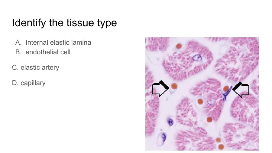
Identify the tissue type
A. Internal elastic lamina
B. endothelial cell
C. elastic artery
D. capillary
AnswerD. capillary
WhyEach marked vessel is a capillary: a single thin endothelial cell around a lumen just wide enough for one red blood cell, lying between cardiac muscle fibres.
Explanation from the source
D. capillary- ID the pericyte around it and thin Thin cell layer around the vessel
How it follows from the main questionLooks like a vessel question, but it is the capillary-rich endomysium of myocardium: a lumen one RBC wide ringed by one endothelial cell = capillary. No elastic lamina, not an artery.
Slide (1)H12B Circulatory II lecture (Dr Bruno) · slide 14Practice Q107Trap: the obvious answer is wrong
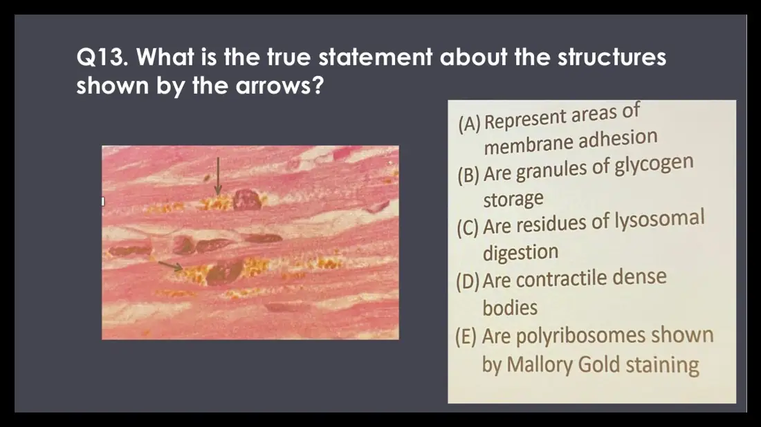
Q13. What is the true statement about the structures shown by the arrows?
(A) Represent areas of membrane adhesion
(B) Are granules of glycogen storage
(C) Are residues of lysosomal digestion
(D) Are contractile dense bodies
(E) Are polyribosomes shown by Mallory Gold staining
AnswerC. Are residues of lysosomal digestion
WhyPerinuclear golden-brown lipofuscin granules are residual bodies of lysosomal digestion, undegradable lipid-protein debris that accumulates in long-lived cardiomyocytes with age.
How it follows from the main questionGolden-brown granules at the nuclear poles are lipofuscin = residues of lysosomal digestion (wear-and-tear pigment). The perinuclear zone also holds glycogen, but glycogen does not appear as golden-brown granules.
10unlocks 6
On a routine H&E section, which feature distinguishes cardiac muscle from skeletal muscle?
A. Fibers made of separate cells joined end to end at intercalated discs
B. A rich capillary network in the connective tissue
C. Cross-striations with alternating A and I bands
D. Sarcomeres between adjacent Z lines
E. Thin filaments that contain actin
AnswerA. Fibers made of separate cells joined end to end at intercalated discs
WhyA cardiac "fiber" is a chain of separate cells joined end to end at intercalated discs; a skeletal fiber is one long multinucleated cell. Striations, A/I bands, sarcomeres, actin and capillaries are shared by both, so they cannot discriminate.
Key factIntercalated discs uniquely identify cardiac muscle: fibers = many cells in series joined end to end (branched; 1–2 central nuclei). Skeletal: one huge multinucleated cell, many peripheral nuclei, no discs. Smooth: spindle cells, one central cigar-shaped nucleus, no striations, no discs. Shared by cardiac and skeletal (never the answer): cross-striations, A and I bands, sarcomeres between Z lines, actin thin filaments, sarcolemma, capillary network. If asked what "uniquely identifies" cardiac muscle, answer intercalated discs.
What it unlocks: 6 questions
No surprises here: every question it answers asks the fact straight out.
How it shows up
2026 lab (patient who died of an arrhythmia): feature that distinguishes cardiac from skeletal muscle on H&E → "cells in series joined by end-to-end connections" / "fibers composed of multiple cells" / "fibers formed by adhesion of multiple cells"
Lecture: intercalated discs as dark transverse lines between cardiac muscle cellsH12A Circulatory I lecture (Dr Bruno) · slide 28Junqueira 10-16: cardiac muscle with central nuclei and intercalated discs (diagram, LM, EM)Junqueira’s Basic Histology, fig 10-16
11unlocks 5TA ×11 not obvious
An antibody against which structure would NOT label the intercalated discs between two cardiomyocytes?
A. Integrin-based hemidesmosomes
B. Desmocollin
C. Desmoglein
D. Cadherin of the fascia adherens
E. Connexons of gap junctions
AnswerA. Integrin-based hemidesmosomes
WhyIntercalated discs hold fasciae adherentes (cadherin; anchor actin), desmosomes (desmoglein + desmocollin; anchor intermediate filaments) and gap junctions (connexons). Hemidesmosomes bind a cell to the basal lamina, not cell to cell, so they are absent.
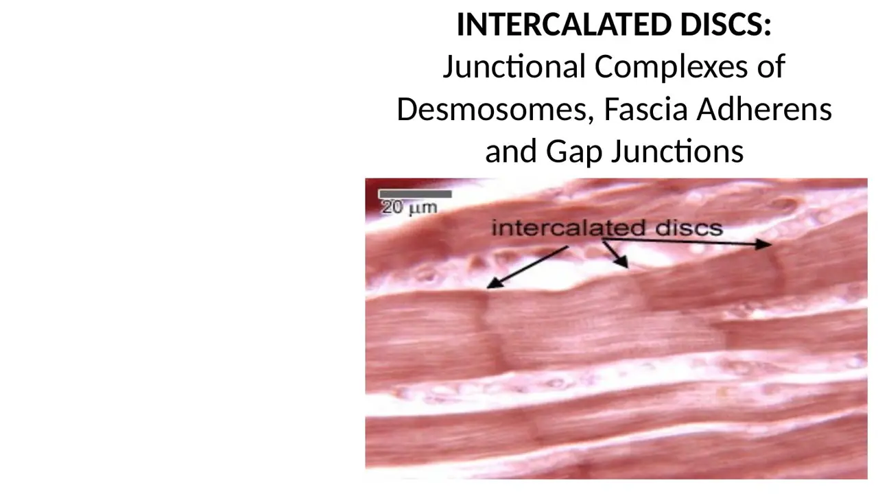
Key factIntercalated disc = junctional complex of three junctions:
Fascia adherens: cadherins; anchors actin thin filaments of the end sarcomere
Desmosome (macula adherens): desmoglein + desmocollin; anchors intermediate filaments; holds cells together during contraction
Gap junction: connexons; electrical coupling
NOT present: hemidesmosomes (cell to basal lamina), zonula occludens (tight junction of epithelia), focal adhesions. (Zona fasciculata is adrenal cortex, not a junction.)
What it unlocks: 6 questions
The 1 question you might not expect it to answer is in the dropdown at the bottom.
How it shows up
2026 interactive session: "The structures indicated by arrows contain a number of constituents, EXCEPT" (hemidesmosomes vs fascia adherens/cadherin, desmoglein, desmocollin, connexons)
"What junctional complexes are involved in the myocardium?" → gap junction, fascia adherens, desmosomes; "What type of cellular complexes are found within the myocardium?" → gap junctions
Arrow on intercalated discs: "a site where multiple junctional complexes are present"
What it lets you eliminate
Hemidesmosomes, zonula occludens/tight junctions, focal adhesions, zona fasciculata
Any option list containing a hemidesmosome or a zonula occludens is not the intercalated-disc trio
Straight-recall questions it answers (5)
TAG24 High-magnification histology of cardiac muscle tissue adjacent to the valve base demonstrates branching striated fibers with centrally located nuclei and intercalated dis… → C. Desmosomes
Lecture diagram: intercalated disc with desmosomes and gap junctionsH12A Circulatory I lecture (Dr Bruno) · slide 292026 session: fascia adherens, desmosome and gap junction (EM and diagrams)Circulatory interactive session 2026 · slide 12Lecture: intercalated discs as dark transverse lines between cardiac muscle cellsH12A Circulatory I lecture (Dr Bruno) · slide 28
Questions it helps you answer (1)
Practice Q86Trap: the obvious answer is wrong
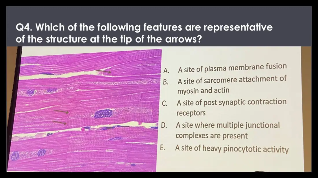
Q4. Which of the following features are representative of the structure at the tip of the arrows?
A. A site of plasma membrane fusion
B. A site of sarcomere attachment of myosin and actin
C. A site of post synaptic contraction receptors
D. A site where multiple junctional complexes are present
E. A site of heavy pinocytotic activity
AnswerD. A site where multiple junctional complexes are present
WhyThe dark step-like transverse lines are intercalated discs, where fasciae adherentes, desmosomes and gap junctions together form a junctional complex between cardiomyocytes.
How it follows from the main questionThe lines are intercalated discs = a site where multiple junctional complexes are present (fascia adherens, desmosomes, gap junctions). Only actin anchors there, not myosin, and the membranes never fuse.
Slide (1)H12A Circulatory I lecture (Dr Bruno) · slide 28
12unlocks 5
A TEM of muscle shows a stepwise dense junction crossing the myofibrils. In its transverse segments, a broad fuzzy dense plaque receives the thin filaments of the last sarcomere; in its longitudinal segments the two membranes are closely apposed. The broad transverse plaque is the:
A. Hemidesmosome
B. Zonula occludens
C. Z disc
D. Fascia adherens
E. Gap junction
AnswerD. Fascia adherens
WhyIn the step-like intercalated disc, transverse segments carry the broad fascia adherens (where actin of the terminal sarcomere inserts) plus discrete desmosomes; longitudinal segments carry the gap junctions. A Z disc lies inside a cell, not at the boundary between two cells.
Key factIntercalated disc on EM: a dark step-like line joining two cells end to end (seeing it = cardiac muscle).
Transverse part (across the fiber): fascia adherens (broad, fuzzy; thin filaments of the end sarcomere insert; only in cardiac muscle) + desmosomes (small discrete dense plaques)
Longitudinal/lateral part (parallel to myofibrils): gap junctions (narrow, closely apposed membranes)
Skeletal muscle EM: regular sarcomeres with no stepwise cell-to-cell junction.
q79: the printed answer list swaps the last two arrows; the page key note uses top = gap junction, middle = fascia adherens, bottom = desmosome.
What it unlocks: 5 questions
No surprises here: every question it answers asks the fact straight out.
How it shows up
TEM with a letter F on the fuzzy transverse plaque: "What is F? Think back to cell junctions" → fascia adherens (only in cardiac muscle)
"What type of muscle is this?" on the same TEM → cardiac (because of the disc)
Diagram with three arrows: identify gap junction, fascia adherens, desmosome
Side-by-side EM: skeletal (regular bands, T tubules) vs cardiac (dark jagged intercalated disc)
What it lets you eliminate
Z disc: inside the cell; hemidesmosome and zonula occludens: not in the disc
2026 session TEM: step-like disc, fascia adherens and desmosome transverse, gap junction lateralCirculatory interactive session 2026 · slide 112026 session: fascia adherens, desmosome and gap junction (EM and diagrams)Circulatory interactive session 2026 · slide 12
13unlocks 4
A mutation that sharply reduces connexin in ventricular myocytes would most directly impair which function?
A. Attachment of cells to the basal lamina
B. Anchoring of actin filaments to the cell membrane
C. Generation of the pacemaker potential
D. Resisting mechanical separation of cells during contraction
E. Cell-to-cell spread of the action potential through low-resistance channels
AnswerE. Cell-to-cell spread of the action potential through low-resistance channels
WhyGap junctions (connexons) are low-resistance ionic channels that pass depolarization from cell to cell, so the myocardium contracts as a coordinated functional syncytium. A = hemidesmosome, B = fascia adherens, C = SA node, D = desmosome.
Key factGap junctions = the only intercalated-disc junction that carries electrical signals: connexon channels → low-resistance pathways ("electrical synapses", ionic continuity) → rapid impulse spread → coordinated contraction. Not electrical: fascia adherens and macula adherens/desmosome (adhesion), endomysium (CT), sarcomere (contraction), T tubules and SR (Ca2+ handling inside one cell). If both "intercalated discs" and "gap junctions" are offered for the channel itself, pick gap junctions.
What it unlocks: 4 questions
No surprises here: every question it answers asks the fact straight out.
How it shows up
2026 lab (asked 3 ways): "Which structure transmits / distributes electrochemical signals between cardiac myocytes to ensure a normal heart rhythm?" (endomysium, fascia adherens, gap junctions, macula adherens, sarcomere)
Physiology-style: "the low-resistance pathways between myocardial cells that allow the spread of action potentials" (gap junctions vs T tubules, SR, intercalated disks, mitochondria)
Matching: "this layer contains junctional complexes; the gap junctions are electrical synapses" → myocardium
What it lets you eliminate
Fascia adherens and macula adherens are adhesive junctions
T tubules and sarcoplasmic reticulum work inside a single cell
Lecture diagram: intercalated disc with desmosomes and gap junctionsH12A Circulatory I lecture (Dr Bruno) · slide 292026 session TEM: step-like disc, fascia adherens and desmosome transverse, gap junction lateralCirculatory interactive session 2026 · slide 11
Endocardium and Purkinje fibers
14unlocks 1TA ×1
Which description fits the innermost layer of the heart wall?
A. Simple cuboidal epithelium on a thick basement membrane
B. Spiral sheets of cardiac muscle joined by intercalated discs
C. Endothelium over a connective tissue layer with scattered smooth muscle, then a deep subendocardial layer that may contain conducting fibers
D. Mesothelium over loose connective tissue with fat and autonomic nerves
E. Masses of dense irregular connective tissue that separate the atria from the ventricles
AnswerC. Endothelium over a connective tissue layer with scattered smooth muscle, then a deep subendocardial layer that may contain conducting fibers
WhyThe endocardium = endothelium + middle fibroelastic CT with smooth muscle + deep subendocardium continuous with the myocardial CT that holds the Purkinje fibers. B = myocardium, D = epicardium, E = cardiac skeleton.
Key factEndocardium (inner layer, continuous with the intima of the great vessels):
endothelium (simple squamous) lining the chamber
middle layer of fibroelastic CT with scattered smooth muscle
deep subendocardium (subendocardial layer) of looser CT continuous with the myocardium's CT; holds veins, nerves and the Purkinje fibers (ventricles)
Heart valves are folds of the endocardium. Matching tags: mesothelium + fat/nerves = epicardium; spiral layers, intercalated discs, gap junctions = myocardium; endothelium + subendocardial conducting fibers = endocardium; masses of DICT = cardiac skeleton; large glycogen-rich subendocardial cells = Purkinje fibers.
What it unlocks: 2 questions
No surprises here: every question it answers asks the fact straight out.
How it shows up
Matching statements to epicardium / myocardium / endocardium / subendocardium ("the deepest layer of endocardial CT containing Purkinje fibers is sometimes called..." → subendocardium)
TA matching descriptors: inner endothelium + subendothelial CT; CT with adipocytes under mesothelium; large glycogen-rich subendocardial cells; masses of DICT between atria and ventricles
Epicardium composition (visceral pericardium); Myocardium (cardiac muscle) on H&E; Purkinje fiber vs contractile cardiomyocyte; Cardiac skeleton: dense irregular CT around the valves
Straight-recall questions it answers (2)
TAG29 Match the descriptors: (A) inner layer of endothelium and subendothelial CT; (B) CT with adipocytes covered by mesothelium; (C) large diameter, abundant glycogen, in the … → Epicardium = B; Purkinje fibers = C; endocardium = A; cardiac skeleton = D
2026 lab: endocardium = endothelium, CT with smooth muscle, deep subendocardiumCirculatory lab 2026 · slide 25TA: endocardium = endothelium, middle CT, deep CT continuous with the myocardiumGreat Falls TA review 2026 · slide 73Lecture: endocardium = endothelium, connective tissue, subendocardial conducting fibersH12A Circulatory I lecture (Dr Bruno) · slide 34
15unlocks 11
A thin layer of flat cells lines a ventricular lumen. Beneath it is pale loose connective tissue with scattered smooth muscle cells and no adipocytes or nerve bundles, then a band of large pale fibers, then dark-pink cardiac muscle. The flat lining cells and the connective tissue under them form the:
A. Cardiac skeleton
B. Endocardium
C. Myocardium
D. Visceral pericardium
E. Epicardium
AnswerB. Endocardium
WhyFlat cells facing the chamber lumen over loose CT with smooth muscle and no fat or nerves, above a band of pale Purkinje fibers, are the endocardium (endothelium + subendothelial CT + subendocardium). The visceral pericardium (epicardium) has mesothelium facing the pericardial cavity over fat.
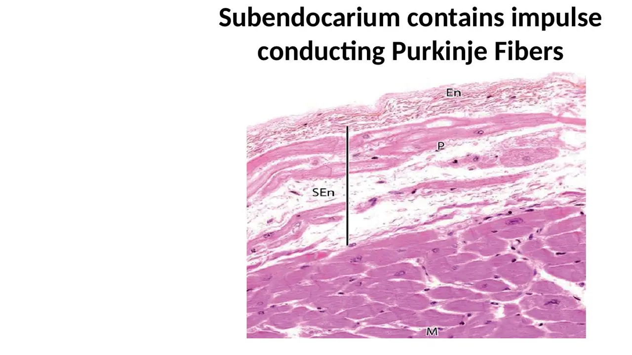
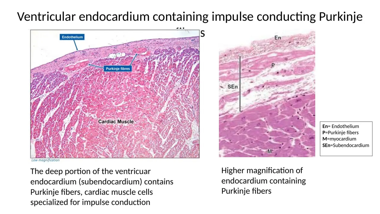
Key factEndocardium micrograph (2026 lab labels): En = endothelium (thin flat cells on the luminal surface) → pale subendothelial CT with smooth muscle → SEn = subendocardium (bracketed looser CT) holding P = Purkinje fibers → M = darker contractile myocardium. Endothelium vs mesothelium (both simple squamous): endothelium faces the chamber lumen with no fat or nerves under it; mesothelium faces the pericardial cavity over fat, nerves and coronary vessels.
q54 (blue bracket) had stray keys from other slides; the page key note (subendocardium) is correct.
What it unlocks: 11 questions
No surprises here: every question it answers asks the fact straight out.
How it shows up
"Identify which layer of the heart" / "What layer is this?" / "Identify the area indicated by the arrow" → endocardium (options epicardium, endocardium, myocardium)
Lab colour labels on the Purkinje image: green box = endothelium, blue bracket = subendocardium; "Label: endothelium, subendothelium, Purkinje fibers, myocardium"; "Label lumen, endocardium, Purkinje fibers, myocardium"
"What is layer C called (2 names)? How do you know it is not mesothelium?" → endothelium/endocardium; no fat or nerves under it
Lippincott autopsy: "Identify the indicated structure" (endocardium vs mesothelium, Purkinje fibers)
What it lets you eliminate
Surface cells over fat or nerves = mesothelium (epicardium), not endothelium
The lining is not "Purkinje fibers"; those are the pale cells beneath it
2026 lab: ventricular endocardium labelled En, SEn, P (Purkinje fibers) and MCirculatory lab 2026 · slide 28Lecture: endocardium micrograph, endothelium over loose CT with smooth muscleH12A Circulatory I lecture (Dr Bruno) · slide 32Junqueira 11-3: endocardium (En), subendocardial layer (SEn) with Purkinje fibers (P), MJunqueira’s Basic Histology, fig 11-3
16unlocks 71 not obvious
In which part of the heart wall would a biopsy most likely sample Purkinje fibers?
A. Subendocardial layer of the endocardium
B. Epicardium
C. Myocardium
D. Visceral pericardium
E. Sinoatrial node
AnswerA. Subendocardial layer of the endocardium
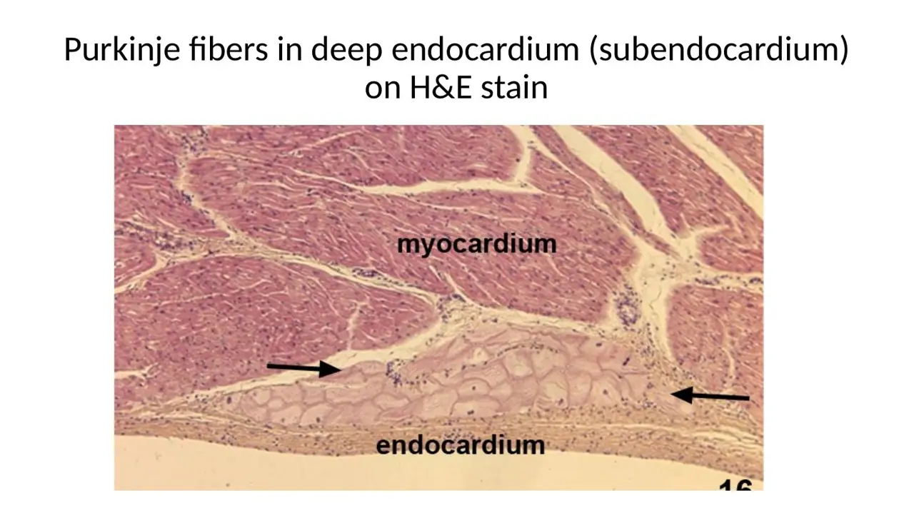
WhyPurkinje fibers run in the subendocardium, the deep layer of the endocardium between the endothelium and the myocardium (ventricles, both sides of the septum). They only contact the myocardium at their ends, and they are never epicardial or pericardial.
Key factPurkinje fibers → subendocardial layer → part of the ENDOCARDIUM (ventricles, along both sides of the interventricular septum toward the apex). If the options say "Endocardium" or "Subendocardium / subendocardial layer", that is the answer (the lecture table words it "subendothelial layer"; same place). So a section showing Purkinje fibers is closer to the endocardium (inner surface). Never epicardium, visceral or parietal pericardium, or "myocardium".
F0718:36 lists Purkinje fibers in the "subendothelial layer" while F0718:33/34 and the 2026 lab say subendocardium; they mean the same location.
What it unlocks: 7 questions
The 1 question you might not expect it to answer is in the dropdown at the bottom.
How it shows up
Dr Bruno lecture quiz Q4: "Where are the Purkinje fibers located?" (epicardium, pericardium, myocardium, endocardium, SA node) and the 2026 lab "Purkinje fibers in this image are located within which layer?"
"Which layer of the heart contains the Purkinje fibers?" answered as endocardium, subendocardial layer or subendocardium
"Are we closer to the epicardium or endocardium?" on a Purkinje image
Trichrome "The highlighted area represents..." (zone with pale Purkinje fibers and nerves in blue CT) → endocardium
What it lets you eliminate
Epicardium, visceral/parietal pericardium, myocardium, SA node: all wrong homes for Purkinje fibers
Lecture: subendocardium (SEn) holds Purkinje fibers (P) under the endothelium (En)H12A Circulatory I lecture (Dr Bruno) · slide 332026 session: Purkinje fibers in the deep endocardium (subendocardium) on H&ECirculatory interactive session 2026 · slide 16Lecture: endocardium = endothelium, connective tissue, subendocardial conducting fibersH12A Circulatory I lecture (Dr Bruno) · slide 34
Questions it helps you answer (1)
Practice Q83One extra step
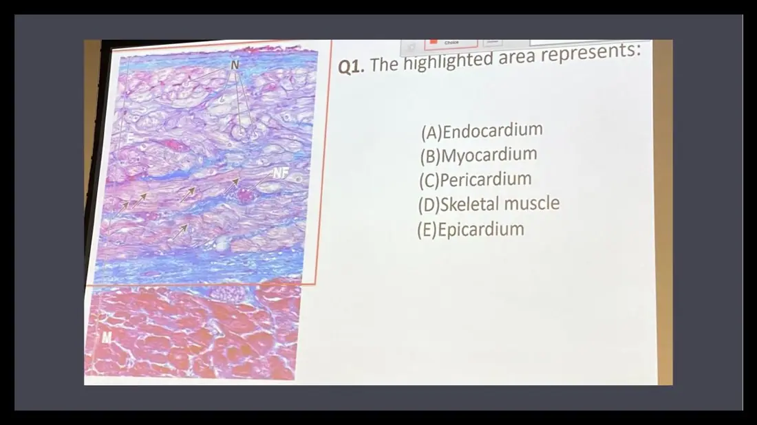
Q1. The highlighted area represents:
(A)Endocardium
(B)Myocardium
(C)Pericardium
(D)Skeletal muscle
(E)Epicardium
AnswerA. Endocardium
WhyThe highlighted zone between the lining and red myocardium holds pale Purkinje fibers, nerves and blue collagen, identifying the endocardium with its subendocardial layer.
How it follows from the main questionThe pale lilac cells inside blue connective tissue are Purkinje fibers (with nerves) → they live in the subendocardium → the highlighted zone is the endocardium, not myocardium or epicardium.
Slides (2)H12A Circulatory I lecture (Dr Bruno) · slide 39H12A Circulatory I lecture (Dr Bruno) · slide 34
17unlocks 9TA ×41 not obvious
Just beneath the ventricular endocardium is a band of cells that are much larger and paler than the neighboring muscle fibers. Which other feature describes these cells?
A. Many peripheral nuclei in a single long cell
B. One large lipid droplet that flattens the nucleus
C. Spindle shape with a cigar-shaped nucleus
D. Cytoplasm packed with myofibrils
E. Abundant glycogen with sparse myofibrils pushed to the periphery
AnswerE. Abundant glycogen with sparse myofibrils pushed to the periphery
WhyLarge, pale cells under the endocardium are Purkinje fibers: abundant glycogen (washed out on H&E, so pale) and few, peripheral myofibrils, with central nuclei. A = skeletal muscle, B = adipocyte, C = smooth muscle, D = contractile cardiomyocyte.
Key factTA rule (If you see → think):
dark pink, striated, many myofibrils, thick ventricular wall, contraction → cardiac muscle (myocardium)
large, pale cells under the endocardium, lots of glycogen, few (peripheral) myofibrils, central nuclei with a clear halo, electrical conduction → Purkinje fibers
Purkinje fibers are larger and paler than contractile fibers; in trichrome they are pale lilac cells inside blue CT above red myocardium. They are poorly contractile, not neurons, and not in the myocardium.
q55 and q57 had stray keys from other slides of the same deck; the page key notes (Purkinje fibers; contractile cardiac muscle) are correct.
What it unlocks: 13 questions
The 1 question you might not expect it to answer is in the dropdown at the bottom.
How it shows up
TA matching: abundant glycogen/lighter stain, force generation, subendocardial location, numerous myofibrils, rapid conduction → Purkinje or cardiac muscle
TA: "cluster of large pale cells beneath the endocardium with fewer myofibrils" (fibroblasts, Purkinje, cardiac muscle, endothelial cells); "population X (eosinophilic, most of wall) vs Y (larger, paler, subendocardial): which statement is correct?"
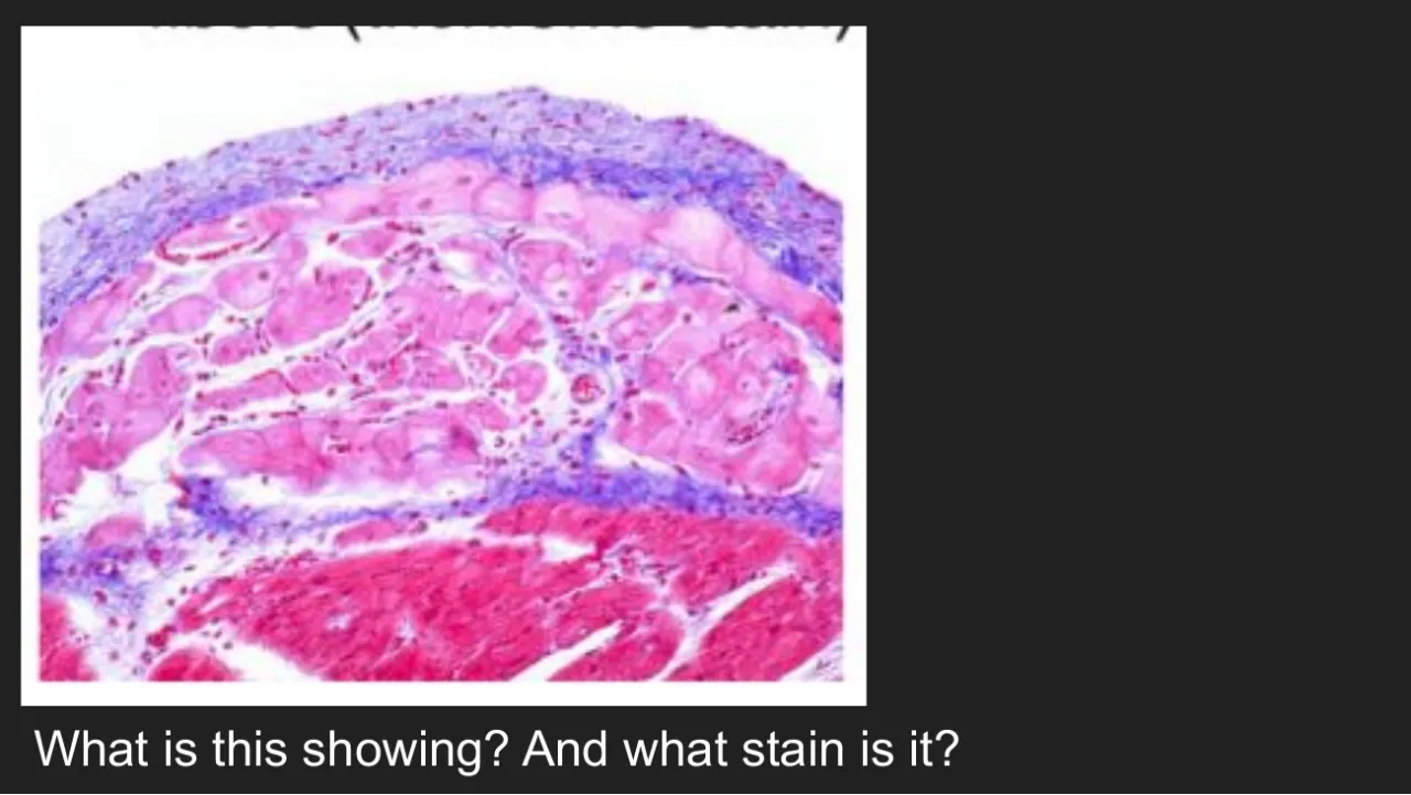
TA trichrome: "What is this showing, and what stain?" → endocardium with Purkinje fibers, trichrome; lab labels "Yellow?" Purkinje vs "Orange?" contractile muscle; "Identify the image with Purkinje fibers"
Lippincott boxed cells (adipose, cardiac muscle, loose CT, Purkinje fibers, smooth muscle); "Which is true: contractile / larger and paler / modified neurons / found in the myocardium"
What it lets you eliminate
Many myofibrils and dark pink → contractile cardiomyocyte
Adipose = empty white rings with a flat peripheral nucleus; smooth muscle = spindle cells; loose CT = fibers with scattered nuclei
"Contractile", "modified neurons", "found in the myocardium" are false statements about Purkinje fibers
Used together with
Purkinje fibers conduct (modified cardiomyocytes)
Straight-recall questions it answers (12)
TAH15 Match each feature with cardiac muscle fiber or Purkinje fiber: 1. abundant glycogen, stains lighter; 2. primary function is force generation; 3. located mainly in the su… → 1 Purkinje; 2 cardiac muscle; 3 Purkinje; 4 cardiac muscle; 5 Purkinje
TAH16 A slide of ventricular tissue shows a cluster of large pale-staining cells beneath the endocardium. The cells contain fewer myofibrils than neighboring muscle fibers. The… → B. Purkinje fibers
TAH17 A ventricular histology section contains two neighboring cell populations: Population X stains intensely eosinophilic and occupies most of the ventricular wall. Populatio… → C. Population Y is specialized for rapid impulse conduction rather than contraction.
TA table: dark pink, many myofibrils = cardiac muscle; large, pale, glycogen-rich = PurkinjeHarlem TA review 2026 · slide 53Lecture: Purkinje fibers are larger and paler than contractile muscle fibersH12A Circulatory I lecture (Dr Bruno) · slide 382026 lab: ventricular endocardium labelled En, SEn, P (Purkinje fibers) and MCirculatory lab 2026 · slide 28
Questions it helps you answer (1)
TA review · TAG36Vignette never names it
What is this showing, and what stain is it? (heart micrograph) Great Falls TA review 2026, slide 80
AnswerEndocardium with Purkinje fibers; trichrome stain (the neighbouring panel is myocardium with intercalated discs)
How it follows from the main questionBlue collagen plus red muscle = trichrome. The large pale cells in the blue subendocardial tissue are Purkinje fibers, so this is endocardium with Purkinje fibers.
18unlocks 2TA ×21 not obvious
Which statement about the large, pale subendocardial fibers of the ventricles is true?
A. They set the heart rate because they fire faster than the SA node
B. They are modified neurons related to the Purkinje cells of the cerebellum
C. They are cardiomyocytes swollen by ischemia
D. They are modified cardiac muscle cells that rapidly conduct the impulse to the ventricular myocardium
E. They are the principal cells of the sinoatrial node
AnswerD. They are modified cardiac muscle cells that rapidly conduct the impulse to the ventricular myocardium
WhyPurkinje fibers are modified cardiac muscle cells of the conduction system that rapidly conduct the impulse from the bundle branches to the ventricular muscle. They share only a name with cerebellar Purkinje neurons, are not SA-node cells or ischemic swelling, and fire slower than the SA node.
Key factPurkinje fibers = modified cardiomyocytes specialized for conduction, not contraction (linked to each other and to contractile cells by desmosomes and gap junctions). Bundle branches → subendocardial Purkinje network near the apex → rapid spread to all ventricular muscle → coordinated contraction from apex to base. Damage → impaired ventricular conduction (not SA-node initiation, not valve support). Intrinsic rate slower than the SA node. Not neurons (≠ cerebellar Purkinje cells).
What it unlocks: 4 questions
The 1 question you might not expect it to answer is in the dropdown at the bottom.
How it shows up
TA: "large, pale conducting fibers beneath the endocardium: best describes these fibers and their function?" (contractile myocardial fibers, smooth muscle, mesothelial cells, fibroblasts, Purkinje fibers for conduction)
TA: "damage to large pale subendocardial fibers impairs...?" (SA initiation, AV valve support, ventricular conduction, vascular resistance, exchange)
Image with arrows on pale fibers: "Which is true?" (modified neuronal cells, SA node component, swollen from coronary disease, conduct the cardiac impulse, subepicardial)
What it lets you eliminate
"Modified neurons", "SA node", "swollen ischemic cells", "subepicardial", "fire faster than the SA node": all false
Used together with
Purkinje fiber vs contractile cardiomyocyte
Straight-recall questions it answers (2)
TAH6 A cardiac histology specimen reveals large, pale conducting muscle fibers located beneath the endocardial lining. Which of the following best describes these fibers and t… → E. Purkinje fibers specialized for conducting electrical impulses through the ventricles
TAH12 A histological examination reveals damage to the large, pale conducting muscle fibers located in the subendocardial region of the ventricles. Which of the following funct… → C. Transmission of electrical impulses through the ventricular conduction system
Slides (3)
2026 lab: conduction system from SA node to AV node, His bundle, branches, Purkinje fibersCirculatory lab 2026 · slide 272026 session: cardiac Purkinje fibers vs cerebellar Purkinje neuronsCirculatory interactive session 2026 · slide 18TA summary of endocardium, epicardium, myocardium and Purkinje fiber featuresGreat Falls TA review 2026 · slide 84
Questions it helps you answer (1)
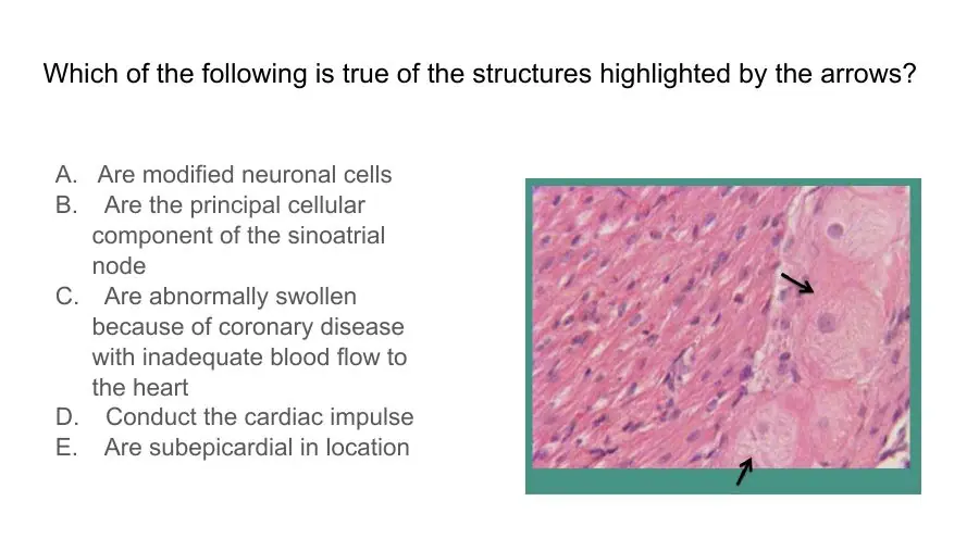
Practice Q27Trap: the obvious answer is wrong
Which of the following is true of the structures highlighted by the arrows?
A. Are modified neuronal cells
B. Are the principal cellular component of the sinoatrial node
C. Are abnormally swollen because of coronary disease with inadequate blood flow to the heart
D. Conduct the cardiac impulse
E. Are subepicardial in location
AnswerD. Conduct the cardiac impulse
WhyThe large, pale fibers with central nuclei and few peripheral myofibrils are Purkinje fibers, modified cardiac muscle cells (not neurons) in the subendocardium that rapidly conduct the impulse to ventricular muscle.
How it follows from the main questionLarge pale cells under the endocardium = Purkinje fibers. They look swollen, but they are normal glycogen-rich modified cardiomyocytes that conduct the cardiac impulse, not neurons or ischemic cells. Also asked as Q38.
Slide (1)H12A Circulatory I lecture (Dr Bruno) · slide 33
19unlocks 61 not obvious
On H&E, a group of subendocardial cells looks large and pale. A PAS stain of the same area turns them deep magenta. The PAS stain is detecting:
A. Myosin
B. Myelin
C. Glycogen
D. Collagen
E. Triglyceride
AnswerC. Glycogen
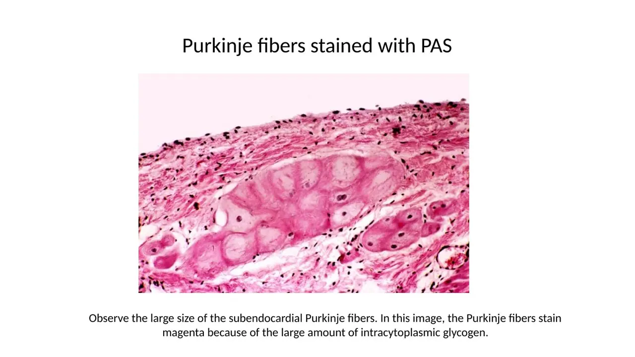
WhyPAS stains glycogen magenta. Purkinje fibers are packed with glycogen, which washes out on H&E (so they look pale) and stains strongly with PAS; so pale H&E + magenta PAS cells beneath the endothelium = Purkinje fibers in the endocardium.
Key factPAS → glycogen → magenta. Glycogen-rich Purkinje fibers stand out deep magenta on PAS; on H&E the same cells look pale/empty. Ordinary cardiomyocytes have only a little glycogen near the nucleus. Vignette chain: pale + large on H&E → magenta on PAS → glycogen → Purkinje fibers → layer = endocardium (subendocardium), not epicardium, visceral or parietal pericardium, or myocardium.
q67: the speaker notes on the source slides say "B" (visceral pericardium); the slide reveal and the page key say D (endocardium), which matches the lecture.
What it unlocks: 6 questions
The 1 question you might not expect it to answer is in the dropdown at the bottom.
How it shows up
2026 interactive session Q5 and rewordings: pale, wide cells on H&E turn magenta with PAS → "Which area of the heart was observed?" → endocardium (distractors epicardium, visceral and parietal pericardium, myocardium)
Sarcoidosis heart-block autopsy: "PAS highlights which substance in the Purkinje fibers?" (collagen, myelin, glycogen, hyaluronic acid, myosin)
"PAS-stained heart wall: identify the cells at the black arrows" → Purkinje fibers; "What is true about these cells?" → subendocardial and contain glycogen
What it lets you eliminate
Triglyceride/lipid → adipocytes; collagen → blue in trichrome; myelin → nerves
Purkinje fibers have fewer (not more) myofibrils and fire slower than the SA node
2026 session: PAS stains the glycogen-rich subendocardial Purkinje fibers magentaCirculatory interactive session 2026 · slide 172026 session: Purkinje fibers in the deep endocardium (subendocardium) on H&ECirculatory interactive session 2026 · slide 16
Questions it helps you answer (1)
Practice Q25One extra step
During light microscopic examination of an H&E stained section of the heart, you notice a group of cells with paler staining, and increased diameter when compared to typical cardiac muscle cells. Determined to classify the cell, you prepare another section of the same area of the heart with PAS staining. This group of cells is now distinguished by a dark magenta color. Which area of the heart was most likely being observed under the microscope?
A. Myocardium
B. Parietal Pericardium
C. Visceral pericardium
D. Epicardium
E. Endocardium
AnswerE. Endocardium
WhyLarge, pale cells that stain magenta with PAS are glycogen-rich Purkinje fibers, which run in the subendocardial layer of the endocardium.
How it follows from the main questionPAS magenta = glycogen → large pale glycogen-rich cells = Purkinje fibers → they sit in the subendocardium → area = endocardium. The question asks the layer, not the cell. Also asked as Q39, Q67.
A Masson trichrome section from the inner left ventricular wall shows a broad zone of blue-staining tissue holding clusters of large, pale lilac cells and small nerve bundles, lying over a band of bright red, densely packed fibers. Which statement is correct?
A. The stain is PAS, and the pale cells are pale because they lack glycogen
B. The blue zone is subendocardial connective tissue holding Purkinje fibers, and the red band is contractile myocardium
C. The pale cells are smooth muscle cells in the media of a coronary artery
D. The pale lilac cells are adipocytes of the epicardium
E. The blue zone is myocardium and the red band is collagen of the cardiac skeleton
AnswerB. The blue zone is subendocardial connective tissue holding Purkinje fibers, and the red band is contractile myocardium
WhyIn trichrome, collagen stains blue and muscle stains red/purple. Blue connective tissue with large pale cells and nerves under the lining is the subendocardium holding Purkinje fibers (part of the endocardium); the red band below is contractile myocardium. A swaps the colours; adipocytes are empty rings in epicardial fat; on PAS the Purkinje fibers would be magenta.
Key factMasson trichrome on heart sections:
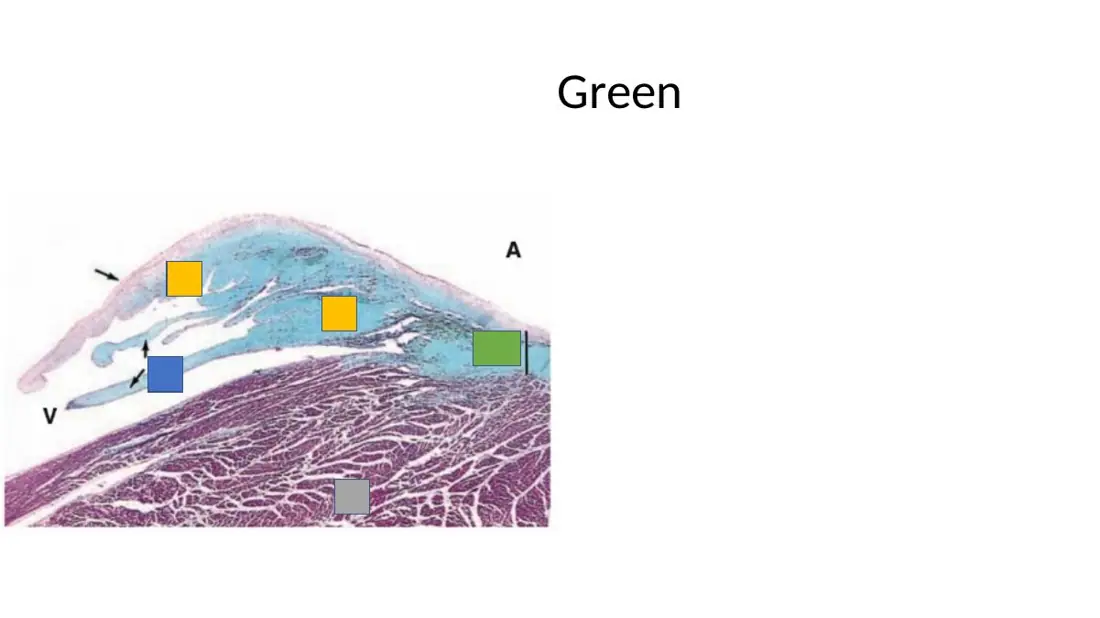
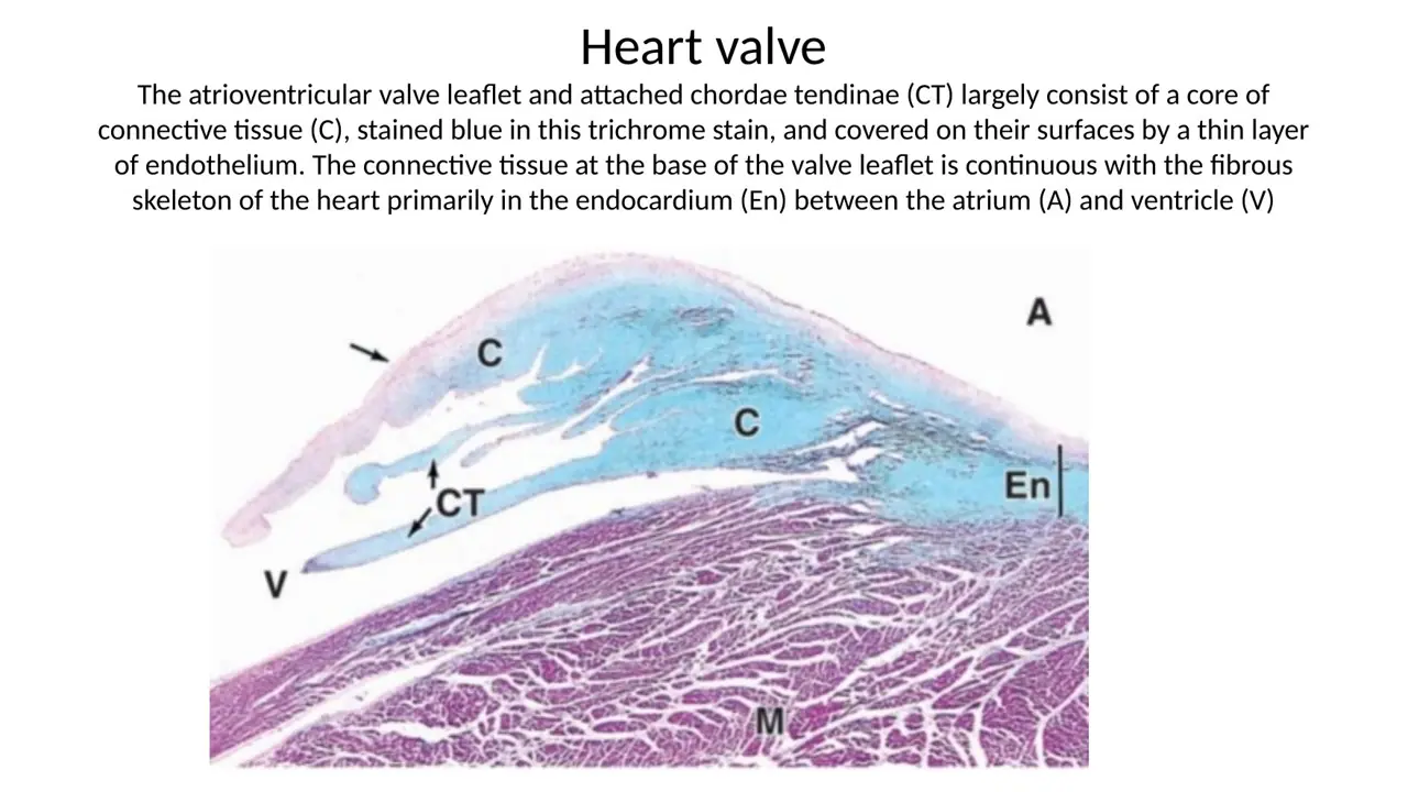
blue (blue-green) = collagen: the subendocardial connective tissue, the dense CT core of valve cusps and chordae tendineae, and the fibrous skeleton at the valve base (lies in the endocardium, labelled En)
red/purple = muscle: the myocardium (e.g., thick ventricular muscle under a valve)
Purkinje fibers = large, pale lilac cells (glycogen washed out, few peripheral myofibrils) inside the blue subendocardial CT, often beside nerves
Read it as: blue zone + pale cells over red muscle = endocardium with Purkinje fibers; blue flap with blue cords over red muscle = AV valve with chordae tendineae. (On PAS the same Purkinje fibers turn magenta.)
q252, q352 and TAG36 all use the 2026 lab trichrome image (F3530:29).
What it unlocks: 10 questions
The 1 question you might not expect it to answer is in the dropdown at the bottom.
How it shows up
TA: "What is this showing, and what stain is it?" on the trichrome Purkinje image → endocardium with Purkinje fibers, trichrome (its neighbouring H&E panel = myocardium with intercalated discs)
2026 lab quiz on the same trichrome image: "The Purkinje fibers in this image are located within which layer?" → endocardium; "What is true about the cells at the arrows?" → subendocardial, contain glycogen
"The highlighted area represents..." on a trichrome field of pale cells and nerves in blue CT next to red muscle → endocardium
Trichrome valve slide: "What is this an image of?", "Identify the circled structure", "Label the slide", "Green?", "Gray?" → AV valve with chordae, endocardium at the base, thick ventricular myocardium
What it lets you eliminate
Blue is never muscle and red is never collagen in trichrome
Pale cells inside blue CT are Purkinje fibers, not adipocytes (empty rings in epicardial fat) or smooth muscle
A blue zone holding Purkinje fibers is endocardium, not myocardium, epicardium or pericardium
TAG36 What is this showing, and what stain is it? (heart micrograph) → Endocardium with Purkinje fibers; trichrome stain (the neighbouring panel is myocardium with intercalated discs)
2026 lab trichrome: blue subendocardial CT with large pale Purkinje fibers over red myocardiumCirculatory lab 2026 · slide 29Lecture trichrome: Purkinje fibers with nerves (N, NF) in blue CT next to red myocardium (M)H12A Circulatory I lecture (Dr Bruno) · slide 39Lecture trichrome: valve cusp with blue dense CT core (C), chordae (CT), En and myocardium (M)H12A Circulatory I lecture (Dr Bruno) · slide 55
Questions it helps you answer (1)
Practice Q64Trap: the obvious answer is wrong
Green
AnswerEndocardium
WhyAt the valve base, dense connective tissue fills the endocardium between atrium and ventricle, forming the fibrous ring of the cardiac skeleton that anchors the valve.
How it follows from the main questionBlue = collagen, but the label marks En: the endocardium at the valve base, where the fibrous skeleton sits and the cusp begins. Answering "cardiac skeleton" or "valve" is the slip.
Slide (1)Circulatory lab 2026 · slide 33
Conduction system
21unlocks 5
Which sequence correctly traces normal cardiac depolarization?
A. Purkinje fibers → bundle branches → bundle of His → AV node → SA node
B. AV node → SA node → atrial myocardium → bundle of His → Purkinje fibers → ventricular myocardium
C. SA node → bundle of His → AV node → Purkinje fibers → atrial myocardium
D. SA node → AV node → ventricular myocardium → Purkinje fibers
E. SA node → atrial myocardium → AV node → bundle of His → bundle branches → Purkinje fibers → ventricular myocardium
AnswerE. SA node → atrial myocardium → AV node → bundle of His → bundle branches → Purkinje fibers → ventricular myocardium
WhyThe SA node fires first (fastest intrinsic rate = pacemaker), the impulse spreads through atrial muscle to the AV node (brief delay so the atria finish emptying), then the bundle of His, the right and left bundle branches, the Purkinje fibers, and finally the ventricular myocardium.
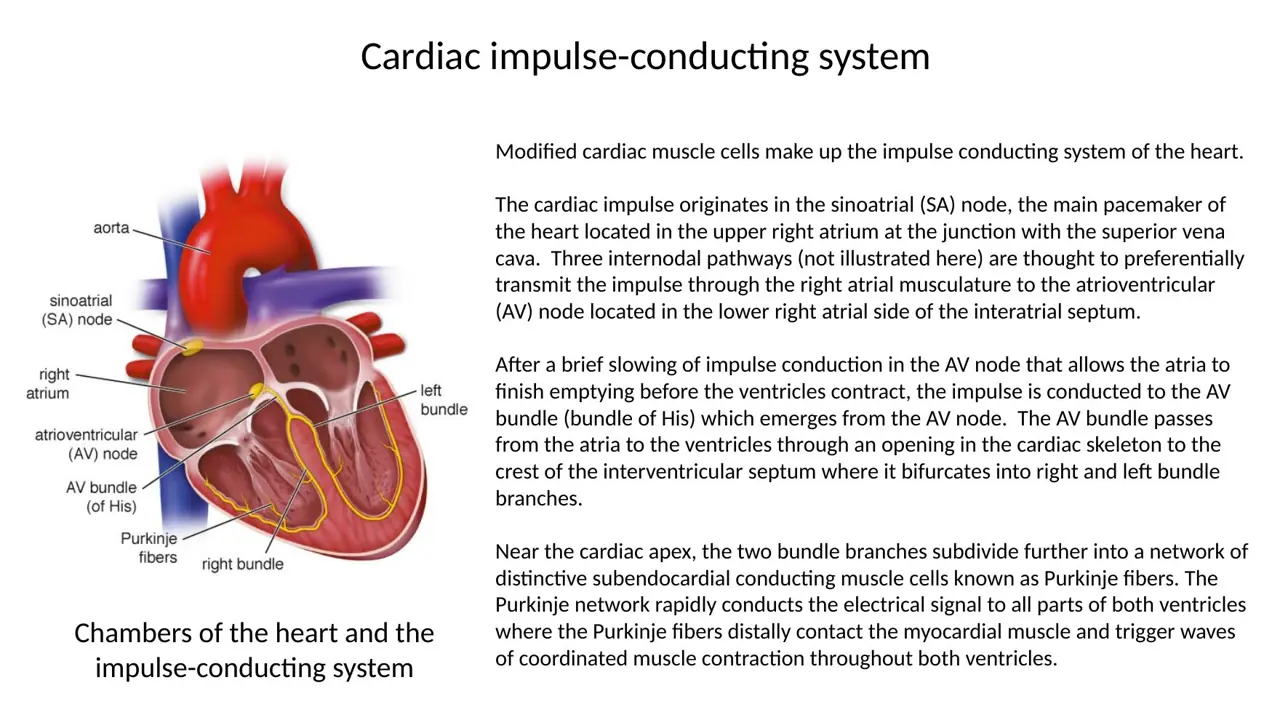
Key factSA node = pacemaker: earliest, fastest spontaneous depolarization (sympathetic fibers only modulate the rate; AV node, His and Purkinje are slower backups). Sequence: SA node → atrial myocardium → AV node (delay) → AV bundle of His → right and left bundle branches → Purkinje fibers → ventricular myocardium. All nodal and conducting cells are modified cardiac muscle cells.
What it unlocks: 5 questions
No surprises here: every question it answers asks the fact straight out.
How it shows up
Dr Bruno lecture quiz Q3: "What is the pacemaker of the heart?" (SA node, AV node, bundle of His, right bundle branch, Purkinje fiber)
Arrhythmia vignette: "Which structure functions as the pacemaker?" with "sympathetic fibers" as a distractor; mapping vignette: earliest spontaneous depolarization near the SVC along the crista terminalis
Ordering 6 items: SA node, atrial myocardium, AV node, bundle of His, Purkinje fiber, ventricular myocardium
What it lets you eliminate
AV node, His bundle, bundle branches, Purkinje fibers: conduct, but are not the pacemaker
Sympathetic fibers change the rate but do not generate the rhythm
2026 lab: conduction system from SA node to AV node, His bundle, branches, Purkinje fibersCirculatory lab 2026 · slide 27Lecture diagram: SA node, AV node, AV bundle, bundle branches and Purkinje fibersH12A Circulatory I lecture (Dr Bruno) · slide 35
22unlocks 51 not obvious
Which pairing of a conduction-system component with its location is correct?
A. Purkinje fibers: epicardium
B. SA node: cardiac skeleton
C. SA node: wall of the left atrium
D. AV bundle of His: interventricular septum
E. AV node: interventricular septum
AnswerD. AV bundle of His: interventricular septum
WhyDr Bruno's table: SA node = wall of the right atrium; AV node = junction between atria and ventricles; AV bundle (His) = interventricular septum; Purkinje fibers = subendothelial (subendocardial) layer. Every other pairing puts a component in the wrong place.
Key factLecture table (F0718:36):
SA node → wall of the right atrium, at its junction with the superior vena cava (crista terminalis)
AV node → junction between atria and ventricles (lower right atrial side of the interatrial septum)
AV bundle (His) → passes through the cardiac skeleton into the interventricular septum, splits into right and left bundle branches
Purkinje fibers → subendothelial/subendocardial layer of the ventricles (endings of the bundle branches)
Both nodes are in the right atrium; the ventricular walls contain the Purkinje fibers.
What it unlocks: 5 questions
The 1 question you might not expect it to answer is in the dropdown at the bottom.
How it shows up
2026 interactive session Q6: "The sinoatrial node is located within..." (right atrial wall, left atrial wall, cardiac skeleton, epicardium, endocardium), asked twice
Matching the four components to wall of right atrium / junction between atria and ventricles / interventricular septum / subendothelial layer (asked as 2 versions)
"Which is true for ventricles?" → walls contain Purkinje fibers of the right and left branches of the AV bundle
What it lets you eliminate
Left atrial wall, cardiac skeleton, epicardium, endocardium are not the SA node's location
Ventricles: at the apex (not the base), receive blood from the atria (not the venae cavae)
Lecture: SA node RA wall; AV node AV junction; His IV septum; Purkinje subendothelialH12A Circulatory I lecture (Dr Bruno) · slide 362026 session: SA and AV nodes in the right atrium, His bundle, subendocardial Purkinje fibersCirculatory interactive session 2026 · slide 21Junqueira 11-2: heart overview with SA node, AV node, His bundle and Purkinje fibersJunqueira’s Basic Histology, fig 11-2
Questions it helps you answer (1)
Practice Q14Looks like another topic
Which of the following is true for ventricles?
a. Located at the base of the heart
b. Myocardial cells contains abundant granules
c. Receive blood directly from the venae cavae and pulmonary veins
d. Walls contain Purkinje fibers of the right and left branches from the atrioventricular bundle
e. Contain more elastic fibers than the atria
Answerd. Walls contain Purkinje fibers of the right and left branches from the atrioventricular bundle
WhyThe right and left bundle branches of the AV bundle end as Purkinje fibers in the ventricular subendocardium; atria, not ventricles, have ANF granules and more elastic fibers.
How it follows from the main questionReads like gross anatomy, but the conduction map answers it: the right and left bundle branches of the AV bundle end as Purkinje fibers in the ventricular walls. The other statements describe atria.
Which pairing of a heart surface with its simple squamous covering is correct?
A. Valve cusps: endothelium on both surfaces
B. Endocardium: mesothelium
C. Epicardium: endothelium
D. Parietal pericardium: endothelium
E. SA node: mesothelium on its free surface
AnswerA. Valve cusps: endothelium on both surfaces
WhyValves are folds of endocardium, so endothelium covers both surfaces. The epicardium and parietal pericardium carry mesothelium; the endocardium carries endothelium. The SA node is buried within the right atrial wall and has no free surface of its own.
Key factSimple squamous linings of the heart:
Mesothelium: epicardium (visceral pericardium) and parietal pericardium; faces the pericardial cavity
Endothelium: endocardium, both faces of every valve cusp, and all vessels (e.g., the aortic intima)
No free surface: the SA node (6–7 mm of paler, thinner nodal fibers around the SA nodal artery, buried subepicardially in the right atrial wall at the crista terminalis/SVC), myocardium, cardiac skeleton
What it unlocks: 2 questions
No surprises here: every question it answers asks the fact straight out.
How it shows up
HY EXCEPT question: "A simple squamous epithelium covers or lines the free surface of all the following EXCEPT" (epicardium, endocardium, mitral valve, aorta, SA node) → SA node
Reworded twin: "A simple squamous epithelium does not cover the immediate surface or lining of which structure?" (same five options)
What it lets you eliminate
Epicardium (mesothelium) and endocardium, mitral valve, aorta (endothelium) all have a simple squamous surface
2026 session: SA node buried subepicardially in the right atrial wall at the crista terminalisCirculatory interactive session 2026 · slide 22Lecture: epicardium = visceral pericardium, mesothelium over loose CT with fat, vessels, nervesH12A Circulatory I lecture (Dr Bruno) · slide 22Lecture: endocardium = endothelium, connective tissue, subendocardial conducting fibersH12A Circulatory I lecture (Dr Bruno) · slide 34
Cardiac skeleton and valves
24unlocks 2TA ×2
Which tissue forms the fibrous rings around the four heart valves, the fibrous trigones and the membranous part of the interventricular septum?
A. Hyaline cartilage
B. Dense regular (tendon-like) connective tissue
C. Dense irregular connective tissue
D. Loose areolar connective tissue
E. Elastic cartilage
AnswerC. Dense irregular connective tissue
WhyThe cardiac skeleton is dense irregular connective tissue: fibrous rings around the valves and the bases of the aorta and pulmonary trunk, the fibrous trigones and the membranous septum. It is not cartilage, tendon-like dense regular CT or loose CT.
four bands (fibrous rings) around the heart valves and the bases of the pulmonary trunk and aorta; fibrous trigones; membranous part of the interventricular septum
surrounds and anchors the valves and extends into the valve cusps and chordae tendineae; gives insertion to atrial and ventricular muscle
On a valve-base section it lies in the endocardium between atrium and ventricle (blue in trichrome).
What it unlocks: 4 questions
No surprises here: every question it answers asks the fact straight out.
How it shows up
"What tissue forms the core structural layer of the cardiac skeleton?" (skeletal muscle, DICT, simple cuboidal, reticular CT)
TA: "specific tissue type of the cardiac skeleton" (hyaline cartilage, elastic cartilage, DICT, dense regular, loose areolar)
TA: "DICT surrounding the valves and bases of the great vessels: identify it and its relationship to the valves" → cardiac skeleton, stabilizes valves, muscle attachment
"What tissue is directly associated with and extends into the heart valves?" → cardiac skeleton
Myocardium, epicardium, His bundle, pericardium do not extend into the valves
Straight-recall questions it answers (4)
TAG22 A histologic section of a cardiac valve shows connective tissue anchored directly to the cardiac skeleton. The cardiac skeleton itself is primarily composed of which spec… → C. Dense irregular connective tissue
TAH8 A histological section of the heart reveals dense irregular connective tissue surrounding the heart valves and bases of the great vessels. Which of the following best ide… → D. Cardiac skeleton, which stabilizes the valves and provides sites for muscle attachment
Lecture: cardiac skeleton = DICT; anchors valves and muscle; insulates atria from ventriclesH12A Circulatory I lecture (Dr Bruno) · slide 472026 lab: fibrous skeleton rings and trigones; extends into valve cusps and chordaeCirculatory lab 2026 · slide 32Cardiac skeleton from above: fibrous rings, trigones and the AV bundle passing throughCourse lecture — cardiac skeleton seen from above with the atria removed: fibrous rings round the four valves, the left and right fibrous trigones, and the atrioventricular bundle (yellow) passing through
25unlocks 1TA ×11 not obvious
Why must the cardiac impulse pass through the AV bundle to reach the ventricles?
A. Epicardial fat insulates the ventricles
B. The dense connective tissue of the cardiac skeleton electrically insulates atrial from ventricular muscle
C. The semilunar valves separate the atria from the ventricles
D. The cardiac skeleton itself generates a delay potential
E. Pericardial fluid blocks all other routes
AnswerB. The dense connective tissue of the cardiac skeleton electrically insulates atrial from ventricular muscle
WhyThe dense CT of the cardiac skeleton does not conduct, so it electrically insulates atria from ventricles; the only electrical bridge is the AV bundle, which pierces the skeleton. The skeleton anchors valves and muscle but never initiates or conducts impulses.
Key factFunctions of the cardiac skeleton (F0718:47): stabilizes valves and anchors muscles; electrically insulates ventricular cells from atrial cells (the AV bundle passing through it is the only path). NOT functions: initiating spontaneous action potentials (SA node), conducting fibers (Purkinje fibers), containing pericardial fluid (pericardial cavity). Lecture wording: "separates atria from ventricles" = AV valves; "separates ventricles from circulation" = semilunar valves.
q74's explanation assigns "separates atria from ventricles" to the AV valves (F0718:51 wording), while TAG29's descriptor "masses of DICT, separates the atria from the ventricles" is the cardiac skeleton. In q74 pick "electrically insulates"; in TAG29 the words "masses of DICT" point to the skeleton.
What it unlocks: 2 questions
The 1 question you might not expect it to answer is in the dropdown at the bottom.
How it shows up
"Which of the following is a function of the cardiac skeleton?" (separates atria from ventricles, electrically insulates, separates ventricles from circulation, contains pericardial fluid, conducting muscle fibers)
TA NOT question: anchoring valves, muscle insertion, electrical insulation, extending into chordae vs "initiating spontaneous action potentials"
Separates ventricles from circulation → semilunar valves
Straight-recall questions it answers (1)
TAG23 The cardiac skeleton plays several vital biomechanical and physiological roles within the heart. Which of the following functions is NOT performed by the cardiac skeleton… → D. Initiating spontaneous action potentials for cardiac depolarization
Slides (2)
Lecture: cardiac skeleton = DICT; anchors valves and muscle; insulates atria from ventriclesH12A Circulatory I lecture (Dr Bruno) · slide 47Cardiac skeleton from above: fibrous rings, trigones and the AV bundle passing throughCourse lecture — cardiac skeleton seen from above with the atria removed: fibrous rings round the four valves, the left and right fibrous trigones, and the atrioventricular bundle (yellow) passing through
Questions it helps you answer (1)
Practice Q74Trap: the obvious answer is wrong
Which of the following is a function of the cardiac skeleton?
A. Separates atria from ventricles
B. Electrically insulates ventricular cells from atrial cells
C. Separates ventricles from circulation
D. Contains the pericardial fluid
E. Conducting muscle fibers
AnswerB. Electrically insulates ventricular cells from atrial cells, also stabilizes valves and anchors muscles
WhyThe dense connective tissue of the cardiac skeleton cannot conduct impulses, so it electrically insulates atria from ventricles, channelling conduction through the AV bundle; it also anchors valves.
Explanation from the source
Separates atria from ventricles→ AV valves | Electrically insulates ventricular cells from atrial cells, also stabilizes valves and anchors muscles | Separates ventricles from circulation→ semilunar valves | Contains the pericardial fluid→ pericardial cavity | Conducting muscle fibers→ Purkinje fibers | **Know the functions of the cardiac skeleton**
How it follows from the main question"Separates atria from ventricles" looks right but is the lecture line for the AV valves ("ventricles from circulation" = semilunar). The skeleton's listed function: electrically insulates ventricular from atrial cells.
Slide (1)H12A Circulatory I lecture (Dr Bruno) · slide 47
26unlocks 4TA ×21 not obvious
Which feature belongs to the semilunar valves but not to the atrioventricular valves?
A. Papillary muscles tense them during ventricular contraction
B. They separate the atria from the ventricles
C. They include the mitral valve
D. Their cusps are tethered by chordae tendineae to papillary muscles
E. They have three cusps and no muscular support
AnswerE. They have three cusps and no muscular support
WhySemilunar valves (aortic, pulmonary) separate the ventricles from the circulation and have three cusps that brace each other like a tripod, with no muscular support. AV valves (tricuspid, mitral) separate atria from ventricles and are braced by chordae tendineae tensed by papillary muscles.
Key factAV valves: tricuspid (right) and bicuspid = mitral (left); separate atria from ventricles; folds of fibrous tissue; closed by blood pressure; papillary muscles tense chordae tendineae so the cusps do not evert. Semilunar valves: pulmonary (RV → pulmonary trunk) and aortic (LV → aorta); separate ventricles from circulation; 3 cusps "like a tripod"; no muscular support. 2 AV + 2 SL valves. All valves close passively by pressure; muscles only brace the AV cusps.
q95 (key: semilunar valves) is loosely worded: no valve is closed by muscle contraction; all close passively by pressure (F0718:51 "blood pressure closes valve"). Its intended point is that only AV valves have muscular (papillary muscle and chordae) support.
What it unlocks: 6 questions
The 1 question you might not expect it to answer is in the dropdown at the bottom.
How it shows up
"What is the difference between AV and semilunar valves?"; "Which is incorrect about semilunar valves?" (the mitral valve is the semilunar valve on the left → false)
Fill-in: "The (blank) valves contain 3 cusps and no muscular support"; "Which valves are NOT actively closed by muscular structures?" → semilunar
TA: "structural composition of AV valves" (folds of fibrous tissue with chordae and papillary muscles) and "AV vs semilunar support"
What it lets you eliminate
"Mitral = semilunar", "4 AV vs 3 SL valves", "semilunar valves have muscular support", "AV valves have three self-supporting cusps": all false
Smooth muscle or Purkinje fibers inside valves: false
Straight-recall questions it answers (5)
TAH7 Which of the following best describes the structural composition of atrioventricular (AV) valves? → C. Folds of fibrous tissue associated with chordae tendineae and papillary muscles
TAH9 Which of the following correctly distinguishes the structural support of atrioventricular valves from that of semilunar valves? → B. AV valves are supported by papillary muscles and chordae tendineae, whereas semilunar valves lack muscular support.
Lecture: semilunar valves, ventricles to circulation, three cusps, no muscular supportH12A Circulatory I lecture (Dr Bruno) · slide 54Lecture: AV valves (tricuspid, mitral); papillary muscles tense the chordae tendineaeH12A Circulatory I lecture (Dr Bruno) · slide 51Lecture diagram: mitral, tricuspid, aortic and pulmonary valves with chordae and papillary muscleH12A Circulatory I lecture (Dr Bruno) · slide 49
Questions it helps you answer (1)
Practice Q95One extra step
Which Heart valves are NOT actively closed by the contraction of muscular structures?
A. Mitral valve
B. Tricuspid valve
C. Semilunar valves
D. Atrioventricular valves
E. All valves are closed by contraction of the heart
AnswerC. Semilunar valves
Key check: Key = Semilunar valves. Strictly, no heart valve is closed by muscle contraction: all four close passively by pressure gradients (papillary muscles only keep AV cusps from everting). The key's intent: only AV valves have muscular (papillary muscle/chordae) support. — Confirmed on re-check; correct answer: C. Semilunar valves
WhySemilunar valves have no papillary muscles or chordae and close purely by arterial back-pressure, whereas AV valves are braced by papillary muscles tensing chordae tendineae.
Explanation from the source
Systole (heart ventricular contraction) causes the Atrioventricular valves to close (Mitral/Tricuspid valves)
How it follows from the main questionTranslate to "which valves lack muscular support": AV valves (mitral, tricuspid) have papillary muscles and chordae; semilunar valves have none. (Strictly, every valve closes passively by pressure.)
Slides (2)H12A Circulatory I lecture (Dr Bruno) · slide 54H12A Circulatory I lecture (Dr Bruno) · slide 51
27unlocks 8TA ×11 not obvious
A flap projecting between atrium and ventricle is covered on both surfaces by simple squamous cells and has a core of collagen-rich dense connective tissue that stains blue with trichrome and contains no capillaries; at its base it is continuous with dense CT between atrium and ventricle. The flap is a(n):
A. Portion of the fibrous pericardium
B. Epicardial fat pad
C. AV valve cusp (a fold of endocardium)
D. Bundle of Purkinje fibers
E. Papillary muscle
AnswerC. AV valve cusp (a fold of endocardium)
WhyValve cusps are folds of endocardium: endothelium on both surfaces around an avascular dense CT core (blue in trichrome) continuous with the cardiac skeleton; chordae tendineae are the same dense CT. Papillary muscles are cardiac muscle, which stains red/purple in trichrome.
Key factHeart valve = fold of endocardium: endothelium on both faces + avascular dense (irregular) CT core (collagen and some elastic fibers; nourished by diffusion) continuous with the fibrous skeleton. Trichrome valve slide labels: arrow = AV valve cusp; C = dense CT core (blue); CT = chordae tendineae (blue cords running to papillary muscles); En = endocardium at the valve base holding the fibrous skeleton; M = thick ventricular myocardium (red/purple); A = atrium, V = ventricle. Valves belong to the endocardium.
q28: keys from another slide ("En, P, SEn, M", "Purkinje fibers") were mixed in; the page key note gives the valve labels used here.
What it unlocks: 9 questions
The 1 question you might not expect it to answer is in the dropdown at the bottom.
How it shows up
"Which tissue forms the central core of heart valves?" → avascular dense CT (distractors cardiac muscle, elastic CT, smooth muscle, vascular loose CT)
Trichrome image: "Identify the circled structure" / "What is this an image of?" → AV valve with chordae tendineae / cardiac skeleton with an AV valve
Lab colour labels on the valve image: green = endocardium, gray = thick ventricular myocardium; "Label the slide" (cusp, C, CT, En, M); "What type of CT? What is CT?" → dense irregular, chordae tendineae
TA: "flap between two chambers, simple squamous on both surfaces, dense CT core rich in collagen and elastic fibers" → endocardial valve
What it lets you eliminate
Cardiac or smooth muscle core, vascular loose CT: wrong for a valve
Epicardium and myocardium do not form valve flaps
Used together with
Cardiac skeleton: dense irregular CT around the valves; AV vs semilunar valves
Straight-recall questions it answers (8)
TAG33 A histology slide shows a flap-like structure between two heart chambers. The structure is covered by simple squamous epithelium on both surfaces and contains a core of d… → C. Endocardial valve
Lecture trichrome: valve cusp with blue dense CT core (C), chordae (CT), En and myocardium (M)H12A Circulatory I lecture (Dr Bruno) · slide 552026 lab: fibrous skeleton rings and trigones; extends into valve cusps and chordaeCirculatory lab 2026 · slide 32
Questions it helps you answer (1)
Practice Q64Trap: the obvious answer is wrong
Green
AnswerEndocardium
WhyAt the valve base, dense connective tissue fills the endocardium between atrium and ventricle, forming the fibrous ring of the cardiac skeleton that anchors the valve.
How it follows from the main questionThat blue band is labelled En: the endocardium at the valve base, where the fibrous skeleton sits, continuing into the cusp (a fold of endocardium). "Cardiac skeleton" or "valve" is the tempting slip.
Slide (1)Circulatory lab 2026 · slide 33
28TA ×2
A chorda tendinea attached to the mitral valve ruptures. Which finding is expected?
A. Aortic regurgitation into the left ventricle
B. Mitral regurgitation: the unsupported leaflet lets blood flow back into the left atrium
C. Mitral stenosis from rigid, calcified leaflets
D. Cardiac tamponade
E. Failure of the SA node to fire
AnswerB. Mitral regurgitation: the unsupported leaflet lets blood flow back into the left atrium
WhyChordae tendineae, tensed by papillary muscles, keep AV cusps from everting during ventricular contraction. Rupture → the mitral cusp flails → incomplete closure = regurgitation (insufficiency) → backflow into the left atrium. Stenosis is narrowing that stops the valve opening fully.
Key factRegurgitation = insufficiency: the valve does not close completely → blood leaks back into the preceding chamber (murmur). Stenosis: stiff, calcified, narrowed valve that does not open fully. AV-valve support chain: papillary muscles → chordae tendineae → cusps. Rupture of chordae or a papillary muscle → cusp everts → mitral regurgitation into the left atrium.
What it unlocks: 2 questions
No surprises here: every question it answers asks the fact straight out.
How it shows up
TA: "rupture of the chordae tendineae attached to the mitral valve → consequence?" (impaired leaflet support with backflow into the left atrium vs aortic valve failure, SA node failure, ↓ aortic elasticity, capillary exchange)
TA: murmur with incomplete valve closure and backflow → "valvular regurgitation (insufficiency)" vs stenosis, MI, complete heart block, tamponade
What it lets you eliminate
Stenosis = failure to open, not failure to close
Aortic and pulmonary valves have no chordae; SA node and pericardium are unrelated
Used together with
AV vs semilunar valves
Straight-recall questions it answers (2)
TAG25 A 48-year-old patient presents with a persistent heart murmur. Echocardiography confirms incomplete valve closure leading to backflow of blood into the preceding chamber.… → B. Valvular regurgitation (insufficiency) caused by abnormal valve closure
TAH11 A structural defect causes rupture of the chordae tendineae attached to the mitral valve. Which of the following consequences is most likely? → B. Impaired support of the mitral valve leaflets, allowing backward blood flow into the left atrium
Slides (2)
Lecture diagram: AV valve cusps, chordae tendineae and papillary muscle, open and closedH12A Circulatory I lecture (Dr Bruno) · slide 50Lecture: AV valves (tricuspid, mitral); papillary muscles tense the chordae tendineaeH12A Circulatory I lecture (Dr Bruno) · slide 51
29unlocks 1TA ×21 not obvious
Bacterial damage strips the endothelium from part of a valve cusp. What is the first intravascular event, and what is the clot that forms on the cusp called?
A. Fibroblast proliferation; hematoma
B. Platelet aggregation; embolus
C. Cardiomyocyte proliferation; vegetation
D. Platelet aggregation; thrombus
E. Smooth muscle migration; atheroma
AnswerD. Platelet aggregation; thrombus
WhyIntact endothelium is an antithrombogenic barrier. When it is injured, platelets aggregate on the exposed surface and build a thrombus, a clot attached to the wall or endocardium. A thrombus fragment that breaks off and travels is an embolus; cardiomyocytes cannot proliferate.
Key factEndothelium (lining the endocardium and the valve cusps) normally maintains an antithrombogenic barrier. Injury → platelet aggregation → thrombus (clot attached to the vessel or endocardial wall); detached, travelling fragment = embolus; atheroma = intimal plaque of arteries; hematoma = blood in tissue. Vegetations on valves (e.g., viridans streptococci entering the blood after dental surgery) = infective endocarditis → the infected layer is the endocardium (valves are folds of it), not epicardium or myocardium.
What it unlocks: 3 questions
The 1 question you might not expect it to answer is in the dropdown at the bottom.
How it shows up
TA: "localized damage to the endothelial monolayer of a valve → immediate intravascular process?" (platelet aggregation vs myocyte proliferation, desmosome dissolution, skeleton hypertrophy, loss of insulation)
TA: "the localized intravascular clot attached to the wall" → thrombus (vs embolus, vegetation, atheroma, hematoma)
Vignette: new palpitations 3 weeks after wisdom-tooth extraction, echo shows valve vegetations → which layer is infected? endocardium (vs epicardium, myocardium, visceral pleura)
What it lets you eliminate
Embolus = detached clot; vegetation = infective mass on a valve; atheroma = arterial plaque
Any answer with cardiomyocyte proliferation is wrong (they do not divide)
TAG26 A histological slide of an endocardial valve shows localized damage to the endothelial cell monolayer lining the valve surface. What immediate intravascular process is di… → A. Aggregation of blood platelets
TAG27 Following endothelial injury on a heart valve cusp, platelet aggregation progresses to form an intravascular clot. What is the correct term for this localized intravascul… → B. Thrombus
Slides (2)
2026 session: endothelial functions, including the antithrombogenic barrierCirculatory interactive session 2026 · slide 25Lecture: coronary thrombosis, occlusive thrombus in a coronary arteryH12A Circulatory I lecture (Dr Bruno) · slide 60
Questions it helps you answer (1)
Practice Q82Vignette never names it
A 19 year old patient comes to you for new onset heart palpitations. He had his wisdom teeth removed three weeks ago. You send him to get an echocardiogram, where you find that he has vegetations growing on his heart valves. What layer of his heart is infected?
A. Epicardium
B. Myocardium
C. Endocardium
D. Visceral Pleura
AnswerC. Endocardium
WhyHeart valves are folds of the endocardium, so bacterial vegetations on the valves after dental bacteremia mean infection of the endocardium (infective endocarditis).
Explanation from the source
Strep. Viridans is part of your normal oral flora. After oral surgery, it can be introduced into the bloodstream where it can travel to the heart and grow on the heart valves, causing strep ENDOcarditis. The valves are lined with endothelium to prevent thrombi. [NOTES] Similar to rheumatic fever, need to spot and treat quickly before irreversible damage.
How it follows from the main questionDental bacteremia → valve vegetations = infective endocarditis. Valves are folds of the endocardium (endothelium over dense CT), so the infected layer is the endocardium, not myocardium or epicardium.
Slide (1)Circulatory lab 2026 · slide 33
Myocardial infarction
30unlocks 1TA ×2
A coronary artery is occluded by thrombus on a ruptured atherosclerotic plaque. Which layer sustains the primary injury, and what happens to its cells?
A. Myocardium: coagulative necrosis of cardiomyocytes
B. Myocardium: proliferation of new cardiomyocytes
C. Epicardium: fat necrosis
D. Endocardium: liquefactive necrosis
E. Fibrous skeleton: dystrophic calcification
AnswerA. Myocardium: coagulative necrosis of cardiomyocytes
WhyCoronary arteries supply the myocardium, so occlusion causes ischemic coagulative necrosis of cardiomyocytes in the muscle layer. The bypass graft is sewn onto epicardial vessels, but the damaged tissue is myocardium; cardiomyocytes cannot proliferate to repair it.
Key factMyocardial infarction = ischemic death of cardiac muscle → the myocardium is the injured layer (not endocardium, epicardium or fibrous skeleton), even though the occluded coronary artery runs in the epicardium. Early histology: coagulative necrosis (hypereosinophilic fibers, loss of nuclei, contraction bands), then inflammation and repair by scar (no regeneration).
What it unlocks: 3 questions
No surprises here: every question it answers asks the fact straight out.
How it shows up
TA: "acute MI from a ruptured plaque: which cardiac layer sustains the primary injury?" (endocardium, epicardium, myocardium, fibrous skeleton)
TA: "which change occurs in the affected cardiac muscle?" (coagulative necrosis vs new lymphatics, proliferation of healthy cardiomyocytes, cartilage, endocardial thickening)
Bypass vignette: "in which layer are you most likely to locate the affected cardiac muscle?" → myocardium (distractors epicardium, endocardium, pericardium, mesothelium)
What it lets you eliminate
Proliferation of healthy cardiomyocytes, cartilage formation, new lymphatics: never
The coronary artery is epicardial, but the infarct is in the myocardium
Straight-recall questions it answers (3)
TAH23 A patient experiences an acute myocardial infarction due to occlusion of a coronary artery by a ruptured atherosclerotic plaque. Which cardiac layer sustains the primary … → C. Myocardium
TAG45 A 67-year-old man experiences an acute myocardial infarction caused by obstruction of a coronary artery. Which of the following changes would most likely occur in the aff… → C. Coagulative necrosis of cardiomyocytes
Lecture: coronary thrombosis, occlusive thrombus in a coronary arteryH12A Circulatory I lecture (Dr Bruno) · slide 60Lecture: myocardial infarction histology with hypereosinophilic fibers and contraction bandsH12A Circulatory I lecture (Dr Bruno) · slide 61
31unlocks 2TA ×21 not obvious
Myocardium from a patient who died with crushing chest pain shows hypereosinophilic fibers, each crossed by several thick, dark, irregular transverse bands within the same cell. These bands are:
A. Z discs
B. Contraction bands of ischemic myocardial injury
C. Intercalated discs
D. Collagen of a healed scar
E. Purkinje fibers
AnswerB. Contraction bands of ischemic myocardial injury
WhyMultiple dense transverse bands within one cardiomyocyte = contraction bands (hypercontracted sarcomeres) → myocardial infarction. Normal intercalated discs are thin step-like lines only at the ends of cells, one per cell junction.
Key factContraction bands: thick, dark eosinophilic transverse bands, many within a single cell, in hypereosinophilic fibers with lost nuclear detail → myocardial infarction (heart attack). Intercalated discs: thin dark step-like lines at cell-to-cell junctions (end to end) → healthy cardiac muscle.
What it unlocks: 4 questions
The 1 question you might not expect it to answer is in the dropdown at the bottom.
How it shows up
"Based off this image, did your patient have..." (heart attack vs breast cancer, parasitic infection, Marfan syndrome, healthy tissue)
"Label healthy cardiac muscle vs myocardial infarction" (intercalated discs vs contraction bands)
TA: chest pain radiating to the left arm, thick dark eosinophilic bands inside cells → contraction bands (vs intercalated discs, Purkinje fibers, Z discs, collagen); sample A (intercalated discs) vs sample B (contraction bands) → MI
What it lets you eliminate
Intercalated discs: one per junction, at cell ends, normal tissue
Z discs: fine regular striations; collagen of a scar comes weeks later
Straight-recall questions it answers (3)
TAG43 A 60-year-old male presents with severe chest pain radiating to his left arm. Histological examination of myocardial tissue reveals multiple thick, dark eosinophilic band… → C. Contraction bands
TAG44 A pathologist examines two myocardial tissue samples. Sample A contains normal branching cardiac muscle cells with intercalated discs. Sample B contains cardiomyocytes wi… → B. Myocardial infarction
Lecture: myocardial infarction histology with hypereosinophilic fibers and contraction bandsH12A Circulatory I lecture (Dr Bruno) · slide 61Lecture: intercalated discs as dark transverse lines between cardiac muscle cellsH12A Circulatory I lecture (Dr Bruno) · slide 28
Questions it helps you answer (1)
Practice Q317Trap: the obvious answer is wrong
Based off this image, did your patient have:
A) Breast cancer
B) Parasitic infection
C) Heart attack
D) Marfan’s syndrome
E) Healthy tissue
AnswerC. Heart attack
WhyMultiple dense transverse contraction bands within single cardiomyocytes are a sign of ischaemic myocardial infarction, not normal intercalated discs at cell ends.
Explanation from the source
Contraction bands (many within one cardiocyte) indicate myocardial infarction. These ARE NOT intercalated discs.
How it follows from the main questionSeveral dark bands within single cells are contraction bands, not intercalated discs (one per cell junction) → ischemic injury → heart attack, not healthy tissue.
Slide (1)H12A Circulatory I lecture (Dr Bruno) · slide 61
32unlocks 31 not obvious
Months after a large myocardial infarction, what has replaced the dead muscle in the infarcted region?
A. Dense collagenous scar tissue
B. New cardiomyocytes produced by hyperplasia
C. Normal myocardium after complete resolution
D. Smooth muscle derived from fibroblasts
E. Hyaline cartilage
AnswerA. Dense collagenous scar tissue
WhyAdult cardiomyocytes cannot divide, so an infarct heals through granulation tissue that matures into a dense collagenous (fibrous) scar (about a month after the event). The scar does not contract, so pump function falls and heart failure can follow.
Key factAdult cardiac muscle cells are permanent (no mitosis):
Infarct → coagulative necrosis → inflammation → granulation tissue → fibrous scar (dense collagen) by about 1 month; scar is non-contractile → ↓ contractility, heart failure
Chronic pressure overload (e.g., hypertension) → the heart enlarges by hypertrophy (bigger cells, more myofibrils), not hyperplasia
What it unlocks: 3 questions
The 1 question you might not expect it to answer is in the dropdown at the bottom.
How it shows up
"Chronic myocardial ischemia → left heart failure: which microscopic change is most responsible?" (hyperplasia, loss of intercalated discs, replacement by dense collagenous tissue, conversion to smooth muscle)
Pathology-style follow-up: lesion 1 month after MI → fibrous scar; heavy heart in long-standing hypertension → hypertrophy
What it lets you eliminate
Hyperplasia or proliferation of cardiomyocytes, complete resolution, conversion to smooth muscle: never
Lecture: myocardial infarction histology with hypereosinophilic fibers and contraction bandsH12A Circulatory I lecture (Dr Bruno) · slide 61
Questions it helps you answer (1)
Practice Q110Looks like another topic
A 20-year-old woman had Goodpasture syndrome which progressed to chronic renal failure. She was 165 cm tall and weighed 55 kg. She had blood pressure measurements in the range of 150/90 to 180/110 mm Hg, but she did not regularly take medications. Laboratory studies showed her blood urea nitrogen was over 100 mg/dL. She required chronic dialysis. She died from heart failure. At autopsy, her heart weighed 540 gm. The size of her heart is most likely to be the result of which of the following processes involving the myocardial fibers?
A. Hypertrophy
B. Fatty change
C. Hyperplasia
D. Edema
AnswerA. Hypertrophy
WhyAdult cardiac myocytes cannot divide, so chronic hypertensive pressure overload enlarges the heart by hypertrophy, increasing myocyte size and myofibrils rather than cell number.
Explanation from the source
Hypertrophy → occurs in tissues that are incapable of replication (myocytes, neurons)
How it follows from the main questionRenal failure → chronic hypertension → pressure overload. Permanent cardiomyocytes cannot divide, so the heart enlarges by hypertrophy (bigger cells, more myofibrils), not hyperplasia, fat or edema.
Arteries, veins and the vessel wall
26 questions · unlock 116 of 120
Vessel wall: the three tunics
33unlocks 5TA ×73 not obvious
A micrograph of an artery wall has the lumen at the top. An arrow marks the thin strip right at the lumen: a single row of flattened nuclei resting on a small amount of connective tissue, just above a thick band of circular smooth muscle. Which layer is marked?
A. Internal elastic lamina
B. Tunica media
C. Tunica intima
D. Tunica adventitia
E. Vasa vasorum
AnswerC. Tunica intima
WhyThe thin luminal layer of endothelium (simple squamous) on subendothelial connective tissue is the tunica intima. The internal elastic lamina is only its outer boundary line, the thick smooth-muscle band below it is the media, and the loose connective tissue with small vessels outside is the adventitia.
Key factThree tunics, from the lumen outward:
Intima = endothelium (simple squamous) + subendothelial connective tissue (a few SM cells in large arteries); in arteries it ends at the internal elastic lamina. Thin strip at the lumen.
Media = circular smooth muscle + elastic fibers/lamellae; the thick middle band of arteries. Nuclei in the media are smooth muscle cells.
Adventitia = loose (to dense irregular) collagenous connective tissue with fibroblasts, vasa vasorum and nerves; outermost, blends into the surroundings (in large veins it also holds longitudinal SM bundles).
To name a layer on a micrograph, find the lumen edge first (images are often upside down), then count outward.
What it unlocks: 12 questions
The 3 questions you might not expect it to answer are in the dropdown at the bottom.
How it shows up
TA micrographs of a vessel wall with an arrow or double arrow: "Identify the layer" (intima, media, adventitia) or "What are the two layers indicated?" (intima and media).
Vignette describing the innermost layer of simple squamous cells on a basement membrane and subendothelial tissue, with internal elastic lamina and vasa vasorum as distractors.
"Which correctly identifies the tissue composition of the three tunics?" with cuboidal or stratified epithelium as traps.
Autopsy aorta: "Identify layer A" (the middle layer) and "identify the cells whose nuclei are marked" in the media (smooth muscle cells).
What it lets you eliminate
Any option giving the intima a cuboidal or stratified epithelium: the lining is always simple squamous endothelium.
Internal elastic lamina is a boundary line and vasa vasorum are vessels inside the adventitia: neither is a tunic.
Fibroblasts, adipocytes, macrophages or cardiac muscle cells as the nuclei of the media: they are smooth muscle cells.
TAG1 A medical student is examining a histological section of an elastic artery under a light microscope. The instructor points to the innermost layer of the vessel wall that … → B. Tunica intima
TAG5 Identify the layer (micrograph of a vessel wall). → Tunica intima
TAG6 Identify the layer (micrograph of a vessel wall). → Tunica media
TAG7 Identify the layer (micrograph of a vessel wall). → Tunica adventitia
TAG8 What are the two layers indicated? (vessel wall micrograph) → Tunica intima and tunica media
Lecture diagram: intima, media, adventitia of a small artery and vein; elastic laminae; vasa vasorumH12B Circulatory II lecture (Dr Bruno) · slide 4Elastic (a) vs muscular (b) artery walls with intima, media, adventitia and IEL labelledH12B Circulatory II lecture (Dr Bruno) · slide 112026 lab aorta: elastic stain of intima, media, adventitia; high power of lamellae and SM cellsCirculatory lab 2026 · slide 37
Questions it helps you answer (3)
TA review · TAG4Answered by elimination
A 68-year-old man with an extensive history of untreated hypertension and smoking presents to the emergency room with severe, tearing chest pain radiating to his back. Computed tomography angiography (CTA) reveals a Stanford Type A aortic dissection. The primary pathological event involves a tear in the innermost lining of the aorta, allowing blood under arterial pressure to surge into and separate the structural layers of the middle vessel tunic. Which tissue component within this middle tunic is most critical for providing the structural resilience required to withstand systolic pressure, and what constitutes the outermost supportive tunic of this vessel? Great Falls TA review 2026, slide 11
How it follows from the main questionUse the outer-tunic half: the adventitia is collagenous connective tissue. Every other option makes it epithelium, endothelium or smooth muscle, so only B (concentric elastic lamellae | dense irregular/loose CT) survives.
TA review · TAG9Trap: the obvious answer is wrong
Identify the layer (micrograph of a vessel wall). Great Falls TA review 2026, slide 22
AnswerTunica adventitia
How it follows from the main questionIn large veins the thick outer coat holds longitudinal smooth-muscle bundles, so muscle here does not mean media. Count from the lumen: thin intima, thin media, then the bundle-filled tunica adventitia.
Practice Q146Trap: the obvious answer is wrong
The autopsy specimen described in Question 16 is examined at higher magnification (shown in the image). Identify the cells whose nuclei are indicated by the arrows.
(A) Adipocytes
(B) Cardiac muscle cells
(C) Fibroblasts
(D) Macrophages
(E) Smooth muscle cells
Answer(E) Smooth muscle cells
WhyElongated nuclei arranged circumferentially within the aortic media are smooth muscle cells, which also synthesize the elastin and collagen of that layer.
Explanation from the source
The answer is E: Smooth muscle cells. Smooth muscle fibers form circumferential layers within the tunica media of large elastic arteries. Smooth muscle fibers typically appear as linear arrays of cells in cross-section (shown in the image). In addition to their contractile function, smooth muscle cells synthesize and secrete extracellular matrix components of the tunica media. These extracellular matrix molecules provide structural support to the vessel wall and help to convert the pulsatile blood flow leaving the heart to more continuous flow of blood in the microcirculation. Fibroblasts and macrophages (choices C and D) may migrate through the tunica media, but they would be difficult to identify without the use of special stains. None of the other cells are present in the tunica media of the vessel wall. Keywords: Arteries, tunica media, smooth muscle cell
How it follows from the main questionThe arrows sit in the media, whose only cells are smooth muscle cells (they make its elastin). Elastic tissue tempts "fibroblasts", but fibroblasts belong to the adventitia → smooth muscle cells. Also asked as Q129.
Slide (1)Circulatory lab 2026 · slide 37
34unlocks 6TA ×1
A femoral artery and its companion femoral vein are sectioned side by side. Which pairing names the most prominent (thickest) tunic of each vessel?
A. Artery: media; vein: media
B. Artery: intima; vein: media
C. Artery: adventitia; vein: media
D. Artery: media; vein: adventitia
E. Artery: adventitia; vein: intima
AnswerD. Artery: media; vein: adventitia
WhyArteries are built around a thick muscular tunica media that withstands and regulates high pressure. Veins have a thin media, and their tunica adventitia is the thickest coat (thickest of all in large veins). "Media for both" is the usual trap.
Key factMost prominent tunic: arteries → tunica media; veins → tunica adventitia (vein media thin, a few SM layers; the adventitia gets thicker as veins get larger). On a micrograph: a wall dominated by a thick muscular middle band = artery; a thin media under a broad collagenous outer coat = vein.
What it unlocks: 7 questions
No surprises here: every question it answers asks the fact straight out.
How it shows up
Dr Bruno's lecture questions: "what tends to be the most prominent layer of the arteries?" and "... of the veins?"
"In large veins the thickest layer is connective tissue with longitudinal collagen and vasa vasorum: which layer?"
Rapid-fire micrograph with a flat intima, little media and a thick adventitia → "vein" (artery, arteriole, venule, myocardium as distractors).
TA capacitance vignette: the wall design that stores blood is a broad adventitia with a thin media.
What it lets you eliminate
"The media of a vein is larger than an artery's" and "vein adventitia is thinner than artery adventitia" are false.
Epicardium and inner/outer elastic membranes are never the answer to "most prominent layer".
Used together with
Muscular artery recognition (q222)
Straight-recall questions it answers (7)
TAG20 A physiology researcher measures total vascular volume and tissue compliance in a human model. He notes that the venous system contains over 60% of total blood volume at … → B. A broad, well-developed tunica adventitia and thin tunica media with high compliance
TA table: adventitia larger in veins, media in arteries; IEL/EEL in arteries; artery types and rolesHarlem TA review 2026 · slide 65Lecture: artery (A) beside vein (V); vein lumen exceeds wall, thin media, thick adventitiaH12B Circulatory II lecture (Dr Bruno) · slide 29
35unlocks 41 not obvious
In an elastic-stained section of a medium-sized vessel, a dark, wavy, scalloped line separates the endothelium-lined inner layer from a thick band of circular smooth muscle, and a thinner dark line marks the outer edge of that band. What is the wavy inner line, and what does finding both lines tell you?
A. Reticular fiber network; the vessel is a venule
B. One of many elastic lamellae; the vessel is an elastic artery
C. Basal lamina; the vessel is a capillary
D. External elastic lamina; the vessel is a vein
E. Internal elastic lamina; the vessel is an artery
AnswerE. Internal elastic lamina; the vessel is an artery
WhyThe internal elastic lamina is a fenestrated elastin sheet at the intima-media border (wavy because the artery contracts after death). An IEL plus an external elastic lamina at the media-adventitia border bracket the media of an artery. An elastic artery would show dozens of lamellae, not two lines.
Key factElastic laminae
Internal elastic lamina (IEL): wavy/scalloped line, dark on elastic stain and glassy pink on H&E, at the intima-media border; counted as part of the intima; most prominent in muscular arteries (the aorta has one too, as its first lamella).
External elastic lamina (EEL): thinner line at the media-adventitia border.
IEL + EEL = artery. Veins lack distinct laminae (at most a thin IEL in the largest veins).
q221 (and the TA table) say veins lack IEL/EEL; the lab manual (F0699:6) and q302's source mention a thin internal elastic membrane in large or larger medium veins. Distinct IEL + EEL around a thick muscular media still means artery.
What it unlocks: 4 questions
The 1 question you might not expect it to answer is in the dropdown at the bottom.
How it shows up
"A wavy, dark line at the junction of the intima and media of a muscular artery (elastic stain): what is it?" with EEL, reticular fibers and basal lamina as distractors.
Aorta image with an arrow on the dark line just under the endothelium → internal elastic lamina.
"What is the major difference between the tunics of arteries and veins?" → arteries have an internal and an external elastic lamina.
Muscular artery micrograph: "What is A?" (IEL) and "many or few elastic fibers in the media?" (few).
What it lets you eliminate
"Veins contain an internal and external elastic lamina" is false.
The basal lamina is not visible as a wavy line in light microscopy; the EEL lies outside the media, never under the endothelium.
Muscular artery with the wavy internal elastic lamina labelled at the intima-media borderCirculatory lab 2025 · slide 44Lecture: muscular (distributing) artery, prominent IEL over many smooth-muscle layersH12B Circulatory II lecture (Dr Bruno) · slide 10Lecture diagram: intima, media, adventitia of a small artery and vein; elastic laminae; vasa vasorumH12B Circulatory II lecture (Dr Bruno) · slide 4
Questions it helps you answer (1)
Practice Q191Trap: the obvious answer is wrong
Red? (Arrow pointing to the darker line below the yellow box)
AnswerInternal Elastic Lamina
WhyThe dark elastic line at the boundary between the intima and media is the internal elastic lamina, the innermost elastic sheet of the arterial wall.
How it follows from the main questionEven in an elastic artery, the first dark elastic sheet at the intima-media border counts as the internal elastic lamina, not just another lamella; the boxed strip above it is the intima.
In an elastic-stained aorta, the dark lamellar media is surrounded by a pale outer zone of loose connective tissue containing small arteries and veins that send branches into the outer media. What are these small vessels, and what do they do?
A. Arteriovenous shunts; they bypass capillary beds for thermoregulation
B. Vasa vasorum; they supply the outer wall, which is too thick to be fed from the lumen
C. Pericytes; they stabilize the endothelium
D. Nervi vasorum; they set the tone of the media
E. Lymphatic vessels; they drain the vessel wall
AnswerB. Vasa vasorum; they supply the outer wall, which is too thick to be fed from the lumen
WhyVasa vasorum ("vessels of the vessels") run in the adventitia of large arteries and veins and feed the adventitia and outer media, which are too far from the lumen for diffusion; they do for the vessel wall what the coronary arteries do for the heart. Nerves are solid bundles with no lumen.
Key factVasa vasorum = small arteries, capillaries and veins in the tunica adventitia of large vessels (elastic arteries, large veins; muscular arteries may have them), branching into the outer media.
Function: oxygen and nutrients to the outer wall (the inner wall is fed from the lumen); analogous to the coronary arteries.
Recognition: small round vessels with lumens (often with RBCs) in the pale loose connective tissue outside the media; also in the adventitia of medium veins and the vena cava.
Not a layer, never in the intima; arterioles and capillaries have none.
What it unlocks: 10 questions
The 3 questions you might not expect it to answer are in the dropdown at the bottom.
How it shows up
Arrow on small vessels in the adventitia of an aorta (H&E or elastic stain) → "Identify" (adipocytes, collagen, elastic fibers, nervi vascularis as distractors).
TA: "small vessels in the outer connective tissue layer of a large vessel: what and where?" and "important feature of large veins within the arrow".
Function: "why do large arteries and veins contain vasa vasorum?"; analogy question → coronary arteries.
Label the layers of a medium vein (D = vasa vasorum) or of an elastic artery ("layer A: how do you know?" → vasa vasorum and nerves = adventitia).
What it lets you eliminate
Options placing vasa vasorum in the intima or media, or pericytes or endothelial cells "in the adventitia".
Lymph drainage, anchoring and blood-pressure sensing are not their job.
Used together with
Three tunics (q117); Elastic artery (aorta) recognition (q181)
Straight-recall questions it answers (7)
TAH3 During histological examination of a large blood vessel, small vessels are observed within the outer connective tissue layer. Which of the following best identifies these… → E. Vasa vasorum located in the tunica adventitia
Lecture: vasa vasorum, small vessels in the adventitia that supply the wall of large vesselsH12B Circulatory II lecture (Dr Bruno) · slide 13Lab manual aorta (H&E, elastic): thin intima, lamellar media, vasa vasorum in the adventitiaLab manual HL7 circulatory · page 8 · slide 8
Questions it helps you answer (3)
TA review · TAG10Trap: the obvious answer is wrong
What important feature of large veins is shown within the arrow? (micrograph) Great Falls TA review 2026, slide 24
AnswerVasa vasorum
How it follows from the main questionThe famous large-vein feature (longitudinal muscle) lies above the arrow; the arrow covers small round vessels in the outer adventitia → vasa vasorum, which large veins have too.
Practice Q216Trap: the obvious answer is wrong
Q15. #1 is most likely?
(A) A muscular artery
(B) A vasa-vasorum
(C) A venule
(D) An arteriole
(E) A capillary
AnswerB. A vasa-vasorum
WhyA small vessel lying in the adventitia outside the elastic media of a large artery is a vasa vasorum, supplying the outer vessel wall.
How it follows from the main questionIt looks like an arteriole, but its location in the adventitia of a large vessel decides it: small vessels there are vasa vasorum, feeding the outer wall.
Slides (2)H12B Circulatory II lecture (Dr Bruno) · slide 13Lab manual HL7 circulatory · page 8Practice Q122One extra step
Vasa vasorum serve a function analogous to that of which of the following?
a. Valves
b. Basal lamina
c. Coronary arteries
d. Endothelial diaphragms
e. Arterioles
Answerc. Coronary arteries
WhyVasa vasorum nourish the outer wall of large vessels just as coronary arteries nourish the myocardium, since neither wall is fed adequately from its lumen.
How it follows from the main questionVasa vasorum feed a wall too thick to be nourished from its own lumen; the heart wall has the same problem and is fed by the coronary arteries ("arterioles" is the look-alike trap).
Slide (1)H12B Circulatory II lecture (Dr Bruno) · slide 13
Elastic arteries
37unlocks 16TA ×32 not obvious
A cross-section of a large vessel shows a thin intima, a very thick media made of dozens of concentric wavy elastic sheets alternating with smooth muscle cells, and a thinner outer coat containing small vessels. Which statement about this vessel is correct?
A. It is a muscular artery; the dark bands are its internal and external elastic laminae
B. It is a medium-sized vein, since its adventitia contains small vessels
C. It is a large vein such as the vena cava; the bands are longitudinal smooth muscle
D. It is a muscular artery such as the popliteal artery
E. It is an elastic (conducting) artery such as the aorta or pulmonary trunk
AnswerE. It is an elastic (conducting) artery such as the aorta or pulmonary trunk
WhyDozens of concentric wavy elastic lamellae filling a very thick media define an elastic artery (aorta, pulmonary trunk and their large branches). A muscular artery such as the popliteal has only two laminae (IEL, EEL) around a smooth-muscle media, and a vena cava's thick layer is the adventitia with smooth-muscle bundles.
Key factElastic (conducting) artery = aorta, pulmonary trunk and arteries, large branches (brachiocephalic, common carotid, subclavian).
Intima: endothelium + connective tissue with some smooth muscle.
Media: very thick; many concentric, fenestrated, wavy elastic lamellae alternating with layers of smooth muscle (the SM cells are the media's only cells and make its elastin). H&E: stacked wavy pink sheets; elastic stain: the whole media is dark ("curly ramen").
Adventitia: relatively thin connective tissue with vasa vasorum and nerves.
vs muscular artery: one wavy IEL + EEL around a smooth-muscle media with little elastic.
q193, q207, q311 and q324 carry page KEY NOTEs correcting deck keys that belonged to neighbouring slides; the corrected answer (aorta/elastic artery) matches the lecture.
What it unlocks: 19 questions
The 2 questions you might not expect it to answer are in the dropdown at the bottom.
How it shows up
Most common vessel ID: "What type of vessel is this?" on an H&E or elastic-stained aorta (elastic artery vs muscular artery, large vein, arteriole, lymphatic duct, postcapillary venule).
"Where would you find this vessel?" → aorta (popliteal artery, femoral vein, portal vein as distractors).
Free-response: "What type of artery is this? How do you know?" → many elastic fibers, very thick media.
"The dark wavy lines in the aortic media (Verhoeff stain) represent ..." → elastic lamellae; "nuclei in the media" → smooth muscle cells.
TA: identify four vessels (elastic artery first) and "Identify the vessel" on an elastic-stained wall.
What it lets you eliminate
Muscular artery when the whole media is full of lamellae; arteriole or postcapillary venule for any thick multilayered wall.
Popliteal, femoral and radial arteries are muscular; femoral and portal are veins.
Used together with
Vasa vasorum (q181); Three tunics (TAG4); Muscular artery recognition, Medium-sized vein, Large vein (TAH18)
Straight-recall questions it answers (16)
TAG13 Identify the vessel (micrograph). → Elastic artery
TAH18 Identify each vessel (four micrographs: two elastic-stained walls, two H&E walls). → Elastic artery (many wavy elastic lamellae); muscular artery (wavy internal elastic lamina over smooth muscle); medium-sized vein; large vein (thick adventitia with smooth-muscle bundles)
2026 lab aorta: elastic stain of intima, media, adventitia; high power of lamellae and SM cellsCirculatory lab 2026 · slide 37Lecture: elastic (conducting) artery, thick media of elastic fibers alternating with smooth muscleH12B Circulatory II lecture (Dr Bruno) · slide 9Elastic artery: diagram and elastic stain with dark wavy lamellae filling the mediaCirculatory lab 2025 · slide 39
Questions it helps you answer (2)
TA review · TAG4Vignette never names it
A 68-year-old man with an extensive history of untreated hypertension and smoking presents to the emergency room with severe, tearing chest pain radiating to his back. Computed tomography angiography (CTA) reveals a Stanford Type A aortic dissection. The primary pathological event involves a tear in the innermost lining of the aorta, allowing blood under arterial pressure to surge into and separate the structural layers of the middle vessel tunic. Which tissue component within this middle tunic is most critical for providing the structural resilience required to withstand systolic pressure, and what constitutes the outermost supportive tunic of this vessel? Great Falls TA review 2026, slide 11
How it follows from the main questionDissection splits the aortic media, and an elastic artery's media is built of concentric elastic lamellae (smooth muscle is the trap); the outermost tunic is connective tissue → B.
Practice Q146Trap: the obvious answer is wrong
The autopsy specimen described in Question 16 is examined at higher magnification (shown in the image). Identify the cells whose nuclei are indicated by the arrows.
(A) Adipocytes
(B) Cardiac muscle cells
(C) Fibroblasts
(D) Macrophages
(E) Smooth muscle cells
Answer(E) Smooth muscle cells
WhyElongated nuclei arranged circumferentially within the aortic media are smooth muscle cells, which also synthesize the elastin and collagen of that layer.
Explanation from the source
The answer is E: Smooth muscle cells. Smooth muscle fibers form circumferential layers within the tunica media of large elastic arteries. Smooth muscle fibers typically appear as linear arrays of cells in cross-section (shown in the image). In addition to their contractile function, smooth muscle cells synthesize and secrete extracellular matrix components of the tunica media. These extracellular matrix molecules provide structural support to the vessel wall and help to convert the pulsatile blood flow leaving the heart to more continuous flow of blood in the microcirculation. Fibroblasts and macrophages (choices C and D) may migrate through the tunica media, but they would be difficult to identify without the use of special stains. None of the other cells are present in the tunica media of the vessel wall. Keywords: Arteries, tunica media, smooth muscle cell
How it follows from the main questionThe aortic media is lamellae alternating with smooth muscle cells, its only cells (they make the elastin). Fibroblasts are tempting for elastic tissue but belong to the adventitia → smooth muscle cells. Also asked as Q129.
Slide (1)Circulatory lab 2026 · slide 37
38unlocks 8TA ×2
Two arteries are stained with Verhoeff elastic stain. In vessel 1 the entire media is crossed by many closely spaced black wavy bands. In vessel 2 a single thick black wavy line lies just under the endothelium, a thinner black line lies at the outer edge of the media, and the media between them is pale. Identify the vessels.
A. 1 = muscular artery; 2 = elastic artery
B. 1 = large vein; 2 = medium vein
C. 1 = elastic artery; 2 = muscular artery
D. 1 = elastic artery; 2 = large vein
E. 1 = arteriole; 2 = muscular artery
AnswerC. 1 = elastic artery; 2 = muscular artery
WhyElastic stains color elastin black or dark purple. A media full of dark elastic lamellae = elastic artery; a pale smooth-muscle media framed by a dark wavy IEL and a thinner EEL = muscular artery. Veins show little organized elastic and no paired laminae.
Key factReading an elastic stain (Verhoeff-van Gieson, Weigert, orcein/resorcin-fuchsin): elastin = black/dark purple; smooth muscle and collagen stay pale (yellow-brown or pink-red).
Dark wavy bands throughout the media = elastic lamellae → elastic artery (aorta).
One dark scalloped line under the endothelium (IEL) + a thinner line outside the media (EEL), pale media between → muscular artery.
Vein: scattered fine elastic fibers, no distinct laminae, adventitia dominant.
In an elastic-stained aorta the pale zone outside the dark media is the adventitia (vasa vasorum live there).
q287 has a page KEY NOTE: muscular artery is correct; the "Elastic artery" key in one source deck belonged to the preceding slide.
What it unlocks: 10 questions
No surprises here: every question it answers asks the fact straight out.
How it shows up
"The dark wavy lines represent which structures?" on a Verhoeff-stained aortic media → elastic lamellae (collagen, reticular fibers, nerve fibers, smooth muscle as distractors).
Lab IDs on elastic stain: elastic artery (dark media throughout) vs muscular artery (Weigert stain, folded IEL + EEL).
TA: four-vessel ID where two walls are elastic-stained; "Identify" an elastic-stained wall → elastic artery.
What it lets you eliminate
Dark wavy lines on an elastic stain are never collagen, reticular fibers, nerves or smooth muscle.
Only two dark lines → not an elastic artery; dark lamellae everywhere → not a muscular artery or vein.
Used together with
Medium-sized vein and Large vein (TAH18)
Straight-recall questions it answers (10)
TAG13 Identify the vessel (micrograph). → Elastic artery
TAH18 Identify each vessel (four micrographs: two elastic-stained walls, two H&E walls). → Elastic artery (many wavy elastic lamellae); muscular artery (wavy internal elastic lamina over smooth muscle); medium-sized vein; large vein (thick adventitia with smooth-muscle bundles)
Elastic artery: diagram and elastic stain with dark wavy lamellae filling the mediaCirculatory lab 2025 · slide 39Muscular artery: diagram and elastic stain with dark IEL and EEL around a pale muscular mediaCirculatory lab 2025 · slide 42
39unlocks 3TA ×1
Which pairing of vessel type and functional role is correct?
A. Elastic artery: stretches in systole and recoils in diastole, keeping blood flowing between beats
B. Arteriole: low-resistance conducting vessel
C. Capillary: capacitance vessel holding most of the blood volume
D. Muscular artery: primary site of vascular resistance
E. Large vein: distributing vessel that directs blood to organs
AnswerA. Elastic artery: stretches in systole and recoils in diastole, keeping blood flowing between beats
WhyElastic (conducting) arteries store the energy of systole in their elastic lamellae and recoil in diastole, smoothing pulsatile output into continuous flow (and pushing blood into the coronaries between beats). Resistance belongs to arterioles, distribution to muscular arteries, exchange to capillaries, capacitance to veins.
Key factElastic fibers/lamellae (elastin on fibrillin microfibrils) let arteries stretch and recoil: the aorta expands in systole and recoils in diastole → continuous downstream flow and maintained diastolic pressure; elastic rebound drives coronary filling between contractions. Functional ladder (for elimination):
Capillary = exchange; Vein = capacitance (most of the blood, low pressure)
What it unlocks: 4 questions
No surprises here: every question it answers asks the fact straight out.
How it shows up
"Elastic fibers in arterial walls enable which function?" → stretch and recoil with pulsatile flow.
"Which wall structure maintains continuous flow in systole and diastole?" → elastic artery, thick media with many lamellae.
TA: thick media of concentric elastic lamellae in the aorta and pulmonary trunk → which role? Distractors are the other rungs of the ladder (resistance, exchange, capacitance, distribution).
What it lets you eliminate
Resistance, exchange, capacitance and organ distribution are the jobs of arterioles, capillaries, veins and muscular arteries: cross them out for elastic arteries.
Elastic tissue is not a rigid scaffold, calcium store or compression-resisting gel.
Straight-recall questions it answers (4)
TAG18 A first-year medical student is reviewing histology slides of various blood vessels. She notes a prominent, thick tunica media rich in concentric elastic lamellae in the … → B. Maintaining continuous downstream blood flow during cardiac diastole
Lecture: elastic rebound of the aorta pushes blood through the coronaries between contractionsH12A Circulatory I lecture (Dr Bruno) · slide 59Lecture: elastic (conducting) artery, thick media of elastic fibers alternating with smooth muscleH12B Circulatory II lecture (Dr Bruno) · slide 9
Muscular arteries and arterioles
40unlocks 14TA ×4
A medium-caliber vessel has a round lumen, a scalloped glassy line just under the endothelium, a media of many layers of circular smooth muscle with little elastic material, and an adventitia thinner than the media. Which choice names the vessel and its most distinctive feature?
A. Medium-sized vein; thick adventitia
B. Muscular artery; smooth muscle in the media
C. Muscular artery; vasa vasorum in the adventitia
D. Muscular artery; prominent internal elastic lamina
WhyMany circular smooth-muscle layers under a prominent wavy internal elastic lamina (plus an external elastic lamina) mark a muscular (distributing) artery. Smooth muscle is common to all arteries and vasa vasorum are inconstant, so neither is the distinctive feature.
Key factMuscular (distributing) artery = most named arteries (coronary, femoral, popliteal, radial; the artery of a neurovascular bundle).
Intima: endothelium + thin connective tissue; prominent wavy IEL (the most distinctive feature).
Media: many layers of circular smooth muscle, little elastic material; thin EEL at its outer edge.
Adventitia: connective tissue, thinner than the media; vasa vasorum may be present.
Round lumen; thick wall relative to the lumen. On elastic stain: one dark scalloped IEL + EEL, pale media.
What it unlocks: 18 questions
No surprises here: every question it answers asks the fact straight out.
How it shows up
Lab IDs: "This is a good example of which vessel?" (vein, lymphatic duct, elastic artery, arteriole, muscular artery) on H&E or Weigert stain.
Free-response: "Muscular or elastic artery? How do you know it is not a vein?" → thick smooth-muscle media, prominent IEL, thin adventitia.
"What vessel is this and what is its most distinctive feature?" → muscular artery, internal elastic lamina (smooth muscle is the trap).
TA: biopsy of a muscular artery: the middle layer is made of → smooth muscle; "Identify the top and bottom structures" → muscular artery over medium vein.
What it lets you eliminate
"Smooth muscle" or "vasa vasorum" as the distinctive feature: present in other arteries too.
Arteriole has only 1-3 SM layers; an elastic artery shows many lamellae; a vein has a thin media.
TAG2 A pathology laboratory receives a biopsy of a large muscular artery. Histological staining highlights the middle layer of the vessel wall, which is notably thick and resp… → C. Smooth muscle cells
TAG11 Identify the vessel (micrograph). → Artery
TAG14 Identify the top and bottom structures (micrograph of two vessels). → Top: muscular artery; bottom: medium vein
TAH18 Identify each vessel (four micrographs: two elastic-stained walls, two H&E walls). → Elastic artery (many wavy elastic lamellae); muscular artery (wavy internal elastic lamina over smooth muscle); medium-sized vein; large vein (thick adventitia with smooth-muscle bundles)
Lecture: muscular (distributing) artery, prominent IEL over many smooth-muscle layersH12B Circulatory II lecture (Dr Bruno) · slide 10Muscular artery with the wavy internal elastic lamina labelled at the intima-media borderCirculatory lab 2025 · slide 44Muscular artery: diagram and elastic stain with dark IEL and EEL around a pale muscular mediaCirculatory lab 2025 · slide 42
41unlocks 5TA ×2
During exercise, blood is shifted away from the gut toward skeletal muscle as the named arteries feeding each organ constrict or dilate. Which structural feature of these vessels makes this possible?
A. Fenestrated endothelium
B. A thick collagenous adventitia
C. Many layers of circular smooth muscle in the tunica media
D. Abundant concentric elastic lamellae in the media
E. Valves formed by folds of the intima
AnswerC. Many layers of circular smooth muscle in the tunica media
WhyMuscular (distributing) arteries have many layers of smooth muscle in the media; contraction and relaxation (vasoconstriction and vasodilation) change lumen size, directing blood to organs and helping set blood pressure. Elastic lamellae give recoil, not control; valves and a thick adventitia belong to veins.
Key factTunica media smooth muscle = vasoconstriction and vasodilation. Muscular arteries are distributing vessels that "respond to stimulus and regulate blood pressure" through a media of many circular smooth-muscle layers (nearly the whole media; the SM cells also make its collagen and elastic fibers). "The middle layer / the layer under the double arrow is mainly composed of ..." → smooth muscle cells (not collagen, elastic lamellae, fibroblasts or reticular fibers).
What it unlocks: 7 questions
No surprises here: every question it answers asks the fact straight out.
How it shows up
"Which tunic is primarily responsible for vasoconstriction and vasodilation?" → tunica media.
TA: "Muscular arteries are distributing vessels that respond to stimuli: which feature?" → numerous smooth-muscle layers in the media.
Stained muscular artery with a double arrow on the media → "primarily composed of" smooth muscle cells.
Described vessel with many concentric SM layers and a prominent IEL → muscular artery that precisely regulates lumen diameter.
What it lets you eliminate
Pericytes, valves, fenestrated endothelium or "absence of smooth muscle" never explain arterial distribution.
"Abundant elastic fibers alternating with smooth muscle" describes elastic arteries (recoil), not distributing ones.
Dense regular connective tissue, epithelium or cartilage are never the media.
Straight-recall questions it answers (7)
TAH4 Muscular arteries are classified as distributing vessels that respond to stimuli and regulate blood pressure. Which structural feature is most directly associated with th… → C. Tunica media composed of numerous layers of smooth muscle
TAG2 A pathology laboratory receives a biopsy of a large muscular artery. Histological staining highlights the middle layer of the vessel wall, which is notably thick and resp… → C. Smooth muscle cells
Lecture: muscular (distributing) artery, prominent IEL over many smooth-muscle layersH12B Circulatory II lecture (Dr Bruno) · slide 10TA table: adventitia larger in veins, media in arteries; IEL/EEL in arteries; artery types and rolesHarlem TA review 2026 · slide 65
Veins and telling vessels apart
42unlocks 7TA ×4
Two vessels of similar outer diameter lie side by side. Vessel 1 has a large, flattened, irregular lumen and a thin wall whose outer collagen coat is thicker than its muscle layer. Vessel 2 has a smaller round lumen, a thick muscular wall and a scalloped inner line. Which statement is correct?
A. 1 is a vein: its lumen diameter exceeds its wall thickness
B. They can only be told apart by the presence of endothelium
C. 2 is a lymphatic vessel, because its lumen is round
D. 1 is an artery, because arteries have larger lumens
E. 2 is a vein, because veins have more smooth muscle
AnswerA. 1 is a vein: its lumen diameter exceeds its wall thickness
WhyJudge the thickness of the media relative to the lumen. An artery has a thick muscular media around a round lumen with a wavy IEL; a vein has a thin wall, a wide, collapsed or irregular lumen (lumen diameter exceeds wall thickness) and an adventitia thicker than its media. Endothelium and connective tissue are in both, so they decide nothing.
Key factArtery vs vein of similar size (wall vs lumen):
Artery: round regular lumen, thick media relative to lumen, wavy IEL, media thicker than adventitia.
Vein: larger, irregular or flattened lumen (often holding blood), lumen diameter exceeds wall thickness, thin media, adventitia thicker than media, may show valves.
Endothelium, smooth muscle and connective tissue occur in both → useless as discriminators.
What it unlocks: 11 questions
No surprises here: every question it answers asks the fact straight out.
How it shows up
TA: "Most useful feature for distinguishing a small muscular artery from a vein of similar diameter?" → thickness of the media relative to lumen size.
Paired-vessel micrographs: "#1 is a vein / #2 is an artery", "Identify the top and bottom structures", "Identify the blood vessels A and B".
"What is true of label 1 (vein)?" → lumen exceeds wall thickness (media thickening and "no valves" are traps).
Presence of endothelium, smooth muscle or connective tissue as the discriminator.
"The media gets thicker as veins get bigger" (it is the adventitia) and "veins do not form valves" are false.
Straight-recall questions it answers (11)
TAH21 Which feature is most useful for distinguishing a small muscular artery from a vein of similar diameter? → C. Thickness of tunica media relative to lumen size
TAG11 Identify the vessel (micrograph). → Artery
TAG12 Identify the vessel (micrograph). → Large vein
TAG14 Identify the top and bottom structures (micrograph of two vessels). → Top: muscular artery; bottom: medium vein
A cross-section of an ulnar neurovascular bundle shows (1) a large, thin-walled, collapsed tube containing blood, (2) a smaller round tube with a thick muscular wall, and (3) solid round bundles with no lumen. Which identification is correct?
A. 1 = large vein; 2 = elastic artery; 3 = lymph node
B. 1 = lymphatic vessel; 2 = arteriole; 3 = nerve
C. 1 = muscular artery; 2 = medium vein; 3 = skeletal muscle
D. 1 = medium vein; 2 = muscular artery; 3 = peripheral nerve
E. 1 = medium vein; 2 = elastic artery; 3 = peripheral nerve
WhyA neurovascular bundle pairs a muscular (distributing) artery with its companion medium-sized vein and a peripheral nerve. The vein is the larger, thinner-walled, irregular lumen; the artery the smaller round one with a thick media and wavy IEL; the nerve is solid with no lumen. Elastic arteries and large veins sit near the heart, not in limb bundles.
Key factNeurovascular bundle (lab): muscular artery + medium-sized vein + peripheral nerve in one connective tissue sheath.
Artery: smaller, round, regular lumen; thick wall; IEL visible even on H&E.
Vein: larger, irregular lumen; wall-to-lumen ratio much smaller than the artery's.
Nerve: solid round bundle(s), no lumen.
So in any limb or neurovascular-bundle stem the artery is muscular (not elastic, not an arteriole) and the vein is medium (not large).
What it unlocks: 6 questions
The 1 question you might not expect it to answer is in the dropdown at the bottom.
How it shows up
"What structure in this image is a vein? A, B or C" (artery, vein, nerve in one field).
"Match it": vein, artery, nerve → the thin-walled lumen, the thick-walled lumen, the solid bundle.
Autopsy neurovascular bundle (upper limb, ulnar) on Weigert or trichrome → muscular artery / medium vein.
TA: two stacked vessels → top muscular artery, bottom medium vein.
What it lets you eliminate
Elastic artery, large vein or arteriole are wrong for vessels in a limb neurovascular bundle.
A structure with no lumen is the nerve, not a collapsed vessel or lymph node.
Used together with
Muscular artery recognition (q297); Medium-sized vein and vein sizes (q302)
Straight-recall questions it answers (5)
TAG14 Identify the top and bottom structures (micrograph of two vessels). → Top: muscular artery; bottom: medium vein
WhyThe artery has a small lumen and thick muscular wall, the vein a larger thin-walled lumen, and the nerve is a solid fibre bundle with no lumen.
Explanation from the source
Look at the lumens! Veins have a larger lumen than arteries. Arteries have a thicker media layer. No lumen for the nerve
How it follows from the main questionRead the adventitial structures like a neurovascular bundle: small lumen, thick wall = artery (1); wider, thin-walled lumen = vein (3); solid, no lumen = nerve (2).
Slide (1)H12B Circulatory II lecture (Dr Bruno) · slide 13
44unlocks 6TA ×2
A high-magnification view of a vessel wall shows a thin intima, a media of only a few circular smooth muscle layers mixed with collagen, and an outer collagen coat that is the thickest of the three layers, without large longitudinal muscle bundles. Which vessel is this?
A. Elastic artery
B. Medium-sized vein
C. Large vein
D. Arteriole
E. Muscular artery
AnswerB. Medium-sized vein
WhyThin intima, a few smooth-muscle layers in the media and an adventitia that is the thickest tunic = medium-sized vein. A muscular artery's thickest layer is its media, and a large vein would show prominent longitudinal smooth-muscle bundles in its adventitia.
Key factVeins by size (lecture): venule → small vein (under 1 mm) → medium vein (under 10 mm) → large vein (over 10 mm). Medium-sized vein (most named limb veins; companion of a muscular artery; has valves):
Intima thin (endothelium, thin subendothelium).
Media thin: a few circular SM layers with collagen.
Adventitia = thickest layer, collagen-rich, without the big longitudinal SM bundles of a large vein.
Venule size differs between lecture slides (F0717:26 says under 0.01 mm, F0717:40 says under 0.001 mm); the small (under 1 mm), medium (under 10 mm) and large (over 10 mm) cut-offs agree. q302's source gives medium veins 0.1-10 mm. q327 has a page KEY NOTE (medium-sized vein correct; "Large vein" belonged to another slide).
What it unlocks: 8 questions
No surprises here: every question it answers asks the fact straight out.
How it shows up
Lab ID with the hint "3 layers at high magnification: which is thickest?" → medium-sized vein (large vein, arteriole, elastic artery, muscular artery as options).
Trichrome of an ulnar neurovascular bundle: thin media between blue collagen → medium vein.
TA: four-vessel ID (medium-sized vein) and "top and bottom structures" (bottom = medium vein); TA tip: know vein sizes.
What it lets you eliminate
Thickest layer = media → artery, not vein; longitudinal SM bundles in the adventitia → large, not medium, vein.
Used together with
Muscular artery recognition (q315, TAG14); Elastic artery, Large vein (TAH18)
Straight-recall questions it answers (8)
TAG14 Identify the top and bottom structures (micrograph of two vessels). → Top: muscular artery; bottom: medium vein
TAH18 Identify each vessel (four micrographs: two elastic-stained walls, two H&E walls). → Elastic artery (many wavy elastic lamellae); muscular artery (wavy internal elastic lamina over smooth muscle); medium-sized vein; large vein (thick adventitia with smooth-muscle bundles)
Medium-sized vein: diagram and micrograph with a thin media and thick collagenous adventitiaCirculatory lab 2025 · slide 47Lecture: vein classes: venule, small vein (under 1 mm), medium vein (under 10 mm), large veinH12B Circulatory II lecture (Dr Bruno) · slide 26
45unlocks 8TA ×3
A section of the inferior vena cava shows a thin intima, a thin media, and a very thick outer layer packed with round islands of smooth muscle (bundles cut in cross-section) and small vessels. What are the muscle islands?
A. Elastic lamellae of the tunica media
B. Circular smooth muscle of the tunica media
C. Nervi vasorum of the tunica adventitia
D. Vasa vasorum of the tunica intima
E. Longitudinal smooth muscle bundles of the tunica adventitia
AnswerE. Longitudinal smooth muscle bundles of the tunica adventitia
WhyA large vein (venae cavae, portal and pulmonary veins) has a thin media and a thick adventitia with longitudinal smooth-muscle bundles, which look like round islands when the vessel is cut across; the small vessels are vasa vasorum. Circular muscle in a thick media is the arterial pattern.
Thin intima; thin media of a few SM layers with abundant collagen.
Very thick adventitia (thickest layer) with prominent longitudinal smooth-muscle bundles (cut across = islands of muscle in collagen) + vasa vasorum.
Low power: big, thin-walled, often collapsed lumen.
What it unlocks: 11 questions
No surprises here: every question it answers asks the fact straight out.
How it shows up
Lab ID with the hint "prominent longitudinal bundles of smooth muscle in the thick adventitia" → large vein.
Vena cava at autopsy: "identify the structures indicated by the arrows" → smooth muscle fibers (elastic bundles, collagen, nervi vascularis, vasa vasorum as distractors).
"In large veins the thickest layer is ..." → adventitia; rapid-fire "vein" slides.
TA: identify a large vein, its adventitia and the vasa vasorum within it; four-vessel ID (large vein).
What it lets you eliminate
Thick muscular media or many elastic lamellae → artery, not large vein.
Elastic artery is the trap for a big vessel: check whether the thick layer is lamellar media or muscle-studded adventitia.
Large vein: diagram and micrograph with longitudinal smooth-muscle bundles in a thick adventitiaCirculatory lab 2025 · slide 50Lab manual vena cava: thin intima and media, thick adventitia with muscle bundles and vasa vasorumLab manual HL7 circulatory · page 6 · slide 6
46unlocks 3TA ×3
A micrograph of the vena cava wall shows, from the lumen outward: a thin band of endothelium over a few circular smooth muscle layers; a very thick zone of collagen with longitudinal muscle bundles; and an outer zone of fat containing several small round vessels. Which statement is correct?
A. Vasa vasorum are found only in elastic arteries such as the aorta
B. The small outer vessels are vasa vasorum, which are more plentiful in large veins than in arteries
C. The small outer vessels are arterioles of the surrounding fat, unrelated to the vein wall
D. The thin luminal band is the intima alone; large veins have no media
E. The very thick muscle-bundle zone is the tunica media
AnswerB. The small outer vessels are vasa vasorum, which are more plentiful in large veins than in arteries
WhyLarge veins carry deoxygenated blood, so their thick walls depend even more on vasa vasorum in the outer adventitia (large veins commonly have more than arteries). The thin luminal band is intima + a thin media, and the thick muscle-bundle zone is the adventitia, not the media.
Key factReading a large-vein (vena cava) wall, lumen outward:
Thin inner band = intima + thin media: endothelium, subendothelium, then only a few circular SM layers with collagen. An arrow over this band = "intima and media".
Very thick zone = adventitia (the thickest layer) with longitudinal smooth-muscle bundles cut across: muscle here does not make it the media.
Outer adventitia and fat: vasa vasorum (small arterioles and venules). Because the luminal blood is deoxygenated, large veins have more vasa vasorum than arteries.
The vein vs artery difference in vasa vasorum is not on the lecture slides; it is from Junqueira 14e ch. 11 ("Because they carry deoxygenated blood, large veins commonly have more vasa vasorum than arteries") and matches TAG10, which calls vasa vasorum an important feature of large veins.
What it unlocks: 6 questions
No surprises here: every question it answers asks the fact straight out.
How it shows up
TA (Great Falls) large-vein sequence on one micrograph: "What are the two layers indicated?" (intima and media), "Identify the layer" over the muscle-bundle zone (adventitia), "What important feature of large veins is shown within the arrow?" (vasa vasorum).
Inferior vena cava with arrows on islands in the thick layer → smooth muscle fibers (vasa vasorum, nervi vascularis, elastic bundles, collagen as distractors).
"In large veins the thickest layer has longitudinal collagen and vasa vasorum: which layer?" → adventitia; rapid-fire vein slide labelled with a thin media and a thick adventitia.
What it lets you eliminate
Calling the muscle-bundle zone the media just because it holds muscle.
"Vasa vasorum occur only in arteries/the aorta" or "small vessels in the outer wall belong to the surrounding tissue": large veins have vasa vasorum, and more of them.
Straight-recall questions it answers (6)
TAG8 What are the two layers indicated? (vessel wall micrograph) → Tunica intima and tunica media
TAG9 Identify the layer (micrograph of a vessel wall). → Tunica adventitia
TAG10 What important feature of large veins is shown within the arrow? (micrograph) → Vasa vasorum
Lab manual vena cava: thin intima and media, thick adventitia with muscle bundles and vasa vasorumLab manual HL7 circulatory · page 6 · slide 6Large vein: diagram and micrograph with longitudinal smooth-muscle bundles in a thick adventitiaCirculatory lab 2025 · slide 50
47unlocks 2TA ×1
More than 60% of the blood volume sits in the venous system at low pressure. Which wall design best explains this capacitance function?
A. Endothelium with pericytes and no adventitia
B. A prominent internal elastic lamina over thick circular smooth muscle
C. A thick media of concentric elastic lamellae
D. A fenestrated endothelium lacking an external elastic lamina
E. A thin media with a broad adventitia around a wide, compliant lumen
AnswerE. A thin media with a broad adventitia around a wide, compliant lumen
WhyVeins are capacitance vessels: a thin media and broad collagenous adventitia around a wide lumen make them compliant, so they hold large volumes at low pressure. Return to the heart relies on valves plus the skeletal-muscle pump, not on a thick media or adventitia generating pressure.
Key factVeins = capacitance vessels: hold most of the blood (over 60%) at low pressure; walls thinner than arteries, thin media, well-developed adventitia, wide compliant lumen. One-way return against gravity = venous valves + skeletal-muscle contraction (muscle pump).
What it unlocks: 3 questions
No surprises here: every question it answers asks the fact straight out.
How it shows up
"A large vessel with a thin media and thick adventitia with collagen and smooth-muscle bundles: primary role?" → support and storage of large volumes at low pressure.
TA: veins hold over 60% of blood volume: which wall architecture? (elastic sheets, pericytes, IEL with thick SM, fenestrated endothelium as distractors).
Saphenous valve incompetence: what normally compensates for low venous pressure? → valves with skeletal-muscle contraction.
What it lets you eliminate
Thick elastic or muscular media describes arteries; pericytes and fenestrations describe capillaries.
Neither the media nor the adventitia of a vein "propels" blood or "generates pressure".
Straight-recall questions it answers (3)
TAG20 A physiology researcher measures total vascular volume and tissue compliance in a human model. He notes that the venous system contains over 60% of total blood volume at … → B. A broad, well-developed tunica adventitia and thin tunica media with high compliance
Lecture: artery (A) beside vein (V); vein lumen exceeds wall, thin media, thick adventitiaH12B Circulatory II lecture (Dr Bruno) · slide 29Lecture: venous valves open with forward flow and close against backflowH12B Circulatory II lecture (Dr Bruno) · slide 30
48unlocks 11TA ×21 not obvious
A biopsy of a dilated subcutaneous leg vein shows thin paired flaps, covered by endothelium on both surfaces, projecting into the lumen. From which part of the wall do these flaps arise?
A. Tunica intima with its subendothelial connective tissue
B. Tunica adventitia
C. Endocardium
D. External elastic lamina
E. Tunica media
AnswerA. Tunica intima with its subendothelial connective tissue
WhyVenous valves are folds of the tunica intima (endothelium over subendothelial connective tissue with collagen and elastic fibers) projecting into the lumen; they prevent backflow and are most numerous in leg veins. When they become incompetent, blood pools and varicose veins form.
Key factVenous valves = paired, thin, cusp-like folds of the intima and subendothelial connective tissue, endothelium on both surfaces, projecting into the lumen (lecture: "Large vein with valve").
In small and medium veins (and some large), most numerous in the lower limb.
Function: one-way flow toward the heart, together with the skeletal-muscle pump.
Damage or incompetence → backflow, pooling, varicose veins.
Micrograph: thin flaps inside a thin-walled vessel = vein with valve (not arterial valves, AV shunt, lymphatic, smooth muscle or subendothelial tissue).
q356: option D ("cusps that project into the lumen") is also true of veins; B (valves in the intimal layer prevent backflow) is the keyed best answer because it names the layer and the function.
What it unlocks: 13 questions
The 1 question you might not expect it to answer is in the dropdown at the bottom.
How it shows up
Varicose-vein vignette: "which layer forms the valves that are damaged?" → tunica intima.
Biopsy of dilated leg veins: "identify the structures indicated by the arrows" → venous valves (free response or with AV shunt, lymphatics, smooth muscle, subendothelial CT as options).
"What is the arrow pointing to and what condition is this from?" → venous valves; varicose veins.
TA: damage to the intimal folds of leg veins → backflow and varicose veins; "feature in the lumen" → a valve.
What it lets you eliminate
Valves never come from the media, adventitia or elastic laminae; "arterial valves" do not exist in arteries.
Varicose-vein stems: "the media is the thickest layer", "the adventitia is thin", "small luminal diameter" are false for veins.
Straight-recall questions it answers (12)
TAG15 What is the feature you see in the lumen of the structure? (vein micrograph) → A valve
TAH10 A patient develops structural damage to the folds of the tunica intima that form valves in the lower extremity veins. Which of the following is the most likely consequenc… → C. Backward blood flow and development of varicose veins
Lecture: large vein with valve, a fold of intima and subendothelial CT projecting into the lumenH12B Circulatory II lecture (Dr Bruno) · slide 31Lecture: venous valves open with forward flow and close against backflowH12B Circulatory II lecture (Dr Bruno) · slide 30
Questions it helps you answer (1)
Practice Q356Vignette never names it
Mrs. Sanchez comes into your office complaining of twisted vessels that goes down her legs. She is concerned because they ache episodically and is worse when she sits or stand for long periods of time. Which of the following is true of this vessel?
A. The tunica media layer is the thickest layer
B. These vessels contain valves in the intimal layer to prevent backflow
C. The adventitia layer of these vessels are thin
D. These vessels contain cusps that projects into the lumen
E. These vessels have small luminal diameter
AnswerB. These vessels contain valves in the intimal layer to prevent backflow
WhyVeins have valves formed by folds of the tunica intima projecting into the lumen to prevent backflow, and their adventitia, not media, is the thickest layer.
Explanation from the source
The tunica ADVENTITIA layer is the thickest layer The adventitia layer of these vessels are THICK These vessels contain VALVES ALONG WITH ENDOTHELIAL CONNECTIVE TISSUE that projects into the lumen Size of veins: Venules < small veins < medium size veins < large veins
How it follows from the main questionTwisted leg vessels = varicose veins. Veins have a thick adventitia, not media (A, C false), and a wide lumen (E false); left: valves in the intimal layer prevent backflow (B).
Slide (1)H12B Circulatory II lecture (Dr Bruno) · slide 29
Vein clinical correlates
49unlocks 8TA ×1
A 56-year-old cashier who stands all day has painful, dilated, tortuous knots on both legs, brown skin discoloration and an ulcer near the medial ankle. Which vessels are affected, and what causes the skin changes?
A. Cutaneous nerves; neuropathic ulceration
B. Small arteries; atherosclerotic occlusion
C. Superficial lymphatic vessels; lymphedema
D. Superficial veins with incompetent valves; slow turbulent flow engorges capillary beds and fluid leaks into the skin
E. Deep veins; arterial ischemia of the skin
AnswerD. Superficial veins with incompetent valves; slow turbulent flow engorges capillary beds and fluid leaks into the skin
WhyVaricose veins are enlarged, tortuous superficial veins (saphenous veins and tributaries) with incompetent valves and/or a dilated wall, common with prolonged standing. Flow is turbulent and slow, the capillary bed is engorged and fluid leaks into the skin → stasis (venous) dermatitis and venous ulcers.
Key factVenous insufficiency and varicose veins (2026 lab):
Vessels: superficial leg veins (great/small saphenous and tributaries), not deep veins, arteries, lymphatics or nerves.
Cause: incompetent venous valves and/or dilation; more common with long periods of standing.
Physiology: turbulent, slow flow, engorged capillary bed, fluid leaks into the skin.
q271: the printed answer names the condition rather than the vessel asked for; the page KEY NOTE (superficial leg veins) is correct.
What it unlocks: 9 questions
No surprises here: every question it answers asks the fact straight out.
How it shows up
Lab case "painful dilated knots in both legs with ulcers": which vessel? (superficial veins) → condition? (venous insufficiency and varicose veins) → complications? (venous dermatitis, venous ulcer).
Same case with options: cutaneous nerves, deep veins, small arteries, superficial lymphatics, superficial veins.
Biopsy follow-up: arrows on valves; "what is the patient suffering from?" → varicose veins with venous dermatitis.
What it lets you eliminate
Deep veins (that is DVT), small arteries, superficial lymphatics and cutaneous nerves are wrong for the visible knots.
Pulmonary embolism, MI and hypotension are not the condition paired with venous valves in these stems.
Straight-recall questions it answers (9)
TAH10 A patient develops structural damage to the folds of the tunica intima that form valves in the lower extremity veins. Which of the following is the most likely consequenc… → C. Backward blood flow and development of varicose veins
2026 lab: varicose veins, enlarged tortuous superficial veins from incompetent valvesCirculatory lab 2026 · slide 582026 lab: complications of varicose veins, venous (stasis) dermatitis and venous ulcerCirculatory lab 2026 · slide 59TA slide: venous insufficiency and varicose veins, slow turbulent flow and stasis dermatitisHarlem TA review 2026 · slide 66
50unlocks 2
A 28-year-old woman collapses with sudden chest pain and dies the next day. Autopsy shows a large thromboembolus at the bifurcation of the right pulmonary artery. Where did it most likely form?
A. Vegetations on the aortic valve
B. A left ventricular mural thrombus
C. Deep veins of the leg
D. Superficial varicose veins of the leg
E. A coronary atherosclerotic plaque
AnswerC. Deep veins of the leg
WhyMost pulmonary emboli come from deep vein thrombosis in the leg: the clot travels up the inferior vena cava, through the right heart, into the pulmonary arteries. Superficial varices may clot but rarely embolize, and left-heart or arterial sources embolize to systemic arteries, not the lungs.
Key factDVT vs varices
Varicose veins are superficial (saphenous); thrombi there rarely embolize, so varices are not a common cause of DVT.
DVT forms in the deep veins of the leg → embolus via IVC → right heart → pulmonary artery (pulmonary embolism).
Left-heart or arterial clots (mural thrombus, plaques, endocarditis) go to systemic arteries.
What it unlocks: 2 questions
No surprises here: every question it answers asks the fact straight out.
How it shows up
Lab question: "Are varices a common cause of deep vein thrombosis in the lower extremities?" → No.
Sudden death with an embolus at the pulmonary artery bifurcation → deep vein thrombosis (endocarditis, complicated plaque, paradoxical embolus, RV mural thrombus as distractors).
What it lets you eliminate
Arterial plaques and left-sided thrombi cannot reach the pulmonary artery; superficial varices are not the usual source.
Lecture: varicose vein with failed valves and backflow beside a normal veinH12B Circulatory II lecture (Dr Bruno) · slide 322026 lab: varicose veins, enlarged tortuous superficial veins from incompetent valvesCirculatory lab 2026 · slide 58
Aortic wall disease
51unlocks 133 not obvious
A tall, thin 20-year-old with very long fingers and hypermobile joints dies of a tearing aortic dissection. Elastic stain of the aorta shows sparse, fragmented elastic tissue. Which layer of the aortic wall is primarily defective?
A. Tunica intima
B. Internal elastic lamina
C. Tunica media
D. Tunica adventitia
E. Vasa vasorum
AnswerC. Tunica media
WhyMarfan syndrome (fibrillin-1 mutation) weakens elastic fibers, because elastin is laid down on fibrillin microfibrils, and frees TGF-β, which activates MMPs that degrade elastic tissue. That tissue is concentrated in the tunica media of elastic arteries, so the aortic media fails → aneurysm and dissection. The IEL is a single sheet, not the elastic mass of the wall.
Key factMarfan syndrome → tunica media of elastic arteries (aorta)
Defect: fibrillin-1, the microfibril scaffold of elastic fibers (elastic fiber = elastin + fibrillin microfibrils); less TGF-β sequestration → MMPs degrade elastic tissue.
Histology: loss/fragmentation of elastic fibers in the media.
Result: aortic aneurysm and dissection (blood dissects into the weakened media).
On a labelled or numbered micrograph pick the thick middle elastic band between the thin luminal intima and the outer adventitia with its vasa vasorum.
Clues: tall, thin, long hypermobile fingers, family history of sudden "tearing" chest pain.
q209's source explanation says Marfan "causes defective elastin synthesis"; the session slide (F3531:8) puts the defect in fibrillin-1, the elastin scaffold. Same answer (tunica media).
What it unlocks: 13 questions
The 3 questions you might not expect it to answer are in the dropdown at the bottom.
How it shows up
Michael Phelps / Marfan vignette (2026 interactive session): "which portion of the aortic wall is most affected?" with intima, IEL, media, adventitia, vasa vasorum labelled on an image.
Same question with a numbered micrograph (1-5): pick the media.
"If a biopsy were made, what would histology show?" → lack of elastic fibers in the tunica media (type III collagen loss = Ehlers-Danlos as trap).
"Which tunic is most affected?" → tunica media of elastic arteries (vs media of veins, intima of distributing arteries, adventitia of arterioles).
What it lets you eliminate
Intima/endothelium (atherosclerosis), IEL, adventitia and vasa vasorum (syphilis) are wrong for Marfan.
Lack of type III collagen, fibrosis, neutrophils or cysts in muscle are not Marfan histology.
2026 session: Marfan syndrome, loss of elastic tissue in the aortic media and aortic dissectionCirculatory interactive session 2026 · slide 82026 lab aorta: elastic stain of intima, media, adventitia; high power of lamellae and SM cellsCirculatory lab 2026 · slide 37
Questions it helps you answer (3)
Practice Q362Looks like another topic
What are the major components that make up the structure found below?
A. Hydroxylated prolines and lysines forming procollagen
B. Hydrated GAGs, proteoglycans, and multiadhesive glycoproteins
C. Fibrillin microfibers and elastin
D. Type III collagen
AnswerC. Fibrillin microfibrils and elastin
WhyElastic fibres and lamellae, like those filling the aortic media, consist of an amorphous elastin core surrounded by fibrillin microfibrils.
Explanation from the source
Elastic Fibers - elastic lamellae in aorta
How it follows from the main questionThese are elastic lamellae, and the Marfan fact gives their make-up: elastin on fibrillin microfibrils, the scaffold Marfan disrupts → fibrillin microfibrils and elastin (C).
Slide (1)Circulatory interactive session 2026 · slide 8Practice Q220Trap: the obvious answer is wrong
A 17 year old male presents to the Emergency Department with excruciating chest pain that radiates to his back. Physical examination is significant for a tall, skinny male, and hypermobility of his fingers. An ultrasound is performed and the diagnosis of aortic aneurysm is made. If a biopsy of the aorta and other large arteries were made, what would be seen on histology?
A. Lack of elastic fibers in the tunica media
B. Lack of Type III collagen
C. Fibrosis with widespread collagen
D. Inflammation with neutrophilic infiltrate
E. Cysts embedded in the muscle wall
AnswerA. Lack of elastic fibers in the tunica media
WhyThe tall, hypermobile patient has Marfan syndrome; fibrillin loss impairs elastin deposition, leaving the aortic tunica media depleted of elastic fibers.
Explanation from the source
Lack of elastic fibers in the tunica media = Marfan Syndrome. FBN1 mutation leads to loss of fibrillin. Fibrillin is needed for deposition of elastin Lack of Type III collagen: Ehlers-Danlos, vascular type
How it follows from the main questionHypermobility tempts Ehlers-Danlos (type III collagen), but tall and skinny with an aortic aneurysm = Marfan → fibrillin defect → lack of elastic fibers in the tunica media.
Slide (1)Circulatory interactive session 2026 · slide 8Practice Q209Vignette never names it
A tall male patient comes to your office with the complaint of tearing chest pain. His father and grandfather were also tall and both died of similar unexpected heart conditions. You diagnose him with aortic dissection and send him for emergency surgery. What layer of his aorta is most deficient?
A. Tunica intima
B. Tunica media
C. Tunica adventitia
AnswerB. Tunica media
WhyTall stature and familial aortic dissection suggest Marfan syndrome, where defective fibrillin weakens the elastic lamellae of the aortic tunica media.
Explanation from the source
This patient has Marfan syndome, which causes defective elastin synthesis. Elastin is a major component of the tunica media in large, conducting, elastic arteries. The aorta is the largest elastic artery, and therefore, it is the most affected by marfans. Even if you don’t know this is Marfan syndrome, the tunica media is the thickest layer of the aorta, and would provide the most structural integrity of the three layers.
How it follows from the main questionTall + familial sudden deaths + dissection = Marfan. The tear starts in the intima, but the deficient layer is the elastic tunica media.
A 70-year-old with long-standing hypertension has sudden tearing chest pain radiating to the back and dies. Which description of the aortic lesion is correct?
A. Longitudinal smooth muscle bundles of the adventitia tore first
B. A lipid-rich plaque formed in the adventitia and ruptured
C. Capillaries of the intima were obliterated by endarteritis
D. Blood collected only between the endothelium and the internal elastic lamina
E. Blood entered through an intimal tear and split the media along failed elastic lamellae
AnswerE. Blood entered through an intimal tear and split the media along failed elastic lamellae
WhyIn aortic dissection blood under arterial pressure passes through an intimal tear and splits the tunica media, whose concentric elastic lamellae are the wall's main structural support. Chronic hypertension (most common) or Marfan degeneration weakens them. Plaques are intimal, and longitudinal muscle bundles belong to large veins.
Key factAortic dissection: intimal tear → blood dissects within the media (often its outer third) along degenerated elastic lamellae.
The concentric elastic lamellae of the media are the main load-bearing component resisting systolic pressure; the outermost tunic is collagenous connective tissue (adventitia).
Causes: chronic hypertension, Marfan (fibrillin), other medial degeneration. "Tearing" chest pain to the back.
Image: a false lumen of blood inside the aortic wall, vs atherosclerosis = intimal plaque narrowing the lumen.
What it unlocks: 4 questions
The 1 question you might not expect it to answer is in the dropdown at the bottom.
How it shows up
Dissecting aneurysm with tearing in the outer third of the media: chronic injury to which structure? → elastic lamellae of the media (adventitial collagen, endothelium, subintimal CT, vasa vasorum as distractors).
TA (Stanford type A): primary component resisting systolic pressure + outermost tunic → concentric elastic lamellae; dense irregular/loose collagenous CT.
"Pick the atherosclerotic vessel" beside a dissecting aorta.
What it lets you eliminate
Smooth muscle, reticular fibers, hyaluronic acid or type IV collagen as the main support of the aortic media: wrong (elastic lamellae).
Epithelium, endothelium or smooth muscle as the outermost tunic: wrong (connective tissue).
TAG4 A 68-year-old man with an extensive history of untreated hypertension and smoking presents to the emergency room with severe, tearing chest pain radiating to his back. Co… → B. Primary Component: Concentric elastic lamellae | Outermost Tunic: Dense irregular/loose collagenous connective tissue
2026 session: Marfan syndrome, loss of elastic tissue in the aortic media and aortic dissectionCirculatory interactive session 2026 · slide 82026 lab aorta: elastic stain of intima, media, adventitia; high power of lamellae and SM cellsCirculatory lab 2026 · slide 37
Questions it helps you answer (1)
Practice Q316Answered by elimination
Pick the atherosclerotic vessel
AnswerLeft image (outlined in red on slide 94); right image = Dissecting aorta :(
WhyThe atherosclerotic vessel has a thick intimal plaque with lipid and calcification narrowing its lumen, while the other image shows blood dissecting within the aortic media.
How it follows from the main questionThe image with blood splitting the wall into a false lumen is the dissecting aorta; the other, with a thick calcified intimal plaque narrowing the lumen, is the atherosclerotic vessel (left).
A 50-year-old with a positive treponemal antibody test has a dilated ascending aorta. Histology shows obliterative inflammation of the small vessels in the adventitia and patchy scarring of the outer media. What is the mechanism of the aneurysm?
A. Endarteritis of the vasa vasorum causing ischemia of the outer media
B. Congenital absence of medial smooth muscle at a branch point
C. Intimal lipid plaque with smooth muscle proliferation
D. Hypertensive smooth muscle hypertrophy of arterioles
E. Fibrillin-1 mutation with medial degeneration
AnswerA. Endarteritis of the vasa vasorum causing ischemia of the outer media
WhyTertiary syphilis causes obliterative endarteritis of the vasa vasorum; the outer media they feed becomes ischemic and necrotic, the wall weakens and the ascending aorta dilates into an aneurysm. Fibrillin defects (Marfan) and congenital smooth-muscle gaps (berry aneurysm) are other aneurysm mechanisms.
Key factLoss of vasa vasorum → aneurysm: vasa vasorum feed the adventitia and outer media of large vessels. If they are obliterated (classic: tertiary syphilis, endarteritis of the vasa vasorum, ascending aorta), the outer media undergoes ischemic necrosis → wall weakening → aortic aneurysm (not hypertension, valve stenosis or MI).
What it unlocks: 2 questions
No surprises here: every question it answers asks the fact straight out.
How it shows up
"Loss of the vasa vasorum in large arteries (e.g. tertiary syphilis) results in" → aneurysm formation.
FTA-positive patient with ascending aortic dilation: mechanism? → endarteritis of the vasa vasorum (atherosclerosis, cystic medial necrosis, fibrillin mutation, hypertension as distractors).
What it lets you eliminate
Hypertension, aortic valve stenosis and myocardial infarction are not the direct result of losing the vasa vasorum.
Lecture: vasa vasorum, small vessels in the adventitia that supply the wall of large vesselsH12B Circulatory II lecture (Dr Bruno) · slide 13Lab manual aorta (H&E, elastic): thin intima, lamellar media, vasa vasorum in the adventitiaLab manual HL7 circulatory · page 8 · slide 8
Atherosclerosis and hypertension
54unlocks 7TA ×21 not obvious
A 60-year-old smoker with hypertension and an LDL of 180 mg/dL has coronary artery disease. By the response-to-injury hypothesis taught in class, which event starts the process that produces their plaques?
A. Proliferation of smooth muscle cells in the intima
B. Proliferation of fibroblasts in the adventitia
C. Formation of a collagen-rich fibrous cap
D. Injury and dysfunction of endothelial cells
E. Degeneration of elastic lamellae in the media
AnswerD. Injury and dysfunction of endothelial cells
WhyAtherogenesis begins with endothelial injury/dysfunction (hypertension, hypercholesterolemia, cigarette toxins), which raises permeability so LDL enters the intima and is oxidized; monocytes become macrophages and then foam cells; later smooth muscle cells migrate from the media into the intima and build the fibrous cap. SM proliferation and the cap are later steps.
Key factResponse-to-injury hypothesis (2026 interactive session): a chronic inflammatory healing response to endothelial injury, in this order:
Monocytes enter → macrophages engulf oxidized LDL → foam cells
Mediators and growth factors (PDGF) → smooth muscle cells migrate from the media into the intima and make matrix → fibrous cap over a lipid/necrotic core
The 1 question you might not expect it to answer is in the dropdown at the bottom.
How it shows up
Session yes/no: "is injury and dysfunction of the endothelial cell likely to play a significant role?" → yes.
"Lipid accumulation in the atheroma was related to chronic injury of which component?" → endothelial cells (adventitia, elastic lamellae, SM cells, subendothelial CT as distractors).
"This disease process initially involved" / TA "which event initiated plaque formation?" → endothelial injury (SM proliferation, intimal plaque, adventitial fibroblasts, cardiac muscle hyperplasia as traps).
Ordering questions (LDL → foam cells → cap) and "source of foam cells" (macrophages) or "cells that build the fibrous cap" (smooth muscle).
What it lets you eliminate
Any sequence that does not start with endothelial injury.
Foam cells come from macrophages (monocytes), not endothelial cells, fibroblasts, neutrophils or pericytes.
Straight-recall questions it answers (8)
TAH22 A coronary artery section demonstrates a large intimal plaque causing marked luminal narrowing. According to the "response-to-injury" hypothesis discussed in class, which… → B. Endothelial cell injury and dysfunction
TAG39 A pathologist examines an artery containing an atherosclerotic plaque. At higher magnification, numerous foam cells are identified within the thickened intima. Which of t… → D. Macrophages
2026 session: response to injury: macrophages become foam cells; SM cells migrate and make matrixCirculatory interactive session 2026 · slide 262026 session: endothelial cell functions (barrier, vascular tone, antithrombotic, leukocyte exit)Circulatory interactive session 2026 · slide 25
Questions it helps you answer (1)
Practice Q244Trap: the obvious answer is wrong
A 69-year-old obese male with an unknown past medical history is rushed to the emergency department by friends after being found unconscious in the bathroom. The patient and his friends were playing a poker game, drinking alcohol, smoking cigarettes, and eating pizza. The patient left the room to use the bathroom and had not returned for approximately 45 minutes. He was found face down on the floor, unconscious and unresponsive. The patient regained consciousness intermittently en route to the emergency department with EMS during CPR. The patient was hemodynamically unstable on arrive despite inotropic agents. Within minutes of arrival, he went into an episode of cardiac arrest and subsequently expired. Autopsy performed revealed a complete thrombotic occlusion of the left main coronary artery. Several atheroma's were identified throughout the coronary vasculature. A histological section is analyzed. A fibrous cap (FC) atheroma with a well-formed necrotic core (NC) is observed and shown below. Which of the following cell types are most directly responsible for the formation of FC?
⚪ A. Foam cells
⚪ B. Monocytes
⚪ C. Macrophages
⚪ D. Smooth muscle cells
⚪ E. Endothelial cells
Answer supplied — the source gives no keyD. Smooth muscle cells
WhyIntimal smooth muscle cells migrate over the lipid-rich necrotic core and secrete collagen and matrix that build the fibrous cap of an atheroma.
How it follows from the main questionFoam cells and macrophages fill the core, but the later step of atherogenesis is smooth muscle migrating from the media into the intima to secrete matrix → fibrous cap = smooth muscle cells.
A coronary atheroma has a necrotic lipid core packed with foamy cells, covered by a dense collagen-rich fibrous cap. Which pairing correctly names the cells that built the cap and the origin of the foamy cells?
A. Cap: pericytes; foamy cells: fibroblasts
B. Cap: smooth muscle cells that migrated from the media; foamy cells: macrophages derived from monocytes
C. Cap: adventitial fibroblasts; foamy cells: neutrophils
D. Cap: macrophages; foamy cells: endothelial cells
E. Cap: endothelial cells; foamy cells: smooth muscle cells
AnswerB. Cap: smooth muscle cells that migrated from the media; foamy cells: macrophages derived from monocytes
WhyMonocytes enter the intima and become macrophages that engulf oxidized LDL → foam cells. Growth factors then make smooth muscle cells migrate from the media into the intima, where they secrete collagen and matrix → the fibrous cap. Fibroblasts belong to the adventitia and pericytes to capillaries, not to plaques.
Key factWho is who among vessel-wall cells
Smooth muscle cell = the only cell of the media. Besides contracting it synthesizes elastin, collagen and proteoglycans (it makes the aortic lamellae). In atherosclerosis it migrates into the intima and builds the fibrous cap; in hypertension it hypertrophies. Nuclei in the media = smooth muscle cells.
Macrophage (from blood monocytes) engulfs oxidized LDL → foam cell (lipid-filled cells of the plaque core, with cholesterol clefts).
Endothelial cell: the lining; its injury starts atherogenesis, but it is not the cap or foam-cell source.
Fibroblast: adventitia, not the media; pericyte: capillaries and venules; neutrophil: acute inflammation, not plaques.
Course model (F3531:26): monocytes become macrophages and lipid-containing foam cells; smooth muscle cells migrate from the media and produce extracellular matrix. Smooth-muscle-derived foam cells exist in pathology texts, but the course and the TA key (TAG39) answer macrophages.
What it unlocks: 8 questions
No surprises here: every question it answers asks the fact straight out.
How it shows up
TA: numerous foam cells in a thickened intima → primary source? (endothelial cells, fibroblasts, neutrophils, macrophages, pericytes).
Sudden-death vignette, atheroma with labelled fibrous cap (FC) and necrotic core (NC) → which cells form the FC? (foam cells, monocytes, macrophages, smooth muscle, endothelium).
High-power aortic media with arrows on nuclei → smooth muscle cells (fibroblasts, macrophages, cardiac muscle, adipocytes as traps).
Stained muscular artery, double arrow on the media → mainly smooth muscle cells; TA: biopsy of a muscular artery's middle layer, and hypertensive small arteries (smooth muscle hypertrophy).
What it lets you eliminate
Fibroblasts as the cells of the media or of the fibrous cap.
Foam cells → answer macrophages (monocyte-derived), not endothelial cells, fibroblasts, neutrophils or pericytes.
Foam cells and macrophages fill the core; they do not build the cap.
Straight-recall questions it answers (8)
TAG39 A pathologist examines an artery containing an atherosclerotic plaque. At higher magnification, numerous foam cells are identified within the thickened intima. Which of t… → D. Macrophages
TAG2 A pathology laboratory receives a biopsy of a large muscular artery. Histological staining highlights the middle layer of the vessel wall, which is notably thick and resp… → C. Smooth muscle cells
TAG41 A histological section of lung tissue reveals small arteries with thickened walls and reduced luminal diameters. The patient has a history of chronic hypertension. Which … → C. Smooth muscle hypertrophy in response to increased pressure
2026 session: response to injury: macrophages become foam cells; SM cells migrate and make matrixCirculatory interactive session 2026 · slide 262026 lab aorta: elastic stain of intima, media, adventitia; high power of lamellae and SM cellsCirculatory lab 2026 · slide 37
56unlocks 4TA ×3
A coronary artery from a patient who died of myocardial infarction shows an eccentric thickening that narrows the lumen. At high power it contains lipid-laden macrophages, needle-shaped cholesterol clefts and proliferating smooth muscle under a fibrous cap. Which diagnosis and layer fit?
A. Aortic dissection; tunica media
B. Cystic medial degeneration; tunica media
C. Hyaline arteriolosclerosis; tunica adventitia
D. Syphilitic endarteritis; vasa vasorum
E. Atherosclerosis; tunica intima
AnswerE. Atherosclerosis; tunica intima
WhyAn atheroma is a fibro-inflammatory lipid plaque in the tunica intima of elastic and muscular arteries: extracellular lipid, foam cells (macrophages), cholesterol clefts, proliferating smooth muscle and a fibrous cap. Narrowing of the coronary lumen and thrombosis on the plaque cause MI. Medial degeneration and dissection are medial lesions.
Key factAtherosclerosis = intimal disease of large elastic and medium muscular arteries (coronary, carotid, aorta).
No surprises here: every question it answers asks the fact straight out.
How it shows up
Autopsy coronary (LAD) cross-section: "which best describes the changes?" → atherosclerosis (arteritis, aneurysm, thrombosis, vasodilation as distractors).
"At high magnification you would see" → lipid deposition and smooth-muscle hyperplasia in the intima (cystic medial necrosis, bacterial colonies, adventitial inflammation, endarteritis of vasa vasorum as traps).
TA: lipid-filled macrophages and cholesterol clefts → atherosclerosis; plaque in a muscular artery → tunica intima; foam cells come from macrophages.
"Pick the atherosclerotic vessel" (intimal plaque) vs a dissecting aorta.
What it lets you eliminate
Hyaline arteriolosclerosis, lymphedema, venous thrombosis and MI (a consequence) are not the plaque diagnosis.
Any option placing the plaque in the media, adventitia, EEL or vasa vasorum.
Straight-recall questions it answers (7)
TAG37 A 65-year-old man with a history of hyperlipidemia undergoes evaluation for coronary artery disease. Histological examination of an affected artery reveals numerous lipid… → B. Atherosclerosis
TAG38 A histological section of a muscular artery reveals an atherosclerotic plaque causing significant narrowing of the vessel lumen. Which layer of the arterial wall is prima… → C. Tunica intima
TAG39 A pathologist examines an artery containing an atherosclerotic plaque. At higher magnification, numerous foam cells are identified within the thickened intima. Which of t… → D. Macrophages
Lecture: coronary thrombosis on an atherosclerotic plaque narrowing the coronary lumenH12A Circulatory I lecture (Dr Bruno) · slide 602026 session: response to injury: macrophages become foam cells; SM cells migrate and make matrixCirculatory interactive session 2026 · slide 26
57unlocks 2TA ×21 not obvious
A 58-year-old with long-standing hypertension has a biopsy showing small arteries and arterioles with thick walls and narrow lumens. Which change best explains the thick walls, and what is its effect?
A. Lipid-filled macrophages in the adventitia; lowers vascular resistance
B. Increased endothelial permeability; raises cardiac output
C. Extra elastic lamellae in the media; lowers vascular resistance
D. Smooth muscle hypertrophy in the tunica media; raises peripheral vascular resistance
E. Loss of lymphatic valves; causes edema
AnswerD. Smooth muscle hypertrophy in the tunica media; raises peripheral vascular resistance
WhyChronic pressure overload makes the smooth muscle of the media in small arteries and arterioles hypertrophy (with some intimal thickening), so walls thicken and lumens narrow. Narrow resistance vessels raise peripheral vascular resistance, the common end result in primary hypertension. Lipid-filled macrophages point to atherosclerosis instead.
Key factHypertension and vessel walls: arterioles and small arteries are the resistance vessels. In hypertension they show smooth-muscle hypertrophy/hyperplasia in the tunica media (± intimal thickening) → thick wall, narrow lumen → ↑ peripheral vascular resistance, the common end result of primary hypertension (not ↑ heart rate, ↑ permeability or arteriolar vasodilation). Hypertension is also a risk factor for atherosclerosis and dissection.
q319's stem calls the change "intimal thickening"; its explanation and the TA keys (TAG40, TAG41) place it in the media (smooth-muscle hypertrophy), which is used here.
What it unlocks: 4 questions
The 1 question you might not expect it to answer is in the dropdown at the bottom.
How it shows up
TA: small arteries with thickened smooth-muscle layers in chronic hypertension → which layer? (tunica media; intima, adventitia, endothelium, IEL as distractors).
TA: lung small arteries with thick walls and narrow lumens → smooth-muscle hypertrophy in response to pressure.
"What caused the wall thickening in these small vessels?" → hypertension (osteogenesis imperfecta, atherosclerosis, Ehlers-Danlos as distractors).
"What contributes most to systemic hypertension?" → increased peripheral vascular resistance.
What it lets you eliminate
Lipid-filled macrophages (atherosclerosis), lymphatic valves, new elastic lamellae or endothelial permeability do not explain hypertensive wall thickening.
Arteriolar vasodilation and increased permeability lower, not raise, pressure.
Straight-recall questions it answers (3)
TAG40 A 58-year-old man with a history of chronic hypertension undergoes a tissue biopsy. Histological examination reveals small arteries with thickened smooth muscle layers an… → B. Tunica media
TAG41 A histological section of lung tissue reveals small arteries with thickened walls and reduced luminal diameters. The patient has a history of chronic hypertension. Which … → C. Smooth muscle hypertrophy in response to increased pressure
Lecture: arterioles, resistance vessels with 1-3 smooth-muscle layers and a very thin adventitiaH12B Circulatory II lecture (Dr Bruno) · slide 12TA table: adventitia larger in veins, media in arteries; IEL/EEL in arteries; artery types and rolesHarlem TA review 2026 · slide 65
Questions it helps you answer (1)
Practice Q319Trap: the obvious answer is wrong
What most likely caused the intimal thickening seen in these small vessels?
A) Osteogenesis imperfecta
B) Hypertension
C) Atherosclerosis
D) Ehlers-danlos syndrome
AnswerB. Hypertension
WhyChronic hypertension makes small arteries and arterioles thicken their walls with smooth-muscle and intimal hyperplasia, narrowing the lumen to withstand higher pressure.
Explanation from the source
Note that the smaller lumen is a result of the smooth muscle thickening. Smooth muscle increases to protect itself against increased pressure (aka hypertension).
How it follows from the main questionThe word "intimal" tempts atherosclerosis, but thick-walled small arteries with narrow lumens are the hypertension pattern: smooth-muscle hypertrophy of the wall that raises resistance → hypertension.
58unlocks 6TA ×2
Which pairing of disease and the vessel-wall site it primarily damages is correct?
A. Tertiary syphilis: vasa vasorum in the adventitia
B. Atherosclerosis: tunica media
C. Berry aneurysm: excess smooth muscle at arterial branch points
D. Chronic hypertension: tunica adventitia
E. Marfan syndrome: tunica intima
AnswerA. Tertiary syphilis: vasa vasorum in the adventitia
WhySyphilis obliterates the vasa vasorum of the adventitia, starving the outer media. Atherosclerosis is intimal, Marfan and dissection are medial, hypertension thickens the media of small arteries and arterioles, and berry aneurysms come from a congenital lack of medial smooth muscle at circle-of-Willis branch points.
Key factLesion → layer map (eliminate by layer):
Intima: atherosclerosis (lipid, foam cells, SM hyperplasia, fibrous cap); venous valves are intimal folds (varicose veins).
Media: Marfan / cystic medial degeneration and dissection (elastic lamellae of elastic arteries); hypertension (SM hypertrophy of small arteries and arterioles); berry aneurysm (congenital SM deficiency at circle-of-Willis branch points).
Adventitia: tertiary syphilis (endarteritis of the vasa vasorum → ischemic outer media → ascending aortic aneurysm).
What it unlocks: 8 questions
No surprises here: every question it answers asks the fact straight out.
How it shows up
Mixed-lesion option lists: "lipid deposition and SM hyperplasia in the intima" vs "cystic medial necrosis", "bacterial colonies in the media", "acute adventitial inflammation", "obliterative endarteritis of the vasa vasorum".
Aneurysm mechanisms side by side: syphilitic (vasa vasorum endarteritis), berry (smooth-muscle deficiency), Marfan (fibrillin), atherosclerotic.
TA: plaque → intima; hypertension → media.
What it lets you eliminate
Cystic medial necrosis "in the tunica intima" is self-contradictory; atherosclerosis "in the media" or "adventitia" is wrong.
Marfan never targets the intima, arterioles or veins.
Straight-recall questions it answers (8)
TAG38 A histological section of a muscular artery reveals an atherosclerotic plaque causing significant narrowing of the vessel lumen. Which layer of the arterial wall is prima… → C. Tunica intima
TAG40 A 58-year-old man with a history of chronic hypertension undergoes a tissue biopsy. Histological examination reveals small arteries with thickened smooth muscle layers an… → B. Tunica media
2026 session: response to injury: macrophages become foam cells; SM cells migrate and make matrixCirculatory interactive session 2026 · slide 262026 session: Marfan syndrome, loss of elastic tissue in the aortic media and aortic dissectionCirculatory interactive session 2026 · slide 8Lecture: vasa vasorum, small vessels in the adventitia that supply the wall of large vesselsH12B Circulatory II lecture (Dr Bruno) · slide 13
Microvasculature and lymphatics
18 questions · unlock 101 of 101
Endothelium
59unlocks 4
A high-power section of the aortic wall shows a single layer of flattened cells with thin, elongated nuclei facing the lumen. Beneath them is a narrow band of loose connective tissue that ends at the first elastic lamella. Which choice correctly names the lining cells and the band beneath them?
A. Simple cuboidal endothelium; internal elastic membrane
B. Simple squamous endothelium; subendothelial layer of the intima
C. Mesothelium; subendothelial layer
D. Mesothelium; tunica media
E. Smooth muscle cells; subendothelial layer
AnswerB. Simple squamous endothelium; subendothelial layer of the intima
WhyEvery blood vessel is lined by endothelium, a simple squamous epithelium, and the loose connective tissue under it is the subendothelial layer. Together they make up the tunica intima. Mesothelium is also simple squamous, but it lines serous cavities like the pericardium and pleura, never a vessel lumen.
Key factEndothelium = simple squamous epithelium (flat cells, thin elongated nuclei) lining every blood vessel, capillary and lymphatic, and the heart (endocardium). Any vascular channel, even a hemangioma (strawberry birthmark), is lined by endothelial cells. Being so thin, it allows rapid exchange between blood and tissue. Tunica intima = endothelium + basal lamina → subendothelial loose CT (with occasional smooth muscle) → internal elastic lamina (in muscular arteries). In the aorta the subendothelium is the pale band between the endothelium and the first elastic lamella. Mesothelium (also simple squamous) lines serous cavities, not vessels.
What it unlocks: 4 questions
No surprises here: every question it answers asks the fact straight out.
How it shows up
Rapid-fire micrograph of a vessel lining: 'what type of epithelium?' or 'top arrows are what cells? (be specific)' → simple squamous endothelial cells
Aorta at high power with a double arrow on the band under the lining → subendothelium (vs elastic lamella, internal elastic membrane, endothelium, media)
Clinical: benign blood-filled channels of a neonate's strawberry hemangioma are lined by which cells → endothelial cells
What it lets you eliminate
Mesothelial cells line serous cavities (epicardium, pleura), never a vessel lumen
Fibroblasts, smooth muscle and inflammatory cells sit beneath or around the lining; they never form it
No vessel is lined by cuboidal or stratified epithelium
Lab manual, aorta: simple squamous endothelium at the lumen, subendothelium, then the mediaLab manual HL7 circulatory · page 8 · slide 8Endothelium drawing and SEM: flat endothelial cells joined by junctional complexes on a basal laminaH12B Circulatory II lecture (Dr Bruno) · slide 7Three tunics of a small artery and vein: intima (endothelium, subendothelium, IEL), media, adventitiaH12B Circulatory II lecture (Dr Bruno) · slide 4
60TA ×1
Healthy endothelial cells release vasodilators such as nitric oxide but also vasoconstrictors such as endothelin, and they can both prevent and promote clotting and inflammation. Which statement best describes normal endothelial function?
A. Only vasodilation, growth inhibition and anticoagulation; the opposite actions occur only in disease
B. Recruitment of leukocytes only, making it purely proinflammatory
C. A balance of opposing actions: dilation vs constriction, growth inhibition vs promotion, antithrombotic vs prothrombotic, anti- vs proinflammatory
D. Gas exchange only, limited to capillaries
E. A passive barrier with no role in vascular tone
AnswerC. A balance of opposing actions: dilation vs constriction, growth inhibition vs promotion, antithrombotic vs prothrombotic, anti- vs proinflammatory
WhyDr Bruno's endothelium slide shows a balance: dilatation, growth inhibition, antithrombosis and anti-inflammation on one side, and constriction, growth promotion, prothrombosis and proinflammation on the other. All eight are normal endothelial functions, so option A is the trap; disease is a shift in the balance, as in atherosclerosis.
Key factNormal endothelial function = a balance (Dr Bruno): Dilatation vs Constriction Growth inhibition vs Growth promotion Antithrombosis vs Prothrombosis Anti-inflammation vs Proinflammation All eight are endothelial functions. The 2026 interactive session adds: selective permeability barrier; vascular tone (secretes vasodilators and vasoconstrictors); antithrombogenic surface under normal conditions; lets inflammatory and immune cells migrate into tissues; angiogenesis in development and repair. Endothelial injury and dysfunction start atherosclerosis (response-to-injury).
What it unlocks: 1 question
No surprises here: every question it answers asks the fact straight out.
How it shows up
TA: 'which of these are functions of the endothelium? dilatation, growth inhibition, constriction, proinflammatory, antiinflammatory' → all of them
Interactive session: does endothelial injury or dysfunction play a role in atherosclerosis? → yes
What it lets you eliminate
Any option that makes the endothelium passive, or allows only one side of each pair (only dilating, only anticoagulant), is wrong
Straight-recall questions it answers (1)
TAG28 Which of the following are considered to be functions of the endothelium? Dilatation; growth inhibition; constriction; proinflammatory; antiinflammatory. → All of them (constriction also comes with growth promotion)
Slides (2)
Dr Bruno: normal endothelial function balances dilation vs constriction, anti vs proinflammation, etc.H12B Circulatory II lecture (Dr Bruno) · slide 62026 session: endothelial functions (barrier, tone, antithrombogenic, leukocyte migration, angiogenesis)Circulatory interactive session 2026 · slide 25
Arterioles
61unlocks 7TA ×2
A small, round vessel has a narrow lumen with endothelial nuclei bulging into it. The endothelium sits directly on a ring of two layers of circular smooth muscle, and only a very thin layer of connective tissue surrounds it. No elastic lamellae are seen. What is this vessel?
A. Medium-sized vein
B. Arteriole
C. Venule
D. Muscular artery
E. Capillary
AnswerB. Arteriole
WhyOne to three smooth muscle layers directly under the endothelium, a very thin adventitia and a lumen that is small for its wall = arteriole. A muscular artery has many smooth muscle layers and a prominent internal elastic lamina. Capillaries and venules have no complete muscle coat.
Key factArteriole (Dr Bruno): Intima = endothelium only (no subendothelial CT or smooth muscle); Media = 1-3 layers of smooth muscle; Adventitia = very thin CT. Look: small, round vessel; narrow lumen, thick wall relative to the lumen; endothelial nuclei often bulge into the lumen. Any complete smooth-muscle ring, however thin, = arteriole (a 'smaller arteriole' still has muscle; a capillary has none). vs muscular artery: many smooth muscle layers + prominent wavy internal elastic lamina vs capillary: endothelium only vs venule: wide lumen, thin wall, full of blood
TAG42 keys 'approximately one to two layers' of smooth muscle; the lecture (F0717:12) and the TAs' own table (TAH:65) say 1-3 layers. Both fit; learn 1-3. q313's explanation mentions a thin internal elastic lamina, but the lecture lists the arteriole intima as endothelium only (q137's stem says no visible IEL); this does not change any answer.
What it unlocks: 9 questions
No surprises here: every question it answers asks the fact straight out.
How it shows up
TA vignette: 'endothelial lining, 1-3 (or 1-2) layers of smooth muscle, very thin outer CT' → arteriole; or a hypertension biopsy asking for the normal arteriole structure → endothelium with about 1-2 smooth muscle layers
Rapid-fire micrograph: classify the vessel (arteriole vs muscular artery, venule, medium vein, capillary)
Labelled field A/B/C: arteriole, smaller arteriole (still muscular), capillary (no muscle)
What it lets you eliminate
Thick media with many elastic lamellae → elastic artery, not arteriole
Large irregular lumen with a thick adventitia → vein
Endothelium with no surrounding muscle → capillary; thin walls + valves + no RBCs → lymphatic
Used together with
Capillary: a tube of endothelium only, one red cell wide, the exchange vessel (q182 C and q312 D are capillaries); Venule vs arteriole: wide, thin-walled, blood-filled vs narrow muscular ring (q312); Lymphatic vessel vs venule: very thin wall, irregular lumen, no red cells, valves (q312)
Straight-recall questions it answers (9)
TAH1 A histological section reveals a small blood vessel containing an endothelial lining, a middle layer of 1–3 layers of smooth muscle, and a very thin outer connective tiss… → C. Arteriole
TAG42 A 62-year-old patient with longstanding hypertension undergoes a renal biopsy. Histological examination reveals thickened arteriolar walls with narrowed lumens. Which of … → D. Endothelium surrounded by approximately one to two layers of smooth muscle
Arterioles: resistance vessels; intima endothelium only, media 1-3 smooth muscle layers, thin adventitiaH12B Circulatory II lecture (Dr Bruno) · slide 12TA table: arteriole = 1-3 smooth muscle layers, endothelium without CT, highest resistance, BP controlHarlem TA review 2026 · slide 65
62unlocks 6TA ×12 not obvious
During exercise, blood flow to the capillary beds of working skeletal muscle rises several-fold while gut flow falls. Which vessels are mainly responsible for this redistribution, and how?
A. Elastic arteries, by elastic recoil that smooths pulse pressure
B. Venules, by valves that prevent backflow
C. Medium veins, by acting as blood reservoirs
D. Capillaries, by contraction of their endothelial cells
E. Arterioles, by changing smooth muscle tone to alter lumen radius and resistance
AnswerE. Arterioles, by changing smooth muscle tone to alter lumen radius and resistance
WhyArterioles are the resistance vessels, the primary site of vascular resistance. Autonomic and local control of their 1-3 smooth muscle layers changes lumen radius, which sets systemic blood pressure and how much blood enters each capillary bed. Elastic arteries are low-resistance conducting vessels, and capillaries have no muscle.
Key factArterioles = resistance vessels, 'the primary site of vascular resistance' (Dr Bruno). Smooth muscle tone (autonomic + local metabolites) → lumen radius ↑ or ↓ → regulates systemic blood pressure and flow into capillary beds (they dilate in exercising muscle). Hypertension → arteriolar wall thickening (smooth muscle proliferation) with a narrowed lumen. Compare: elastic arteries = conducting, low resistance, recoil smooths pulse pressure; muscular arteries = distributing; capillaries = exchange; veins = blood reservoirs; postcapillary venules = leukocyte exit.
What it unlocks: 7 questions
The 2 questions you might not expect it to answer are in the dropdown at the bottom.
How it shows up
Fill-in: 'the muscle tone of (blank) resists blood flow ... and controls blood flow to (blank)' → arterioles, capillaries
Vignette: football players or exercising muscle → which component directs more blood to muscle capillaries → arterioles (not aorta, heart, medium arteries, pulmonary arteries)
Structure to function: '1-2 smooth muscle layers, no IEL: how does this relate to function?' → regulates flow into capillary beds; TA hypertension remodeling → precise autonomic regulation of radius = systemic resistance
TAG19 A patient with long-standing essential hypertension exhibits structural remodeling of small, peripheral resistance vessels. Histological analysis of these arterioles demo… → C. Allows precise autonomic regulation of luminal radius to generate systemic resistance
Arterioles: resistance vessels; intima endothelium only, media 1-3 smooth muscle layers, thin adventitiaH12B Circulatory II lecture (Dr Bruno) · slide 12TA table: arteriole = 1-3 smooth muscle layers, endothelium without CT, highest resistance, BP controlHarlem TA review 2026 · slide 65
Questions it helps you answer (2)
Practice Q201Answered by elimination
Which of the following is true about the associated image?
A- Endothelial layer has connective tissue, but no smooth muscle
B- It is the most resistant to blood pressure
C- Adventitia has vaso vasorum
D- There is no smooth muscle in media layer
AnswerB. B- It is the most resistant to blood pressure
WhyArterioles, with 1-3 smooth-muscle layers and no vasa vasorum, are resistance vessels, the primary site of vascular resistance in the circulation.
Explanation from the source
This is an arteriole, the primary site of vascular resistance and the smallest artery Remember a distinctive feature of all arteries is the presence of smooth muscle in their media layer, arterioles only have 1-3 layers of smooth muscle in the media layer so it’s not as prominent as the bigger arteries but it’s still there. The intima layer has no smooth muscle or connective tissue, just endothelium The adventitia is very thin because these are the smallest arteries and thus there arent any vaso vasorum Order of largest to smallest: elastic arteries → muscular arteries → small arteries → arterioles
How it follows from the main questionRecognize an arteriole: intima has no CT (A false), no vasa vasorum (C false), 1-3 SM layers (D false). Left: B, "most resistant", i.e. the primary site of vascular resistance.
Slide (1)H12B Circulatory II lecture (Dr Bruno) · slide 12Practice Q128Vignette never names it
A 66-year-old man diagnosed with type II diabetes 10 years earlier presents with an aching pain in the muscles of his lower extremities. He says the pain is relieved by rest and worsened by physical activity. His lower limbs appear cold, pale, discolored, and he has a sore on the skin of his left heel. He has a weak tibial pulse on both sides and poor skin filling from dermal capillaries. The problems with blood distribution in this patient’s leg are most likely associated with what vascular structures?
a. Veins and venules
b. Arterioles
c. Branches of the aorta
d. Lymphatic vessels
e. Ventricles
Answerb. Arterioles
WhyDiabetes thickens and narrows arterioles, the resistance vessels that control flow into capillary beds, causing claudication, cold pale limbs and poor skin filling.
How it follows from the main questionPoor capillary-bed filling points to the vessels that control flow into capillary beds, the resistance vessels → arterioles. "Branches of the aorta" tempts because muscular arteries are called distributing.
Slide (1)H12B Circulatory II lecture (Dr Bruno) · slide 12
Capillaries
63unlocks 9TA ×2
In myocardium, a tiny channel between cardiac muscle fibers is lined by a single flattened endothelial cell on a basal lamina, has no smooth muscle or adventitia, and holds red cells in single file. What is it, and what is its main function?
A. Capillary; exchange of gases, nutrients and wastes
B. Venule; exit of leukocytes
C. Small artery; distribution of blood
D. Lymphatic capillary; drainage of interstitial fluid
E. Arteriole; regulation of vascular resistance
AnswerA. Capillary; exchange of gases, nutrients and wastes
WhyA wall of endothelium only (plus basal lamina and pericytes), with no media or adventitia and a lumen just wide enough for one red cell, is a capillary, the exchange vessel. Even one smooth muscle ring would make it an arteriole. Lymphatic capillaries contain no red cells.
Key factCapillary (Dr Bruno): Intima = endothelium only (+ basal lamina); Media = none (only pericytes); Adventitia = none. Diameter about one RBC → red cells pass in single file. Smallest vessel; fine detail needs EM. Function: exchange vessels. The thin wall allows rapid exchange of gases, nutrients and wastes. LM look: a thin pink endothelial tube with an elongated nucleus, running between muscle fibers or fat cells, or looping up into dermal papillae ('capillary loops'). One smooth muscle layer = arteriole; an empty thin channel with no RBCs = lymphatic.
What it unlocks: 11 questions
No surprises here: every question it answers asks the fact straight out.
How it shows up
Myocardium or skeletal muscle micrograph with an arrow on a tiny channel or single-file RBCs → capillary (vs arteriole, venule, lymphatic, small artery)
TA: 'identify the structure in the middle' → capillary; 'vessel with no media or adventitia, just endothelium + basement membrane: what function?' → rapid exchange of gases, nutrients and wastes
EM bonus 'what is this?' → one endothelial cell wrapped around one RBC = capillary
Labelled fields: the capillary is the smallest endothelial tube with no CT or muscle
What it lets you eliminate
Any smooth muscle → not a capillary (arteriole or larger)
'High-pressure conduit', 'autonomic tone', 'anchoring to stroma', 'prevents backflow' → not capillary functions
Used together with
Where each capillary type is found (q197, q256, q348: muscle and connective tissue have continuous capillaries); Arteriole: endothelium on 1-3 smooth muscle layers, very thin adventitia (q182); Venule vs arteriole: wide, thin-walled, blood-filled vs narrow muscular ring (q312, q346)
Straight-recall questions it answers (11)
TAG3 A researcher is studying small, penetrating blood vessels in human tissue samples under high magnification. Under the microscope, she identifies a tiny vessel lacking a d… → B. Rapid exchange of gases, nutrients, and metabolic wastes
TAG17 Identify the structure in the middle of the slide (micrograph). → A capillary
Capillaries: exchange vessels; endothelium only, no media or adventitia; pericytes (P) outsideH12B Circulatory II lecture (Dr Bruno) · slide 14Lab manual, myocardial capillaries; continuous, fenestrated, sinusoid; precapillary sphincter textLab manual HL7 circulatory · page 4 · slide 4Microvasculature field: venule (V), arteriole (A), lymphatic (L) and capillaries (C)H12B Circulatory II lecture (Dr Bruno) · slide 22
64unlocks 28TA ×11 not obvious
An electron micrograph of a capillary shows thin endothelium perforated by many small pores, each spanned by a thin membrane. Beneath the endothelium is a basal lamina. Which choice correctly names the capillary and the two labelled features?
A. Continuous capillary; marginal fold and basal lamina
B. Sinusoid; tight junction and discontinuous basal lamina
C. Fenestrated capillary; pore diaphragm and continuous basal lamina
D. Fenestrated capillary; gap junction and basal lamina
E. Fenestrated capillary; pore diaphragm and discontinuous basal lamina
AnswerC. Fenestrated capillary; pore diaphragm and continuous basal lamina
WhyPores (fenestrae) closed by thin diaphragms over a continuous basal lamina = fenestrated capillary. A discontinuous basal lamina is found only in sinusoids, which is the trap in option E. Marginal folds and tight junctions without pores mark a continuous capillary.
Key factContinuous: unbroken endothelium joined by tight junctions with marginal folds (small flaps of endothelial cytoplasm projecting into the lumen at cell junctions), pinocytotic vesicles, no fenestrae, continuous basal lamina; smallest diameter. Fenestrated: endothelium with many pores (fenestrae) bridged by thin diaphragms; basal lamina still continuous; intermediate diameter. Discontinuous = sinusoid: largest, irregular lumen; gaps between endothelial cells + large fenestrae; discontinuous basal lamina. The lab's sinusoid TEM is a liver sinusoid: a thin, gappy lining with hepatocyte microvilli in the space of Disse behind it; on SEM the fenestrae cluster into sieve plates (hepatic sinusoid). An SEM of a wall full of large irregular holes = sinusoid. Pericytes occur on all types, so they never identify the type.
Several page key notes fix mis-assigned source keys (q144, q184, q227, q270, q328: continuous vs sinusoid answers that belonged to neighbouring slides); the answers here follow the key notes, which match the lab slides.
What it unlocks: 29 questions
The 1 question you might not expect it to answer is in the dropdown at the bottom.
How it shows up
Unlabelled TEM: 'what type of capillary?' with continuous, fenestrated, discontinuous, lymphatic capillary and postcapillary venule as options; TA shows three EMs to name in turn
Numbered TEM of a fenestrated capillary: #1 = diaphragm of pore, #2 = basal lamina (the trap is 'discontinuous basal lamina')
'You can tell this capillary is (blank) because it has (blank)' → continuous, marginal folds (not pericytes); arrows on marginal folds
Description: large irregular spaces with gaps that pass proteins and even blood cells → discontinuous; spleen/islet/lung items that ask for the structure of the local capillary
What it lets you eliminate
'Discontinuous basal lamina' is right only for sinusoids
'Pericytes' never identifies the type, since all types have them
'Discontinuous capillary with continuous endothelium but interrupted basal lamina' is a made-up combination
Gap junction, tight junction or intercellular cleft as the pore label in a fenestrated EM → wrong (pores carry diaphragms)
Used together with
Where each capillary type is found (every 'which organ?' TEM item and the organ-first items q144, q156, q169, q170, q184)
Straight-recall questions it answers (28)
TAH19 Identify each capillary (three electron micrographs). → Continuous capillary; discontinuous (sinusoidal) capillary; fenestrated capillary
Key check: The image is the lab's SEM of a rat hepatic sinusoid with fenestrae in sieve plates, so the organ is the liver, not the pancreas. Answer used: A. Liver
WhyClusters of open fenestrae grouped into sieve plates, with some larger gaps, are the hallmark of hepatic sinusoid endothelium seen by scanning electron microscopy.
How it follows from the main questionMany pores tempt 'fenestrated → pancreas' (the printed key's error). Pores clustered into sieve plates with larger gaps = hepatic sinusoid lining → Liver.
Slide (1)Circulatory lab 2026 · slide 54
65unlocks 272 not obvious
Which organ and capillary-type pairing is INCORRECT?
A. Lung alveolar septa: continuous
B. Bone marrow: sinusoidal (discontinuous)
C. Pancreatic islets: fenestrated
D. Small intestinal villi: sinusoidal (discontinuous)
E. Brain: continuous
AnswerD. Small intestinal villi: sinusoidal (discontinuous)
WhyIntestinal villi absorb through fenestrated capillaries. Sinusoids occur in the liver, spleen and bone marrow (and lymph nodes). Brain, lung, muscle and connective tissue have continuous capillaries, and endocrine tissue such as the islets has fenestrated ones.
Key factContinuous (most common): muscle (skeletal and cardiac), brain and nervous tissue, connective tissue (including fat), lung, exocrine glands, skin. In the brain, tight junctions form the blood-brain barrier → lowest permeability. Fenestrated: endocrine glands (e.g. pancreatic islets), pancreas, intestinal villi, kidney glomerulus. Sinusoid (discontinuous): liver, spleen, bone marrow, lymph nodes, certain endocrine glands. Organ clues in images: wide thin-walled spaces among purple blood-forming cells and fat = bone marrow sinusoids; TEM with hepatocyte microvilli (space of Disse) or SEM sieve plates = liver. Logic: the brain must be picky (continuous); hormones and absorbed nutrients need quick entry (fenestrated); whole cells must cross (sinusoid).
q340: printed key is Pancreas, but the image is the lab's SEM of a rat hepatic sinusoid with sieve plates (F3530:54), so the answer is Liver (the page key note agrees). The lab table also lists lymph nodes and 'certain endocrine glands' as sinusoid sites; the practice questions treat endocrine glands and pancreatic islets as fenestrated. Kidney is not on the lab table, but q261 and TAG21 key the glomerulus as fenestrated.
What it unlocks: 27 questions
The 2 questions you might not expect it to answer are in the dropdown at the bottom.
How it shows up
TEM of a capillary + 'which organ might you find this vessel in?' (brain, intestines, liver, muscle, endocrine gland, bone marrow, pancreas, spleen)
Organ first: BBB drugs or 'brain tight junctions', lung tumor margin, pancreatic islets, spleen, liver biopsy, bone-marrow rapid fire → name the type or its structure
Lists and EXCEPT: 'where are continuous capillaries located?', 'organs with fenestrated capillaries' (intestines, kidney, pancreas), 'sinusoids in all EXCEPT…' (small intestine); 'capillary permeability is lowest in the…' (brain)
Single-file RBCs between skeletal muscle fibers, or capillaries between fat cells → continuous capillaries
What it lets you eliminate
Brain, muscle, lung, connective tissue and skin are never fenestrated or sinusoidal
Small intestine, pancreatic islets and kidney glomerulus → fenestrated, never sinusoid in these questions
Liver, spleen and bone marrow are never continuous
Used together with
Three capillary types by their wall: continuous vs fenestrated vs sinusoid (EM recognition) (name the TEM before choosing the organ); Capillary: a tube of endothelium only, one red cell wide, the exchange vessel (q197, q256, q348); Pericyte: a contractile stem cell outside the endothelium, inside its basal lamina (q210)
2026 lab table: the three capillary types, basement membrane, fenestrae, diameter and locationsCirculatory lab 2026 · slide 46SEM of a liver sinusoid: endothelial fenestrae clustered into sieve platesCirculatory lab 2026 · slide 54
Questions it helps you answer (2)
Practice Q210Answered by elimination
Which of the following would be associated with the capillaries of the blood brain barrier?
A. Fenestrations
B. Discontinuous Basement Membrane
C. Smooth muscle
D. Pore diaphragms
E. Pericytes
AnswerE. Pericytes (Continuous Capillaries)
WhyBrain capillaries are continuous, without fenestrations or gaps, and are closely invested by pericytes, which help limit permeability and support the endothelium.
Explanation from the source
Fenestrations (Fenestrated) | Discontinuous Basement Membrane (Discontinuous) | Smooth muscle (Arteries/Arterioles) | Pore diaphragms (Fenestrated) | The blood brain barrier is composed of tightly regulated continuous capillaries surrounded by pericytes. Pericytes limit the permeability of capillaries and promote regeneration after damage. Fenestrated capillaries (A, D) are found in endocrine organs to facilitate transport. Discontinuous capillaries (C) are found in the bone marrow and liver.
How it follows from the main questionBrain capillaries are continuous → cross out fenestrations, pore diaphragms and discontinuous BM; capillaries have no smooth muscle → only pericytes remain (they wrap continuous capillaries).
Slide (1)Circulatory lab 2026 · slide 46Practice Q156Vignette never names it
A 68-year-old man with a history of smoking presents with chest pain and mild fever. The patient is subsequently diagnosed with lung cancer. Examination of the surgical specimen reveals normal tissue along the tumor margin (shown in image). Which of the following best describes structural features of the capillary indicated by the arrow?
(A) Continuous endothelial cells with continuous basal lamina
(B) Discontinuous endothelial cells with discontinuous basal lamina
(C) Endothelial cells with fenestrations covered by diaphragms
(D) Fenestrated endothelial cells with continuous basal lamina
(E) Sinusoidal capillary with discontinuous basal lamina
Answer(A) Continuous endothelial cells with continuous basal lamina
WhyAlveolar-septum capillaries are continuous capillaries, with an uninterrupted endothelium joined by tight junctions and a continuous basal lamina.
Explanation from the source
The answer is A: Continuous endothelial cells with continuous basal lamina. Three types of capillaries can be distinguished on the basis of their ultrastructural morphology: continuous, fenestrated, and discontinuous. Continuous capillaries have an uninterrupted endothelial lining, surrounded by a continuous layer of basal lamina. Endothelial cells are linked by tight junctions (zonula occludens). Continuous capillaries are found in the lungs, CNS, thymus, skeletal muscle, and bone. Fenestrated capillaries (choices C and D) have many small windows (fenestra) that facilitate the diffusion of biological molecules across the endothelium. The fenestrations may, or may not, be covered by a thin “diaphragm.” The basal lamina is continuous in fenestrated capillaries. Fenestrated capillaries with diaphragms are found in the intestines, endocrine organs, and kidney tubules (i.e., peritubular capillaries). Fenestrated capillaries without diaphragms facilitate the ultrafiltration of blood in the renal glomerulus. Discontinuous capillaries (also referred to as sinusoidal capillaries) display an incomplete endothelial lining and a fragmented basal lamina. These capillaries (choices B and E) facilitate communication between parenchymal cells and blood. Discontinuous capillaries are found in the liver, spleen, and bone marrow. Keywords: Capillaries, endothelial cells
How it follows from the main questionIgnore the cancer: the tissue is lung (alveolar septa), and lung capillaries are continuous → continuous endothelium with a continuous basal lamina (not fenestrated or sinusoidal).
Compared with the fenestrated capillaries of a renal glomerulus, the sinusoids of the liver and bone marrow are specialized to:
A. Regulate vascular resistance by smooth muscle contraction
B. Filter fluid and small solutes while holding back cells and most proteins
C. Allow maximal molecular exchange, passage of plasma proteins and movement of whole blood cells across the wall
D. Prevent backflow with endothelial valves
E. Restrict passage to lipid-soluble molecules only
AnswerC. Allow maximal molecular exchange, passage of plasma proteins and movement of whole blood cells across the wall
WhySinusoids (largest diameter, intercellular gaps, discontinuous basal lamina) allow maximal molecular exchange and movement of blood cells: new blood cells leave the marrow and plasma proteins cross in the liver. Fenestrated capillaries (option B) filter fluid and small solutes quickly but keep cells and proteins in. Continuous capillaries (option E, the brain) are the most restrictive.
Key factPermeability rises continuous → fenestrated → sinusoid. Continuous: most selective; tight junctions; transport by pinocytotic vesicles. Brain = blood-brain barrier = lowest permeability (only lipid-soluble drugs like alcohol, nicotine and caffeine cross easily). Fenestrated: rapid exchange and filtration of water and small solutes (hormones leaving endocrine cells, absorbed nutrients in the gut, glomerular filtrate) while retaining cells and most proteins. Sinusoid: 'greater diameter than most capillaries; maximal molecular exchange and movement of blood cells' (Dr Bruno) → plasma proteins and whole cells cross (new blood cells enter the circulation from marrow). All capillaries are exchange vessels. None regulate resistance, since they have no smooth muscle.
What it unlocks: 4 questions
No surprises here: every question it answers asks the fact straight out.
How it shows up
TA: bone-marrow capillaries with large diameters, gaps and a discontinuous BM → which function? → maximal molecular exchange and movement of blood cells across the wall
TA: glomerular fenestrated capillary vs liver sinusoid → fenestrated filters fluid and solutes but keeps cells and proteins; sinusoid gaps pass plasma proteins and blood cells
'Capillary permeability is lowest within the…' → brain; liver vessels whose gaps pass proteins and cells → discontinuous
What it lets you eliminate
Smooth muscle functions (resistance, pumping, autonomic tone) never belong to capillaries
'Prevents backflow' or 'valves' → veins and lymphatics, not capillaries
'Thick adventitia maintains pressure' → never a capillary
Straight-recall questions it answers (4)
TAH5 A histological section of bone marrow reveals capillaries with large diameters, intercellular gaps, and discontinuous basement membranes. Which function is most directly … → D. Maximal molecular exchange and movement of blood cells across the vessel wall
TAG21 A 45-year-old male undergoes a kidney biopsy following persistent proteinuria. Electron microscopy reveals glomerular capillaries featuring an endothelial wall perforated… → B. Fenestrated: Permits high fluid/solute filtration while retaining cells/proteins | Sinusoid: Large gaps allow passage of intact plasma proteins and blood cells
Bone marrow sinusoids: greater diameter; maximal molecular exchange and movement of blood cellsH12B Circulatory II lecture (Dr Bruno) · slide 21Discontinuous capillary (sinusoid): larger diameter and openings, intercellular gaps, discontinuous BMH12B Circulatory II lecture (Dr Bruno) · slide 19Fenestrated capillary: numerous openings in the capillary wall, continuous basement membraneH12B Circulatory II lecture (Dr Bruno) · slide 18
67unlocks 91 not obvious
On a TEM of a capillary, one nucleus bulges into the lumen next to a red cell. A second, flattened cell lies on the outer surface of the endothelium, enclosed by the same basal lamina. What is the second cell?
A. Endothelial cell
B. Macrophage
C. Smooth muscle cell of a tunica media
D. Pericyte, a contractile mesenchymal cell that can act as an adult stem cell
E. Fibroblast
AnswerD. Pericyte, a contractile mesenchymal cell that can act as an adult stem cell
WhyA cell outside the endothelium but inside its basal lamina is a pericyte, while the nucleus that bulges into the lumen belongs to the endothelial cell. Pericytes are contractile mesenchymal adult stem cells that maintain the endothelium and can form smooth muscle and fibroblasts during repair. Fibroblasts and macrophages lie outside the basal lamina.
Key factPericytes: small cells with long processes wrapped around capillaries and postcapillary venules, outside the endothelium but enclosed in (sharing) its basal lamina. Nature: contractile mesenchymal adult stem cells → can become smooth muscle cells or fibroblasts and help vessel repair; they support and maintain endothelial cells and limit permeability (they surround the brain's continuous capillaries at the BBB). EM: the endothelial nucleus bulges into the lumen (it forms the wall); the pericyte nucleus sits outside, hugging the endothelium. LM: elongated cells ('P') on the outer capillary surface. Not: terminally differentiated, joined in a gap-junction layer, multinucleated, or like cardiac myocytes.
q208: the keyed choice (C. Maintain endothelial cells) is right, but its annotation says 'podocyte'. The arrowed cell is a pericyte; podocytes are kidney glomerular epithelial cells.
What it unlocks: 9 questions
The 1 question you might not expect it to answer is in the dropdown at the bottom.
How it shows up
TEM arrow on a cell wrapping a capillary → pericyte, keyed as 'an adult stem cell' or 'stem cell' (vs erythrocyte, leukocyte, macrophage, mast cell, fibroblast, endothelial cell)
'Identify the nucleus of the endothelial cell' or 'how do you tell a pericyte from an endothelial cell?' → the endothelial nucleus protrudes into the lumen; the pericyte nucleus lies outward
'True of pericytes?' → associated with the basal lamina of capillary endothelium; 'function of the arrowed cell?' → maintain endothelial cells
'Which is associated with blood-brain-barrier capillaries?' → pericytes; LM 'what is P?' → pericyte
What it lets you eliminate
'Terminally differentiated', 'forms multinucleated fibers', 'joined by gap junctions like myocardium' → false for pericytes
Capillary: a tube of endothelium only, one red cell wide, the exchange vessel (q183: the vessel is a capillary); Where each capillary type is found (q210: brain capillaries are continuous)
TEM of a capillary: endothelial cell (E), pericyte (P) inside the basal lamina (BL), lumen (L)H12B Circulatory II lecture (Dr Bruno) · slide 15Pericytes attached to capillary walls outside the endothelial cells (LM and SEM)Circulatory lab 2026 · slide 51Capillaries: exchange vessels; endothelium only, no media or adventitia; pericytes (P) outsideH12B Circulatory II lecture (Dr Bruno) · slide 14
Questions it helps you answer (1)
Practice Q120One extra step
What is the arrow pointing to?
A. An adult stem cell
B. Erythrocyte
C. A specialized epithelial-like cell
D. Leukocyte
AnswerA. An adult stem cell - PERICYTE!
WhyA pericyte wraps the capillary endothelium inside a shared basal lamina; these mesenchymal cells act as adult stem cells for vessel repair.
Explanation from the source
Carefully look at the image and see what is actually surrounding the lumen!
How it follows from the main questionA cell outside the endothelium but inside its basal lamina = pericyte; 'pericyte' is not offered, but pericytes are mesenchymal adult stem cells → An adult stem cell. Also asked as Q121.
Slides (2)H12B Circulatory II lecture (Dr Bruno) · slide 15H12B Circulatory II lecture (Dr Bruno) · slide 14
Venules
68unlocks 9TA ×11 not obvious
Two small vessels lie side by side. Vessel 1 has a wide, oval lumen packed with red cells and a wall of endothelium with a few pericytes. Vessel 2 has a narrow, round lumen with bulging endothelial nuclei and a ring of smooth muscle. Which identification is correct?
A. 1 = arteriole, 2 = venule
B. 1 = venule, 2 = arteriole
C. 1 = lymphatic vessel, 2 = arteriole
D. 1 = capillary, 2 = small artery
E. 1 = venule, 2 = capillary
AnswerB. 1 = venule, 2 = arteriole
WhyVenules have a large diameter and relatively thin wall (endothelium + pericytes, later a few smooth muscle cells) and usually contain blood. Arterioles have a narrow lumen and thick wall of 1-3 smooth muscle layers. A lymphatic would also be thin-walled, but it would have no red cells.
Key factVenules (Dr Bruno): between capillaries and veins; large diameter, relatively thin wall. Postcapillary venule wall = endothelium + pericytes; muscular venules add 1-2 smooth muscle layers. Usually full of RBCs, with an oval or irregular lumen. Arteriole: small round lumen, thick wall relative to lumen (1-3 smooth muscle layers), endothelial nuclei bulge in. Rule: in the same field, the wider, thinner-walled, blood-filled vessel is the venule or vein, and the small thick muscular ring is the arteriole. Venules have a larger lumen than arterioles. (Thin-walled + no RBCs + irregular = lymphatic; the smallest endothelial tube = capillary.)
The lecture gives venule size inconsistently (F0717:26: venule <0.01 mm; F0717:40: <0.001 mm). No practice question asks it, but check it if the TAs' 'know vein sizes' tip matters.
What it unlocks: 10 questions
The 1 question you might not expect it to answer is in the dropdown at the bottom.
How it shows up
TA micrograph of two small vessels: name the top and bottom → venule and arteriole
Hernia specimen submucosa: arrow on a wide, blood-filled vessel with a very thin wall → venule (vs arteriole, capillary, muscular artery, small vein)
Liver portal area: the small thick-walled vessel = arteriole, the wide thin-walled one = venule, next to a cuboidal bile ductule
What it lets you eliminate
'Venules have a smaller lumen than arterioles' → false
A thin-walled vessel full of red cells is not a lymphatic
A vessel with a complete muscular ring is not a capillary or postcapillary venule
Used together with
Arteriole: endothelium on 1-3 smooth muscle layers, very thin adventitia (q312, q346); Capillary: a tube of endothelium only, one red cell wide, the exchange vessel (q312, q346); Lymphatic vessel vs venule: very thin wall, irregular lumen, no red cells, valves (q312)
Straight-recall questions it answers (9)
TAG16 Identify the top and bottom structures (micrograph of two small vessels). → Top: venule; bottom: arteriole
Venules: large diameter, thin wall with a pericyte (P), beside a small thick-walled arteriole (A)H12B Circulatory II lecture (Dr Bruno) · slide 28Microvasculature field: venule (V), arteriole (A), lymphatic (L) and capillaries (C)H12B Circulatory II lecture (Dr Bruno) · slide 22Lymphatic vessels (asterisks): thin, irregular, no RBCs, next to a small vein, venule, arteriole, arteryCirculatory lab 2026 · slide 66
Questions it helps you answer (1)
Practice Q126Trap: the obvious answer is wrong
During light microscopic examination of a tissue, you note a vessel that has no smooth muscle but a large amount of connective tissue at its periphery. Which of the following vessels are you examining?
a. Arteriole
b. Venule
c. Elastic artery
d. Capillary
e. Large vein
Answerb. Venule
WhySmall venules (postcapillary type) have only endothelium and pericytes, no smooth muscle, yet are surrounded by a relatively large amount of connective tissue.
How it follows from the main questionNo smooth muscle leaves capillary and venule. A capillary is endothelium + pericytes only, while the postcapillary venule sits in surrounding connective tissue → venule.
Slides (2)H12B Circulatory II lecture (Dr Bruno) · slide 27H12B Circulatory II lecture (Dr Bruno) · slide 28
69unlocks 2
A patient has an infected skin wound draining pus. Neutrophils reach the tissue by rolling along, adhering to and squeezing through the endothelium of which vessel?
A. Lymphatic capillary
B. Precapillary arteriole
C. Muscular artery
D. Capillary bed
E. Postcapillary venule
AnswerE. Postcapillary venule
WhyPostcapillary venules (endothelium + pericytes) are the primary site where WBCs adhere and leave the circulation. In inflammation their endothelium responds to histamine and serotonin (plasma leakage) and expresses selectins. Capillaries are exchange vessels, not the main exit.
Key factPostcapillary venules (Dr Bruno): 'primary site where WBCs adhere and leave circulation (infection or tissue damage)'. Intima: endothelium + pericytes; no real media. Their endothelium responds to histamine and serotonin (plasma leakage → edema) and expresses adhesion molecules (selectins) → leukocyte rolling, adhesion and diapedesis. Not capillaries, sinusoids, lymphatics, lymph nodes, arterioles or large venules.
What it unlocks: 2 questions
No surprises here: every question it answers asks the fact straight out.
How it shows up
Vignette (abscess, pneumonia, infected wound): diapedesis or 'structure WBCs use to leave the circulation' → postcapillary venules
Postcapillary venules: primary site where WBCs adhere and leave the circulation; endothelium + pericytesH12B Circulatory II lecture (Dr Bruno) · slide 27
Microcirculation
70unlocks 2
In a microvascular bed, rings of a few smooth muscle cells sit where true capillaries branch off a metarteriole. When they contract, blood bypasses the capillaries through the thoroughfare channel. What are these rings, and what do they do?
A. Precapillary sphincters; act as switches that regulate blood flow into the capillary bed
B. Venous valves; prevent backflow from veins into capillaries
C. Capillary endothelial cells; contract to close the lumen
D. Postcapillary venules; regulate leukocyte exit
E. Pericytes; form the blood-brain barrier
AnswerA. Precapillary sphincters; act as switches that regulate blood flow into the capillary bed
WhyPrecapillary sphincters are a few circular smooth muscle cells at the origin of capillaries (from arterioles or metarterioles). Contracting or relaxing them switches perfusion of each capillary bed off or on. Capillary endothelium and venules have no such muscular control.
Key factMicrovascular bed = arteriole → metarteriole → true capillaries → postcapillary venule (+ a thoroughfare channel). Precapillary sphincters = rings of a few smooth muscle cells where capillaries leave the arteriole or metarteriole. Relaxed → capillary bed perfused; contracted → blood bypasses the bed via the thoroughfare channel. They directly regulate flow into the capillary bed ('switches'). Arterioles set overall resistance and distribute flow between organs; the sphincters fine-tune each bed. If a question asks what directly controls flow into a particular capillary bed and offers both, pick precapillary sphincters.
What it unlocks: 2 questions
No surprises here: every question it answers asks the fact straight out.
How it shows up
Direct: 'function of precapillary sphincters?' → act as switches that regulate blood flow into capillary beds
Vignette: which structure directly regulates blood flow to the microvascular bed of the heart → precapillary sphincters (vs arteriole endothelial cells, capillary endothelial cells, AV-shunt capsule, postcapillary venules)
What it lets you eliminate
'Controls flow from veins to capillaries' → wrong direction
'Prevents flow through metarterioles' or 'regulates volume through arterioles' → wrong location
Endothelial cells (arteriole or capillary) are not the contractile regulators
In which arrangement does blood pass through two capillary beds in series, as when blood from the intestinal capillaries travels in a vein to the sinusoids of the liver?
A. Portal system
B. Thoroughfare channel
C. Arteriovenous shunt
D. Vasa vasorum
E. Simple pathway
AnswerA. Portal system
WhyA portal system runs artery → capillary bed → portal vessel → second capillary bed → vein, and the hepatic portal vein carries gut blood to the liver sinusoids. An AV shunt skips the capillary bed entirely (e.g. the glomus body), and a simple pathway has only one bed.
Key factDr Bruno's three pathways: Simple: artery → arteriole → capillary bed → venule → vein. Arteriovenous shunt: arteriole joins the venule directly, bypassing the capillary bed (e.g. the glomus body in fingertip dermis, for thermoregulation). Portal system: capillary bed → portal vessel → second capillary bed → vein. Venous portal: liver (gut capillaries → hepatic portal vein → liver sinusoids). Arterial portal: kidney (afferent arteriole → glomerular capillaries → efferent arteriole → peritubular capillaries or vasa recta). Afferent = arriving (moving toward a structure); efferent = exiting. The glomerulus is fed by the afferent arteriole and drained by the efferent arteriole. In a lymph node, afferent lymphatics pierce the capsule into the subcapsular sinus, and efferent lymphatics leave at the hilum.
The glomus body, renal glomerulus and lymph node questions (q237, q238, q243) are only partly CP1 material; the TAs stressed knowing afferent vs efferent arterioles.
What it unlocks: 5 questions
No surprises here: every question it answers asks the fact straight out.
How it shows up
'One site where a portal system is found' → liver (vs bone marrow, pancreas, spleen)
Kidney micrograph: blood from the glomerular capillary tuft drains into which vessel → efferent arteriole (TA tip: afferent vs efferent arterioles are important)
Glomus tumor: a normal glomus body is an arteriovenous shunt
Lymph node capsule: a thin, valved vessel entering the node → afferent lymphatic vessel
What it lets you eliminate
Glomerular blood never drains into a venule first; it leaves by the efferent arteriole
'Afferent' never means leaving and 'efferent' never means arriving
In connective tissue next to a small artery and vein is a vessel with an extremely thin endothelial wall and an irregular, partly collapsed lumen. The lumen contains pale pink fluid and a few lymphocytes but no red blood cells, and thin paired leaflets project into it. What is this vessel?
A. Medium-sized vein
B. Venule
C. Capillary
D. Arteriole
E. Lymphatic vessel
AnswerE. Lymphatic vessel
WhyLymphatic vessels have thinner walls and less distinct layers than blood vessels of similar size. Their lumens are irregular or collapsed, they carry pale proteinaceous lymph with lymphocytes and no red cells, and they have thin valves. A venule is also thin-walled but contains RBCs.
Key factLymphatic vessel look: very thin wall (endothelium with little CT or smooth muscle; layers indistinct), poorly defined basement membrane, irregular, often collapsed or slit-like lumen, lightly staining pale pink proteinaceous lymph (sometimes with lymphocytes), NO red blood cells, and valves (thin paired infoldings of intima covered by endothelium). Discriminator: a venule is thin-walled too but has RBCs and a rounder lumen; a vein has a media and blood; an arteriole has a muscular ring. Seen beside arteries and veins in CT, and as large thin channels at the lymph node hilum.
q186's printed key says 'blind ended (right side), valve'. Per the page key note, no blind end is visible; the identifying clues are the very thin wall, the valve and the absence of RBCs.
What it unlocks: 7 questions
No surprises here: every question it answers asks the fact straight out.
How it shows up
TA vignette listing thin wall relative to lumen, irregular or collapsed lumen, no erythrocytes, poorly defined BM (sometimes 'structures that prevent backflow') → lymphatic vessel (vs venule, arteriole, capillary, muscular artery)
'Identify lymphatic vessels in this image' (asterisks next to a small vein, venule, arteriole and small artery)
Lymph node hilum: large thin-walled channels with pale lymph → lymphatic vessels (vs arterioles, small arteries, veins, venules)
Longitudinal section with valve leaflets: 'what type of vessel, how do you know?'
What it lets you eliminate
Any vessel with red cells in it is not a lymphatic
A thin wall alone does not prove lymphatic, since venules are thin too; check for RBCs
Used together with
Venule vs arteriole: wide, thin-walled, blood-filled vs narrow muscular ring (q312); Arteriole: endothelium on 1-3 smooth muscle layers, very thin adventitia (q312); Capillary: a tube of endothelium only, one red cell wide, the exchange vessel (q312)
Straight-recall questions it answers (7)
TAH20 A vessel in a tissue section has the following characteristics: Thin wall relative to lumen diameter Irregular, partially collapsed lumen No erythrocytes visible within t… → D. Lymphatic vessel
TAG47 A histological section of connective tissue reveals thin-walled vessels with irregular lumens, no red blood cells, and structures that prevent fluid from flowing backward… → D. Lymphatic vessel
Lymphatic vessels (asterisks): thin, irregular, no RBCs, next to a small vein, venule, arteriole, arteryCirculatory lab 2026 · slide 66Lymphatic vessels: thin walls, valves, muscle-aided flow, no RBCs; drain via thoracic or right ductCirculatory lab 2026 · slide 62Lymphatic vessel in longitudinal section: valve of thin paired intimal folds prevents backflowCirculatory lab 2026 · slide 64
73unlocks 2TA ×1
Which feature distinguishes lymphatic capillaries from blood capillaries?
A. They collapse when interstitial pressure rises
B. They lack an endothelial lining
C. They are open-ended and have very thick walls
D. They are blind-ended tubes of overlapping endothelial cells without tight junctions, with an incomplete basal lamina and anchoring filaments
E. They are less permeable than blood capillaries
AnswerD. They are blind-ended tubes of overlapping endothelial cells without tight junctions, with an incomplete basal lamina and anchoring filaments
WhyLymphatic capillaries are close-ended (blind-ended), very thin tubes. Their overlapping endothelial cells lack tight junctions, the basal lamina is incomplete, and anchoring filaments hold them open. Interstitial fluid and proteins therefore enter easily: they are more permeable than blood capillaries and stay open, rather than collapsing, when tissue pressure rises.
Key factLymphatic capillaries (Dr Bruno + 2026 lab): close-ended (blind-ended), very thin endothelial tubes. The endothelial cells overlap and lack tight junctions; the basal lamina is incomplete or discontinuous. Interstitial fluid enters through openings between endothelial cells; anchoring filaments (collagen and elastin) hold the tubes open as tissue swells; endothelial flaps stop backflow. → more permeable than blood capillaries (they take up proteins, debris and chylomicrons). Found in most tissues but absent from the CNS (and bone marrow). They begin blind and join into valved collecting vessels.
What it unlocks: 3 questions
No surprises here: every question it answers asks the fact straight out.
How it shows up
TA: impaired lymphatic drainage → normal structure of the draining vessels → blind-ended vessels with overlapping endothelial cells
Post-mastectomy arm swelling + 'describe the structure damaged' → vessels that arise from blind endings and have one-way valves (vs veins: carry blood toward the heart, also valved)
'Lymph capillaries differ from systemic blood capillaries in that they…' → are absent in the central nervous system
What it lets you eliminate
'Open-ended', 'very thick', 'less permeable', 'not lined by endothelium', 'collapse with high interstitial pressure' → all false
'Thick muscular walls', 'elastic lamellae', 'continuous capillaries with many erythrocytes', 'thick adventitia' → blood vessels, not lymphatics
Used together with
Lymphedema = blocked or removed lymphatics (filariasis, axillary node dissection) (q177: the damaged vessels are lymphatics)
Straight-recall questions it answers (3)
TAG48 A 45-year-old patient presents with chronic swelling of the lower extremity due to impaired lymphatic drainage. Which of the following best describes the normal structura… → B. Blind-ended vessels with overlapping endothelial cells
Lymphatic capillaries: close-ended, very thin, incomplete basal lamina, anchoring filamentsH12B Circulatory II lecture (Dr Bruno) · slide 372026 lab: lymphatic capillaries are blind-ended, lack tight junctions, have a discontinuous basal laminaCirculatory lab 2026 · slide 63
74unlocks 3
Lymph from the right foot returns to the bloodstream. Which statement about its route and propulsion is correct?
A. It drains to the right lymphatic duct, propelled by a thick muscular media in the lymphatic wall
B. It drains to the right lymphatic duct; lymphatic vessels have no valves
C. It drains to the thoracic duct; flow depends on valves and compression by contracting skeletal muscle and surrounding tissues
D. It drains to the thoracic duct, driven mainly by arterial pressure transmitted through the capillaries
E. It enters the inferior vena cava directly through venous valves
AnswerC. It drains to the thoracic duct; flow depends on valves and compression by contracting skeletal muscle and surrounding tissues
WhyThe thoracic duct drains both legs, the abdomen and the left upper body. Only the right upper body (right arm and the right side of the head, neck and thorax) drains to the right lymphatic duct. Thin-walled lymphatics move lymph by valves plus external compression from contracting skeletal muscle and surrounding tissue.
Key factDrainage: lymph → lymph nodes → larger lymphatics → back to the blood via the thoracic duct (both lower limbs, abdomen, left arm, left side of head and thorax) or the right lymphatic duct (right arm and right side of head, neck and thorax only). Propulsion: lymphatic walls are thin with little muscle, so flow depends on external force: compression by contracting skeletal muscle and surrounding tissues, plus valves (as in veins) that keep flow one-way. Valves are found in lymph vessels and veins; arteries, arterioles and capillaries have none (and the inferior vena cava has none).
What it unlocks: 3 questions
No surprises here: every question it answers asks the fact straight out.
How it shows up
'Correct statement' items mixing vessel facts: 'lymph from the right lower leg is drained by the thoracic duct'; 'compression of lymph vessels by surrounding tissue is important for lymphatic drainage'
'Which of these has one-way valves?' (inferior vena cava, capillaries, femoral artery, arterioles, lymph vessels) → lymph vessels
What it lets you eliminate
'Right lymphatic duct' for anything in the legs, abdomen or left side → false
Arteries, arterioles and capillaries have no valves
Companion false statements in the same items: 'veins have the more prominent media', 'venous smooth muscle restricts venous return', 'external elastic lamina separates intima from media'
Lymphatic vessels: thin walls, valves, muscle-aided flow, no RBCs; drain via thoracic or right ductCirculatory lab 2026 · slide 62TA slide: longitudinal section of a lymphatic vessel (LV) showing a valveHarlem TA review 2026 · slide 62
75unlocks 2
A section through a jejunal villus shows a clear central channel lined by thin endothelium, with no red cells, running up the core of the villus. After a fatty meal, absorbed lipid leaves the villus mainly through which route?
A. Arterioles of the submucosa
B. The lacteal, a blind-ended lymphatic capillary
C. Sinusoidal capillaries of the villus
D. Venules draining to the portal vein
E. Fenestrated blood capillaries of the villus
AnswerB. The lacteal, a blind-ended lymphatic capillary
WhyFat is packaged as chylomicrons, which are too large for blood capillaries, so it enters the lacteal, the central blind-ended lymphatic capillary of each villus. The villus blood capillaries are fenestrated and carry sugars and amino acids to the portal vein, and villi have no sinusoids.
Key factLacteal = blind-ended lymphatic capillary in the core of each small intestinal villus: a clear central channel with a thin endothelial (simple squamous) lining, pale lymph (sometimes with WBCs), and no RBCs. It absorbs dietary fat as chylomicrons (too big for blood capillaries) → lymph → thoracic duct. Villus blood capillaries are fenestrated and carry sugars and amino acids to the portal vein.
What it unlocks: 2 questions
No surprises here: every question it answers asks the fact straight out.
How it shows up
Jejunum or ileum villus micrograph with an arrow on the central channel → lymphatic capillary (vs arteriole, capillary, small vein, venule)
'What is true about fat absorption in the GI tract?' → fat is absorbed via lacteals in the villi (vs sinusoidal or fenestrated capillaries, GLUT2)
What it lets you eliminate
'Sinusoidal capillaries in the villi' → villi have none
'Fat taken up by fenestrated capillaries' → those carry water-soluble nutrients, not chylomicrons
Lab manual, ileum villus: the lacteal, a blind-ended lymphatic capillary lined by endotheliumLab manual HL7 circulatory · page 7 · slide 7
76unlocks 6TA ×11 not obvious
One patient returns from South Asia with massive swelling of the scrotum and one leg caused by filarial roundworms. Another develops non-pitting arm swelling with orange-peel skin months after a mastectomy with axillary lymph node removal. Both conditions result from obstruction or loss of which vessels?
A. Lymphatic vessels
B. Postcapillary venules
C. Muscular arteries of the limb
D. Capillaries of the skin
E. Limb and axillary veins
AnswerA. Lymphatic vessels
WhyBoth are lymphedema. Filarial worms lodge in and block lymphatic vessels (elephantiasis), and axillary node dissection cuts off the arm's lymphatic drainage, so protein-rich fluid accumulates (non-pitting, peau d'orange). Venous obstruction causes congestion and varicosities, arterial blockage causes ischemia, and postcapillary venules are where leukocytes exit.
Key factLymphedema = swelling from failed lymph drainage → protein-rich interstitial fluid accumulates → non-pitting edema, skin thickening (peau d'orange), fibrosis. Causes tested: lymphatic filariasis (filarial roundworms such as Wuchereria bancrofti obstruct lymphatic vessels and nodes → elephantiasis of the leg or scrotum); mastectomy with axillary lymph node removal (or radiation) → surgical disruption of the arm's lymphatic drainage → arm lymphedema on that side. The answer is always lymphatic vessels, not arteries, veins, capillaries or the thoracic duct.
What it unlocks: 7 questions
The 1 question you might not expect it to answer is in the dropdown at the bottom.
How it shows up
Filariasis vignette (massive scrotal and leg edema, photo of elephantiasis) → which vessels are obstructed → lymphatic vessels (vs mesenteric or skin capillaries, limb arteries, limb veins, femoral artery, postcapillary venule)
Mastectomy + axillary node removal → arm swelling: which vessels are damaged, or what caused it → surgical disruption of the arm's lymphatic drainage by node removal
Follow-up on the same patient: describe the damaged structure → vessels that arise from blind endings and have one-way valves
What it lets you eliminate
Arteries → ischemia, not edema; veins → venous congestion and varicose veins; capillaries and venules → wrong level
In the mastectomy item, 'thoracic duct damage', 'angiogenesis', 'vessel hypertrophy' and 'cancer growth blocking drainage' are the wrong mechanisms; it is surgical removal of the nodes
Used together with
Lymphatic capillaries: blind-ended, overlapping endothelium, incomplete basal lamina (q177 asks for the blind-ended, valved structure)
Straight-recall questions it answers (6)
TAG46 A 52-year-old woman develops persistent swelling of her right arm several months after undergoing a mastectomy with axillary lymph node removal. Damage to which of the fo… → C. Lymphatic vessels
Elephantiasis: lymphatic filariasis causing massive lymphedema of the legH12B Circulatory II lecture (Dr Bruno) · slide 39
Questions it helps you answer (1)
Practice Q177Vignette never names it
Which of the following describes the structure most likely damaged in this patient?
A) Vessels which arise from blind endings and have one-way valves
B) Forces blood at high pressure into large elastic vessels
C) Vessels which carry blood away from the heart and have a thick layer of smooth muscle
D) Vessels which carry blood towards the heart which have one way valves
AnswerA. Vessels which arise from blind endings and have one-way valves
WhyRemoving axillary nodes damages lymphatic vessels, which begin as blind-ended capillaries and carry lymph through valved vessels, so arm lymph drainage fails.
Explanation from the source
Lymphatic vessels for the arm are vulnerable to damage during mastectomies for breast cancer treatment. The destroyed lymphatic drainage causes lymphedema, which is local swelling caused by increased lymph in the tissues.
How it follows from the main questionUnilateral arm swelling = lymphedema (classically after axillary node removal) → damaged lymphatic vessels → the description with blind endings and one-way valves; valved veins carry blood, not lymph.
Tracing the airway from the nose into the lung, which segment is the LAST one of the conducting portion?
A. Respiratory bronchiole
B. Alveolar duct
C. Terminal bronchiole
D. Large bronchiole
E. Segmental bronchus
AnswerC. Terminal bronchiole
WhyThe conducting portion runs from the nasal cavity to the terminal bronchioles, and no alveoli open from a terminal bronchiole's wall. The respiratory bronchiole is the first segment with alveoli in its wall, so it starts the respiratory portion; segmental bronchi and large bronchioles conduct air but are not the most distal.
Key factConducting portion (conditions and moves air, no gas exchange): nasal cavity → nasopharynx → larynx → trachea → bronchi → bronchioles → terminal bronchioles (last conducting segment). Respiratory portion (gas exchange): respiratory bronchioles (first segment with alveoli in the wall) → alveolar ducts → alveolar sacs → alveoli; type I and II pneumocytes belong here. So 'gas exchange occurs in terminal bronchioles' = false, and 'the respiratory portion begins at' = respiratory bronchioles.
What it unlocks: 21 questions
No surprises here: every question it answers asks the fact straight out.
How it shows up
Direct recall: 'most distal part of the conducting portion' (Vidal self-assess slide, Lippincott cystic-fibrosis vignette, TA Great Falls Q10)
'The respiratory portion begins at…' / 'which structures make up the respiratory portion?' (TA Harlem 25, Great Falls Q11)
Select-all lists of conducting structures; 'all are respiratory portion EXCEPT' (terminal bronchiole is the odd one out among pneumocytes, alveoli, respiratory bronchiole)
True/false: 'gas exchange occurs in the terminal bronchioles' (false)
What it lets you eliminate
Anything with alveoli (respiratory bronchiole, alveolar duct, sac, alveolus) is never conducting
Bronchi and larger bronchioles are conducting but never 'most distal'
Type I/II pneumocytes are respiratory-portion cells, so in 'EXCEPT respiratory portion' items the terminal bronchiole is the answer
Straight-recall questions it answers (21)
TAH25 The respiratory portion of the respiratory system begins at which of the following locations in the lungs? → D. Respiratory bronchioles
TAG58 Q10. Which of the following structures belongs to the conducting portion of the respiratory system? → D. Terminal bronchiole
TAG59 Q11. Which structures make up the respiratory portion of the respiratory system? → C. Respiratory bronchioles, alveolar ducts, and alveoli
Vidal: conducting portion = nasal cavity to terminal bronchioles; respiratory = resp. bronchioles to alveoliH13 Respiratory lecture (Dr Vidal) · slide 42026 lab: terminal bronchiole (conducting) opening into a respiratory bronchiole; the line marks the borderRespiratory lab 2026 · slide 21
78unlocks 2
Which statement about the divisions of the respiratory tract is correct?
A. The upper respiratory tract ends at the terminal bronchioles
B. The respiratory portion includes the trachea and bronchi
C. The upper and lower tracts correspond exactly to the conducting and respiratory portions
D. The upper/lower boundary lies between the false and true vocal cords
E. The larynx is the first structure of the lower respiratory tract
AnswerE. The larynx is the first structure of the lower respiratory tract
WhyAnatomically the upper tract is the nasal cavity, paranasal sinuses and pharynx, and the lower tract begins at the larynx (boundary = pharynx/larynx). This does not match the functional split: the lower tract holds both conducting airways (larynx to terminal bronchioles) and the respiratory portion, and the terminal/respiratory bronchiole junction is the functional, not the anatomic, boundary.
They do not correspond: the conducting portion spans the whole upper tract and much of the lower tract.
What it unlocks: 2 questions
No surprises here: every question it answers asks the fact straight out.
How it shows up
'The transition from the upper to the lower respiratory tract is located between…' (pharynx and larynx; distractors terminal/respiratory bronchioles, false/true vocal cords, trachea/large bronchi, middle/superior concha)
Figure 17-1 question: 'do the 2 anatomic divisions correspond to the 2 functional portions?' (no)
What it lets you eliminate
Terminal vs respiratory bronchiole is the functional boundary, not upper/lower
False vs true vocal cords and trachea vs bronchi are not boundaries of either scheme
Which of the following would be correctly labeled as part of the 'bronchial tree'?
A. Alveolar duct
B. Alveolar sac
C. Trachea
D. Bronchiole
E. Larynx
AnswerD. Bronchiole
WhyThe bronchial tree is the branching system of intrapulmonary conducting airways: the bronchi that branch from the primary bronchi down through the bronchioles. Larynx and trachea are extrapulmonary conducting airways, and alveolar ducts and sacs belong to the respiratory portion.
Key factBronchial tree = intrapulmonary air-conducting passages: primary (main) → secondary (lobar) → tertiary (segmental) bronchi → bronchioles (lobule) → terminal bronchioles (about 10 generations of bronchi first). Not in it: larynx, trachea (extrapulmonary conducting) and alveolar ducts, sacs, alveoli (respiratory portion).
What it unlocks: 2 questions
No surprises here: every question it answers asks the fact straight out.
How it shows up
PBL vignette: 'proper usage of the term bronchial tree… which structure is a component?' (TA Harlem 26, Lippincott)
The distractors are always larynx, trachea, alveolar ducts and alveolar sacs
What it lets you eliminate
Larynx and trachea: conducting but extrapulmonary
Alveolar ducts and sacs: respiratory portion
Straight-recall questions it answers (2)
TAH26 During a problem-based learning session, your facilita- tor asks about proper usage of the term “bronchial tree.” Which of the following structures is a component of the … → C. Bronchioles
2026 lab: bronchial tree, primary (main), secondary (lobar), tertiary (segmental) bronchi to bronchiolesRespiratory lab 2026 · slide 11Vidal: bronchial tree from trachea and bronchi down to terminal and respiratory bronchiolesH13 Respiratory lecture (Dr Vidal) · slide 20
Nasal cavity and sinuses
80unlocks 6TA ×1
Biopsies are taken from the nasal vestibule, the inferior concha and the superior concha. Which pairing is correct?
A. Superior concha: keratinized stratified squamous epithelium
B. Superior concha: olfactory epithelium
C. Inferior concha: olfactory epithelium
D. Outer vestibule: respiratory epithelium
E. Middle concha: keratinized stratified squamous epithelium
AnswerB. Superior concha: olfactory epithelium
WhyThe vestibule is skin (keratinized stratified squamous with vibrissae), the middle and inferior conchae and most of the cavity carry respiratory epithelium, and only the superior concha and roof carry olfactory epithelium. Putting bipolar olfactory cells on the inferior or middle concha is the classic false statement.
Key factNasal cavity, outside → in:
Vestibule: outer part = skin (keratinized stratified squamous, vibrissae, sebaceous and sweat glands); inner part = transition zone to respiratory epithelium
Respiratory region (most of the cavity, middle and inferior conchae; also nasopharynx and sinuses): respiratory epithelium
Olfactory region: superior concha + roof → olfactory epithelium with bipolar neurons
Loss of smell after a cold = the olfactory region was affected.
q487 matches the inner vestibule to respiratory epithelium; Vidal (F0712:5) calls the inner vestibule a 'transition zone' (to respiratory epithelium). Of the three choices offered, respiratory is still the right match, so no real conflict.
What it unlocks: 7 questions
No surprises here: every question it answers asks the fact straight out.
How it shows up
Matching: outer vestibule / inner vestibule / superior concha → keratinized SS / respiratory / olfactory
'Samples from superior and inferior conchae' and 'which is NOT true: the middle and inferior conchae contain bipolar cells'
TA Great Falls Q8: 'where is olfactory epithelium primarily located?' (superior conchae and roof)
Tall olfactory-epithelium image: 'vestibule, superior or middle concha?'; transient anosmia after a URI: which region was affected?
What it lets you eliminate
Bipolar/olfactory cells in the middle or inferior conchae → false
Olfactory epithelium in trachea, larynx, pharynx or alveoli → never
Used together with
Olfactory vs respiratory epithelium
Straight-recall questions it answers (7)
TAG56 Q8. Where is olfactory epithelium primarily located? → D. Over the superior nasal conchae and roof of the nasal cavity
Vidal: nasal cavity, vestibule skin, superior concha olfactory, middle/inferior respiratory; functionsH13 Respiratory lecture (Dr Vidal) · slide 52026 lab: respiratory epithelium in most of the nasal cavity; olfactory on superior conchae and roofRespiratory lab 2026 · slide 4
81unlocks 2
Which pairing of a nasal mucosal component with its function is correct?
A. Nasal MALT plasma cells: secrete IgG onto the surface
B. Goblet cells: release plasma cells against pathogens
C. Vascular loops of the lamina propria: warm the inhaled air
D. Vibrissae: secrete surfactant
E. Olfactory epithelium: humidifies the air with goblet-cell mucus
AnswerC. Vascular loops of the lamina propria: warm the inhaled air
WhyNasal mucosa conditions air: the vascular loops / venous plexus warm it, secretions humidify, mucus traps particles, and plasma cells supply IgA. Goblet cells release mucus (not plasma cells), MALT plasma cells make IgA (not IgG), and olfactory epithelium has no goblet cells.
Humidifies: secretions of goblet cells and seromucous glands
Warms: vascular loops / large venous plexus in the lamina propria
Traps particles: mucus, swept by cilia to the pharynx
IgA secretion by plasma cells
Together = 'conditioning of inhaled air' (humidification, warming, filtration).
What it unlocks: 2 questions
No surprises here: every question it answers asks the fact straight out.
How it shows up
'Nasal mucosal damage: which combination of functions is impaired?' (humidification, warming, filtration; distractors airflow pressure/speech, taste, odors/resonance)
Select-all true statements about the nasal cavity (plexus warms; goblet cells humidify and trap; MALT gives IgA; olfactory epithelium has bipolar neurons and no goblet cells)
What it lets you eliminate
Taste, speech production, airflow pressure and odor detection are not conditioning
IgG, 'goblet cells release plasma cells' and 'unipolar olfactory neurons' are planted errors
A teenager with a cold has a 'stuffy nose.' Which change best explains the obstruction?
A. Engorged venous swell bodies with edema of the lamina propria
B. Squamous metaplasia of the nasal epithelium
C. Infiltration of the lamina propria by chronic inflammatory cells
D. Overproduction of mucus by goblet cells
E. Increased secretion by mucous glands of the lamina propria
AnswerA. Engorged venous swell bodies with edema of the lamina propria
WhyThe hallmark of the nasal lamina propria is a rich vascular plexus with large venous sinusoids, the swell bodies. In colds and allergy they engorge and leak plasma, so the lamina propria swells and narrows the passage; extra mucus explains a runny nose, not the blockage.
Key factNasal lamina propria hallmark = rich vascular plexus (capillary loops + large venous plexus = swell bodies) that warms the air (Vidal: large venous plexus in nasal cavity and sinuses). Cold/allergy → swell bodies engorge + edema → swelling of the lamina propria → congestion.
What it unlocks: 3 questions
No surprises here: every question it answers asks the fact straight out.
How it shows up
Lippincott pair: 'hallmark of the nasal lamina propria' (rich vascular plexus; distractors elastic fibers, loose CT, lymphocytes, reticular fibers), then 'mechanism of the congestion' (swelling of the lamina propria)
Distractors: more mucous/serous gland secretion, goblet-cell mucus, chronic inflammatory infiltrate
What it lets you eliminate
Secretion options explain rhinorrhea, not obstruction
Loose CT, lymphocytes, elastic or reticular fibers occur in many lamina propriae, so they are not the nasal hallmark
Vidal: nasal cavity, sinuses, nasopharynx: thin respiratory epithelium, few glands, large venous plexusH13 Respiratory lecture (Dr Vidal) · slide 11Vidal: BRS table of support, glands, epithelium, cilia and goblet cells for each airway segmentH13 Respiratory lecture (Dr Vidal) · slide 46
83unlocks 3
A patient has recurrent sinusitis. Which epithelium normally lines their maxillary sinus, and what keeps the sinus clear?
A. Simple squamous; macrophage phagocytosis
B. Nonkeratinized stratified squamous; swallowing
C. Keratinized stratified squamous; desquamation
D. Pseudostratified ciliated columnar with goblet cells; ciliary sweeping of mucus out through the ostium
E. Simple columnar with microvilli; absorption of mucus
AnswerD. Pseudostratified ciliated columnar with goblet cells; ciliary sweeping of mucus out through the ostium
WhyParanasal sinuses are lined by a thin respiratory epithelium (pseudostratified ciliated columnar, few goblet cells) continuous with the nasal cavity. Cilia sweep sinus mucus out through the narrow ostia, so loss of ciliary function lets mucus stagnate and causes chronic sinusitis; microvilli and goblet cells cannot move mucus.
Key factNasal cavity, paranasal sinuses and nasopharynx: thin respiratory epithelium (pseudostratified ciliated columnar, few goblet cells), few small glands, large venous plexus. Sinus mucus is cleared by cilia → defective cilia (e.g. PCD) → mucus stasis → chronic sinusitis.
What it unlocks: 3 questions
No surprises here: every question it answers asks the fact straight out.
How it shows up
'Which epithelium normally lines the paranasal sinuses?' (ciliated pseudostratified columnar with goblet cells)
Image: 'normal sinus: epithelium type?'
'Loss of function of which cell or structure causes chronic sinusitis?' (cilia; distractors basal cells, goblet cells, microvilli, mucous glands)
What it lets you eliminate
Stratified squamous options belong to the vestibule, vocal folds and lingual epiglottis
Microvilli, goblet cells and glands don't move mucus
Used together with
Ciliated cells: motile 9+2 cilia drive the mucociliary escalator
Vidal: nasal cavity, sinuses, nasopharynx: thin respiratory epithelium, few glands, large venous plexusH13 Respiratory lecture (Dr Vidal) · slide 11Vidal: primary ciliary dyskinesia, organs affected; normal vs faulty cilia with mucus buildupH13 Respiratory lecture (Dr Vidal) · slide 13
Olfactory mucosa
84unlocks 7TA ×21 not obvious
Which pairing of an olfactory epithelial cell with its role is correct?
A. Brush cell: replaces worn-out olfactory neurons
B. Olfactory neuron: bipolar receptor cell with non-motile cilia on its dendritic knob
C. Basal cell: bipolar neuron whose axon forms CN I
D. Supporting cell: detects odorants with motile cilia
E. Goblet cell: moistens the olfactory surface
AnswerB. Olfactory neuron: bipolar receptor cell with non-motile cilia on its dendritic knob
WhySmell is detected by olfactory neurons, true bipolar neurons with one dendrite ending in a knob of long non-motile cilia (odorant receptors) and one axon through the cribriform plate (CN I). Supporting cells are glia-like cells with microvilli, basal cells are the stem cells, and olfactory epithelium has no goblet cells.
Key factOlfactory epithelium (pseudostratified, no goblet cells), 3 cells:
Olfactory (receptor) neurons = bipolar neurons, the cells that detect odorants: their dendrites reach the luminal surface and end in knobs with non-motile cilia bearing odorant receptors; axons → lamina propria → cribriform plate → olfactory nerve (CN I) → olfactory bulb. True neurons that are continually replaced.
Supporting (sustentacular) cells: tall, microvilli, ion channels; mechanical and metabolic support = neuroglia-like; the most numerous cell
Basal cells: stem cells that replace neurons and supporting cells
What it unlocks: 9 questions
The 1 question you might not expect it to answer is in the dropdown at the bottom.
How it shows up
'Which cell is responsible for the sense of smell?' (neurons / bipolar cell; TA Harlem 28, Vidal self-assess), then 'what type of neuron?' (bipolar, not pseudounipolar, multipolar, motor, interneuron)
TA Great Falls Q9: 'which combination characterizes olfactory epithelium?' (bipolar neurons, sustentacular cells, basal cells)
'Which cell is functionally like neuroglia?' (supporting cells); 'which statement is true?' (receptor cells are neurons)
What it lets you eliminate
Goblet, brush and ciliated cells belong to respiratory epithelium, never the olfactory answer
Pseudounipolar, multipolar, motor, interneuron → wrong neuron type
'Receptor cells are sloughed and not replaced' → false
Used together with
The five cell types of respiratory epithelium
Straight-recall questions it answers (8)
TAH28 A biopsy of the olfactory mucosa is examined by light microscopy (shown in the image). Which of the following cells in the olfactory epithelium is responsible for the sen… → D. Neurons
TAG57 Q9. Which combination of cells characterizes olfactory epithelium? → E. Bipolar olfactory neurons, sustentacular cells, and basal cells
Which of the following statements about the olfactory system is true?
(A) The receptor cells are neurons
(B) The receptor cells are sloughed off and are not replaced
(C) Axons of cranial nerve (CN) I are A-delta fibers
(D) Axons from receptor cells synapse in the prepiriform cortex
(E) Fractures of the cribriform plate can cause inability to detect ammonia
Answer(A) The receptor cells are neurons
WhyOlfactory receptor cells are true bipolar neurons that are regularly replaced from basal stem cells, which is why the other statements fail.
Explanation from the source
Cranial nerve (CN) I innervates the olfactory epithelium. Its axons are C fibers. Fracture of the cribriform plate can tear the delicate olfactory nerves and thereby eliminate the sense of smell (anosmia); however, the ability to detect ammo- nia is left intact. Olfactory receptor cells are unique in that they are true neurons that are continuously replaced from undifferentiated stem cells.
How it follows from the main questionOlfactory receptor cells are true bipolar neurons, continually replaced from basal cells → 'the receptor cells are neurons'. The fracture option tempts, but ammonia is still sensed (trigeminal irritant fibers).
Slide (1)Respiratory lab 2026 · slide 6
85unlocks 6
A very tall pseudostratified epithelium from the nasal roof shows an apical row of oval nuclei, a broad middle band of round nuclei, and small nuclei on the basement membrane. The middle band belongs to which cells?
A. Bowman's gland cells
B. Supporting (sustentacular) cells
C. Goblet cells
D. Basal cells
E. Olfactory receptor neurons
AnswerE. Olfactory receptor neurons
WhyIn olfactory epithelium the supporting-cell nuclei form the apical row, the olfactory neuron nuclei the irregular middle rows, and small basal-cell nuclei sit on the thin basement membrane. Goblet cells are absent, and Bowman's glands lie below in the lamina propria.
Key factOlfactory epithelium on a slide: very tall pseudostratified columnar, many rows of nuclei, no goblet cells, a fuzzy surface of non-motile cilia + mucus, thin basement membrane.
Apical nuclei = supporting cells
Middle rows = olfactory (bipolar) neurons
Small nuclei on the BM = basal (stem) cells
Below: Bowman's glands + nerve bundles in the lamina propria. Site: superior concha and roof.
What it unlocks: 6 questions
No surprises here: every question it answers asks the fact straight out.
How it shows up
Labelled Junqueira micrograph (ON, S, B, C, LP, M): 'identify the organ / name the epithelium' (olfactory)
Arrows on cells: 'blue arrows = olfactory receptor cell nuclei'; 'boxes over cells on the BM = olfactory mucosa, basal cells'
'Epithelium with bipolar neurons, supporting cells and basal stem cells is classified as…' (pseudostratified)
Multi-part lab item: A neurons, B supporting cells, C basal cells, olfactory, superior concha
2026 lab: olfactory mucosa labelled, basal cells (B), olfactory neurons (ON), supporting cells (S)Respiratory lab 2026 · slide 5Vidal: olfactory vs respiratory epithelium side by side; Bowman's glands and nerves vs cilia and goblet cellsH13 Respiratory lecture (Dr Vidal) · slide 12Vidal: olfactory mucosa, supporting, olfactory and basal cell nuclei over Bowman's glands and nervesH13 Respiratory lecture (Dr Vidal) · slide 10
86unlocks 61 not obvious
Two nasal biopsies are compared. Which feature identifies the olfactory one rather than the respiratory one?
A. Serous glands and nerve bundles in the lamina propria, with no goblet cells in the epithelium
B. Nuclei at several levels (pseudostratified)
C. A thick, glassy basement membrane
D. A dense carpet of motile cilia
E. Goblet cells scattered among columnar cells
AnswerA. Serous glands and nerve bundles in the lamina propria, with no goblet cells in the epithelium
WhyBoth epithelia are pseudostratified, so that never decides it. Respiratory epithelium has goblet cells, motile cilia and a thick basement membrane; olfactory epithelium is taller with no goblet cells, only non-motile olfactory cilia, a thin BM, and Bowman's glands + olfactory nerve bundles below.
Key factOlfactory vs respiratory epithelium (both pseudostratified):
Goblet cells: none vs many
Cilia: non-motile, on neuron knobs (no ciliated cells) vs motile, on ciliated columnar cells
Cells: bipolar neurons, supporting, basal vs ciliated, goblet, brush (chemosensory), small granule, basal
The BRS table on Vidal's slide F0712:46 marks olfactory epithelium 'ciliated: yes' (it means the non-motile olfactory cilia), while Vidal's slide F0712:9 says 'absence of goblet cells and ciliated cells'. Follow the lecture: olfactory epithelium lacks goblet cells and (motile) ciliated cells, which is how q466, q467 and q535 are keyed.
What it unlocks: 6 questions
The 1 question you might not expect it to answer is in the dropdown at the bottom.
How it shows up
'Which epithelium lacks goblet and ciliated cells but has receptor neurons and supporting cells?' (olfactory)
Biopsy from the superior nasal cavity with no goblet cells and Bowman's glands → olfactory mucosa; T/F 'olfactory mucosa has ciliated and goblet cells' (false)
Side-by-side images: respiratory (C, G, thick BM) vs olfactory (SC, BC, BG); 'identify the epithelium and location'
'Two Touro students arguing': swapped statements (goblet cells in olfactory, Bowman's glands or olfactory nerve in respiratory) are the traps; the true one is that respiratory epithelium has neurosensory (brush) cells
What it lets you eliminate
Any statement putting goblet cells in olfactory epithelium, or Bowman's glands/olfactory nerve in respiratory epithelium, is false
Vidal: olfactory vs respiratory epithelium side by side; Bowman's glands and nerves vs cilia and goblet cellsH13 Respiratory lecture (Dr Vidal) · slide 122025 review: olfactory neurons with non-motile cilia; motile cilia belong to respiratory epitheliumRespiratory supplemental exam review 2025 · slide 3
Questions it helps you answer (1)
Practice Q515Answered by elimination
Two Touro students were seen arguing after Histo lab on how to differentiate olfactory epithelium from respiratory epithelium. Being the peacemaker you are, you step in and say:
A. “Olfactory epithelium have goblet cells, respiratory epithelium does not”
B. “Respiratory epithelium have Bowman’s glands to help dissolve odorants”
C. “Respiratory epithelium contain the olfactory nerve which will later synapse on the olfactory bulb”
D. “Respiratory epithelium contain neurosensory cells that detect oxygen”
E. “I’m reporting you both for being loud in the hallway”
AnswerD. “Respiratory epithelium contain neurosensory cells that detect oxygen”
WhyRespiratory epithelium contains chemosensory brush and neuroendocrine cells; the other statements reverse features, since goblet cells are respiratory and Bowman's glands and olfactory nerve are olfactory.
How it follows from the main questionCross out the swaps: goblet cells are respiratory, not olfactory; Bowman's glands and the olfactory nerve are olfactory. Left standing: respiratory epithelium has neurosensory (chemosensory brush) cells.
Which structure in the olfactory lamina propria secretes a watery fluid that dissolves odorants and rinses the receptor surface?
A. Supporting cells
B. Goblet cells
C. Brunner's glands
D. Seromucous glands
E. Bowman's (olfactory) glands
AnswerE. Bowman's (olfactory) glands
WhyBowman's glands are serous glands in the olfactory lamina propria and the hallmark of olfactory mucosa; their watery secretion (with odorant-binding proteins, lysozyme, IgA) dissolves odorants and flushes them away. Goblet cells are absent from olfactory epithelium, and Brunner's glands are duodenal.
Key factBowman's (olfactory) glands: serous glands in the lamina propria of olfactory mucosa, ducts opening on the surface. Secrete watery fluid that dissolves odorants, plus odorant-binding proteins, lysozyme and IgA; the constant flow clears old odorants. On a slide: clusters of pink serous acini among nerve bundles under a tall epithelium = distinctive hallmark of olfactory mucosa (olfactory mucosa = olfactory epithelium + Bowman's glands + olfactory nerve).
q713's BONUS key says 'Brunner glands in the lamina propria'; that is wrong (Brunner's glands are duodenal). The page's key note already corrects it to Bowman's glands, which is what Vidal (F0712:8) and the 2026 lab (F3532:6) teach.
What it unlocks: 8 questions
The 1 question you might not expect it to answer is in the dropdown at the bottom.
How it shows up
Interactive session Q2: 'distinct, characteristic feature of the olfactory lamina propria' (Bowman's glands; distractors hyaline cartilage, thick collagen, scant capillaries, no nerves)
Arrow on a gland in olfactory mucosa (TA Harlem 29: olfactory gland vs mucous gland, nerve, vessel, lymphatic nodule); red box 'what is it?'
'Which part secretes watery fluid and dissolves odorants?'; fill-in 'the middle of the slide shows Bowman's glands'
'Which best describes the olfactory mucosa?' (olfactory epithelium + Bowman's glands)
What it lets you eliminate
'Mucous gland': Bowman's glands are serous
Brunner's glands are duodenal (one printed key used this by mistake)
'Absence of nerves': the olfactory lamina propria is full of nerve bundles
Used together with
Olfactory epithelium: three cells and their jobs
Straight-recall questions it answers (7)
TAH29 A portion of olfactory mucosa is obtained at autopsy and examined by light microscopy (shown in the image). Identify the structure indicated by the arrow. → E. Olfactory gland
2026 lab: olfactory mucosa, supporting cells, bipolar neurons, basal stem cells, serous Bowman's glandsRespiratory lab 2026 · slide 6Vidal: olfactory mucosa, thin BM, lamina propria with serous Bowman's glands and olfactory nerveH13 Respiratory lecture (Dr Vidal) · slide 8Vidal: olfactory vs respiratory epithelium side by side; Bowman's glands and nerves vs cilia and goblet cellsH13 Respiratory lecture (Dr Vidal) · slide 12
Questions it helps you answer (1)
Practice Q364Trap: the obvious answer is wrong
In the tissue depicted in this micrograph, the surface facing the lumen contains [blank 1] . The cells where these projections originate are [blank 2] . The middle of this slide shows the [blank 3] , which produce mucus to coat epithelial surface.
WhyThe luminal processes are dendrites (knobs with non-motile cilia) of bipolar olfactory neurons, and the lamina propria glands are serous Bowman's glands.
Explanation from the source
This is a medium power view of the olfactory epithelium that contains a meshwork of sustentacular microvilli and olfactory non-motile cilia on the surface. The epithelium is pseudostratified columnar and it is hard to differentiate between the olfactory receptor cells, sustentacular cells and basal epithelial cells. However, the nuclei of the sustentacular cells tend to be in the top portion of the layer, the nuclei of the olfactory receptor cells tend to be in the middle portion, and the nuclei of the basal epithelial cells tend to be in the bottom portion. One can also see the an afferent nerve bundle. The dendrites of the olfactory neurons are sometimes called "non-motile cilia", due to the fact that at light microscopy they resemble cilia. However, they are in fact dendrites.
Below the epithelial layer there is a lamina propria. This lamina propria is composed of connective tissue, and is where we find the bowman’s glands, which produce mucus to trap odorants and eventually wash them away. Below the bowman’s glands is a bundle of nerves, formed by the axons of the bipolar neurons, which will run through the cribriform plate to the olfactory cortex via the olfactory nerve.
How it follows from the main questionSurface = dendrites (knobs with cilia) of bipolar neurons. The glands are Bowman's glands; don't pick goblet cells because the stem says 'mucus': olfactory epithelium has no goblet cells.
Slide (1)Respiratory lab 2026 · slide 6
88unlocks 9TA ×1
After COVID, one patient's smell returns within months. Another patient fractures the ethmoid bone and never recovers smell. Which pair best explains the two outcomes?
A. Supporting cells regenerate; supporting cells are crushed
B. Goblet cells recover; Bowman's glands are destroyed
C. Brush cells regenerate; olfactory bulb neurons divide
D. Basal stem cells replace olfactory neurons; olfactory axons are sheared at the cribriform plate
E. Olfactory neurons divide; the nasal vestibule is scarred
AnswerD. Basal stem cells replace olfactory neurons; olfactory axons are sheared at the cribriform plate
WhyBasal cells are stem cells that replace olfactory neurons (every 1–2 months), so smell lost to a cold, COVID or toxic fumes comes back. The neurons' axons pass through the cribriform plate of the ethmoid; a fracture shears them and scarring blocks regrowth, so that anosmia is permanent. Mature olfactory neurons don't divide themselves.
Key fact
Temporary anosmia (URI, COVID, toxic fumes, mild trauma): only receptor neurons are damaged → basal stem cells replace them (normally every 1–2 months; supporting cells less often) → smell returns
Permanent anosmia: cribriform plate (ethmoid) fracture tears the olfactory axons (CN I filaments) → neurons disconnected from the brain
TA: 'smell is restored as long as we have basal cells, and as long as they stay connected to the brain.'
What it unlocks: 10 questions
No surprises here: every question it answers asks the fact straight out.
How it shows up
Interactive-session vignette: URI, smell returns in a week → which cells are responsible? (basal stem cells; distractors olfactory neurons, sustentacular, goblet, ciliated cells)
TA Great Falls: smell lost after COVID and restored → basal cells
Baseball or boxer injury to the nose with ethmoid fracture → the damaged structure is the bipolar neuron (its axon)
Free response: why is fume-related loss temporary but ethmoid trauma permanent?
What it lets you eliminate
Olfactory neurons don't divide; supporting, goblet and ciliated cells don't make neurons
Goblet and ciliated cells aren't even in olfactory epithelium
Straight-recall questions it answers (10)
TAG72 Your friend lost their sense of smell after an infection with COVID. After a few months, it was restored. What cell contributed to this? → B. Basal cells
An airway lining shows tall cells with apical cilia, pale goblet cells, and nuclei at several heights, all resting on a thick basement membrane. How is it classified?
WhyNuclei at several levels while every cell touches the basement membrane = pseudostratified; add cilia, goblet cells and a thick BM and it is respiratory epithelium. It only looks stratified because the basal cells don't reach the lumen.
Key factRespiratory epithelium = pseudostratified ciliated columnar with goblet cells: nuclei at different heights, all cells on one thick basement membrane, apical motile cilia, pale goblet cells, small basal cells along the BM. Lines the 'respiratory tract': nasal cavity (respiratory region), paranasal sinuses, nasopharynx, larynx (except vocal folds and lingual epiglottis), trachea, bronchi. A nasal polyp keeps this lining. Not to confuse: pseudostratified with stereocilia = epididymis.
What it unlocks: 16 questions
No surprises here: every question it answers asks the fact straight out.
How it shows up
Image ID: 'Name that epithelium' / 'what type of epithelium is this?' (pseudostratified columnar; distractors stratified squamous, cuboidal, columnar, urothelium)
'The lining of the trachea is best classified as…'; 'normal respiratory epithelium before smoker's metaplasia' (with cilia, not stereocilia)
Trichrome micrograph with C, G and a blue BM → 'respiratory epithelium'; 'how do you know?' (cilia, goblet cells); 'where would you find it?' (respiratory tract)
2026 lab: respiratory epithelium, ciliated, goblet, brush, small granule (Kulchitsky) and basal cellsRespiratory lab 2026 · slide 7Vidal: respiratory epithelium in trichrome (cilia C, goblet G, thick BM) and SEM of the surfaceH13 Respiratory lecture (Dr Vidal) · slide 7Vidal: respiratory epithelium, pseudostratified ciliated columnar, thick BM, its five cell typesH13 Respiratory lecture (Dr Vidal) · slide 6
90unlocks 3TA ×1
Which cell is NOT a normal component of the pseudostratified epithelium lining the trachea?
A. Basal cell
B. Club cell
C. Goblet cell
D. Brush cell
E. Small granule (Kulchitsky) cell
AnswerB. Club cell
WhyRespiratory epithelium has five cell types: ciliated columnar, goblet, basal, brush and small granule cells. Club cells are non-ciliated dome-shaped cells of the bronchioles, not of the trachea or bronchi.
Key factRespiratory epithelium, 5 cells (Vidal: ciliated, goblet and basal about 30% each; lab: ciliated most abundant):
Ciliated columnar: reach the lumen, motile cilia
Goblet: apical mucin cup, no cilia
Basal: small cells on the BM that don't reach the lumen; stem cells for the others
Brush: blunt microvilli, chemosensory
Small granule (Kulchitsky): neuroendocrine
H&E shows about 3 (ciliated, goblet, basal); brush and small granule cells need EM or immunostains. NOT here: club cells (bronchioles), olfactory neurons and sustentacular cells (olfactory), pneumocytes (alveoli).
What it unlocks: 4 questions
No surprises here: every question it answers asks the fact straight out.
How it shows up
TA Great Falls Q1: 'which combination of cell types is found in typical respiratory epithelium?' (ciliated, goblet, brush, small granule, basal)
'Which is not part of the respiratory epithelium?' (club cells)
Image: 'how many cell types are present, how many can you identify here?' (5; about 3); 'identify ciliated, goblet and basal cells'
Free response: list the olfactory and respiratory cell types
What it lets you eliminate
Options containing pneumocytes, club cells, olfactory neurons/sustentacular cells, endothelium or fibroblasts are not respiratory epithelium
Straight-recall questions it answers (4)
TAG49 Q1. 1. Which combination of cell types is found in typical respiratory epithelium lining most of the nasal cavities and conducting airways? → C. Ciliated columnar cells, goblet cells, brush cells, small granule cells, and basal cells
Vidal: respiratory epithelium, pseudostratified ciliated columnar, thick BM, its five cell typesH13 Respiratory lecture (Dr Vidal) · slide 62026 lab: respiratory epithelium, ciliated, goblet, brush, small granule (Kulchitsky) and basal cellsRespiratory lab 2026 · slide 72026 lab: TEM of respiratory epithelium, ciliated (C), goblet (G) and basal (B) cellsRespiratory lab 2026 · slide 9
91unlocks 7TA ×11 not obvious
What is the principal function of the most numerous cell type in tracheal epithelium?
A. Secreting mucin glycoproteins into the lumen
B. Secreting peptide hormones
C. Coordinated beating that moves mucus and trapped particles toward the pharynx
D. Detecting odorants
E. Absorbing fluid by pinocytosis
AnswerC. Coordinated beating that moves mucus and trapped particles toward the pharynx
WhyThe most numerous cells are ciliated columnar cells, each with 250–300 motile cilia (9+2 microtubule axonemes). Their coordinated beating moves the mucus blanket toward the pharynx, the mucociliary escalator. Mucin comes from goblet cells, odorant detection from non-motile olfactory cilia, hormones from small granule cells.
Key factCiliated columnar cells (most numerous): 250–300 motile cilia each = 9+2 microtubule axoneme with dynein arms; long, unbranched; beat in coordination → move mucus and trapped particles toward the oropharynx (mucociliary escalator). It also carries out alveolar macrophages (dust cells) that have phagocytosed inhaled particles. Compare: stereocilia and brush border = actin-cored microvilli (non-motile); olfactory cilia = non-motile receptors. Motile cilia protect the airway; the digestive tract has none.
What it unlocks: 8 questions
The 1 question you might not expect it to answer is in the dropdown at the bottom.
How it shows up
TA Harlem 31 / Lippincott: 'principal function of ciliated columnar cells' (coordinated motion of mucus; distractors mucus degradation, pinocytosis, anti-inflammatory molecules, pathogen receptors)
Lab Q4 image: 'what is at the apical surface?' (motile cilia vs brush border, stereocilia, non-motile, olfactory cilia)
SEM 'what is labeled C?' (cilia) and 'its characteristics' (unbranched, motile)
'Which protective feature is absent from the digestive tract?' (cilia); 'apical modification with a 9+2 microtubule assembly'; brown pigment in lung → macrophages, phagocytosis
What it lets you eliminate
Brush border, stereocilia, microvilli: actin cores, not motile, never the mucus mover
Non-motile/olfactory cilia: only on olfactory neurons
Straight-recall questions it answers (7)
TAH31 Which of the following describes the principal function of ciliated columnar epithelial cells in respiratory epithelium of the lungs? pathogens → A. Coordinated motion of mucus
2026 lab: SEM of airway surface, ciliated cells (C), goblet cells (G), brush cells (B), mucusRespiratory lab 2026 · slide 8Vidal: primary ciliary dyskinesia, organs affected; normal vs faulty cilia with mucus buildupH13 Respiratory lecture (Dr Vidal) · slide 13Vidal: dust cells phagocytose particles and are removed by mucociliary clearance or lymphaticsH13 Respiratory lecture (Dr Vidal) · slide 42
Questions it helps you answer (1)
Practice Q394One extra step
Which feature involved in protection of the respiratory tract is absent from the digestive tract?
a. Goblet cells
b. Cilia
c. Lymphoid nodules
d. Secretory IgA
e. Tight junctions
Answerb. Cilia
WhyMotile cilia that drive the mucociliary escalator are unique to the airway lining; goblet cells, lymphoid nodules, secretory IgA and tight junctions also protect the gut.
How it follows from the main questionThe gut also has goblet cells, lymphoid nodules, IgA and tight junctions, and its surface has microvilli. Only the motile cilia of the mucociliary escalator are airway-specific → cilia.
92unlocks 5TA ×12 not obvious
A respiratory epithelial cell has a narrow base, no cilia, and an apical cup of pale, foamy granules that stain magenta with PAS. What does it secrete?
A. Odorant-binding proteins
B. Lysozyme only
C. Surfactant phospholipids
D. Serotonin and peptide hormones
E. Mucin glycoproteins
AnswerE. Mucin glycoproteins
WhyThis is a goblet cell. Its granules hold mucin glycoproteins, which are mostly carbohydrate, so they look pale in H&E and stain with PAS. Surfactant comes from type II pneumocytes (club cells make a surfactant-like material), peptide hormones from small granule cells.
Key factGoblet cell: columnar, narrow base, basal nucleus, apical cup of mucinogen granules, no cilia. Product = mucin glycoproteins, heavily glycosylated = mostly carbohydrate → pale/foamy in H&E, magenta with PAS. SEM: smooth dome dotted with short microvilli between tufts of cilia. Mucus also comes from seromucous glands below. Where: respiratory epithelium and intestinal epithelium (ileum, colon, appendix); absent from stratified squamous linings (esophagus, vocal folds).
What it unlocks: 6 questions
The 2 questions you might not expect it to answer are in the dropdown at the bottom.
How it shows up
TA Great Falls Q2: 'which cell produces mucin glycoproteins?' and 'which stain shows goblet cells?' (PAS)
Arrow on a pale cup-shaped cell in tracheal epithelium → goblet cell (distractors basal, brush, ciliated, small granule)
'What do these glandular cells produce?' (carbohydrates); SEM 'what is labeled G?'
What it lets you eliminate
Brush cells (microvilli tuft), basal cells (on the BM), small granule cells (invisible in H&E) have no pale apical cup
Proteins, lipids, nucleic acids: mucin is mainly carbohydrate
Straight-recall questions it answers (4)
TAG50 Q2. Which cell type produces mucin glycoproteins that contribute to the mucus layer of the respiratory tract? → D. Goblet cell
2026 lab: respiratory epithelium, ciliated, goblet, brush, small granule (Kulchitsky) and basal cellsRespiratory lab 2026 · slide 72026 lab: SEM of airway surface, ciliated cells (C), goblet cells (G), brush cells (B), mucusRespiratory lab 2026 · slide 8Vidal: respiratory epithelium in trichrome (cilia C, goblet G, thick BM) and SEM of the surfaceH13 Respiratory lecture (Dr Vidal) · slide 7
Questions it helps you answer (2)
Practice Q484Trap: the obvious answer is wrong
What do the glandular cells in the image below produce?
A) Proteins
B) Carbohydrates
C) Lipids
D) Nucleic acids
AnswerB. Carbohydrates
WhyGoblet cells secrete mucus made of heavily glycosylated mucins, so their product is mostly carbohydrate, which is why it stains pale in H&E.
Explanation from the source
These are Goblet cells in the upper respiratory tract. They produce mucus, which is composed of complex carbohydrates. You can also find Goblet cells along the small intestine.
How it follows from the main questionThese are goblet cells; their mucin glycoproteins are so heavily glycosylated that the product is mostly carbohydrate (pale in H&E, PAS-positive). 'Glycoprotein' tempts toward protein, but the answer is carbohydrates.
Slide (1)Respiratory lab 2026 · slide 7Practice Q586Looks like another topic
4. Which of the following lacks goblet cells?
a. nasal cavity
b. ileum
c. trachea
d. esophagus
e. vermiform appendix
AnswerD. esophagus
WhyThe esophagus has nonkeratinized stratified squamous epithelium without goblet cells, while nasal and tracheal respiratory epithelium and intestinal epithelium contain them.
How it follows from the main questionGoblet cells sit in respiratory epithelium (nasal cavity, trachea) and intestinal epithelium (ileum, appendix); stratified squamous linings have none → esophagus.
93unlocks 4
An immunostain of bronchial epithelium with an antibody to a peptide hormone turns a few basally placed cells brown. These cells are:
A. Goblet cells
B. Brush cells
C. Club cells
D. Small granule (Kulchitsky) cells
E. Ciliated columnar cells
AnswerD. Small granule (Kulchitsky) cells
WhySmall granule (Kulchitsky) cells are the airway's neuroendocrine cells (diffuse neuroendocrine system, about 3%), with dense-core granules of peptide hormones and amines. On H&E they look like basal cells, so they are demonstrated by immunostaining for their hormones.
Key factSmall granule cells = Kulchitsky cells = neuroendocrine (DNES): about 3% of respiratory epithelium, basally placed, dense-core granules (100–300 nm) with peptide hormones/amines (e.g. a calcitonin-like peptide, serotonin). Hard to tell from basal cells on H&E → shown by immunohistochemistry (antibody to the hormone → brown).
What it unlocks: 4 questions
No surprises here: every question it answers asks the fact straight out.
How it shows up
Lab Q5: 'an immunostain of respiratory epithelium for a peptide hormone would highlight…' (small granule cells)
'Which cell of the respiratory system is a neuroendocrine cell?' (seminar on lung endocrine function)
Image with brown cells: 'what stain demonstrates them?' (immunostain, antibody to the protein)
Vidal: respiratory epithelium, pseudostratified ciliated columnar, thick BM, its five cell typesH13 Respiratory lecture (Dr Vidal) · slide 62026 lab: respiratory epithelium, ciliated, goblet, brush, small granule (Kulchitsky) and basal cellsRespiratory lab 2026 · slide 7
94unlocks 2
On SEM of the tracheal surface, a few cells show a small tuft of short, blunt projections instead of long cilia. These cells are best described as:
A. Chemosensory brush cells with blunt microvilli
B. Basal stem cells
C. Club cells with secretory granules
D. Ciliated cells with immotile cilia
E. Goblet cells releasing mucus
AnswerA. Chemosensory brush cells with blunt microvilli
WhyBrush cells are sparse columnar cells whose apex bears blunt microvilli; they carry chemosensory receptors and synapse with afferent nerve endings (Vidal: 'neurosensory cells'). Cilia are long and flowing, goblet cells show smooth domes, and basal cells never reach the surface.
Key factBrush cells: few columnar cells with an apical tuft of short blunt microvilli (not cilia); chemosensory receptors with synaptic contact to afferent nerves = the 'neurosensory' cells of respiratory epithelium. SEM trio: long flowing cilia (ciliated cells), smooth domes (goblet cells), small bristly tuft (brush cell).
What it unlocks: 2 questions
No surprises here: every question it answers asks the fact straight out.
How it shows up
'The apical surface of brush cells bears…' (blunt microvilli; distractors cilia, stereocilia, mucus droplets)
Labelled SEM A/B/C: goblet cell, cilia, brush cell; 'what apical modification does C have?' (microvilli)
What it lets you eliminate
Stereocilia are long microvilli of the epididymis and inner ear, not the airway
2026 lab: SEM of airway surface, ciliated cells (C), goblet cells (G), brush cells (B), mucusRespiratory lab 2026 · slide 82026 lab: respiratory epithelium, ciliated, goblet, brush, small granule (Kulchitsky) and basal cellsRespiratory lab 2026 · slide 7
95unlocks 11TA ×12 not obvious
Which apical specialization is correctly matched with its structure and the effect of its loss?
A. Motile airway cilia: carry odorant receptors; loss causes anosmia
B. Olfactory cilia: non-motile, carry odorant receptors; loss causes anosmia
C. Brush-border microvilli: dynein-driven beating; loss causes bronchiectasis
D. Brush-cell microvilli: sweep mucus toward the pharynx; loss causes chronic sinusitis
E. Stereocilia: 9+2 microtubules that beat to move sperm; loss causes male infertility
AnswerB. Olfactory cilia: non-motile, carry odorant receptors; loss causes anosmia
WhyOlfactory cilia on the neurons' dendritic knobs are long and non-motile; they hold the odorant receptors, so losing them causes anosmia. Motile 9+2 cilia with dynein arms move mucus (their failure → infections, plus infertility through sperm flagella), while stereocilia and microvilli are actin-cored, non-motile projections that never beat.
Key factFour look-alike apical projections:
Motile cilia (airway ciliated cells; also oviduct): long, unbranched, 9+2 microtubule axoneme + dynein arms on a basal body; beat → mucociliary clearance. Defect (PCD/Kartagener) → sinusitis, otitis, bronchiectasis; sperm flagella share the axoneme → infertility
Olfactory cilia: long, non-motile, on bipolar-neuron dendritic knobs; carry the odorant receptors → loss = anosmia
Stereocilia: long, often branched actin-cored microvilli, non-motile; epididymis/ductus deferens (absorption) and inner-ear hair cells; never in the airway
Microvilli: short actin cores (fimbrin/villin); intestinal brush border (absorption; loss → malabsorption, celiac) and the blunt tuft of airway brush cells (chemosensory)
Only motile cilia move mucus; only olfactory cilia smell.
Stereocilia and intestinal microvilli are Foundations (epithelium unit) facts; the CP1 practice questions use them only as distractors. The motile (respiratory) vs non-motile (olfactory) cilia contrast is stated on the 2025 review slide F1262:3.
What it unlocks: 12 questions
The 2 questions you might not expect it to answer are in the dropdown at the bottom.
How it shows up
TA Harlem 45: pair each defect with its correlate (olfactory-cilia loss → anosmia; dynein-arm dysfunction → impaired clearance and infections); the traps swap them or say motile cilia detect odorants
Lab Q4 image: 'what is at the apical surface?' (motile cilia vs brush border, stereocilia, non-motile, olfactory cilia); 'characteristics?' (unbranched, motile)
Infertility + chronic infections → cilia, not stereocilia; normal respiratory epithelium is pseudostratified 'with cilia', not 'with stereocilia'
Kartagener 'expected presentation' with malabsorption (microvilli) and epididymal absorption (stereocilia) as distractors; brush cells bear blunt microvilli; 'apical modification with a 9+2 assembly vs actin core with fimbrin'
What it lets you eliminate
Stereocilia and microvilli have actin cores and never beat → never the cause of PCD and never the mucus mover
Any option where motile cilia detect odorants, or olfactory cilia move mucus, is reversed
9+2 + dynein arms = cilium or flagellum only; 'actin core anchored by fimbrin' = microvillus
Used together with
Recognizing respiratory epithelium; Olfactory epithelium: three cells and their jobs
Straight-recall questions it answers (9)
TAH45 Which pairing of a structural defect with its clinical correlate is correct? → A. Loss of olfactory receptor neuron cilia → anosmia; dysfunction of motile ciliary dynein arms → impaired mucociliary clearance with recurrent infections
2025 review: olfactory neurons with non-motile cilia; motile cilia belong to respiratory epitheliumRespiratory supplemental exam review 2025 · slide 32026 lab: SEM of airway surface, ciliated cells (C), goblet cells (G), brush cells (B), mucusRespiratory lab 2026 · slide 8Vidal: primary ciliary dyskinesia, organs affected; normal vs faulty cilia with mucus buildupH13 Respiratory lecture (Dr Vidal) · slide 13
Questions it helps you answer (2)
Practice Q413Vignette never names it
With a 5-year history of chronic respiratory infections, a 23-yearold, non-smoking man is referred to an otolaryngologist. A bronchial biopsy indicates altered structures in the epithelial cells. Which of the following, if altered to reduce function, is most likely involved in this patient’s condition?
a. Hemidesmosomes
b. Cilia
c. Basolateral cell membrane folds
d. Microvilli
e. Tight junctions
AnswerB. b. Cilia
WhyDefective cilia stop mucociliary clearance, so mucus and microbes stay in the bronchi, causing recurrent infections in a non-smoker (primary ciliary dyskinesia).
How it follows from the main questionRecurrent airway infections without smoking = failed mucociliary clearance → the motile cilia are faulty. Hemidesmosomes, basolateral folds, microvilli (absorption) and tight junctions don't move mucus. Also asked as Q574.
Which feature involved in protection of the respiratory tract is absent from the digestive tract?
a. Goblet cells
b. Cilia
c. Lymphoid nodules
d. Secretory IgA
e. Tight junctions
Answerb. Cilia
WhyMotile cilia that drive the mucociliary escalator are unique to the airway lining; goblet cells, lymphoid nodules, secretory IgA and tight junctions also protect the gut.
How it follows from the main questionGut epithelium also has goblet cells, lymphoid nodules, IgA and tight junctions, but its apical projections are microvilli. Only motile cilia are airway-only protection → cilia.
Cilia defects and metaplasia
96unlocks 8TA ×11 not obvious
Electron microscopy of airway cilia from a boy with recurrent sinusitis shows a normal 9+2 microtubule pattern, yet the cilia do not beat. Which component is most likely defective?
A. Nuclear lamins
B. Actin filaments
C. Basal bodies
D. Dynein arms
E. Tubulin polymerization
AnswerD. Dynein arms
WhyCilia move because dynein arms (ATPase motors) slide the doublet microtubules of the 9+2 axoneme. In primary ciliary dyskinesia (DNAI1/DNAH5 mutations) the cilia are present with a normal-looking 9+2 layout but are immotile. Actin and lamins aren't in the axoneme, and absent basal bodies or failed tubulin polymerization would mean no cilia at all.
Key factMotile cilium = axoneme of 9+2 microtubules (9 doublets + central pair) with dynein arms (the motor), radial spokes, nexin. Primary ciliary dyskinesia (Kartagener): mutations such as DNAI1, DNAH5 → defective dynein arms → cilia and sperm flagella present but immotile → persistent respiratory infections and infertility. 'Which structure is affected?' = cilia / microtubules / 9+2 axoneme / dynein arms; never actin, intermediate filaments, lamins, microvilli or stereocilia.
What it unlocks: 9 questions
The 1 question you might not expect it to answer is in the dropdown at the bottom.
How it shows up
Fill-in chain: defect in the dynein arm of the axoneme, a 9+2 formation of microtubules → immotile → persistent infections and infertility
EM cross-section of a cilium missing its arms: 'what disease is this?' (Kartagener/PCD) or 'what structure is affected?' (9+2 microtubules)
'Genetic disorder of ciliary motility: which structure is affected?' (microtubules; distractors actin, lamins, myosin, desmin)
'Kartagener is due to…' (DNAI1/DNAH5 → defective dynein arms; not absent ciliated cells, lysosomes, T cells, antitrypsin); TA Harlem 45 pairs it with olfactory-cilia loss → anosmia
What it lets you eliminate
Actin, myosin, desmin, lamins, intermediate filaments: not part of the axoneme
'Absence of ciliated cells': in PCD the cilia are present but immotile
TAH45 Which pairing of a structural defect with its clinical correlate is correct? → A. Loss of olfactory receptor neuron cilia → anosmia; dysfunction of motile ciliary dynein arms → impaired mucociliary clearance with recurrent infections
Vidal: primary ciliary dyskinesia, organs affected; normal vs faulty cilia with mucus buildupH13 Respiratory lecture (Dr Vidal) · slide 13
Questions it helps you answer (1)
Practice Q507Vignette never names it
What disease is this?
AnswerKartagener Syndrome or Primary Cilia Syndrome
WhyLoss of dynein arms on the 9+2 axoneme makes cilia immotile, the defect of Kartagener syndrome (primary ciliary dyskinesia).
Explanation from the source
Cilia are made of microtubules Microtubules has a specific 9+2 conformation and when dynein arms are not present, you lose the mechanical function of these microbules
How it follows from the main questionA 9+2 axoneme whose doublets lack their dynein arms = cilia that can't beat → primary ciliary dyskinesia (Kartagener syndrome).
A man has a productive cough with a fourth chest infection this year, chronic sinusitis, past ear infections and infertility; on chest x-ray the cardiac shadow points right. What is the diagnosis?
A. Squamous metaplasia from smoking
B. Asthma
C. Cystic fibrosis
D. Alpha-1 antitrypsin deficiency
E. Primary ciliary dyskinesia (Kartagener syndrome)
WhyRecurrent sinopulmonary and ear infections + infertility + situs inversus/dextrocardia = Kartagener syndrome, a form of primary ciliary dyskinesia. Immotile cilia can't clear mucus (bronchiectasis, infections) and immotile sperm cause infertility; cystic fibrosis also causes infections and male infertility but not dextrocardia.
Key factPCD / Kartagener: immotile cilia and flagella →
'Which structure?' → cilia (not stereocilia, microvilli, desmosomes, tight junctions, hemidesmosomes). TA: 'Touro asks this on any exam that they can.'
What it unlocks: 6 questions
The 2 questions you might not expect it to answer are in the dropdown at the bottom.
How it shows up
TA Great Falls: chronic infections + couple can't conceive + cardiac shadow points right → PCD (Kartagener)
Infertile man with chronic respiratory and middle-ear infections → structure = cilia (distractor stereocilia)
Non-smoker with 5 years of infections and altered bronchial epithelium → cilia
'Expected presentation of Kartagener' (can't clear mucus; not malabsorption = microvilli, not epididymal absorption = stereocilia); 'where is the problem?' (respiratory tract)
Cystic fibrosis: infections + male infertility, but no dextrocardia
Straight-recall questions it answers (3)
TAG73 A lad comes to clinic with a thick, sputumous cough and an infection. He said it is fourth one this year. Records show he and his partner have had issues with conceivin… → C. Primary Ciliary Dyskinesia
Vidal: primary ciliary dyskinesia, organs affected; normal vs faulty cilia with mucus buildupH13 Respiratory lecture (Dr Vidal) · slide 13
Questions it helps you answer (2)
Practice Q508Vignette never names it
Where is there a problem?
AnswerRespiratory Tract
WhyCiliated respiratory epithelium depends on beating cilia to clear mucus, so immotile cilia cause sinusitis, bronchiectasis and infections of the respiratory tract.
How it follows from the main questionArm-less 9+2 axoneme = Kartagener; immotile cilia can't run the mucociliary escalator, so the main problem is the respiratory tract (sinusitis, bronchitis, bronchiectasis), with sperm flagella also affected.
Slide (1)H13 Respiratory lecture (Dr Vidal) · slide 13Practice Q413Vignette never names it
With a 5-year history of chronic respiratory infections, a 23-yearold, non-smoking man is referred to an otolaryngologist. A bronchial biopsy indicates altered structures in the epithelial cells. Which of the following, if altered to reduce function, is most likely involved in this patient’s condition?
a. Hemidesmosomes
b. Cilia
c. Basolateral cell membrane folds
d. Microvilli
e. Tight junctions
AnswerB. b. Cilia
WhyDefective cilia stop mucociliary clearance, so mucus and microbes stay in the bronchi, causing recurrent infections in a non-smoker (primary ciliary dyskinesia).
How it follows from the main questionRecurrent infections without smoking point to failed mucociliary clearance, i.e. cilia (PCD). Hemidesmosomes, basolateral folds, microvilli and tight junctions don't clear mucus. Also asked as Q574.
A bronchial biopsy from a patient with a 40-pack-year history shows patches where the lining is several layers of flattened cells without cilia or goblet cells. What has happened?
A. Invasive carcinoma, irreversible by definition
B. Pseudostratified ciliated columnar → stratified squamous (squamous metaplasia)
C. Columnar → transitional epithelium (urothelial metaplasia)
D. Goblet cells have turned into type I pneumocytes
E. Stratified squamous → pseudostratified columnar (columnar metaplasia)
WhyChronic smoke immobilizes cilia and injures the lining, and the basal stem cells switch to a sturdier stratified squamous epithelium = squamous metaplasia. Losing ciliated and goblet cells abolishes mucociliary clearance; the change is still reversible and is not carcinoma, though dysplasia can follow. The reverse direction (squamous → columnar) is the esophageal Barrett pattern.
Key factSquamous metaplasia (smoking, air pollution): pseudostratified ciliated columnar → stratified squamous (lab shorthand: 'columnar to squamous'). Mechanism: airway basal (stem) cells redifferentiate into squamous cells. Consequence: no cilia or goblet cells → impaired mucociliary clearance → infections; can progress to dysplasia → squamous cell carcinoma.
TAG74 keys D 'Columnar to Squamous' but also offers F 'Pseudo to Squamous'. The lecture/lab wording (F3532:32) is 'pseudostratified ciliated columnar to stratified squamous', so F is just as defensible; the TA key follows the lab shorthand on F1264:29 ('columnar to squamous'). On the exam, choose whichever option names the change from (pseudostratified) columnar to squamous.
What it unlocks: 10 questions
The 1 question you might not expect it to answer is in the dropdown at the bottom.
How it shows up
Lab case 'Alfredo, 30 pack-years, chronic cough: what will the respiratory mucosa / a bronchial biopsy show?' (squamous metaplasia)
Biopsy picture: 'what change in cell type?' (columnar to squamous; distractors squamous to columnar, columnar to pseudo, to alveolar epithelium)
Fill-in: healthy ciliated pseudostratified columnar → metaplastic stratified squamous; 'explain the mechanism' (vs emphysema elastase and asthma remodeling)
TA Harlem 44: basal cells redifferentiate, loss of cilia impairs clearance; TA Great Falls image after 30 pack-years
What it lets you eliminate
Squamous → columnar is reversed for the smoker's airway
Conversions into pneumocytes or of glands into cartilage are nonsense options
TAH44 A biopsy of a large bronchus from a long-term smoker shows that the normal pseudostratified ciliated columnar epithelium has been replaced by stratified squamous epitheli… → B. Basal (stem) cells have redifferentiated into a stratified squamous phenotype, and the loss of ciliated cells impairs mucociliary clearance
TAG74 You receive a biopsy of someone with a 30 year pack history. What change have you noticed, being told that this is from the respiratory tract? → D. Columnar to Squamous
2026 lab: squamous metaplasia, pseudostratified ciliated columnar replaced by stratified squamousRespiratory lab 2026 · slide 322025 lab: chronic smokers, columnar to squamous, squamous metaplasia of the bronchial liningRespiratory lab 2025 · slide 29
Questions it helps you answer (1)
Practice Q690Trap: the obvious answer is wrong
A 65 year old male patient, current smoker for the past 48 years, with a history of COPD and CHF, presents to your office for a new onset of cough. He denies any recent colds or sick contacts, and states that the cough is mild but constant throughout the day and nothing makes it better. While performing a bronchoscopy you collect biopsies from the bronchial mucosa. The pathologist is out of town, vacationing somewhere in Europe, so the lab sends you the slides for interpretation (picture below). Explain the mechanism of your suspected diagnosis.
A. Smoking activates alveolar macrophages which degrade elastin and destroy the alveolar walls leaving no space for gas exchange.
B. Respiratory bronchial epithelium transforms to stratified squamous epithelium to protect the underlying mucosa from harmful chemicals.
C. Stratified squamous epithelium transforms to respiratory bronchial epithelium to accommodate for the smoking particles that need to be cleared from the bronchial mucosa.
D. Smoking leads to hypertrophic smooth muscle, increase in goblet cells, excess in mucous and squamous metaplasia.
AnswerB. Respiratory bronchial epithelium transforms to stratified squamous epithelium to protect the underlying mucosa from harmful chemicals. (SQUAMOUS METAPLASIA)
WhyChronic smoke makes bronchial respiratory epithelium change to protective stratified squamous epithelium, which is squamous metaplasia, not emphysema or asthma remodelling.
Explanation from the source
Emphysema | SQUAMOUS METAPLASIA | That would be considered Columnar metaplasia | Asthma
How it follows from the main questionBronchial biopsy in a smoker = squamous metaplasia: respiratory epithelium → stratified squamous to resist chemicals. COPD tempts toward emphysema (alveoli, not bronchial mucosa); the reverse direction and asthma remodeling are wrong.
Slide (1)Respiratory lab 2026 · slide 32
99unlocks 3
Which statement about metaplasia is correct?
A. It is disordered growth of atypical cells that is always irreversible
B. It requires malignant transformation of stem cells
C. One fully differentiated cell type is replaced by another, and the change can reverse when the irritant is removed
D. It is an increase in the number of cells of the same type
E. It occurs only in the respiratory tract
AnswerC. One fully differentiated cell type is replaced by another, and the change can reverse when the irritant is removed
WhyMetaplasia = one mature cell type replaced by another mature type after chronic injury (squamous in smokers' airways; columnar in reflux esophagus). It is reversible: stop the smoke, stop the metaplasia, so cancer risk falls. More cells of the same type is hyperplasia; atypical disordered growth is dysplasia.
Key factMetaplasia: reversible replacement of one fully differentiated cell type by another, sturdier one after repeated insult (TA: 'one mature cell type to another mature cell type'). Examples: airway ciliated columnar → stratified squamous (smoking); esophagus squamous → columnar (reflux). Reversible if the stimulus stops; persistent injury → dysplasia → carcinoma. Hyperplasia = ↑ cell number; dysplasia = disordered atypia; neoplasia = autonomous growth.
What it unlocks: 3 questions
No surprises here: every question it answers asks the fact straight out.
How it shows up
'Replacement of pseudostratified ciliated columnar by stratified squamous is…' (metaplasia vs hyperplasia, dysplasia, neoplasia, aplasia)
TA review: metaplasia = one mature cell type to another; reversible when the insult (smoke) stopsGreat Falls TA review 2026 · slide 1812026 lab: squamous metaplasia, pseudostratified ciliated columnar replaced by stratified squamousRespiratory lab 2026 · slide 32
Larynx
100unlocks 14
An opera singer develops hoarseness. Which epithelium normally covers the structure that vibrates to produce their voice?
A. Nonkeratinized stratified squamous
B. Pseudostratified ciliated columnar
C. Stratified columnar
D. Simple squamous
E. Keratinized stratified squamous
AnswerA. Nonkeratinized stratified squamous
WhyPhonation comes from the vocal folds (true cords), which take constant friction from vibration and fast airflow, so they are covered by nonkeratinized stratified squamous epithelium. The rest of the larynx, including the vestibular folds, has respiratory epithelium, and keratinization appears only with chronic injury.
Key factVocal folds (true vocal cords): nonkeratinized stratified squamous epithelium (resists abrasion from vibration and rapid airflow); core = vocal (elastic) ligament + vocalis (skeletal) muscle; they produce phonation. NOT pseudostratified (T/F 'vocal cords are pseudostratified' = false) and not keratinized when healthy. A singer's nodule sits on this same epithelium.
What it unlocks: 14 questions
No surprises here: every question it answers asks the fact straight out.
How it shows up
Direct: 'epithelium of the true vocal cords / vocal fold?' (many wordings; lab Q6 'non-keratinizing stratified squamous')
Vignettes: opera singer, hoarse aspiring singer, ENT biopsy of a nodule, 'pimp' question on rounds → stratified squamous (nonkeratinized)
T/F: 'vocal cords are lined with pseudostratified epithelium' (false)
2026 lab: larynx coronal, vestibular fold, ventricle, vocal cord, vocalis; respiratory vs squamous mucosaRespiratory lab 2026 · slide 10Vidal: laryngeal mucosa, stratified squamous on lingual epiglottis and vocal fold, respiratory elsewhereH13 Respiratory lecture (Dr Vidal) · slide 18Vidal: larynx, false cord with pseudostratified epithelium and glands, true fold with SS and vocalisH13 Respiratory lecture (Dr Vidal) · slide 16
101unlocks 1
A singer's nodule is best described as:
A. Hypofunction of the seromucous glands of the vestibular fold
B. Injury to the respiratory epithelium of the lingual epiglottis
C. Wear-and-tear injury of the respiratory epithelium of the vestibular fold
D. A callus-like reactive thickening, sometimes keratinized, of the stratified squamous epithelium at the midpoint of the vocal fold
E. An irreversible malignant change of the vocal fold epithelium
AnswerD. A callus-like reactive thickening, sometimes keratinized, of the stratified squamous epithelium at the midpoint of the vocal fold
WhyVocal cord (singer's) nodules come from repetitive voice overuse and look like calluses: thickened, sometimes keratinized stratified squamous epithelium at the midpoint of the vocal folds. They are reactive and reversible, and they involve the vocal fold, not the vestibular fold (respiratory epithelium).
Key factSinger's nodule (vocal cord nodule): overuse or misuse of the voice → callus-like growth at the midpoint of the true vocal fold; histology = thickened nonkeratinized stratified squamous epithelium that may become keratinized (wear and tear); benign, reversible (voice rest). Most common in women aged 20–50.
What it unlocks: 1 question
No surprises here: every question it answers asks the fact straight out.
How it shows up
'A singer's nodule is associated with…' (wear and tear ± keratinization of vocal-fold SS; distractors vestibular fold, seromucous glands, epiglottis, 'irreversible')
Interactive session Q3: laryngoscopy shows a nodule → the epithelium lining the fold and nodule (nonkeratinized SS)
What it lets you eliminate
Vestibular-fold options: wrong fold, which has respiratory epithelium
Vidal: singer's nodules, callus-like growths at the midpoint of the vocal foldsH13 Respiratory lecture (Dr Vidal) · slide 192026 session: vocal cord nodule on laryngoscopy and its stratified squamous surface on H&ERespiratory interactive session 2026 · slide 12
102unlocks 6TA ×1
A coronal section shows an upper pair of folds covered by ciliated epithelium over seromucous glands and lymphoid nodules, a recess, then a lower pair of folds covered by stratified squamous epithelium over skeletal muscle. Name the organ and the lower fold's muscle.
A. Esophagus; muscularis externa
B. Larynx; vocalis
C. Trachea; trachealis
D. Nasopharynx; superior constrictor
E. Epiglottis; hyoglossus
AnswerB. Larynx; vocalis
WhyVestibular (false) folds carry respiratory epithelium over seromucous glands and lymphoid nodules; below them is the ventricle; the vocal (true) folds carry nonkeratinized stratified squamous epithelium over the vocal ligament and vocalis skeletal muscle. An abrupt squamous-to-respiratory change with cartilage and skeletal muscle means larynx; the trachea is uniformly respiratory and the esophagus has no respiratory epithelium or cartilage.
No surprises here: every question it answers asks the fact straight out.
How it shows up
Lab Q7: 'the vestibular folds are lined by…' (pseudostratified columnar)
Labelled image (VF, V, VC, VM): 'identify the location' (larynx); 'which letter has SS, respiratory epithelium, skeletal muscle, and what is the muscle?' (vocalis)
TA Harlem 43: abrupt SS over a cord with skeletal muscle next to ciliated epithelium with glands, plus cartilage → larynx (not trachea, nasal vestibule, terminal bronchiole, esophagus)
'Phonation is initiated and shaped by…' (vocal folds; distractors epiglottis, oropharynx, ventricle, ventricular folds)
What it lets you eliminate
Ventricular/vestibular folds don't vibrate for phonation
Esophagus has no respiratory epithelium or cartilage; nasal vestibule is keratinized skin with hairs
Straight-recall questions it answers (7)
TAH43 A pictomicrograph taken from a single organ shows an abrupt transition: one surface is covered by nonkeratinized stratified squamous epithelium, over a cord-like structur… → B. Larynx
2026 lab: larynx coronal, vestibular fold, ventricle, vocal cord, vocalis; respiratory vs squamous mucosaRespiratory lab 2026 · slide 10Vidal: larynx, false cord with pseudostratified epithelium and glands, true fold with SS and vocalisH13 Respiratory lecture (Dr Vidal) · slide 16Lab manual: larynx, hyaline cartilages, respiratory epithelium except nonkeratinized SS vocal foldsLab manual HL8 respiratory · page 4 · slide 4
103unlocks 41 not obvious
A child has H. influenzae epiglottitis. Which description of the normal epiglottis is correct?
A. Fibrocartilage core covered by transitional epithelium
B. Elastic cartilage core covered entirely by keratinized stratified squamous epithelium
C. Skeletal muscle core covered by simple columnar epithelium
D. Hyaline cartilage core covered entirely by respiratory epithelium
E. Elastic cartilage core; stratified squamous epithelium on the lingual surface, changing to respiratory epithelium on the lower laryngeal surface
AnswerE. Elastic cartilage core; stratified squamous epithelium on the lingual surface, changing to respiratory epithelium on the lower laryngeal surface
WhyThe epiglottis has an elastic cartilage core. Its lingual surface and upper laryngeal tip meet swallowed food and are nonkeratinized stratified squamous; lower on the laryngeal surface it changes to respiratory epithelium, so epiglottitis damages both. Hyaline cartilage is the thyroid, cricoid and trachea.
Key factEpiglottis: elastic cartilage core (the other laryngeal cartilages are hyaline), seromucous glands, taste buds. Epithelium: lingual (anterior) surface + upper laryngeal tip = nonkeratinized stratified squamous ('multiple layers of flat cells') → lower laryngeal (posterior) surface = respiratory epithelium. Vidal's 'two locations with stratified squamous epithelium' = lingual epiglottis + vocal folds.
What it unlocks: 4 questions
The 1 question you might not expect it to answer is in the dropdown at the bottom.
How it shows up
'The anterior and posterior superior epiglottis under H&E appears as…' (multiple layers of flat cells)
Epiglottitis vignette → damaged SS plus respiratory epithelium along the laryngeal surface
'What type of cartilage is imaged?' (elastic; epiglottis inset)
Labelled larynx/epiglottis: which surfaces are squamous vs respiratory
What it lets you eliminate
Hyaline = thyroid, cricoid, trachea; not the epiglottis
Keratinized and transitional epithelia never cover the epiglottis
The anterior and posterior superior part of the epiglottis contains cells that under H+E stain appear:
A. Cuboidal boxy cells that have the same height and width.
B. Single layer of columnar cells with microvilli on the the apical surface.
C. A single layer of flat, widened cells.
D. Multiple layers of flat, widened cells.
E. Single layer of cells with different heights with cilia on the apical surface.
AnswerD. Multiple layers of flat, widened cells
WhyThe lingual surface and upper laryngeal surface of the epiglottis face food abrasion and carry nonkeratinized stratified squamous epithelium, many layers of flattened cells.
Explanation from the source
Cuboidal boxy cells that have the same height and width. (cuboidal epithelium found in glands and terminal bronchioles) | Single layer of columnar cells with microvilli on the the apical surface. (intestinal lumen epithelium specialized for absorption) | A single layer of flat, widened cells. (squamous cell epithelium found in vascular tissue specialized for gas exchange, also Type I Pneumocytes) | Multiple layers of flat, widened cells (non-keratinized stratified squamous epithelium found in the pharyngeal surface of the epiglottis) | Single layer of cells of different heights with cilia on the apical surface (respiratory epithelium found in the laryngeal surface of the epiglottis).
How it follows from the main questionThe lingual surface and the upper laryngeal surface are nonkeratinized stratified squamous = 'multiple layers of flat cells'. The ciliated, uneven-height respiratory lining starts only lower on the laryngeal side.
A soccer ball strikes the front of the neck, yet the trachea does not collapse. What keeps it open, and what fills the gap at the back of each support?
A. Elastic cartilage rings; adventitia
B. Tonically contracted smooth muscle; hyaline cartilage
C. C-shaped hyaline cartilage rings; trachealis smooth muscle
D. Dense connective tissue of the submucosa; skeletal muscle
WhyAbout 15–20 C-shaped hyaline cartilage rings between submucosa and adventitia brace the anterior and lateral wall and keep the lumen open. Their open ends face the esophagus and are bridged by the trachealis, a band of smooth muscle with fibroelastic tissue. Elastic cartilage belongs to the epiglottis, and the esophagus gives no support.
Key factTrachea: about 15–20 C-shaped (incomplete) hyaline cartilage rings with perichondrium, between submucosa and adventitia → keep the airway open (anterior and lateral wall). Posterior gap next to the esophagus = trachealis smooth muscle + elastic/collagen CT, so the esophagus can expand. On a high-power view of the posterior wall, the arrowed band of spindle cells = smooth muscle.
What it unlocks: 6 questions
No surprises here: every question it answers asks the fact straight out.
How it shows up
Autopsy trachea: 'which structure is essential for maintaining an open air passage?' (cartilaginous rings; TA Harlem 27), then 'arrow on the posterior wall' (smooth muscle)
Soccer ball to the neck → hyaline cartilage (distractors elastic cartilage, 'rigid skeletal muscle', tonic smooth muscle, mucus)
Distractors: adventitial CT, dense submucosa, proximity of the esophagus, elastic fibers in the lamina propria
Esophagus, adventitia, submucosa and elastic lamina propria don't hold the lumen open
Straight-recall questions it answers (6)
TAH27 A 50-year-old woman with lung cancer develops respiratory insufficiency and expires. A transverse section of the trachea is examined at autopsy. The posterior aspect of t… → A. Cartilaginous rings
A wide airway shows respiratory epithelium on a thick, glassy basement membrane, a lamina propria, seromucous glands, and one large C-shaped hyaline cartilage with perichondrium; no lung tissue surrounds it. Which structure is it?
A. Trachea
B. Large bronchiole
C. Esophagus
D. Intrapulmonary bronchus
E. Larynx
AnswerA. Trachea
WhyRespiratory epithelium over a thick basement membrane, seromucous glands and a large C-shaped hyaline ring (trachealis at the open end), with no surrounding alveoli, is the trachea. An intrapulmonary bronchus has cartilage plates with lung all around, bronchioles have no cartilage, and the larynx has folds with squamous epithelium and skeletal muscle.
Key factTrachea, lumen → out: respiratory epithelium on a thick basement membrane (visible by LM; thicker in smokers and asthma) → lamina propria (elastic fibers, lymphoid tissue, few glands) → submucosa with seromucous glands → perichondrium + hyaline cartilage C-ring (trachealis posteriorly) → adventitia. Low power: one wide lumen ringed by a single large cartilage, no lung tissue around.
What it unlocks: 5 questions
No surprises here: every question it answers asks the fact straight out.
How it shows up
Lab Q1 cross-section: trachea vs extrapulmonary/intrapulmonary bronchus, bronchiole, respiratory bronchiole
'Is this larynx, trachea, primary, secondary or tertiary bronchus?' (trachea)
Described slide: respiratory epithelium, lamina propria, lymphoid tissue, seromucous glands, incomplete hyaline ring → trachea
'Label the layers A–E'; arrowheads under the epithelium → basement membrane
What it lets you eliminate
Lung tissue around the airway → intrapulmonary bronchus, not trachea
No cartilage → bronchiole; squamous lining without cartilage → esophagus
In the tracheal wall, a cluster of acini has pale, foamy cytoplasm with flattened nuclei pressed to the base, beside darker pink acini. What are the pale acini, and where else are mucus-secreting cells found in this wall?
A. Bowman's glands; the perichondrium
B. Adipocytes; the lamina propria only
C. Mucous acini of seromucous glands; goblet cells in the epithelium
D. Serous acini; chondrocytes of the cartilage matrix
E. A lymphoid follicle; the adventitia
AnswerC. Mucous acini of seromucous glands; goblet cells in the epithelium
WhyPale, foamy acini with flattened basal nuclei are mucous; eosinophilic acini and demilunes are serous; together they form seromucous glands in the lamina propria/submucosa. Mucus also comes from goblet cells in the epithelium, so glandular cells sit in both layers. Bowman's glands exist only in olfactory mucosa.
Key factAirway glands = seromucous (mixed): mucous tubules/acini (pale, foamy, flattened basal nuclei) capped by serous demilunes (eosinophilic, round nuclei); ducts open onto the epithelium. Found in the lamina propria/submucosa of nasal cavity, larynx, trachea and bronchi (to the smallest bronchi); gone at the bronchioles. Glandular cells are in both the epithelium (goblet cells) and the connective tissue (glands).
Minor wording difference: the 2026 lab (F3532:13, q606) places the seromucous glands in the lamina propria, Vidal (F0712:24) in the submucosa. The answers don't change: glands sit in the connective tissue under the epithelium.
What it unlocks: 5 questions
The 1 question you might not expect it to answer is in the dropdown at the bottom.
How it shows up
TA Harlem 24 / Lippincott: oval in tracheal H&E → mucous gland (distractors serous gland, artery, lymphoid follicle, nerve)
Lab Q3: 'glandular cells are visible in which layer(s)?' (both epithelium and lamina propria)
'What is the composition of the submucosal glands?' (seromucous); arrows in a bronchial wall → mucous glands
What it lets you eliminate
Serous acini stain pink, not pale
No glands in the cartilage matrix or perichondrium; Bowman's glands are olfactory only
Used together with
How the wall changes down the tree (Table 19.1)
Straight-recall questions it answers (4)
TAH24 A segment of trachea is obtained at autopsy. Paraffin sections are stained with H&E and examined by light microscopy (shown in the image). Identify the structure within t… → C. Mucous gland
Q3. Glandular cells are visible in which layer(s)?
a. Epithelium only
b. Lamina propria only
c. Both epithelium and lamina propria
d. Cartilage matrix
e. Perichondrium
Answerc. Both epithelium and lamina propria
WhyAirway mucus comes from goblet cells within the epithelium and from seromucous glands in the underlying connective tissue, so both layers contain glandular cells.
How it follows from the main questionSeromucous glands sit in the connective tissue under the epithelium, but goblet cells are glandular cells too, inside the epithelium → both epithelium and lamina propria.
What is the main function of the secretions of tracheal seromucous glands and goblet cells?
A. Moistening, warming and cleaning inhaled air (conditioning)
B. Substrate for inflammatory cell adhesion
C. Production of IgA antibody
D. Source of pulmonary surfactant
E. Polypeptide hormone secretion
AnswerA. Moistening, warming and cleaning inhaled air (conditioning)
WhyMucous and serous secretions coat the conducting airways, moistening and cleaning inhaled air and trapping particles for ciliary removal, i.e. conditioning of inhaled air. Surfactant comes from type II pneumocytes, hormones from small granule cells, and IgA from plasma cells.
Key factMucus (goblet cells + mucous glands) and watery serous secretion coat almost the whole conducting surface → condition inhaled air: moisten, help warm, trap particles, pollen and pathogens for the mucociliary escalator, protect the epithelium. Who makes what: surfactant → type II pneumocytes (club cells: surfactant-like); peptide hormones → small granule cells; IgA → plasma cells.
What it unlocks: 2 questions
No surprises here: every question it answers asks the fact straight out.
How it shows up
Lippincott pair: identify the gland in the oval → 'which best describes its function?' (conditioning of inhaled air)
Which feature distinguishes an intrapulmonary bronchus from the extrapulmonary part of a primary bronchus?
A. Seromucous glands in the wall
B. A connective tissue adventitia
C. Respiratory epithelium with goblet cells
D. No cartilage at all
E. Isolated irregular plates of hyaline cartilage instead of rings
AnswerE. Isolated irregular plates of hyaline cartilage instead of rings
WhyExtrapulmonary primary bronchi look like the trachea, with cartilage rings. Inside the lung the rings break up into irregular hyaline cartilage plates around the whole circumference. Glands, respiratory epithelium, smooth muscle and adventitia occur in both, and 'no cartilage' would make it a bronchiole.
Key factTrachea + extrapulmonary primary bronchi: cartilage rings (C-shaped like the trachea; the 2026 lab calls them complete rings in primary bronchi). Intrapulmonary bronchi (lobar, segmental…): rings replaced by isolated irregular hyaline plates all around + a distinct spiral smooth-muscle layer. Shared by both, so never distinguishing: respiratory epithelium, goblet cells, seromucous glands, smooth muscle, adventitia.
Small wording difference: the 2026 lab (F3532:14) says 'complete ring of cartilage in primary bronchi', while the BRS table (F0712:46) and q392's explanation say C-shaped rings like the trachea. The tested point (rings outside the lung vs plates inside) is the same.
What it unlocks: 3 questions
No surprises here: every question it answers asks the fact straight out.
How it shows up
'Which feature distinguishes a bronchus within the lung from the primary bronchi?' (irregular cartilage plates)
Segmentectomy vignette, tumor-margin image of an intrapulmonary bronchus → 'presence of cartilage plates'
Distractors: absence of submucosal glands, adventitia, lack of cartilage, loss of respiratory epithelium, goblet cells, smooth muscle
What it lets you eliminate
Glands, cilia, goblet cells and smooth muscle are present in both → never the distinguishing feature
A lung section shows a large airway with folded respiratory epithelium, a band of smooth muscle, scattered seromucous glands and separate plates of hyaline cartilage, with alveoli all around it. Which structure is it?
A. Large bronchiole
B. Terminal bronchiole
C. Trachea
D. Intrapulmonary (segmental) bronchus
E. Extrapulmonary bronchus
AnswerD. Intrapulmonary (segmental) bronchus
WhyCartilage plates + glands mean a bronchus (bronchioles have neither), and lung tissue all around means it is intrapulmonary. The trachea and extrapulmonary bronchi have rings and no surrounding alveoli.
Key factIntrapulmonary (lobar/segmental = tertiary) bronchus on a slide: folded respiratory epithelium + smooth-muscle layer + seromucous glands + separate hyaline cartilage plates + lung tissue around (lacy empty alveoli with thin septa; a pulmonary artery branch often alongside). Decision path: cartilage? yes → trachea or bronchus; plates + alveoli around → intrapulmonary; ring + no lung → trachea/extrapulmonary. No cartilage or glands → bronchiole.
What it unlocks: 13 questions
The 1 question you might not expect it to answer is in the dropdown at the bottom.
How it shows up
Lab Q8 / interactive Q5: 'which structure is pictured / dominates the field?' (intrapulmonary bronchus; distractors trachea, extrapulmonary bronchus, bronchiole, alveolus, alveolar duct, respiratory or terminal bronchiole)
'Bronchus or bronchiole? How do you know?' (segmented cartilage, glands); 'intra- or extrapulmonary?' (look at the surrounding alveolar tissue)
Junqueira Fig 17-7 'Identify' (tertiary/segmental bronchus); 'tubule with smooth muscle and cartilage in alveolar tissue'
Bonus: 'what tissue is this?' (lung, from the alveoli)
What it lets you eliminate
Alveoli around → not trachea or extrapulmonary bronchus
Cartilage present → not a bronchiole or any respiratory-portion structure
WhyThin-walled alveoli with delicate septa surrounding the vessels identify the tissue as lung.
How it follows from the main questionThe lacy network of empty spaces with thin septa around the vessels is alveoli, the same lung tissue that marks an airway as intrapulmonary → lung.
Slide (1)Respiratory lab 2026 · slide 24
110unlocks 3
In an intrapulmonary bronchus, a continuous band of eosinophilic spindle cells lies between the lamina propria and the cartilage plates, and the mucosa is thrown into folds. What is the band?
A. Basement membrane
B. Smooth muscle layer (muscularis mucosae)
C. Lamina propria
D. Perichondrium
E. Dense connective tissue
AnswerB. Smooth muscle layer (muscularis mucosae)
WhyIntrapulmonary airways gain a distinct layer of spirally arranged smooth muscle (muscularis mucosae) between lamina propria and submucosa. Its contraction, e.g. after death, throws the mucosa into the folds seen in sections; the basement membrane and cartilage plates don't fold the mucosa.
Key factBronchi: spiral bundles of smooth muscle (Vidal) forming a distinct layer, the 'muscularis mucosae', between lamina propria and submucosa/cartilage; continuous in large bronchi, discontinuous in smaller ones, persisting through the bronchioles to the alveolar-duct openings. Folded mucosa in sections = postmortem contraction of this muscle.
What it unlocks: 3 questions
No surprises here: every question it answers asks the fact straight out.
How it shows up
Lippincott: 'identify the structure indicated by the arrow' in the bronchial wall (muscularis mucosa; distractors basement membrane, dense CT, lamina propria, submucosa)
'Which component is responsible for the folded appearance of the bronchial mucosa?' (muscularis mucosa; distractors BM, cartilage plates, adventitial elastic fibers, lamina propria)
What it lets you eliminate
The basement membrane is a thin line under the epithelium; cartilage plates lie outside the muscle
In lung tissue, an airway under 1 mm across has folded simple columnar epithelium and a thick layer of smooth muscle, but no cartilage plates and no glands. Identify it.
A. Intrapulmonary bronchus
B. Respiratory bronchiole
C. Alveolar duct
D. Bronchiole
E. Trachea
AnswerD. Bronchiole
WhyBronchioles (under 1 mm) have folded mucosa, ciliated simple columnar to cuboidal epithelium, well-developed smooth muscle and no cartilage and no glands. A bronchus would show cartilage plates and glands, and a respiratory bronchiole's wall is interrupted by alveoli.
Key factBronchiole (Vidal): lumen under 1 mm, folded mucosa, ciliated/non-ciliated columnar → cuboidal epithelium, well-developed smooth muscle, no cartilage, no glands, dense CT adventitia; complete wall without alveolar outpockets. Large bronchiole (lab): strongly folded epithelium, prominent smooth muscle, only fibrous CT around. (A true tertiary bronchus would still have cartilage plates.)
q732's keyed option reads 'Tertiary bronchus / Large bronchiole'. By the lecture and lab (F3532:14, F3532:15) these are different: a tertiary (segmental) bronchus has cartilage plates and glands, while the pictured airway (no cartilage) is a large bronchiole. The option is right only because of its 'large bronchiole' half.
What it unlocks: 5 questions
No surprises here: every question it answers asks the fact straight out.
How it shows up
Described slide: 'well-developed smooth muscle, lumen under 1 mm, folded mucosa, ciliated/non-ciliated epithelium' → bronchiole
Image ID: arrow or oval on a small airway in lung (bronchiole; distractors extrapulmonary/segmental bronchus, pulmonary artery, respiratory bronchiole)
Lab Q9: large bronchiole (folded E, smooth muscle at arrows, no cartilage or glands)
What it lets you eliminate
Any cartilage or glands → bronchus; alveoli opening from the wall → respiratory bronchiole or beyond
Pulmonary artery: muscular wall but no folded epithelium
2026 lab: large bronchiole, folded epithelium (E), prominent smooth muscle (M), no glands or cartilageRespiratory lab 2026 · slide 15Vidal: bronchiole, lumen under 1 mm, folded mucosa, well-developed smooth muscle, no cartilage or glandsH13 Respiratory lecture (Dr Vidal) · slide 29Junqueira Fig 17-9: bronchioles, folded epithelium and prominent smooth muscle, no cartilage or glandsJunqueira’s Basic Histology, fig 17-9
112unlocks 2
Compared with a large bronchiole, the epithelium of a terminal bronchiole is:
A. Taller and pseudostratified, with more goblet cells
B. Stratified squamous
C. The same, with the same share of goblet cells
D. Simple squamous, made of type I cells
E. Low cuboidal, mostly non-ciliated and dominated by club cells, with no goblet cells
AnswerE. Low cuboidal, mostly non-ciliated and dominated by club cells, with no goblet cells
WhyFrom large to terminal bronchioles the epithelium goes low columnar → cuboidal, ciliated → mostly non-ciliated, and few → predominantly club cells; goblet cells occur only in larger bronchioles. Pseudostratified with many goblet cells is a bronchus; simple squamous is alveolar.
Large bronchioles: ciliated low columnar, few club cells, few goblet cells
Terminal bronchioles: cuboidal, mostly non-ciliated, predominantly club cells, no goblet cells, plus a few brush and small granule cells and stem cells
Large vs terminal bronchiole on a slide: size relative to neighboring alveoli (large ≫ alveolar sacs) + epithelial height.
What it unlocks: 2 questions
No surprises here: every question it answers asks the fact straight out.
How it shows up
Free response: 'which epithelium lines the bronchioles, and which cell types are present?'
'How can we tell a large bronchiole from a terminal bronchiole?' (size against the alveoli; epithelial height)
What it lets you eliminate
Pseudostratified with many goblet cells = trachea/bronchi
2026 lab: bronchiolar cells, ciliated cuboidal and dome-shaped club cells; large-to-terminal changesRespiratory lab 2026 · slide 17Junqueira Fig 17-9: bronchioles, folded epithelium and prominent smooth muscle, no cartilage or glandsJunqueira’s Basic Histology, fig 17-9Vidal: BRS table of support, glands, epithelium, cilia and goblet cells for each airway segmentH13 Respiratory lecture (Dr Vidal) · slide 46
113unlocks 13TA ×1
An EM of a terminal bronchiole shows ciliated cuboidal cells alternating with taller non-ciliated cells whose rounded apices bulge above them and contain granules and abundant SER. What are the non-ciliated cells?
A. Brush cells
B. Goblet cells
C. Club (Clara) cells
D. Small granule (Kulchitsky) cells
E. Type II pneumocytes
AnswerC. Club (Clara) cells
WhyClub cells are non-ciliated cuboidal cells with dome-shaped apices bulging into the lumen, apical secretory granules and abundant SER. They are bronchiolar: few in larger bronchioles and predominant in terminal and respiratory bronchioles. Goblet cells are gone by the terminal bronchiole, and type II cells line alveoli.
Key factClub cells (bronchiolar exocrine cells; 'Clara cell' is the old name): non-ciliated cuboidal, dome-shaped apex bulging into the lumen, apical secretory granules, round central nucleus, abundant SER. Location: bronchioles only; few in large bronchioles, a regular and then predominant cell in terminal bronchioles, and still present in respiratory bronchioles (Vidal: 'in terminal & respiratory bronchioles'). Not in trachea, bronchi, olfactory epithelium or among type I cells, and not one of the respiratory-epithelium cell types. 'First appear at…' → terminal bronchioles (or 'bronchioles' in free response).
'Where do club cells first appear?' is keyed two ways: q450 and q524 say terminal bronchioles (Lippincott, matching Vidal's slide F0712:31 'in terminal & respiratory bronchioles'), q539 says 'bronchioles'. The 2026 lab slide F3532:17 and the BRS table (F0712:46) show a few club cells already in large bronchioles. If both 'large bronchioles' and 'terminal bronchioles' are offered, the course key is terminal bronchioles.
What it unlocks: 14 questions
No surprises here: every question it answers asks the fact straight out.
How it shows up
Image ID: 'abundant non-ciliated cells with dome-shaped apices containing granules in this terminal bronchiole' (club cell vs Kulchitsky, goblet, brush); lab Q10/Q11, TA Great Falls Q5
'The cells in the image are found in…' (respiratory epithelium of terminal bronchioles); arrow in bronchiolar epithelium
'Clara cells first appear at…' (terminal bronchioles; free response 'bronchioles')
NOT-true items: 'found in the lower trachea and primary bronchus' (false); 'non-ciliated cuboidal cell' (true)
What it lets you eliminate
Goblet cells: mucin cup, absent from terminal bronchioles
Kulchitsky cells: basal, invisible on H&E
Type II pneumocytes and dust cells: alveoli, not airways
Straight-recall questions it answers (14)
TAG53 Q5. Which cells are especially abundant in the epithelium of terminal bronchioles and have dome-shaped apical surfaces containing secretory granules? → D. Club cells
2026 lab: bronchiolar cells, ciliated cuboidal and dome-shaped club cells; large-to-terminal changesRespiratory lab 2026 · slide 17Junqueira Fig 17-10: terminal bronchiole and club cells with bulging granular domes; their functionsJunqueira’s Basic Histology, fig 17-10Vidal: club cells in terminal and respiratory bronchioles, non-ciliated, granules, SER, stem cellsH13 Respiratory lecture (Dr Vidal) · slide 31
114unlocks 5
Which set of functions belongs to club cells?
A. Mucin secretion and ciliary clearance
B. Surfactant-like secretion, P450 detoxification in SER, IgA secretory component and antimicrobial peptides, and stem-cell renewal
C. Peptide hormone secretion and chemosensation
D. Gas exchange and phagocytosis of inhaled dust
E. Odorant binding and replacement of olfactory neurons
AnswerB. Surfactant-like secretion, P450 detoxification in SER, IgA secretory component and antimicrobial peptides, and stem-cell renewal
WhyClub cells protect the bronchioles: they secrete surfactant-like lipoproteins and mucins, detoxify inhaled xenobiotics with cytochrome P450 enzymes in their SER, secrete IgA secretory component, lysozyme/antimicrobial peptides and cytokines, and act as progenitor cells. True surfactant is the type II pneumocyte's job, and gas exchange/phagocytosis belong to type I cells and macrophages.
Key factClub cell functions (Vidal + lab):
Secrete surfactant-like lipoproteins/phospholipids and mucins (help keep bronchioles from collapsing); TA: 'type II pneumocytes do surfactant'
Detoxify inhaled xenobiotics: abundant SER with cytochrome P450
Local immunity: secretory component of IgA, antimicrobial peptides (lysozyme), cytokines
Stem/progenitor cells for bronchiolar epithelium
They don't exchange gas, don't phagocytose and are not ciliated.
What it unlocks: 5 questions
No surprises here: every question it answers asks the fact straight out.
How it shows up
Lab: 'what functions do bronchiolar exocrine (club) cells have?'; image 'what is C, what is its function, which part of the tract?' (terminal bronchiole)
'Describe Clara cells: appearance, role, location'
'Which best describes the structure or function of Clara cells?' (non-ciliated cuboidal; distractors 'minimal SER', 'participates in gas exchange', 'transient phagocytic cell')
What it lets you eliminate
'Minimal SER', gas exchange, phagocytosis → wrong (type I cells, macrophages)
Vidal: club cells in terminal and respiratory bronchioles, non-ciliated, granules, SER, stem cellsH13 Respiratory lecture (Dr Vidal) · slide 31Junqueira Fig 17-10: terminal bronchiole and club cells with bulging granular domes; their functionsJunqueira’s Basic Histology, fig 17-10TA review: club cells (not Clara), surfactant-like secretion, P450 detox, progenitors; type II = surfactantGreat Falls TA review 2026 · slide 174
Asthma
115unlocks 5TA ×11 not obvious
During an asthma attack, why do bronchioles narrow more than bronchi?
A. Bronchioles lack cartilage and have a proportionately thick smooth muscle layer
B. Bronchioles are lined by thick stratified squamous epithelium
C. Bronchioles have more seromucous glands
D. Bronchioles have complete cartilage rings that constrict
E. Bronchioles have no smooth muscle, so they collapse passively
AnswerA. Bronchioles lack cartilage and have a proportionately thick smooth muscle layer
WhyBronchioles have no cartilage (and no glands) to hold them open and a relatively thick, well-developed smooth muscle layer, so bronchospasm after mast-cell degranulation can nearly close the lumen. Epinephrine and β2 agonists relax that smooth muscle.
Key factAsthma attack = bronchospasm of bronchiolar smooth muscle: specific antigen → mast cell degranulation (mucosa/BALT; histamine, leukotrienes) → bronchoconstriction + edema + mucus. Bronchioles suffer most because they have no cartilage and a proportionately thick smooth muscle layer. Treatment: β2 agonists (salbutamol) / epinephrine act directly on airway smooth muscle → relaxation → wider lumen. Among airway segments the terminal bronchiole (conducting, no cartilage) is the asthma target, not alveoli or alveolar ducts.
What it unlocks: 6 questions
The 1 question you might not expect it to answer is in the dropdown at the bottom.
How it shows up
Lab case: 'why are the bronchioles most susceptible to bronchospasm?' (no cartilage, relatively thick smooth muscle)
TA Great Falls Q14: asthma narrowing → 'proportionately prominent smooth muscle layer and lack cartilage'
'Which cell/location/function match this asthmatic?' (mast cells in BALT → bronchoconstriction and edema)
'Salbutamol acts directly on…' (smooth muscle); 'which segment is most affected in asthma?' (terminal bronchiole)
What it lets you eliminate
Cartilage rings in bronchioles, SS lining, olfactory neurons, 'no smooth muscle' → all false
Conducting portion ends at the terminal bronchiole
Straight-recall questions it answers (5)
TAG62 Q14. A patient with asthma develops narrowing of the bronchioles due to contraction of smooth muscle. Which histological characteristic helps explain the effect of this c… → A. Bronchioles have a proportionately prominent smooth muscle layer and lack cartilage.
2025 lab case: asthma = bronchospasm of bronchiolar smooth muscle via mast cells; epinephrine relaxes itRespiratory lab 2025 · slide 30Vidal: bronchiole, lumen under 1 mm, folded mucosa, well-developed smooth muscle, no cartilage or glandsH13 Respiratory lecture (Dr Vidal) · slide 29Vidal: bronchial asthma, thick smooth muscle and BM, goblet cells, mucus plug, inflammatory cellsH13 Respiratory lecture (Dr Vidal) · slide 36
Questions it helps you answer (1)
Practice Q595Answered by elimination
Q5. Asthma is a disease characterized by episodes of acute reversible expiratory airflow obstruction due to airway hyperreactivity in response to various triggers. Early in the disease, the airway hyperreactivity may be associated with only minor histologic alteration, but in chronic disease airway remodeling occurs and histologic changes include thickening of the airway wall, narrowing of the lumen, increase in size and number of mucous glands and goblet cells, and hyperplasia of smooth muscle. Which of the following segments of the respiratory tract is most affected in asthma?
Show figure
A. Alveolus
B. Alveolar duct
C. Respiratory bronchioles
D. Trachea
E. Terminal bronchiole
Answer supplied — the source gives no keyE. Terminal bronchiole
WhyAsthma remodels conducting airways with thick smooth muscle and no cartilage, and among the choices the terminal bronchiole is the conducting segment affected.
How it follows from the main questionAsthma narrows cartilage-free airways with thick smooth muscle. Cross out alveolus, alveolar duct, respiratory bronchiole (respiratory portion) and trachea (rings hold it open) → terminal bronchiole.
A biopsy of an airway wall contains a plate of hyaline cartilage. It could have come from any of the following EXCEPT:
A. Segmental bronchus
B. Trachea
C. Larynx
D. Terminal bronchiole
E. Primary bronchus
AnswerD. Terminal bronchiole
WhyHyaline cartilage is found in the larynx, the trachea (C-rings) and all bronchi (rings, then plates), and it ends with the bronchi. Bronchioles, terminal and respiratory bronchioles and everything distal have no cartilage and no glands, so a cartilage-containing section can never be a bronchiole.
TA Great Falls Q3: feature that distinguishes trachea from bronchioles (C-shaped hyaline rings + seromucous glands); TA Harlem 41: ciliated epithelium, goblet cells, glands, cartilage → trachea or large bronchus
Two-slide comparison: conducting (cartilage, smooth muscle) vs respiratory (no cartilage, alveoli)
What it lets you eliminate
Cartilage or glands in the picture → cross out every bronchiole, terminal/respiratory bronchiole, alveolar duct and alveolus
'Bronchioles have rings of cartilage' (asthma distractor) is always false
Used together with
Recognizing respiratory epithelium; Conducting portion ends at the terminal bronchiole
Straight-recall questions it answers (6)
TAG51 Q3. Which histological feature helps distinguish the trachea from bronchioles? → C. The trachea contains C-shaped hyaline cartilage rings and seromucous glands.
TAH41 A pictomicrograph of a conducting airway shows a tall, pseudostratified columnar epithelium with prominent apical cilia, numerous interspersed mucus-filled goblet cells, … → B. Trachea (or large bronchus)
Vidal: BRS table of support, glands, epithelium, cilia and goblet cells for each airway segmentH13 Respiratory lecture (Dr Vidal) · slide 46Vidal: Netter, cartilage plates in bronchi vs none in bronchioles, with companion cross-sectionsH13 Respiratory lecture (Dr Vidal) · slide 28Vidal: bronchiole, lumen under 1 mm, folded mucosa, well-developed smooth muscle, no cartilage or glandsH13 Respiratory lecture (Dr Vidal) · slide 29
118unlocks 13TA ×21 not obvious
Which pairing of an airway site with its normal lining epithelium is INCORRECT?
A. Vestibular (false) vocal fold: pseudostratified ciliated columnar
B. Terminal bronchiole: pseudostratified ciliated columnar with goblet cells
C. Lingual surface of the epiglottis: nonkeratinized stratified squamous
D. Outer nasal vestibule: keratinized stratified squamous
E. Alveolar duct: simple squamous
AnswerB. Terminal bronchiole: pseudostratified ciliated columnar with goblet cells
WhyRespiratory epithelium (pseudostratified ciliated columnar with goblet cells) ends at the bronchi; bronchioles thin to simple ciliated columnar, and terminal bronchioles are simple cuboidal with club cells and no goblet cells. The other pairs are right: skin in the outer vestibule, squamous on the lingual epiglottis (and true cords), respiratory epithelium on the false cords, simple squamous in alveolar ducts.
Nonkeratinized SS: the two airway SS sites = true vocal folds and lingual/upper epiglottis
Pseudostratified ciliated columnar + goblet cells (respiratory epithelium): nasal cavity, sinuses, nasopharynx, rest of the larynx incl. vestibular folds, trachea, bronchi (last level). Olfactory epithelium (superior concha) is also pseudostratified but has no goblet cells
Simple ciliated columnar → cuboidal: bronchioles → terminal bronchioles (club cells, no goblet cells); respiratory bronchioles cuboidal between alveoli
Simple squamous: alveolar ducts, sacs, alveoli (type I cells)
So goblet cells + pseudostratified lining = trachea/bronchus level; an abrupt SS/respiratory junction with skeletal muscle = larynx.
Lab slide F3532:15 calls the large bronchiole's epithelium 'respiratory epithelium' (ciliated but simple columnar); Vidal's bronchiole slide (F0712:29) and the BRS table (F0712:46) put the pseudostratified → simple change at bronchus → bronchiole, which is how q536 and q538 are keyed. Vidal's summary slide (F0712:60) lists the 'two locations with stratified squamous epithelium'.
What it unlocks: 15 questions
The 1 question you might not expect it to answer is in the dropdown at the bottom.
How it shows up
TA Harlem 41: described tall ciliated pseudostratified epithelium with goblet and basal cells + glands + cartilage → trachea or large bronchus (not terminal/respiratory bronchiole, alveolar duct, vocal cord)
TA Harlem 43: abrupt nonkeratinized SS next to ciliated epithelium with glands, skeletal muscle and cartilage → larynx
Review free response: last level with respiratory epithelium (bronchi), where it turns simple columnar/cuboidal, bronchiolar lining; outer vestibule / inner vestibule / superior concha matching; main nasal and sinus lining
Epiglottis and larynx: which surfaces are squamous vs respiratory; T/F 'vocal cords are pseudostratified'; two-slide conducting vs respiratory comparison
What it lets you eliminate
Goblet cells or a pseudostratified lining → cross out bronchioles, terminal/respiratory bronchioles, alveolar ducts
Stratified squamous in a healthy airway → only the vestibule (keratinized), true cords or lingual epiglottis; in the trachea or bronchi it means metaplasia
Simple squamous → only alveolar ducts, sacs, alveoli
Used together with
Conducting portion ends at the terminal bronchiole
Straight-recall questions it answers (14)
TAH41 A pictomicrograph of a conducting airway shows a tall, pseudostratified columnar epithelium with prominent apical cilia, numerous interspersed mucus-filled goblet cells, … → B. Trachea (or large bronchus)
TAH43 A pictomicrograph taken from a single organ shows an abrupt transition: one surface is covered by nonkeratinized stratified squamous epithelium, over a cord-like structur… → B. Larynx
Vidal: BRS table of support, glands, epithelium, cilia and goblet cells for each airway segmentH13 Respiratory lecture (Dr Vidal) · slide 46Vidal: laryngeal mucosa, stratified squamous on lingual epiglottis and vocal fold, respiratory elsewhereH13 Respiratory lecture (Dr Vidal) · slide 182026 lab: Table 19.1, epithelium shortening from trachea to terminal bronchiole, with cell columnsRespiratory lab 2026 · slide 12
Questions it helps you answer (1)
Practice Q492Trap: the obvious answer is wrong
The anterior and posterior superior part of the epiglottis contains cells that under H+E stain appear:
A. Cuboidal boxy cells that have the same height and width.
B. Single layer of columnar cells with microvilli on the the apical surface.
C. A single layer of flat, widened cells.
D. Multiple layers of flat, widened cells.
E. Single layer of cells with different heights with cilia on the apical surface.
AnswerD. Multiple layers of flat, widened cells
WhyThe lingual surface and upper laryngeal surface of the epiglottis face food abrasion and carry nonkeratinized stratified squamous epithelium, many layers of flattened cells.
Explanation from the source
Cuboidal boxy cells that have the same height and width. (cuboidal epithelium found in glands and terminal bronchioles) | Single layer of columnar cells with microvilli on the the apical surface. (intestinal lumen epithelium specialized for absorption) | A single layer of flat, widened cells. (squamous cell epithelium found in vascular tissue specialized for gas exchange, also Type I Pneumocytes) | Multiple layers of flat, widened cells (non-keratinized stratified squamous epithelium found in the pharyngeal surface of the epiglottis) | Single layer of cells of different heights with cilia on the apical surface (respiratory epithelium found in the laryngeal surface of the epiglottis).
How it follows from the main questionThe lingual surface and the upper laryngeal surface are nonkeratinized stratified squamous = 'multiple layers of flat cells'. The ciliated, uneven-height respiratory lining starts only lower on the laryngeal side.
As bronchi branch into smaller bronchi and then bronchioles, which change occurs?
A. Smooth muscle disappears before the terminal bronchioles
B. Goblet cells and glands become more numerous
C. Cartilage becomes complete rings
D. The epithelium becomes taller and stratified
E. Cartilage, glands and goblet cells dwindle and disappear, while smooth muscle stays prominent and elastic fibers persist
AnswerE. Cartilage, glands and goblet cells dwindle and disappear, while smooth muscle stays prominent and elastic fibers persist
WhyDown the tree cartilage, glands and goblet cells shrink and are gone by the bronchioles, and the epithelium shortens (pseudostratified → simple columnar → cuboidal). Smooth muscle stays prominent into the respiratory bronchioles and elastic fibers continue to the alveoli, so elastic fibers increase as a proportion of the wall.
Key factTable 19.1, trachea → alveoli:
Goblet cells: fade out in the bronchi (a few in the largest bronchioles, none in terminal bronchioles)
Glands and hyaline cartilage: end with the bronchi (last cartilage = bronchi)
Epithelium: pseudostratified ciliated columnar through the bronchi (last 'respiratory epithelium' = bronchi) → simple ciliated columnar → cuboidal in bronchioles (change at bronchus → bronchiole)
Cilia: persist into respiratory bronchioles; smooth muscle: prominent in bronchioles, persists to alveolar-duct openings
Elastic fibers: all the way into the alveoli → the component that increases proportionally
The 2026 lab slide F3532:15 labels the large bronchiole's epithelium 'respiratory epithelium' (it is ciliated, but simple columnar), and Lippincott (q450's explanation) says larger bronchioles have typical respiratory epithelium. Vidal's bronchiole slide (F0712:29) and the BRS table (F0712:46) put the change from pseudostratified to simple columnar/cuboidal at the bronchus → bronchiole transition, which is how q536 ('bronchi') and q538 are keyed.
What it unlocks: 10 questions
The 1 question you might not expect it to answer is in the dropdown at the bottom.
How it shows up
'Which component increases as a proportion of the wall from trachea to alveoli?' (elastic fibers; distractors cilia, smooth muscle, cartilage, goblet cells)
TA Great Falls Q4: 'structural changes as bronchi become smaller' (cartilage plates and glands decrease, smooth muscle remains prominent)
Review free response: last area with respiratory epithelium (bronchi), last hyaline cartilage (bronchi), where the epithelium becomes simple columnar/cuboidal (bronchi → bronchioles)
What it lets you eliminate
'Cartilage/glands more abundant distally' and 'epithelium taller' are reversed
'Smooth muscle disappears before the terminal bronchioles' is false
Stratified keratinized squamous never lines the normal tree
Straight-recall questions it answers (9)
TAG52 Q4. Which statement correctly describes the structural changes that occur as bronchi become smaller? → B. Cartilage plates decrease in size, glands become less prominent, and smooth muscle remains prominent.
Vidal: Table 19.1, goblet cells, cilia, glands, cartilage, smooth muscle and elastic fibers down the treeH13 Respiratory lecture (Dr Vidal) · slide 21Vidal: BRS table of support, glands, epithelium, cilia and goblet cells for each airway segmentH13 Respiratory lecture (Dr Vidal) · slide 462026 lab: Table 19.1, epithelium shortening from trachea to terminal bronchiole, with cell columnsRespiratory lab 2026 · slide 12
Questions it helps you answer (1)
Practice Q389Trap: the obvious answer is wrong
Which of the following components increase(s) as a proportion of the respiratory tract wall from trachea to alveoli?
a. Cilia
b. Elastic fibers
c. Smooth muscle
d. Cartilage
e. Goblet cells
Answerb. Elastic fibers
WhyAs cartilage, glands, goblet cells and cilia disappear distally, elastic fibers become relatively more prominent, ending in the elastic alveolar septa that drive recoil.
How it follows from the main questionCartilage, goblet cells and cilia disappear; smooth muscle tempts (prominent in bronchioles) but stops at alveolar-duct openings. Only elastic fibers continue into the alveoli → elastic fibers.
Respiratory portion: alveoli, pneumocytes, blood-air barrier and surfactant
31 questions · unlock 150 of 150
Respiratory portion: the segments
120unlocks 2TA ×1
Which feature is the main structural reason the lung can exchange O2 and CO2 efficiently?
A. Capillaries of the interalveolar septa lying directly against the alveolar air spaces
B. Repeated branching of the bronchial tree
C. Dual (pulmonary and bronchial) arterial supply
D. Pulmonary veins running alongside the bronchioles
E. Smooth-muscle knobs at the openings of alveolar ducts
AnswerA. Capillaries of the interalveolar septa lying directly against the alveolar air spaces
WhyAir in an alveolus is separated from blood in the septal pulmonary capillaries by only a few hundred nanometres, so the close association of alveolar spaces and pulmonary capillaries is the basis of diffusion. A dual blood supply and branching are true but do not explain exchange; pulmonary veins run in the interlobular septa, not with the bronchioles.
Key factGas exchange begins in respiratory bronchioles (first alveoli), but the alveoli are the major site (about 300 million, huge surface). Structural basis: each interalveolar septum is a dense capillary plexus sandwiched between alveolar air spaces, so alveolar air lies a fraction of a micrometre from capillary blood = close association of alveolar spaces and pulmonary capillaries.
What it unlocks: 3 questions
No surprises here: every question it answers asks the fact straight out.
How it shows up
Which anatomic relationship is the structural basis for gas exchange (TA review question, same as the BRS item)
Which component is the MAJOR site of gas exchange: alveoli (respiratory bronchiole is where it begins, not the major site)
Distractor set to expect: dual blood supply, arteries with veins, pulmonary veins with the bronchial tree, continuous branching
What it lets you eliminate
'Dual blood supply' and 'branching of the tree' are true statements but never the basis of exchange
'Begins at' = respiratory bronchiole; 'major site' = alveoli; bronchi and terminal bronchioles never exchange gas
Straight-recall questions it answers (3)
TAH30 Which of the following anatomic relationships best describes the structural basis for gas exchange in the lungs? tree capillaries → B. Close association of alveolar spaces and pulmonary capillaries
Junqueira fig 17-13: septal capillaries, alveolar pores, and the air-blood barrier layersJunqueira’s Basic Histology, fig 17-13Lecture: alveolar ducts/alveoli simple squamous; smooth muscle only at openings; septa rich in capillariesH13 Respiratory lecture (Dr Vidal) · slide 372026 lab table: respiratory bronchioles, alveolar ducts/sacs, alveoli; what tapers off down the treeRespiratory lab 2026 · slide 20
121unlocks 22TA ×12 not obvious
A lung section shows a small airway with no cartilage or glands, lined by simple cuboidal cells including dome-shaped nonciliated cells, with a thin band of smooth muscle. Its wall is repeatedly interrupted by thin-walled alveoli opening directly into the lumen. What is this airway?
A. Respiratory bronchiole
B. Large bronchiole
C. Terminal bronchiole
D. Intrapulmonary bronchus
E. Alveolar duct
AnswerA. Respiratory bronchiole
WhyCuboidal and club-cell lining with smooth muscle and alveoli interrupting the wall = respiratory bronchiole. A terminal bronchiole has the same lining but a complete, uninterrupted wall; an alveolar duct has almost no wall of its own, only a row of alveolar openings.
Key factTerminal bronchiole (last conducting): simple cuboidal (some ciliated) with many club cells, 1–2 layers of smooth muscle, no cartilage, no glands, no goblet cells; complete wall with no alveoli opening into it; feeds two or more respiratory bronchioles. Respiratory bronchiole (first respiratory): same cuboidal/club-cell lining (some cilia) plus smooth muscle bundles, but the wall is interrupted by alveoli opening into the lumen, so gas exchange begins here. The one discriminator: alveoli in the wall. Cilia, club cells and smooth muscle are shared, glands and cartilage are absent from both.
What it unlocks: 23 questions
The 2 questions you might not expect it to answer are in the dropdown at the bottom.
How it shows up
Micrograph with an oval, outline or pointer on a small airway: complete wall = terminal bronchiole; wall broken by alveolar outpocketings = respiratory bronchiole
'Which feature distinguishes terminal from respiratory bronchioles' (alveoli; not cilia, club cells, glands or smooth muscle), also as free response
Fill-in: respiratory bronchioles still contain cilia, smooth muscle and alveoli in the wall
What it lets you eliminate
Cartilage plates or glands present → bronchus, never a bronchiole
Cilia, club cells and smooth muscle cannot separate terminal from respiratory bronchiole (both have them)
No continuous wall at all, only alveolar openings → alveolar duct, not respiratory bronchiole
Used together with
Conducting portion ends at the terminal bronchiole; Alveolar duct vs alveolar sac vs alveolus (the distal sequence); How the wall changes down the tree (Table 19.1); Type I pneumocyte: thin squamous cell over ~95% of the alveolus; Type II pneumocyte = the surfactant secretor; Centriacinar (smokers, respiratory bronchioles) vs panacinar (α1-antitrypsin) emphysema
Straight-recall questions it answers (20)
TAG60 Q12. A histological section shows a small airway with simple cuboidal epithelium, numerous club cells, and smooth muscle. It has no cartilage, no submucosal glands, and n… → A. Terminal bronchiole
Lecture: respiratory bronchiole begins the respiratory portion; alveoli in recesses of its wallH13 Respiratory lecture (Dr Vidal) · slide 342026 lab: terminal bronchiole (complete wall, conducting) next to respiratory bronchiole (alveoli in wall)Respiratory lab 2026 · slide 212026 lab: respiratory bronchiole cross section: club-cell epithelium, smooth muscle, alveoli in the wallRespiratory lab 2026 · slide 22
Questions it helps you answer (2)
Practice Q596Looks like another topic
Q6. A 75 year-old man with a 50 pack-year history of cigarette smoking had been short of breath for several years due to emphysema characterized by permanent enlargement of the airspaces distal to the terminal bronchiole with destruction of the alveolar walls. His pattern of emphysema termed centrilobular or centriacinar is most common in cigarette smokers and features involvement of the respiratory bronchioles with sparing of the alveolar ducts, alveolar sacs and alveoli. In the histologic image presented, which segment is involved in centriacinar emphysema?
A. 1
B. 2
C. 3
D. 4
E. 5
AnswerB. 2
WhyCentriacinar emphysema destroys the respiratory bronchioles at the centre of the acinus while sparing distal ducts, sacs and alveoli.
How it follows from the main questionIt is really an image ID: past the complete-walled terminal bronchiole (1), the segment whose wall is broken by alveoli is the respiratory bronchiole → 2.
Slide (1)Respiratory lab 2026 · slide 26Practice Q475One extra step
Fill in the blanks: Respiratory zone does __________. ● Respiratory bronchioles ○ Contains _____, smooth muscle, and alveolar sacs in the walls ● Alveolar Ducts terminate as alveolar sacs, which are lined by alveoli ● Alveoli: ○ Each lobe has about 300 million alveoli ○ Exchange of O2 and CO2 within large surface area ○ Contain type 2 pneumocytes which synthesize surfactant in order to _________. ○ Type I is responsible for __________.
WhyThe respiratory zone does gas exchange; respiratory bronchioles keep some ciliated cells, type II cells make surfactant to reduce surface tension, and type I cells exchange gas.
How it follows from the main questionRespiratory bronchioles keep the terminal bronchiole's cuboidal lining, which still has some ciliated cells (cilia are shared, so they never separate the two) → blank = cilia. Also asked as Q476.
In a low-power lung field, an elongated air passage has no continuous wall of its own: its sides are a row of alveolar openings whose rims carry small knobs of smooth muscle, and it ends in a blind space surrounded by a cluster of alveoli. What is the elongated passage?
A. Alveolus
B. Alveolar sac
C. Alveolar duct
D. Terminal bronchiole
E. Respiratory bronchiole
AnswerC. Alveolar duct
WhyA corridor whose 'wall' is entirely alveolar openings with smooth-muscle knobs at the rims is an alveolar duct. The blind space it ends in is an alveolar sac; a respiratory bronchiole still has stretches of cuboidal-lined wall between scattered alveoli.
Respiratory bronchiole: a real wall (cuboidal + club cells, smooth muscle) with scattered alveoli
Alveolar duct: long 'hallway' with no wall of its own, lined by alveolar openings; smooth-muscle knobs at the rims of the openings (the last smooth muscle in the tree)
Alveolar sac: blind common space at the end of a duct into which a cluster of alveoli opens ('room')
Alveolus: one small pocket bounded by thin interalveolar septa; opens into a respiratory bronchiole, duct or sac
What it unlocks: 17 questions
The 1 question you might not expect it to answer is in the dropdown at the bottom.
How it shows up
Numbered spaces 1–5 on a low-power lung field: which number is the alveolar duct, the alveolar sac, the alveolus
Pointer or letter on a long passage lined by alveolar openings → alveolar duct
Key check: Source key 'Respiratory bronchioles - scattered' stops too early: per the course (BRS table/lecture) smooth muscle continues into the alveolar ducts as knobs at the alveolar openings; it ends there. — Confirmed on re-check; correct answer: Alveolar ducts (as smooth-muscle knobs at the alveolar openings)
Key check: The key stops at respiratory bronchioles, but the lecture/BRS table puts the last smooth muscle in the alveolar ducts at alveolar openings. Answer used: Alveolar ducts (as smooth-muscle knobs at the alveolar openings)
WhySmooth muscle continues past respiratory bronchioles into the alveolar ducts, where it forms knobs at the alveolar openings and then ends.
How it follows from the main questionAlveolar ducts are recognized by smooth-muscle knobs at the rims of their alveolar openings, the last smooth muscle in the tree → alveolar ducts (the printed 'respiratory bronchioles' stops one level short).
Alveolar lining cells: type I and type II pneumocytes
123unlocks 7TA ×1
Which statement about the epithelium lining the alveoli is correct?
A. It is mostly type II cells, which cover most of the alveolar surface
B. It is pseudostratified columnar with goblet cells
C. It is stratified squamous to resist friction from airflow
D. It is simple cuboidal with cilia that sweep debris toward the bronchioles
E. It is a simple squamous epithelium made mostly of thin type I cells that cover about 95% of the surface and do not divide
AnswerE. It is a simple squamous epithelium made mostly of thin type I cells that cover about 95% of the surface and do not divide
WhyAlveoli are lined by a simple squamous epithelium: thin type I pneumocytes cover about 95% of the surface (gas exchange) and cannot divide, while cuboidal type II cells are more numerous but cover only about 5%. Alveoli have no cilia and no goblet cells.
Key factType I pneumocyte: extremely thin squamous cell (cytoplasm as thin as ~25 nm), flat dark nucleus, covers ~95% of the alveolar surface; function = gas exchange, forms the alveolar side of the blood-air barrier; joined by occluding (tight) junctions; cannot divide (replaced by type II cells). Alveolar lining = simple squamous epithelium of type I + type II pneumocytes; no cilia, no goblet cells.
What it unlocks: 8 questions
No surprises here: every question it answers asks the fact straight out.
How it shows up
Which epithelium lines the alveoli → simple squamous
Alveoli are composed of what cell type → type I and type II pneumocytes
Pairing of cell, structure and function → type I = squamous, tight junctions, alveolar side of the blood-air barrier
Fill-in or free response: type I is responsible for gas exchange
NOT question: cilia are not found in alveoli
What it lets you eliminate
Cuboidal, ciliated, pseudostratified or stratified options for the alveolar lining
Swap traps: 'type II is the attenuated cell forming the diffusion surface', 'type I is cuboidal'
Used together with
Conducting portion ends at the terminal bronchiole; Terminal vs respiratory bronchiole: alveoli in the wall decide it; How the wall changes down the tree (Table 19.1); Type II pneumocyte = the surfactant secretor; Dust cells and anthracosis (carbon goes to the hilar lymph nodes)
Straight-recall questions it answers (8)
TAG63 Which of the following correctly pairs an alveolar cell type with its microscopic structure and primary function? → C. Type I pneumocyte — Squamous cell joined by tight junctions; forms the alveolar side of the blood-air barrier.
Lecture: type I squamous with occluding junctions; type II cuboidal, lamellar bodies, surfactant, stem cellsH13 Respiratory lecture (Dr Vidal) · slide 40Lecture: alveolar ducts/alveoli simple squamous; smooth muscle only at openings; septa rich in capillariesH13 Respiratory lecture (Dr Vidal) · slide 372026 lab: alveolar wall with type I, type II, capillaries, macrophages; carbon-laden dust cellsRespiratory lab 2026 · slide 27
124unlocks 1TA ×1
After injury to the alveolar epithelium, protein-rich interstitial fluid floods the air spaces. Which junction normally joins type I pneumocytes and prevents this leak?
A. Hemidesmosome
B. Macula adherens (desmosome)
C. Zonula adherens
D. Zonula occludens
E. Gap junction
AnswerD. Zonula occludens
WhyZonula occludens (tight, occluding junctions) seal neighboring type I cells and keep interstitial fluid out of the alveoli; losing the seal gives pulmonary edema. Desmosomes, adherens junctions and cadherins only anchor cells, hemidesmosomes anchor cells to the basal lamina, and gap junctions pass ions.
Key factType I pneumocytes are joined by occluding junctions = zonula occludens = tight junctions → a watertight seal that stops interstitial fluid leaking into the alveolar air space (prevents edema). Lecture F0712:40: 'Type I cells – squamous cells with occluding junctions'.
What it unlocks: 2 questions
No surprises here: every question it answers asks the fact straight out.
How it shows up
'The squamous cells covering 95% of the alveolar surface are joined by junctions that prevent leakage of interstitial fluid…' (TA review and BRS, same wording)
Pairing question: type I = squamous cell joined by tight junctions forming the alveolar side of the barrier
What it lets you eliminate
Cadherins, zonula adherens, macula adherens = adhesion, not sealing
Hemidesmosomes = cell to basal lamina
'Tight junctions or gap junctions between epithelial and endothelial cells' do not exist: the two cells share a fused basal lamina instead
Straight-recall questions it answers (2)
TAH34 The squamous epithelial cells that cover most (95%) of the surface area of the pulmonary alveoli are connected to one another through junctions that prevent the leakage o… → E. Zonula occludens
Lecture: type I squamous with occluding junctions; type II cuboidal, lamellar bodies, surfactant, stem cellsH13 Respiratory lecture (Dr Vidal) · slide 40TA slide: TEM of a type II cell and diagram of surfactant made via RER, Golgi, MVB, lamellar bodiesGreat Falls TA review 2026 · slide 160Junqueira fig 17-16: TEM of a type II cell with lamellar bodies, RER, Golgi, MVB, microvilliJunqueira’s Basic Histology, fig 17-16
125unlocks 11TA ×2
A severe viral pneumonia selectively destroys one alveolar cell type, and the patient's alveoli then tend to collapse at end-expiration. Which cell was lost?
A. Type I pneumocyte
B. Type II pneumocyte
C. Club cell
D. Alveolar macrophage
E. Goblet cell
AnswerB. Type II pneumocyte
WhyType II pneumocytes make and secrete surfactant; without it surface tension rises and alveoli collapse. Type I cells do gas exchange, macrophages phagocytose (they clear surfactant, not make it), and club and goblet cells are airway cells.
Key factType II pneumocyte (septal cell, great alveolar cell): cuboidal, rounded cell in the alveolar wall, often at septal junctions, round nucleus, foamy cytoplasm (lamellar bodies), apical microvilli; more numerous than type I cells but covers only ~5% of the surface. Function: synthesizes, stores and secretes surfactant, which lowers surface tension so alveoli stay open; also the alveolar stem cell. The only alveolar surfactant source: not type I cells, club cells, goblet cells, macrophages or 'pores of Kohn'. Loss of type II cells → ↑ alveolar tendency to collapse.
What it unlocks: 13 questions
No surprises here: every question it answers asks the fact straight out.
How it shows up
Function of type II alveolar cells → secretion of surfactant (vs cytokines, fluid transport, IgA, phagocytosis)
Premature baby lacking surfactant: what secretes it
Cell with foamy cytoplasm or lamellar inclusions that secretes surfactant → type II pneumocyte
COVID destroys type II cells → increased alveolar tendency to collapse
Three-cell summary options: only the option where type II secretes surfactant is right
What it lets you eliminate
Any option in which type I cells, macrophages, club cells or goblet cells make alveolar surfactant
Airway outcomes (thick mucus, ciliary loss, bronchospasm) are not what follows type II loss
Used together with
Conducting portion ends at the terminal bronchiole; Terminal vs respiratory bronchiole: alveoli in the wall decide it; Type I pneumocyte: thin squamous cell over ~95% of the alveolus; Lamellar bodies = surfactant storage granules of type II cells; Dust cells and anthracosis (carbon goes to the hilar lymph nodes)
Straight-recall questions it answers (13)
TAH32 Which of the following best describes the function of type II alveolar cells? → E. Secretion of surfactant
TAH35 A premature infant born at 28 weeks' gestation develops progressive tachypnea, grunting, and diffuse atelectasis. The underlying deficiency involves a product normally sy… → B. Type II pneumocyte
Lecture: type I squamous with occluding junctions; type II cuboidal, lamellar bodies, surfactant, stem cellsH13 Respiratory lecture (Dr Vidal) · slide 402026 lab: alveolar wall with type I, type II, capillaries, macrophages; carbon-laden dust cellsRespiratory lab 2026 · slide 27TA slide: type I vs type II vs macrophage; surfactant, DPPC, Laplace, glucocorticoidsGreat Falls TA review 2026 · slide 152
126unlocks 4TA ×3
Weeks after toxic smoke inhalation caused diffuse alveolar damage with extensive loss of type I cells, a patient's alveolar lining is restored. Which cells divided to replace both type I and type II pneumocytes?
A. Type I pneumocytes
B. Type II pneumocytes
C. Capillary endothelial cells
D. Alveolar macrophages (dust cells)
E. Club cells
AnswerB. Type II pneumocytes
WhyType I cells cannot divide; type II pneumocytes proliferate (type II hyperplasia) and differentiate into new type I and type II cells. Club cells repair bronchioles, not alveoli, and dust cells are monocyte-derived phagocytes.
Key factAfter alveolar injury (viral pneumonia, chemotherapy, toxic smoke, pesticides = diffuse alveolar damage), type II pneumocytes are the progenitor (stem) cells: they divide and differentiate into both type I and type II cells. Type I cells are terminally differentiated. Club cells are stem cells only for the bronchioles. TA: 'This will be on the exam (at least once)'.
What it unlocks: 7 questions
No surprises here: every question it answers asks the fact straight out.
How it shows up
Vignette (viral pneumonia during chemotherapy, pesticide spraying, smoke inhalation, COVID) with alveolar damage → which cells regenerate the alveolar epithelium
Lecture self-assessment: after viral alveolar damage the alveolar cells are replenished by…
Contrast with dust cells: type II cells make surfactant and act as progenitors; macrophages phagocytose and leave the lung
What it lets you eliminate
Type I alveolar cells (cannot divide)
Clara/club cells (bronchiolar stem cells), brush cells, enterochromaffin or small granule cells
Dust cells, fibroblasts, endothelium, and 'alveolar cells cannot be replaced'
Used together with
Dust cells and anthracosis (carbon goes to the hilar lymph nodes)
Straight-recall questions it answers (7)
TAG64 How do alveolar macrophages (dust cells) and Type II pneumocytes differ in their primary functions and ultimate fates? → B. Macrophages phagocytose foreign debris and exit via the mucociliary escalator, lymphatics, or remain in septa; Type II pneumocytes produce surfactant and act as progenitor stem cells to regenerate the alveolar epithelium[2].
TAG65 A patient recovering from severe toxic smoke inhalation shows significant loss of Type I pneumocytes. Which cell type undergoes mitosis to regenerate both Type I and Type… → B. Type II pneumocytes
TAH33 A 50-year-old woman with leukemia undergoes chemotherapy. During treatment, she develops increasing cough and shortness of breath. Sputum cultures are negative, and the p… → E. Type II alveolar cells
Lecture: type I squamous with occluding junctions; type II cuboidal, lamellar bodies, surfactant, stem cellsH13 Respiratory lecture (Dr Vidal) · slide 40TA slide: type I vs type II vs macrophage; surfactant, DPPC, Laplace, glucocorticoidsGreat Falls TA review 2026 · slide 152
127unlocks 11TA ×22 not obvious
In an H&E section of alveolar walls, arrowheads mark plump, rounded cells with round nuclei and pale, foamy cytoplasm that sit within the wall where septa meet and bulge into the air space. Arrows mark flat, dark, elongated nuclei in thin stretches of wall. What do the arrowheads mark?
A. Kulchitsky (small granule) cells
B. Alveolar macrophages
C. Type II pneumocytes
D. Capillary endothelial cells
E. Type I pneumocytes
AnswerC. Type II pneumocytes
WhyPlump, rounded cells within the wall, clustered at septal junctions, with foamy cytoplasm (lamellar bodies) are type II pneumocytes. The flat, dark, elongated nuclei are type I cells. Macrophages lie free in the lumen or inside the septum rather than forming the lining.
Key factType I on H&E: thin, flat, dark, elongated nucleus that is part of the alveolar wall; cytoplasm too thin to see. Type II on H&E: plump, cuboidal cell IN the wall, round nucleus, pale foamy (vacuolated) cytoplasm, clusters at septal junctions and bulges into the alveolus. Rule of thumb: flat in the wall = type I; plump in the wall = type II; plump and free in the lumen = macrophage.
TAG71 (Great Falls, RDS with red and green arrows) is keyed 'E. Green arrow, RDS', but the identical image on the 2026 interactive-session slide F3533:26 labels the red arrows 'Type II pneumocytes' and the green arrow 'Type I pneumocyte'; practice q648 (same image) is keyed 'RDS; red arrow'. Follow the faculty slide: the answer is red arrow, RDS (D).
What it unlocks: 13 questions
The 2 questions you might not expect it to answer are in the dropdown at the bottom.
How it shows up
Arrows vs arrowheads on the same alveolar wall: arrows (flat nuclei) = type I, arrowheads (rounded cells) = type II
Described micrograph: cuboidal vacuolated cells in the wall among thin squamous cells → type II (TA review)
'How would you distinguish the two pneumocytes' free response
RDS image with colored arrows: the arrow on the rounded septal-corner cell marks the cell that fails in RDS
What it lets you eliminate
Kulchitsky, brush, club and goblet cells are not alveolar-wall cells
A cell that is part of the wall is not a macrophage
Used together with
Lamellar bodies = surfactant storage granules of type II cells; Light microscopy: macrophage = plump cell free in the lumen; capillary = septal vessel with RBCs; Neonatal RDS = immature type II pneumocytes → too little surfactant
Straight-recall questions it answers (11)
TAH42 In a photomicrograph of lung parenchyma, the alveolar wall is lined predominantly by extremely thin, attenuated squamous cells, among which sit occasional rounded, cuboid… → B. Type II pneumocyte
Junqueira fig 17-14: alveolar walls with type I (I), type II (II), capillaries (C), macrophages (M)Junqueira’s Basic Histology, fig 17-142026 session: type II cells (red arrows) vs type I (green arrow); TEM of a type II cell with lamellar bodiesRespiratory interactive session 2026 · slide 26Lecture: labelled type I and type II alveolar cells and pigment-laden alveolar macrophages in the lumenH13 Respiratory lecture (Dr Vidal) · slide 43
Questions it helps you answer (2)
Practice Q648Vignette never names it
What is the diagnosis and which colored arrow indicates the impaired cell type?
A. Asthma; green arrow
B. Asthma; red arrow
C. RDS; green arrow
D. RDS; red arrow
E. Bronchitis; green arrow
F. Bronchitis; red arrow
AnswerD. RDS; red arrow
WhyRespiratory distress syndrome of premature infants results from immature type II pneumocytes, the rounded septal cells, failing to make enough surfactant.
Explanation from the source
RDS = Respiratory distress syndrome
How it follows from the main questionRed arrows sit on plump cells at septal corners = type II; green marks a thin flat wall cell = type I. An alveolar-cell disease is RDS → RDS; red arrow.
Slides (2)Respiratory interactive session 2026 · slide 26Respiratory interactive session 2026 · slide 27TA review · TAG71Vignette never names it
A premature infant suddenly becomes hypoxic. It was decided that there was a failure of a cell in the lungs. Please match. (Lung micrograph: a red arrow and a green arrow point to two different cells of the alveolar wall.) Great Falls TA review 2026, slide 171
A. Red arrow, asthma
B. Green arrow, asthma
C. Red and green arrows, RDS
D. Red arrow, RDS
E. Green arrow, RDS
AnswerD. Red arrow, RDS
Key check: The TA slide keys this as "Green arrow, RDS", but the green arrow points to a thin, flat type I cell; the red arrows point to the rounded type II pneumocytes at the septal corners, the cells that fail in RDS. The 2026 respiratory interactive session uses the same image and keys "RDS; red arrow" (practice Q648).
How it follows from the main questionPremature + hypoxia = RDS, a type II failure. Red arrows mark plump septal-corner cells = type II; green marks a thin type I cell → red arrow, RDS (D).
128unlocks 51 not obvious
Two electron micrographs of alveolar walls: cell X is a thin cytoplasmic sheet with a flattened nucleus stretched over a capillary; cell Y is a rounded cell with short apical microvilli and several granules filled with concentric, stacked membranes. Which identification is correct?
A. X = type I pneumocyte, Y = club cell
B. X = endothelial cell, Y = alveolar macrophage
C. X = fibroblast, Y = mast cell
D. X = type I pneumocyte, Y = type II pneumocyte
E. X = type II pneumocyte, Y = type I pneumocyte
AnswerD. X = type I pneumocyte, Y = type II pneumocyte
WhyOn TEM, type I = attenuated sheet on the air side of the capillary (the gas-exchange cell); type II = cuboidal cell with apical microvilli and lamellar bodies. A macrophage would hold phagocytosed debris rather than stacked lamellae, and club cells live in bronchioles.
Key factTEM recognition:
Type I: very thin, attenuated cytoplasm with few organelles and a flat nucleus, draped over capillaries → the alveolar layer of the blood-air barrier (gas exchange)
Type II: plump cuboidal cell bulging into the alveolus, often at a septal corner; apical microvilli, RER, Golgi and lamellar bodies (concentric or parallel stacked membranes); attached to type I cells by junctional complexes
q495: arrow A sits on the thin rim over the top capillary (the source panel F3532:29 labels that capillary's cell 'E', endothelial); the page key 'A: Type I pneumocyte' fits the thin outer layer, and B is clearly the type II cell.
What it unlocks: 5 questions
The 1 question you might not expect it to answer is in the dropdown at the bottom.
How it shows up
Lab quiz: two TEMs, 'type 1 pneumocyte (top); type 2 pneumocyte (bottom)' or the reverse
'What types of cells are these? What is the red arrow?' → type I (flat) and type II (round) with lamellar bodies
EM with letters A/B: which cell of the blood-air barrier does gas exchange → the thin type I layer, not the lamellar-body cell
What it lets you eliminate
A cell with lamellar bodies is never type I and never part of the thin barrier
Club cells and mast cells are not in alveolar walls on these images
Used together with
Blood-air barrier: type I cell + fused basal laminae + continuous endothelium; Reading the blood-air barrier on TEM (A epithelium, B fused lamina, C endothelium)
2026 lab TEMs: type I cell, fused basement membranes, endothelial cell, type II cells, fibroblastRespiratory lab 2026 · slide 29Junqueira fig 17-16: TEM of a type II cell with lamellar bodies, RER, Golgi, MVB, microvilliJunqueira’s Basic Histology, fig 17-16Lecture: type I squamous with occluding junctions; type II cuboidal, lamellar bodies, surfactant, stem cellsH13 Respiratory lecture (Dr Vidal) · slide 40
Questions it helps you answer (1)
Practice Q495Looks like another topic
A 10 year old female with history of asthma presents to the ER with progressive dyspnea that started while playing soccer. The patient is hyperventilating and appears cyanotic. Asthmatic patients experience thickening of their bronchial mucosa and excess mucous travelling down the alveolar sacs blocking gas exchange. Which cell in the blood-air barrier is involved in alveolar gas exchange?
A. A: Type I pneumocyte
B. A: Type II pneumocyte
C. A: Alveolar Macrophage
D. B: Type I pneumocytes
E. B: Type II pneumocyte
F. B: Alveolar Macrophage
AnswerA. A: Type I pneumocyte
WhyThe thin cell layer at A lining the capillary is the type I pneumocyte that forms the blood-air barrier; B, with lamellar bodies and microvilli, is a type II cell.
Explanation from the source
Type I Pneumocytes: Gas exchange, squamous cells with tight junctions Type II Pneumocytes: Cuboidal cells with lamellar bodies. Make Surfactant. Responsible for replenishing the alveolar pneumocytes (Stem cell ability) Alveolar Macrophages: aka Dust Cells, Monocytes of the respiratory system. (Anthracosis) Pores of Kohn: Connect adjacent alveoli to equalize air pressure. Responsible for spreading pneumonia across alveoli.
How it follows from the main questionIgnore the asthma story. The gas-exchange cell is type I, the thin sheet over the capillary (A); B, the plump cell with lamellar bodies, is type II → A: type I pneumocyte.
Slide (1)Respiratory lab 2026 · slide 29
129unlocks 10TA ×2
A TEM shows a cuboidal alveolar cell containing membrane-bound granules, 100–400 nm across, filled with closely stacked, onion-like membrane lamellae. What do these granules contain?
A. Platelet dense-granule contents (ADP, calcium)
B. Hydrolytic enzymes for phagocytosis
C. Mucin glycoproteins
D. Serotonin and other amines
E. Surfactant phospholipids and proteins
AnswerE. Surfactant phospholipids and proteins
WhyGranules of stacked membrane lamellae are lamellar bodies, the storage organelle for surfactant (DPPC and other phospholipids, cholesterol, surfactant proteins), unique to type II pneumocytes and released by exocytosis. Serotonin and dense-core granules belong to neuroendocrine cells and platelets, mucin granules to goblet cells.
Key factLamellar body = membrane-bound granule (100–400 nm) packed with concentric or parallel stacked membrane lamellae ('onion', or the TAs' 'croissant-looking vacuole', on TEM; makes the cytoplasm look foamy on H&E). Contents = surfactant (phospholipids, mainly DPPC, plus cholesterol and surfactant proteins), released by exocytosis at the apical surface. Lamellar bodies are the hallmark of the type II pneumocyte (TA: 'be able to recognize a lamellar body').
What it unlocks: 12 questions
No surprises here: every question it answers asks the fact straight out.
How it shows up
EM with a circle or arrow on a layered granule: identify it (lamellar body / 'layered structure filled with phospholipids') vs serotonin granules, mitochondria, platelet granules
'Name the cell, the distinguishing organelle and its product' → type II pneumocyte, lamellar body, surfactant
'What do these vacuoles contain and what cell makes it?' → surfactant, type II
Storage organelle for newly made surfactant (TA review) → lamellar bodies, not RER, peroxisomes or zymogen granules
Picture of a cell with lamellar bodies: 'what is its function?' → decrease surface tension
What it lets you eliminate
Mitochondria have cristae, not concentric lamellae
Serotonin, dense-core or zymogen granules are other cells' granules
A lamellar-body cell is never type I, a dust cell or a club cell
Used together with
Type II pneumocyte = the surfactant secretor; Light microscopy: type I (flat, in the wall) vs type II (plump, in the wall, at corners); Light microscopy: macrophage = plump cell free in the lumen; capillary = septal vessel with RBCs
Straight-recall questions it answers (12)
TAH35 A premature infant born at 28 weeks' gestation develops progressive tachypnea, grunting, and diffuse atelectasis. The underlying deficiency involves a product normally sy… → B. Type II pneumocyte
TAH37 In alveolar type II pneumocytes, newly synthesized surfactant is packaged and stored in a characteristic organelle before being secreted into the alveolar space. What is … → A. Lamellar bodies
2026 session: type II cells (red arrows) vs type I (green arrow); TEM of a type II cell with lamellar bodiesRespiratory interactive session 2026 · slide 26Junqueira fig 17-16: TEM of a type II cell with lamellar bodies, RER, Golgi, MVB, microvilliJunqueira’s Basic Histology, fig 17-16TA slide: TEM of a type II cell and diagram of surfactant made via RER, Golgi, MVB, lamellar bodiesGreat Falls TA review 2026 · slide 160
130unlocks 1TA ×1
Surfactant protein B is made in a type II pneumocyte. Which pathway correctly takes it to the alveolar lumen?
WhySurfactant contains protein, so it starts on the RER, passes through the Golgi, is packaged via multivesicular bodies into lamellar bodies and leaves by exocytosis. TA tip: if unsure, 'it has protein, so rough ER'.
Key factType II pneumocyte surfactant pathway: proteins (SP-A, B, C, D) on the RER, phospholipids (phosphatidylcholine, DPPC) in the ER → Golgi → multivesicular bodies → fuse into lamellar bodies (storage) → exocytosis → spreads as a film over the alveolar surface. What the type II cell secretes (lecture F0712:41): DPPC, cholesterol, surfactant proteins A, B, C, D.
What it unlocks: 2 questions
No surprises here: every question it answers asks the fact straight out.
How it shows up
TA review: which organelle pathway assembles, stores and releases surfactant (RER and Golgi → multivesicular bodies → lamellar bodies → exocytosis)
'Two key lipids secreted from lamellar bodies' → DPPC and cholesterol
What it lets you eliminate
Pathways with no protein synthesis (smooth ER only), peroxisomes or passive diffusion
Granules 'without membrane lamellae'
Straight-recall questions it answers (2)
TAG67 Which organelle pathway inside Type II pneumocytes is specifically responsible for assembling, storing, and releasing pulmonary surfactant? → B. Rough ER and Golgi apparatus processing into multivesicular bodies and lamellar bodies before exocytosis
TA slide: TEM of a type II cell and diagram of surfactant made via RER, Golgi, MVB, lamellar bodiesGreat Falls TA review 2026 · slide 160Lecture: type II pneumocytes secrete DPPC, cholesterol and surfactant proteins A, B, C, DH13 Respiratory lecture (Dr Vidal) · slide 41Junqueira fig 17-16: TEM of a type II cell with lamellar bodies, RER, Golgi, MVB, microvilliJunqueira’s Basic Histology, fig 17-16
Alveolar macrophages (dust cells)
131unlocks 9TA ×11 not obvious
In a lung section, A marks a flat nucleus forming part of the alveolar wall, B marks a cuboidal cell within the wall, and C marks large rounded cells with brown granular cytoplasm lying free in the air space, separated from the wall. Which choice identifies A, B and C?
A. A type II, B type I, C macrophages
B. A type I, B type II, C plasma cells
C. A endothelial cell, B type II, C neutrophils
D. A type I, B type II, C macrophages
E. A type I, B macrophage, C type II
AnswerD. A type I, B type II, C macrophages
WhyCells free in the alveolar lumen (a gap from the wall), large and often pigment-laden, are alveolar macrophages (dust cells). Flat nuclei in the wall = type I; plump cells in the wall = type II. Small septal channels holding orange-red erythrocytes are capillaries.
Key factAlveolar macrophage (dust cell): large, rounded, abundant cytoplasm, often filled with black carbon or golden-brown hemosiderin; lies free in the alveolar lumen or inside the interalveolar septum; monocyte-derived. Pneumocytes are part of the wall: type I flat, type II plump. Capillaries: thin septal channels containing orange-red erythrocytes.
q504: the printed answer 'capillary endothelial cell nucleus' was corrected on the page to 'capillaries with erythrocytes' (C labels the red cells in the septal capillaries of Junqueira fig 17-14).
What it unlocks: 10 questions
The 1 question you might not expect it to answer is in the dropdown at the bottom.
How it shows up
Lab quiz and interactive session: 'identify the cell types A, B, C' (plump inside the lumen = macrophage, flat part of the wall = type I, plump part of the wall = type II)
Colored circles: capillary, macrophage, type I, type II (+ bonus: lamellar bodies)
'Bonus: what is C?' on the Junqueira alveolar-wall figure → capillaries with erythrocytes
TA review: macrophages sit in the alveolar spaces and septa and look dark from carbon or hemosiderin; type II cells are cuboidal in the wall
What it lets you eliminate
Neutrophils, plasma cells and basophils are not the pigment-laden alveolar cells
A free-floating cell in the alveolus is never a pneumocyte; a cell forming the wall is never a macrophage
Used together with
Light microscopy: type I (flat, in the wall) vs type II (plump, in the wall, at corners); Lamellar bodies = surfactant storage granules of type II cells
Straight-recall questions it answers (9)
TAG66 Which of the following best contrasts alveolar macrophages (dust cells) with Type II pneumocytes regarding their location and microscopic appearance? → B. Alveolar macrophages reside in the alveolar spaces and septa and often appear dark due to ingested carbon or hemosiderin; Type II pneumocytes are cuboidal cells in the alveolar wall containing membrane-bound lamellar bodies.
Lecture: labelled type I and type II alveolar cells and pigment-laden alveolar macrophages in the lumenH13 Respiratory lecture (Dr Vidal) · slide 432026 lab: alveolar wall with type I, type II, capillaries, macrophages; carbon-laden dust cellsRespiratory lab 2026 · slide 272026 session: alveolus, capillary, type I, type II and alveolar macrophages (dust cells) labelledRespiratory interactive session 2026 · slide 15
Questions it helps you answer (1)
Practice Q420One extra step
Label the correct cell type involved in the previous question:
A. A
B. B
C. C
AnswerC. C: Macrophages
WhyThe rounded, pigment-filled cells free in the alveolar lumen (C) are alveolar macrophages, unlike the flat type I (A) and cuboidal type II (B) cells in the wall.
How it follows from the main questionThe previous question's cells are heart failure cells = macrophages. A and B are flat and plump cells in the wall (type I, type II); C are pigmented cells free in the lumen → C.
At dissection, the hilar lymph nodes of a city-dwelling smoker are jet black, and lung sections show large cells crammed with black particles in the interalveolar septa. What are these cells, and how did the pigment reach the nodes?
A. Alveolar macrophages carrying inhaled carbon through pulmonary lymphatics (anthracosis)
B. Melanocytes that migrated from the skin
C. Type II pneumocytes, by exocytosis into capillaries
D. Hemosiderin-laden heart failure cells
E. Metastatic carcinoma cells
AnswerA. Alveolar macrophages carrying inhaled carbon through pulmonary lymphatics (anthracosis)
WhyInhaled carbon is engulfed by alveolar macrophages (dust cells), which collect in septal connective tissue and travel in lymphatics to peribronchial and hilar lymph nodes, turning them black = anthracosis. Hemosiderin is golden-brown iron from red cells, not black carbon.
Key factAlveolar macrophages = dust cells: from blood monocytes; sit in the alveolar lumen and interalveolar septa; phagocytose inhaled particles (carbon, dust, pollen), microbes and erythrocytes. Exit: the mucociliary escalator (coughed or swallowed) or lymphatics to peribronchial and hilar lymph nodes. Anthracosis = black carbon in macrophages in the lung and hilar nodes of smokers, city dwellers and coal miners; innocuous.
What it unlocks: 10 questions
No surprises here: every question it answers asks the fact straight out.
How it shows up
Biopsy with large cells laden with carbon → macrophages (vs type I/II pneumocytes, fibroblasts, erythrocytes)
'Identify the cells containing pigment' → dust cells (the option 'hemosiderin' is a pigment, not a cell)
Black hilar lymph nodes at dissection or surgery → carbon in macrophages / anthracosis (vs lipofuscin, melanin, hemosiderin, metastasis)
Chronic smoker's lung with black pigment → anthracosis
Fate questions: macrophages leave via the mucociliary escalator or lymphatics
What it lets you eliminate
Black pigment = carbon; golden-brown = hemosiderin; lipofuscin is yellow-brown wear-and-tear pigment in long-lived cells
Pneumocytes never carry phagocytosed carbon
Used together with
Type I pneumocyte: thin squamous cell over ~95% of the alveolus; Type II pneumocyte = the surfactant secretor; Type II pneumocyte = alveolar stem cell (type I cannot divide)
Straight-recall questions it answers (10)
TAG64 How do alveolar macrophages (dust cells) and Type II pneumocytes differ in their primary functions and ultimate fates? → B. Macrophages phagocytose foreign debris and exit via the mucociliary escalator, lymphatics, or remain in septa; Type II pneumocytes produce surfactant and act as progenitor stem cells to regenerate the alveolar epithelium[2].
2026 session: anthracosis, carbon engulfed by macrophages that collect near lymphatics and the hilumRespiratory interactive session 2026 · slide 162026 lab: alveolar wall with type I, type II, capillaries, macrophages; carbon-laden dust cellsRespiratory lab 2026 · slide 27Lecture: dust cells phagocytose RBCs and particles; leave via mucociliary clearance or lymphaticsH13 Respiratory lecture (Dr Vidal) · slide 42
133unlocks 5
A patient with chronic left-sided heart failure has a lung biopsy with many large alveolar cells full of golden-brown granules that stain blue with Prussian blue. What are these cells, and what is the pigment?
A. Dust cells storing carbon
B. Neutrophils with myeloperoxidase granules
C. Type II pneumocytes storing lipofuscin
D. Plasma cells with Russell bodies
E. Alveolar macrophages storing hemosiderin from phagocytosed red cells
AnswerE. Alveolar macrophages storing hemosiderin from phagocytosed red cells
WhyCongested capillaries leak red cells; alveolar macrophages engulf them and store the iron as hemosiderin (golden-brown, Prussian-blue positive, ferritin aggregates in lysosomes) = heart failure cells. Carbon in dust cells is black and iron-negative.
Key factHeart failure cells = alveolar macrophages that have phagocytosed extravasated erythrocytes in congestive (left) heart failure, and store the iron as hemosiderin: golden-brown granules (ferritin aggregates in lysosomes), Prussian blue positive. Lecture F0712:42: macrophages 'phagocytose erythrocytes and particulate matter… heart failure cells – in congestive cardiac failure'.
What it unlocks: 5 questions
No surprises here: every question it answers asks the fact straight out.
How it shows up
Image of alveolar cells phagocytosing red cells → macrophages (vs neutrophils, plasma cells, basophils)
'Extra credit: name the pigment' → hemosiderin, golden-brown, iron-containing
Left heart failure with iron-filled macrophages: which cytoplasmic component stores the iron → hemosiderin inclusions (vs lipid droplets, glycogen, peroxisomes)
Heart-failure autopsy lung with pigment-laden cells → macrophages
What it lets you eliminate
Type I/II pneumocytes, goblet cells, neutrophils and plasma cells do not store red-cell iron
Lipid droplets, glycogen and peroxisomes do not store iron
Lecture: dust cells phagocytose RBCs and particles; leave via mucociliary clearance or lymphaticsH13 Respiratory lecture (Dr Vidal) · slide 42Lecture: labelled type I and type II alveolar cells and pigment-laden alveolar macrophages in the lumenH13 Respiratory lecture (Dr Vidal) · slide 43
134unlocks 2TA ×3
Which statement correctly contrasts alveolar macrophages (dust cells) with type II pneumocytes?
A. Type II pneumocytes cover about 95% of the alveolar surface; dust cells are attached to the capillary basement membrane
B. Both are attenuated squamous cells joined by tight junctions that form the blood-air barrier
C. Dust cells are monocyte-derived phagocytes in the alveolar lumen and septa that leave via the mucociliary escalator or lymphatics; type II pneumocytes are cuboidal wall cells that secrete surfactant and divide to replace type I and II cells
D. Dust cells divide to replace damaged type I cells; type II pneumocytes are coughed up and swallowed after engulfing debris
E. Dust cells secrete surfactant from lamellar bodies; type II pneumocytes phagocytose inhaled carbon
AnswerC. Dust cells are monocyte-derived phagocytes in the alveolar lumen and septa that leave via the mucociliary escalator or lymphatics; type II pneumocytes are cuboidal wall cells that secrete surfactant and divide to replace type I and II cells
WhyDust cells are monocyte-derived phagocytes, free in the alveoli and in the septa; loaded with carbon or hemosiderin, they leave by the mucociliary escalator or lymphatics. Type II cells stay in the wall: surfactant and stem cell. Every distractor swaps a job, a shape or a location between the cells.
Key fact
Type I pneumocyte: squamous, about 95% of the surface, tight junctions; gas exchange (alveolar side of the blood-air barrier); cannot divide
Type II pneumocyte: cuboidal cell in the wall at septal corners, apical microvilli, lamellar bodies; secretes surfactant and is the stem cell for type I and II; stays in the wall
Alveolar macrophage (dust cell): monocyte-derived, free in the alveolar lumen or inside the septa, dark with carbon or brown with hemosiderin; phagocytoses particles, microbes and red cells (and clears used surfactant); leaves via the mucociliary escalator (swallowed) or lymphatics to the hilar nodes
TA: 'Make sure you know these three cells and their different functions.'
What it unlocks: 5 questions
No surprises here: every question it answers asks the fact straight out.
How it shows up
TA review: how dust cells and type II pneumocytes differ in function and fate (escalator or lymphatics vs surfactant + progenitor)
TA review: contrast their location and look (lumen and septa, dark pigment vs cuboidal wall cell with lamellar bodies)
TA review: pair a cell with its structure and function (type I = squamous, tight junctions, barrier; distractors give type I microvilli or type II a 25 nm cytoplasm)
Biopsy described as thin, cuboidal and pigment-laden cells: which option gets all three right; free response: the function of each cell
What it lets you eliminate
Any option in which macrophages make surfactant, divide to replace pneumocytes, or line 95% of the surface
Any option in which type II cells phagocytose, ride the escalator, or leave through venules
'Binucleated columnar macrophage' or 'type II cells floating in pleural fluid': invented
Straight-recall questions it answers (5)
TAG63 Which of the following correctly pairs an alveolar cell type with its microscopic structure and primary function? → C. Type I pneumocyte — Squamous cell joined by tight junctions; forms the alveolar side of the blood-air barrier.
TAG64 How do alveolar macrophages (dust cells) and Type II pneumocytes differ in their primary functions and ultimate fates? → B. Macrophages phagocytose foreign debris and exit via the mucociliary escalator, lymphatics, or remain in septa; Type II pneumocytes produce surfactant and act as progenitor stem cells to regenerate the alveolar epithelium[2].
TAG66 Which of the following best contrasts alveolar macrophages (dust cells) with Type II pneumocytes regarding their location and microscopic appearance? → B. Alveolar macrophages reside in the alveolar spaces and septa and often appear dark due to ingested carbon or hemosiderin; Type II pneumocytes are cuboidal cells in the alveolar wall containing membrane-bound lamellar bodies.
TA slide: type I vs type II vs macrophage; surfactant, DPPC, Laplace, glucocorticoidsGreat Falls TA review 2026 · slide 152Lecture: dust cells phagocytose RBCs and particles; leave via mucociliary clearance or lymphaticsH13 Respiratory lecture (Dr Vidal) · slide 42TA slide: alveolar walls with type I, type II, capillaries, and dust cells in alveoli or septaGreat Falls TA review 2026 · slide 157
Surfactant and neonatal RDS
135unlocks 81 not obvious
Which statement about pulmonary surfactant is correct?
A. Its loss increases lung compliance
B. It forms a phospholipid-rich film over a thin aqueous layer that lowers surface tension, preventing alveolar collapse at expiration and easing inspiration
C. It is secreted directly into the alveoli by alveolar macrophages
D. It has no role in local defense against bacteria
E. It raises surface tension, which holds the alveoli open
AnswerB. It forms a phospholipid-rich film over a thin aqueous layer that lowers surface tension, preventing alveolar collapse at expiration and easing inspiration
WhySurfactant is a phospholipid film over a thin aqueous layer that lowers surface tension, so alveoli don't collapse at end-expiration and inflate with less force (↑ compliance, ↓ work of breathing). Its loss decreases compliance; it comes from type II cells (macrophages only clear it), and SP-A and SP-D help defense.
Key factSurfactant (from type II pneumocytes) spreads over the whole alveolar surface as a film of phospholipids and proteins over a thin aqueous layer. Function: DECREASES surface tension at the air–liquid interface → prevents alveolar collapse at expiration (atelectasis), lets alveoli inflate with less force → ↑ compliance, ↓ work of breathing. SP-A and SP-D also aid innate defense. Loss (premature lung, type II injury, shallow post-operative breathing) → ↑ surface tension → atelectasis, ↓ compliance, ↑ work of breathing, hypoxia.
What it unlocks: 8 questions
The 1 question you might not expect it to answer is in the dropdown at the bottom.
How it shows up
Function of the surfactant given to a premature infant → reduce surface tension at the air–water interface
'Which is true of surfactant' with traps: made by type I cells or macrophages, increases surface tension, completely hydrophobic, no effect on bacteria
Post-operative shallow breathing with lower-lobe collapse → loss of surfactant, ↑ surface tension
2026 session: surfactant film lowers surface tension, prevents collapse at exhalation, eases inspirationRespiratory interactive session 2026 · slide 272025 review: surfactant is a phospholipid-lipoprotein film over a thin aqueous phaseRespiratory supplemental exam review 2025 · slide 29TA slide: RDS = type II cells; lamellar body holds surfactant; steroids to mother; lowers tensionGreat Falls TA review 2026 · slide 173
Questions it helps you answer (1)
Practice Q470Answered by elimination
Which of the following are components of surfactant?
A. Major basic protein and histamine
B. Lipoproteins and mucins
C. Perforin granzymes
D. Nucleic acids
AnswerB. Lipoproteins and mucins
WhySurfactant is a phospholipid-protein (lipoprotein) film, so this is the only fitting choice; major basic protein, perforin and nucleic acids belong to other cells.
How it follows from the main questionSurfactant is a phospholipid–protein (lipoprotein) film. Basic protein and histamine, perforin and nucleic acids belong to other cells → lipoproteins and mucins, though mucins are not really surfactant.
Slide (1)Respiratory lab 2026 · slide 18
136unlocks 1TA ×1
By the law of Laplace (collapsing pressure P = 2T/r), how does surfactant stabilize alveoli of different sizes during expiration?
A. It enlarges alveolar radius so that collapsing pressure rises
B. It thickens the alveolar fluid layer to speed diffusion
C. It lowers surface tension more as an alveolus shrinks, so small alveoli do not empty into larger ones
D. It acts only in large alveoli, where collapsing pressure is highest
E. It raises surface tension in small alveoli so they stay inflated
AnswerC. It lowers surface tension more as an alveolus shrinks, so small alveoli do not empty into larger ones
WhyCollapsing pressure rises as radius falls (P = 2T/r), so without surfactant small alveoli would empty into larger ones at end-expiration. The film concentrates as the alveolus shrinks, so it lowers T most in small alveoli; in RDS the small alveoli collapse first.
Key factLaplace: collapsing pressure = 2 × surface tension / radius → small alveoli have the highest collapsing pressure. Surfactant lowers surface tension more as alveolar radius decreases → stops small alveoli emptying into large ones, prevents end-expiratory collapse, keeps FRC. No surfactant (neonatal RDS) → collapse of small alveoli, ↓ compliance, ↓ PaO2, rapid breathing.
What it unlocks: 2 questions
No surprises here: every question it answers asks the fact straight out.
How it shows up
TA review: which surfactant mechanism explains neonatal atelectasis → lowers surface tension more as radius decreases
BRS physiology: findings expected in a 25-week infant with RDS → collapse of the small alveoli (not ↑ compliance, normal PO2 or L:S > 2)
What it lets you eliminate
'Increases surface tension', 'raises radius to increase collapsing pressure', 'stiffens the wall', 'thickens the fluid layer'
Straight-recall questions it answers (2)
TAH36 A neonate develops respiratory distress and diffuse atelectasis. Deficiency of which surfactant component most directly explains the alveolar collapse and by what mechani… → A. Surfactant lowers surface tension more as alveolar radius decreases, preventing end-expiratory collapse
TA slide: type I vs type II vs macrophage; surfactant, DPPC, Laplace, glucocorticoidsGreat Falls TA review 2026 · slide 1522026 session: surfactant film lowers surface tension, prevents collapse at exhalation, eases inspirationRespiratory interactive session 2026 · slide 27
137unlocks 12TA ×12 not obvious
An exogenous surfactant instilled into a preterm infant's trachea depends mainly on which amphipathic phospholipid?
A. Phosphatidylinositol
B. Sphingomyelin
C. Phosphatidylethanolamine
D. Dipalmitoyl phosphatidylcholine
E. Phosphatidylserine
AnswerD. Dipalmitoyl phosphatidylcholine
WhyDPPC (dipalmitoyl phosphatidylcholine = dipalmitoyl lecithin) is over 80% of surfactant phospholipid: choline head in the fluid, two saturated palmitate tails toward the air, so it lowers surface tension. Sphingomyelin is only the comparison lipid in the L:S ratio; PS, PE and PI are minor.
Key factSurfactant = lipoprotein complex secreted by type II cells: DPPC, cholesterol and surfactant proteins A, B, C, D (lecture F0712:41). DPPC = dipalmitoyl phosphatidylcholine = dipalmitoyl lecithin: the major (>80%), amphipathic, surface-active phospholipid; its category is phosphatidylcholine (PC, lecithin). Its deficiency → atelectasis and infant RDS. If both 'lecithin' and 'DPPC' are offered, choose DPPC.
q470 is keyed 'Lipoproteins and mucins'; mucins are not part of alveolar surfactant (the wording comes from the club-cell slide F3532:18). It is still the only option that contains a surfactant component (lipoprotein), so pick it by elimination.
What it unlocks: 13 questions
The 2 questions you might not expect it to answer are in the dropdown at the bottom.
How it shows up
'Major phospholipid of the fluid layer lining the alveoli' among DPPE, DPPC, DPPS, DPPG, DPPI
'More than 80% of surfactant phospholipid is…' / 'key amphipathic molecule' / 'should predominate in mature lungs'
Which phospholipid category, when absent, causes atelectasis and IRDS → phosphatidylcholine
Major component of the surfactant solution given to a premature newborn → DPPC
'Two key lipids from lamellar bodies' → DPPC and cholesterol; 'components of surfactant' → lipoproteins
What it lets you eliminate
PS, PE, PI, PG, sphingomyelin, ceramide, sphingosine, diacylglycerol are never the main surfactant lipid
Major basic protein, histamine, perforin, nucleic acids are not surfactant components
Straight-recall questions it answers (11)
TAG68 Which of the following components of pulmonary surfactant is primarily responsible for dramatically lowering alveolar surface tension at the air-epithelium interface? → A. Dipalmitoylphosphatidylcholine (DPPC)
Lecture: type II pneumocytes secrete DPPC, cholesterol and surfactant proteins A, B, C, DH13 Respiratory lecture (Dr Vidal) · slide 41TA slide: type I vs type II vs macrophage; surfactant, DPPC, Laplace, glucocorticoidsGreat Falls TA review 2026 · slide 152
Questions it helps you answer (2)
Practice Q527Trap: the obvious answer is wrong
A 27 week pregnant women presents to the Emergency room with preeclampsia. The doctors are worried about the fetal lungs being underdeveloped. Which insufficiency is important in the diagnosis of infant respiratory distress syndrome (IRDS)?
A. Phosphatidylserine (PS)
B. Phosphatidylethanolamine (PE)
C. Dipalmitoyl phosphatidylcholine, DPPC
D. Lecithin: sphingomyelin ratio
AnswerC. Dipalmitoyl phosphatidylcholine, DPPC
WhyInfant respiratory distress syndrome reflects too little dipalmitoyl phosphatidylcholine (DPPC), the main surface-active phospholipid secreted by immature type II pneumocytes.
How it follows from the main questionThe L:S ratio is the test, not the missing substance. What is insufficient in IRDS is the main surfactant phospholipid → DPPC.
Which of the following are components of surfactant?
A. Major basic protein and histamine
B. Lipoproteins and mucins
C. Perforin granzymes
D. Nucleic acids
AnswerB. Lipoproteins and mucins
WhySurfactant is a phospholipid-protein (lipoprotein) film, so this is the only fitting choice; major basic protein, perforin and nucleic acids belong to other cells.
How it follows from the main questionSurfactant = DPPC-rich phospholipid + surfactant proteins, a lipoprotein complex; the other pairs belong to eosinophils/mast cells, killer cells or nuclei → lipoproteins and mucins (mucins themselves are not surfactant).
Slide (1)Respiratory lab 2026 · slide 18
138unlocks 12TA ×42 not obvious
An infant born at 27 weeks develops grunting, nasal flaring, intercostal retractions and cyanosis within minutes of birth, and the chest x-ray shows a ground-glass pattern. Which defect explains the disease?
A. Absence of elastic fibers from the alveolar septa
B. Type I pneumocytes fail to form the blood-air barrier
C. Primary ciliary dyskinesia of alveolar cilia
D. Club cells fail to secrete mucins
E. Type II pneumocytes are present but not mature enough to make adequate surfactant
AnswerE. Type II pneumocytes are present but not mature enough to make adequate surfactant
WhyNeonatal RDS (hyaline membrane disease) is surfactant deficiency because the type II pneumocytes are immature (not fully differentiated) → high surface tension → alveoli collapse at end-expiration → hypoxia. Type I cells are present, alveoli have no cilia, and club cells are bronchiolar.
Key factNeonatal (infant) respiratory distress syndrome = hyaline membrane disease: premature infant → type II pneumocytes present but not fully differentiated → too little surfactant → ↑ surface tension → alveolar collapse (atelectasis) at end-expiration, ↓ compliance, hypoxia; fibrin-rich hyaline membranes line the alveoli and ducts. Clinical: tachypnea, grunting, nasal flaring, retractions, cyanosis, ground-glass x-ray. Site injured = alveoli. 'Which cell is too immature' → type II pneumocyte.
Timing varies among sources: the BRS explanation for q409/q459 says type II cells do not appear until after about week 35, whereas the TA (TAH35, TAG:152) gives surfactant synthesis from about 20–24 weeks and mature levels by about 32–35 weeks. The answer (type II pneumocyte) is the same either way. TAH35 lists 'Type II pneumocyte' twice (B and E), a TA-deck typo; B is keyed. TAG71: see the light-microscopy type I vs type II item (TA key says green arrow; the faculty slide makes it the red arrow).
What it unlocks: 16 questions
The 2 questions you might not expect it to answer are in the dropdown at the bottom.
How it shows up
Premature infant with hypoxia or respiratory difficulty diagnosed with RDS: which cells are too immature → type II pneumocytes (asked again and again: lab, interactive session, TA reviews, BRS)
Primary pathophysiology → reduced surfactant synthesis by immature type II cells, ↑ surface tension
Late preterm (35 wk): failure of type II cells to complete differentiation (not failure of progenitors to proliferate)
Which component is injured → alveoli (not bronchioles, bronchi or trachea)
Image with colored arrows: RDS + the arrow on the rounded type II cell
What it lets you eliminate
Type I pneumocytes, macrophages, fibroblasts, erythrocytes, club, goblet or brush cells are never 'the immature cell' in RDS
Light microscopy: type I (flat, in the wall) vs type II (plump, in the wall, at corners)
Straight-recall questions it answers (14)
TAG55 Q7. A premature infant has difficulty inflating the alveoli because of inadequate pulmonary surfactant. Which cellular deficiency most directly explains this condition? → B. Incomplete differentiation of type II pneumocytes
TAG71 A premature infant suddenly becomes hypoxic. It was decided that there was a failure of a cell in the lungs. Please match. (Lung micrograph: a red arrow and a green arrow… → D. Red arrow, RDS
TAH35 A premature infant born at 28 weeks' gestation develops progressive tachypnea, grunting, and diffuse atelectasis. The underlying deficiency involves a product normally sy… → B. Type II pneumocyte
TAH46 A preterm infant born at 28 weeks develops grunting, retractions, and hypoxemia shortly after birth, with a ground-glass pattern on chest X-ray. Histology of the lung sho… → A. Deficient surfactant production by immature type II pneumocytes, raising alveolar surface tension and causing end-expiratory changes
Lecture: hyaline membrane disease in premature infants due to surfactant defect (RDS)H13 Respiratory lecture (Dr Vidal) · slide 44TA slide: RDS = type II cells; lamellar body holds surfactant; steroids to mother; lowers tensionGreat Falls TA review 2026 · slide 1732026 session: type II cells (red arrows) vs type I (green arrow); TEM of a type II cell with lamellar bodiesRespiratory interactive session 2026 · slide 26
Questions it helps you answer (2)
Practice Q648Vignette never names it
What is the diagnosis and which colored arrow indicates the impaired cell type?
A. Asthma; green arrow
B. Asthma; red arrow
C. RDS; green arrow
D. RDS; red arrow
E. Bronchitis; green arrow
F. Bronchitis; red arrow
AnswerD. RDS; red arrow
WhyRespiratory distress syndrome of premature infants results from immature type II pneumocytes, the rounded septal cells, failing to make enough surfactant.
Explanation from the source
RDS = Respiratory distress syndrome
How it follows from the main questionNo history: arrows on alveolar wall cells mean an alveolar disease → RDS, from immature type II cells, the plump septal-corner cells under the red arrows → RDS; red arrow.
Slides (2)Respiratory interactive session 2026 · slide 26Respiratory interactive session 2026 · slide 27Practice Q397Trap: the obvious answer is wrong
After 35 weeks of gestation, a 5 lb 5 oz girl is born to a 30-year-old gravid 2, para 2 (G2P2) woman. The infant has rapid and labored breathing that is viewed as transient tachypnea of the newborn. The infant’s 1- and 5-min APGAR scores are 8 and 9, respectively. She has respiratory distress, with a normal pulse and no heart murmurs. She is transported to the neonatal intensive care unit with worsening tachypnea. In this infant which of the following is likely to be involved?
a. Failure of the type I pneumocytes to form complete blood-air barriers
b. Absence of elastic fibers from the bronchiolar walls and interalveolar septa
c. Failure of type II pneumocytes to complete differentiation and become fully functional
d. Failure of type II pneumocyte progenitors to proliferate adequately during gestation
e. Inadequate development of the parietal and visceral pleura
Answerc. Failure of type II pneumocytes to complete differentiation and become fully functional
WhyLate-preterm lungs have enough type II pneumocytes that have not fully differentiated, so surfactant output is inadequate even though progenitors proliferated.
How it follows from the main questionPreterm distress = surfactant shortfall. Type II cells are present but not fully differentiated; 'progenitors failed to proliferate' is the trap → type II cells failed to complete differentiation.
A premature infant becomes hypoxic soon after birth. An H&E section of alveoli shows a red arrow on a plump, rounded cell bulging from the corner where two septa meet, and a green arrow on a long, thin, flat nucleus lining an alveolus. Which choice pairs the diagnosis with the arrow on the failing cell?
A. Neonatal RDS; both arrows
B. Neonatal RDS; red arrow
C. Asthma; red arrow
D. Neonatal RDS; green arrow
E. Primary ciliary dyskinesia; green arrow
AnswerB. Neonatal RDS; red arrow
WhyPremature + hypoxia = neonatal RDS: the type II pneumocytes are too immature to make surfactant. On H&E the type II cell is the plump, rounded cell at a septal corner (red arrow); the thin flat nucleus (green) is a type I cell, which is present and not the defect. Asthma and ciliary dyskinesia are airway diseases, not alveolar-cell failures.
Key factThe picture version of RDS: find the cell that makes surfactant.
Type II (the failing cell in RDS): plump, rounded or cuboidal, round nucleus, foamy cytoplasm, sits at septal corners and bulges into the alveolus (lamellar bodies on EM)
Type I: thin, flat, elongated nucleus stretched along the wall; present in RDS and not the defect
Choosing the disease: arrows on alveolar-wall cells in a premature infant (or 'which disease fits the impaired cell') → RDS; asthma, bronchitis and ciliary dyskinesia are airway diseases. On the 2026 slide F3533:26 the red arrows are labelled type II and the green arrow type I.
The TA deck keys TAG71 as "E. Green arrow, RDS", but the green arrow sits on a thin type I cell. The 2026 interactive-session slide F3533:26 labels the red arrows type II pneumocytes, and q648 (same image) is keyed red, so the answer is the red arrow; the page follows the faculty slide.
What it unlocks: 2 questions
No surprises here: every question it answers asks the fact straight out.
How it shows up
Alveolar micrograph with colored arrows and no history: 'What is the diagnosis and which colored arrow indicates the impaired cell type?' (asthma, RDS or bronchitis × red or green)
TA review: premature infant suddenly hypoxic, 'failure of a cell in the lungs', match the arrow and the disease (red, green or both arrows; asthma or RDS)
The same image follows the RDS question on the 2026 interactive-session slides, with the red arrows labelled type II
What it lets you eliminate
Asthma, bronchitis and ciliary dyskinesia are airway diseases, never 'the failing alveolar cell'
'Both arrows': type I cells are present and working in RDS; only type II is immature
A long, thin, flat nucleus is never a type II cell
Straight-recall questions it answers (2)
TAG71 A premature infant suddenly becomes hypoxic. It was decided that there was a failure of a cell in the lungs. Please match. (Lung micrograph: a red arrow and a green arrow… → D. Red arrow, RDS
2026 session: type II cells (red arrows) vs type I (green arrow); TEM of a type II cell with lamellar bodiesRespiratory interactive session 2026 · slide 26TA slide: RDS = type II cells; lamellar body holds surfactant; steroids to mother; lowers tensionGreat Falls TA review 2026 · slide 173Lecture: hyaline membrane disease in premature infants due to surfactant defect (RDS)H13 Respiratory lecture (Dr Vidal) · slide 44
140unlocks 5TA ×1
A lung section from a preterm infant shows collapsed alveoli and bright pink, glassy bands lining the alveolar ducts and the remaining air spaces. What are these bands made of?
A. Fibrin mixed with necrotic alveolar epithelial (type II) cell debris
WhyHyaline membranes are eosinophilic layers of fibrin plus necrotic epithelial debris (the lecture says from type II pneumocytes) lining collapsed alveoli and alveolar ducts, the histology of neonatal RDS. They are not mucus, collagen or surfactant; surfactant is what is missing.
Key factHyaline membrane disease slide: collapsed alveoli (atelectasis) next to dilated ducts and air spaces lined by pink, glassy hyaline membranes = fibrin + necrotic epithelial (type II) cell debris (lecture F0712:44). Lung maturity: surfactant synthesis starts about 20–24 weeks and matures about 32–35 weeks; amniotic lecithin:sphingomyelin ratio of 2 or more = mature, below 2 = RDS risk. Prevention and treatment: antenatal glucocorticoids (betamethasone or dexamethasone to the mother) speed type II maturation; exogenous surfactant (DPPC-based) into the newborn's airway.
What it unlocks: 6 questions
No surprises here: every question it answers asks the fact straight out.
How it shows up
Autopsy of a preterm infant: alveoli lined by eosinophilic, proteinaceous material → RDS from immature type II cells
TA review: collapsed alveoli alternating with over-distended air spaces and hyaline membranes lining alveolar ducts → surfactant deficiency
Biochemistry-style items: L:S ratio below 2 → steroids to the mother; L:S 1.5 at birth → expect labored breathing and grunting
TA tip: synthetic surfactant for the baby, betamethasone or dexamethasone for the mother
What it lets you eliminate
Hyaline membranes are not mucus (goblet cells), collagen (fibrosis) or surfactant
L:S ratio ≥ 2 means mature lungs, so it cannot accompany RDS
Straight-recall questions it answers (6)
TAH46 A preterm infant born at 28 weeks develops grunting, retractions, and hypoxemia shortly after birth, with a ground-glass pattern on chest X-ray. Histology of the lung sho… → A. Deficient surfactant production by immature type II pneumocytes, raising alveolar surface tension and causing end-expiratory changes
Hyaline membrane disease: collapsed alveoli and pink hyaline membranes lining air spacesCourse lecture — hyaline membrane diseaseLecture: hyaline membrane disease in premature infants due to surfactant defect (RDS)H13 Respiratory lecture (Dr Vidal) · slide 44TA slide: RDS = type II cells; lamellar body holds surfactant; steroids to mother; lowers tensionGreat Falls TA review 2026 · slide 173
Blood-air barrier, septum and pores of Kohn
141unlocks 9TA ×3
Bleomycin damages the thinnest part of the alveolar wall, where O2 crosses from air to blood. Which sequence lists that barrier from the alveolar air inward?
A. Alveolar macrophage → elastic fibers → endothelium
B. Ciliated epithelium → basement membrane → endothelium
C. Type I pneumocyte → two separate basal laminae with interstitium → fenestrated endothelium
D. Surfactant film → type I pneumocyte cytoplasm → fused basal laminae → continuous capillary endothelium
E. Type II pneumocyte → interstitial connective tissue → endothelium
AnswerD. Surfactant film → type I pneumocyte cytoplasm → fused basal laminae → continuous capillary endothelium
WhyAt the thin portion the basal laminae of the type I cell and the endothelium fuse into one, so gas crosses only surfactant, type I cytoplasm, the fused basal lamina and continuous (non-fenestrated) endothelium. Type II cells, macrophages, fibroblasts, and two separate laminae with interstitium belong to the thick portion, not the barrier.
Key factBlood-air barrier = respiratory membrane = alveolar-capillary membrane (air → blood): thin surfactant layer → attenuated type I pneumocyte cytoplasm → fused basal laminae of the type I cell and endothelium → thin continuous (non-fenestrated) capillary endothelium; about 0.2 µm at its thinnest. NOT in it: type II pneumocytes, macrophages, fibroblasts or interstitial connective tissue, smooth muscle, cilia. Damage (bleomycin, ARDS) → leaky barrier, edema, fibrosis.
The lecture (F0712:48) lists four elements (thin endothelium, fused basal lamina, alveolar cell, thin layer of surfactant), while the TA questions ask for three layers (type I cell, fused basal laminae, endothelium). Both are consistent; an option that adds the surfactant layer in front is still correct by the lecture.
What it unlocks: 12 questions
No surprises here: every question it answers asks the fact straight out.
How it shows up
Order from alveolar lumen to capillary lumen → type I cell, fused basal laminae, capillary endothelial cell
'Important adaptation of the barrier' → fusion of epithelial and endothelial basal laminae (not gap or tight junctions between them, microvilli, sinusoids)
Which is NOT a component → type II pneumocyte
TA reviews: three components / three precise layers (non-fenestrated endothelium); 'respiratory membrane' = blood-air barrier
Bleomycin vignette: structural site of damage → fused basal laminae
What it lets you eliminate
Any option with type II cells, macrophages, fibroblasts, elastic fibers, smooth muscle, cilia, goblet or club cells in the barrier
Fenestrated or sinusoidal capillaries, and 'two basement membranes with interstitium' (that is the thick portion)
Used together with
TEM: type I (thin sheet) vs type II (microvilli + lamellar bodies)
Straight-recall questions it answers (12)
TAG54 Q6. Which components form the principal blood–air barrier? → C. Type I alveolar epithelium, fused basal laminae, and capillary endothelium
TAG69 Which list correctly identifies the three precise structural layers comprising the blood-air barrier (respiratory membrane)? → B. Attenuated Type I pneumocyte cytoplasm, fused basal laminae of the Type I cell and endothelial cell, and non-fenestrated capillary endothelial cytoplasm
TAH38 A first year medical student is asked to list, from the air spaces to the blood, the three layers that gas must cross at the thinnest portion of the alveolar wall. Which … → B. Type 1 pneumocyte → fused basement membrane → capillary endothelial cell
A TEM of an alveolar septum shows a capillary containing an erythrocyte. Label A is on the thin cytoplasmic layer facing the alveolar air, B on the single dark line beneath it, and C on the thin cytoplasm lining the capillary lumen. Which identification is correct?
A. A capillary endothelium, B basement membrane, C alveolar epithelium
B. A type II pneumocyte, B collagen fibers, C fibroblast
C. A surfactant, B type I cell, C elastic lamina
D. A respiratory epithelium, B capillary epithelium, C club cell
E. A alveolar (type I) epithelium, B fused basement membrane, C capillary endothelium
AnswerE. A alveolar (type I) epithelium, B fused basement membrane, C capillary endothelium
WhyRead from air to blood: the outer thin sheet is type I alveolar epithelium, the single shared line is the fused basement membrane, and the layer touching the capillary lumen is capillary endothelium. A rounded cell with lamellar bodies on the same image is type II and is not part of the barrier.
Key factOn TEM, first find the alveolus (empty air) and the capillary lumen (red cells). Across the thin side between them: type I cytoplasm (air side) → one fused basal lamina → endothelial cytoplasm (blood side). Bulges with a nucleus are endothelial or type I nuclei; a cuboidal cell with lamellar bodies = type II; regions with fibroblasts, collagen and elastic fibers = thick portion (interstitium), not the barrier.
q676: the printed answer 'Endo → Collagen → Epi' is imprecise; the thin barrier has fused basal laminae, and collagen belongs to the thick portion (page KEY NOTE agrees).
What it unlocks: 5 questions
The 1 question you might not expect it to answer is in the dropdown at the bottom.
How it shows up
EM with A, B, C: identify the layers (free response and MCQ; distractors swap 'epithelium' and 'endothelium' or add club cells)
'Identify the layers of the alveolar-capillary membrane' on a TEM
'Identify type 1 pneumocyte, type 2 pneumocyte, endothelial cell and blood-air barrier' on one TEM
EM with letters on a capillary and a lamellar-body cell: which cell of the barrier does gas exchange
What it lets you eliminate
'Capillary epithelium' and 'alveolar endothelium' are wrong terms (capillaries have endothelium, alveoli have epithelium)
Collagen, fibroblasts and type II cells are never one of the three barrier layers
Used together with
TEM: type I (thin sheet) vs type II (microvilli + lamellar bodies)
2026 lab TEMs: type I cell, fused basement membranes, endothelial cell, type II cells, fibroblastRespiratory lab 2026 · slide 29TA slide: TEM of the blood-air barrier: type I cell, fused basal lamina, endothelium, fibroblastGreat Falls TA review 2026 · slide 166EM: thin portion (barrier) vs thick portion (interstitium) of the alveolar septumCourse lecture — thin and thick portions of the alveolar septum, electron micrograph
Questions it helps you answer (1)
Practice Q495Looks like another topic
A 10 year old female with history of asthma presents to the ER with progressive dyspnea that started while playing soccer. The patient is hyperventilating and appears cyanotic. Asthmatic patients experience thickening of their bronchial mucosa and excess mucous travelling down the alveolar sacs blocking gas exchange. Which cell in the blood-air barrier is involved in alveolar gas exchange?
A. A: Type I pneumocyte
B. A: Type II pneumocyte
C. A: Alveolar Macrophage
D. B: Type I pneumocytes
E. B: Type II pneumocyte
F. B: Alveolar Macrophage
AnswerA. A: Type I pneumocyte
WhyThe thin cell layer at A lining the capillary is the type I pneumocyte that forms the blood-air barrier; B, with lamellar bodies and microvilli, is a type II cell.
Explanation from the source
Type I Pneumocytes: Gas exchange, squamous cells with tight junctions Type II Pneumocytes: Cuboidal cells with lamellar bodies. Make Surfactant. Responsible for replenishing the alveolar pneumocytes (Stem cell ability) Alveolar Macrophages: aka Dust Cells, Monocytes of the respiratory system. (Anthracosis) Pores of Kohn: Connect adjacent alveoli to equalize air pressure. Responsible for spreading pneumonia across alveoli.
How it follows from the main questionRead the TEM from the air side: the thin sheet over the capillary (A) is type I, the gas-exchange cell; B with lamellar bodies is type II, not barrier → A: type I pneumocyte.
Slide (1)Respiratory lab 2026 · slide 29
143TA ×2
A patient with ARDS has protein-rich alveolar edema. Which statement correctly contrasts the thin and thick portions of the interalveolar septum?
A. The thin portion holds the fibroblasts and elastin that give recoil; the thick portion is where gas diffuses
B. The thick portion stores surfactant
C. The thin portion (type I cell, fused basal lamina, endothelium) is for gas diffusion; the thick portion has interstitial fibroblasts, elastic and collagen fibers and macrophages for support and recoil
D. Only the thick portion contains capillaries
E. Both portions have two separate basal laminae with interstitium between them
AnswerC. The thin portion (type I cell, fused basal lamina, endothelium) is for gas diffusion; the thick portion has interstitial fibroblasts, elastic and collagen fibers and macrophages for support and recoil
WhyWhere a capillary touches the epithelium the laminae fuse (thin portion = blood-air barrier, diffusion); elsewhere an interstitium of fibroblasts, elastic and collagen fibers and macrophages separates them (thick portion: scaffold, elastic recoil, tissue fluid). Swapping the two roles is the classic trap.
Key factInteralveolar septum = the whole wall between two alveoli: capillary plexus + interstitium, covered on both faces by alveolar epithelium.
Thin portion = blood-air barrier: type I cell + fused basal laminae + endothelium → gas diffusion
Thick portion: separate basal laminae with interstitial connective tissue (fibroblasts and myofibroblasts, elastic and collagen/reticular fibers, macrophages, pericytes) → mechanical support, elastic recoil, interstitial fluid; its elastic fibers are what emphysema destroys
What it unlocks: 2 questions
No surprises here: every question it answers asks the fact straight out.
How it shows up
TA review: what the septum contains that the blood-air barrier does not → an interstitial compartment with fibroblasts, elastin and collagen
TA review (ARDS): the barrier is for rapid diffusion, the broader septum gives structural stability and recoil
TA review on barrier layers: 'two basement membranes with intervening interstitium' describes the thick portion, so it is the wrong barrier answer
What it lets you eliminate
'The barrier contains the fibroblasts that make elastin' and 'the septum is the main diffusion surface' are swaps
Neither portion stores surfactant (type II lamellar bodies do)
Straight-recall questions it answers (2)
TAH39 Compared with the blood-air barrier, the interalveolar septum is a broader structure. Which statement best captures what the alveolar septum contains that the thin blood-… → D. An interstitial connective-tissue compartment with fibroblasts, elasin, and collagen fibers
TAH40 A patient with ARDS develops protein-rich alveolar edema and impaired oxygenation. This reflects disruption of the structure specialized for gas diffusion. Which comparis… → B. The blood-air barrier is specialized for rapid gas diffusion, while the broader septum (with its elastin/collagen fibers) provides structural stability and recoil
Slides (3)
EM: thin portion (barrier) vs thick portion (interstitium) of the alveolar septumCourse lecture — thin and thick portions of the alveolar septum, electron micrographLecture: alveolar ducts/alveoli simple squamous; smooth muscle only at openings; septa rich in capillariesH13 Respiratory lecture (Dr Vidal) · slide 372026 lab TEMs: type I cell, fused basement membranes, endothelial cell, type II cells, fibroblastRespiratory lab 2026 · slide 29
144unlocks 5TA ×2
Pneumococcal infection spreads through an entire lung lobe as fluid and bacteria pass between neighboring alveoli. Which structure provides this route, and what is its normal role?
A. Alveolar capillaries; normally carry blood into the alveoli
B. Pores of Kohn; normally equalize air pressure and allow collateral air flow between alveoli
C. Lymphatics of the alveolar walls; normally drain surfactant
D. Visceral pleura; normally lubricates the lung surface
E. Blood-air barrier; normally exchanges O2 and CO2
AnswerB. Pores of Kohn; normally equalize air pressure and allow collateral air flow between alveoli
WhyAlveolar pores of Kohn connect neighboring alveoli: they equalize air pressure and allow collateral air circulation if a small airway is blocked, but they also let bacteria, fibrin and inflammatory cells pass from alveolus to alveolus → lobar pneumonia. They carry air, not water, blood or impulses.
Key factPores of Kohn (alveolar pores): small openings in the interalveolar septa that connect adjacent alveoli. Normal: equalize air pressure between alveoli; collateral ventilation (air reaches alveoli whose own airway is obstructed). Clinical: main route for alveolus-to-alveolus spread of infection → lobar pneumonia (bacteria, fluid, fibrin, inflammatory cells).
What it unlocks: 7 questions
No surprises here: every question it answers asks the fact straight out.
How it shows up
'What is the function of the alveolar pores' → equalize air pressure (vs exchange of water, impulses, blood entry)
'Function of pores of Kohn' → collateral circulation of air if obstructed
Lobar pneumonia, passage of fluid and bacteria between alveoli → pores of Kohn (vs blood-air barrier, capillaries, macrophages, visceral pleura)
Streptococcus spreads through a lobe via → pores of Kohn (vs cadherins, tight-junction destruction, facilitated diffusion)
TA reviews: pores connect neighboring alveoli, equalize pressure, collateral airflow; route for lobar pneumonia
What it lets you eliminate
Pores of Kohn are not in the nose, trachea, bronchi or glands, and never secrete surfactant
They do not let blood or water into the alveoli
Straight-recall questions it answers (7)
TAG61 Q 13. Which statement best describes alveolar pores, also called pores of Kohn? → E. They connect neighboring alveoli, helping equalize air pressure and permitting collateral airflow.
TAG70 An electron micrograph reveals a 0.2 μm thick interface where gas exchange occurs, alongside adjacent septal regions containing collagen, elastic fibers, and small openin… → B. Alveolar pores of Kohn — equalize air pressure and allow collateral air circulation between adjacent alveoli
Lecture: pores of Kohn connect neighboring alveoli and equalize air pressureH13 Respiratory lecture (Dr Vidal) · slide 472026 session: alveolar pores of Kohn on SEM and in a respiratory-membrane diagramRespiratory interactive session 2026 · slide 31Junqueira fig 17-13: septal capillaries, alveolar pores, and the air-blood barrier layersJunqueira’s Basic Histology, fig 17-13
Emphysema
145unlocks 3
A 68-year-old long-term smoker has a barrel chest, hyperinflated lungs and progressive dyspnea. Biopsy shows a few abnormally large air spaces with thin, broken walls and no fibrosis. Which component was destroyed?
A. Alveolar septa (walls) and their elastic fibers
B. Submucosal mucous glands
C. Bronchial smooth muscle
D. Submucosal cartilage plates
E. Respiratory epithelial cilia
AnswerA. Alveolar septa (walls) and their elastic fibers
WhyEmphysema = permanent enlargement of air spaces distal to the terminal bronchiole by destruction of alveolar walls (septa) without fibrosis: fewer, larger air sacs, less surface, ↓ elastic recoil → air trapping and barrel chest. Squamous metaplasia and gland hyperplasia are airway changes of smoking, and fibrosis would thicken the walls.
Key factEmphysema (a COPD): irreversible enlargement of air spaces distal to the terminal bronchiole due to destruction of alveolar walls (interalveolar septa), with no significant fibrosis → fewer, larger, floppy air sacs; ↓ elasticity/recoil, ↓ gas-exchange surface, air trapping (barrel chest, flat diaphragm, hyperinflation). Cause: mostly smoking → elastase destroys alveolar elastin (lecture F0712:45: 'destruction of alveolar walls, decreased elasticity').
What it unlocks: 3 questions
No surprises here: every question it answers asks the fact straight out.
How it shows up
Smoker with hyperinflation diagnosed with emphysema: injury to which component → alveolar septa
Control vs emphysematous lung image: 'what is emphysema?' free response
Smoker with barrel chest and a biopsy of huge air spaces: diagnosis and etiology → emphysema (vs pneumonia, pulmonary hypertension, fibrosis)
What it lets you eliminate
Cartilage, glands, cilia and bronchial smooth muscle are airway structures, not the emphysema lesion
Fibrosis and inflammatory infiltrate describe other diseases, not emphysema
Lecture: emphysema, destroyed alveolar walls; smoking, loss of antitrypsin, macrophage elastaseH13 Respiratory lecture (Dr Vidal) · slide 452025 review: emphysema with large air spaces and broken septa; antitrypsin loss and elastaseRespiratory supplemental exam review 2025 · slide 32
146unlocks 1
A smoker's emphysema mainly involves the center of each acinus and spares the alveolar ducts, sacs and alveoli farther out. Which segment is destroyed in this pattern?
A. Segmental bronchus
B. Alveolar duct
C. Alveolar sac
D. Respiratory bronchiole
E. Terminal bronchiole
AnswerD. Respiratory bronchiole
WhyCentriacinar (centrilobular) emphysema, the smoker's pattern, destroys the respiratory bronchioles at the center of the acinus and spares distal ducts, sacs and alveoli. Panacinar emphysema (whole acinus) is the α1-antitrypsin-deficiency pattern. The terminal bronchiole lies proximal to the acinus.
Key factAcinus = everything distal to one terminal bronchiole (respiratory bronchiole → alveolar duct → alveolar sac → alveoli).
2026 interactive session: respiratory bronchiole, alveolar ducts, alveolar sacs and alveoli labelledRespiratory interactive session 2026 · slide 23Lecture: respiratory bronchiole begins the respiratory portion; alveoli in recesses of its wallH13 Respiratory lecture (Dr Vidal) · slide 34
147unlocks 6
A heavy smoker with normal α1-antitrypsin genes develops emphysema. At the molecular level, what has happened in the lung?
A. Smoke stimulated fibroblasts to overproduce elastin
B. Smoke oxidants oxidized a methionine in α1-antitrypsin, lowering its activity, while recruited macrophages and neutrophils released more elastase
C. Smoke increased lysyl oxidase cross-linking of collagen
D. α1-antitrypsin activity increased and elastase activity decreased
E. Smoke caused a point mutation that blocks α1-antitrypsin secretion from the liver
AnswerB. Smoke oxidants oxidized a methionine in α1-antitrypsin, lowering its activity, while recruited macrophages and neutrophils released more elastase
WhySmoke recruits and activates macrophages and neutrophils that release elastase, and its oxidants oxidize the reactive-site methionine of α1-antitrypsin, so AAT activity falls. Unopposed elastase then digests alveolar elastin. A secretion-blocking mutation is the genetic form, not the smoker's mechanism.
Key factProtease–antiprotease balance: elastase (from macrophages and neutrophils) digests elastin; α1-antitrypsin (AAT, a liver-made serine-protease inhibitor) blocks it. Smoking → more macrophages and neutrophils = ↑ elastase, and oxidants oxidize AAT's methionine = ↓ AAT activity → elastin destruction → emphysema. Lecture F0712:45: 'Smoking → loss of antitrypsin → elastase from macrophages destroy elastin'. Answer wordings: 'decreased α1-antitrypsin activity', 'increased macrophage activity', 'methionine oxidation of AAT'.
q513 is keyed 'increased macrophage activity' (matching the lecture's 'elastase from macrophages'), while the genetic-deficiency questions (q381, q382, q570) key 'neutrophil elastase'. Both cells release elastase; choose whichever source the options offer.
What it unlocks: 6 questions
No surprises here: every question it answers asks the fact straight out.
How it shows up
Two brothers with the same AAT deficiency, the smoker gets emphysema earlier → smoke increases protease-mediated elastin destruction and impairs antiprotease protection
Smoker with barrel chest and flat diaphragms: molecular cause → decreased α1-antitrypsin activity (not trypsin, not sulfhydryl reduction)
Tobacco free radicals oxidize a methionine of which enzyme → α1-antitrypsin
'Which is true of the mechanism' → increased macrophage activity (not lack of surfactant, ↑ antitrypsin, ↓ elastase)
What it lets you eliminate
'Increased antitrypsin' or 'decreased elastase' is always backwards
Fibrillin, tropoelastin, lysyl oxidase, type IV collagen and vitamin C options are distractors
Lecture: emphysema, destroyed alveolar walls; smoking, loss of antitrypsin, macrophage elastaseH13 Respiratory lecture (Dr Vidal) · slide 452025 review: emphysema with large air spaces and broken septa; antitrypsin loss and elastaseRespiratory supplemental exam review 2025 · slide 32
148unlocks 6
A 32-year-old never-smoker whose sibling also has lung disease has panacinar emphysema and a markedly reduced α1-globulin band on serum protein electrophoresis. Uncontrolled activity of which enzyme destroys this patient's alveolar walls?
A. Neutrophil elastase
B. Alkaline phosphatase
C. Collagenase from fibroblasts
D. Lysyl oxidase
E. Prolyl hydroxylase
AnswerA. Neutrophil elastase
WhyA low α1-globulin band = α1-antitrypsin deficiency (misfolded AAT retained in the liver). Without its inhibitor, neutrophil elastase digests alveolar elastin → early, panacinar emphysema even without smoking. Lysyl oxidase and prolyl hydroxylase build collagen and elastin; collagenase and vitamin C are distractors.
Key factα1-antitrypsin (AAT) deficiency (inherited; young non-smoker, family history, Northern European, ↓ α1-globulin peak): misfolded AAT is retained in hepatocytes (→ cirrhosis), so too little reaches the lung (→ emphysema) → unopposed neutrophil elastase destroys alveolar elastin → early-onset panacinar emphysema. Elastase normally turns over elastin and kills microbes; AAT keeps it in check. Imbalance = tissue destruction.
What it unlocks: 6 questions
No surprises here: every question it answers asks the fact straight out.
How it shows up
Non-smoker with emphysema and a misfolded circulating protease inhibitor: increased activity of → elastase released by neutrophils
Lecture: emphysema, destroyed alveolar walls; smoking, loss of antitrypsin, macrophage elastaseH13 Respiratory lecture (Dr Vidal) · slide 45
Lung vessels and pleura
149unlocks 21 not obvious
In a lung section, a thin-walled branch of the pulmonary artery runs alongside a respiratory bronchiole. Which structures does this artery ultimately supply?
A. The walls of the segmental bronchi
B. The visceral pleura only
C. The capillaries of the alveoli and alveolar septa
D. The cartilage plates of the intrapulmonary bronchi
E. The connective tissue at the hilum
AnswerC. The capillaries of the alveoli and alveolar septa
WhyBranches of the pulmonary artery follow the airways and break up into the capillary network of the alveolar septa for gas exchange. Airway walls, hilar connective tissue and pleura are fed by bronchial (systemic) arteries, which reach the respiratory bronchioles and anastomose there with pulmonary branches.
Key factPulmonary arteries (functional, deoxygenated blood from the right ventricle): branch with the bronchial tree (a branch sits next to each bronchiole and respiratory bronchiole) → capillaries of the alveolar septa → pulmonary veins, which run separately in the interlobular septa → left atrium. Bronchial arteries (nutritive, systemic): supply bronchi, bronchioles, connective tissue and pleura down to the respiratory bronchioles, where they anastomose with pulmonary artery branches.
What it unlocks: 2 questions
The 1 question you might not expect it to answer is in the dropdown at the bottom.
How it shows up
Pulmonary (functional) and bronchial (nutritive) arteries anastomose at which level → respiratory bronchioles
Tumor-margin image: the artery beside the airway supplies → alveoli and alveolar septa
Distractors in the gas-exchange question: dual blood supply, pulmonary veins with the bronchial tree
What it lets you eliminate
Bronchi, hilar connective tissue and large bronchioles belong to the bronchial arteries
Pulmonary veins do not travel with the bronchioles
Lecture: low-power lung with pulmonary artery, terminal and respiratory bronchiole, alveolar ductH13 Respiratory lecture (Dr Vidal) · slide 38TA slide: respiratory bronchiole runs with a pulmonary artery branch; veins course separatelyGreat Falls TA review 2026 · slide 1402026 lab: terminal bronchiole (complete wall, conducting) next to respiratory bronchiole (alveoli in wall)Respiratory lab 2026 · slide 21
Questions it helps you answer (1)
Practice Q642Vignette never names it
[Question 56] A lung carcinoma is resected, and the tumor margin is carefully examined in the pathology department (shown in the image). The arteriole indicated by the arrow on the image is a branch from which of the following arteries? [Question 57] The artery indicated by the dagger on the image shown in Question 56 supplies blood to which of the following structures of the respiratory system?
(A) Alveoli and alveolar septa
(B) Connective tissue at hilum of lungs
(C) Large-diameter bronchioles
(D) Segmental bronchi
(E) Terminal bronchioles
AnswerA. Alveoli and alveolar septa
WhyPulmonary arteries follow the airways but carry deoxygenated blood to the alveolar septal capillaries for gas exchange; airway walls get bronchial arteries.
Explanation from the source
Pulmonary arteries and their branches travel with corresponding segments of the conducting airways and bring deoxygenated blood from the right ventricle to capillaries in the alveolar septa for gas exchange. Oxygenated blood leaves alveoli via pulmonary venous capillaries and returns to the left atrium of the heart via the four pulmonary veins. Tributaries of the pulmonary veins travel in the connective septa, between bronchopulmonary segments, at a distance from the pulmonary arteries and conducting airways. The other structures listed receive blood from the bronchial arteries. Keywords: Pulmonary artery, alveoli
How it follows from the main questionAn artery travelling with the airways is a pulmonary artery branch, which breaks into the septal capillaries → alveoli and alveolar septa (airway walls and hilum get bronchial arteries).
Slide (1)Respiratory lab 2026 · slide 28
150unlocks 1
A section of the outer surface of the lung shows a single layer of flat cells resting on thin connective tissue rich in collagen and elastic fibers, vessels and lymphatics. How is this covering classified?
WhyThe lung is covered by visceral pleura, a serous membrane of simple squamous mesothelium on thin connective tissue rich in collagen and elastic fibers; parietal pleura looks the same. One layer of flat cells = simple squamous.
Key factPleura (visceral on the lung, parietal on the thoracic wall) = serosa: simple squamous mesothelium on a thin connective tissue layer rich in collagen and elastic fibers, with blood vessels and lymphatics; the pleural cavity lies between the two layers.
What it unlocks: 1 question
No surprises here: every question it answers asks the fact straight out.
How it shows up
Image of the lung surface: 'classify the epithelium' → simple squamous (mesothelium of the visceral pleura)
What it lets you eliminate
Flat cells in one layer can only be simple squamous; cuboidal, stratified and pseudostratified options are out