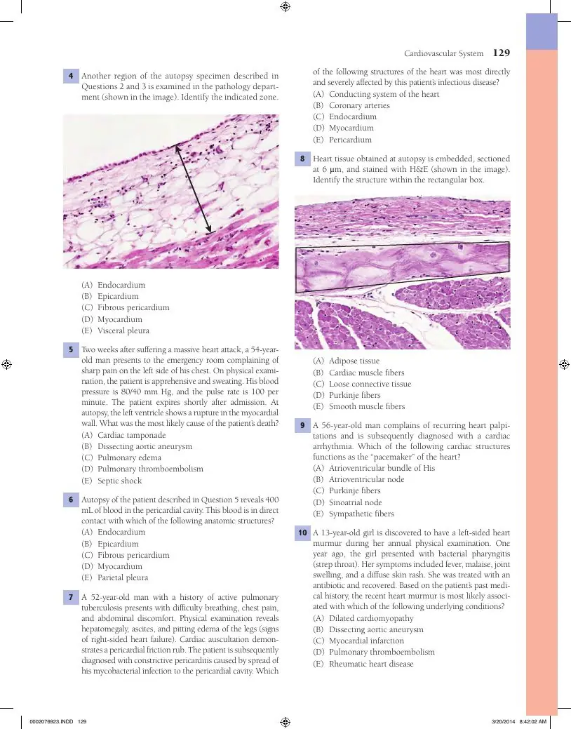
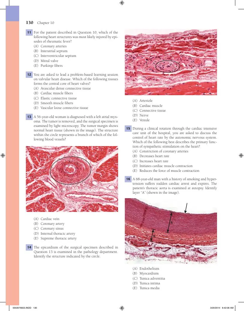
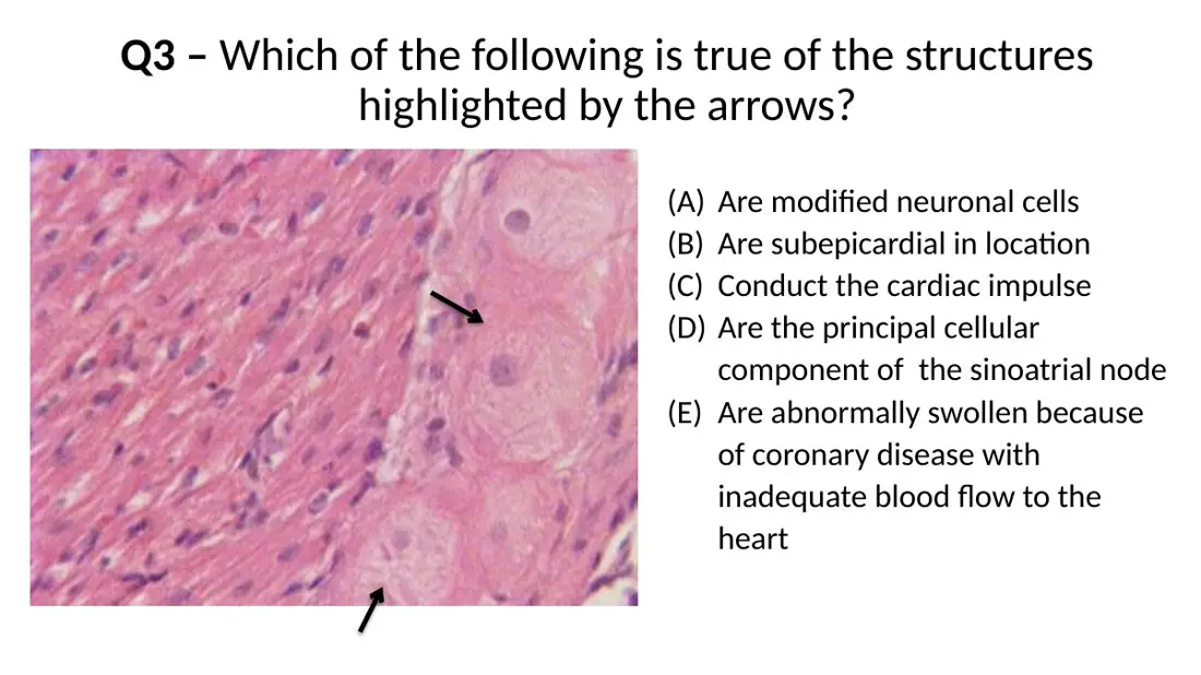
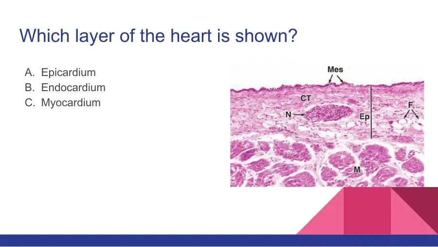
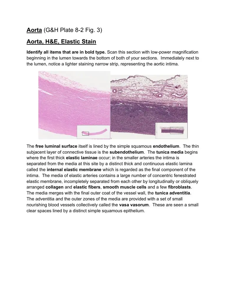
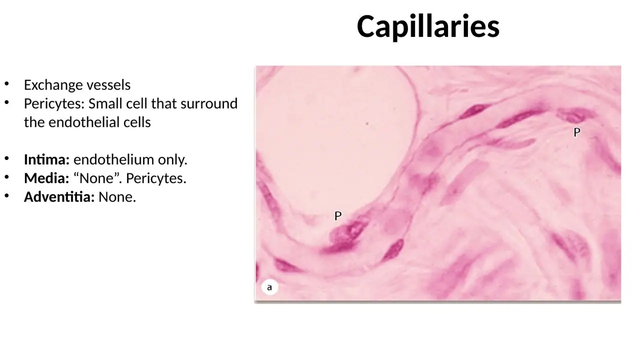
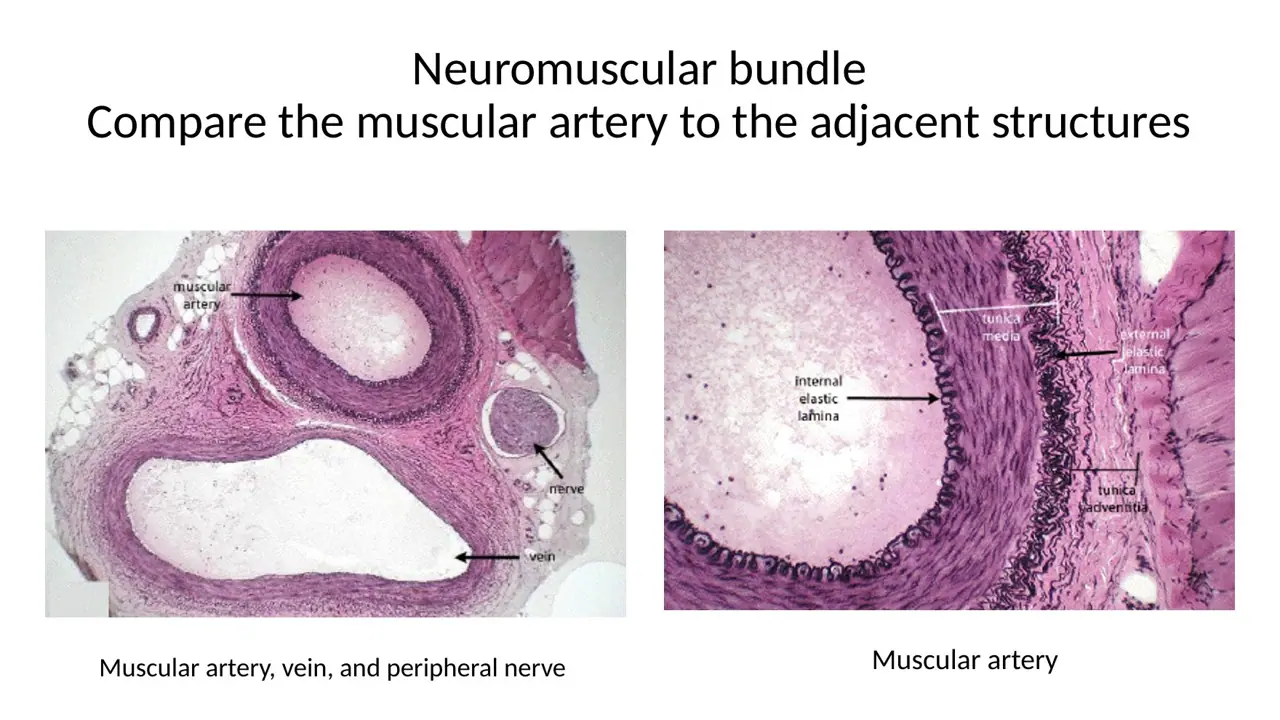
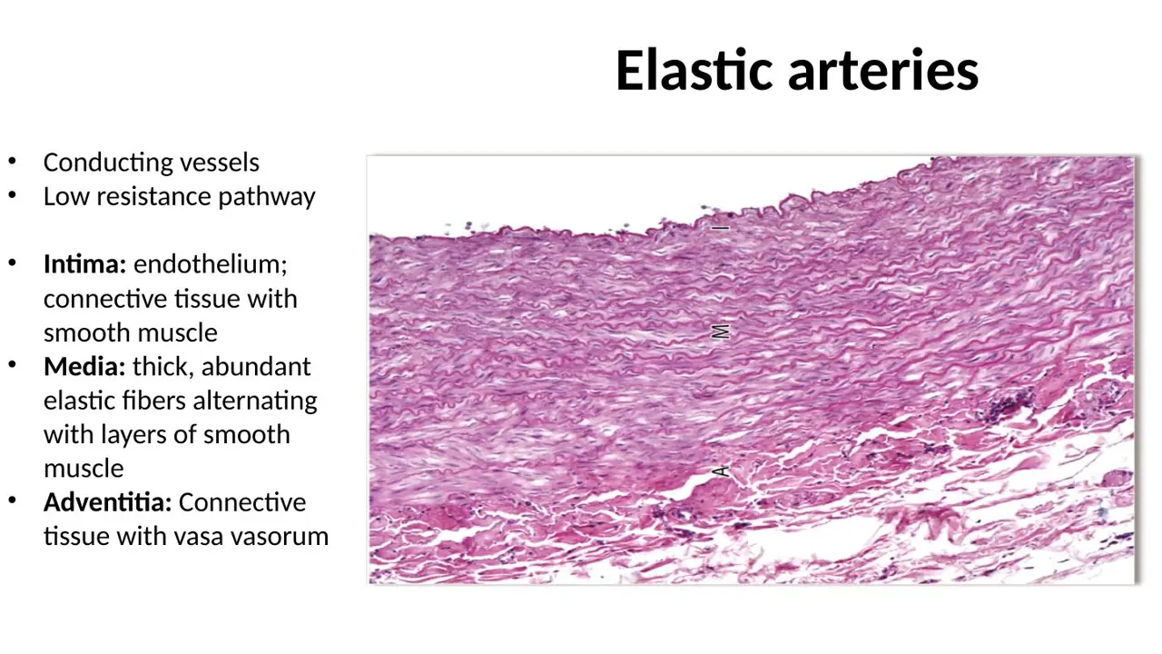
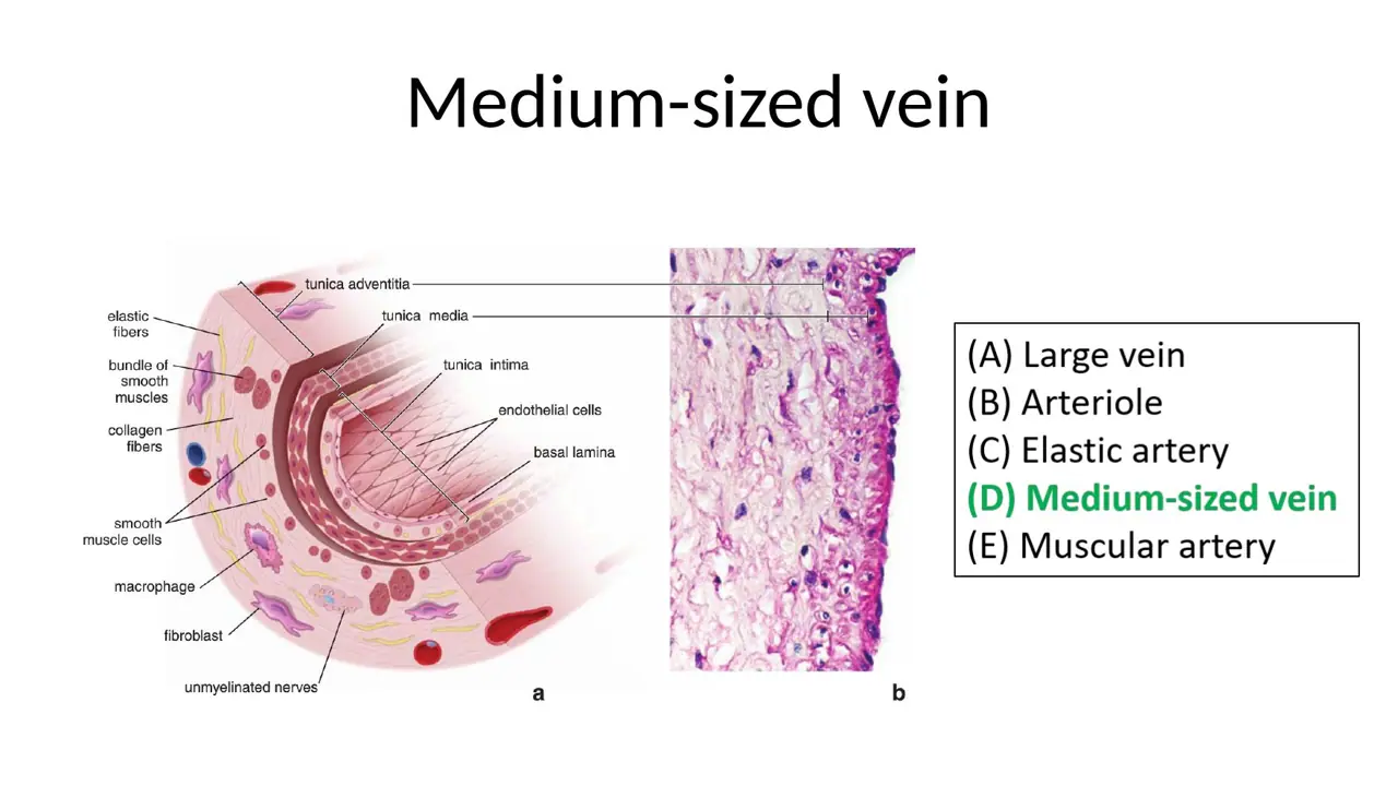
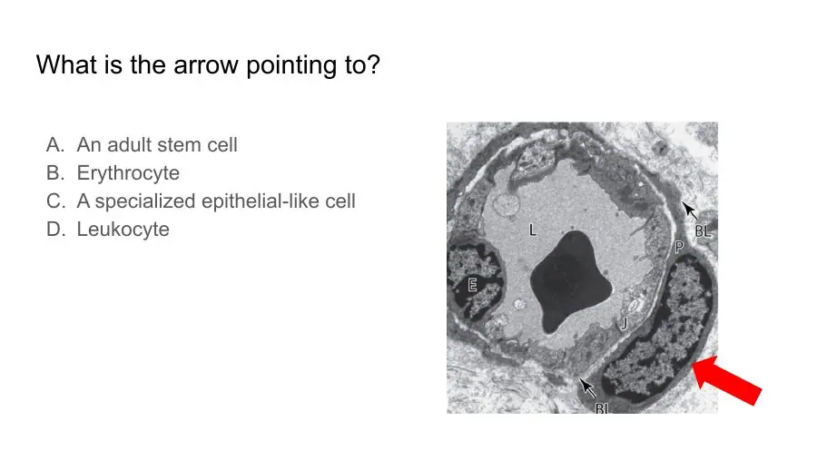
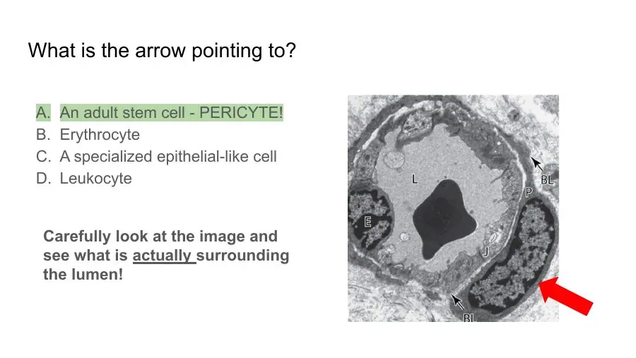
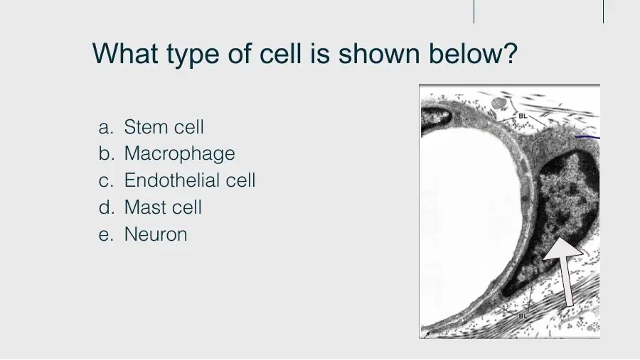
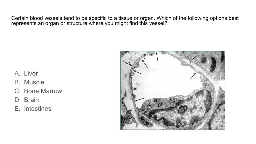
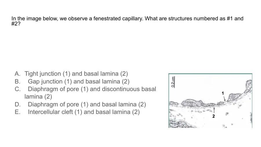
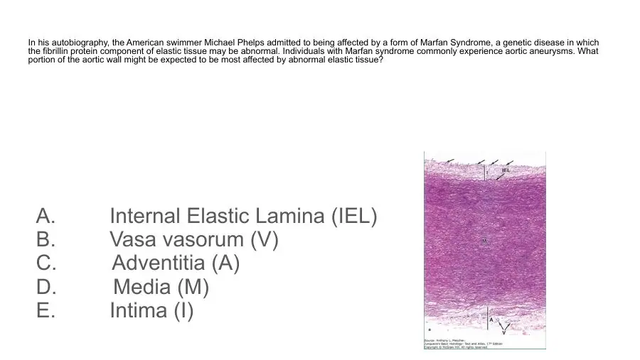
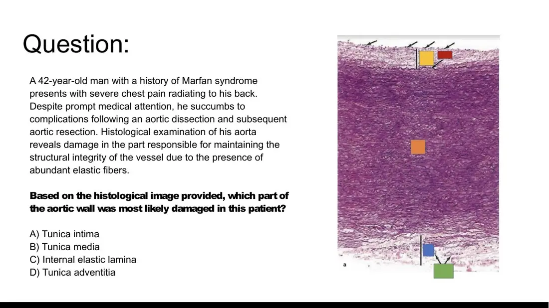
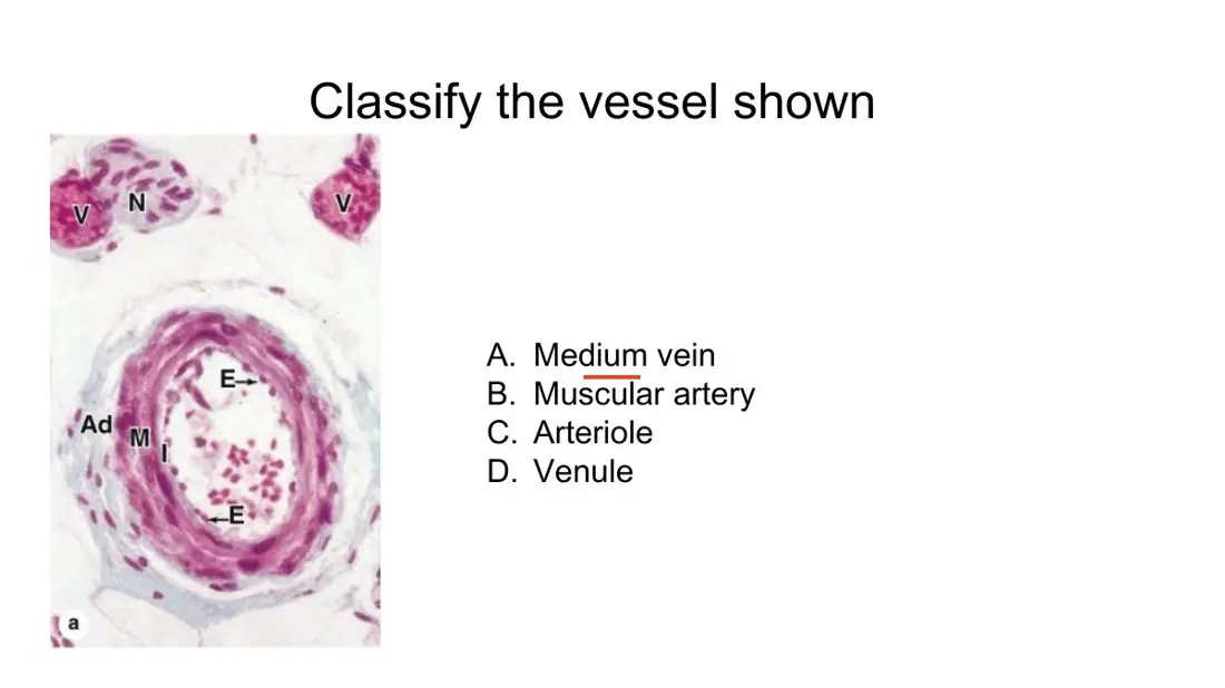
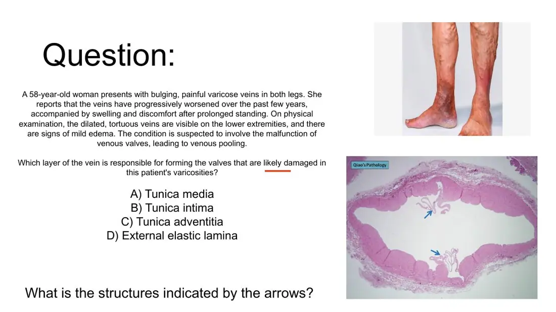
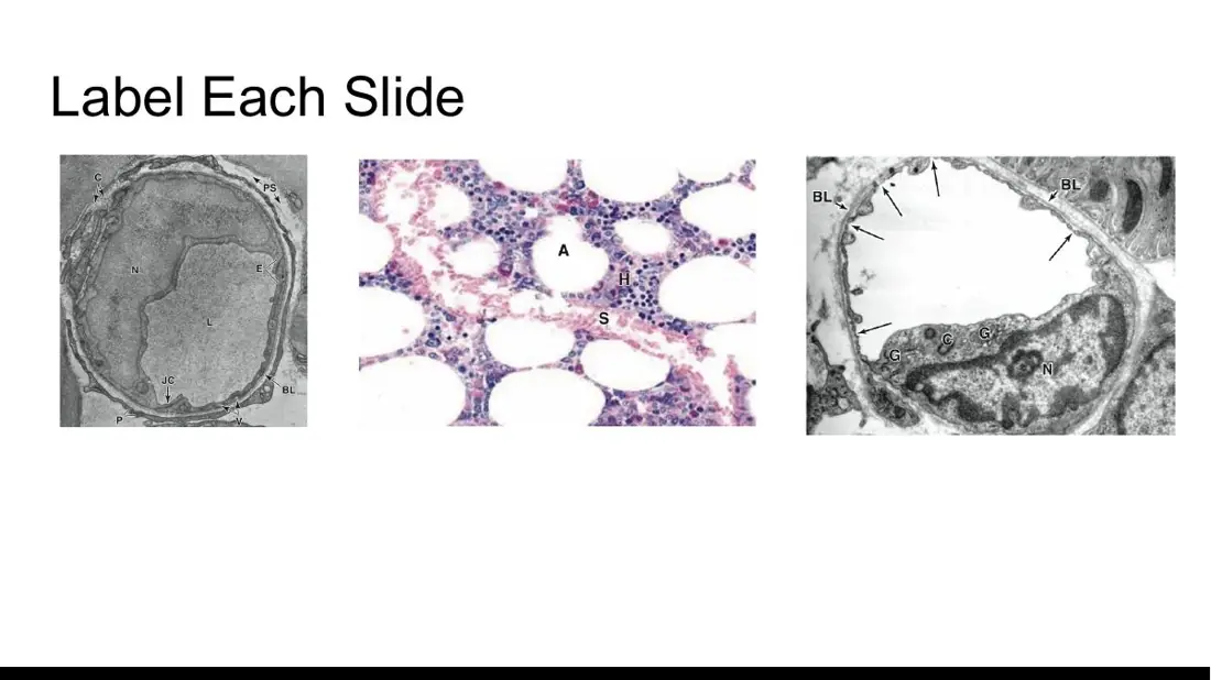
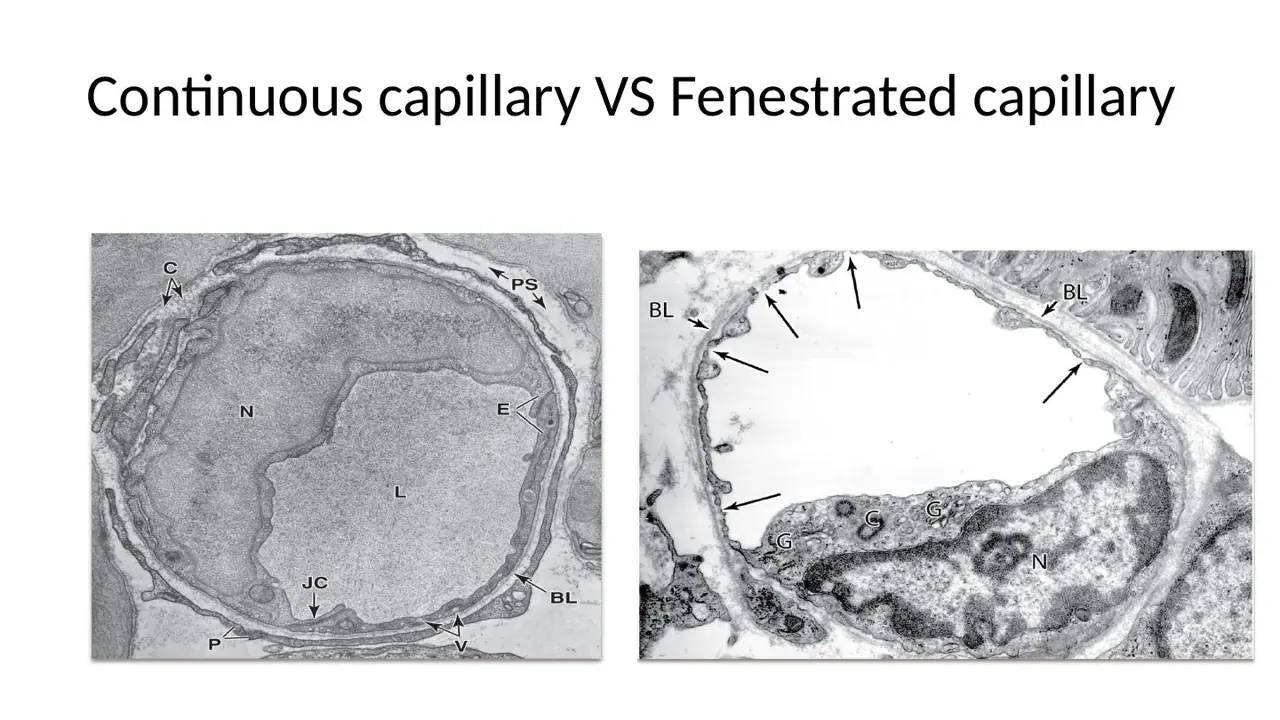
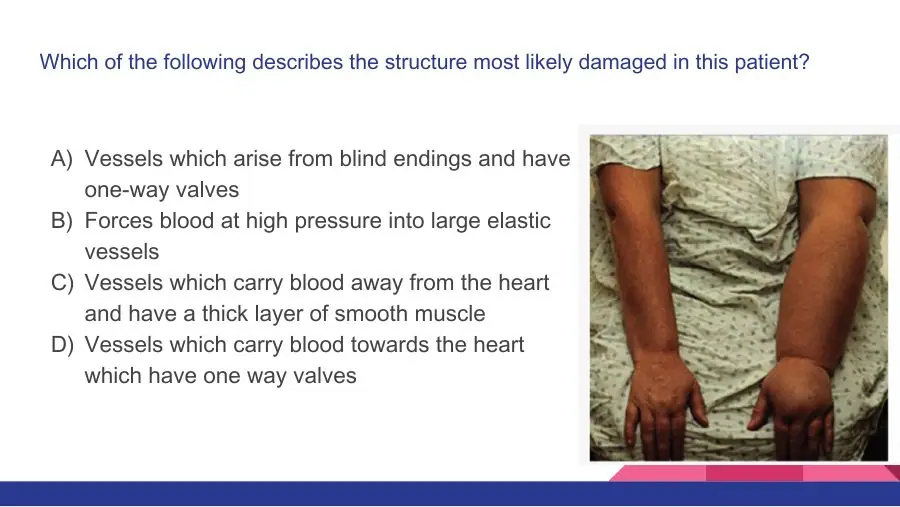
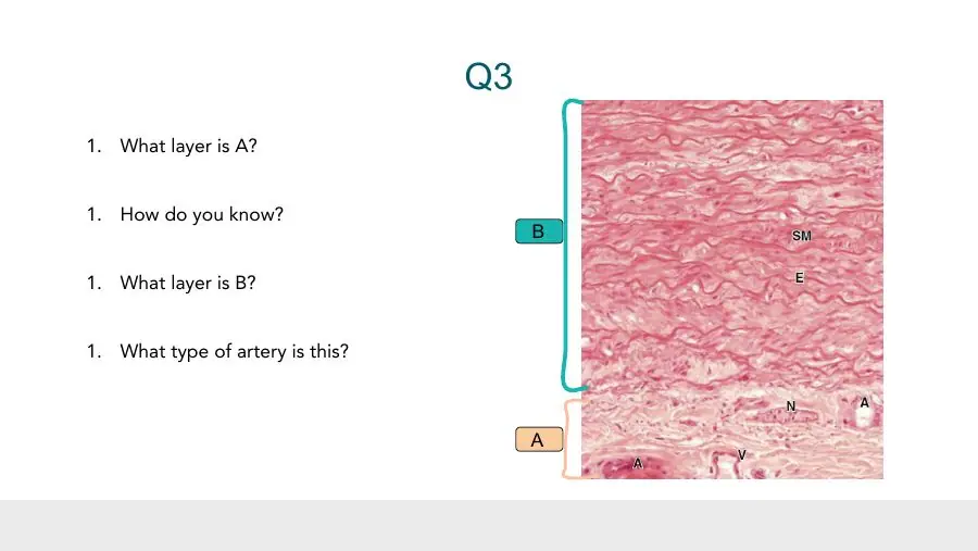
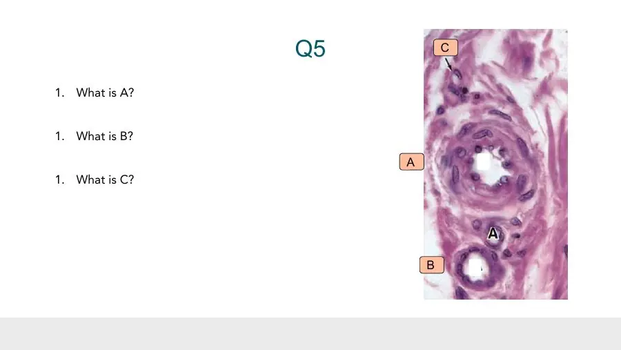
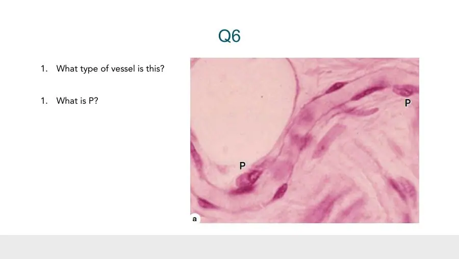
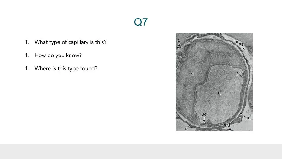
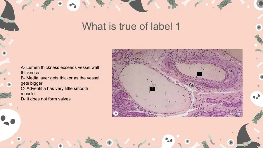
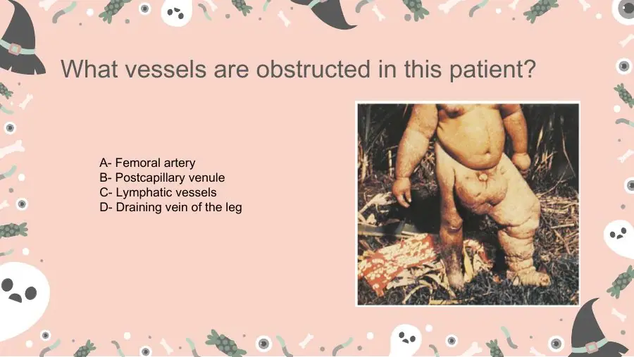
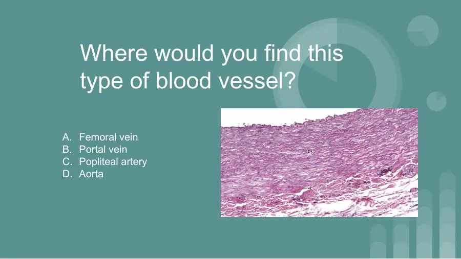
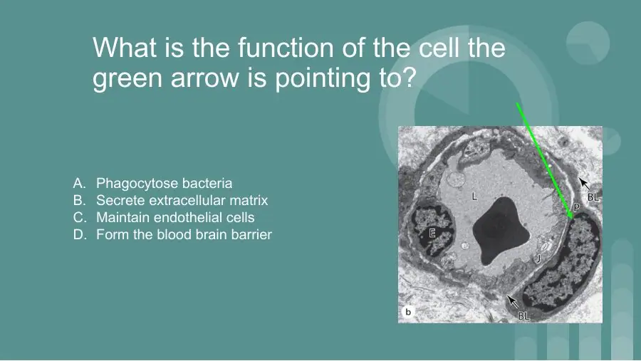
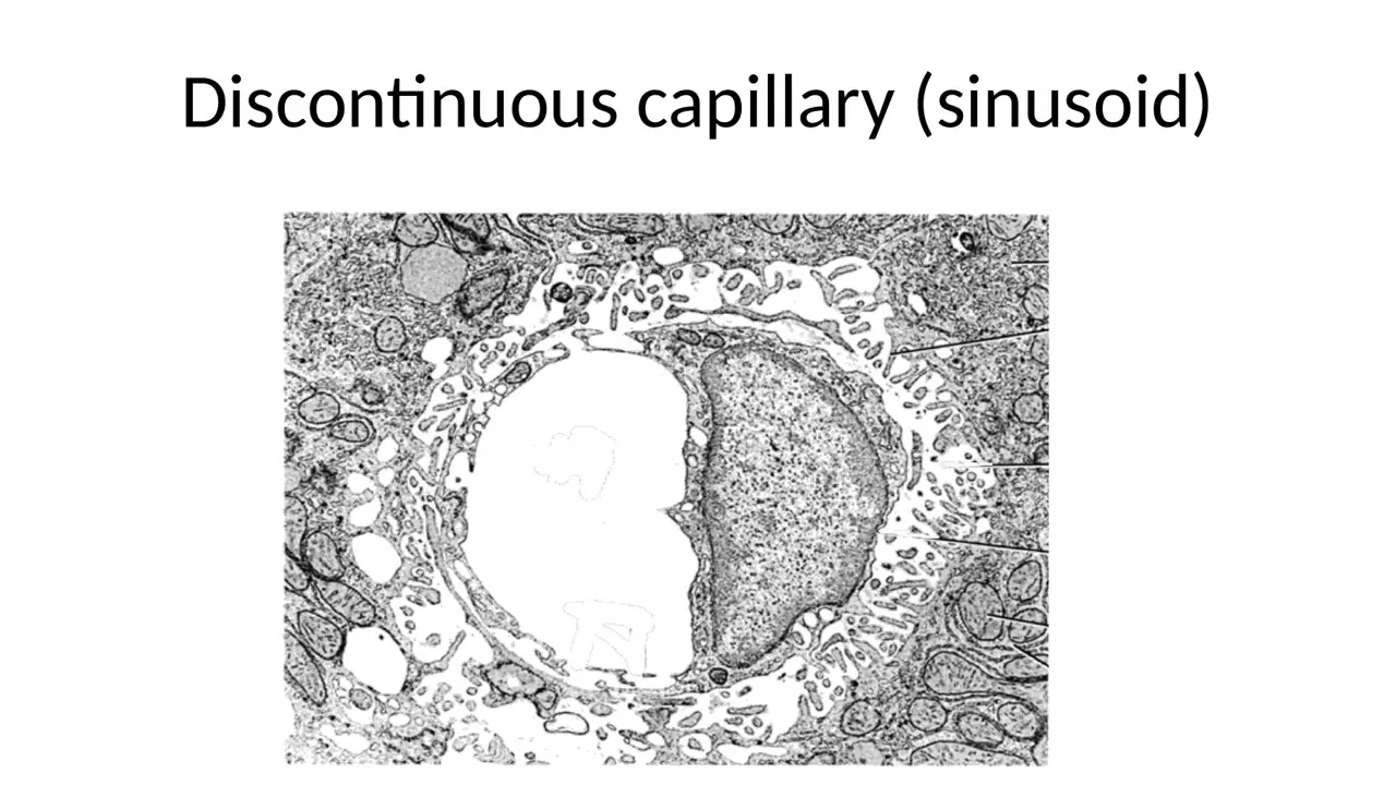
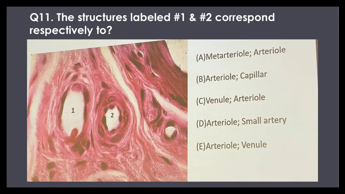
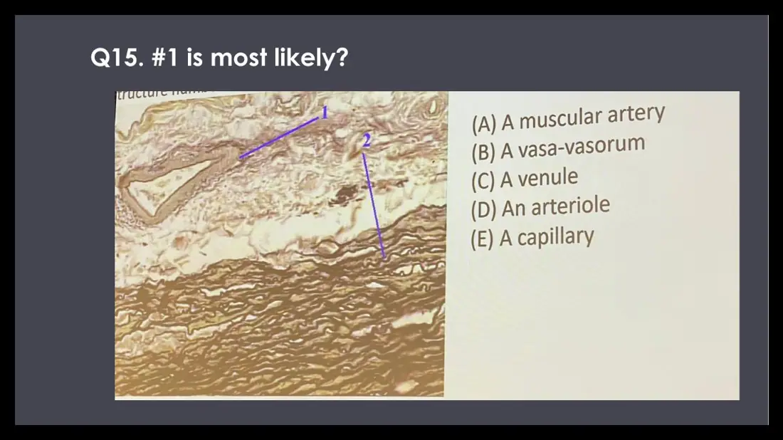
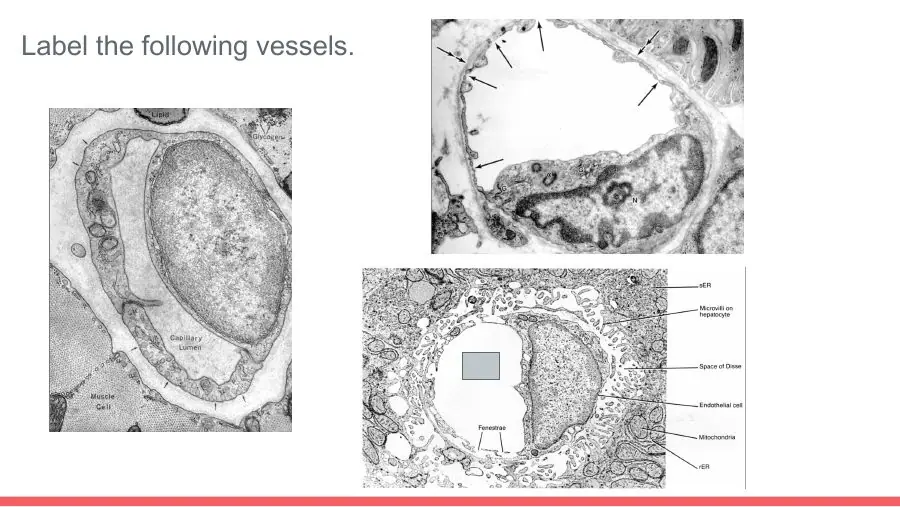
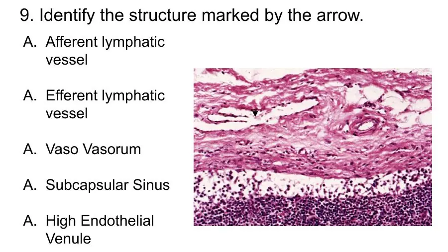
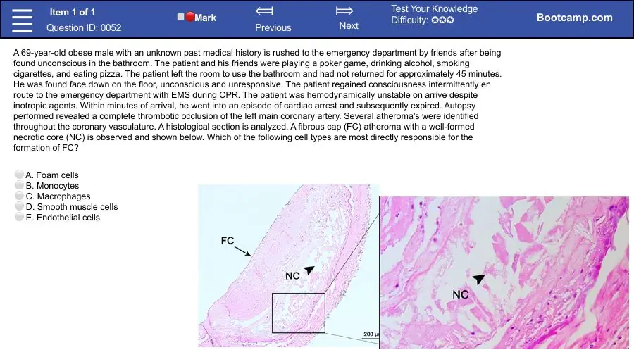
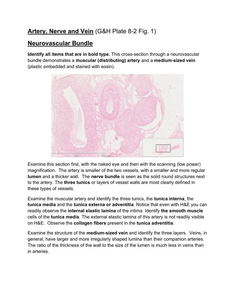
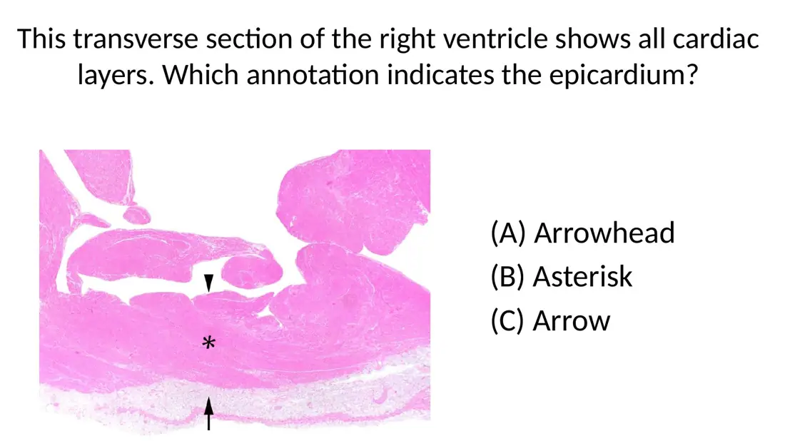
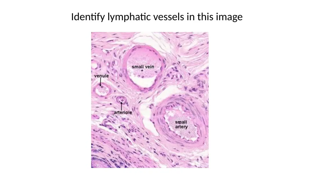
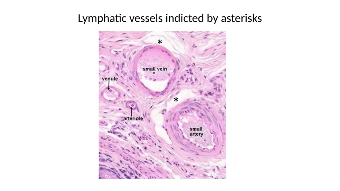
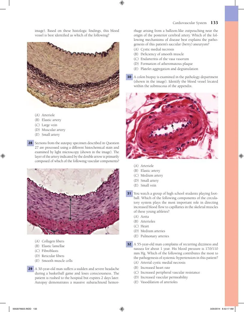
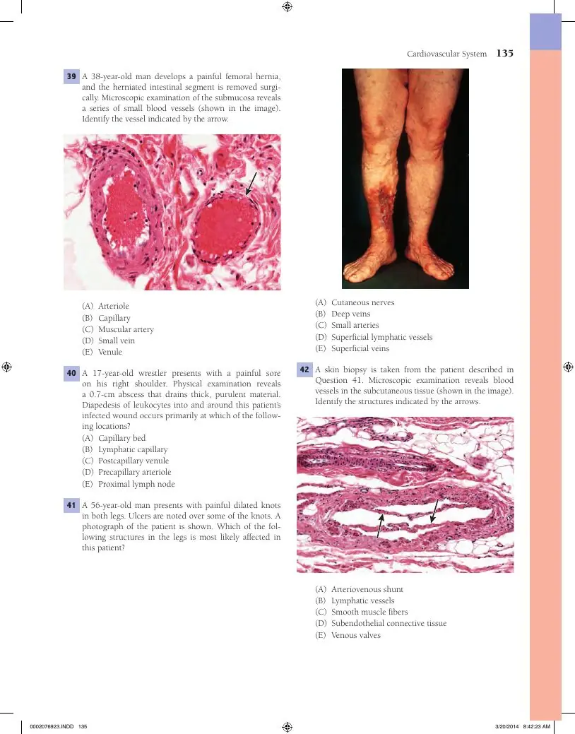
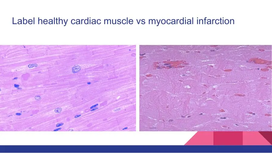
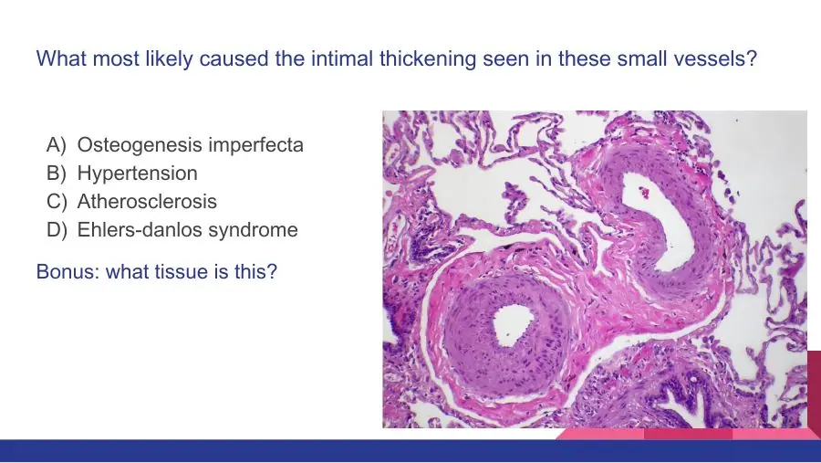
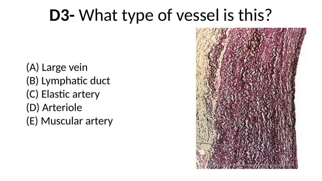
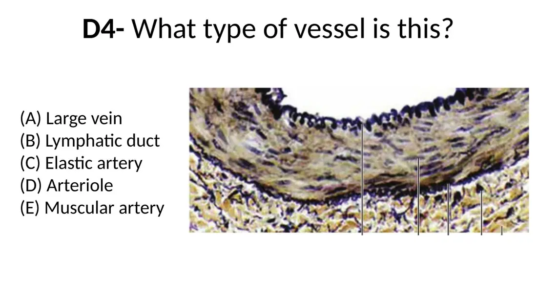
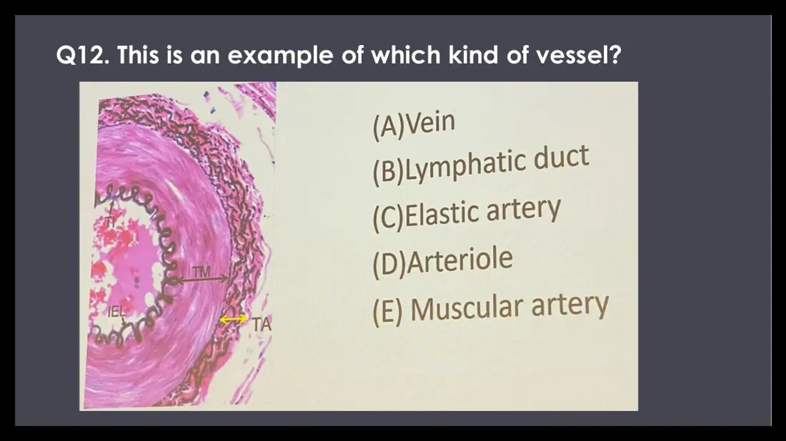
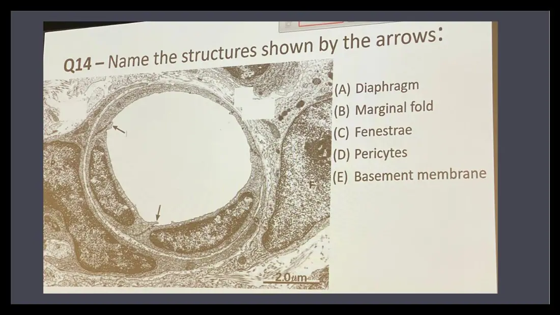
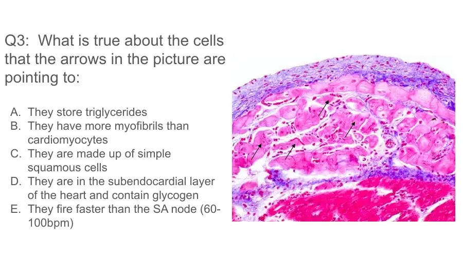
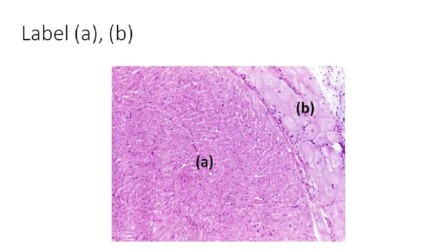
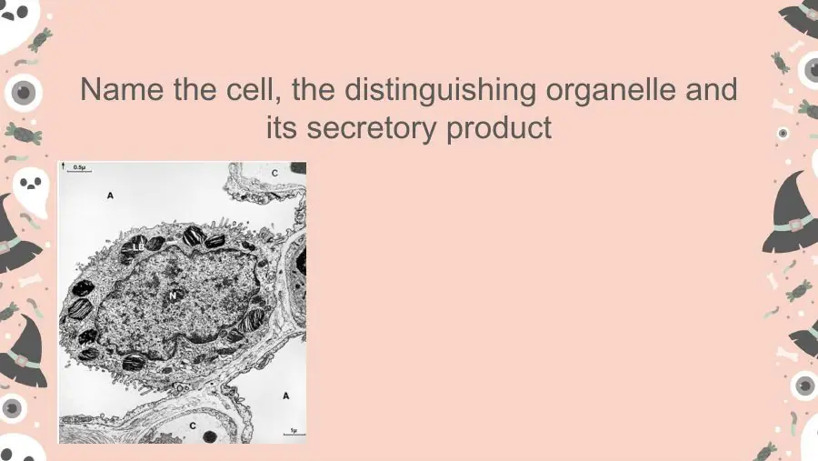
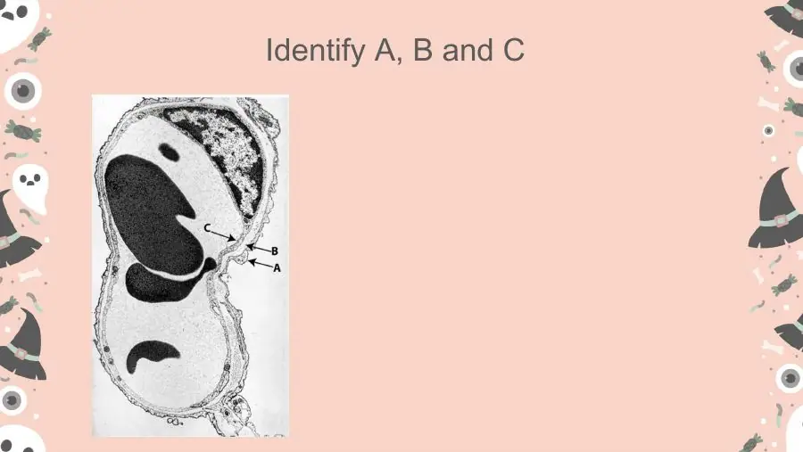
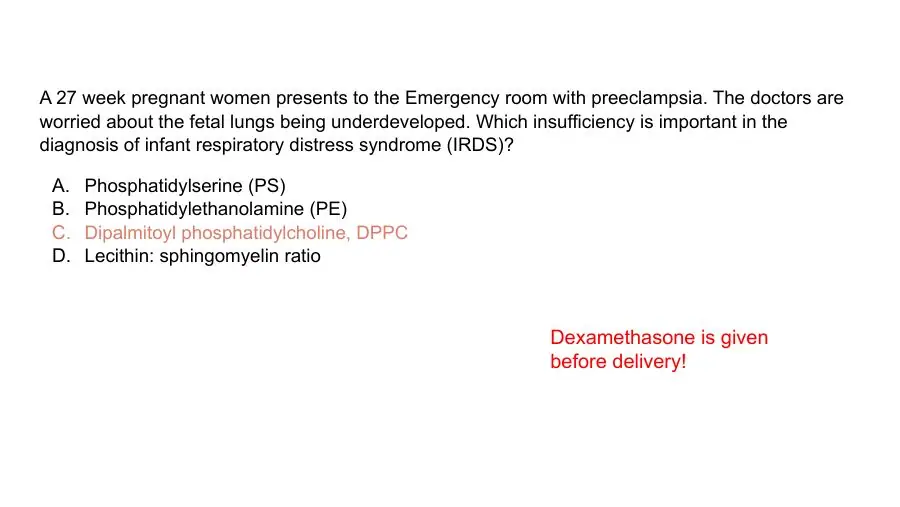
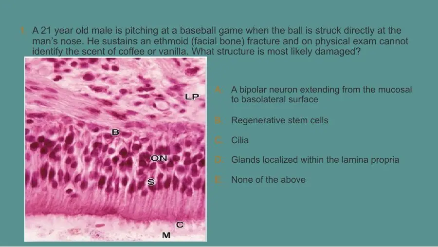
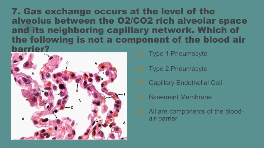
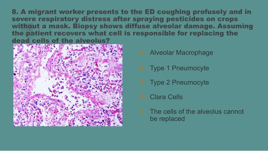
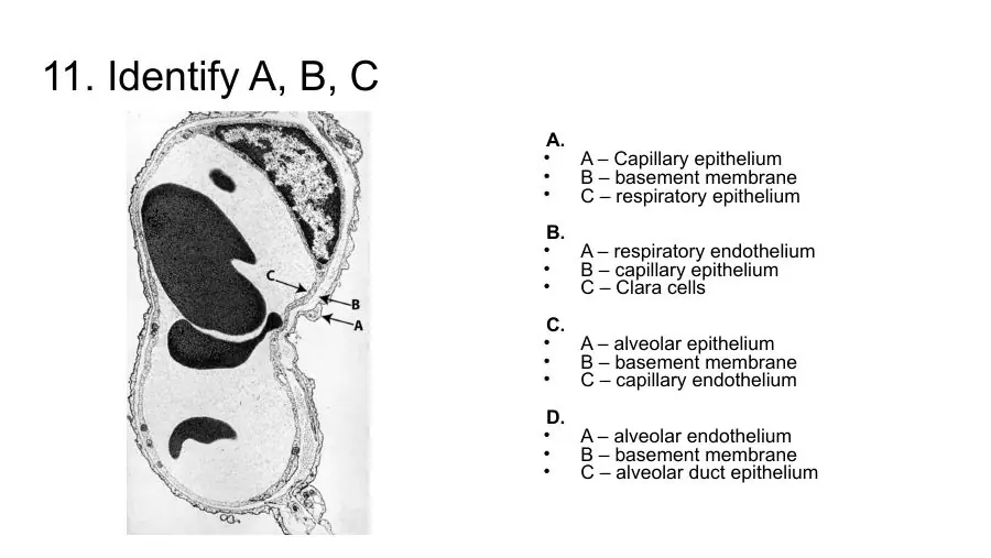
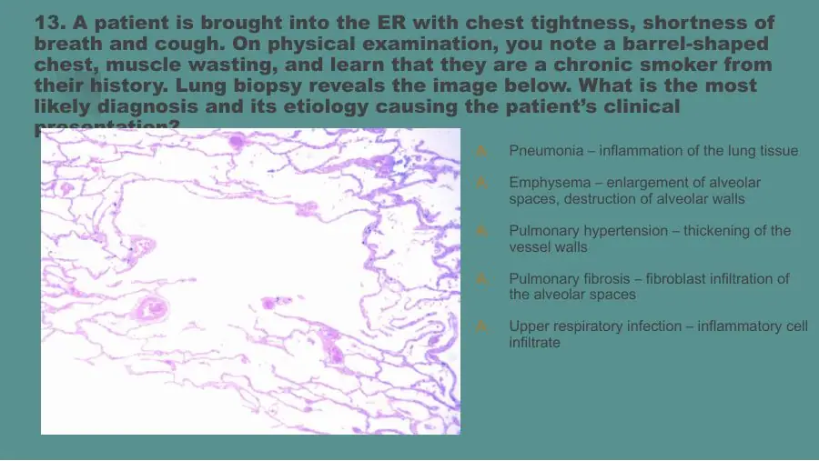
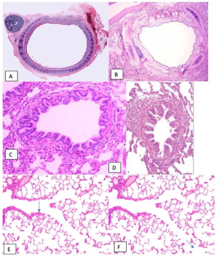
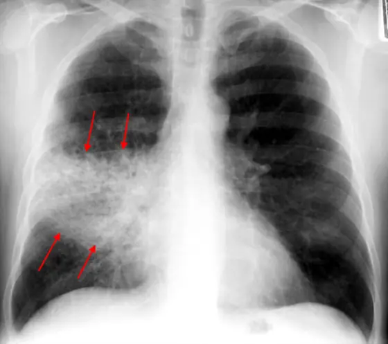
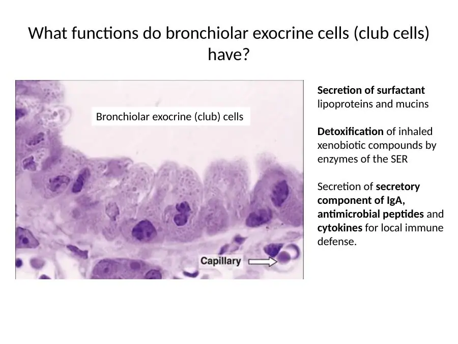
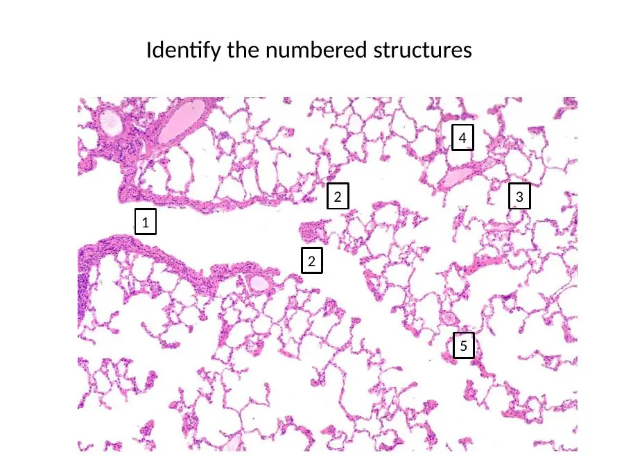
Every question found for this course's CP1 topics in Canvas, the lecture decks, the SGA drives, the TouroCOM resources and the textbooks, recorded as written. A question is listed once only when two sources word it exactly the same. Under each question, "Slides" opens the lecture and lab slides that lead to its answer. Each section opens with a Learning questions dropdown: the three-choice teaching questions from the Cardio-Pulm 1 question deck, each with its own "Slides" dropdown. Only CP1 material is kept (checked against the CP1 exam scope document); questions that are partly outside CP1 sit in a "Borderline" dropdown at the end of each section.
HIST-7A Circulatory system: general organization & the heart
102 questions · 69 concepts · 21 high-yield
Learning questions 85
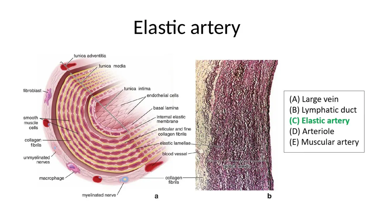
The circulatory system and the three layers of the heart wall
L1Learning
What are the two components of the circulatory system?
A. Blood vascular (cardiovascular) system + lymphatic vascular system
B. Blood vascular (cardiovascular) system + lymphoid organs (nodes, spleen, thymus)
C. Pulmonary circulation + systemic circulation, with no lymphatic part
AnswerA. Blood vascular (cardiovascular) system + lymphatic vascular system
WhyThe circulatory system = the cardiovascular (blood vascular) system (heart, arteries, capillaries, veins) + the lymphatic vascular system.
Explanation
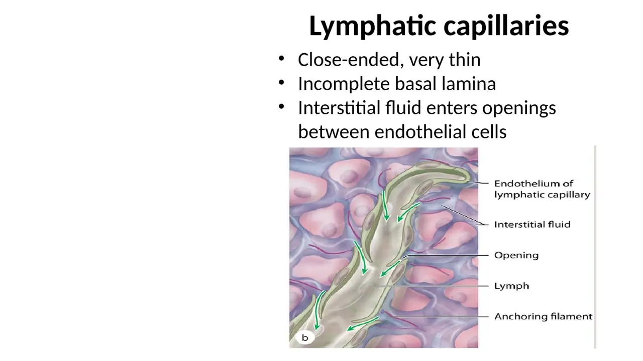
The blood vascular system is the heart plus the closed loop of arteries, capillaries and veins. The lymphatic vascular system is a one-way set of blind-ended lymphatic capillaries and larger lymphatic vessels that collect excess tissue fluid and return it to the veins through the thoracic and right lymphatic ducts.
Lymphoid organs (nodes, spleen, thymus) belong to the immune system; only the lymphatic vessels are part of the circulatory system in this course.
Pulmonary + systemic circulation is how the blood vascular system itself is divided; it leaves out the lymphatic half altogether.
Slide (1)H12A Circulatory I lecture (Dr Bruno) · slide 6L2Learning
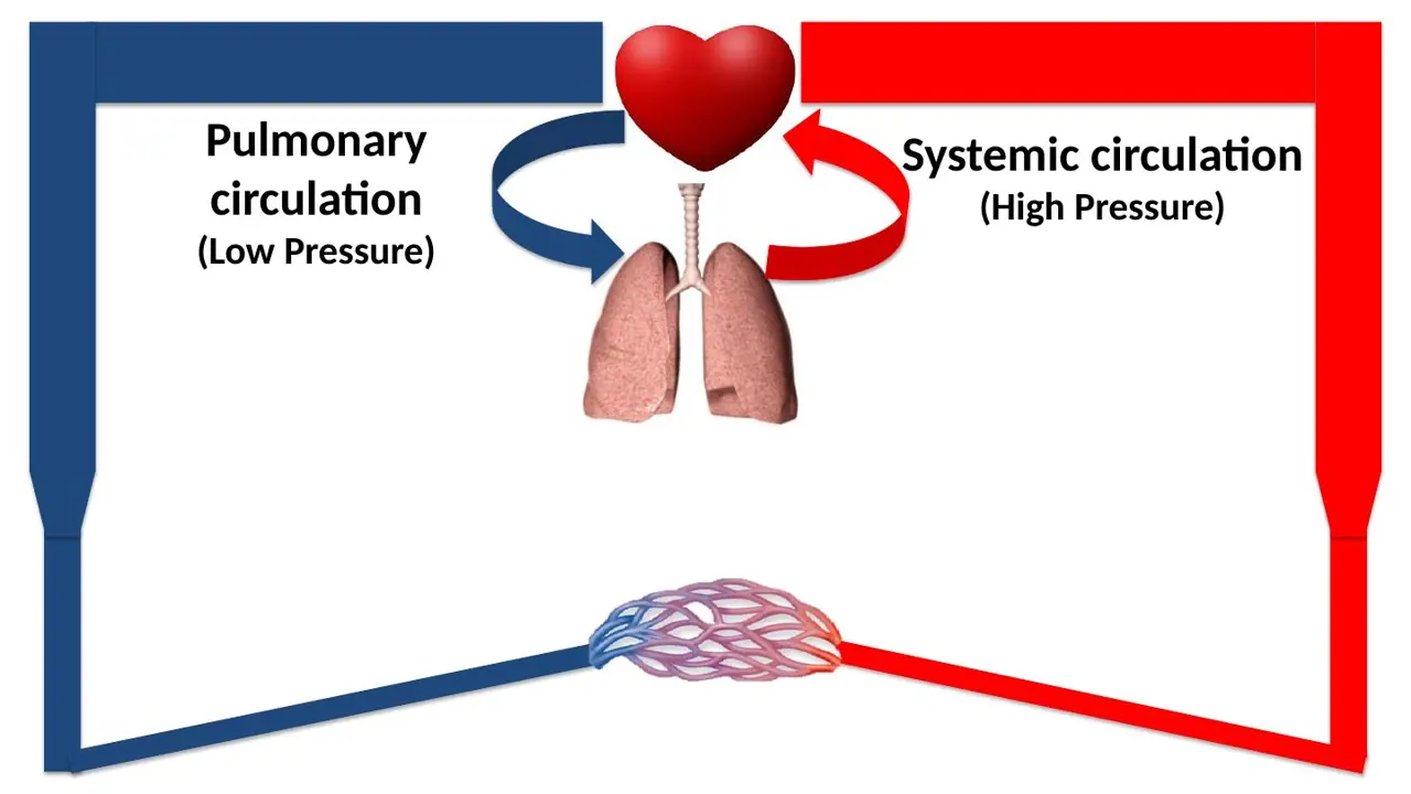
Which circulation works at high pressure and which at low pressure?
A. Pulmonary circulation high; systemic circulation low
B. Both run at the same high pressure, since both ventricles eject the same volume
C. Systemic circulation high; pulmonary circulation low
WhyThe left ventricle drives the high-pressure systemic circuit; the right ventricle drives the low-pressure pulmonary circuit through the lungs.
Explanation
Both ventricles eject the same volume each beat, but the resistance they pump against differs. The systemic circuit (left ventricle → aorta → body) has a high resistance, so it needs a high pressure (about 120/80 mmHg). The pulmonary circuit (right ventricle → pulmonary trunk → lungs) has a low resistance and runs at roughly a sixth of that pressure.
The reverse pairing swaps the two circuits.
Same pressure: equal volume does not mean equal pressure; pressure depends on the resistance downstream.
This is why the left ventricular wall is about three times thicker than the right.
Slide (1)H12A Circulatory I lecture (Dr Bruno) · slide 7L3Learning
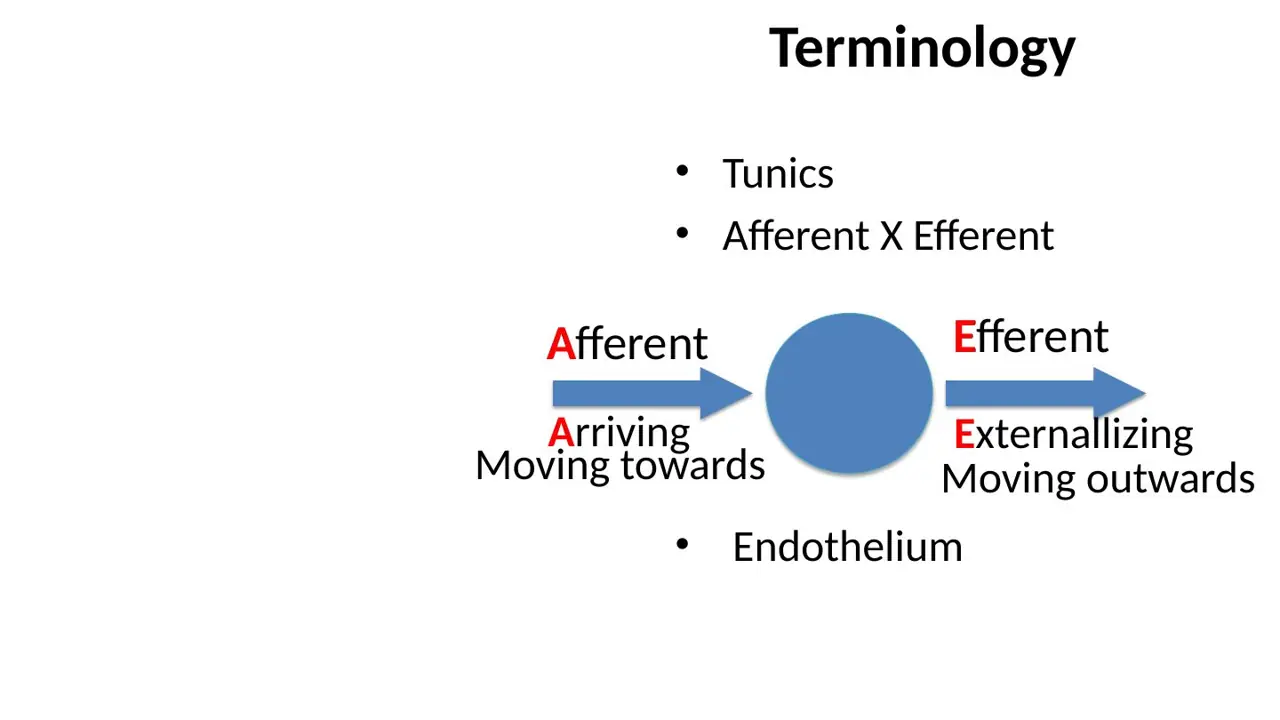
In the lecture's vessel terminology, what do afferent and efferent mean?
A. Afferent: arriving, moving towards a structure Efferent: exiting, moving outward from it
B. Afferent: exiting, moving outward from a structure Efferent: arriving, moving towards it
C. Afferent: carrying oxygenated blood Efferent: carrying oxygen-poor blood
AnswerA. Afferent: arriving, moving towards a structure Efferent: exiting, moving outward from it
WhyAfferent = Arriving (moving towards); efferent = Exiting (moving outward). The words describe direction, not oxygen content.
Explanation
The terms describe the direction of flow relative to the structure being discussed. A vessel that carries blood or lymph towards an organ is afferent; one that carries it away is efferent.
The swapped option reverses the definitions.
Oxygenated vs oxygen-poor confuses direction with content: an afferent vessel can carry either kind of blood.
Hook from the slide: Afferent = Arriving, Efferent = Externalising.
Slide (1)H12A Circulatory I lecture (Dr Bruno) · slide 4L4Learning
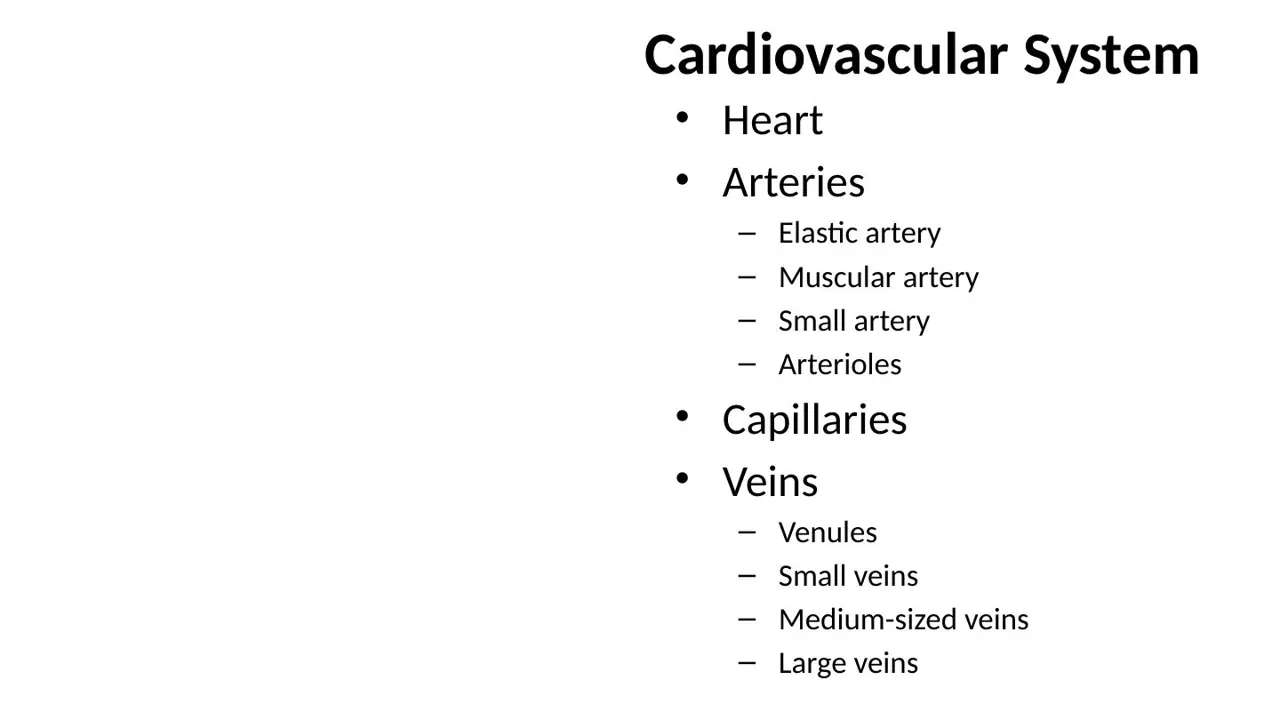
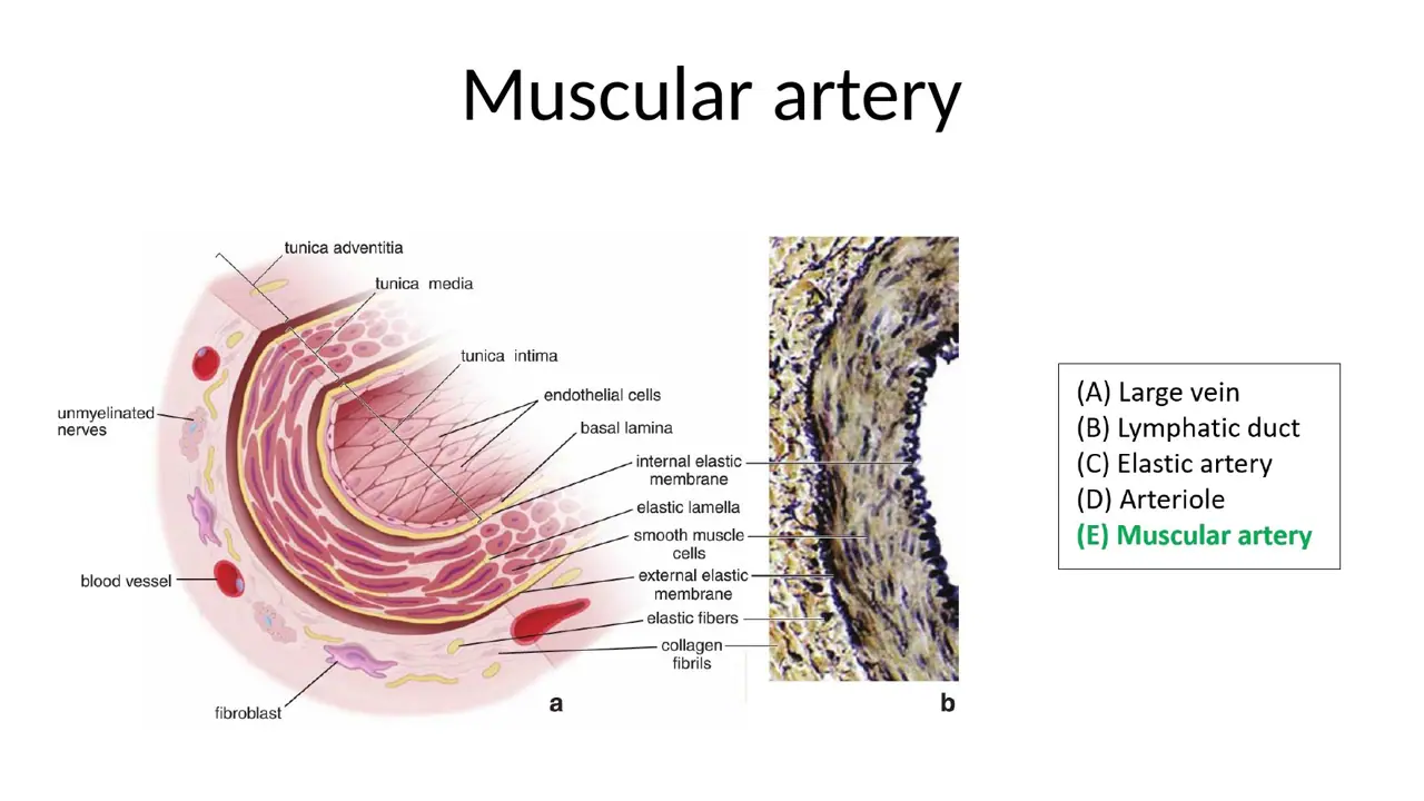
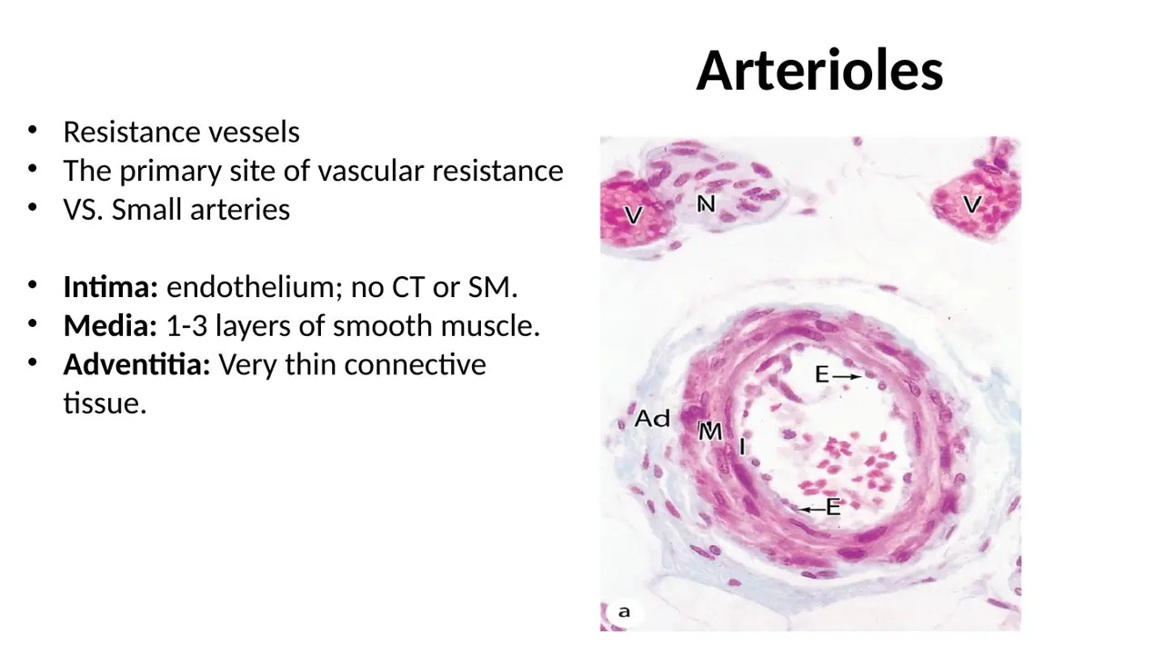
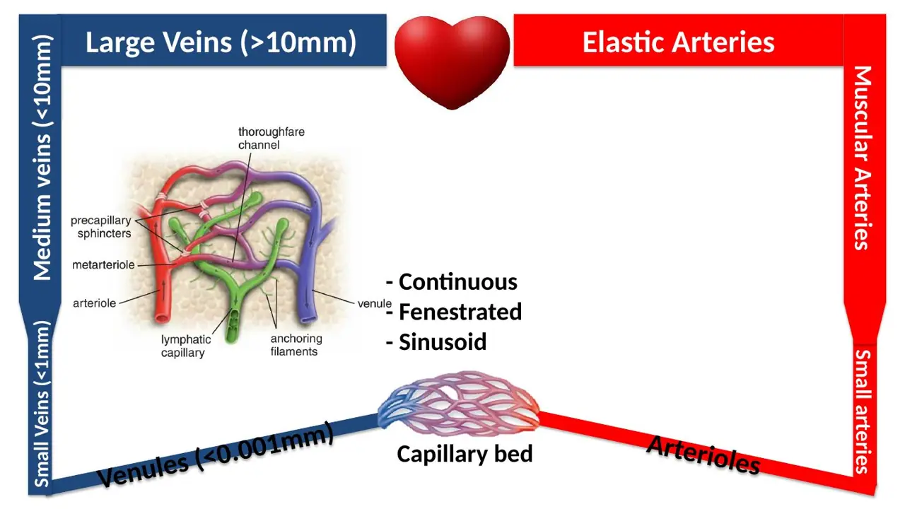
Going outward from the heart, how are the arteries subdivided?
A. Muscular artery → elastic artery → small artery → arteriole
B. Elastic artery → muscular artery → small artery → arteriole
C. Elastic artery → small artery → muscular artery → arteriole
WhyArteries shrink in steps: elastic (aorta) → muscular (distributing) → small artery → arteriole, which feeds the capillaries.
Explanation
The lecture's list of cardiovascular elements runs heart → arteries → capillaries → veins. The elastic arteries (aorta, pulmonary trunk) leave the heart; they branch into muscular arteries that distribute blood to organs, then into small arteries, and finally arterioles, the resistance vessels just before the capillaries. Veins run the other way: venules → small veins → medium-sized veins → large veins.
Putting muscular before elastic reverses the first step: the aorta, right at the heart, is the elastic one.
Putting small artery before muscular breaks the steady fall in size.
Slide (1)H12A Circulatory I lecture (Dr Bruno) · slide 8L5Learning
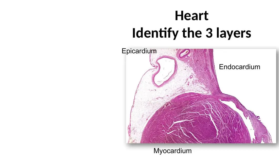
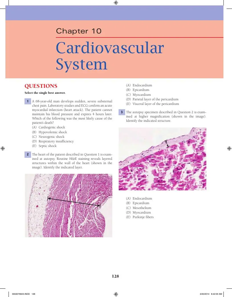
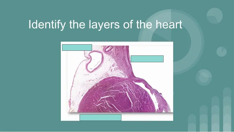
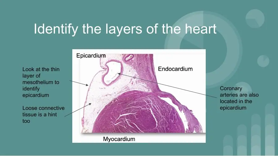
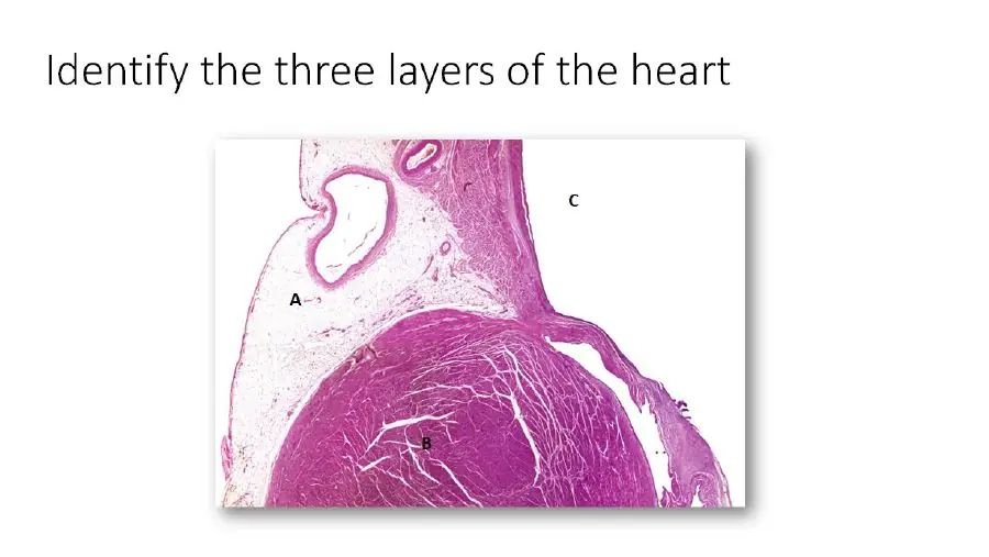
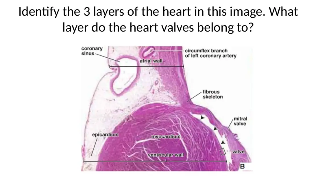
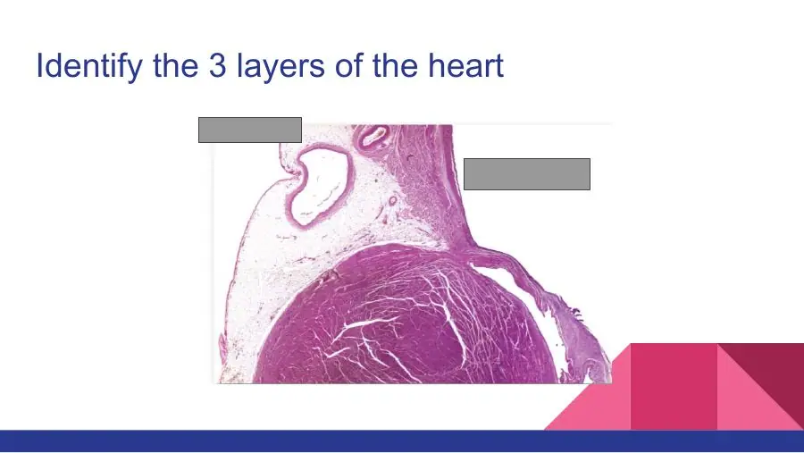
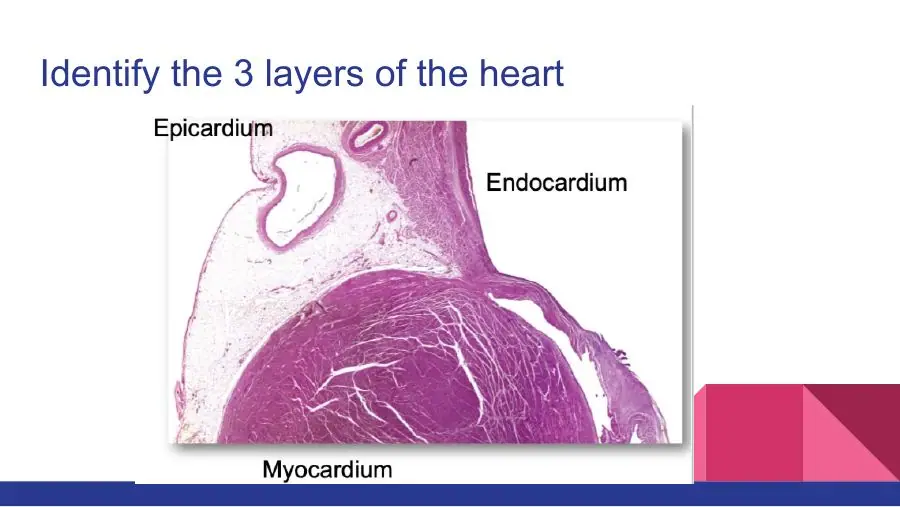
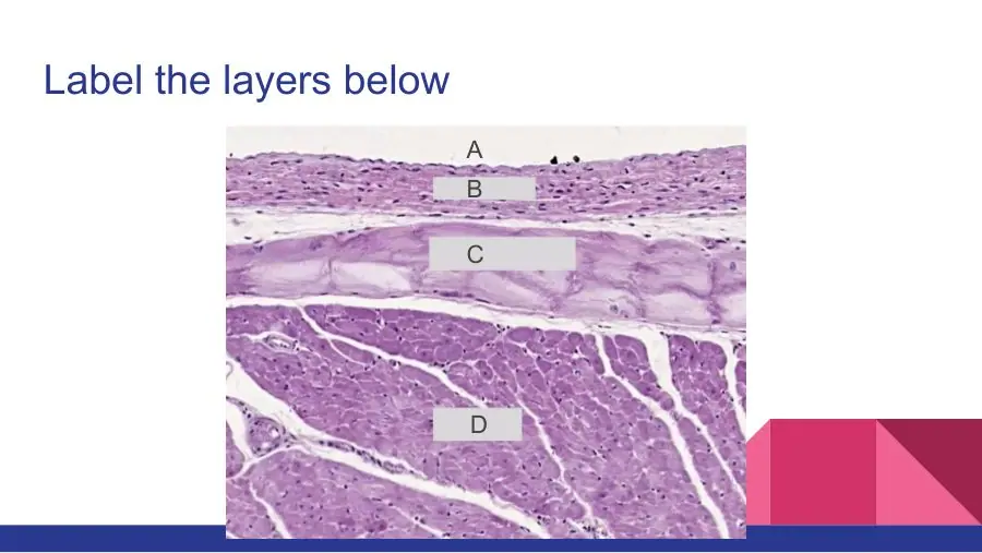
Name the three layers of the heart wall, from inside out.
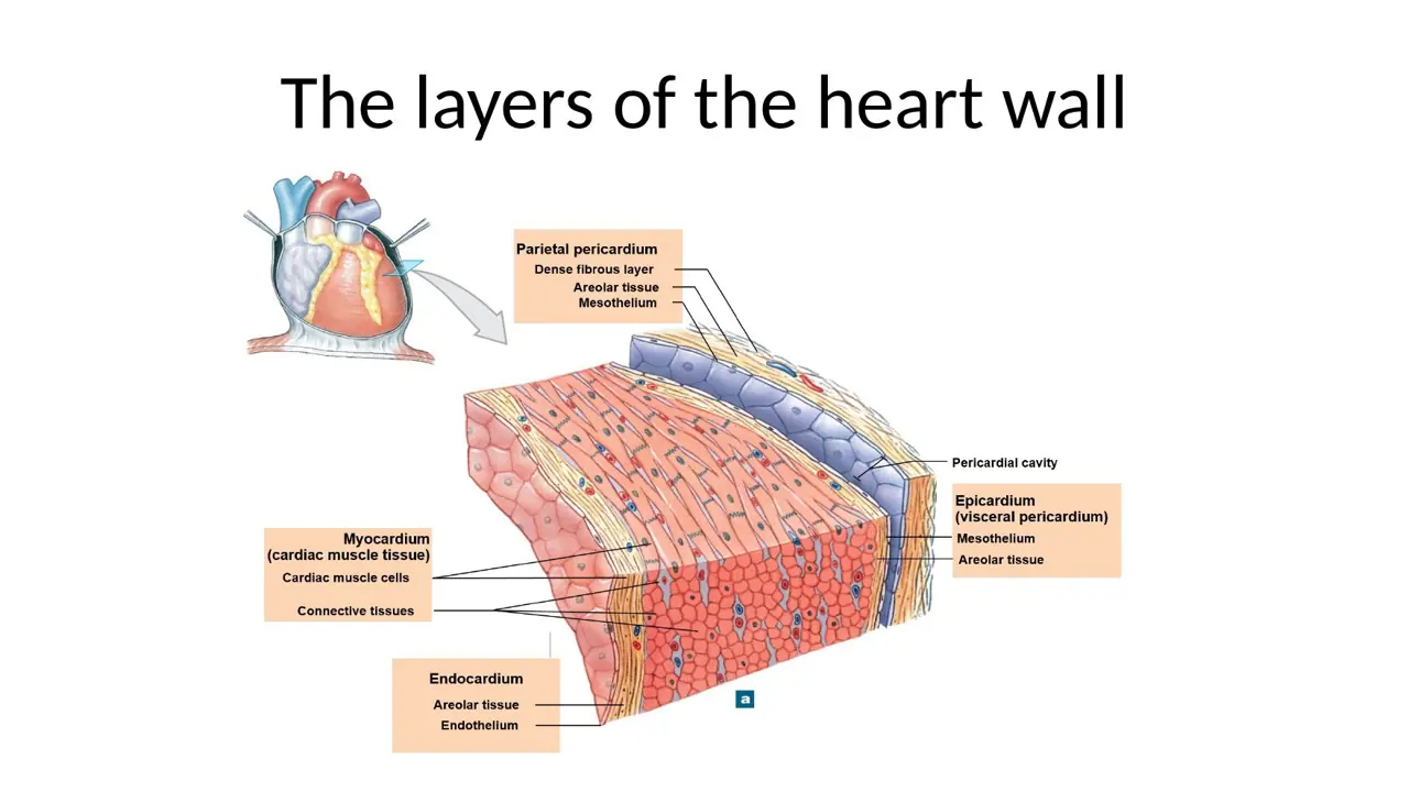
Endo- = inside, myo- = muscle, epi- = upon. The endocardium lines the chambers and faces the blood; the myocardium is the thick muscular middle; the epicardium covers the outer surface and faces the pericardial cavity.
Epicardium → myocardium → endocardium is the right sequence read from outside in, not inside out.
Endocardium → epicardium → myocardium puts the covering layer in the middle; the muscle always sits between the two thin layers.
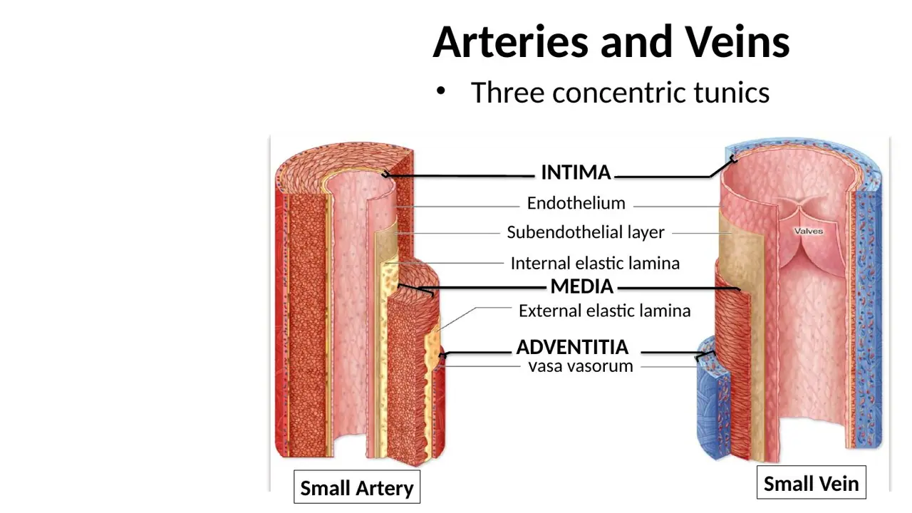
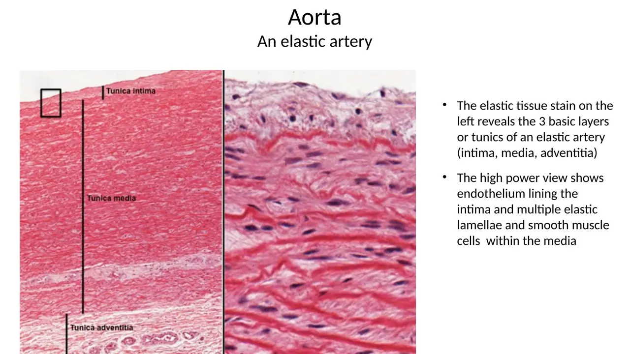
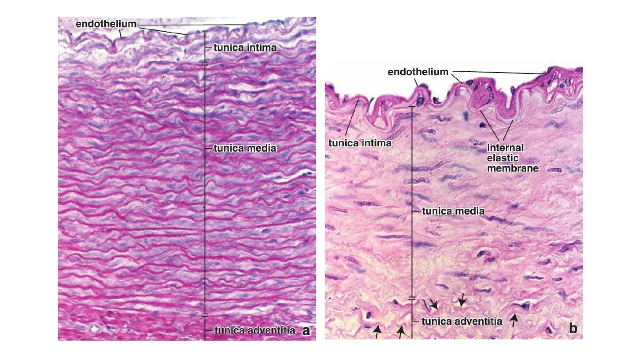
The heart wall is a modified vessel wall: endocardium ≈ tunica intima, myocardium ≈ tunica media, epicardium ≈ tunica adventitia.
The endocardium is continuous with which layer of the great vessels?
A. Tunica media
B. Tunica adventitia
C. Tunica intima
AnswerC. Tunica intima
WhyThe endocardium's endothelium and subendothelial tissue continue straight into the tunica intima of the aorta, pulmonary trunk and great veins.
Explanation
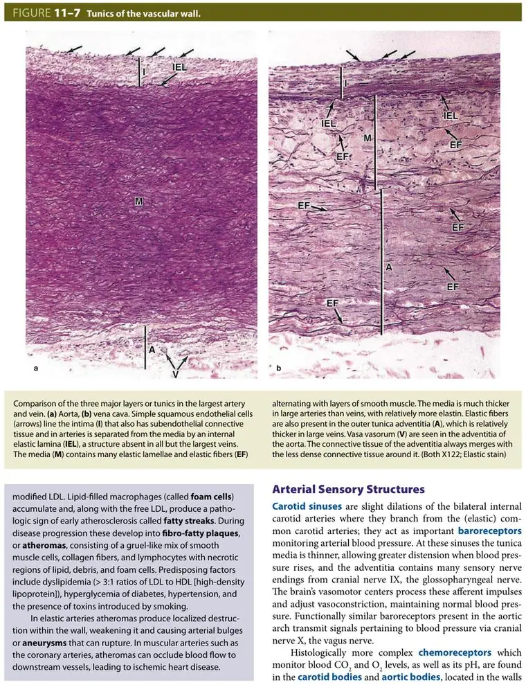
Both the endocardium and the tunica intima are an endothelium (simple squamous epithelium) on a thin layer of connective tissue, and they face the blood. Where the great vessels join the heart, one simply runs into the other, so the endothelium lining blood is one unbroken sheet from the heart to the capillaries.
Tunica media is the vessel's muscular/elastic middle coat; its heart equivalent is the myocardium.
Tunica adventitia is the outer connective-tissue coat; its heart equivalent is the epicardium.
Which layer is the thickest part of the heart wall, and which vessel tunic does it correspond to?
A. Endocardium (connective tissue) — equivalent of the tunica media
B. Myocardium (cardiac muscle) — equivalent of the tunica adventitia
C. Myocardium (cardiac muscle) — equivalent of the tunica media
AnswerC. Myocardium (cardiac muscle) — equivalent of the tunica media
WhyThe myocardium, closely packed cardiac muscle, is by far the thickest layer; like the vessel media, it is the contractile middle coat.
Explanation
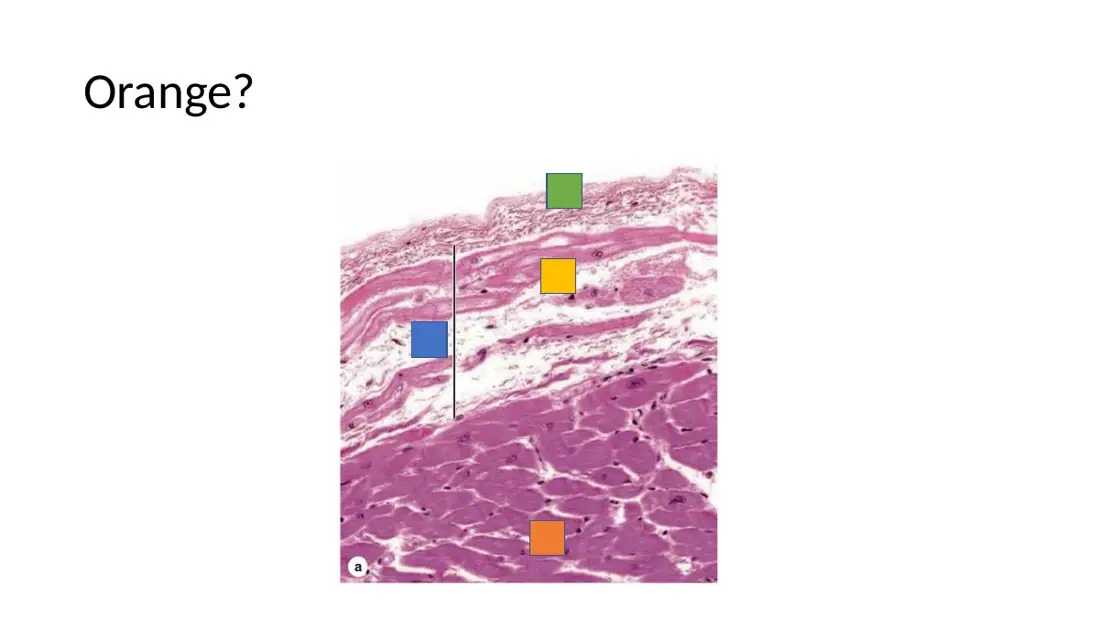
The myocardium is the muscular wall of the heart: cardiac muscle cells arranged in spiral layers round the chambers, grouped into fascicles by connective-tissue septa that carry vessels and nerves. It does the pumping, as the smooth muscle of the tunica media does in an artery.
The endocardium is a thin lining (endothelium plus connective tissue), not the bulk of the wall; it matches the intima.
The adventitia is the outer connective-tissue coat; the epicardium, not the myocardium, corresponds to it.
Slide (1)Lab manual HL7 circulatory · page 11
Pericardium and epicardium
L8Learning
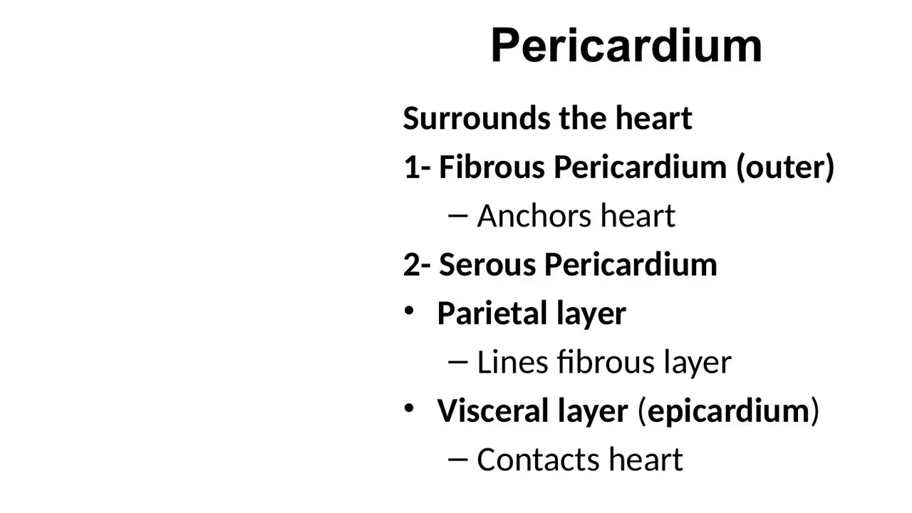
What does the fibrous pericardium do, and what is its key mechanical property?
A. Elastic outer sac that cushions the heart; recoils after stretch
B. Tough outer sac that anchors the heart; cannot stretch quickly
C. Thin serous membrane that secretes fluid; stretches readily
AnswerB. Tough outer sac that anchors the heart; cannot stretch quickly
WhyThe fibrous pericardium is the dense outer sac that anchors the heart; it is inextensible over minutes to hours, which is why sudden fluid causes tamponade.
Explanation
The fibrous pericardium is a sac of dense collagenous connective tissue attached to the diaphragm and the roots of the great vessels. It holds the heart in place and stops it overfilling. Because collagen barely stretches, the sac cannot expand to make room for fluid that collects suddenly.
Elastic and recoiling describes elastic tissue, not this collagen sac.
Secreting fluid is the job of the serous pericardium's mesothelium, which lines the inside of the fibrous sac and covers the heart.
Slide (1)H12A Circulatory I lecture (Dr Bruno) · slide 14L9Learning
A shard of glass pierces the chest wall and enters the left ventricle. In what order does it cross the layers?
The pericardium has two parts. The fibrous pericardium is the outer sac. The serous pericardium is a closed double layer of mesothelium: its parietal layer lines the inside of the fibrous sac, then folds back at the great vessels to become the visceral layer, which sticks to the heart surface and is the epicardium. The thin pericardial cavity lies between the two serous layers.
Putting the visceral layer first inverts the serous pair: the visceral layer is the one on the heart.
Putting the parietal layer outside the fibrous sac is wrong: it lines the fibrous sac from the inside.
Picture a fist (the heart) pushed into a balloon: the layer touching the fist is visceral, the outer balloon wall is parietal.
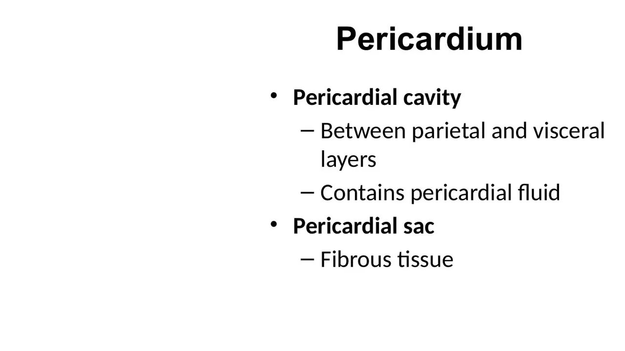
Where is the pericardial cavity, and what does it contain?
A. Between the parietal and visceral serous layers; a thin film of lubricating fluid
B. Between the fibrous pericardium and the parietal serous layer; a thin film of lubricating fluid
C. Between the visceral serous layer and the myocardium; a thin film of lubricating fluid
AnswerA. Between the parietal and visceral serous layers; a thin film of lubricating fluid
WhyThe pericardial cavity is the potential space between the parietal and visceral serous layers; it holds a small amount of pericardial fluid.
Explanation
Normally the cavity is only a slit holding a few tens of millilitres of serous fluid made by the mesothelium. The fluid lets the beating heart slide against the sac without friction.
The fibrous pericardium and parietal layer are fused to each other; there is no space between them.
The visceral layer and myocardium are joined by the loose subepicardial connective tissue (with fat and coronary vessels), not separated by a cavity.
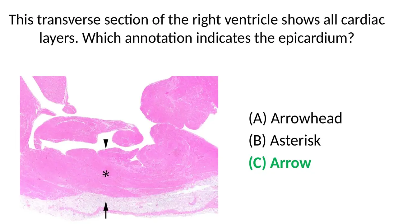
Because both walls of the cavity are serous layers, any blood or fluid that collects there touches the epicardium directly.
Slide (1)H12A Circulatory I lecture (Dr Bruno) · slide 15L11Learning
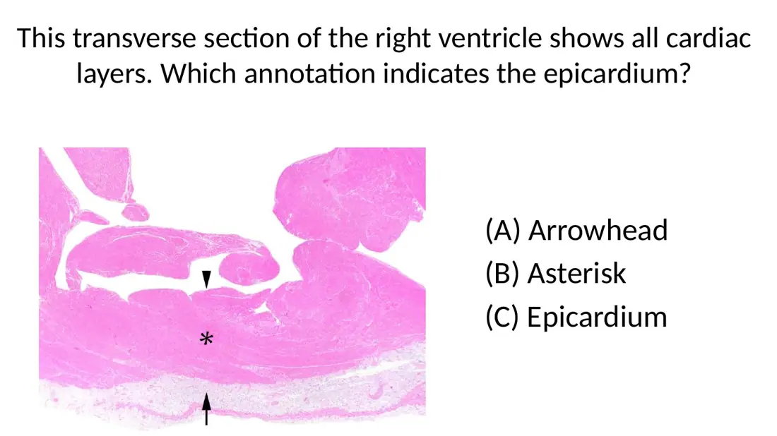
The epicardium is the same layer as which of these?
A. The parietal layer of the serous pericardium
B. The visceral layer of the serous pericardium
C. The fibrous pericardium
AnswerB. The visceral layer of the serous pericardium
WhyThe epicardium is the visceral pericardium: the serous layer stuck to the heart surface.
Explanation
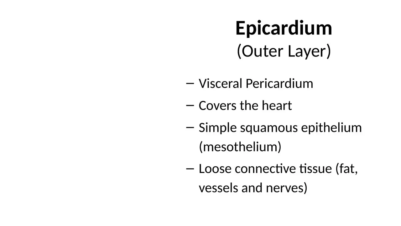
The serous pericardium has two layers. The visceral one covers the heart and is called the epicardium; it is mesothelium on loose connective tissue that holds fat, coronary vessels and nerves. The parietal one lines the inside of the fibrous sac.
The parietal layer faces the epicardium across the pericardial cavity but is part of the sac, not the heart wall.
The fibrous pericardium is the outermost dense sac and has no mesothelium of its own.
Hook: visceral = on the viscus (the heart itself).
Slide (1)H12A Circulatory I lecture (Dr Bruno) · slide 14L12Learning
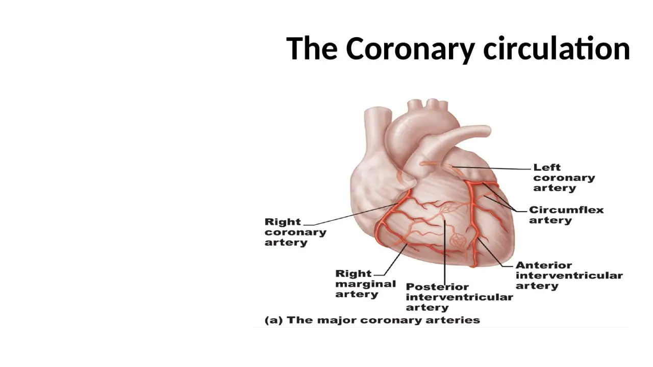
In which layer of the heart wall do the main coronary arteries and veins run?
A. Epicardium
B. Myocardium
C. Endocardium
AnswerA. Epicardium
WhyThe coronary arteries and veins (and the cardiac nerves) run in the epicardium, embedded in its fat, and send branches inward into the myocardium.
Explanation
The epicardium's loose connective tissue holds variable amounts of fat, and the large coronary branches lie in this fat on the heart surface, for example the anterior interventricular artery in the fat of the anterior interventricular sulcus. Smaller branches then dive into the myocardium.
The myocardium contains only the smaller penetrating branches and a dense capillary bed, not the main vessels.
The endocardium is a thin lining nourished largely by diffusion from the chamber blood.
This is why a coronary bypass graft is sewn onto the surface of the heart.
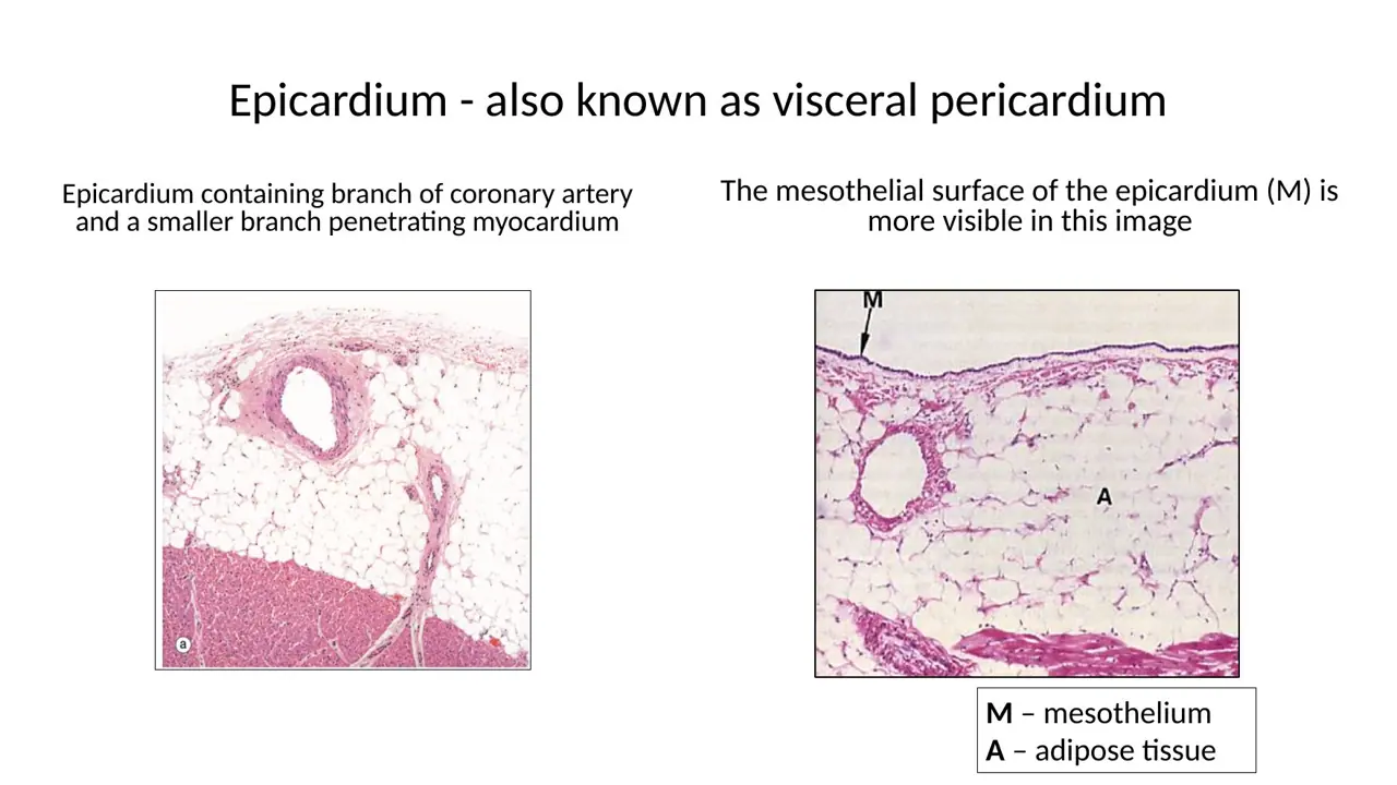
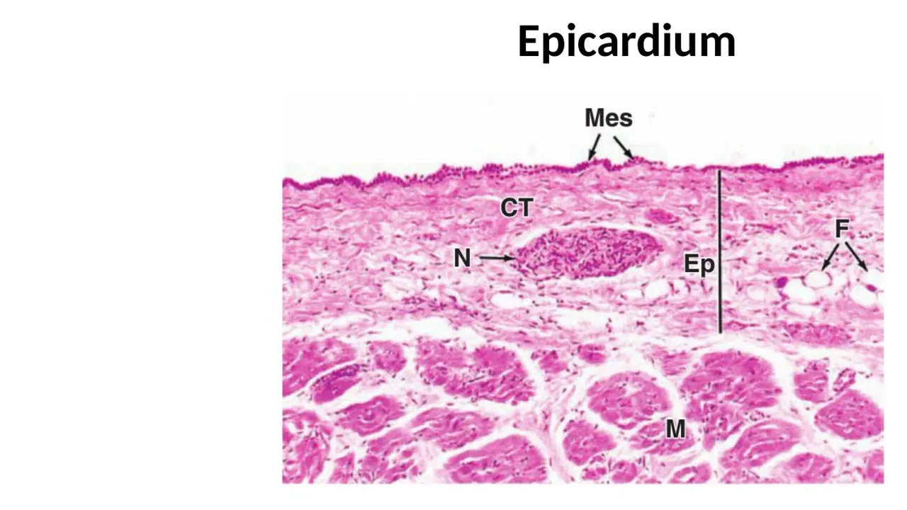
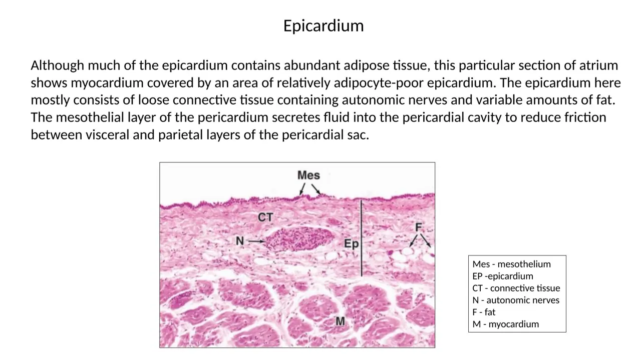
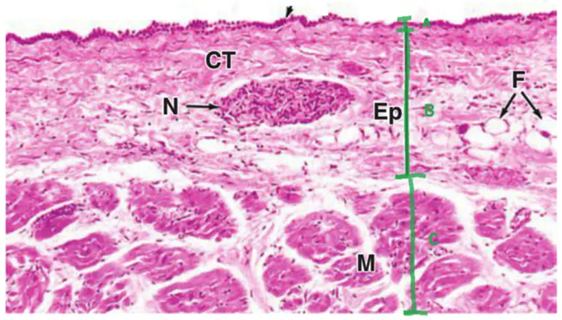
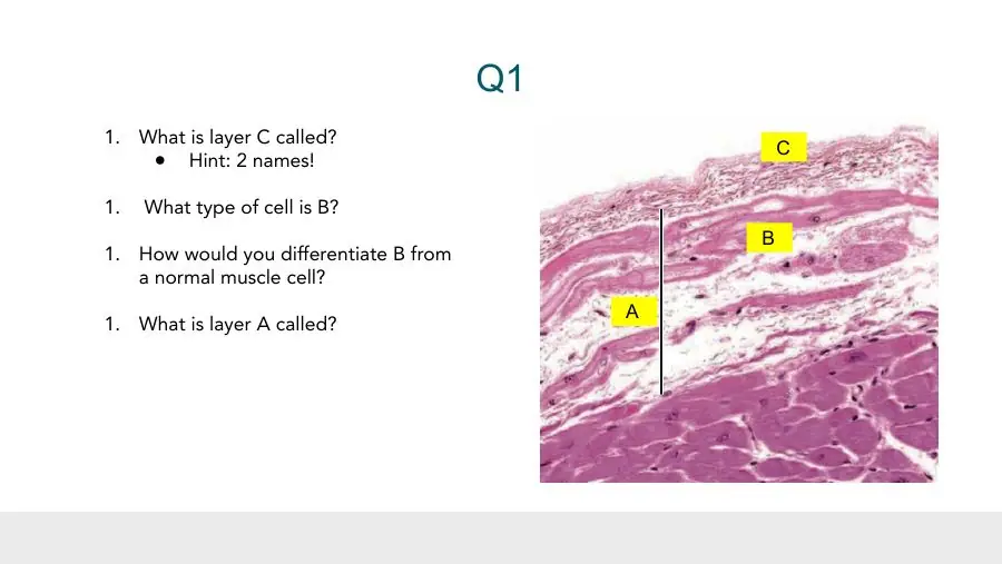
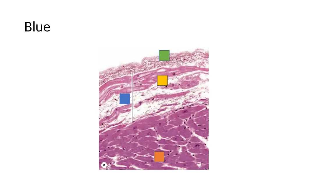
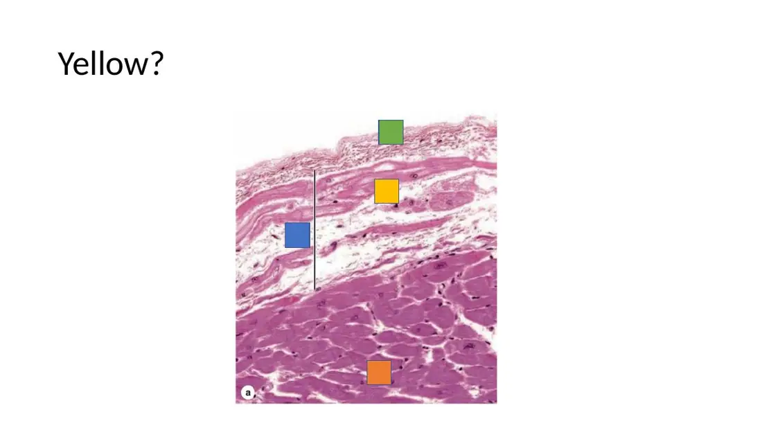
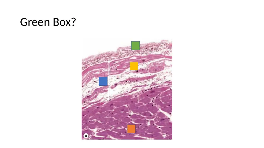
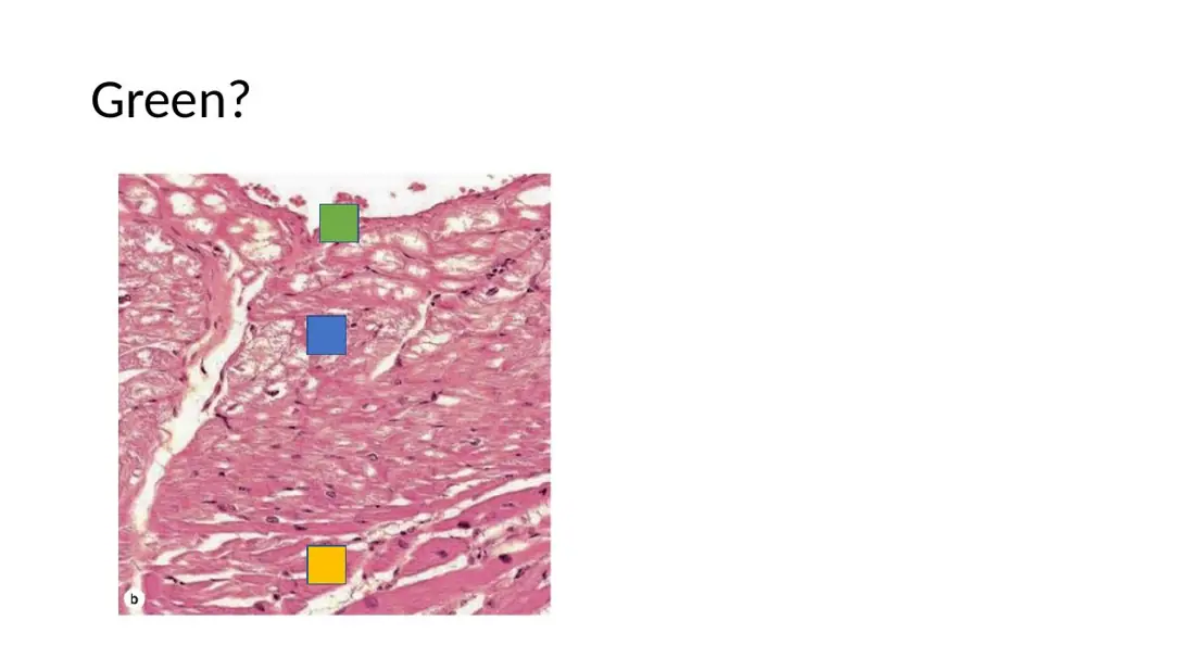
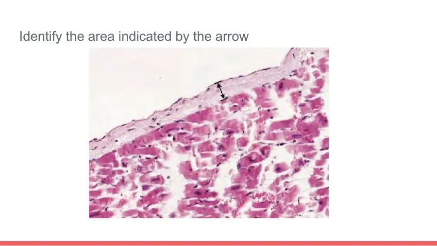
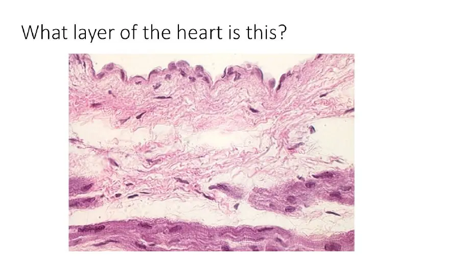
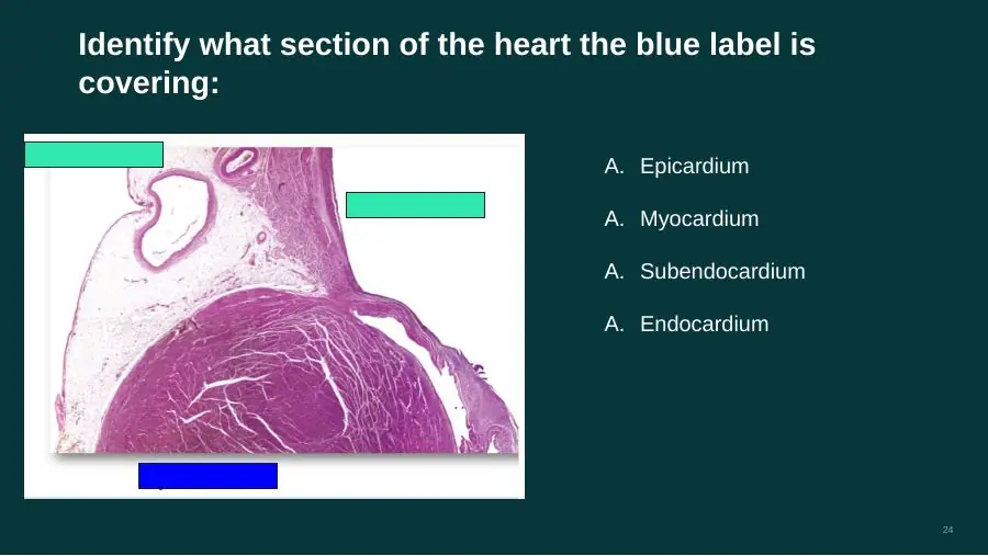
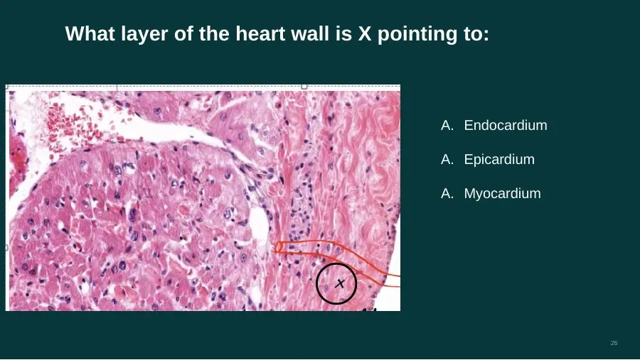
Under the microscope, what does the epicardium consist of?
A. Mesothelium over loose connective tissue holding fat, nerves and coronary vessels
B. Endothelium over loose connective tissue holding pale Purkinje fibres
C. Dense fibrous tissue over areolar tissue, with no epithelial covering
AnswerA. Mesothelium over loose connective tissue holding fat, nerves and coronary vessels
WhyEpicardium = simple squamous mesothelium on loose connective tissue with fat, autonomic nerves and coronary vessels.
Explanation
On the slide the epicardium is the pale outer band of the heart wall. Its free surface is a single row of flat mesothelial cells. Beneath them is loose connective tissue (fibro-elastic) with fat cells, nerve bundles and branches of the coronary arteries and veins, and below that the cardiac muscle begins.
Endothelium with Purkinje fibres describes the endocardium on the inner side of the wall.
Dense fibrous tissue without a surface epithelium describes the fibrous pericardium, the outer sac.
What secretes pericardial fluid, and why is it needed?
A. The endothelium of the coronary vessels — it stops the two layers rubbing
B. The mesothelium of the serous pericardium — it stops the two layers rubbing
C. The fibrous pericardium — it nourishes the outer myocardium
AnswerB. The mesothelium of the serous pericardium — it stops the two layers rubbing
WhyThe mesothelial cells of both serous layers secrete a lubricating fluid that lets the beating heart slide inside the sac without friction.
Explanation
Mesothelium is the simple squamous epithelium that lines the body's serous cavities. In the heart it covers the epicardium and lines the parietal pericardium, and it secretes a thin serous fluid into the pericardial cavity. The fluid lubricates the surfaces as the heart moves with each beat.
Endothelium lines blood vessels and the heart chambers; it faces blood, not the pericardial cavity.
The fibrous pericardium is dense connective tissue with no secretory epithelium, and the fluid does not feed the myocardium (the coronary vessels do).
Mesothelium vs endothelium: what do they share, and how do they differ?
A. Both simple squamous Mesothelium lines the heart and vessels Endothelium lines a body cavity (pericardial sac)
B. Both simple cuboidal Mesothelium lines a body cavity (pericardial sac) Endothelium lines the heart and vessels
C. Both simple squamous Mesothelium lines a body cavity (pericardial sac) Endothelium lines the heart and vessels
AnswerC. Both simple squamous Mesothelium lines a body cavity (pericardial sac) Endothelium lines the heart and vessels
WhyBoth are simple squamous epithelia: mesothelium lines serous cavities (pericardium, pleura, peritoneum); endothelium lines the heart and every blood vessel.
Explanation
The two are named by where they sit, not by their shape. In the heart wall, the mesothelium is the surface of the epicardium and of the parietal pericardium, facing the pericardial cavity. The endothelium is the surface of the endocardium, facing the blood in the chambers, and it continues into the intima of every vessel.
The swapped option reverses where each one lies.
Cuboidal is wrong for both: each is a single layer of flat cells.
Slides (2)H12A Circulatory I lecture (Dr Bruno) · slide 22H12A Circulatory I lecture (Dr Bruno) · slide 34L16Learning
In section, what are the layers of the parietal pericardium, from outside in?
A. Mesothelium → areolar tissue → dense fibrous layer facing the cavity
B. Dense fibrous layer → areolar tissue → mesothelium facing the cavity
C. Dense fibrous layer → mesothelium → areolar tissue facing the cavity
WhyThe sac is dense fibrous tissue outside, then areolar (loose) tissue, then mesothelium on the surface that faces the pericardial cavity.
Explanation
The epithelium is always on the free surface, the side that faces the cavity. So the parietal pericardium has its mesothelium on the inside, supported by a thin areolar layer, with the tough dense fibrous layer on the outside. The epicardium mirrors this across the cavity: mesothelium facing the cavity, areolar tissue beneath, then myocardium.
Putting mesothelium on the outside turns the sac inside out; the outside of the sac faces the mediastinum, not a serous cavity.
Putting areolar tissue on the cavity side would leave the cavity without its lining epithelium.
About 400 mL of blood is found in the pericardial cavity at autopsy. Which structure is it in direct contact with?
A. Fibrous pericardium
B. Myocardium
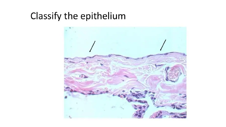
C. Epicardium
AnswerC. Epicardium
WhyThe pericardial cavity lies between the parietal serous layer and the epicardium (visceral serous layer), so blood in it touches the epicardium.
Explanation
The two walls of the pericardial cavity are the two serous layers. The inner wall is the epicardium, whose mesothelium covers the heart; the outer wall is the parietal serous layer lining the fibrous sac. Blood that escapes into the cavity (haemopericardium) therefore bathes the epicardial surface.
The fibrous pericardium is outside the parietal serous layer, so the blood is separated from it by that mesothelial lining.
The myocardium lies beneath the epicardium and is not exposed to the cavity unless the wall has ruptured.
Slides (2)H12A Circulatory I lecture (Dr Bruno) · slide 15H12A Circulatory I lecture (Dr Bruno) · slide 14L18Learning
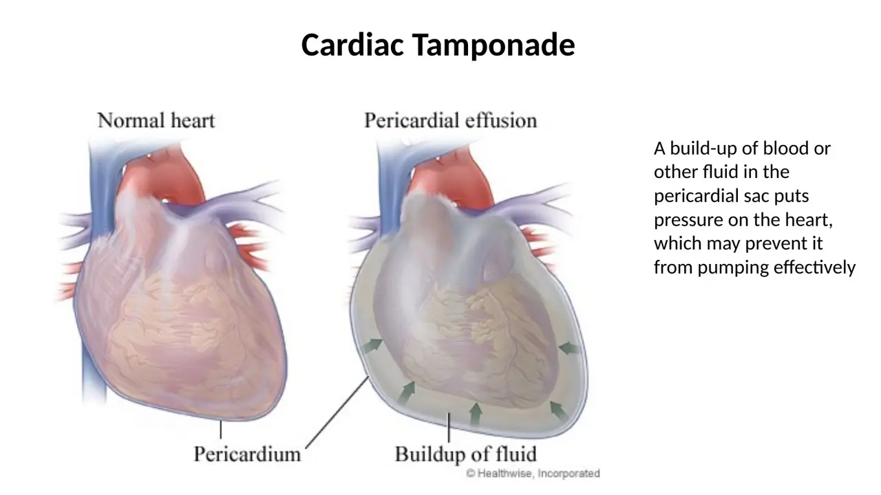
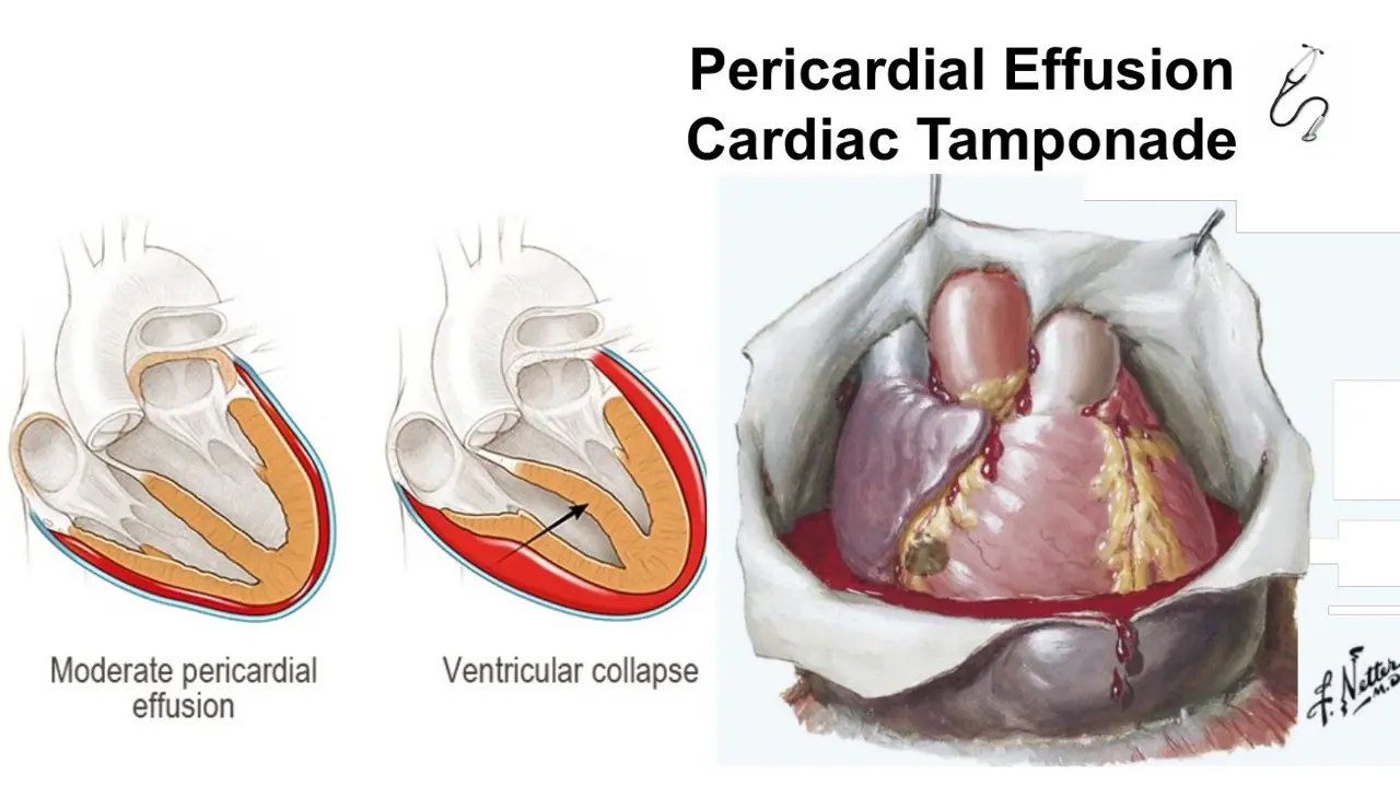
What is the mechanism of cardiac tamponade?
A. Fluid or blood fills the pericardial cavity → the fibrous sac stretches → the heart dilates and cannot contract
B. Fluid or blood fills the pericardial cavity → the fibrous sac cannot stretch → the heart is squeezed and cannot fill
C. The pericardium scars and thickens → the heart is encased and cannot fill, with no fluid present
AnswerB. Fluid or blood fills the pericardial cavity → the fibrous sac cannot stretch → the heart is squeezed and cannot fill
WhyIn tamponade, fluid in the sac cannot push the inextensible fibrous pericardium outward, so it presses on the heart and stops the chambers filling.
Explanation
The pericardial cavity normally holds only a thin film of fluid. When blood or fluid collects quickly, the fibrous pericardium cannot expand, so the pressure in the sac rises and compresses the heart, first the thin-walled right atrium and ventricle. Less filling means a smaller stroke volume, falling blood pressure and, without drainage, death.
If the sac stretched, there would be room for the fluid and no compression; it is the failure to stretch that matters.
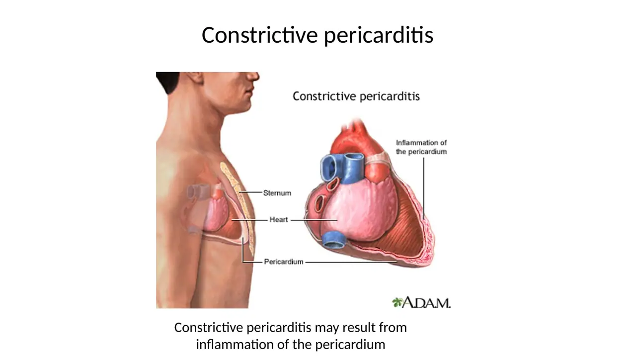
Scarring and thickening with no fluid is constrictive pericarditis, a different disease that also limits filling.
What is the difference between a pericardial effusion and cardiac tamponade?
A. Effusion: enough fluid to compress the heart and stop it filling Tamponade: fluid in the pericardial sac
B. Effusion: fluid in the myocardium Tamponade: fluid in the pericardial sac
C. Effusion: fluid in the pericardial sac Tamponade: enough fluid to compress the heart and stop it filling
AnswerC. Effusion: fluid in the pericardial sac Tamponade: enough fluid to compress the heart and stop it filling
WhyEffusion just means extra fluid in the sac; tamponade is when that fluid raises the pressure enough to compress the heart.
Explanation
An effusion can be small and harmless, or large but slow enough for the sac to adapt. It becomes tamponade once the pressure in the sac stops the chambers filling, shown on the slide as a moderate effusion progressing to ventricular collapse.
The swapped option reverses the two terms: tamponade is the severe, compressive end of an effusion.
Fluid in the myocardium is myocardial oedema; an effusion is by definition in the pericardial cavity.
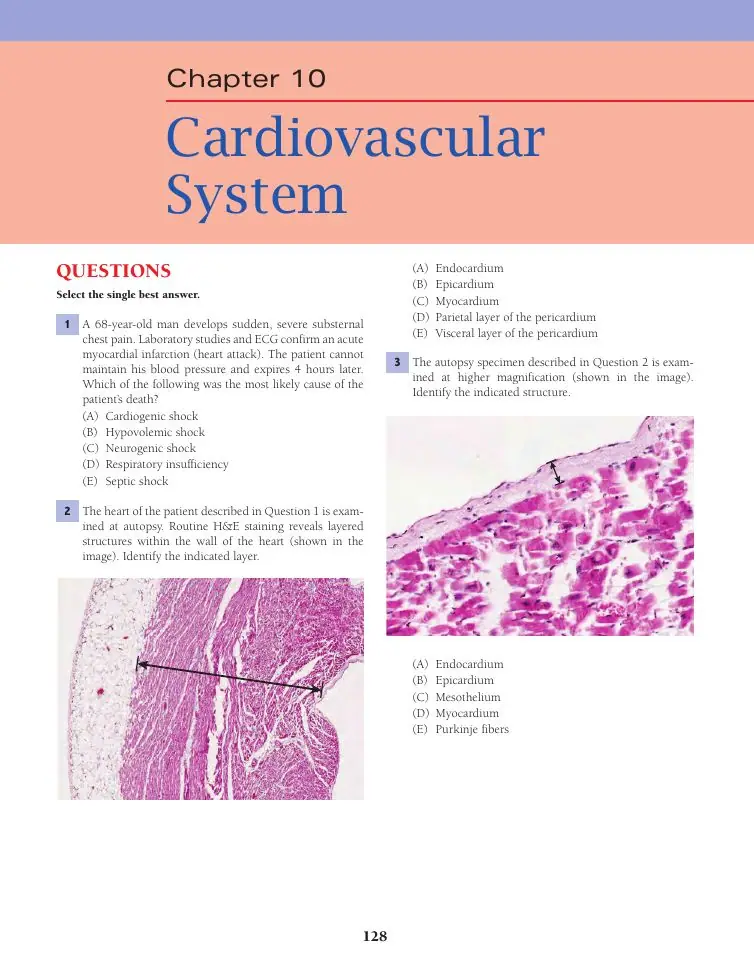
Two weeks after a massive heart attack, a man has sharp chest pain, BP 80/40 and pulse 100, and dies. Autopsy shows a rupture in the left ventricular wall. What killed him?
A. Pulmonary oedema
B. Cardiac tamponade
C. Pulmonary thromboembolism
AnswerB. Cardiac tamponade
WhyThe softened infarct ruptured; blood poured into the pericardial cavity (haemopericardium) and compressed the heart: cardiac tamponade.
Explanation
After a myocardial infarction the dead muscle is soft while it is being removed and before the scar is strong, typically in the first one to two weeks. A free-wall rupture lets ventricular blood under high pressure flood the pericardial cavity. The inextensible fibrous pericardium cannot expand, so the heart cannot fill: low blood pressure, fast pulse and sudden death.
Pulmonary oedema comes from left heart failure raising pulmonary venous pressure; it does not follow from a hole that empties the ventricle into the sac.
Pulmonary thromboembolism starts from deep vein thrombi in the legs, not from a ruptured ventricle.
Slide (1)H12A Circulatory I lecture (Dr Bruno) · slide 17L21Learning
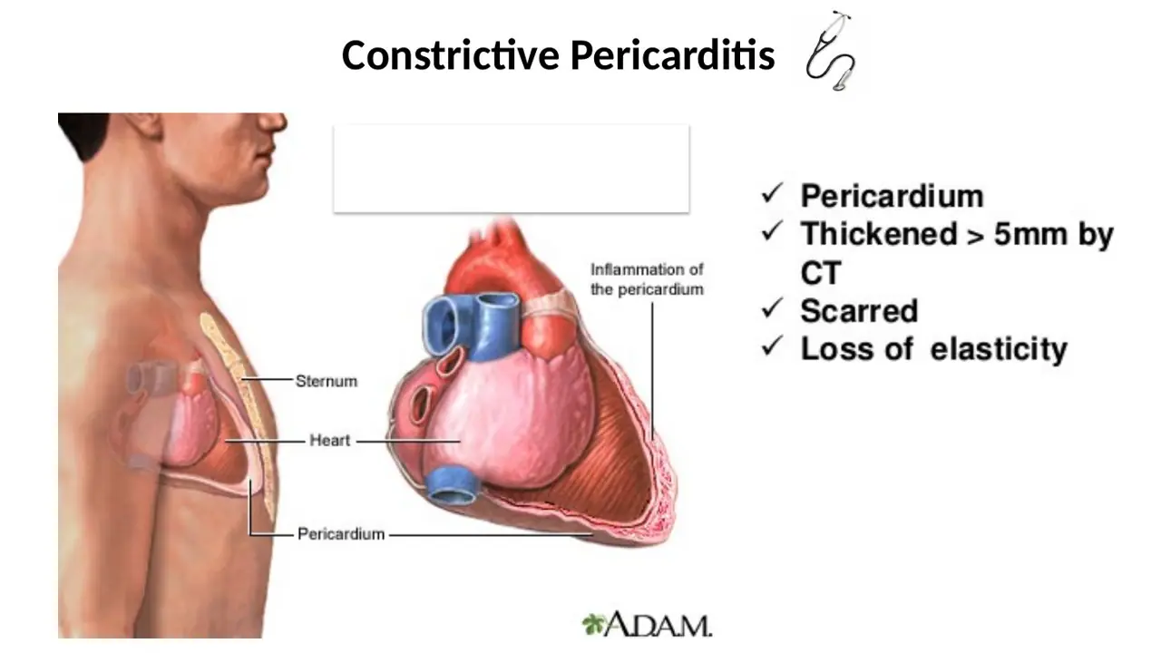
What is constrictive pericarditis?
A. Pericardium filled with fluid and stretched → restricted outflow from the ventricles
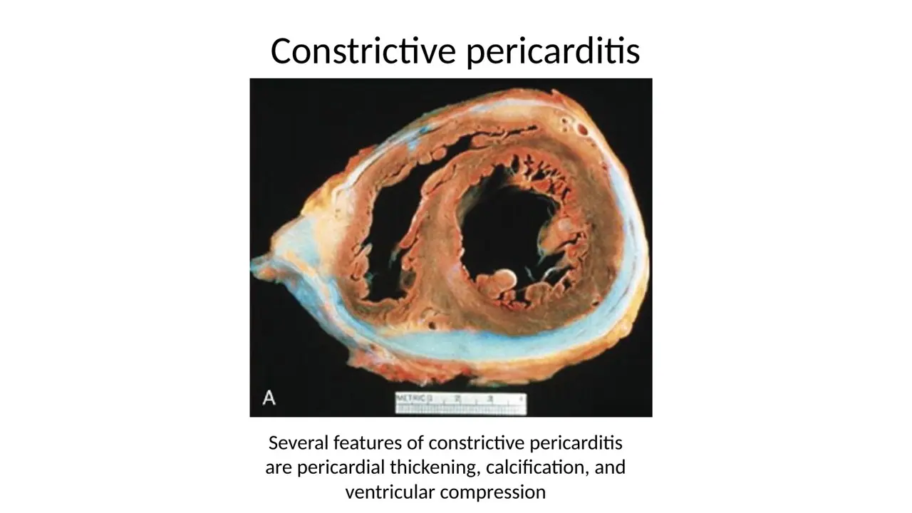
B. Pericardium scarred and thickened (over 5 mm), with loss of elasticity → restricted filling of the heart
C. Myocardium scarred and thickened, with loss of elasticity → restricted contraction of the ventricles
AnswerB. Pericardium scarred and thickened (over 5 mm), with loss of elasticity → restricted filling of the heart
WhyIn constrictive pericarditis the pericardium becomes a thick (>5 mm), scarred, inelastic shell that limits venous inflow, with no fluid needed.
Explanation
Chronic inflammation of the pericardium (for example after tuberculosis) replaces it with dense scar tissue, sometimes calcified. The rigid shell stops the heart expanding in diastole, so blood backs up in the veins; the heart looks 'quiet' because it barely moves. The problem is filling, not pumping.
Fluid-filled and stretched describes an effusion, and the problem in tamponade is also filling, not outflow.
Scarred myocardium is a disease of the muscle; constrictive pericarditis spares the muscle.
B. Pericardiocentesis (draining fluid with a needle)
C. Coronary artery bypass grafting
AnswerA. Pericardiectomy (removing the pericardium)
WhyCutting away the rigid shell (pericardiectomy) frees the heart; on imaging its movement returns to normal.
Explanation
The disease is a thick scarred sac, so the cure is to remove it. The lab's moving images show the same patient's heart barely moving before surgery and beating normally after pericardiectomy.
Pericardiocentesis (needle drainage) treats tamponade, where the problem is fluid; there is no fluid to drain in constriction.
Bypass grafting treats blocked coronary arteries, which are not the problem here.
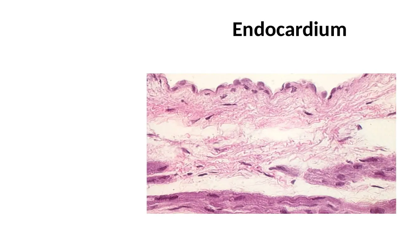
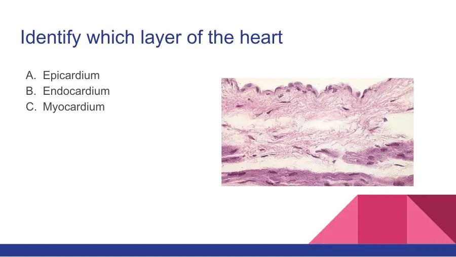
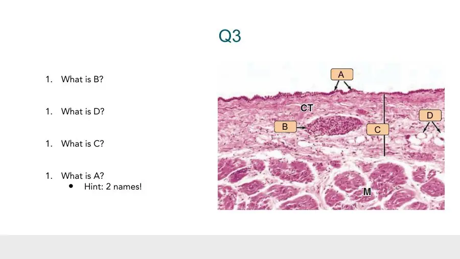
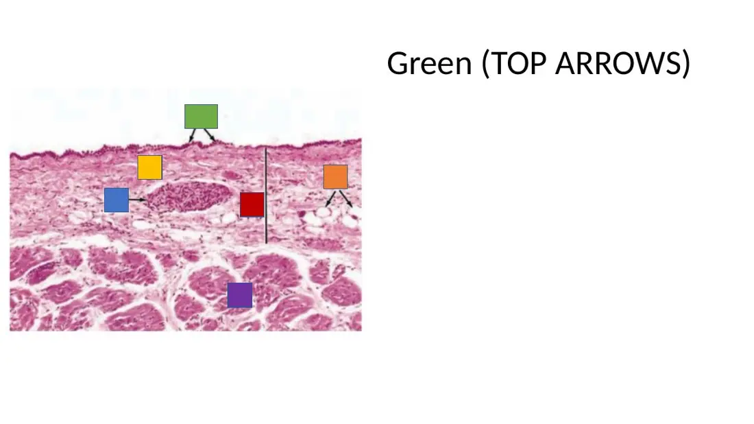
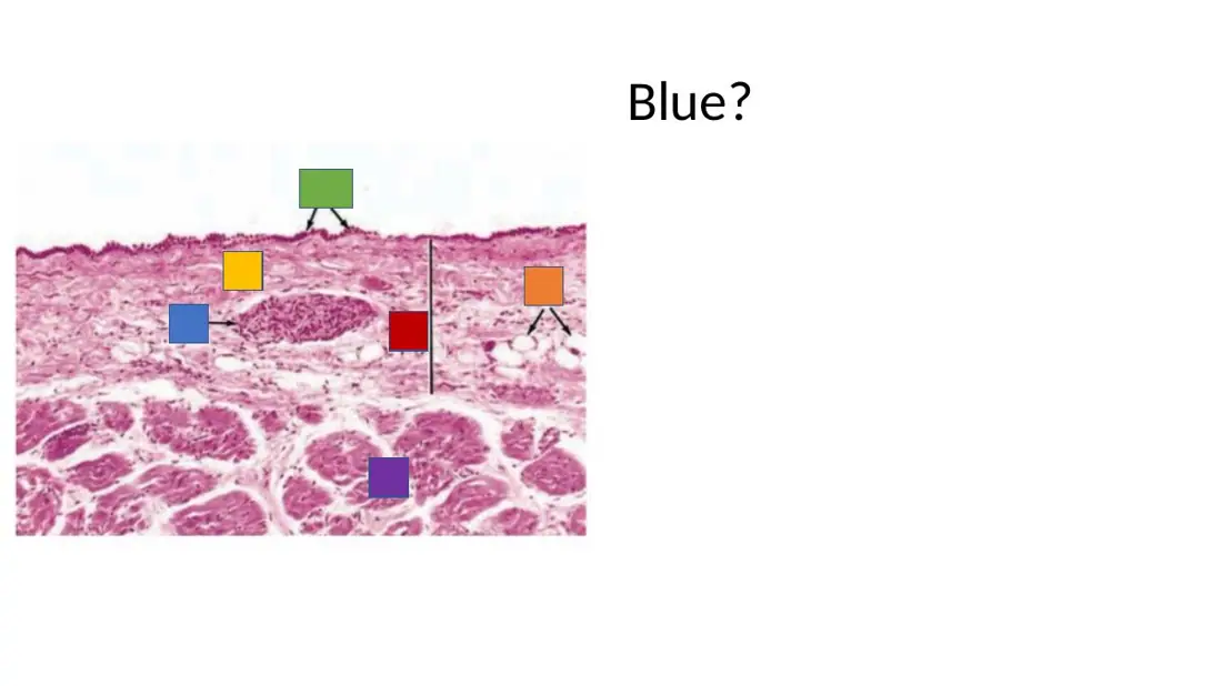
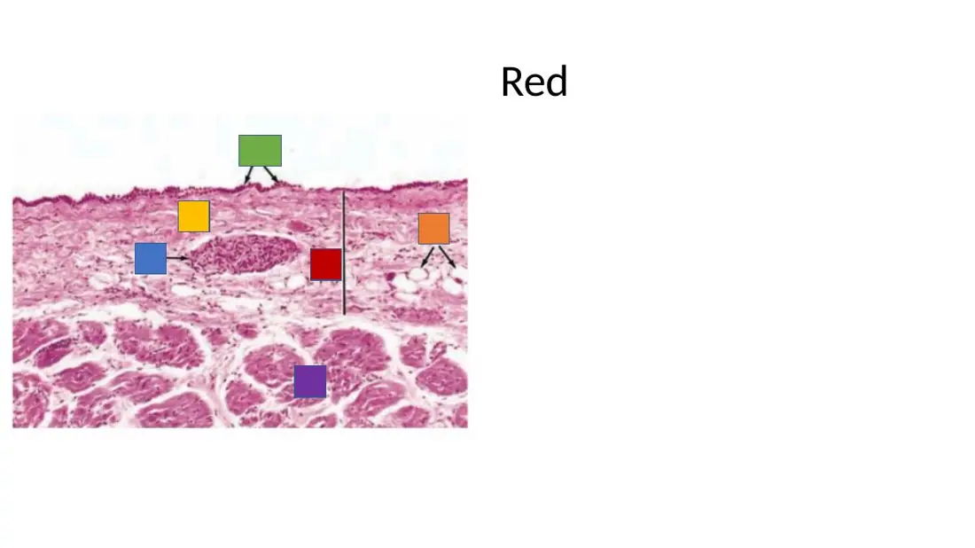
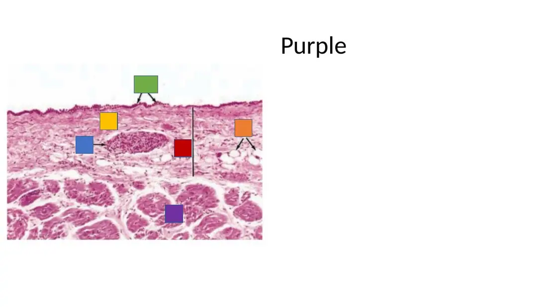
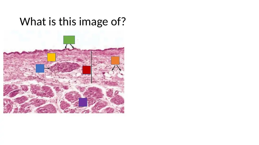
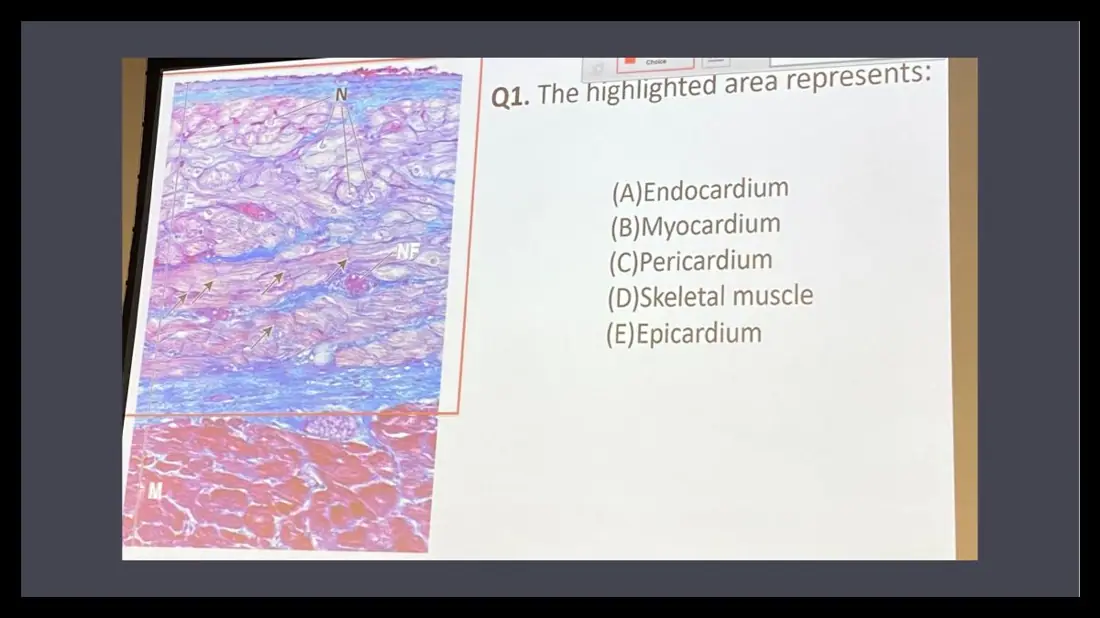
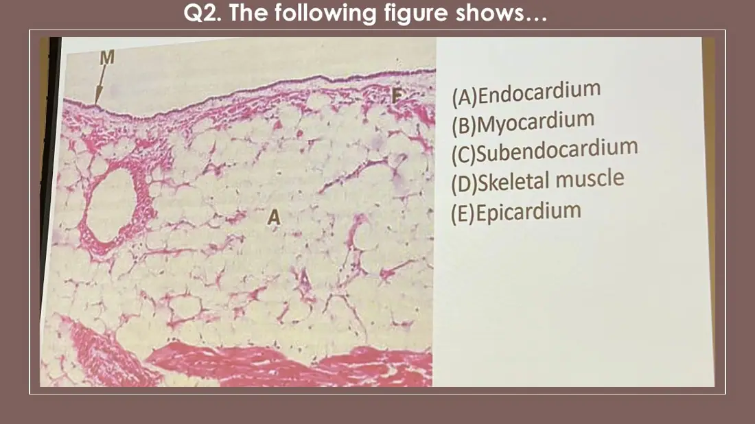
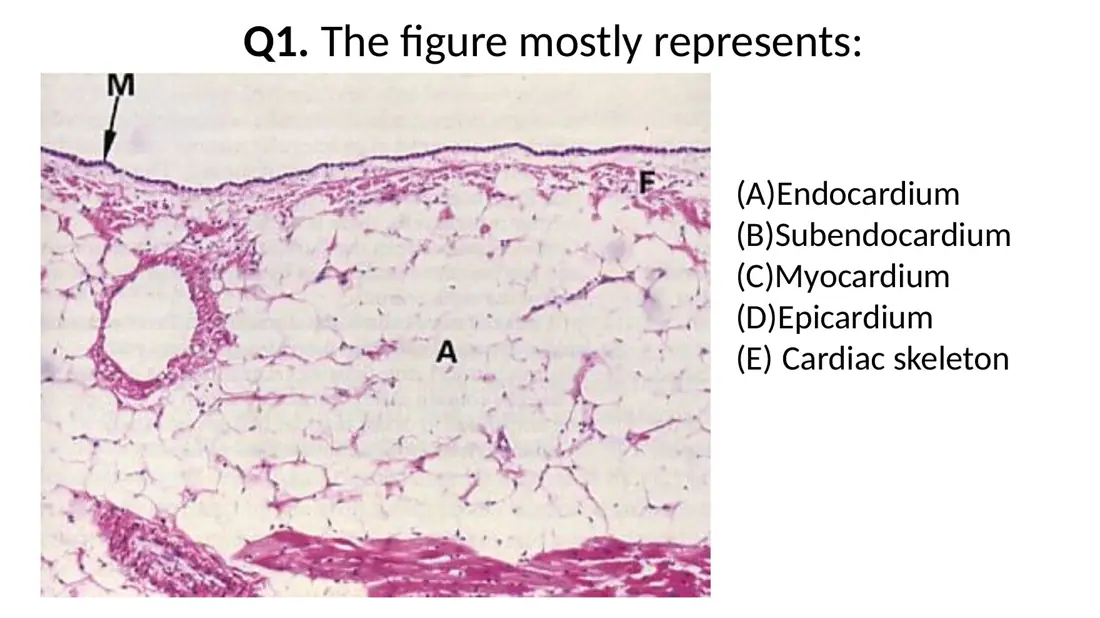
An H&E section of atrial wall shows a row of flat cells on the free surface, over loose tissue with fat cells, a nerve bundle and a small artery; cardiac muscle lies beneath. Which layer is this?
A. Epicardium
B. Endocardium
C. Parietal pericardium
AnswerA. Epicardium
WhyFlat surface cells over fat, nerves and vessels, with muscle directly below, is the epicardium: mesothelium on subepicardial loose connective tissue.
Explanation
Three clues point to the epicardium: a mesothelium on the surface, adipocytes (empty round spaces) and a nerve in the loose connective tissue, and cardiac muscle directly underneath. Coronary branches also run here.
The endocardium also has flat surface cells (endothelium), but its connective tissue holds no fat or nerve bundles; it may hold pale Purkinje fibres, and it faces a chamber full of blood.
The parietal pericardium has mesothelium too, but under it lies dense fibrous tissue, not cardiac muscle.
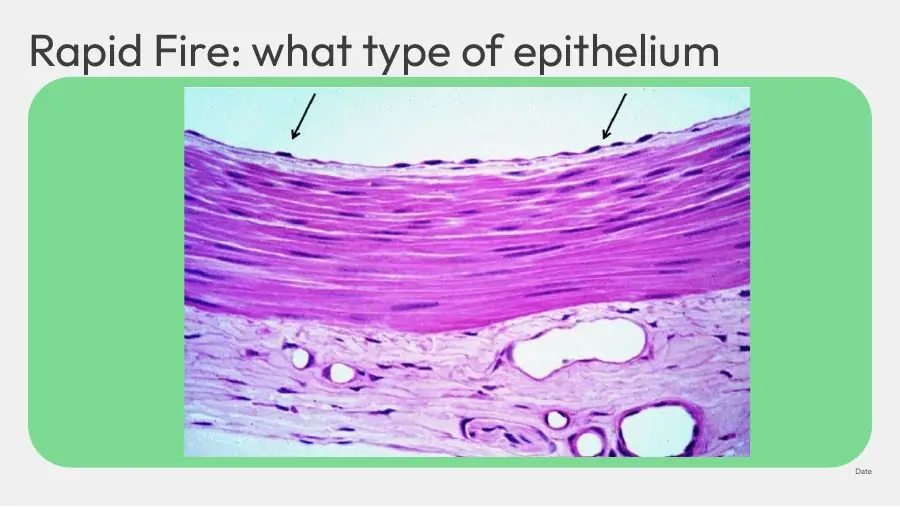
In a heart section, how can you tell that a layer of flat surface cells is endothelium rather than mesothelium?
A. It faces the pericardial cavity, and the connective tissue beneath has fat and nerve bundles
B. Its cells are cuboidal, while mesothelial cells are flat
C. It faces a chamber lumen, and the connective tissue beneath has no fat or nerve bundles
AnswerC. It faces a chamber lumen, and the connective tissue beneath has no fat or nerve bundles
WhyBoth are flat single layers; the endothelium faces the chamber blood and sits on thin connective tissue without fat or nerves (often with Purkinje fibres below).
Explanation
The cells themselves look the same: one layer of flattened cells with flat nuclei. What gives them away is the company they keep. Endothelium lines the endocardium, faces the lumen of a chamber and sits on thin connective tissue that runs into the subendocardium, where pale Purkinje fibres may appear. Mesothelium covers the epicardium, faces the pericardial cavity and sits on loose tissue with fat lobules, nerves and coronary vessels.
Facing the pericardial cavity with fat and nerves below is the description of mesothelium.
Cuboidal is wrong: both are simple squamous epithelia.
Slides (2)H12A Circulatory I lecture (Dr Bruno) · slide 33H12A Circulatory I lecture (Dr Bruno) · slide 21
Myocardium and intercalated discs
L25Learning
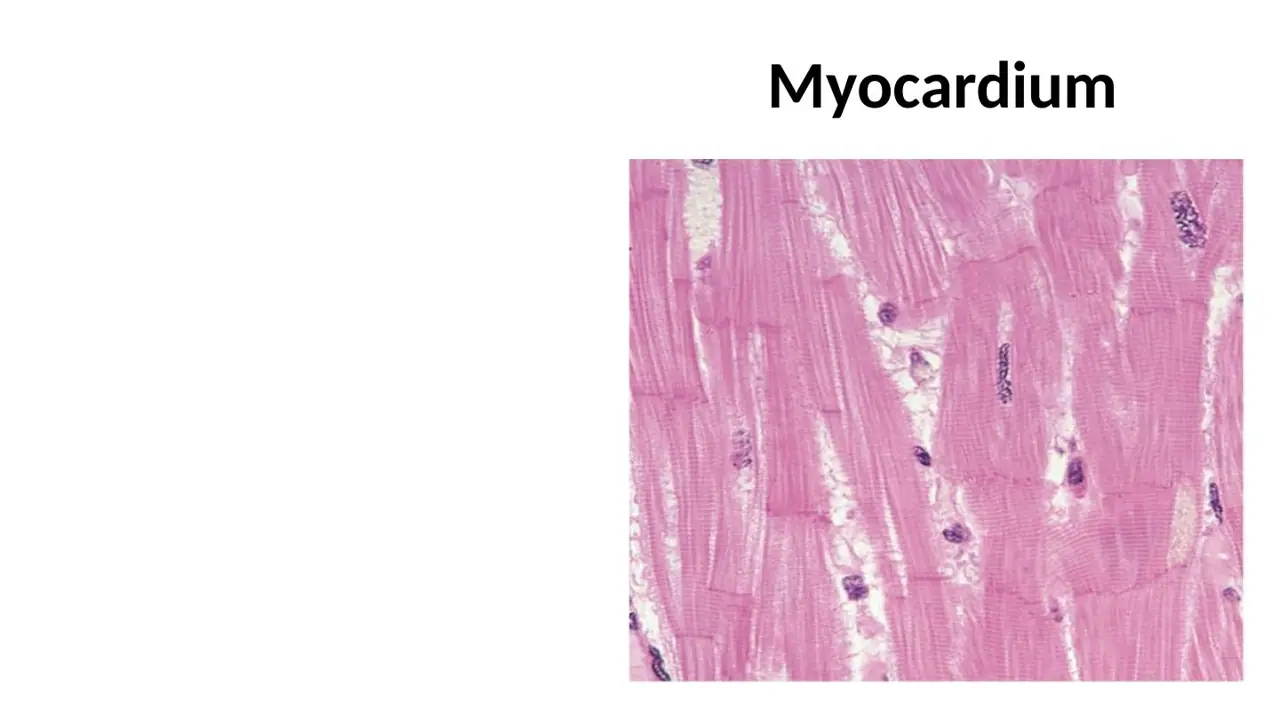
How are the cardiac muscle cells of the myocardium arranged?
A. In concentric, spiral layers round the chambers, grouped into fascicles by sheets of elastic laminae with no vessels
B. In concentric, spiral layers round the chambers, grouped into fascicles by connective-tissue septa carrying vessels and nerves
C. In straight longitudinal sheets from base to apex, forming one continuous mass with no septa
AnswerB. In concentric, spiral layers round the chambers, grouped into fascicles by connective-tissue septa carrying vessels and nerves
WhyMyocardial cells wind round the chambers in spiral layers; connective-tissue septa carrying vessels and nerves group them into fascicles.
Explanation
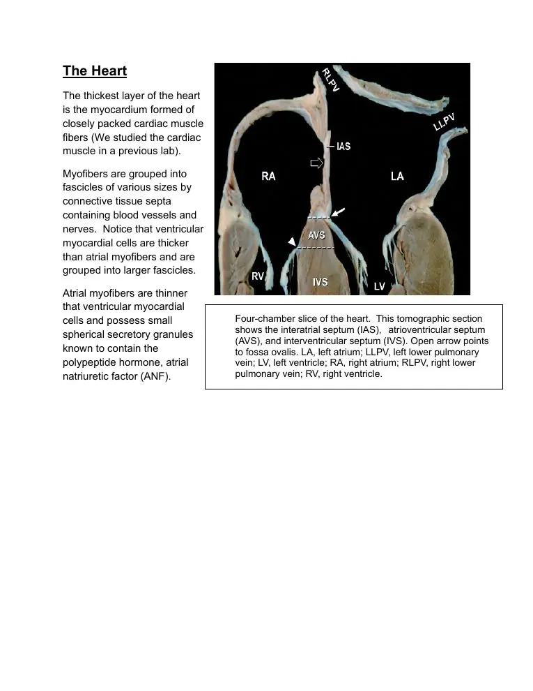
The spiral arrangement wrings blood out of the ventricles: when the layers contract, the chamber shortens and twists, squeezing blood toward the outflow valves. Between the fascicles, thin connective-tissue septa carry the rich capillary network and autonomic nerves that the working muscle needs. Ventricular fascicles are larger than atrial ones.
Elastic laminae without vessels is wrong twice: the septa are ordinary connective tissue, and they carry the vessels and nerves.
Straight longitudinal sheets would only shorten the chamber, not wring it; the slide shows spiral bands.
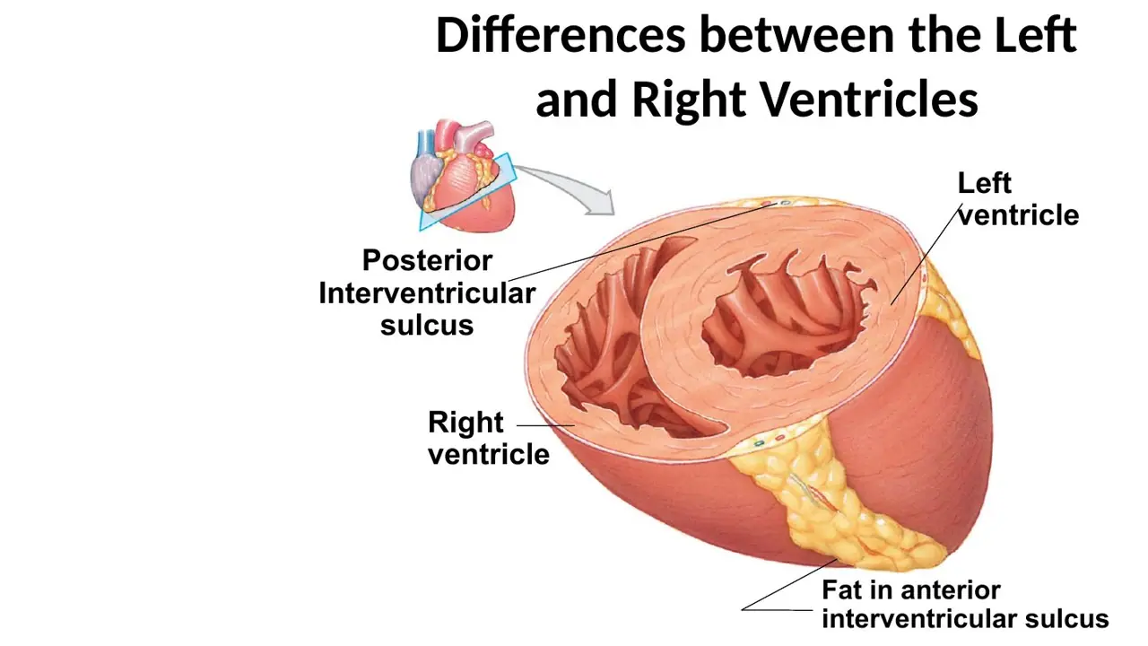
Why is the left ventricular wall much thicker than the right?
A. It pumps into the high-pressure systemic circulation
B. It pumps into the low-pressure pulmonary circulation
C. It must hold a larger volume of blood than the right ventricle
AnswerA. It pumps into the high-pressure systemic circulation
WhyThe left ventricle must generate systemic pressure (~120 mmHg), so its wall is about three times thicker than the right's.
Explanation
Wall thickness follows the pressure a chamber must produce. The left ventricle ejects into the aorta against the high systemic resistance; the right ventricle ejects the same volume into the low-resistance pulmonary circuit. In cross-section the left ventricle is a thick ring and the right a thin crescent wrapped against the interventricular septum.
The pulmonary circulation is the right ventricle's target, and it is low pressure.
Both ventricles hold and eject the same volume; otherwise blood would pile up in one circuit.
The slide also shows epicardial fat in the anterior interventricular sulcus, where the anterior interventricular artery runs.
Slides (2)H12A Circulatory I lecture (Dr Bruno) · slide 24H12A Circulatory I lecture (Dr Bruno) · slide 7L27Learning
How do ventricular muscle cells compare with atrial muscle cells?
A. Atrial cells are thicker and grouped into larger fascicles
B. Ventricular cells are thicker and grouped into larger fascicles
C. Ventricular cells are thinner but grouped into larger fascicles
AnswerB. Ventricular cells are thicker and grouped into larger fascicles
WhyVentricular myocytes are thicker than atrial ones and form larger fascicles, matching the heavier work of the ventricles.
Explanation
The ventricles do most of the pumping, so their cells are wider and packed into bigger bundles. Atrial myocytes are thinner, and they have a second job: they carry small secretory granules of atrial natriuretic factor.
The atrial-thicker option reverses the sizes.
Thinner ventricular cells contradicts the lab manual: ventricular cells are the thicker ones.
What do atrial muscle cells store in small secretory granules?
A. Atrial natriuretic factor (ANF)
B. Renin
C. Antidiuretic hormone (ADH)
AnswerA. Atrial natriuretic factor (ANF)
WhyAtrial myocytes store the peptide hormone atrial natriuretic factor in small granules near the nucleus, so the atria are also an endocrine organ.
Explanation
When the atria are stretched by a high blood volume, the cells release ANF (also called atrial natriuretic peptide). The granules are tiny and are seen with the electron microscope rather than on routine H&E.
Renin is made by the juxtaglomerular cells of the kidney, not by heart muscle.
ADH is made by hypothalamic neurons and released from the posterior pituitary.
A. Makes the kidney retain more sodium and water, raising blood volume and pressure
B. Constricts arterioles, raising blood pressure
C. Makes the kidney excrete more sodium and water, lowering blood volume and pressure
AnswerC. Makes the kidney excrete more sodium and water, lowering blood volume and pressure
WhyANF is released when the atria are overstretched and makes the kidneys lose sodium and water (natriuresis), so blood volume and pressure fall.
Explanation
'Natriuretic' means causing sodium loss in the urine. ANF increases the glomerular filtration rate and blocks sodium reabsorption, and it also relaxes vascular smooth muscle. All its effects lower blood volume and pressure, the opposite of the renin–angiotensin–aldosterone system.
Retaining sodium and water is what aldosterone and ADH do.
Constricting arterioles is the action of angiotensin II and sympathetic stimulation; ANF dilates.
L30Learning
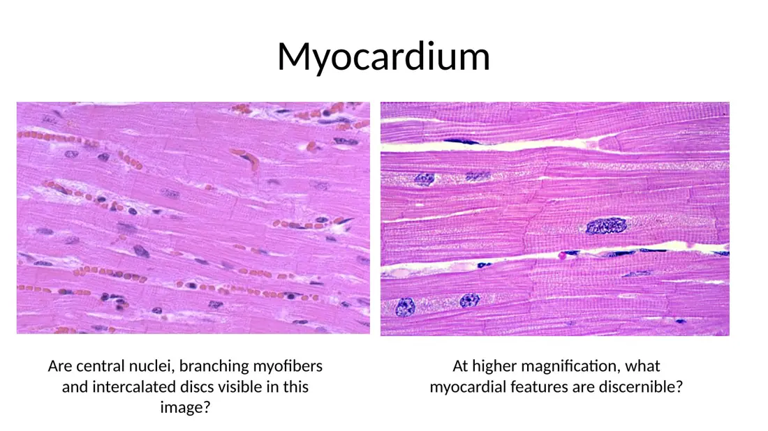
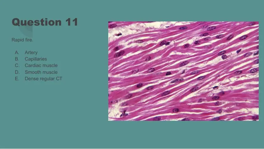
Which three features together identify cardiac muscle on a slide?
A. Striated Central nucleus Intercalated discs
B. Non-striated Central nucleus Intercalated discs
C. Striated Peripheral nuclei Intercalated discs
AnswerA. Striated Central nucleus Intercalated discs
WhyCardiac muscle is striated like skeletal muscle, but has one or two central nuclei and intercalated discs joining the branched cells end to end.
Explanation
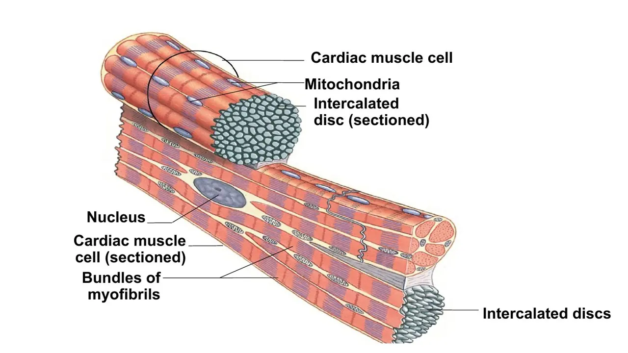
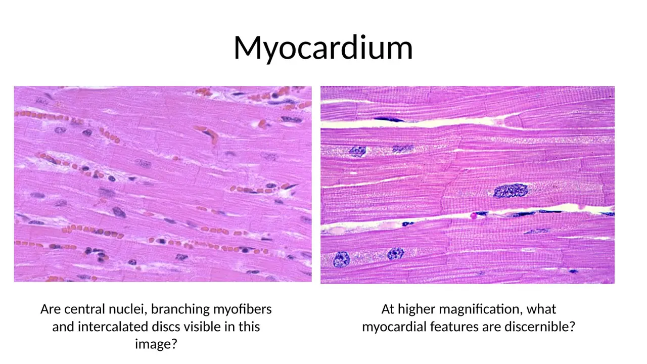
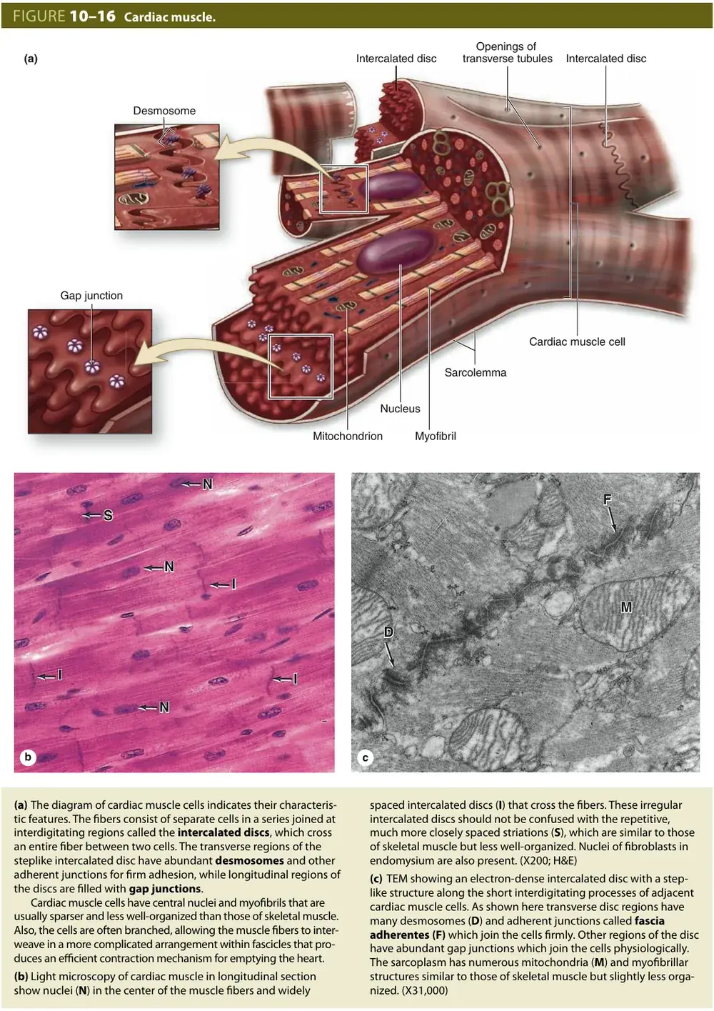
In longitudinal section, cardiac muscle shows fine cross-striations, oval nuclei sitting in the middle of each cell (often with a pale zone at each pole), branching cells, and dark, irregular, step-like intercalated discs crossing the fibres at intervals.
Non-striated with central nuclei describes smooth muscle (which has no intercalated discs).
Peripheral nuclei describes skeletal muscle, whose long multinucleated fibres push their nuclei under the cell membrane.
Cardiac vs skeletal muscle: how do the nuclei differ?
A. Cardiac: one (or two), at the periphery Skeletal: many, central
B. Cardiac: one (or two), central Skeletal: many, at the periphery
C. Cardiac: many, at the periphery Skeletal: one (or two), central
AnswerB. Cardiac: one (or two), central Skeletal: many, at the periphery
WhyEach cardiac cell has one or two central nuclei; a skeletal fibre is one huge cell with many peripheral nuclei.
Explanation
A skeletal muscle fibre forms by fusion of many myoblasts, so it is multinucleated and its nuclei lie just under the sarcolemma. A cardiac 'fibre' is a chain of separate cells joined end to end, each with its own one or two nuclei in the centre, surrounded by myofibrils.
Cardiac peripheral / skeletal central puts both nuclei in the wrong place.
Cardiac many / skeletal one swaps the whole pattern.
Cardiac vs skeletal muscle: how do the cell shapes differ?
A. Cardiac: long, unbranched fibres Skeletal: short, branching cells joined end to end
B. Cardiac: short, branching cells joined end to end Skeletal: long, unbranched fibres
C. Cardiac: short, unbranched cells joined side to side Skeletal: long, branching fibres
AnswerB. Cardiac: short, branching cells joined end to end Skeletal: long, unbranched fibres
WhyCardiac cells are short and branched, linked end to end into a network; skeletal fibres are long, unbranched cylinders.
Explanation
The branching lets cardiac cells form a three-dimensional meshwork that contracts as a unit, which suits emptying a chamber. A skeletal muscle fibre is one long cylindrical cell running much of the muscle's length.
The swapped option gives each tissue the other's shape.
Side-to-side, unbranched cardiac cells and branching skeletal fibres are both wrong: cardiac cells branch and meet end to end at intercalated discs.
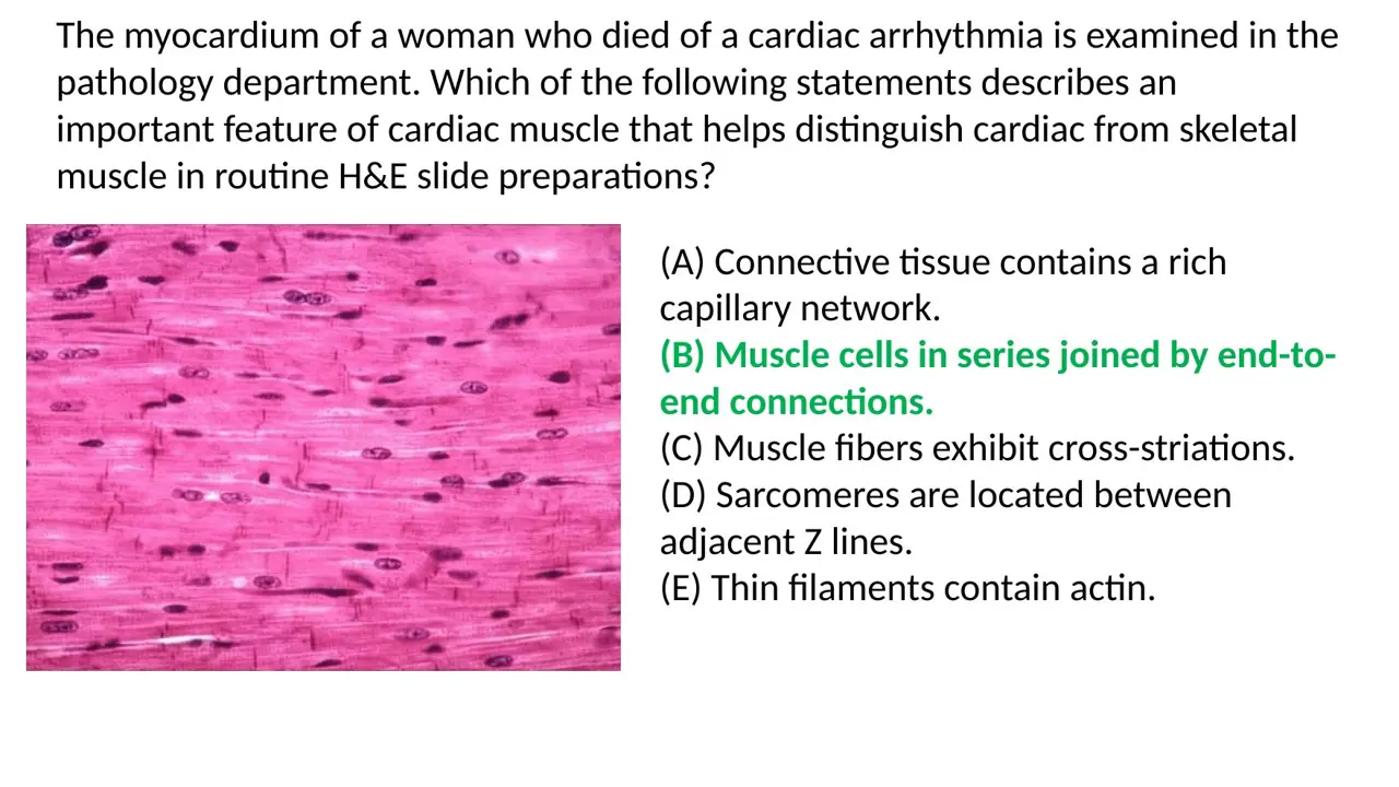
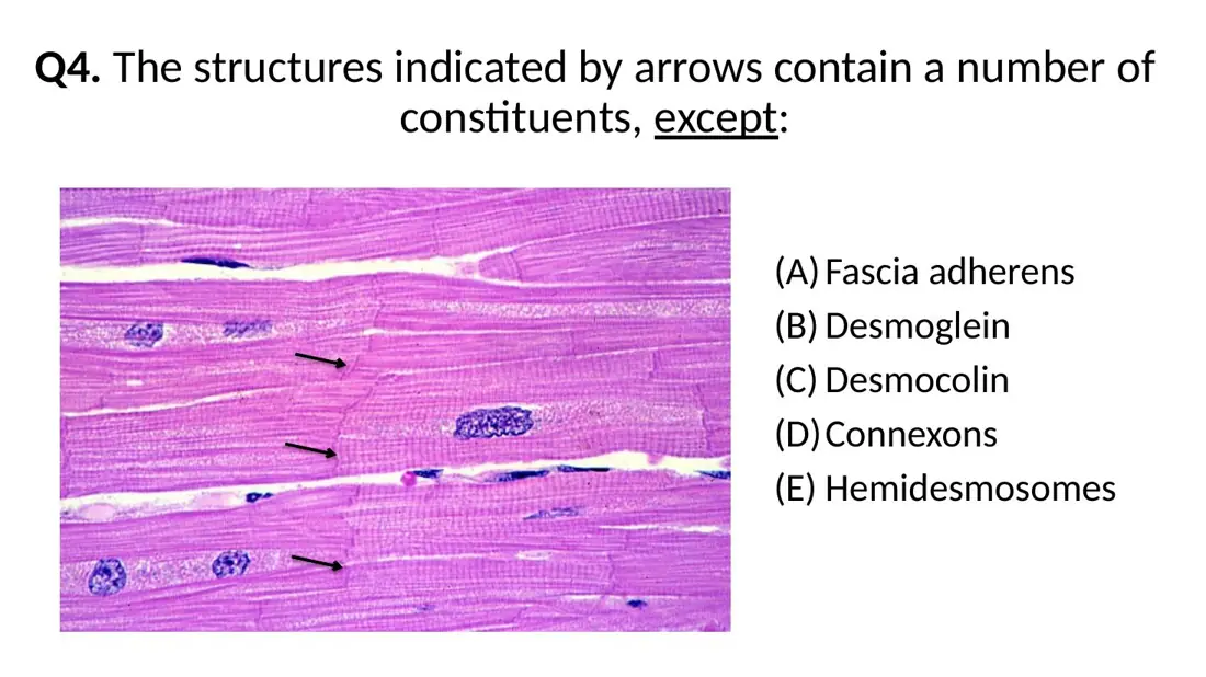
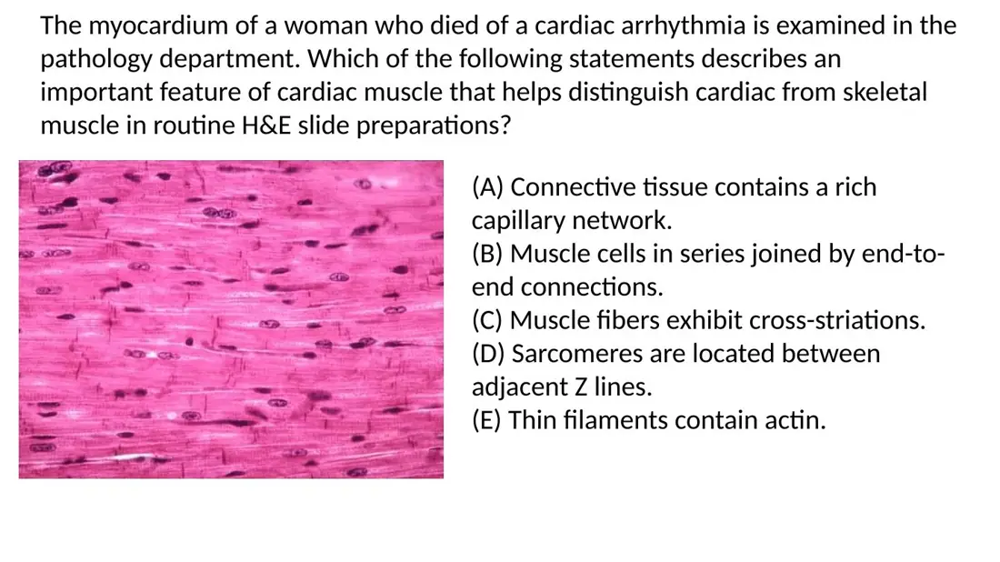
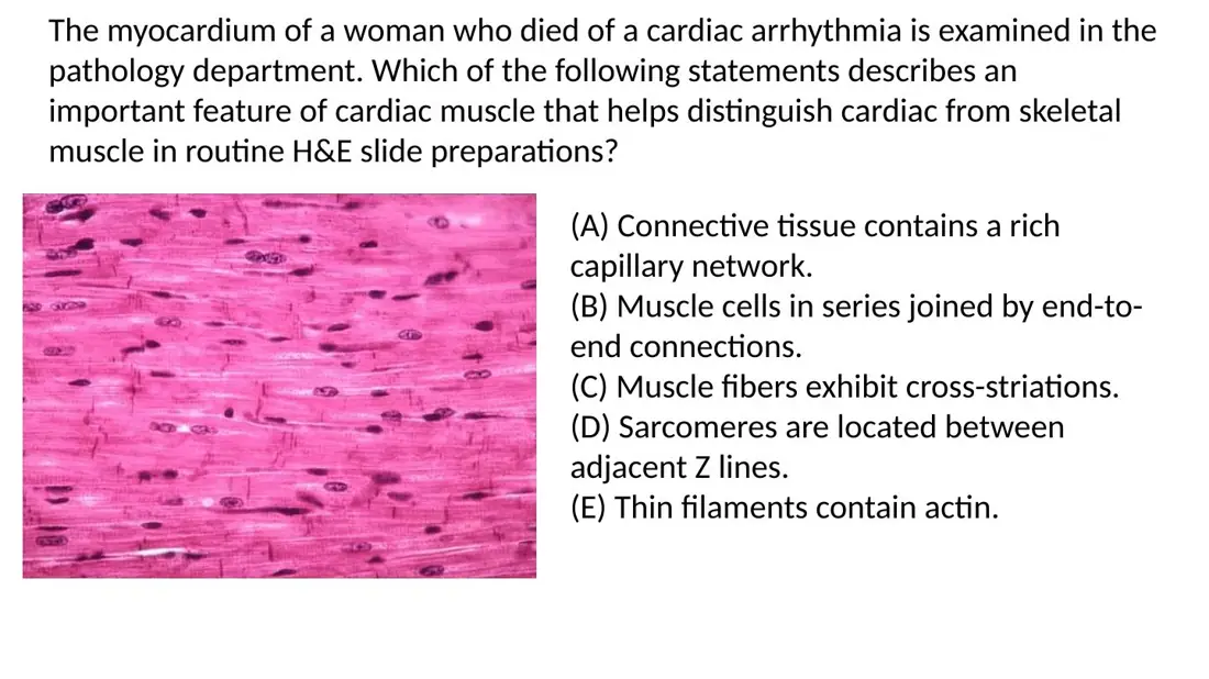
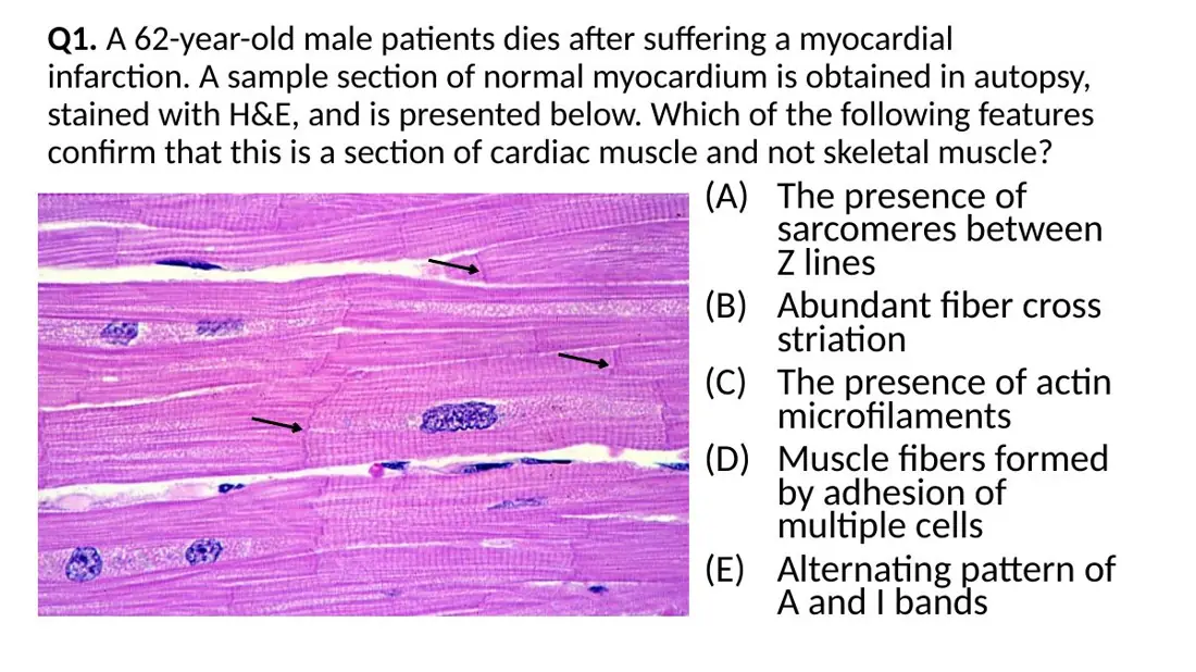
In routine H&E, which feature distinguishes cardiac from skeletal muscle?
A. Fibres showing regular cross-striations along their length
B. Sarcomeres lying between adjacent Z lines in each myofibril
C. Muscle cells in series, joined by end-to-end connections
AnswerC. Muscle cells in series, joined by end-to-end connections
WhyCardiac 'fibres' are chains of separate cells joined end to end at intercalated discs; skeletal fibres are single multinucleated cells.
Explanation
The end-to-end junctions are the intercalated discs, the dark step-like lines crossing cardiac fibres. Skeletal muscle has nothing like them because each fibre is one continuous cell.
Cross-striations are present in both cardiac and skeletal muscle, so they cannot tell them apart.
Sarcomeres between Z lines is the structure of every striated muscle, cardiac and skeletal alike.
Exam tip: when an option is true of both tissues, it cannot be the distinguishing feature.
A. Dark, irregular cross-lines joining cells end to end — also seen in skeletal muscle
B. Pale longitudinal lines between myofibrils — unique to cardiac muscle
C. Dark, irregular cross-lines joining cells end to end — unique to cardiac muscle
AnswerC. Dark, irregular cross-lines joining cells end to end — unique to cardiac muscle
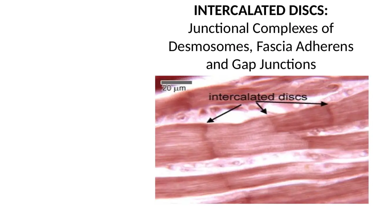
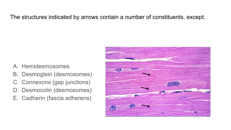
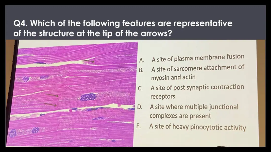
WhyIntercalated discs are dark, step-like lines across the fibres, more widely spaced than the striations, found only in cardiac muscle.
Explanation
Each disc marks the boundary between two cardiac cells, so the discs occur at irregular intervals of one cell length, much farther apart than the closely spaced, regular striations. They stain darkly because the junctions are dense with protein.
Skeletal muscle has no intercalated discs; its fibres are single cells with no end-to-end boundaries.
Pale longitudinal lines run the wrong way: the discs cross the fibre.
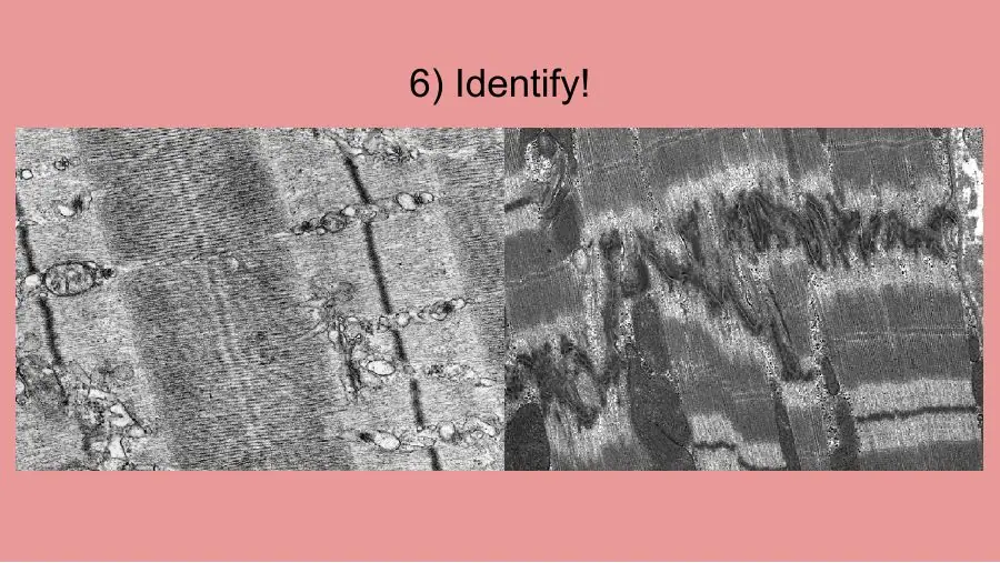
Do not confuse them with contraction bands in infarcted muscle: those are many dense bands within a single cell.
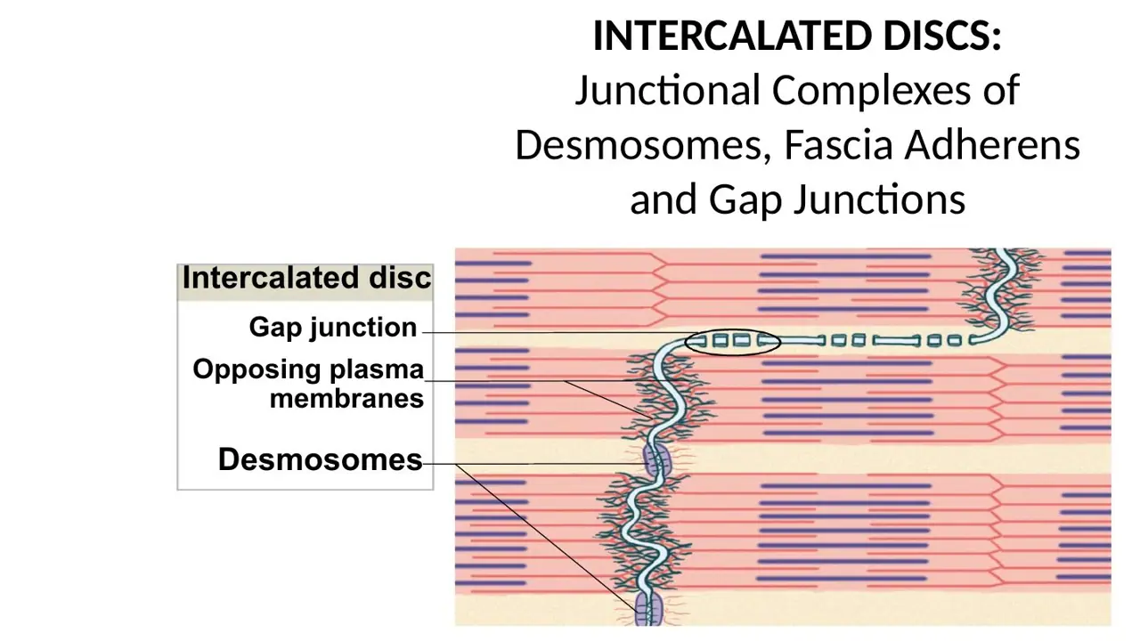
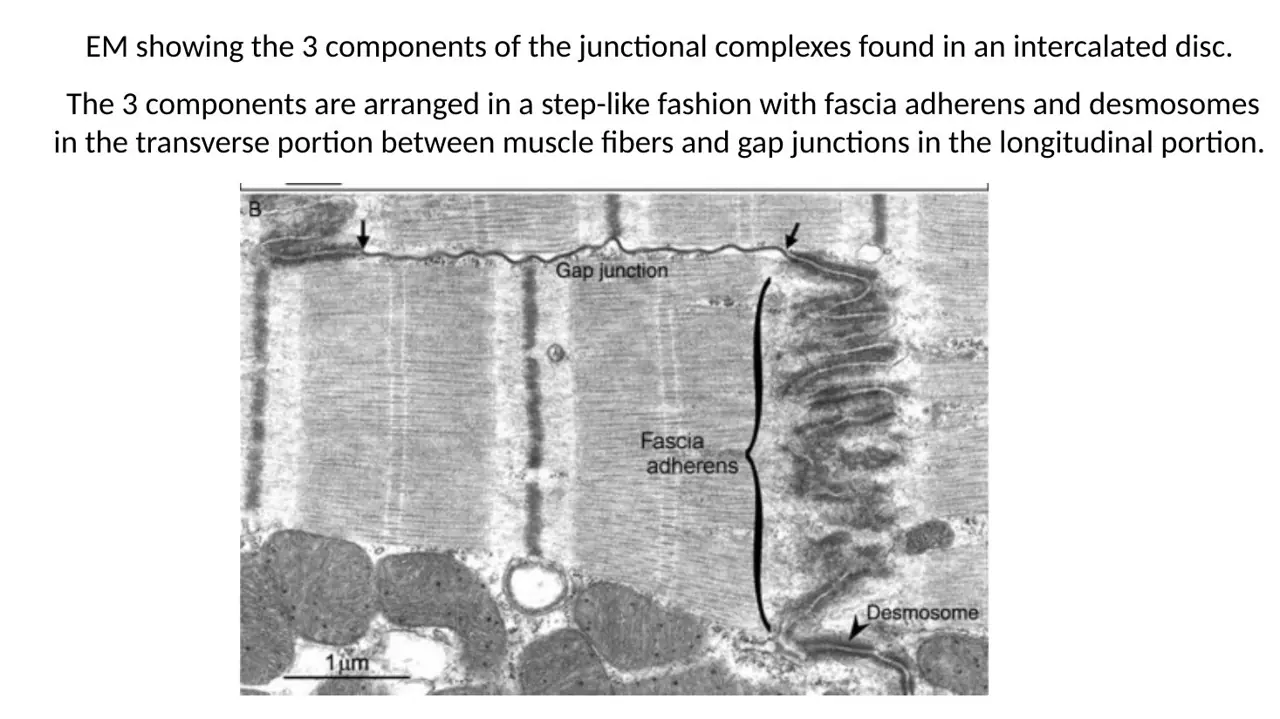
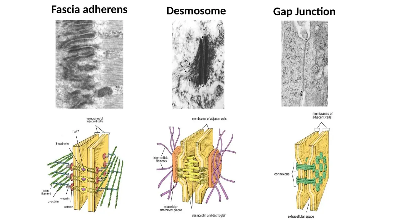
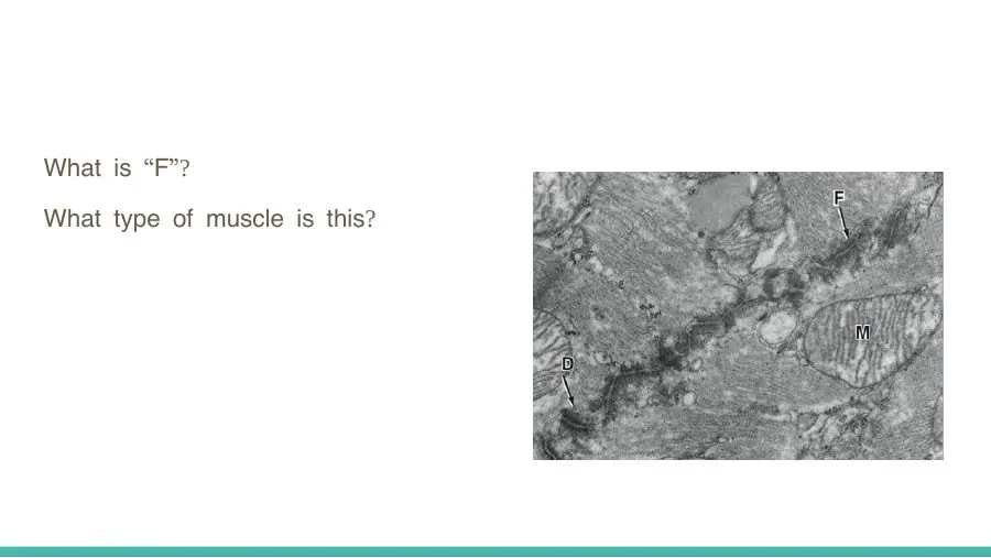
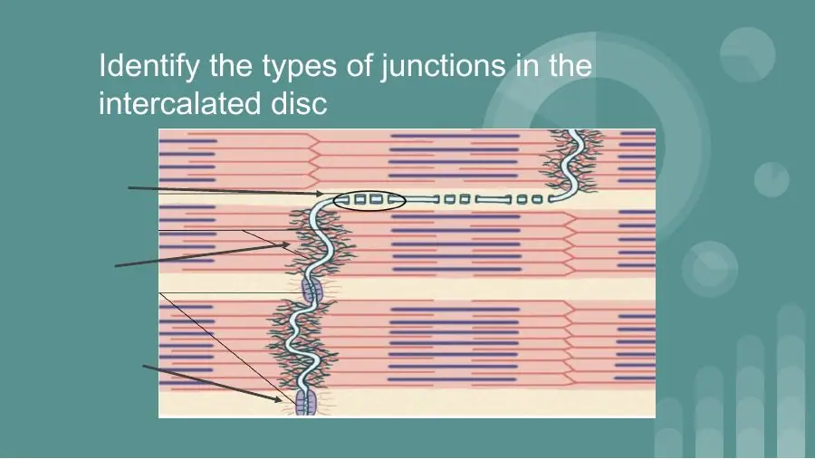
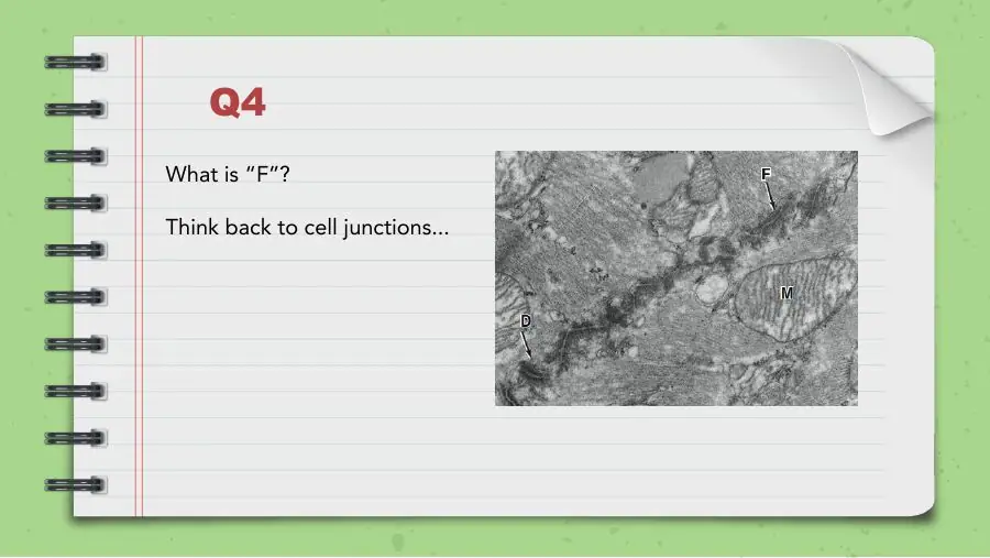
Which three junctions make up an intercalated disc?
A. Fascia adherens Desmosomes Gap junctions
B. Tight junctions Desmosomes Gap junctions
C. Fascia adherens Hemidesmosomes Gap junctions
AnswerA. Fascia adherens Desmosomes Gap junctions
WhyAn intercalated disc is a junctional complex of fasciae adherentes and desmosomes (holding) plus gap junctions (signalling).
Explanation
In the electron microscope the disc runs like a staircase. Its transverse parts (the 'treads') carry the fasciae adherentes, where actin filaments anchor, and the desmosomes, which bind the cells against the pull of contraction. Its lateral parts (the 'risers') carry the gap junctions, which pass ions from cell to cell.
Tight junctions seal epithelia; there are none in the intercalated disc.
Hemidesmosomes fix a cell to its basement membrane, not to a neighbouring cell.
What do the fasciae adherentes and desmosomes of the intercalated disc do?
A. Let the electrical signal pass from cell to cell
B. Hold the cells together against the pull of contraction
C. Anchor the cells to the basement membrane
AnswerB. Hold the cells together against the pull of contraction
WhyFasciae adherentes anchor actin filaments at the cell ends and desmosomes bind the cells, so contraction cannot pull them apart.
Explanation
Each contraction pulls hard on the ends of every cardiac cell. The fascia adherens is where the actin of the last sarcomere attaches to the cell membrane, passing the force to the next cell; desmosomes act as spot welds anchored to intermediate filaments.
Passing the electrical signal is the job of the gap junctions in the same disc.
Anchoring to the basement membrane is done by hemidesmosomes, which are not part of the disc.
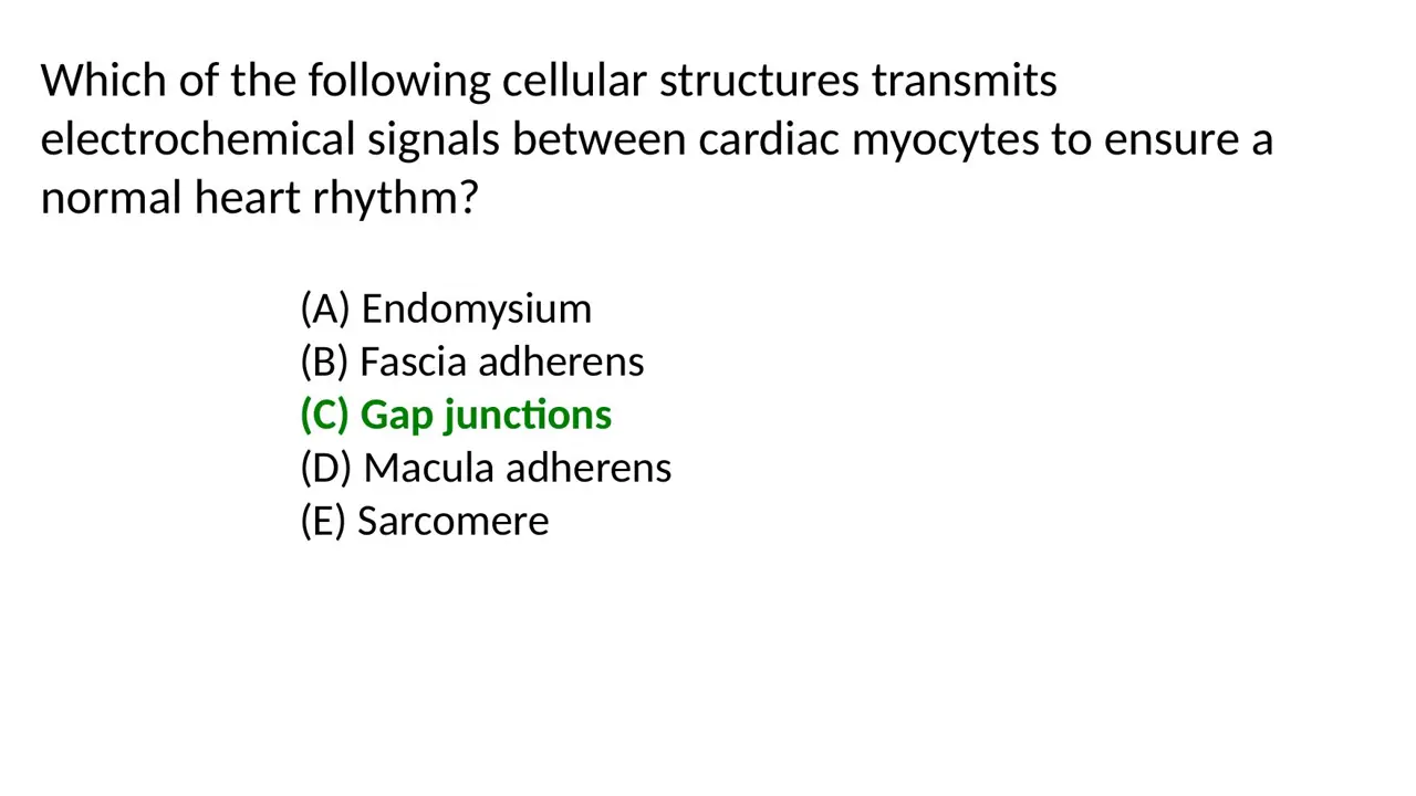
Which component of the intercalated disc spreads electrical signals between cardiac myocytes to keep a normal rhythm?
A. Gap junctions
B. Fasciae adherentes
C. Desmosomes (maculae adherentes)
AnswerA. Gap junctions
WhyGap junctions are low-resistance channels that let ions, and so the action potential, pass straight from one cardiac cell to the next.
Explanation
A gap junction is a cluster of connexon channels that join the cytoplasm of two neighbouring cells. Current flows through them easily, so depolarisation spreads quickly through the myocardium and the cells contract together as a functional syncytium.
Fasciae adherentes anchor actin filaments; they hold, they do not conduct.
Desmosomes (maculae adherentes) are spot welds for mechanical strength.
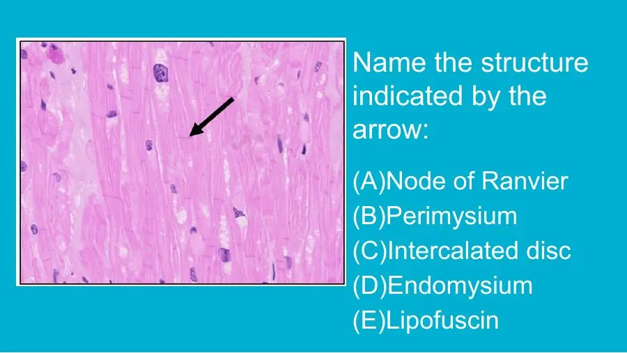
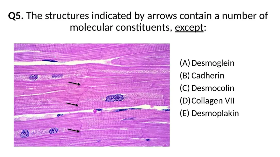
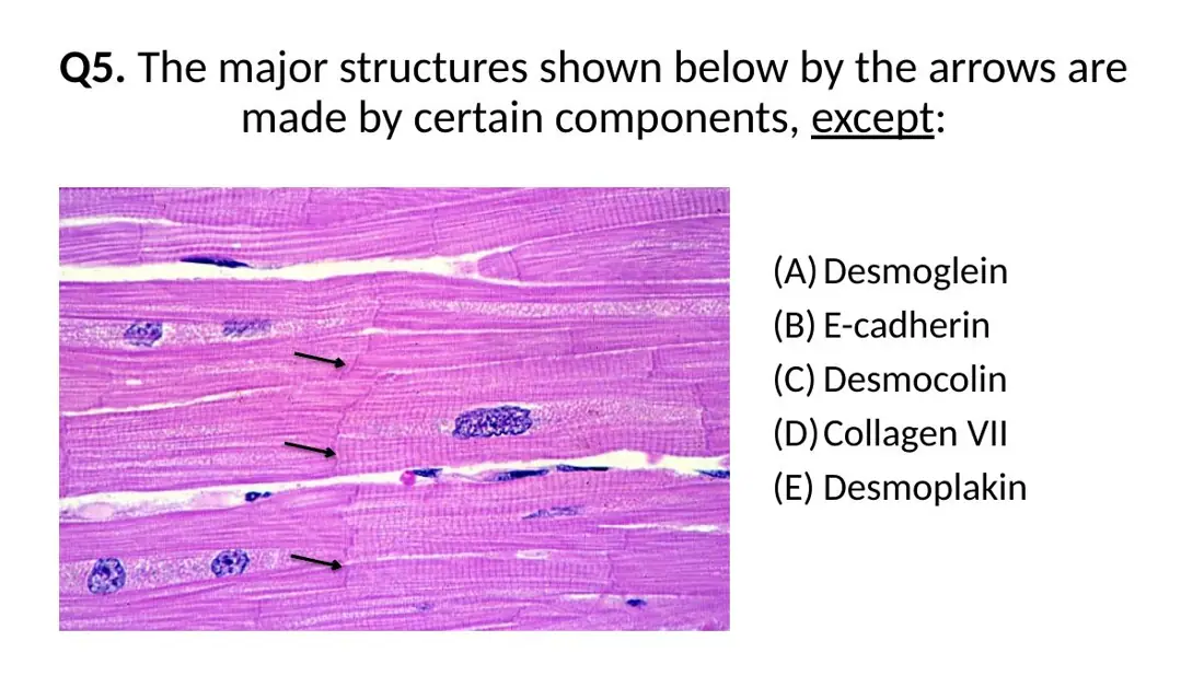
Intercalated discs contain all of these molecules EXCEPT:
A. Collagen VII
B. Desmoglein and desmocollin
C. Cadherin
AnswerA. Collagen VII
WhyCollagen VII forms anchoring fibrils beneath epithelia; the disc's junctions use desmogleins/desmocollins (desmosomes), cadherins (fascia adherens) and connexins (gap junctions).
Explanation
Each junction of the intercalated disc has its own adhesion proteins. Desmosomes bind neighbouring cells with the cadherin-family proteins desmoglein and desmocollin, held inside by desmoplakin. The fascia adherens uses N-cadherin linked to actin. Gap junctions are built from connexins.
Collagen VII is an extracellular matrix protein that anchors the basal lamina of skin to the dermis; it plays no part in cell-to-cell junctions.
The other two options are genuine disc proteins.
Likewise, hemidesmosomes are never part of an intercalated disc.
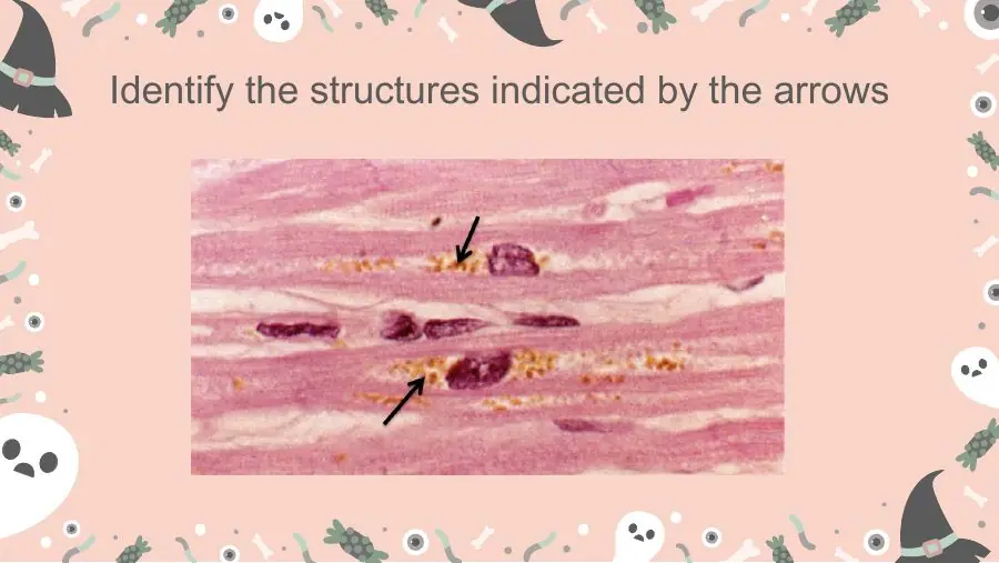
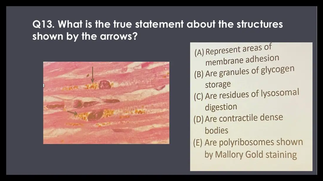
In cardiac muscle from an elderly person, yellow-brown granules cluster at the poles of the central nuclei. What are they?
A. Glycogen granules: stored fuel for contraction
B. Atrial natriuretic factor granules: stored hormone
C. Lipofuscin: undigested residues of lysosomal digestion
AnswerC. Lipofuscin: undigested residues of lysosomal digestion
WhyLipofuscin, the 'wear-and-tear' pigment, is indigestible material left in residual bodies after lysosomal digestion; it builds up near the nucleus with age.
Explanation
Cardiac myocytes live for the whole lifetime and do not divide, so the leftovers of years of lysosomal digestion accumulate as golden-brown lipofuscin granules in the pale zone at each pole of the nucleus.
Glycogen washes out in routine processing and leaves pale, clear sarcoplasm, not brown granules (it is plentiful in Purkinje fibres).
ANF granules lie in atrial cells but are tiny, colourless and visible only by electron microscopy.
L40Learning
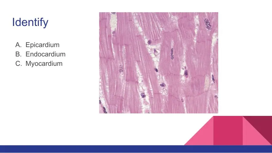
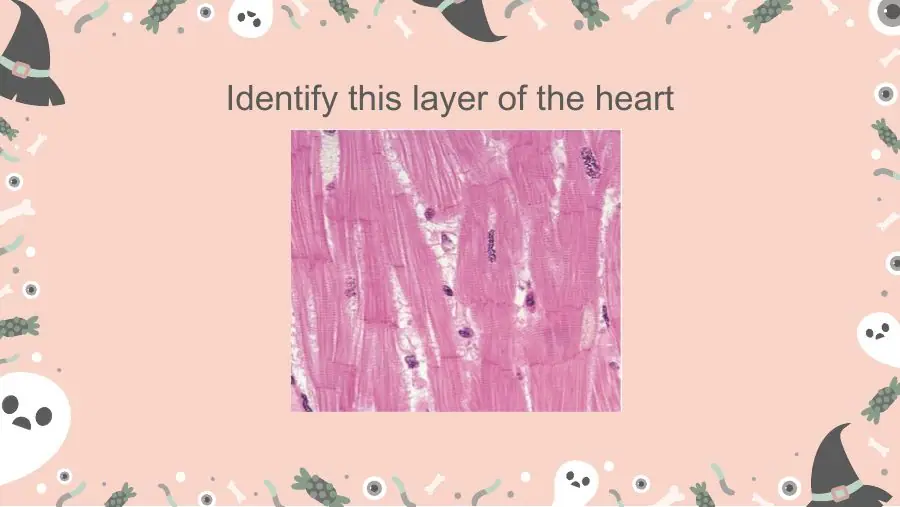
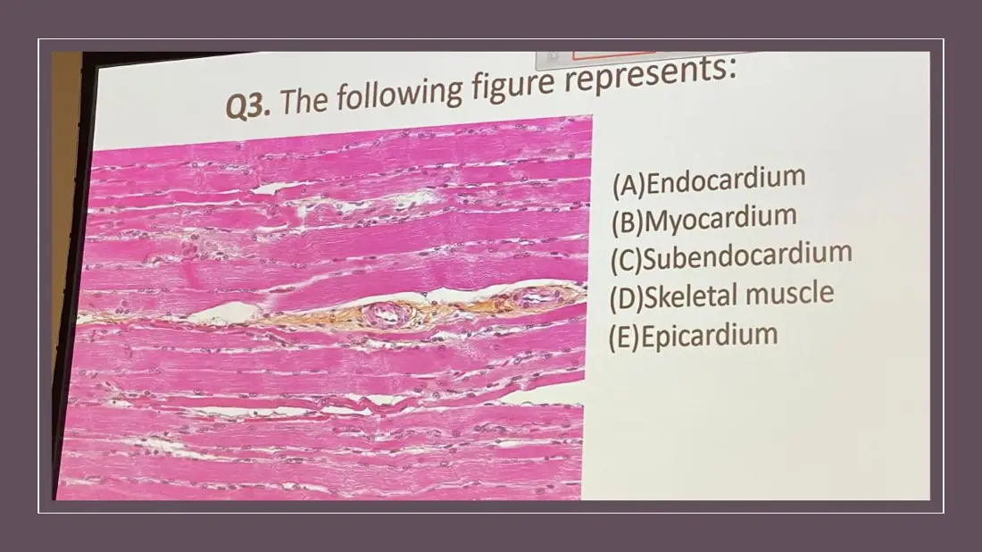
A section shows a thick layer of branching striated cells with central nuclei, joined end to end by dark cross-lines, with capillaries between them. Which heart layer is this?
A. Endocardium
B. Epicardium
C. Myocardium
AnswerC. Myocardium
WhyBranching striated cells with central nuclei and intercalated discs filling the field is the myocardium.
Explanation
The myocardium is almost entirely cardiac muscle: striated, branched cells with one or two central nuclei, intercalated discs, and a rich capillary network in the thin connective tissue between fascicles.
The endocardium is a thin layer of endothelium and connective tissue on the luminal side; its only muscle-like cells are the pale Purkinje fibres.
The epicardium is mesothelium over loose tissue with fat, nerves and coronary vessels on the outer surface.
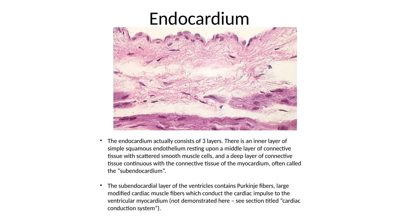
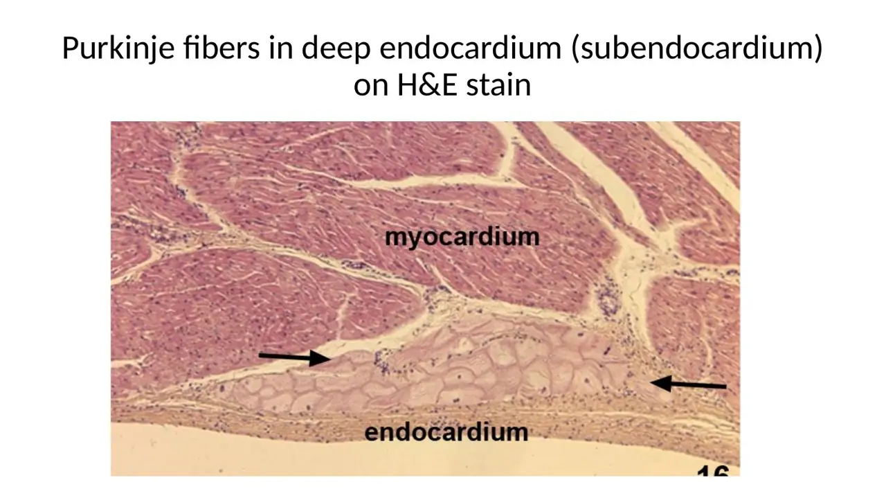
WhyEndocardium = endothelium + a middle layer of connective tissue with some smooth muscle + a deeper subendocardium that blends into the myocardium.
Explanation
The inner lining of the heart has three parts: a simple squamous endothelium facing the blood; a middle myoelastic layer of connective tissue with scattered smooth muscle cells; and the subendocardial layer, loose connective tissue continuous with the myocardium's connective tissue. The subendocardium of the ventricles carries the Purkinje fibres.
Mesothelium with fat describes the epicardium on the outside of the heart.
Endothelium directly on muscle leaves out the connective-tissue layers in which the Purkinje fibres run.
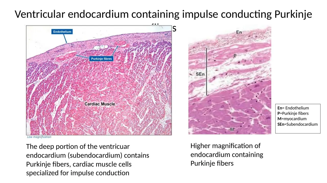
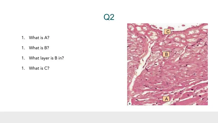
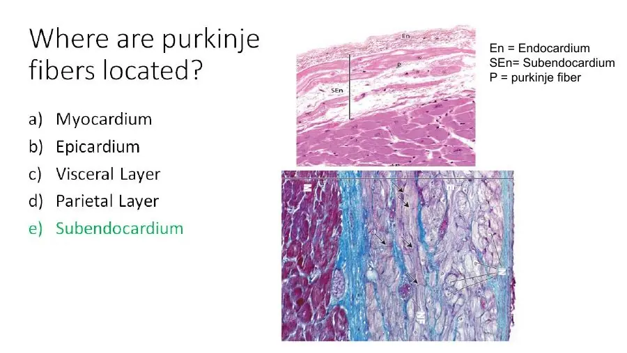
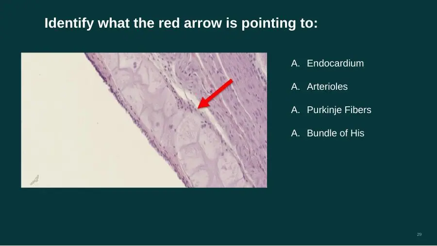
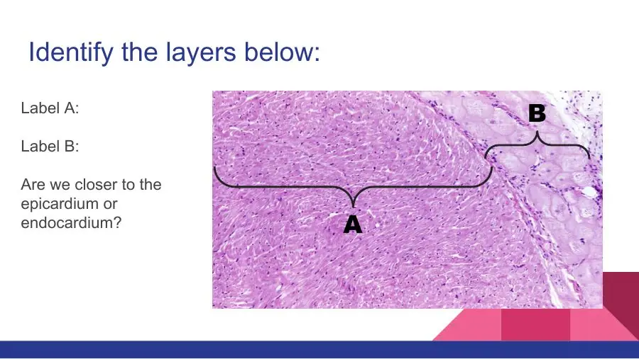
Which layer of the heart wall contains the Purkinje fibres?
A. Myocardium (among the contractile fibres)
B. Epicardium (its subepicardial fatty layer)
C. Endocardium (its subendocardial layer)
AnswerC. Endocardium (its subendocardial layer)
WhyPurkinje fibres run in the subendocardial layer, the deepest part of the endocardium, just above the myocardium.
Explanation
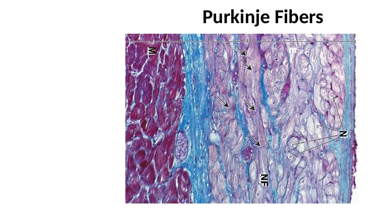
The subendocardium is loose connective tissue between the endothelium-covered lining and the cardiac muscle. In the ventricles it carries the network of Purkinje fibres that spreads the impulse; from there the fibres contact the contractile muscle. The lecture slide calls it the 'subendothelial layer', meaning the same place.
The myocardium is the contractile muscle the Purkinje fibres feed into, not the layer they run in.
The epicardium carries coronary vessels, fat and nerves on the outer surface.
Slide (1)H12A Circulatory I lecture (Dr Bruno) · slide 33L43Learning
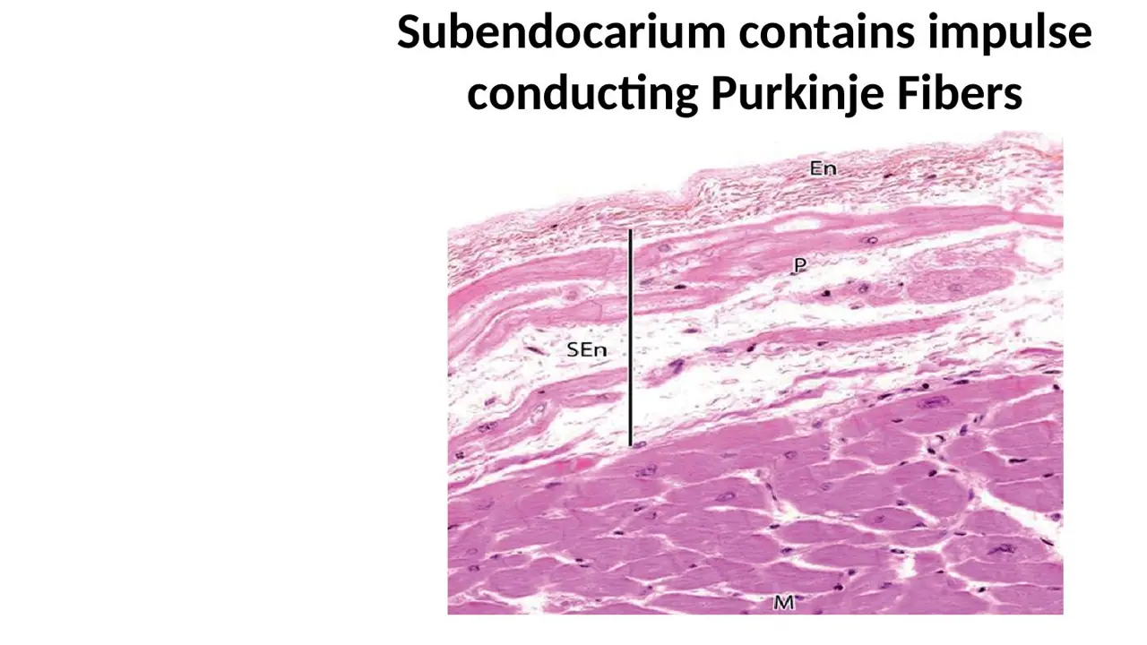
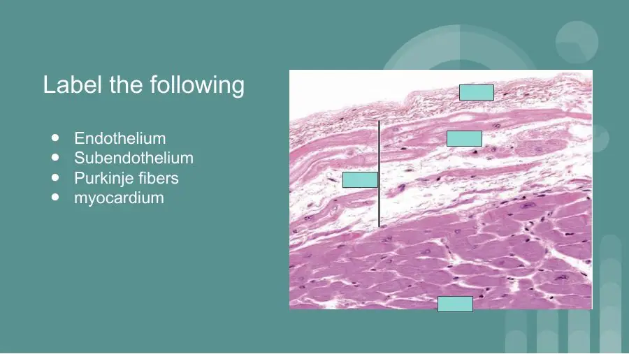
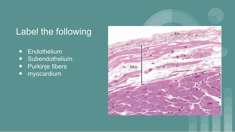
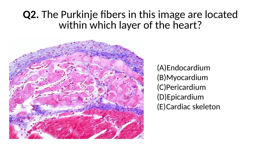
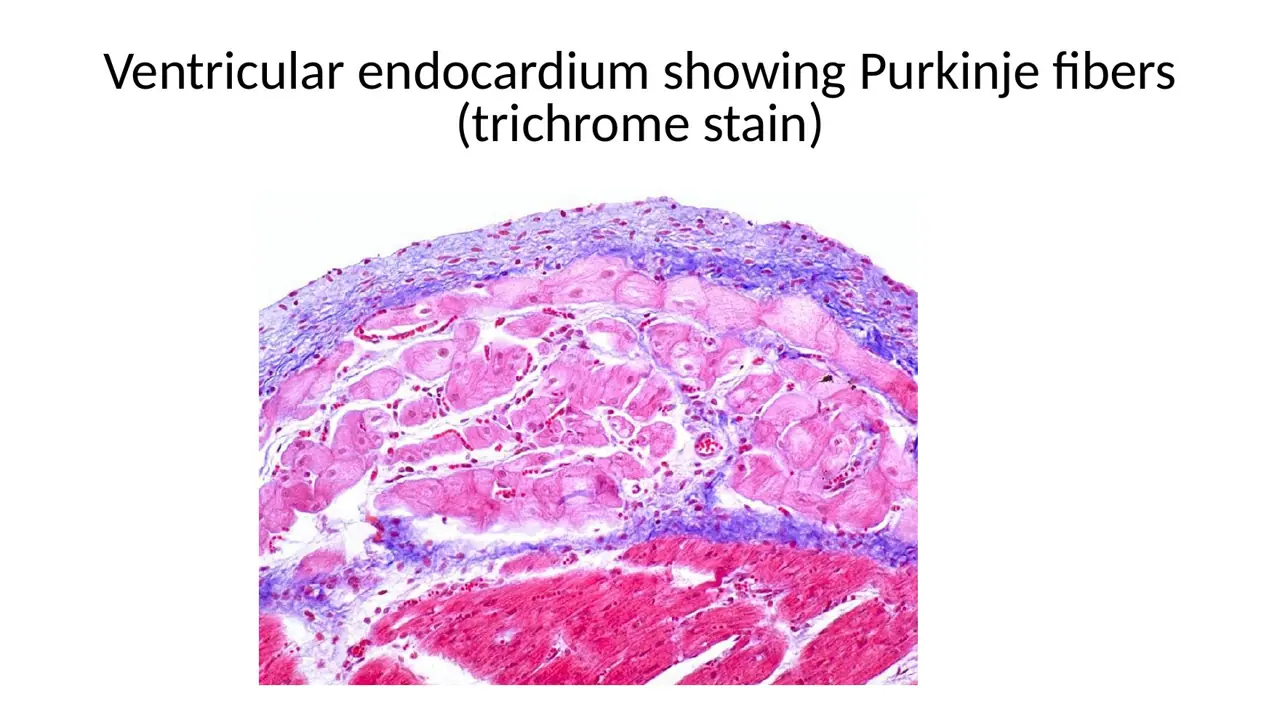
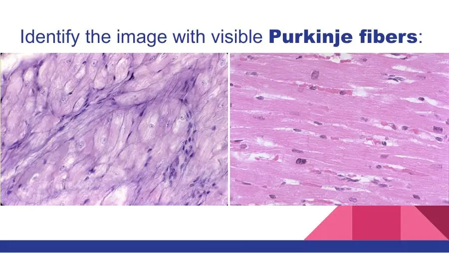
An H&E section of ventricle shows a thin endothelium, then a band of connective tissue holding large pale fibres with central nuclei, then deep-pink cardiac muscle. Name the band and the pale fibres.
A. Subepicardium; adipocytes
B. Myoelastic layer; smooth muscle cells
C. Subendocardium; Purkinje fibres
AnswerC. Subendocardium; Purkinje fibres
WhyLarge pale fibres in the connective tissue between endothelium and myocardium are Purkinje fibres in the subendocardium.
Explanation
Read the section from the lumen: endothelium (En), then the subendocardium (SEn) with Purkinje fibres (P), then the myocardium (M). Purkinje fibres are wider than the contractile cells and pale because their glycogen dissolves during processing.
Subepicardium with adipocytes is on the other side of the wall, under a mesothelium; fat cells are empty rings with flattened peripheral nuclei, not fibres with central nuclei.
The myoelastic layer lies just under the endothelium; its smooth muscle cells are thin and spindle-shaped, not large and pale.
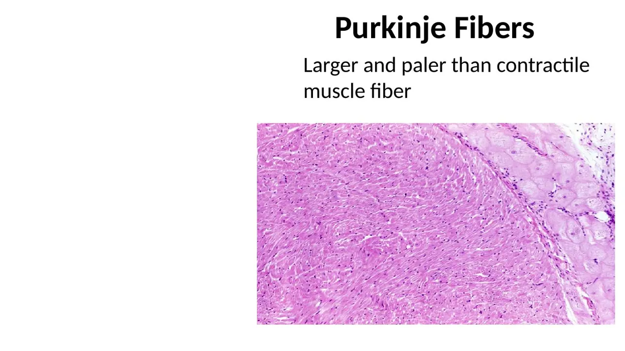
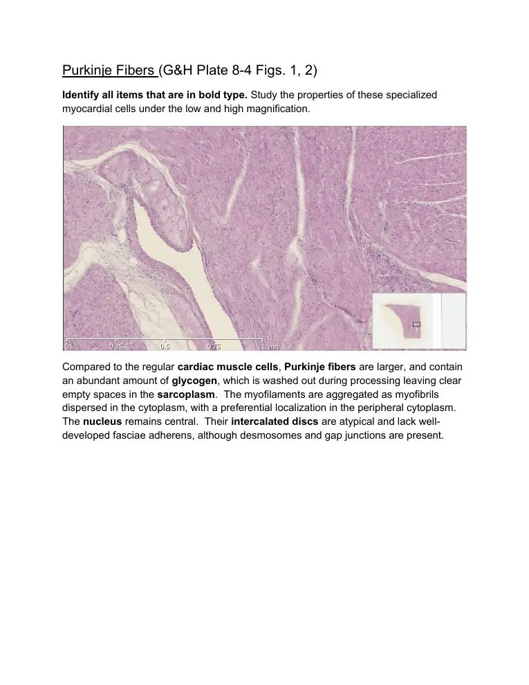
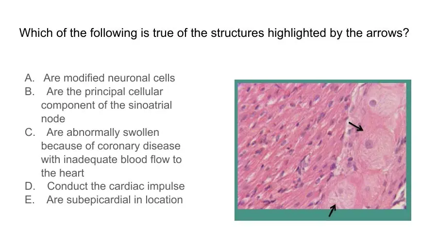
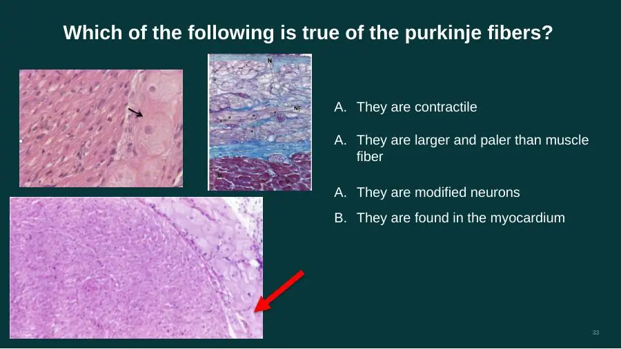
How do Purkinje fibres look compared with ordinary cardiac muscle cells?
A. Smaller and darker — little glycogen, more myofibrils
B. Larger and paler — abundant glycogen, fewer myofibrils
C. Larger and darker — abundant myoglobin, more myofibrils
AnswerB. Larger and paler — abundant glycogen, fewer myofibrils
WhyPurkinje fibres are larger and paler than contractile cells: they are packed with glycogen and have fewer myofibrils.
Explanation
Purkinje fibres are modified cardiac muscle cells built for conduction, not force. Much of their cytoplasm is glycogen instead of myofibrils, so in H&E they look swollen and pale beside the dense pink working muscle; in a trichrome stain they are pale lavender above the deep-red myocardium.
Smaller and darker with more myofibrils describes the contractile cells, not the conducting ones.
Myoglobin and extra myofibrils would make them darker; their pallor comes from glycogen.
Giveaway: big pale cells just under the endocardium = Purkinje fibres.
Why do Purkinje fibres look pale in routine sections?
A. Lipid dissolves in processing → clear sarcoplasm round a peripheral nucleus, myofibrils packed centrally
B. Glycogen washes out in processing → clear sarcoplasm round a central nucleus, myofibrils pushed to the edge
C. Glycogen stains intensely → nucleus pushed to the edge by packed myofibrils
AnswerB. Glycogen washes out in processing → clear sarcoplasm round a central nucleus, myofibrils pushed to the edge
WhyTheir abundant glycogen dissolves during processing, leaving clear sarcoplasm round a central nucleus, with the sparse myofibrils at the cell periphery.
Explanation
The glycogen occupies the middle of the cell. When it is removed by routine fixation and staining, empty-looking sarcoplasm is left around the centrally placed nucleus, and the few myofibrils sit mainly near the cell edge.
Lipid is not what fills them, and their nucleus stays central.
Glycogen does not stain with H&E; it shows up only with a special stain such as PAS, where it turns magenta.
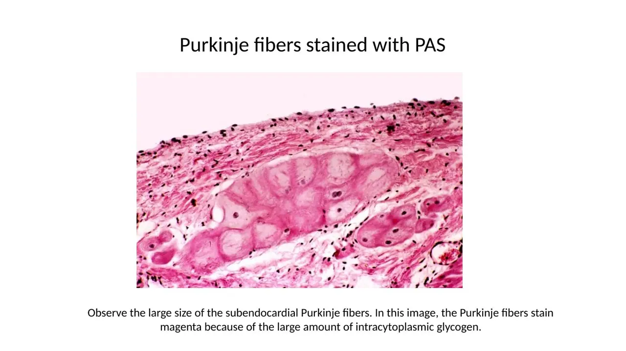
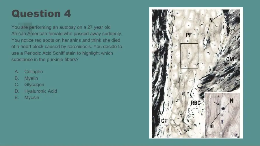
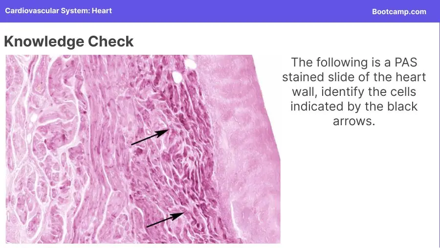
A PAS stain turns the large pale subendocardial fibres dark magenta. Which substance in them does PAS reveal?
A. Lipid
B. Collagen
C. Glycogen
AnswerC. Glycogen
WhyPAS (periodic acid–Schiff) stains glycogen magenta, and Purkinje fibres are full of it.
Explanation
The glycogen that washes out in routine H&E, leaving Purkinje fibres pale, is kept and coloured by PAS. So the same fibres that look empty in H&E turn deep magenta in PAS, which confirms both their identity and their location in the endocardium.
Lipid is dissolved by routine processing and is shown with fat stains on frozen sections, not PAS.
Collagen is blue in trichrome; PAS colours it only faintly. The fibres here are muscle cells, not connective tissue.
How do the intercalated discs of Purkinje fibres differ from typical ones?
A. Fasciae adherentes and desmosomes present; gap junctions absent
B. Tight junctions replace gap junctions; desmosomes well developed
C. Desmosomes and gap junctions present; fasciae adherentes poorly developed
AnswerC. Desmosomes and gap junctions present; fasciae adherentes poorly developed
WhyPurkinje fibre discs are atypical: they keep desmosomes and gap junctions but lack well-developed fasciae adherentes.
Explanation
The fascia adherens anchors actin of the contractile apparatus; Purkinje fibres have few myofibrils and do little contracting, so they need little of it. They do need gap junctions, because conducting the impulse quickly is their whole job.
No gap junctions would stop them conducting.
Tight junctions are not found in intercalated discs of any kind.
Slide (1)Lab manual HL7 circulatory · page 9
The conduction system
L48Learning
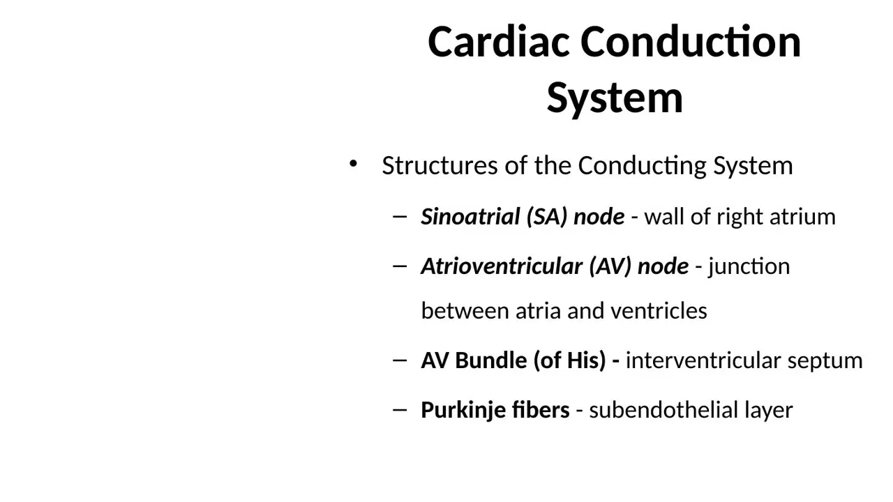
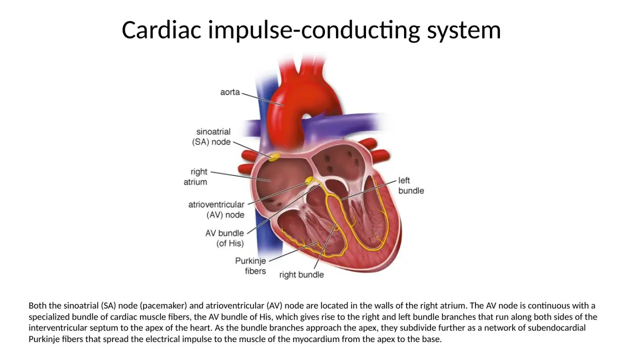
What are the cells of the cardiac conduction system?
A. Modified cardiac muscle cells, specialised for conduction rather than contraction
B. Specialised neurons of the autonomic nervous system
C. Modified smooth muscle cells, specialised for conduction rather than contraction
AnswerA. Modified cardiac muscle cells, specialised for conduction rather than contraction
WhyThe nodes, the AV bundle and the Purkinje fibres are all modified cardiac muscle cells that generate and conduct the impulse instead of contracting strongly.
Explanation
The SA and AV nodes are small masses of specialised myocardial tissue in the right atrial wall; the AV bundle, bundle branches and Purkinje fibres are conducting muscle cells joined by gap junctions. They contain few myofibrils.
Neurons: the heart beats without its nerves; autonomic nerves only speed or slow the rate set by the SA node. 'Purkinje cells' of the cerebellum are neurons, but Purkinje fibres of the heart are muscle.
Smooth muscle occurs in the endocardium's myoelastic layer and in vessel walls, but it does not conduct the heartbeat.
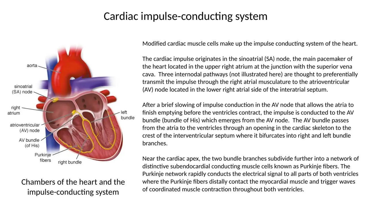
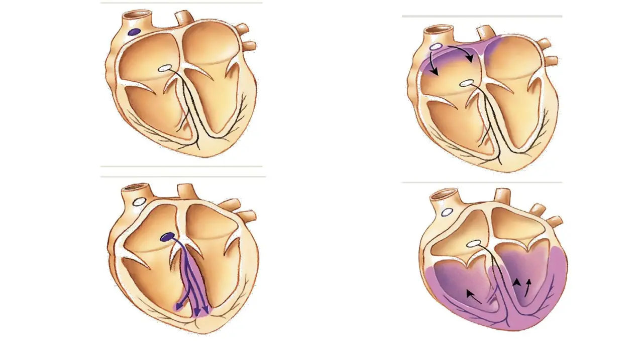
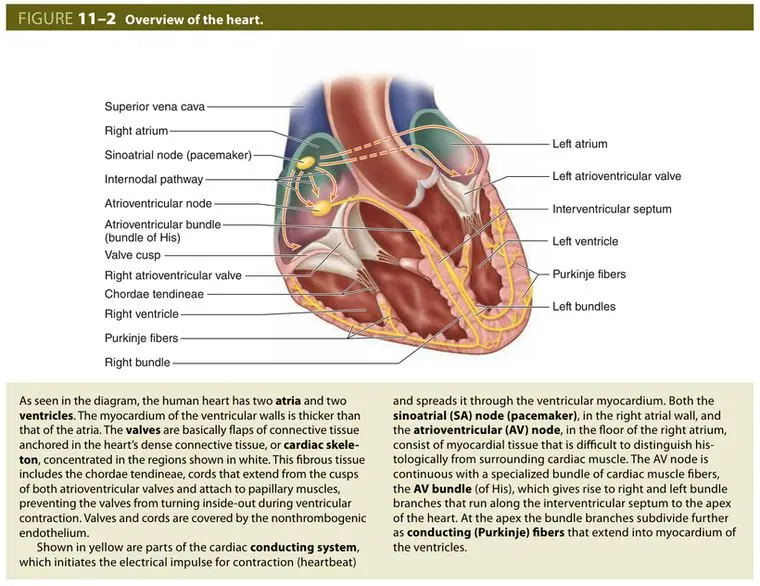
A. SA node → AV bundle (of His) → AV node → right and left bundle branches → Purkinje fibres
B. SA node → AV node → AV bundle (of His) → right and left bundle branches → Purkinje fibres
C. AV node → SA node → AV bundle (of His) → right and left bundle branches → Purkinje fibres
AnswerB. SA node → AV node → AV bundle (of His) → right and left bundle branches → Purkinje fibres
WhySA node fires → atria → AV node (delay) → bundle of His through the skeleton → bundle branches in the septum → Purkinje fibres → ventricles.
Explanation
The impulse starts in the SA node in the right atrial wall and spreads over both atria. It is funnelled into the AV node, which delays it, then into the AV bundle (of His), the only path through the insulating cardiac skeleton. The bundle splits into right and left branches in the interventricular septum; about halfway down these become the subendocardial Purkinje network that reaches all of both ventricles.
Putting the bundle of His before the AV node skips the delay: the AV node feeds the bundle, not the other way round.
Starting at the AV node makes it the pacemaker; the SA node fires first because it is the fastest.
After the SA node fires, which tissue depolarises before the AV node?
A. Ventricular myocardium
B. Bundle of His
C. Atrial myocardium
AnswerC. Atrial myocardium
WhyOrder: SA node → atrial myocardium → AV node → bundle of His → Purkinje fibres → ventricular myocardium.
Explanation
The SA node's impulse spreads cell to cell through the atrial muscle (through gap junctions), so the atria contract first. The wave converges on the AV node in the floor of the right atrium, which passes it on after a delay.
The ventricular myocardium is the last to depolarise, after the Purkinje fibres.
The bundle of His comes after the AV node, carrying the impulse through the cardiac skeleton.
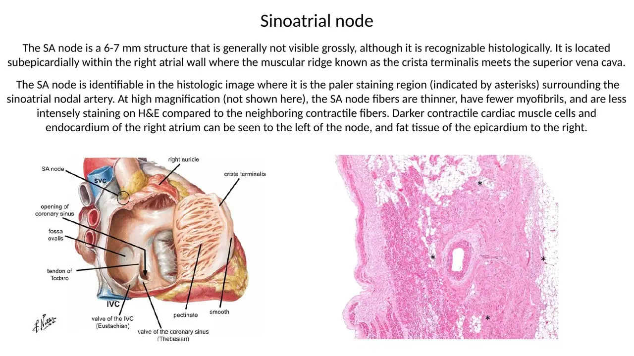
Where is the sinoatrial (SA) node, and what is its role?
A. Wall of the left atrium — pacemaker of the heart
B. Interventricular septum — pacemaker of the heart
C. Wall of the right atrium — pacemaker of the heart
AnswerC. Wall of the right atrium — pacemaker of the heart
WhyThe SA node sits in the right atrial wall near the entrance of the superior vena cava and is the pacemaker: it fires fastest and sets the heart rate.
Explanation
The SA node is a small mass of specialised cardiac muscle under the epicardium at the junction of the superior vena cava with the right atrium. Its cells depolarise spontaneously faster than any other part of the conduction system, so each beat starts there. When it fails, an artificial pacemaker can take over.
The left atrium has no node; both nodes are on the right side.
The interventricular septum carries the AV bundle and its branches, not the pacemaker.
Electrophysiological mapping shows the earliest spontaneous depolarisation in a small nodal structure in the upper right atrial wall, where the superior vena cava enters. What is it?
A. Sinoatrial (SA) node
B. Atrioventricular (AV) node
C. Bundle of His
AnswerA. Sinoatrial (SA) node
WhyThe first site to fire, at the superior vena cava–right atrium junction, is the SA node, the pacemaker.
Explanation
The SA node lies along the crista terminalis where the superior vena cava enters the right atrium. Because its cells reach threshold fastest, every normal beat begins there.
The AV node lies lower, in the floor of the right atrium at the atrioventricular junction; it fires spontaneously only if the SA node fails, and more slowly.
The bundle of His is in the interventricular septum and only conducts what the AV node passes to it.
A. Floor of the left atrium, at the junction between atria and ventricles
B. Upper interventricular septum, below the cardiac skeleton
C. Floor of the right atrium, at the junction between atria and ventricles
AnswerC. Floor of the right atrium, at the junction between atria and ventricles
WhyThe AV node lies in the floor of the right atrium, at the atrioventricular junction, just above the cardiac skeleton.
Explanation
The lecture places the AV node at the junction between atria and ventricles; the textbook puts it more precisely in the floor of the right atrium. It collects the atrial impulse and passes it into the AV bundle, which pierces the cardiac skeleton.
The left atrium has no nodal tissue.
The upper interventricular septum below the skeleton is where the AV bundle runs and divides, after it has left the node.
A. It lets the atria finish emptying before the ventricles contract
B. It lets the ventricles finish emptying before the atria contract
C. It lets the ventricles contract before the atria fill
AnswerA. It lets the atria finish emptying before the ventricles contract
WhyThe AV-node delay gives the atria time to finish pushing blood into the ventricles before the ventricles contract.
Explanation
The atria contract first and top up the ventricles at the end of their filling. If the impulse reached the ventricles at once, they would contract while the atria were still emptying, and the AV valves would close on incomplete filling.
Ventricles emptying before the atria contract reverses the sequence: atrial contraction comes first.
Ventricles contracting before the atria fill has nothing to do with the delay; the atria fill throughout ventricular contraction.
A. Through an opening in the cardiac skeleton → interventricular septum → splits into right and left bundle branches
B. Around the cardiac skeleton → right atrial wall → splits into anterior and posterior branches
C. Through an opening in the cardiac skeleton → left ventricular free wall → splits straight into Purkinje fibres
AnswerA. Through an opening in the cardiac skeleton → interventricular septum → splits into right and left bundle branches
WhyThe AV bundle passes through the cardiac skeleton into the interventricular septum, where it divides into right and left bundle branches.
Explanation
The bundle is the only muscular link between atria and ventricles. After piercing the fibrous skeleton it runs along the top of the muscular interventricular septum and bifurcates. About halfway down the septum the branches break up into the subendocardial Purkinje network.
It cannot go around the skeleton: the skeleton insulates the atria from the ventricles all the way round, and the bundle runs in the septum, not the atrial wall.
It does not enter the left ventricular free wall; it divides in the septum first, and both ventricles are reached through their own branch.
A. Carry the signal slowly to both atria, ending on contractile muscle → atria contract together
B. Carry the signal rapidly to all parts of both ventricles, ending on contractile muscle → ventricles contract together
C. Carry the signal rapidly to the right ventricle only → ventricles contract one after the other
AnswerB. Carry the signal rapidly to all parts of both ventricles, ending on contractile muscle → ventricles contract together
WhyThe His–Purkinje network rapidly delivers the impulse to all parts of both ventricles, so the ventricular muscle contracts in one coordinated wave.
Explanation
Purkinje fibres are the terminal branches of the right and left bundle branches, running in the subendocardium of both ventricles. They conduct faster than ordinary myocardium and end on the contractile cells, triggering contraction throughout both ventricles almost at once.
The atria are reached directly from the SA node through atrial muscle, not through Purkinje fibres.
Both bundle branches carry Purkinje fibres, so both ventricles are activated together.
A simple squamous epithelium covers or lines the free surface of all of these EXCEPT:
A. The mitral valve
B. The sinoatrial node
C. The epicardium
AnswerB. The sinoatrial node
WhyThe SA node sits inside the right atrial wall and reaches neither surface; valves are covered by endothelium and the epicardium by mesothelium.
Explanation
Every free surface of the heart has a simple squamous epithelium: endothelium on the endocardium and both faces of every valve, mesothelium on the epicardium, and endothelium continuing into the aorta's intima. The SA node is a buried mass of specialised muscle at the superior vena cava–right atrium junction, under the epicardium, so it has no free surface of its own.
The mitral valve is a fold of endocardium; endothelium covers both its surfaces.
The epicardium is covered by mesothelium, which is also simple squamous.
A. The sound of the heart valves closing, from a microphone on the chest
B. The blood flow through the coronary arteries, from electrodes on the skin
C. The spread of the electrical impulse through the heart, from electrodes on the skin
AnswerC. The spread of the electrical impulse through the heart, from electrodes on the skin
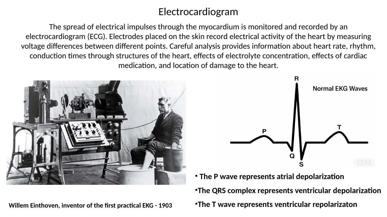
WhyAn ECG records the voltage differences that the spreading impulse produces, measured between skin electrodes; Einthoven built the first practical one in 1903.
Explanation
As the impulse spreads through the myocardium, it creates voltage differences that reach the body surface. Electrodes at different points record them. The tracing shows heart rate, rhythm, conduction times through each part of the conduction system, effects of electrolytes and drugs, and the location of damaged muscle.
Valve sounds are heard with a stethoscope or recorded by phonocardiography.
Coronary flow is studied by angiography or perfusion scans, not by the ECG.
Slide (1)Circulatory lab 2026 · slide 30
The cardiac skeleton
L59Learning
What tissue forms the cardiac skeleton?
A. Dense irregular connective tissue
B. Dense regular connective tissue
C. Elastic connective tissue
AnswerA. Dense irregular connective tissue
WhyThe cardiac skeleton is dense irregular connective tissue: thick collagen bundles running in many directions.
Explanation
The skeleton must resist pull from every direction, as valves snap shut and muscle contracts on all sides, so its collagen bundles are woven in many directions, like the dermis. It lies mainly in the endocardium at the junction of atria and ventricles.
Dense regular tissue has parallel bundles that resist pull in one direction only, as in tendons.
Elastic tissue stretches and recoils, as in the aortic wall; the skeleton has to stay rigid to hold the valve openings in shape.
Slide (1)H12A Circulatory I lecture (Dr Bruno) · slide 47L60Learning
How is the cardiac skeleton arranged?
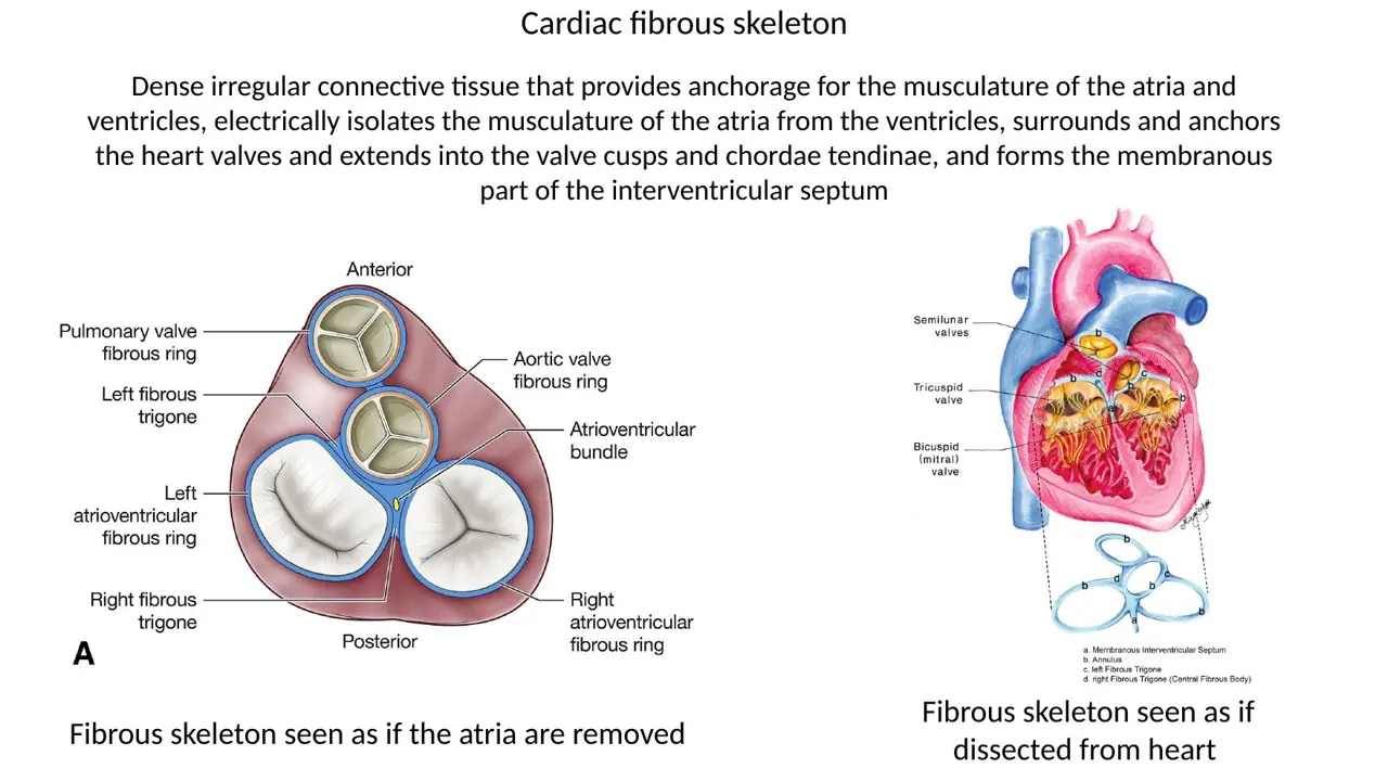
A. Four fibrous rings (annuli) around the four valves, joined by the left and right fibrous trigones
B. Four cartilaginous rings around the four valves, joined by the left and right fibrous trigones
C. Four fibrous rings (annuli) around the four valves, joined by the anterior and posterior papillary muscles
AnswerA. Four fibrous rings (annuli) around the four valves, joined by the left and right fibrous trigones
WhyThe skeleton is four fibrous rings round the valve openings (including the bases of the aorta and pulmonary trunk), linked by the fibrous trigones and the membranous septum.
Explanation
Seen from above with the atria removed, the skeleton is a flat plate between atria and ventricles: rings around the tricuspid, mitral, aortic and pulmonary openings, joined in the middle by the right and left fibrous trigones. The membranous part of the interventricular septum hangs down from it. The AV bundle passes through the right fibrous trigone.
Cartilage: the human cardiac skeleton is fibrous connective tissue, not cartilage.
Papillary muscles are muscular projections inside the ventricles; they pull on the chordae and are not part of the skeleton.
Into which structures does the cardiac skeleton extend?
A. Valve cusps Papillary muscles Muscular part of the interventricular septum
B. Valve cusps Purkinje fibres Membranous part of the interventricular septum
C. Valve cusps Chordae tendineae Membranous part of the interventricular septum
AnswerC. Valve cusps Chordae tendineae Membranous part of the interventricular septum
WhyThe skeleton's collagen runs into the valve cusps and chordae tendineae and forms the membranous interventricular septum.
Explanation
The dense connective tissue core of each valve leaflet is continuous with the fibrous ring it hangs from, and the chordae tendineae are cords of the same collagen. The small upper, membranous part of the interventricular septum is skeleton too.
Papillary muscles and the muscular septum are cardiac muscle, not connective tissue.
Purkinje fibres are modified muscle cells in the subendocardium; only the AV bundle passes through the skeleton.
Which part of the interventricular septum is formed by the cardiac skeleton?
A. The muscular part: large, lower part running toward the apex
B. The membranous part: large, lower part running toward the apex
C. The membranous part: small, uppermost, just below the aortic valve
AnswerC. The membranous part: small, uppermost, just below the aortic valve
WhyThe membranous septum is a small, thin, fibrous area at the top of the septum below the aortic valve; the rest is the thick muscular septum.
Explanation
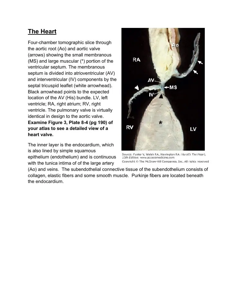
In a four-chamber slice, nearly all of the interventricular septum is thick muscle. Only a small, thin patch at its top, just below the aortic root, is fibrous. This membranous septum is part of the cardiac skeleton; the AV bundle runs along its lower edge as it reaches the muscular septum.
The muscular part is cardiac muscle, not skeleton.
The membranous part is small and high, not the large lower portion.
A. Anchors the valves Generates the pacemaker impulse Electrically couples atria to ventricles
B. Anchors the valves Insertion for atrial and ventricular muscle Electrically insulates atria from ventricles
C. Anchors the coronary arteries Insertion for atrial and ventricular muscle Conducts the impulse from atria to ventricles
AnswerB. Anchors the valves Insertion for atrial and ventricular muscle Electrically insulates atria from ventricles
WhyThe skeleton stabilises the valves, gives the atrial and ventricular muscle something to pull on, and electrically insulates the atria from the ventricles.
Explanation
The fibrous rings keep each valve opening the right shape so the cusps meet. The atrial and ventricular muscle bundles insert on the skeleton. Because connective tissue has no gap junctions, the skeleton blocks the atrial impulse everywhere except at the AV bundle.
Pacemaking and coupling are done by the SA node and the AV node/bundle; the skeleton does the opposite of coupling.
The skeleton does not conduct; and the coronary arteries are anchored in the epicardium, not by the skeleton.
Slide (1)H12A Circulatory I lecture (Dr Bruno) · slide 47L64Learning
What is the only electrical path from the atria to the ventricles?
A. The AV bundle (of His), which pierces the cardiac skeleton
B. Purkinje fibres, which run through the fibrous trigones
C. Gap junctions spread evenly across the whole cardiac skeleton
AnswerA. The AV bundle (of His), which pierces the cardiac skeleton
WhyThe insulating skeleton leaves one gap: the AV bundle passing through it, so every impulse must go through the AV node and bundle.
Explanation
This arrangement makes the AV node the gatekeeper. It delays the impulse so the atria can empty, and it limits how fast the ventricles can be driven if the atria beat abnormally fast. If the skeleton did not insulate, impulses could cross anywhere and the ventricles would contract out of step with the atria.
Purkinje fibres are downstream, in the ventricular subendocardium; they do not cross the skeleton.
Connective tissue has no gap junctions; that is exactly why it insulates.
WhyValves keep flow one-way: atrioventricular valves between atria and ventricles, semilunar valves between ventricles and the aorta or pulmonary trunk.
Explanation
There are two types of heart valve. The two AV valves (tricuspid on the right, mitral on the left) stop blood going back into the atria when the ventricles contract. The two semilunar valves (pulmonary and aortic) stop blood falling back into the ventricles when they relax.
The swapped option reverses the two types.
The cardiac skeleton surrounds the AV openings but does not close them; the AV valves hang from it.
Slides (2)H12A Circulatory I lecture (Dr Bruno) · slide 51H12A Circulatory I lecture (Dr Bruno) · slide 54L66Learning
Name the atrioventricular valves.
A. Right: bicuspid (mitral) Left: tricuspid
B. Right: tricuspid Left: bicuspid (mitral)
C. Right: pulmonary Left: aortic
AnswerB. Right: tricuspid Left: bicuspid (mitral)
WhyThe tricuspid valve guards the right AV opening; the bicuspid (mitral) valve guards the left.
Explanation
The right AV valve has three cusps, the left two. The mitral valve is named for its resemblance to a bishop's mitre.
Mitral on the right swaps the sides.
Pulmonary and aortic are the semilunar valves at the exits of the ventricles.
Hook: 'Tricuspid on the Right'; the Left has the mitraL.
Slide (1)H12A Circulatory I lecture (Dr Bruno) · slide 51L67Learning
Name the semilunar valves and where each sits.
A. Pulmonary: left ventricle → pulmonary trunk Aortic: right ventricle → aorta
B. Pulmonary: right ventricle → aorta Aortic: left ventricle → pulmonary trunk
C. Pulmonary: right ventricle → pulmonary trunk Aortic: left ventricle → aorta
AnswerC. Pulmonary: right ventricle → pulmonary trunk Aortic: left ventricle → aorta
WhyThe pulmonary semilunar valve guards the right ventricle's exit into the pulmonary trunk; the aortic valve guards the left ventricle's exit into the aorta.
Explanation
Each semilunar valve sits at the root of the great artery leaving a ventricle. The right ventricle feeds the lungs through the pulmonary trunk; the left feeds the body through the aorta.
The first wrong option swaps which ventricle each valve serves.
The second swaps which artery each valve opens into.
The mitral valve is an AV valve, never a semilunar one.
Slide (1)H12A Circulatory I lecture (Dr Bruno) · slide 54L68Learning
What forms the core of a heart valve?
A. Vascular loose connective tissue, covered on both surfaces by endothelium
B. Avascular dense connective tissue, covered on both surfaces by endothelium
C. Cardiac muscle, covered on both surfaces by endothelium
AnswerB. Avascular dense connective tissue, covered on both surfaces by endothelium
WhyA valve is a fold of endocardium with an avascular dense connective tissue core (the fibrosa), covered by endothelium; it moves passively.
Explanation
The collagen core (blue in trichrome) is continuous with the fibrous ring of the cardiac skeleton. It has no blood vessels: the thin leaflet is nourished by diffusion from the blood on both sides. Valves contain no muscle and open and close only with the pressure difference across them.
Vascular loose tissue would be too weak and would bleed; the core is dense and avascular.
Cardiac muscle stays in the myocardium and papillary muscles; the leaflet itself does not contract.
Because they are avascular, infected valves are hard for white cells and antibiotics to reach.
Which layer of the heart wall do the valves belong to?
A. Myocardium (extensions of the muscle), with a core continuous with the papillary muscles
B. Endocardium (folds of the lining), with a core continuous with the cardiac skeleton
C. Epicardium (folds of the serous layer), with a core continuous with the pericardium
AnswerB. Endocardium (folds of the lining), with a core continuous with the cardiac skeleton
WhyValves are folds of endocardium: endothelium over a connective-tissue core anchored in the cardiac skeleton.
Explanation
At the atrioventricular junction the skeleton lies mainly within the endocardium, and each leaflet grows out of it: its dense collagen core continues from the fibrous ring, and its endothelial covering continues from the endocardium of the atrium and ventricle.
The myocardium stops at the base of the valve; the papillary muscles attach to the chordae, not to the leaflet core.
The epicardium covers the outside of the heart and never reaches the valve openings.
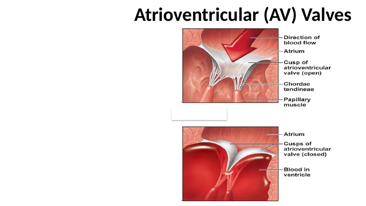
What closes the atrioventricular valves at the start of ventricular contraction?
A. Contraction of the papillary muscles pulls the cusps together
B. Contraction of muscle within the cusps draws them shut
C. Rising ventricular blood pressure pushes the cusps together
AnswerC. Rising ventricular blood pressure pushes the cusps together
WhyAV valves close passively: when ventricular pressure exceeds atrial pressure, blood pressure pushes the cusps shut.
Explanation
The lecture lists the two forces: 'blood pressure closes valve' and 'papillary muscles tense chordae tendineae'. The pressure gradient does the closing; the papillary muscles, contracting with the rest of the ventricle, only keep the closed cusps from being blown back into the atrium.
Papillary muscles pull the cusps toward the ventricle through the chordae; they brace the closed valve rather than shutting it.
The cusps contain no muscle: they are dense connective tissue covered by endothelium.
Slides (2)H12A Circulatory I lecture (Dr Bruno) · slide 51H12A Circulatory I lecture (Dr Bruno) · slide 50L71Learning
What stops the atrioventricular valve cusps turning inside out into the atria?
A. Papillary muscles contract → tense the chordae tendineae
B. Papillary muscles relax → slacken the chordae tendineae
C. The three cusps brace against one another like a tripod
AnswerA. Papillary muscles contract → tense the chordae tendineae
WhyDuring ventricular contraction the papillary muscles contract too and tense the chordae tendineae, holding the closed AV cusps against the pressure.
Explanation
The chordae tendineae are collagen cords running from the free edges of the AV cusps to the papillary muscles on the ventricular wall. As ventricular pressure rises, the papillary muscles shorten in step and keep the cords taut, so the cusps meet but cannot evert (prolapse) into the atria. If a papillary muscle ruptures after an infarct, the valve flips back and blood regurgitates into the atrium.
Slack chordae would let the cusps blow back.
Bracing like a tripod is how the semilunar valves stay shut; they have no chordae or papillary muscles.
Semilunar valves have no muscular support. How do they stay shut against the back-pressure?
A. Their three cusps fill with blood and brace against one another like a tripod
B. Papillary muscles tense chordae tendineae attached to each cusp
C. Smooth muscle in the cusps contracts during diastole
AnswerA. Their three cusps fill with blood and brace against one another like a tripod
WhyThe semilunar valves have no chordae or papillary muscles; their three cusps fill with back-flowing blood and press together, supporting each other like a tripod.
Explanation
When the ventricle relaxes, blood in the aorta or pulmonary trunk falls back toward it, fills the pocket-shaped cusps and pushes them together at the centre. The three cusps meet along their thick edges, with the nodules sealing the middle, and hold one another up.
Chordae and papillary muscles belong to the AV valves only.
Smooth muscle is not present in the cusps; valve movement is passive.
Exam point: 'three cusps, no muscular support' = semilunar.
Slide (1)H12A Circulatory I lecture (Dr Bruno) · slide 54L73Learning
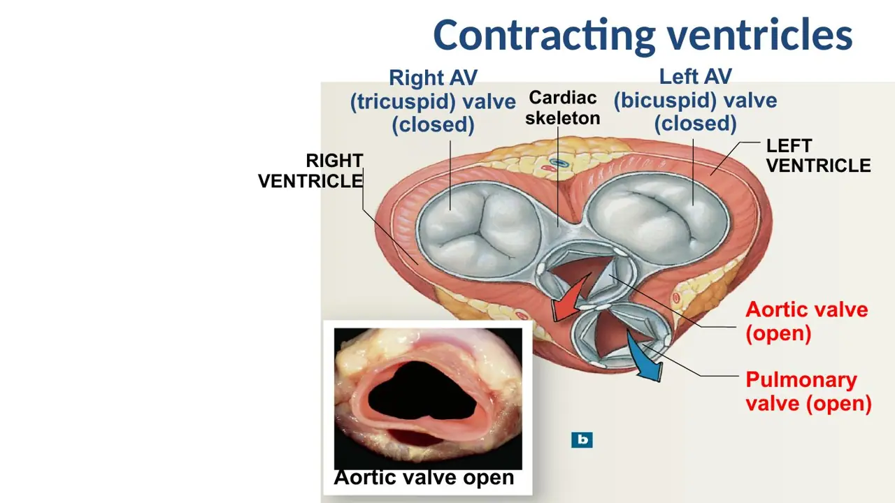
While the ventricles are contracting and ejecting blood, which valves are open?
A. Tricuspid and mitral open; aortic and pulmonary closed
B. Aortic and pulmonary open; tricuspid and mitral closed
C. Mitral and aortic open; tricuspid and pulmonary closed
AnswerB. Aortic and pulmonary open; tricuspid and mitral closed
WhyDuring ejection the semilunar valves are open and the AV valves closed; in relaxation it is the reverse.
Explanation
The two ventricles work together, so valves of the same type always do the same thing on both sides. When the ventricles contract, their pressure rises above atrial pressure (closing the tricuspid and mitral valves) and above arterial pressure (opening the aortic and pulmonary valves). When they relax, the AV valves open to let them fill and the semilunar valves close.
AV open, semilunar closed is the picture of relaxed, filling ventricles.
Mitral and aortic open together mixes the two states; a valve and its partner on the other side always move together.
Slide (1)H12A Circulatory I lecture (Dr Bruno) · slide 53L74Learning
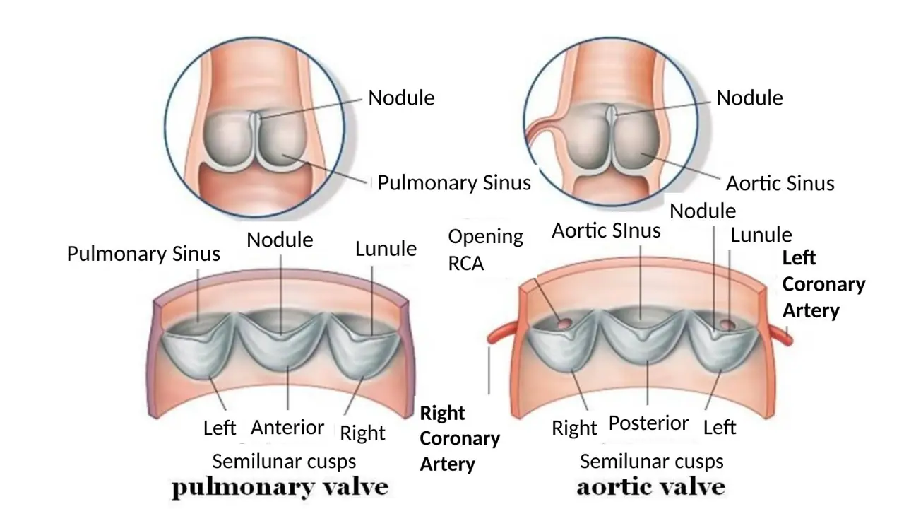
Name the parts of a semilunar cusp and what lies behind it.
A. Lunule at the middle of the free edge; nodule on either side Sinus: a bulge of the vessel wall behind each cusp
B. Nodule at the middle of the free edge; lunule on either side Chordae tendineae: cords behind each cusp
C. Nodule at the middle of the free edge; lunule on either side Sinus: a bulge of the vessel wall behind each cusp
AnswerC. Nodule at the middle of the free edge; lunule on either side Sinus: a bulge of the vessel wall behind each cusp
WhyEach cusp has a central nodule on its free edge, thin crescent lunules on either side, and a pocket of vessel wall behind it, the aortic or pulmonary sinus.
Explanation
When the valve closes, the lunules of neighbouring cusps press against each other and the three nodules meet in the centre to seal the gap. Behind each cusp the vessel wall bulges out as a sinus; blood fills the sinuses during diastole and keeps the cusps from sticking to the wall.
Swapping nodule and lunule reverses them: the nodule is the single thickening in the middle.
Chordae tendineae belong to the AV valves; nothing tethers a semilunar cusp.
Slide (1)H12A Circulatory I lecture (Dr Bruno) · slide 57L75Learning
Behind which cusps of the aortic valve do the coronary arteries open?
A. Right and posterior cusps; the left cusp has no coronary opening
B. Anterior and left cusps; the right cusp has no coronary opening
C. Right and left cusps; the posterior cusp has no coronary opening
AnswerC. Right and left cusps; the posterior cusp has no coronary opening
WhyThe right coronary artery opens from the right aortic sinus and the left coronary artery from the left; the posterior (non-coronary) sinus has none.
Explanation
The aortic valve has right, left and posterior cusps, each with a sinus behind it. The coronary ostia sit in the right and left aortic sinuses, just above the valve. The pulmonary valve's cusps are named left, anterior and right, and it has no coronary openings.
The posterior sinus is the non-coronary one, so it cannot be one of the two.
Anterior is a pulmonary cusp name; the aortic valve has no anterior cusp.
Slide (1)H12A Circulatory I lecture (Dr Bruno) · slide 57L76Learning
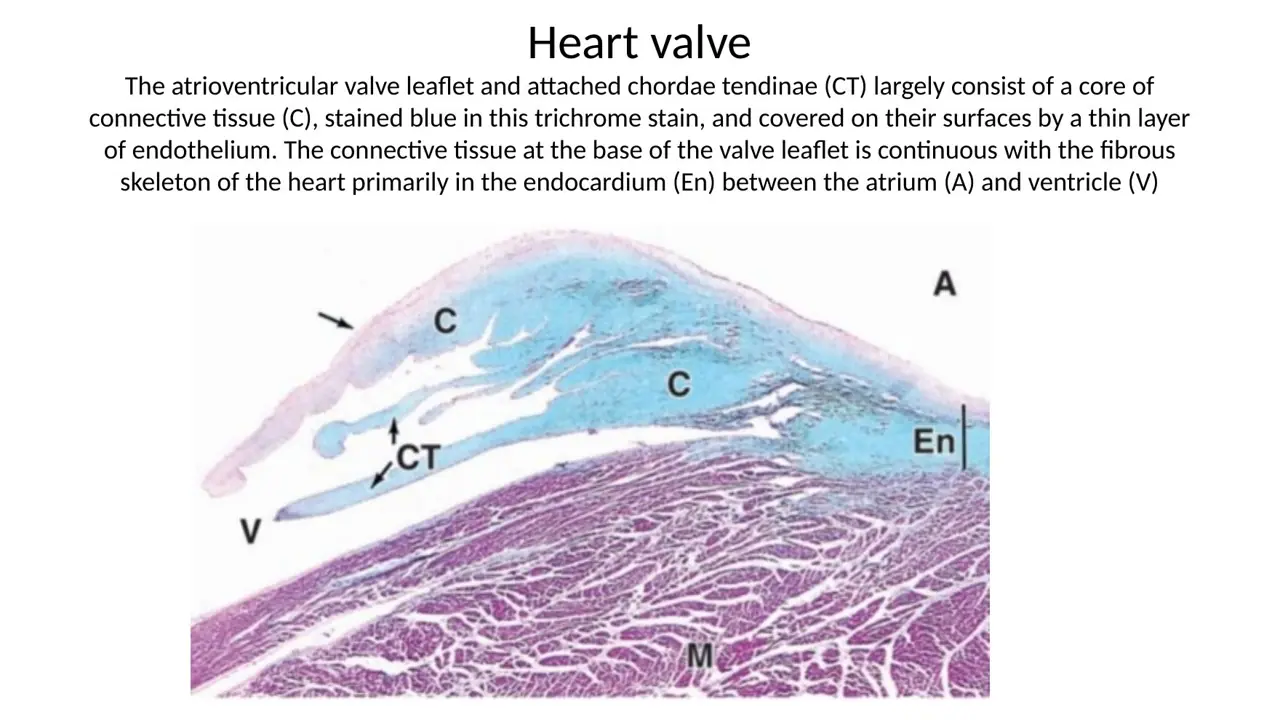
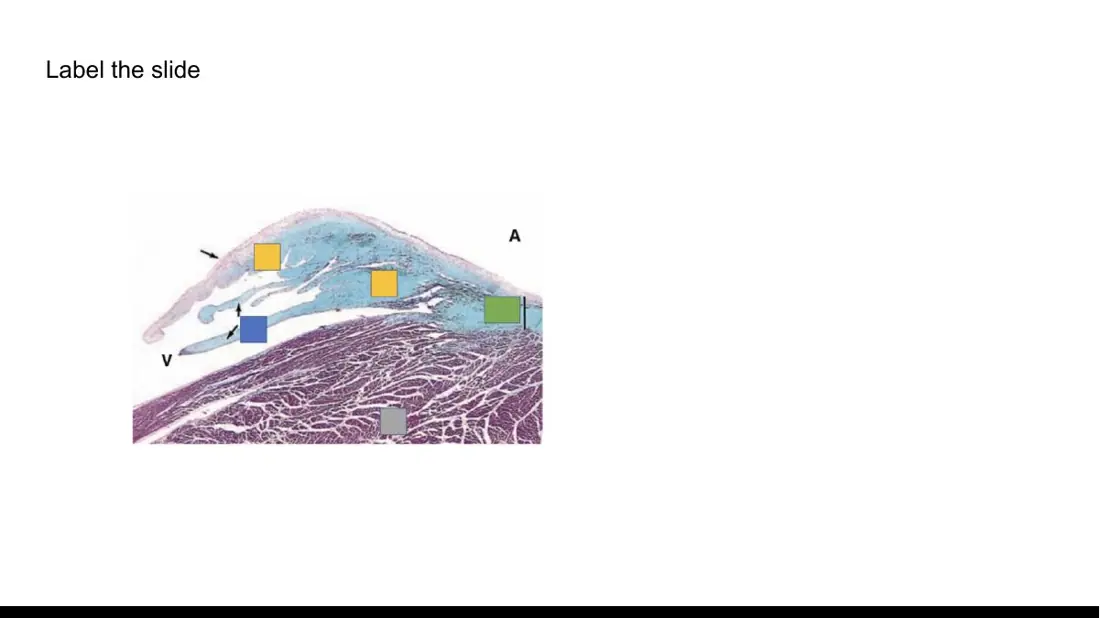
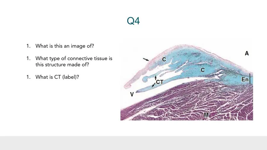
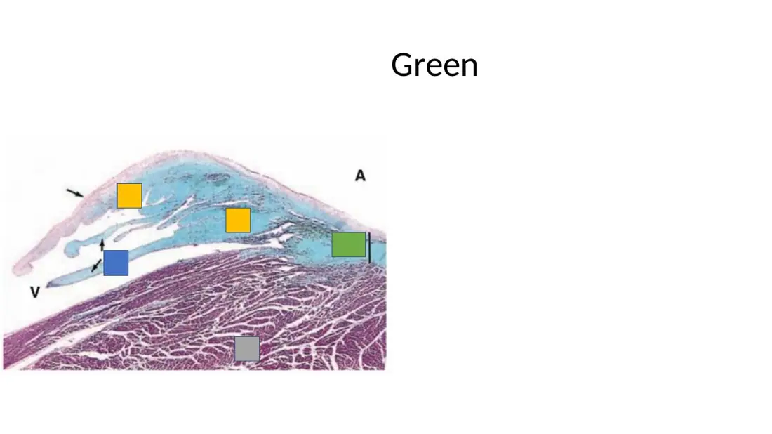
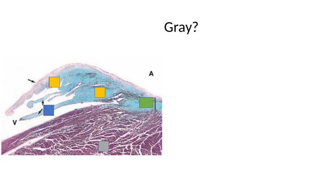
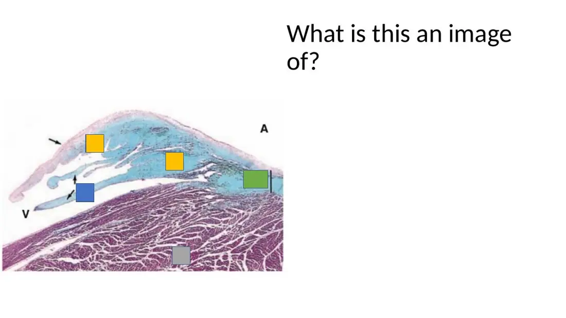
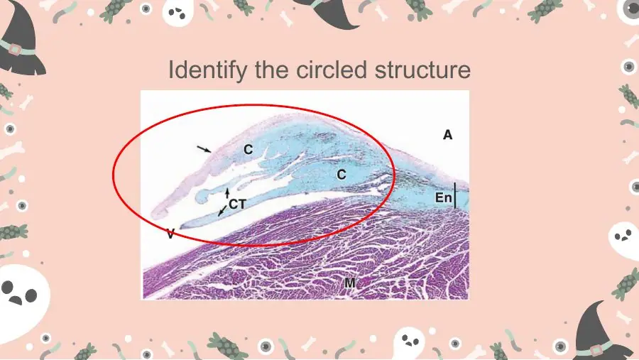
A trichrome section shows a thin flap with a blue-staining core, covered by a thin cellular lining, with thin blue cords attached to it; its base blends with blue tissue between atrium and ventricle. What is it?
A. An atrioventricular valve leaflet with chordae tendineae
B. A semilunar valve cusp attached to its aortic sinus wall
C. A papillary muscle with a tendon arising from its tip
AnswerA. An atrioventricular valve leaflet with chordae tendineae
WhyBlue (collagen) core, endothelial covering and attached chordae tendineae = an AV valve leaflet, its base continuous with the fibrous skeleton.
Explanation
Trichrome stains collagen blue and muscle red/purple. A leaflet whose dense connective-tissue core (C) is continuous at its base with the fibrous skeleton in the endocardium (En), between atrium (A) and ventricle (V), with cords (CT) attached, is an atrioventricular valve. The chordae are the cords tying its free edge to the papillary muscles.
A semilunar cusp has no chordae tendineae, and it hangs from the root of the aorta or pulmonary trunk, not between atrium and ventricle.
A papillary muscle would stain red like the myocardium; only its chordae are blue.
Three weeks after a dental extraction, a 19-year-old has palpitations, and echocardiography shows vegetations on his heart valves. Which layer of the heart is infected?
A. Endocardium
B. Myocardium
C. Epicardium
AnswerA. Endocardium
WhyHeart valves are folds of endocardium, so vegetations on them are infective endocarditis.
Explanation
Mouth bacteria (often viridans streptococci) can enter the blood during dental work and settle on the valves, which are covered by endothelium and have an avascular connective-tissue core. Clumps of bacteria, platelets and fibrin grow there as vegetations and can damage the valve.
Myocardium: infection of the muscle is myocarditis, not lesions on the valves.
Epicardium: infection of the outer surface is pericarditis; the epicardium never covers a valve.
Slide (1)Circulatory lab 2026 · slide 33
Coronary circulation and myocardial infarction
L78Learning
Where do the right and left coronary arteries arise?
A. From the aortic arch, beside the brachiocephalic trunk
B. From the aortic sinuses, just above the aortic valve
C. From the left ventricle, just below the aortic valve
AnswerB. From the aortic sinuses, just above the aortic valve
WhyThe coronary arteries are the first branches of the aorta, opening from the right and left aortic sinuses just above the valve cusps.
Explanation
The aortic sinuses are the three bulges of the aortic root behind the valve cusps. The right coronary artery leaves the right sinus and the left coronary artery the left sinus, so the heart muscle is the first tissue supplied with freshly ejected blood.
The aortic arch is farther along and gives off the vessels to the head and arms.
Nothing arises below the valve: that is still inside the ventricle.
Slides (2)H12A Circulatory I lecture (Dr Bruno) · slide 59H12A Circulatory I lecture (Dr Bruno) · slide 57L79Learning
When do the coronary arteries mainly fill, and what drives the flow?
A. Between contractions — the stretched elastic aortic wall recoils and pushes blood in
B. During contraction — the squeeze of the ventricle pushes blood directly into them
C. Between contractions — the pulmonary trunk recoils and pushes blood in
AnswerA. Between contractions — the stretched elastic aortic wall recoils and pushes blood in
WhyCoronary flow is greatest between contractions (diastole): the elastic rebound of the stretched aortic wall drives blood into the coronary openings.
Explanation
During ejection the aorta stretches and stores energy in its elastic wall. When the ventricle relaxes and the aortic valve closes, the wall recoils and keeps the aortic pressure high, pushing blood into the coronary arteries from the aortic sinuses. During contraction, the squeezing myocardium compresses its own vessels, so flow, especially to the left ventricle, falls.
The ventricular squeeze squashes the intramural coronary vessels rather than filling them.
The pulmonary trunk does not give rise to the coronary arteries.
A fast heart rate shortens diastole and so shortens the time for coronary filling.
Slide (1)H12A Circulatory I lecture (Dr Bruno) · slide 59L80Learning
What are the main branches of the left coronary artery?
A. Anterior interventricular artery Circumflex artery
B. Anterior interventricular artery Right marginal artery
C. Circumflex artery Posterior interventricular artery
WhyThe left coronary artery divides into the anterior interventricular (left anterior descending) and the circumflex arteries.
Explanation
The anterior interventricular artery runs down the anterior interventricular sulcus, in epicardial fat, supplying the front of both ventricles and most of the septum. The circumflex runs round the left side in the coronary (atrioventricular) groove.
The right marginal artery is a branch of the right coronary artery.
The posterior interventricular artery usually comes from the right coronary artery.
Slide (1)H12A Circulatory I lecture (Dr Bruno) · slide 58L81Learning
What are the main branches of the right coronary artery?
A. Anterior interventricular artery Circumflex artery
B. Right marginal artery Usually the posterior interventricular artery
C. Left marginal artery Usually the anterior interventricular artery
AnswerB. Right marginal artery Usually the posterior interventricular artery
WhyThe right coronary artery gives the right marginal artery and, in most hearts, the posterior interventricular artery.
Explanation
The right coronary artery runs in the right coronary groove, gives the right marginal artery along the lower border of the heart, and usually continues as the posterior interventricular artery in the posterior interventricular sulcus (a 'right-dominant' heart).
The anterior interventricular and circumflex arteries are the two branches of the left coronary artery.
A left marginal artery arises from the circumflex, a left coronary branch.
Slide (1)H12A Circulatory I lecture (Dr Bruno) · slide 58L82Learning
What is a myocardial infarction?
A. Death of heart muscle when a coronary artery is blocked, usually by a thrombus
B. Brief coronary spasm that reduces flow without killing any muscle
C. Scarring of the pericardium that restricts filling of the heart
AnswerA. Death of heart muscle when a coronary artery is blocked, usually by a thrombus
WhyMyocardial infarction = death of myocardium in the territory of a blocked coronary artery, most often blocked by a thrombus on an atherosclerotic plaque.
Explanation
A clot that blocks a coronary artery (coronary thrombosis) cuts off oxygen to the muscle it supplies. After about 20–30 minutes of severe ischaemia the cells start to die. The slides show a coronary artery whose lumen is filled by a thrombus and the infarcted muscle.
Spasm without cell death describes angina-type ischaemia, which is reversible.
Pericardial scarring is constrictive pericarditis, a different disease.
Slides (2)H12A Circulatory I lecture (Dr Bruno) · slide 60H12A Circulatory I lecture (Dr Bruno) · slide 61L83LearningBorderline
One month after a myocardial infarction, what has replaced the dead heart muscle?
A. New cardiac muscle cells formed by division of the surviving myocytes
B. New smooth muscle cells converted from the surviving myocytes
C. A scar of dense collagenous connective tissue
AnswerC. A scar of dense collagenous connective tissue
WhyAdult cardiac myocytes cannot divide, so an infarct heals as a fibrous (collagen) scar that does not contract.
Explanation
After macrophages clear the dead muscle, granulation tissue grows in and fibroblasts lay down collagen. By about a month the infarct is largely fibrous tissue, which matures into a dense, pale collagen scar over the following weeks (complete by about two months). Because the scar cannot contract, the heart pumps less well, and a large scar can lead to heart failure.
Myocyte division (hyperplasia) does not happen in the adult heart to any useful degree; the muscle is a permanent tissue.
Myocytes do not turn into smooth muscle.
L84LearningBorderline
A young woman with years of untreated severe hypertension dies of heart failure; her heart weighs 540 g. What process in the myocardial fibres enlarged it?
A. Hyperplasia (myocytes divide to increase in number)
B. Fatty change (fat droplets accumulate in each myocyte)
C. Hypertrophy (each myocyte grows larger)
AnswerC. Hypertrophy (each myocyte grows larger)
WhyCardiac myocytes cannot divide, so a heart working against high pressure enlarges by hypertrophy: each cell adds myofibrils and grows.
Explanation
A raised afterload makes the left ventricle work harder. It responds by thickening its wall: each muscle cell makes more sarcomeres and becomes larger, while the number of cells stays about the same.
Hyperplasia needs cells that can divide; adult cardiac muscle cells, like neurons, essentially cannot.
Fatty change makes cells pale and vacuolated but does not double the weight of the heart.
L85LearningBorderline
In myocardium from a patient who died of a heart attack, single myocytes show many dense pink transverse bands close together. What are they?
A. Intercalated discs of normal muscle
B. Contraction bands of infarcted (dying) muscle
C. Lipofuscin granules of ageing muscle
AnswerB. Contraction bands of infarcted (dying) muscle
WhyMany dense bands within one cell are contraction bands, a sign of myocardial infarction; intercalated discs occur only between cells.
Explanation
When dying cardiac muscle is flooded with calcium (for example when blood flow returns), its myofibrils hypercontract into thick, dense, eosinophilic bands crowded together across the cell. Healthy muscle shows only one intercalated disc at each end of a cell.
Intercalated discs are thin, step-like lines at the cell borders, roughly one cell length apart.
Lipofuscin is golden-brown granules at the nuclear poles, not bands across the cell.
Slide (1)H12A Circulatory I lecture (Dr Bruno) · slide 61
1High-yield
Two weeks after suffering a massive heart attack, a 54-year-old man presents to the emergency room complaining of sharp pain on the left side of his chest. On physical examination, the patient is apprehensive and sweating. His blood pressure is 80/40 mm Hg, and the pulse rate is 100 per minute. The patient expires shortly after admission. At autopsy, the left ventricle shows a rupture in the myocardial wall. What was the most likely cause of the patient’s death?
(A) Cardiac tamponade
(B) Dissecting aortic aneurysm
(C) Pulmonary edema
(D) Pulmonary thromboembolism
(E) Septic shock
AnswerA. Cardiac tamponade
WhyRupture of a softened infarct floods the pericardial cavity with blood (hemopericardium), compressing the heart and blocking ventricular filling, which is cardiac tamponade.
Explanation from the source
Rupture of a myocardial infarct causes blood to fill the peritoneal cavity (hemopericardium). This medical catastrophe restricts the motion of the heart and is referred to as cardiac tamponade. When it happens, it typically occurs within the first 3 weeks following myocardial infarction. Pulsus paradoxus (>10 mm Hg drop in arterial blood pressure with inspiration) is commonly observed in patients with hemopericardium. Although aortic dissections (choice B) can break through to the pericardium, they do not rupture the heart wall. None of the other cardiovascular diseases are associated with rupture of the heart wall and cardiac tamponade. Keywords: Hemopericardium, cardiac tamponade
ConceptVentricular rupture after MI causes hemopericardium and cardiac tamponade
SGA Drive › Histology Fall 2023 › Histology Exam 2 › Histology Exam 2 › Professor Reviews › Histology - 2022 Exam 2 - Braun review (DHahn).pdf · 3: Heart has three layers: Endo, Myo, and Pericardium / Cross section of your heart / There’s also fat = Blood vessels / On a Histology slide, you’ll see adipose tissue in the Epicardium / And Blood Vessels / Endothelium (and Sub-Endo) is very thin / You don’t see fat or BV / The coronary is on the surface of the heart / This should help you understand what you expect to see / Myocardium is the thickest part of eh heart. Muscle is here (Cardiac muscle) / Distinctives feature is intercalated disks / Striated muscle with these special disks
Correct key: Endocardium; Myocardium; Epicardium. Heart wall = endocardium (inner), myocardium (middle), epicardium (outer = visceral pericardium). The Bruno lecture key is right; the Braun review's 'Endo, Myo and Pericardium' is loose wording, since the outer layer of the heart wall itself is the epicardium.
WhyFrom inside out: the endocardium (endothelium plus subendocardial tissue), the thick cardiac-muscle myocardium, and the epicardium, which is the visceral serous pericardium over fat and coronary vessels.
ConceptIdentify the three heart-wall layers on a low-power section
Slide (1)H12A Circulatory I lecture (Dr Bruno) · slide 193
Q2- Where are the main blood vessels which supply the heart located?
(A) Epicardium
(B) Pericardium
(C) Myocardium
(D) Endocardium
(E) Endomysium
Answer supplied — the source gives no key(A) Epicardium — the coronary arteries and veins run in the subepicardial connective tissue and fat of the epicardium.
WhyThe coronary arteries and cardiac veins run in the subepicardial loose connective tissue and fat of the epicardium, beneath its mesothelium, sending branches inward into the myocardium.
ConceptCoronary vessels run in the epicardium
Slide (1)Circulatory lab 2026 · slide 94
Q3- What is the pacemaker of the heart?
(A) Sinoatrial node
(B) Atrioventricular node
(C) Bundle of His
(D) Right bundle branch
(E) Purkinje fiber
Answer supplied — the source gives no key(A) Sinoatrial node
WhyThe sinoatrial node in the right atrial wall depolarizes spontaneously faster than any other conducting tissue, so it sets the heart rate as the pacemaker; the AV node and His–Purkinje system are slower backups.
ConceptThe sinoatrial node is the pacemaker of the heart
Slide (1)Circulatory lab 2026 · slide 275
Q4- Where are the Purkinje fibers located?
(A) Epicardium
(B) Pericardium
(C) Myocardium
(D) Endocardium
(E) Sinoatrial node
Answer supplied — the source gives no key(D) Endocardium — Purkinje fibers lie in the subendocardial layer of the endocardium.
WhyPurkinje fibers lie in the subendocardial layer of the endocardium, the loose connective tissue between the endothelial lining and the myocardium, from which they enter the ventricular muscle.
ConceptPurkinje fibers lie in the subendocardial layer of the endocardium
Slides (2)H12A Circulatory I lecture (Dr Bruno) · slide 33H12A Circulatory I lecture (Dr Bruno) · slide 346
Q4. The structures indicated by arrows contain a number of constituents, except:
A. Fascia adherens
B. Desmoglein
C. Desmocolin
D. Connexons
E. Hemidesmosomes
AnswerE. Hemidesmosomes
WhyIntercalated discs contain fasciae adherentes, desmosomes (built of desmoglein and desmocollin) and gap junctions (connexons). Hemidesmosomes anchor cells to the basal lamina, not cell to cell, so they are absent.
Explanation from the source
EM showing the 3 components of the junctional complexes found in an intercalated disc. The 3 components are arranged in a step-like fashion with fascia adherens and desmosomes in the transverse portion between muscle fibers and gap junctions in the longitudinal portion.
ConceptIntercalated discs contain desmosomes, fascia adherens and gap junctions, not hemidesmosomes
Q6 – The sinoatrial node is located within which of the following structures?
A. Right atrial wall
B. Left atrial wall
C. Cardiac skeleton
D. Epicardium
E. Endocardium
AnswerA. Right atrial wall
WhyThe sinoatrial node sits in the right atrial wall near the entrance of the superior vena cava, where it generates the impulse that starts each heartbeat.
Explanation from the source
Both the sinoatrial (SA) node (pacemaker) and atrioventricular (AV) node are located in the walls of the right atrium. The AV node is continuous with a specialized bundle of cardiac muscle fibers, the AV bundle of His, which gives rise to the right and left bundle branches that run along both sides of the interventricular septum to the apex of the heart. As the bundle branches approach the apex, they subdivide further as a network of subendocardial Purkinje fibers that spread the electrical impulse to the muscle of the myocardium from the apex to the base. Sinoatrial nodeThe SA node is a 6-7 mm structure that is generally not visible grossly, although it is recognizable histologically. It is located subepicardially within the right atrial wall where the muscular ridge known as the crista terminalis meets the superior vena cava. The SA node is identifiable in the histologic image where it is the paler staining region (indicated by asterisks) surrounding the sinoatrial nodal artery. At high magnification (not shown here), the SA node fibers are thinner, have fewer myofibrils, and are less intensely staining on H&E compared to the neighboring contractile fibers. Darker contractile cardiac muscle cells and endocardium of the right atrium can be seen to the left of the node, and fat tissue of the epicardium to the right.
Match the descriptions below to the GREEN letters in the picture:
• This layer of loose connective tissue and adipose tissue contains blood vessels and nerves.
• The surface layer of simple squamous mesothelium produces lubricant fluid which prevents friction within the pericardium.
• This layer contains junctional complexes. The gap junctions within these complexes provide ionic continuity between cells that serve as “electrical synapses” promoting rapid impulse conduction through the muscle cells.
AnswerA → The surface layer of simple squamous mesothelium produces lubricant fluid which prevents friction within the pericardium. B → This layer of loose connective tissue and adipose tissue contains blood vessels and nerves. C → This layer contains junctional complexes. The gap junctions within these complexes provide ionic continuity between cells that serve as “electrical synapses” promoting rapid impulse conduction through the muscle cells.
WhyA is the surface mesothelium secreting lubricating fluid, B the epicardial loose connective tissue with fat, vessels and nerves, and C myocardium with gap-junction-bearing intercalated discs.
Explanation from the source
The surface layer of simple squamous mesothelium produces lubricant fluid which prevents friction within the pericardium. A
This layer contains junctional complexes. The gap junctions within these complexes provide ionic continuity between cells that serve as “electrical synapses” promoting rapid impulse conduction through the muscle cells. C
This layer of loose connective tissue and adipose tissue contains blood vessels and nerves. B
Match the following statements with the correct layers of the heart (epicardium, myocardium, endocardium).
A. Simple squamous epithelium (mesothelium) supported by loose connective tissue containing blood vessels and nerves
B. Spiral layers of cardiac muscle fibers
C. Simple squamous epithelium (endothelium) supported by connective tissue continuous with the myocardium
D. Layer characterized by intercalated discs
E. The deepest layer of the endocardial connective tissue containing Purkinje fibers is sometimes called
• Endocardium
• Subendocardium
• Myocardium
• Epicardium
AnswerA → Epicardium B → Myocardium C → Endocardium D → Myocardium E → Subendocardium
WhyMesothelium on vessel-rich tissue is epicardium, spiral cardiac muscle with intercalated discs is myocardium, endothelium on connective tissue is endocardium, and its Purkinje-bearing deepest part is subendocardium.
Explanation from the source
Simple squamous epithelium (mesothelium) supported by loose connective tissue containing blood vessels and nerves - epicardium
Spiral layers of cardiac muscle fibers -myocardium
Simple squamous epithelium (endothelium) supported by connective tissue continuous with the myocardium - endocardium
Layer characterized by intercalated discs -myocardium
The deepest layer of the endocardial connective tissue containing Purkinje fibers is sometimes called - subendocardium
ConceptMatch layer descriptions to epicardium, myocardium, endocardium, subendocardium
List the following layers in correct order from inside to outside.
A. 1- Most inner
B. 2
C. 3
D. 4- Most outer
• Visceral pericardium (epicardium)
• Parietal pericardium
• Myocardium
• Endocardium
AnswerA → Endocardium B → Myocardium C → Visceral pericardium (epicardium) D → Parietal pericardium
WhyFrom the lumen outward come endocardium, myocardium, visceral pericardium (epicardium), and then, across the pericardial cavity, the parietal pericardium lining the fibrous sac.
ConceptHeart wall layers from inside out to the parietal pericardium
Slide (1)Circulatory lab 2026 · slide 311
Please match each structure of the cardiac conduction system to where it could be found.
A. Sinoatrial (SA) Node
B. Atrioventricular (AV) Node
C. AV bundle (of His)
D. Purkinje Fibers
• Interventricular septum
• Junction between atria and ventricles
• Subendothelial layer
• Wall of right atrium
AnswerA → Wall of right atrium B → Junction between atria and ventricles C → Interventricular septum D → Subendothelial layer
WhyThe SA node sits in the right atrial wall, the AV node at the atrioventricular junction (interatrial septum near the coronary sinus), the His bundle runs down the interventricular septum, and Purkinje fibers lie subendothelially (subendocardial layer).
Explanation from the source
Sinoatrial (SA) Node (b)
Atrioventricular (AV) Node (c)
AV bundle (of His) (d)
Purkinje Fibers (a)
ConceptLocations of SA node, AV node, His bundle and Purkinje fibers
Slide (1)H12A Circulatory I lecture (Dr Bruno) · slide 3612High-yield
Put the list of components of the cardiac conduction system into the correct order in which they generate and conduct waves of depolarization that stimulate myocardial contraction:
A. 1st
B. 2nd
C. 3rd
D. 4th
E. 5th
F. 6th
• SA Node
• Ventricle Myocardium
• AV node
• Bundle of His
• Purkinje Fiber
• Atrium Myocardium
AnswerA → SA Node B → Atrium Myocardium C → AV node D → Bundle of His E → Purkinje Fiber F → Ventricle Myocardium
WhyThe impulse starts at the SA node, spreads through atrial myocardium to the AV node (delay), then the His bundle, Purkinje fibers, and finally ventricular myocardium.
ConceptConduction sequence from SA node to ventricular myocardium
Slide (1)Circulatory lab 2026 · slide 2713
Describe the appearance of the nuclei of cardiac myocytes.
Answer supplied — the source gives no keyOne (occasionally two) oval nucleus located centrally in each cell, often with a pale perinuclear clear zone (sarcoplasm containing glycogen/lipofuscin); in cross-section the central nucleus is surrounded by myofibrils.
WhyEach cardiac myocyte has one or two oval central nuclei, often with a pale perinuclear zone, unlike the many peripheral nuclei of skeletal muscle fibers.
ConceptCardiac myocytes have one or two central oval nuclei
Slide (1)Circulatory lab 2026 · slide 1914
Which of the following is true for ventricles?
a. Located at the base of the heart
b. Myocardial cells contains abundant granules
c. Receive blood directly from the venae cavae and pulmonary veins
d. Walls contain Purkinje fibers of the right and left branches from the atrioventricular bundle
e. Contain more elastic fibers than the atria
Answerd. Walls contain Purkinje fibers of the right and left branches from the atrioventricular bundle
WhyThe right and left bundle branches of the AV bundle end as Purkinje fibers in the ventricular subendocardium; atria, not ventricles, have ANF granules and more elastic fibers.
ConceptVentricular walls contain Purkinje fibers from the bundle branches
[Question 1] A 68-year-old man develops sudden, severe substernal chest pain. Laboratory studies and ECG confirm an acute myocardial infarction (heart attack). The patient cannot maintain his blood pressure and expires 4 hours later. Which of the following was the most likely cause of the patient’s death? [Question 2] The heart of the patient described in Question 1 is examined at autopsy. Routine H&E staining reveals layered structures within the wall of the heart (shown in the image). Identify the indicated layer.
(A) Endocardium
(B) Epicardium
(C) Myocardium
(D) Parietal layer of the pericardium
(E) Visceral layer of the pericardium
AnswerC. Myocardium
WhyThe thick middle layer of branching, striated cardiac muscle with central nuclei and intercalated discs is the myocardium, the contractile layer that dies in a myocardial infarction.
Explanation from the source
The heart is a folded endothelial tube with a thickened muscular wall that pumps blood through the circulatory system. The principal functional component of the heart is the myocardial layer (myocardium) that is composed of cardiac muscle. The myocardium is covered externally by an epicardium (choice B), consisting of a layer of mesothelium and associated adipose connective tissue. The inner (luminal) surface of the myocardium is lined by endocardium (choice A), consisting of a layer of endothelium and associated connective tissue. Parietal layer of pericardium (choice D) lines the fibrous pericardial sac and is not visible in this section. Visceral layer of pericardium (choice E) refers to the mesothelial cell layer of epicardium. The myocardium is considerably thicker in the ventricles, than in the atria, reflecting the needs of the ventricles to propel blood through pulmonary and systemic circulations. Keywords: Heart, myocardium
ConceptThick muscular middle layer of the heart wall (image) is the myocardium
Slide (1)H12A Circulatory I lecture (Dr Bruno) · slide 1916
[Question 1] A 68-year-old man develops sudden, severe substernal chest pain. Laboratory studies and ECG confirm an acute myocardial infarction (heart attack). The patient cannot maintain his blood pressure and expires 4 hours later. Which of the following was the most likely cause of the patient’s death? [Question 2] The heart of the patient described in Question 1 is examined at autopsy. Routine H&E staining reveals layered structures within the wall of the heart (shown in the image). Identify the indicated layer. [Question 3] The autopsy specimen described in Question 2 is examined at higher magnification (shown in the image). Identify the indicated structure.
(A) Endocardium
(B) Epicardium
(C) Mesothelium
(D) Myocardium
(E) Purkinje fibers
AnswerA. Endocardium
WhyThe thin luminal layer of endothelium on subendothelial connective tissue (with some smooth muscle), over a subendocardial layer that can hold Purkinje fibers, is the endocardium, lining the heart chambers.
Explanation from the source
Endocardium lines the luminal surface of the heart chambers. It is composed of a layer of endothelium and subendothelial connective tissue. Smooth muscle fibers can be found in the subendothelial connective tissue. Purkinje fibers (choice E) are specialized cardiomyocytes that conduct action potentials in the heart. None of the other tissue layers describe histologic features of the endocardium. Keywords: Heart, endocardium
ConceptThin luminal endothelium plus connective tissue (image) is the endocardium
Slides (2)H12A Circulatory I lecture (Dr Bruno) · slide 32H12A Circulatory I lecture (Dr Bruno) · slide 3417
[Question 1] A 68-year-old man develops sudden, severe substernal chest pain. Laboratory studies and ECG confirm an acute myocardial infarction (heart attack). The patient cannot maintain his blood pressure and expires 4 hours later. Which of the following was the most likely cause of the patient’s death? [Question 2] The heart of the patient described in Question 1 is examined at autopsy. Routine H&E staining reveals layered structures within the wall of the heart (shown in the image). Identify the indicated layer. [Question 3] The autopsy specimen described in Question 2 is examined at higher magnification (shown in the image). Identify the indicated structure. [Question 4] Another region of the autopsy specimen described in Questions 2 and 3 is examined in the pathology department (shown in the image). Identify the indicated zone.
(A) Endocardium
(B) Epicardium
(C) Fibrous pericardium
(D) Myocardium
(E) Visceral pleura
AnswerB. Epicardium
WhyAdipose-rich connective tissue on the heart's outer surface, covered by mesothelium (visceral serous pericardium) and carrying coronary vessels and nerves, is the epicardium.
Explanation from the source
Epicardium covers the external surface of the heart and is in direct contact with the potential space provided by the pericardial cavity. Epicardium is composed of (1) a layer of the mesothelium (visceral layer of serous pericardium) and (2) an underlying adipose connective tissue. The adipose tissue functions as a cushion for the heart as it moves within the pericardial sac. Blood vessels and nerves supplying the heart travel through the epicardium. Endocardium (choice A) lines the luminal surface of the heart and is typically devoid of adipose tissue. Fibrous pericardium (choice C) is composed of dense connective tissue and is not visible in this image. Visceral pleura (choice E) represents a layer of mesothelium that is adherent to the surface of the lungs. Keywords: Heart, epicardium
ConceptMesothelium-covered adipose connective tissue on the heart surface is the epicardium
Slide (1)Circulatory lab 2026 · slide 918
[Question 5] Two weeks after suffering a massive heart attack, a 54-year-old man presents to the emergency room complaining of sharp pain on the left side of his chest. On physical examination, the patient is apprehensive and sweating. His blood pressure is 80/40 mm Hg, and the pulse rate is 100 per minute. The patient expires shortly after admission. At autopsy, the left ventricle shows a rupture in the myocardial wall. What was the most likely cause of the patient’s death? [Question 6] Autopsy of the patient described in Question 5 reveals 400 mL of blood in the pericardial cavity. This blood is in direct contact with which of the following anatomic structures?
(A) Endocardium
(B) Epicardium
(C) Fibrous pericardium
(D) Myocardium
(E) Parietal pleura
AnswerB. Epicardium
WhyBlood from a ruptured ventricle fills the pericardial cavity, which is lined on the heart side by the mesothelium of the visceral serous pericardium, the surface of the epicardium; the fibrous pericardium is outside the parietal layer.
Explanation from the source
The pericardial sac is lined by visceral and parietal mesothelia that together are referred to as serous pericardium. The visceral layer of serous pericardium represents the mesothelial layer of the epicardium. The parietal layer of serous pericardium lines the inner surface of the fibrous pericardium (choice C). The space between the visceral and parietal layers of serous pericardium defines the pericardial cavity. In normal condition, a thin layer of serous fluid within the pericardial cavity (about 50 mL) provides lubrication for movement of the heart during contraction. Myocardial rupture, pericarditis, or chest injuries can cause excess fluid or blood to accumulate in the pericardial cavity. Accumulation of excess fluid or blood restricts ventricular filling, a life-threatening condition referred to as cardiac tamponade. Pericardiocentesis is often required to drain excess pericardial fluid to relieve pressure on the heart. None of the other structures are in direct contact with the pericardial cavity. Keywords: Cardiac tamponade, heart, epicardium
ConceptBlood in the pericardial cavity contacts the epicardium (visceral serous pericardium)
Slides (2)H12A Circulatory I lecture (Dr Bruno) · slide 15H12A Circulatory I lecture (Dr Bruno) · slide 1419
[Question 13] A 56-year-old woman is diagnosed with a left atrial myxoma. The tumor is removed, and the surgical specimen is examined by light microscopy. The tumor margin shows normal heart tissue (shown in the image). The structure within the circle represents a branch of which of the following blood vessels? [Question 14] The epicardium of the surgical specimen described in Question 13 is examined in the pathology department. Identify the structure indicated by the circle.
(A) Arteriole
(B) Cardiac muscle
(C) Connective tissue
(D) Nerve
(E) Venule
AnswerD. Nerve
WhyNerves and major vessels supplying the heart travel in the epicardium; a bundle of wavy fibers with elongated Schwann-cell nuclei and no lumen or red cells is a nerve (autonomic and visceral afferent fibers).
Explanation from the source
As mentioned above, the nerves and major blood vessels supplying the heart travel within the epicardium. Nerves innervating the heart contain sympathetic, parasympathetic, and visceral afferent (sensory) fibers. An arteriole (choice A) is visible to the right of the nerve. The walls of arterioles are thicker than venules (choice E). Red blood cells are seen within the lumens of the blood vessels. Cardiac muscle fibers (choice B) are seen at the lower portion of the image. Connective tissue fibers (choice C) are seen in longitudinal, cross-, and oblique sections throughout the epicardium. Keywords: Heart, epicardium
ConceptNerve bundle in the epicardial connective tissue (image)
Slide (1)Circulatory lab 2026 · slide 1020
A 46-year-old man undergoes electrophysiologic mapping for palpitations. The earliest spontaneous depolarization is recorded from a small nodal structure located in the superior wall of the right atrium near the entrance of the superior vena cava, along the crista terminalis. Which structure is being identified?
A. Atrioventricular (AV) node
B. Bundle of His
C. Purkinje fibers
D. Sinoatrial (SA) node
E. Right bundle branch
AnswerD. Sinoatrial (SA) node
WhyThe SA node lies in the superior right atrial wall near the superior vena cava entrance along the crista terminalis, and it shows the earliest, fastest spontaneous depolarization, making it the pacemaker.
Explanation from the source
AKA Pacemaker of the Heart
ConceptThe sinoatrial node is the pacemaker of the heart
Question: What is the difference between AV and semilunar valves?
A. AV valves separate the ventricles from viculation
B. Semilunar valves have muscular support
C. There are 4 AV valves compared to 3 semilunar valves
D. AV valves separate atria from ventricles
AnswerD. AV valves separate atria from ventricles
WhyAtrioventricular valves (tricuspid and mitral) lie between atria and ventricles, whereas semilunar valves guard ventricular outflow into the aorta and pulmonary trunk.
ConceptAV valves separate atria from ventricles
Slide (1)H12A Circulatory I lecture (Dr Bruno) · slide 5122
Question: Which of the following is incorrect about semilunar valves?
A. They separate ventricles from circulation
B. They have no muscular support
C. They have 3 cusps
D. The pulmonary semilunar valve is between the right ventricle and pulmonary trunk
E. The mitral valve is the semilunar valve on the left
AnswerE. The mitral valve is the semilunar valve on the left
WhyThe mitral (bicuspid) valve is the left atrioventricular valve; the left-sided semilunar valve is the aortic valve, so this is the false statement about semilunar valves.
ConceptThe mitral valve is an AV valve, not a semilunar valve (NOT question)
Slides (2)H12A Circulatory I lecture (Dr Bruno) · slide 54H12A Circulatory I lecture (Dr Bruno) · slide 5123
Which layer of the heart contains the purkinje fibers?
A. Epicardium
B. Pericardium
C. Subendocardial layer
D. Myocardium
AnswerC. Subendocardial layer
WhyPurkinje fibers run in the subendocardial layer, the loose connective tissue joining the endocardium to the myocardium; they are not in the epicardium or pericardium.
ConceptPurkinje fibers lie in the subendocardial layer of the endocardium
Slide (1)H12A Circulatory I lecture (Dr Bruno) · slide 3324High-yield
The structures indicated by arrows contain a number of constituents, except:
A. Hemidesmosomes
B. Desmoglein (desmosomes)
C. Connexons (gap junctions)
D. Desmocolin (desmosomes)
E. Cadherin (fascia adherens)
AnswerA. Hemidesmosomes
WhyIntercalated discs join cardiomyocytes cell to cell by fasciae adherentes, desmosomes and gap junctions; hemidesmosomes anchor cells to basal lamina, so they are absent.
ConceptIntercalated discs contain desmosomes, fascia adherens and gap junctions, not hemidesmosomes
During light microscopic examination of an H&E stained section of the heart, you notice a group of cells with paler staining, and increased diameter when compared to typical cardiac muscle cells. Determined to classify the cell, you prepare another section of the same area of the heart with PAS staining. This group of cells is now distinguished by a dark magenta color. Which area of the heart was most likely being observed under the microscope?
A. Myocardium
B. Parietal Pericardium
C. Visceral pericardium
D. Epicardium
E. Endocardium
AnswerE. Endocardium
WhyLarge, pale cells that stain magenta with PAS are glycogen-rich Purkinje fibers, which run in the subendocardial layer of the endocardium.
ConceptPale, large, PAS-positive (glycogen-rich) Purkinje fibers lie in the endocardium
Which of the following is true of the structures highlighted by the arrows?
A. Are modified neuronal cells
B. Are the principal cellular component of the sinoatrial node
C. Are abnormally swollen because of coronary disease with inadequate blood flow to the heart
D. Conduct the cardiac impulse
E. Are subepicardial in location
AnswerD. Conduct the cardiac impulse
WhyThe large, pale fibers with central nuclei and few peripheral myofibrils are Purkinje fibers, modified cardiac muscle cells (not neurons) in the subendocardium that rapidly conduct the impulse to ventricular muscle.
ConceptPurkinje fibers (image) are modified cardiomyocytes that conduct the impulse
Slide (1)H12A Circulatory I lecture (Dr Bruno) · slide 3328
Label the slide
AnswerCT: Chordae Tendinae C:Dense Connective Tissue of the Chordae Tendinae En: Endocardium M: Thick ventricular myoacardium Pitcture of a Cardiac skeleton with an AV calve
Sources disagree on the key:
SGA Drive › Mod Exams (Fall 2025) › Cardio-Pulm Exam 2 › Histo-Path › Past Content and Practice Q_s › Histo Circulatory Review 2025.pptx · 4: En: endocardium P: Purkinje cells SEn: subendocardium M: myocardium
SGA Drive › Mod Exams (Fall 2025) › Cardio-Pulm Exam 2 › Histo-Path › Past Content and Practice Q_s › Histo Circulatory Review 2025.pptx · 9: CT: Chordae Tendinae C:Dense Connective Tissue of the Chordae Tendinae En: Endocardium M: Thick ventricular myoacardium Pitcture of a Cardiac skeleton with an AV calve
Correct key: Arrow = AV valve cusp; yellow C = dense connective tissue of valve/chordae; blue CT = chordae tendineae; green En = endocardium (fibrous skeleton/annulus); grey M = ventricular myocardium. Junqueira trichrome of an AV valve on the cardiac skeleton (answer slide 10). Unit 9 keys (2025 review and 2024 TA review) are right; the 'En, P, SEn, M' and 'Purkinje fibers' keys belong to the slide-4 Purkinje image.
WhyIn trichrome the AV valve cusp and chordae tendineae stain blue as dense collagenous tissue continuous with the fibrous skeleton in the endocardium, above purple ventricular myocardium.
Explanation from the source
The fibrous cardiac skeleton consists of dense irregular connective tissue, primarily in the endocardium (En), which anchors the valves and surrounds the two atrioventricular canals, maintaining their proper shape. The micrograph shows a section through a cusp of an atrioventricular valve (arrow) and attached chordae tendineae (CT). These structures are largely dense connective tissue (C) covered with a thin layer of endothelium. The collagen-rich connective tissue of the valves is stained pale blue here and is continuous with the fibrous ring of connective tissue at the base of the valves, which fills the endocardium between the atrium (A) and ventricle (V). The thick ventricular myocardium (M) is also shown. (X20; Masson trichrome)
The heart of the patient described in Question 1 is examined at autopsy. Routine H&E staining reveals layered structures within the wall of the heart (shown in the image). Identify the indicated layer.
(A) Endocardium
(B) Epicardium
(C) Myocardium
(D) Parietal layer of the pericardium
(E) Visceral layer of the pericardium
Answer(C) Myocardium
WhyThe thickest layer, built of branching cardiac muscle fibers between the fatty epicardium and the thin endocardium, is the myocardium, the contractile wall of the heart.
Explanation from the source
The answer is C: Myocardium. The heart is a folded endothelial tube with a thickened muscular wall that pumps blood through the circulatory system. The principal functional component of the heart is the myocardial layer (myocardium) that is composed of cardiac muscle. The myocardium is covered externally by an epicardium (choice B), consisting of a layer of mesothelium and associated adipose connective tissue. The inner (luminal) surface of the myocardium is lined by endocardium (choice A), consisting of a layer of endothelium and associated connective tissue. Parietal layer of pericardium (choice D) lines the fibrous pericardial sac and is not visible in this section. Visceral layer of pericardium (choice E) refers to the mesothelial cell layer of epicardium. The myocardium is considerably thicker in the ventricles, than in the atria, reflecting the needs of the ventricles to propel blood through pulmonary and systemic circulations. Keywords: Heart, myocardium
ConceptThick muscular middle layer of the heart wall (image) is the myocardium
Slide (1)H12A Circulatory I lecture (Dr Bruno) · slide 1930High-yield
The autopsy specimen described in Question 2 is examined at higher magnification (shown in the image). Identify the indicated structure.
(A) Endocardium
(B) Epicardium
(C) Mesothelium
(D) Myocardium
(E) Purkinje fibers
Answer(A) Endocardium
WhyThe thin pale layer on the luminal surface, made of endothelium and subendothelial connective tissue over the cardiac muscle, is the endocardium.
Explanation from the source
The answer is A: Endocardium. Endocardium lines the luminal surface of the heart chambers. It is composed of a layer of endothelium and subendothelial connective tissue. Smooth muscle fibers can be found in the subendothelial connective tissue. Purkinje fibers (choice E) are specialized cardiomyocytes that conduct action potentials in the heart. None of the other tissue layers describe histologic features of the endocardium. Keywords: Heart, endocardium
ConceptThin luminal endothelium plus connective tissue (image) is the endocardium
Slides (2)H12A Circulatory I lecture (Dr Bruno) · slide 32H12A Circulatory I lecture (Dr Bruno) · slide 3431
Another region of the autopsy specimen described in Questions 2 and 3 is examined in the pathology department (shown in the image). Identify the indicated zone.
(A) Endocardium
(B) Epicardium
(C) Fibrous pericardium
(D) Myocardium
(E) Visceral pleura
Answer(B) Epicardium
WhyThe outer zone of adipose connective tissue covered by mesothelium (visceral serous pericardium) and resting on the myocardium is the epicardium, the cushioning outer tunic.
Explanation from the source
The answer is B: Epicardium. Epicardium covers the external surface of the heart and is in direct contact with the potential space provided by the pericardial cavity. Epicardium is composed of (1) a layer of the mesothelium (visceral layer of serous pericardium) and (2) an underlying adipose connective tissue. The adipose tissue functions as a cushion for the heart as it moves within the pericardial sac. Blood vessels and nerves supplying the heart travel through the epicardium. Endocardium (choice A) lines the luminal surface of the heart and is typically devoid of adipose tissue. Fibrous pericardium (choice C) is composed of dense connective tissue and is not visible in this image. Visceral pleura (choice E) represents a layer of mesothelium that is adherent to the surface of the lungs. Keywords: Heart, epicardium
ConceptMesothelium-covered adipose connective tissue on the heart surface is the epicardium
Slide (1)Circulatory lab 2026 · slide 932
Two weeks after suffering a massive heart attack, a 54-yearold man presents to the emergency room complaining of sharp pain on the left side of his chest. On physical examination, the patient is apprehensive and sweating. His blood pressure is 80/40 mm Hg, and the pulse rate is 100 per minute. The patient expires shortly after admission. At autopsy, the left ventricle shows a rupture in the myocardial wall. What was the most likely cause of the patient’s death?
(A) Cardiac tamponade
(B) Dissecting aortic aneurysm
(C) Pulmonary edema
(D) Pulmonary thromboembolism
(E) Septic shock
Answer(A) Cardiac tamponade
WhyRupture of a softened infarct fills the pericardial sac with blood (hemopericardium); the rigid fibrous pericardium cannot stretch, so pressure compresses the heart and stops ventricular filling, causing cardiac tamponade and shock.
Explanation from the source
The answer is A: Cardiac tamponade. Rupture of a myocardial infarct causes blood to fill the peritoneal cavity (hemopericardium). This medical catastrophe restricts the motion of the heart and is referred to as cardiac tamponade. When it happens, it typically occurs within the first 3 weeks following myocardial infarction. Pulsus paradoxus (>10 mm Hg drop in arterial blood pressure with inspiration) is commonly observed in patients with hemopericardium. Although aortic dissections (choice B) can break through to the pericardium, they do not rupture the heart wall. None of the other cardiovascular diseases are associated with rupture of the heart wall and cardiac tamponade. Keywords: Hemopericardium, cardiac tamponade
ConceptVentricular rupture after MI causes hemopericardium and cardiac tamponade
Autopsy of the patient described in Question 5 reveals 400 mL of blood in the pericardial cavity. This blood is in direct contact with which of the following anatomic structures?
(A) Endocardium
(B) Epicardium
(C) Fibrous pericardium
(D) Myocardium
(E) Parietal pleura
Answer(B) Epicardium
WhyBlood in the pericardial cavity touches the mesothelium of the visceral serous pericardium, which is the surface of the epicardium; the parietal layer lines the fibrous pericardium.
Explanation from the source
The answer is B: Epicardium. The pericardial sac is lined by visceral and parietal mesothelia that together are referred to as serous pericardium. The visceral layer of serous pericardium represents the mesothelial layer of the epicardium. The parietal layer of serous pericardium lines the inner surface of the fibrous pericardium (choice C). The space between the visceral and parietal layers of serous pericardium defines the pericardial cavity. In normal condition, a thin layer of serous fluid within the pericardial cavity (about 50 mL) provides lubrication for movement of the heart during contraction. Myocardial rupture, pericarditis, or chest injuries can cause excess fluid or blood to accumulate in the pericardial cavity. Accumulation of excess fluid or blood restricts ventricular filling, a lifethreatening condition referred to as cardiac tamponade. Pericardiocentesis is often required to drain excess pericardial fluid to relieve pressure on the heart. None of the other structures are in direct contact with the pericardial cavity. Keywords: Cardiac tamponade, heart, epicardium
ConceptBlood in the pericardial cavity contacts the epicardium (visceral serous pericardium)
Slides (2)H12A Circulatory I lecture (Dr Bruno) · slide 15H12A Circulatory I lecture (Dr Bruno) · slide 1434High-yield
A 56-year-old man complains of recurring heart palpitations and is subsequently diagnosed with a cardiac arrhythmia. Which of the following cardiac structures functions as the “pacemaker” of the heart?
(A) Atrioventricular bundle of His
(B) Atrioventricular node
(C) Purkinje fibers
(D) Sinoatrial node
(E) Sympathetic fibers
Answer(D) Sinoatrial node
WhyThe sinoatrial node in the right atrial wall has the fastest spontaneous depolarization rate, so it sets the heart rhythm as the pacemaker.
Explanation from the source
The answer is D: Sinoatrial node. Rhythmic cardiac muscle contractions are initiated and propagated by the conducting system of the heart. The conducting system of the heart consists of the (1) sinoatrial (SA) node, (2) atrioventricular (AV) node, (3) AV bundle of His, and (4) right and left bundle branches. The SA node, situated at the junction of superior vena cava and the right atrium, is composed of specialized nodal cardiac muscle cells. Because the SA node generates electrical impulses and has the fastest depolarization rate, it is commonly referred to as the “pacemaker” of the heart. Impulses from the SA node are conducted to the AV node (choice B) through an internodal pathway. The AV node is located in the interatrial septum near the orifice of the coronary sinus. The AV node generates impulses at a considerably lower rate than the SA node and, therefore, cannot function as pacemaker of the heart. The AV bundle of His (choice A) and Purkinje fibers (choice C) transmit impulses from the AV node to ventricular cardiac muscle. Sympathetic fibers (choice E) regulate heart rate, but they do not initiate cardiac muscle contraction. Keywords: Heart, sinoatrial node
ConceptThe sinoatrial node is the pacemaker of the heart
You are asked to lead a problem-based learning session on valvular heart disease. Which of the following tissues forms the central core of heart valves?
(A) Avascular dense connective tissue
(B) Cardiac muscle fibers
(C) Elastic connective tissue
(D) Smooth muscle fibers
(E) Vascular loose connective tissue
Answer(A) Avascular dense connective tissue
WhyValve cusps have a core of avascular dense connective tissue continuous with the fibrous skeleton and covered by endothelium, getting nutrients by diffusion from blood.
Explanation from the source
The answer is A: Avascular dense connective tissue. Mitral, tricuspid, aortic, and pulmonary heart valves have an avascular, dense connective tissue core (termed fibrosa) that is continuous with collagen fibers forming the fibrous skeleton of the heart. These valves are covered by a layer of loose connective tissue on the atrial or blood vessel side and by endothelium on the ventricular side. Heart valves are normally lacking in blood vessels, since they are continuously exposed to oxygen and nutrients in the blood. None of the other tissues characterize the central core of heart valves. Keywords: Heart, valves
ConceptHeart valve core is avascular dense connective tissue
Slide (1)Circulatory lab 2026 · slide 3336
A 56-year-old woman is diagnosed with a left atrial myxoma. The tumor is removed, and the surgical specimen is examined by light microscopy. The tumor margin shows normal heart tissue (shown in the image). The structure within the circle represents a branch of which of the following blood vessels?
(A) Cardiac vein
(B) Coronary artery
(C) Coronary sinus
(D) Internal thoracic artery
(E) Supreme thoracic artery
Answer(B) Coronary artery
WhyThe thick-walled vessel with concentric smooth muscle in the heart wall is a branch of the coronary arteries, which arise from the aortic sinuses and supply the myocardium.
Explanation from the source
The answer is B: Coronary artery. The photomicrograph shows an arteriole and an accompanying venule embedded in adipose connective tissue. The heart receives arterial blood supply from the right and left coronary arteries— branches off the initial part of the ascending aorta. The coronary arteries give rise to numerous branches that travel within the adipose tissue of the epicardium near the surface of the heart. Cardiac veins (choice A) accompany branches of the coronary arteries. These veins drain to the coronary sinus (choice C) on the posterior wall of the heart, which, in turn, empties into the right atrium. None of the other blood vessels supply the heart. Keywords: Arteries, coronary arteries
ConceptMuscular arterial vessels in the heart wall are coronary artery branches
Slide (1)Circulatory lab 2026 · slide 937
The epicardium of the surgical specimen described in Question 13 is examined in the pathology department. Identify the structure indicated by the circle.
(A) Arteriole
(B) Cardiac muscle
(C) Connective tissue
(D) Nerve
(E) Venule
Answer(D) Nerve
WhyThe circled bundle of wavy fibers with elongated nuclei and no lumen is an autonomic nerve, which travels with the coronary vessels in the epicardium.
Explanation from the source
The answer is D: Nerve. As mentioned above, the nerves and major blood vessels supplying the heart travel within the epicardium. Nerves innervating the heart contain sympathetic, parasympathetic, and visceral afferent (sensory) fibers. An arteriole (choice A) is visible to the right of the nerve. The walls of arterioles are thicker than venules (choice E). Red blood cells are seen within the lumens of the blood vessels. Cardiac muscle fibers (choice B) are seen at the lower portion of the image. Connective tissue fibers (choice C) are seen in longitudinal, cross-, and oblique sections throughout the epicardium. Keywords: Heart, epicardium
ConceptNerve bundle in the epicardial connective tissue (image)
Which of the following is true of the structures highlighted by the arrows?
A. Are modified neuronal cells
B. Are subepicardial in location
C. Conduct the cardiac impulse
D. Are the principal cellular component of the sinoatrial node
E. Are abnormally swollen because of coronary disease with inadequate blood flow to the heart
AnswerC. Conduct the cardiac impulse
WhyThe large, pale fibers with central nuclei and sparse peripheral myofibrils are Purkinje fibers, modified cardiac muscle cells of the subendocardium that conduct the impulse.
ConceptPurkinje fibers (image) are modified cardiomyocytes that conduct the impulse
Slide (1)H12A Circulatory I lecture (Dr Bruno) · slide 3339High-yield
During light microscopic examination of an H&E stained section of the heart, you notice a group of cells with paler staining, and increased diameter when compared to typical cardiac muscle cells. Determined to classify the cell, you prepare another section of the same area of the heart with PAS staining. This group of cells is now distinguished by a dark magenta color. Which area of the heart was most likely being observed under the microscope?
A. Epicardium
B. Visceral pericardium
C. Parietal Pericardium
D. Endocardium
E. Myocardium
AnswerD. Endocardium
WhyLarge, pale Purkinje fibers are packed with glycogen, which stains magenta with PAS, and they lie in the subendocardial layer of the endocardium.
Explanation from the source
Compare Purkinje fibers stained with H&E (left) and PAS (right). The PAS stains glycogen magenta.
ConceptPale, large, PAS-positive (glycogen-rich) Purkinje fibers lie in the endocardium
A simple squamous epithelium covers or lines the free surface of all the following structures, except…
A. Epicardium
B. Endocardium
C. Mitral valve
D. Aorta
E. Sinoatrial node
AnswerE. Sinoatrial node
WhyEpicardium (mesothelium) and endocardium, mitral valve and aorta (endothelium) all have simple squamous surfaces, but the SA node lies within the right atrial wall without a free surface.
Explanation from the source
Sinoatrial nodeThe SA node is located within the right atrial wall at the superior vena cava junction. It does not reach the squamous epithelial surface of the epicardium (mesothelium) or the endocardium (endothelium). See the subepicardial location of the SA node in B.
ConceptSA node is buried in the atrial wall, with no simple squamous surface (EXCEPT)
What tissue forms the core structural layer of the cardiac skeleton?
A. Skeletal muscle
B. Dense irregular connective tissue
C. Simple cuboidal epithelium
D. Reticular connective tissue
AnswerB. Dense irregular connective tissue
WhyThe cardiac skeleton is dense irregular connective tissue forming fibrous rings, trigones and the membranous septum that anchor valves and muscle and insulate atria from ventricles.
Explanation from the source
Valves consist of central fibrosa (dense CT), covered by endocardium.
ConceptThe cardiac skeleton is dense irregular connective tissue
Slide (1)H12A Circulatory I lecture (Dr Bruno) · slide 4742High-yield
What tissue is directly associated with and extends into the heart valves?
A. Myocardium
B. Epicardium
C. Atrioventricular bundle of His
D. Cardiac skeleton
E. Pericardium
AnswerD. Cardiac skeleton
WhyValve cusps are connective-tissue flaps anchored in, and continuous with, the dense cardiac skeleton (fibrous annuli), which also extends into the chordae tendineae.
Explanation from the source
The valves consist of connective tissue flaps anchored in the heart’s dense connective tissue, or cardiac skeleton…(Junqueira)
ConceptThe cardiac skeleton extends into and anchors the heart valves
Slide (1)Circulatory lab 2026 · slide 3243
A 68-year-old develops left-sided heart failure due to chronic myocardial ischemia. Which microscopic change in cardiac muscle is most responsible?
A. Hyperplasia of cardiomyocytes
B. Loss of intercalated discs
C. Replacement of damaged myocardium by dense collagenous tissue
D. Conversion to smooth muscle
AnswerC. Replacement of damaged myocardium by dense collagenous tissue
WhyAdult cardiac myocytes cannot divide, so dead myocardium is replaced by dense collagenous scar that does not contract, lowering pump function and leading to failure.
ConceptInfarcted myocardium is replaced by collagenous scar, not regenerated
44
Which layer of the heart is shown?
A. Epicardium
B. Endocardium
C. Myocardium
AnswerA. Epicardium
WhyA surface mesothelium over loose connective tissue holding a nerve and fat cells, lying on myocardium, identifies the epicardium (visceral pericardium).
Explanation from the source
Reference: slide 22 in 12A
ConceptEpicardium (image): mesothelium over connective tissue with nerve and fat
Slides (2)H12A Circulatory I lecture (Dr Bruno) · slide 21H12A Circulatory I lecture (Dr Bruno) · slide 2245
Identify
A. Epicardium
B. Endocardium
C. Myocardium
AnswerC. Myocardium
WhyBranching striated fibers with central nuclei joined end to end by intercalated discs identify cardiac muscle, the myocardium.
Explanation from the source
Reference slide 28 slides 12A
ConceptBranched striated fibers with central nuclei and intercalated discs (image) = myocardium
Slide (1)H12A Circulatory I lecture (Dr Bruno) · slide 2746
Identify which layer of the heart
A. Epicardium
B. Endocardium
C. Myocardium
AnswerB. Endocardium
WhyA thin endothelium on loose connective tissue with smooth-muscle cells, lying over cardiac muscle, identifies the endocardium that lines the heart chambers.
Explanation from the source
Reference slides 24 in slides 12A
ConceptEndothelium over connective tissue with muscle beneath (image) = endocardium
Slides (2)H12A Circulatory I lecture (Dr Bruno) · slide 32H12A Circulatory I lecture (Dr Bruno) · slide 3447
What is layer C called? Hint: 2 names! What type of cell is B? How would you differentiate B from a normal muscle cell? What is layer A called?
AnswerHow do you know that C is not the mesothelium? There are no nerves of fat in the underlying CT / Endothelium, endocardium / Purkinje fibers / Larger and paler, more fusiform / Subendocardium
WhyC is the endocardial endothelium (no fat or nerves beneath, so not mesothelium), B are larger, paler Purkinje fibers, and A is the subendocardium holding them.
WhyOrdinary cardiac muscle (A) is myocardium, the large pale glycogen-rich Purkinje fibers (B) lie in the subendocardium, and C is the endocardial endothelium.
ConceptLabel image: myocardium, Purkinje fibers in subendocardium, endothelium
Slide (1)Circulatory lab 2026 · slide 2849
What is B? What is D? What is C? What is A? Hint: 2 names!
WhyThe epicardium (C) is loose connective tissue with a nerve (B) and fat (D) beneath a surface mesothelium (A), which is the visceral serous pericardium.
What is this an image of? What type of connective tissue is this structure made of? What is CT (label)?
AnswerHeart valve / Dense, irregular / Chordae tendineae → what are these connected to?
WhyThe blue trichrome flap is an AV valve of dense irregular connective tissue, and CT marks the chordae tendineae, which tether the cusps to papillary muscles.
ConceptHeart valve: dense irregular connective tissue with chordae tendineae
56. The low-resistance pathways between myocardial cells that allow for the spread of action potentials are the
(A) gap junctions
(B) T tubules
(C) sarcoplasmic reticulum (SR)
(D) intercalated disks
(E) mitochondria
AnswerA. gap junctions
WhyGap junctions in the lateral parts of intercalated discs are connexon channels forming low-resistance ionic pathways that let action potentials spread from cell to cell.
Explanation from the source
The gap junctions occur at the intercalated disks between cells and are low-resistance sites of current spread.
ConceptGap junctions are the low-resistance paths that spread myocardial action potentials
Slide (1)Circulatory lab 2026 · slide 2352
What is “F”?
AnswerFascia adherens
WhyF marks the fascia adherens, the dense transverse part of the intercalated disc where thin (actin) filaments of the terminal sarcomeres anchor at the cell membrane.
ConceptTEM: F marks the fascia adherens of the intercalated disc
Correct key: Subendocardium (subendocardial layer). Blue bracket spans the loose CT layer between endocardial endothelium and myocardium holding Purkinje fibers = subendocardium; the other keys (Purkinje fibers, chordae tendineae) belong to other slides of the deck with different images.
WhyThe bracketed layer of loose connective tissue between the endocardial endothelium and the myocardium, holding Purkinje fibers, is the subendocardium (subendocardial layer).
ConceptBracketed loose CT layer holding Purkinje fibers is the subendocardium
Correct key: Purkinje fibers (conducting fibers). Yellow marks the large pale fibers within the subendocardium = Purkinje fibers; the other listed answers are keys for 'Yellow?' on other slides/images in the same deck.
WhyThe large, pale fibers with sparse peripheral myofibrils in the subendocardium are Purkinje fibers, the impulse-conducting cardiac muscle cells.
ConceptLarge pale fibers in the subendocardium (image) are Purkinje fibers
Slide (1)Circulatory lab 2026 · slide 2856
Green Box?
AnswerEndothelium
WhyThe flat cells on the luminal surface of the endocardium are endothelium, a simple squamous lining continuous with the intima of the great vessels.
ConceptSurface lining of the endocardium is endothelium
Correct key: Contractile cardiac muscle (myocardium). Orange sits in the deep layer of ordinary cardiac muscle fibers below the subendocardium = contractile myocardium; 'fat cells' and 'tunica media' are keys for other 'Orange?' slides.
WhyThe compact, darker fibers below the subendocardium are ordinary contractile cardiomyocytes of the myocardium, smaller and more eosinophilic than the pale Purkinje fibers.
ConceptDeep darker cardiac fibers below the subendocardium are contractile myocardium
Slide (1)Circulatory lab 2026 · slide 2858
Green?
AnswerEndothelium
WhyThe thin layer of flat cells lining the chamber surface above the pale Purkinje fibers is the endocardial endothelium, a simple squamous epithelium.
ConceptEndothelium lining the endocardial surface (Purkinje image b)
Slide (1)Circulatory lab 2026 · slide 2859
Green (TOP ARROWS)
AnswerMesothelium
WhyThe flat surface cells of the epicardium facing the pericardial cavity are mesothelium, the visceral serous pericardium that secretes lubricating pericardial fluid.
Explanation from the source
The top layers tha tlines pericardial space
ConceptSurface simple squamous layer of the epicardium is mesothelium
Correct key: Autonomic nerve (in epicardium). Image is the epicardium (mesothelium, CT, fat, myocardium below); blue arrow points to a nerve bundle in the subepicardial CT. 'Outer tunica adventitia' is the key for a different (vessel) slide.
WhyThe oval bundle of wavy fibers in the subepicardial connective tissue is an autonomic nerve, which runs with the coronary vessels in the epicardium.
ConceptNerve bundle in the epicardium (image)
Slide (1)Circulatory lab 2026 · slide 1061
Red
AnswerEpicardium
WhyA surface mesothelium over loose connective tissue with fat, nerves and coronary vessels, resting on the myocardium, identifies the epicardium (visceral layer of serous pericardium).
ConceptEpicardium (image): mesothelium over connective tissue with nerve and fat
Slide (1)Circulatory lab 2026 · slide 1062
Purple
AnswerMyocardium
WhyThe bundles of cardiac muscle fibers beneath the fatty epicardial connective tissue form the myocardium, the thick contractile middle layer of the heart wall.
ConceptMuscle bundles beneath the epicardium are the myocardium
Slide (1)Circulatory lab 2026 · slide 1063
What is this image of?
AnswerThe external tunic of the heart, the epicardium, is the site of the coronary vessels and contains considerable adipose tissue. This section of atrium shows part of the myocardium (M) and epicardium (Ep).
WhyThe outer tunic of the heart, the epicardium, is loose connective tissue with abundant adipose tissue and the coronary vessels, covered by mesothelium and lying over the myocardium.
Explanation from the source
The external tunic of the heart, the epicardium, is the site of the coronary vessels and contains considerable adipose tissue. This section of atrium shows part of the myocardium (M) and epicardium (Ep). The epicardium consists of loose connective tissue (CT) containing autonomic nerves (N) and variable amounts of fat (F). The epicardium is the visceral layer of the pericardium and is covered by the simple mesothelium (Mes) that also lines the pericardial space. The mesothelial cells secrete a lubricant fluid that prevents friction as the beating heart contacts the parietal pericardium on the other side of the pericardial cavity. (X100; H&E)
ConceptEpicardium (image): mesothelium over connective tissue with nerve and fat
Slide (1)Circulatory lab 2026 · slide 1064
Green
AnswerEndocardium
WhyAt the valve base, dense connective tissue fills the endocardium between atrium and ventricle, forming the fibrous ring of the cardiac skeleton that anchors the valve.
ConceptEndocardium at the valve base holds the fibrous skeleton (trichrome)
Slide (1)Circulatory lab 2026 · slide 3365
Gray?
AnswerThick ventricular myoacardium
WhyThe purple-red cardiac muscle beneath the blue valve in trichrome is the thick ventricular myocardium, much thicker than atrial muscle because ventricles pump against higher pressure.
ConceptMuscle beneath the AV valve (trichrome) is thick ventricular myocardium
Slide (1)Circulatory lab 2026 · slide 3366
What is this an image of?
AnswerCardiac skeleton with an AV valve
WhyBlue (collagen-stained) flaps of dense connective tissue anchored to the fibrous cardiac skeleton and tethered by chordae tendineae are an AV valve, a fold of endocardium with an avascular collagen core.
ConceptTrichrome image of an AV valve with chordae tendineae on the cardiac skeleton
Slide (1)Circulatory lab 2026 · slide 3367
During light microscopic examination of an H&E stained section of the heart, you notice a group of cells with a faint eosinophilic staining, central nucleus and increased diameter when compared to a cardiac muscle cell. Determined to classify the cell, you prepare another section of the same area of the heart with PAS staining. This group of cells is now distinguished by a dark magenta color. Which area of the heart was most likely being observed under the microscope?
A. Epicardium
B. Visceral pericardium
C. Parietal Pericardium
D. Endocardium
E. Myocardium
AnswerD. Endocardium
Key check: Speaker notes on slides 20–22 say "B" (Visceral pericardium), contradicting the green reveal (D. Endocardium). Endocardium is correct: PAS-positive, glycogen-rich, pale, large-diameter Purkinje fibres lie in the subendocardial layer of the endocardium. — Confirmed on re-check; correct answer: D. Endocardium
WhyLarge, pale fibers with central nuclei that turn magenta with PAS are glycogen-rich Purkinje fibers, which lie in the subendocardial layer of the endocardium, not the pericardium.
ConceptPale, large, PAS-positive (glycogen-rich) Purkinje fibers lie in the endocardium
A simple squamous epithelium does not cover the immediate surface or lining of which structure?
A. Epicardium
B. Endocardium
C. Mitral valve
D. Aorta
E. Sinoatrial node
AnswerE. Sinoatrial node
WhyThe epicardium is covered by mesothelium, and the endocardium, mitral valve and aorta are lined by endothelium, both simple squamous; the SA node is buried within the right atrial wall and has no free surface.
ConceptSA node is buried in the atrial wall, with no simple squamous surface (EXCEPT)
The pericardial space is lined by which of the following layers of the heart?
A. Endocardium and myocardium
B. Myocardium and visceral pericardium
C. Visceral pericardium and parietal pericardium
D. Parietal pericardium and fibrous pericardium
E. Endocardium and fibrous pericardium
AnswerC. Visceral pericardium and parietal pericardium
WhyThe pericardial cavity is the space between the visceral pericardium (epicardium) and the parietal serous layer lining the fibrous pericardium, and it holds lubricating fluid.
Explanation from the source
**The parietal pericardium and the visceral pericardium (AKA epicardium) are separated by the pericardial cavity which contains the pericardial fluid**
ConceptThe pericardial cavity lies between visceral and parietal serous pericardium
Slide (1)H12A Circulatory I lecture (Dr Bruno) · slide 1570High-yield
Which of the following layers of the heart is lined by mesothelium (a simple squamous epithelium?
A. Fibrous pericardium
B. Visceral pericardium (epicardium)
C. Myocardium
D. Endocardium
E. Purkinje fibers
AnswerB. Visceral pericardium (epicardium)
WhyThe visceral pericardium (epicardium) is covered by mesothelium, a simple squamous epithelium, whereas the endocardium is lined by endothelium.
Explanation from the source
**The visceral pericardium (epicardium) is lined by mesothelium and contains loose connective tissue with fat, nerves, and blood vessels**
ConceptThe epicardium (visceral pericardium) is covered by mesothelium
Slide (1)H12A Circulatory I lecture (Dr Bruno) · slide 2271
Identify this layer of the heart
AnswerMYOCARDIUM
WhyBranching, striated fibers with central nuclei joined end to end by intercalated discs are cardiac muscle, which forms the myocardium.
Explanation from the source
** Intercalated discs are present in myocardium**
ConceptBranched striated fibers with central nuclei and intercalated discs (image) = myocardium
Slide (1)H12A Circulatory I lecture (Dr Bruno) · slide 2772
Which of the following layers of the heart contains Purkinje Fibers?
A. Fibrous pericardium
B. Parietal pericardium
C. Visceral pericardium (epicardium)
D. Myocardium
E. Endocardium
AnswerE. Endocardium→ specifically in the subendocardial layer
WhyPurkinje fibers are housed in the subendocardial layer of the endocardium, the loose connective tissue between the endocardial lining and the myocardium.
Explanation from the source
**Myocardium and endocardium with the subendocardial layer in between**
ConceptPurkinje fibers lie in the subendocardial layer of the endocardium
Slides (2)H12A Circulatory I lecture (Dr Bruno) · slide 33H12A Circulatory I lecture (Dr Bruno) · slide 3473
Which of the following layers contains the muscular wall of the heart?
A. Parietal pericardium
B. Fibrous pericardium
C. Visceral pericardium (epicardium)
D. Myocardium
E. Endocardium
AnswerD. Myocardium
WhyThe myocardium is the thick middle layer of cardiac muscle that forms the contractile, muscular wall of the heart.
ConceptThe cardiac-muscle wall of the heart is the myocardium
Slide (1)H12A Circulatory I lecture (Dr Bruno) · slide 3174High-yield
Which of the following is a function of the cardiac skeleton?
A. Separates atria from ventricles
B. Electrically insulates ventricular cells from atrial cells
C. Separates ventricles from circulation
D. Contains the pericardial fluid
E. Conducting muscle fibers
AnswerB. Electrically insulates ventricular cells from atrial cells, also stabilizes valves and anchors muscles
WhyThe dense connective tissue of the cardiac skeleton cannot conduct impulses, so it electrically insulates atria from ventricles, channelling conduction through the AV bundle; it also anchors valves.
Explanation from the source
Separates atria from ventricles→ AV valves | Electrically insulates ventricular cells from atrial cells, also stabilizes valves and anchors muscles | Separates ventricles from circulation→ semilunar valves | Contains the pericardial fluid→ pericardial cavity | Conducting muscle fibers→ Purkinje fibers | **Know the functions of the cardiac skeleton**
ConceptThe cardiac skeleton electrically insulates atria from ventricles
Slide (1)H12A Circulatory I lecture (Dr Bruno) · slide 4775High-yield
Match the corresponding structures of the conducting system
WhyThe SA node lies in the right atrial wall, the AV node at the atrioventricular junction, the His bundle in the interventricular septum, and Purkinje fibers subendocardially.
ConceptLocations of SA node, AV node, His bundle and Purkinje fibers
Slide (1)H12A Circulatory I lecture (Dr Bruno) · slide 3676High-yield
Identify the circled structure
AnswerAV VALVE WITH CHORDA TENDINEAE ATTACHED
WhyThe blue trichrome flap of collagen-rich dense connective tissue tethered by chordae tendineae is an AV valve, a fold of the endocardium.
Explanation from the source
**Trichrome staining here is blue since the valves contain connective tissue with lots of collagen**
ConceptTrichrome image of an AV valve with chordae tendineae on the cardiac skeleton
The _____ valves contain 3 cusps and no muscular support
A. Atrioventricular
B. Semilunar
AnswerB. Semilunar
WhySemilunar valves (aortic, pulmonary) have three pocket-like cusps closed by arterial back-pressure, with no papillary muscles or chordae, unlike the AV valves.
Explanation from the source
** Know the difference between the 2 categories of valves**
ConceptSemilunar valves have three cusps and no muscular support
Slide (1)H12A Circulatory I lecture (Dr Bruno) · slide 5478High-yield
Identify the layers of the heart
AnswerEpicardium; Endocardium; Myocardium
WhyOuter epicardium is pale loose tissue with fat and coronary vessels under mesothelium, the middle myocardium is thick cardiac muscle, and the inner endocardium lines the chamber.
Explanation from the source
Look at the thin layer of mesothelium to identify epicardium Loose connective tissue is a hint too Coronary arteries are also located in the epicardium
ConceptIdentify the three heart-wall layers on a low-power section
Slide (1)H12A Circulatory I lecture (Dr Bruno) · slide 1979
Identify the types of junctions in the intercalated disc
AnswerGap Junction; Desmosome; Fascia Adherens
Key check: Read top to bottom, the printed list swaps the last two: the middle arrow points to the fuzzy fascia adherens and the bottom arrow to a discrete desmosome plaque. Answer used: Top arrow: gap junction; middle arrow: fascia adherens; bottom arrow: desmosome
WhyIntercalated discs have transverse parts with fasciae adherentes (actin anchoring) and desmosomes (adhesion), and lateral parts with gap junctions for ionic coupling.
ConceptIntercalated disc junction types on a diagram
AnswerEn (top box: Endothelium); P (second box: Purkinje fibers); SEn (bracketed layer: Subendothelium); M (bottom box: myocardium)
WhyFrom the lumen down lie the endothelium (En), subendocardial connective tissue (SEn) holding pale Purkinje fibers (P), then ordinary contractile myocardium (M).
Which layer of the heart contains the coronary arteries?
A. Endocardium
B. Myocardium
C. Epicardium
D. Pericardium
E. Endomysium
AnswerC. Epicardium
WhyThe main coronary arteries and cardiac veins travel in the loose connective tissue and fat of the epicardium, just beneath its mesothelial surface.
ConceptCoronary vessels run in the epicardium
Slide (1)Circulatory lab 2026 · slide 982
A 19 year old patient comes to you for new onset heart palpitations. He had his wisdom teeth removed three weeks ago. You send him to get an echocardiogram, where you find that he has vegetations growing on his heart valves. What layer of his heart is infected?
A. Epicardium
B. Myocardium
C. Endocardium
D. Visceral Pleura
AnswerC. Endocardium
WhyHeart valves are folds of the endocardium, so bacterial vegetations on the valves after dental bacteremia mean infection of the endocardium (infective endocarditis).
Explanation from the source
Strep. Viridans is part of your normal oral flora. After oral surgery, it can be introduced into the bloodstream where it can travel to the heart and grow on the heart valves, causing strep ENDOcarditis. The valves are lined with endothelium to prevent thrombi. [NOTES] Similar to rheumatic fever, need to spot and treat quickly before irreversible damage.
ConceptValve vegetations (infective endocarditis) involve the endocardium
Slide (1)Circulatory lab 2026 · slide 3383
Q1. The highlighted area represents:
(A)Endocardium
(B)Myocardium
(C)Pericardium
(D)Skeletal muscle
(E)Epicardium
AnswerA. Endocardium
WhyThe highlighted zone between the lining and red myocardium holds pale Purkinje fibers, nerves and blue collagen, identifying the endocardium with its subendocardial layer.
ConceptTrichrome: subendocardial zone with Purkinje fibers and nerves is endocardium
Slides (2)H12A Circulatory I lecture (Dr Bruno) · slide 39H12A Circulatory I lecture (Dr Bruno) · slide 3484
Q2. The following figure shows…
(A)Endocardium
(B)Myocardium
(C)Subendocardium
(D)Skeletal muscle
(E)Epicardium
AnswerE. Epicardium
WhyA surface mesothelium (M) over abundant adipose tissue (A), vessels and fibroelastic tissue on the muscle identifies the epicardium, the site of coronary vessels and epicardial fat.
ConceptMesothelium over abundant adipose tissue and vessels is the epicardium
Slide (1)Circulatory lab 2026 · slide 985
Q3. The following figure represents:
(A)Endocardium
(B)Myocardium
(C)Subendocardium
(D)Skeletal muscle
(E)Epicardium
AnswerB. Myocardium
WhyLong striated fibers with central nuclei, branching and a richly vascular endomysium indicate cardiac muscle, so the field is myocardium rather than skeletal muscle or a lining layer.
ConceptCardiac muscle fibers with interstitial vessels form the myocardium
Slide (1)H12A Circulatory I lecture (Dr Bruno) · slide 2786
Q4. Which of the following features are representative of the structure at the tip of the arrows?
A. A site of plasma membrane fusion
B. A site of sarcomere attachment of myosin and actin
C. A site of post synaptic contraction receptors
D. A site where multiple junctional complexes are present
E. A site of heavy pinocytotic activity
AnswerD. A site where multiple junctional complexes are present
WhyThe dark step-like transverse lines are intercalated discs, where fasciae adherentes, desmosomes and gap junctions together form a junctional complex between cardiomyocytes.
ConceptThe intercalated disc is a site of multiple junctional complexes
Slide (1)H12A Circulatory I lecture (Dr Bruno) · slide 2887
Which of the following cardiac structures functions as the pacemaker of the heart?
A. Atrioventricular bundle of His
B. Atrioventricular node
C. Purkinje fibers
D. Sinoatrial node
E. Sympathetic fibers
AnswerD. Sinoatrial node
WhyThe sinoatrial node has the fastest intrinsic rate of spontaneous depolarization, so it is the pacemaker; sympathetic fibers only modulate that rate, and the AV node and His–Purkinje system conduct.
ConceptThe sinoatrial node is the pacemaker of the heart
Slide (1)Circulatory lab 2026 · slide 2788
What type of cellular complexes are found within the myocardium?
A. Gap junctions
B. Zona occludens
C. Hemidesmosomes
D. Zona fasciculata
AnswerA. Gap junctions
WhyCardiomyocytes are coupled through gap junctions in the intercalated discs, while zonula occludens and hemidesmosomes belong to epithelia and zona fasciculata is adrenal cortex.
ConceptMyocardial cells are coupled by gap junctions
Slide (1)H12A Circulatory I lecture (Dr Bruno) · slide 2889
Two weeks after suffering a massive heart attack, a 54 year old man presents to the emergency room complaining of sharp pain on the left side of his chest. On physical exam, the patient is apprehensive and sweating. His blood pressure is 80/40, and pulse rate is 100 per minute. The patient expires shortly after admission. At autopsy, the left ventricle shows a rupture in the myocardial wall. What was the most likely cause of death?
A. Cardiac tamponade
B. Dissecting aortic aneurysm
C. Pulmonary edema
D. Pulmonary thromboembolism
E. Septic shock
AnswerA. Cardiac tamponade
WhyRupture of the infarcted left ventricle fills the pericardial cavity with blood (hemopericardium), which compresses the heart and blocks filling, giving cardiac tamponade with hypotension and tachycardia.
ConceptVentricular rupture after MI causes hemopericardium and cardiac tamponade
Key check: Key order (superficial→deep): Adipose tissue 1, Fibrous pericardium 2, Serous pericardium 3, Myocardium 4, Endocardium 5. 'Adipose tissue' as most superficial fits only the mediastinal/pericardial fat outside the fibrous pericardium (as drawn on the TA's slide-22 figure). Epicardial (subepicardial) fat, which carries the coronary vessels, lies deep to the visceral serous pericardium, i.e. between serous pericardium and myocardium (Fibrous → Serous → Adipose → Myocardium → Endocardium). — Confirmed on re-check; correct answer: Fibrous pericardium (1) -> Serous pericardium (2) -> Adipose tissue (3) -> Myocardium (4) -> Endocardium (5)
Key check: The key puts adipose tissue outermost, but the epicardial fat carrying the coronary vessels lies deep to the visceral serous pericardium, just above the myocardium. Answer used: Fibrous pericardium 1; Serous pericardium 2; Adipose tissue 3; Myocardium 4; Endocardium 5
WhyFrom outside in lie the fibrous pericardium, the serous pericardium (parietal layer, cavity, visceral mesothelium), subepicardial adipose tissue, myocardium, then endocardium lining the chambers.
ConceptOrder heart coverings superficial to deep, placing epicardial fat correctly
Which of the following tissues forms the central core of heart valves?
A. Avascular dense connective tissue
B. Cardiac muscle fibers
C. Elastic connective tissue
D. Smooth muscle fibers
E. Vascular loose connective tissue
AnswerA. Avascular dense connective tissue
WhyValve cusps have a core of avascular dense connective tissue continuous with the fibrous cardiac skeleton and covered by endothelium; they receive nutrients by diffusion from the blood.
ConceptHeart valve core is avascular dense connective tissue
Slide (1)Circulatory lab 2026 · slide 3392
Identify the area indicated by the arrow
AnswerEndocardium
WhyThe arrow points to the thin luminal layer of endothelium on subendothelial connective tissue overlying the cardiac muscle, which is the endocardium.
ConceptThin luminal endothelium plus connective tissue (image) is the endocardium
Slides (2)H12A Circulatory I lecture (Dr Bruno) · slide 32H12A Circulatory I lecture (Dr Bruno) · slide 3493
Blood in the pericardial cavity would be in contact with which of the following?
A. Endocardium
B. Epicardium
C. Fibrous pericardium
D. Myocardium
E. Parietal pleura
AnswerB. Epicardium
WhyThe pericardial cavity lies between the parietal and visceral serous pericardium; the visceral layer is the mesothelial surface of the epicardium, so blood there contacts the epicardium.
ConceptBlood in the pericardial cavity contacts the epicardium (visceral serous pericardium)
Slides (2)H12A Circulatory I lecture (Dr Bruno) · slide 15H12A Circulatory I lecture (Dr Bruno) · slide 1494
What is “F”? Think back to cell junctions...
AnswerFascia adherens - only in cardiac muscle
WhyF marks the fascia adherens, the dense transverse part of the intercalated disc where actin filaments of the terminal sarcomeres anchor, found only in cardiac muscle.
Explanation from the source
Fascia adherens - only in cardiac muscle
ConceptTEM: F marks the fascia adherens of the intercalated disc
Which Heart valves are NOT actively closed by the contraction of muscular structures?
A. Mitral valve
B. Tricuspid valve
C. Semilunar valves
D. Atrioventricular valves
E. All valves are closed by contraction of the heart
AnswerC. Semilunar valves
Key check: Key = Semilunar valves. Strictly, no heart valve is closed by muscle contraction: all four close passively by pressure gradients (papillary muscles only keep AV cusps from everting). The key's intent: only AV valves have muscular (papillary muscle/chordae) support. — Confirmed on re-check; correct answer: C. Semilunar valves
WhySemilunar valves have no papillary muscles or chordae and close purely by arterial back-pressure, whereas AV valves are braced by papillary muscles tensing chordae tendineae.
Explanation from the source
Systole (heart ventricular contraction) causes the Atrioventricular valves to close (Mitral/Tricuspid valves)
ConceptSemilunar valves lack papillary-muscle and chordae support
Slides (2)H12A Circulatory I lecture (Dr Bruno) · slide 54H12A Circulatory I lecture (Dr Bruno) · slide 5196
30 year old male was found at the scene of an automobile accident with a piece of glass penetrating into his chest. If the glass penetrates the heart, what is the order of which the knife punctures from the pericardium to the endocardium?
WhyA penetrating object passes the fibrous pericardium, parietal serous layer, pericardial cavity, visceral serous layer (epicardium), then myocardium and finally the endocardium lining the chamber.
Explanation from the source
Fibrous pericardium anchors heart. Visceral layer of serous pericardium is in contact with the epicardium and are sometimes used interchangeably. Pericardial cavity contains PERICARDIAL FLUID.
ConceptOrder of layers from fibrous pericardium inward to endocardium
AnswerA – Epicardium; B – Myocardium; C – Endocardium
WhyA is the outer epicardium (fat and coronary vessels under mesothelium), B the thick muscular myocardium, and C the thin inner endocardium lining the chamber.
ConceptIdentify the three heart-wall layers on a low-power section
Slide (1)H12A Circulatory I lecture (Dr Bruno) · slide 1998
To ensure conductivity amongst cells, the myocardium cells communicate via ____. What are the junctional complexes involved in the myocardium?
A. Fascia adherens, Hemidesmosome, desmosomes
B. Gap junction, fascia adherens, desmosomes
C. Fascia adherens, zonula occludens, desmosomes
D. Gap junction, fascia adherens, hemidesmosome
E. Hemidesmosome, zonula occludens, gap junction
AnswerB. Gap junction, fascia adherens, desmosomes
WhyIntercalated discs combine fasciae adherentes and desmosomes for adhesion with gap junctions for electrical coupling, while hemidesmosomes and zonulae occludentes are absent.
Explanation from the source
Where are the junctional complexes located? In the intercalated disc! Why gap junction? So electrical signals travel quickly so that the heart can contract in syncytium.
ConceptIntercalated disc junctions are gap junctions, fasciae adherentes and desmosomes
Slide (1)H12A Circulatory I lecture (Dr Bruno) · slide 2899
Where are purkinje fibers located?
Show figure
a) Myocardium
b) Epicardium
c) Visceral Layer
d) Parietal Layer
e) Subendocardium
AnswerE. Subendocardium
WhyPurkinje fibers run in the subendocardium, the loose connective tissue between the endocardial endothelium and the myocardium; they are larger and paler than ordinary cardiomyocytes, with abundant glycogen and few myofibrils.
Explanation from the source
En = Endocardium SEn= Subendocardium P = purkinje fiber
ConceptPurkinje fibers lie in the subendocardial layer of the endocardium
Slide (1)H12A Circulatory I lecture (Dr Bruno) · slide 33100
What layer of the heart is this?
AnswerEndocardium
WhyA thin endothelium on loose connective tissue, sitting over cardiac muscle, is the endocardium, the lining of the heart chambers.
Explanation from the source
Notice endothelial lining at top
ConceptEndothelium over connective tissue with muscle beneath (image) = endocardium
Slides (2)H12A Circulatory I lecture (Dr Bruno) · slide 32H12A Circulatory I lecture (Dr Bruno) · slide 34101
A 54-year old man visits his physician due to episodic burning substernal chest pain. His pain increases with activity and improves with rest. A perfusion scan reveals a myocardial infarction (loss of oxygen supply to cardiac muscle) following occlusion of a large coronary artery. A bypass surgery is scheduled to restore blood flow to the affected areas. During surgery, in which of the following layers of the heart wall are you most likely to locate the affected cardiac muscle?
A. Epicardium
B. Endocardium
C. Myocardium
D. Pericardium
E. Mesothelium
Answer supplied — the source gives no keyC. Myocardium
WhyInfarction kills cardiac muscle, which makes up the thick middle myocardium; the bypass graft is sewn to coronary arteries in the epicardium, but the oxygen-starved, damaged layer is the myocardium.
ConceptThe cardiac-muscle wall of the heart is the myocardium
Slide (1)H12A Circulatory I lecture (Dr Bruno) · slide 31102
Name the structure indicated by the arrow:
(A)Node of Ranvier
(B)Perimysium
(C)Intercalated disc
(D)Endomysium
(E)Lipofuscin
AnswerC. Intercalated disc
WhyThe dark transverse line crossing a fiber where two cardiomyocytes meet end to end is an intercalated disc, the hallmark of cardiac muscle.
Each needs a CP1 histology fact plus depth from outside the CP1 lectures (mostly pathology or physiology); the reason is under each one.
103Borderline
You will notice that cardiac muscle fibers resemble those of skeletal muscle. List the unique morphologic features of cardiac muscle at the LM and EM levels?
Answer supplied — the source gives no keyLM: branched cells joined end-to-end by intercalated discs (dark step-like lines), one or two central nuclei, perinuclear clear zone, rich capillary network; EM: intercalated discs with fasciae adherentes and desmosomes (transverse parts) and gap junctions (lateral parts), T-tubules at Z lines forming diads with sparse SR, numerous large mitochondria, and granules of atrial natriuretic peptide in atrial cells.
WhyCardiac muscle has branched cells with one or two central nuclei joined end to end by intercalated discs (fasciae adherentes, desmosomes, gap junctions), plus diads and abundant mitochondria.
Cardiac-muscle morphology (central nuclei, intercalated discs) is taught in H12A, but this prompt comes from the MSK-I muscle-tissue lab manual (generic muscle comparison).
The structures indicated by arrows contain a number of molecular constituents, except:
A. Desmoglein
B. Cadherin
C. Desmocolin
D. Collagen VII
E. Desmoplakin
AnswerD. Collagen VII
WhyIntercalated discs contain cadherins, desmoglein, desmocollin and desmoplakin of fasciae adherentes and desmosomes, while collagen VII forms anchoring fibrils beneath basal laminae, not cell-cell junctions.
Explanation from the source
The intercalated disc is a junctional complex. This higher magnification EM shows the 3 types of cell junction that make up an intercalated disc. The 3 components are arranged in a step-like fashion with fascia adherens and desmosomes transversely oriented between muscle fibers and gap junctions parallel to their long axis. Fascia adherens (contains cadherin protein) Desmosome (contains desmocollin, desmoglein, and desmoplakin protein) Gap Junction (contains connexin protein) Hemidesmosome In hemidesmosomes, collagen type VII forms anchoring fibril loops that bind the basal lamina to underlying connective tissue. Therefore, collagen type VII does not contribute to formation of the intercalated disc.
Recognizing intercalated discs is CP1, but the molecular make-up of junctions (collagen VII anchoring fibrils) comes from the earlier cell-junction lecture.
The major structures shown below by the arrows are made by certain components, except:
A. Desmoglein
B. E-cadherin
C. Desmocolin
D. Collagen VII
E. Desmoplakin
AnswerD. Collagen VII
WhyIntercalated discs contain cadherins, desmoglein, desmocollin and desmoplakin of fasciae adherentes and desmosomes, while collagen VII forms anchoring fibrils beneath basal laminae, not cell-cell junctions.
Needs recognition of intercalated discs (HIST-7A, IN) plus the molecular make-up of cell junctions (desmosome cadherins/desmoplakin vs hemidesmosome anchoring fibrils) from the earlier epithelium/cell-junction lecture.
AnswerLIPOFUSCIN GRANULES (aging or “wear-and-tear” pigment
WhyGolden-brown granules clustered at the nuclear poles of cardiomyocytes are lipofuscin, indigestible residues of lysosomal digestion that accumulate with age as wear-and-tear pigment.
Cardiac-muscle cell inclusion (lipofuscin) shown in the heart section of the review; lipofuscin is not on this year's CP1 slide list (muscle/cell-biology detail), heart-context only.
107Borderline
Q13. What is the true statement about the structures shown by the arrows?
(A) Represent areas of membrane adhesion
(B) Are granules of glycogen storage
(C) Are residues of lysosomal digestion
(D) Are contractile dense bodies
(E) Are polyribosomes shown by Mallory Gold staining
AnswerC. Are residues of lysosomal digestion
WhyPerinuclear golden-brown lipofuscin granules are residual bodies of lysosomal digestion, undegradable lipid-protein debris that accumulates in long-lived cardiomyocytes with age.
Cardiac-muscle cell inclusion (lipofuscin) asked in the circulatory lab clickers; lipofuscin is not on this year's CP1 slide list (muscle/cell-biology detail), heart-context only.
108Borderline
6) Identify!
AnswerLeft to right: Skeletal muscle EM (note the t-tubules and distinct lines and bands of the sarcomere), cardiac muscle EM (note the dark jagged line - these are intercalated disks)
WhyThe right panel's dark, stepwise jagged line crossing the myofibrils is an intercalated disc, marking cardiac muscle, while the left panel shows regular skeletal-muscle sarcomeres without one.
Explanation from the source
Left to right: Skeletal muscle EM (note the t-tubules and distinct lines and bands of the sarcomere), cardiac muscle EM (note the dark jagged line - these are intercalated disks)
The cardiac intercalated disc is CP1, but identifying skeletal-muscle EM features is MSK I.
A 47 y.o man had chest pain persisting for 3 hrs. A radiographic imaging procedure procedure showed an infarction involving the posterior left ventricular free wall. The cardiologist on staff confirmed an acute myocardial infarction. Which of the following pathologic findings would most likely be seen in left ventricular lesion 1 month later?
A. Chronic inflammation
B. Coagulative necrosis
C. Complete resolution
D. Fibrous scar
AnswerD. Fibrous scar
WhyCardiac myocytes are permanent cells that cannot regenerate, so by about a month granulation tissue has matured into a collagenous fibrous scar replacing the infarct.
Explanation from the source
Remember myocardial tissue is permanent so the inflammation won’t resolve, also because it’s been 4 weeks repair is completed and the injury site will be replaced by fibrous tissue.
Infarct time course is general pathology; histology lists cardiac infarction as a correlate (myocardium is permanent tissue -> scar).
110Borderline
A 20-year-old woman had Goodpasture syndrome which progressed to chronic renal failure. She was 165 cm tall and weighed 55 kg. She had blood pressure measurements in the range of 150/90 to 180/110 mm Hg, but she did not regularly take medications. Laboratory studies showed her blood urea nitrogen was over 100 mg/dL. She required chronic dialysis. She died from heart failure. At autopsy, her heart weighed 540 gm. The size of her heart is most likely to be the result of which of the following processes involving the myocardial fibers?
A. Hypertrophy
B. Fatty change
C. Hyperplasia
D. Edema
AnswerA. Hypertrophy
WhyAdult cardiac myocytes cannot divide, so chronic hypertensive pressure overload enlarges the heart by hypertrophy, increasing myocyte size and myofibrils rather than cell number.
Explanation from the source
Hypertrophy → occurs in tissues that are incapable of replication (myocytes, neurons)
Hypertension is a Topic 7 correlate and the stem asks about myocardial fibers, but cell adaptation (hypertrophy vs hyperplasia of non-dividing cardiac myocytes) is General Pathology.
HIST-7B Vessels & lymphatics
120 questions · 98 concepts · 29 high-yield
Learning questions 99
The three tunics and the vasa vasorum
L1Learning
Name the three tunics of a blood-vessel wall, from the lumen outward.
A. Tunica media → tunica intima → tunica adventitia
B. Tunica intima → tunica media → tunica adventitia
C. Tunica adventitia → tunica media → tunica intima
AnswerB. Tunica intima → tunica media → tunica adventitia
WhyFrom the lumen outward: intima (endothelium), media (smooth muscle and elastic tissue), adventitia (connective tissue).
Explanation
A tunic is a concentric coat of the vessel wall. The intima lines the lumen (endothelium plus a thin subendothelial layer); the media is the middle coat of smooth muscle and elastic tissue; the adventitia (tunica externa) is the outer connective-tissue coat that blends with the surrounding tissue.
Media first is impossible: the endothelium always faces the blood, and it belongs to the intima.
Adventitia first reverses the order; adventitia means the coat that comes from outside.
Almost every vessel-identification question is really asking which of these three coats dominates. Hook: I-M-A, from inside out.
Slide (1)H12B Circulatory II lecture (Dr Bruno) · slide 4L2Learning
What makes up the tunica intima?
A. Endothelium on its basal lamina + a thin subendothelial layer of loose connective tissue
B. Endothelium on its basal lamina + a thick layer of circular smooth muscle
C. Loose connective tissue with collagen, nerves and vasa vasorum
AnswerA. Endothelium on its basal lamina + a thin subendothelial layer of loose connective tissue
WhyThe intima is the endothelium (simple squamous) plus a thin subendothelial connective-tissue layer; in arteries the internal elastic lamina closes it off.
Explanation
The endothelium is a single layer of flat cells resting on a basal lamina. Beneath it lies the subendothelial layer, a thin band of loose connective tissue (in large arteries it also holds a few smooth muscle cells). In arteries the internal elastic lamina marks the outer edge of the intima.
A thick layer of circular smooth muscle is the tunica media, the next coat out.
Loose connective tissue with nerves and vasa vasorum describes the adventitia.
Which tunic does every blood vessel have, right down to a capillary?
A. Tunica adventitia
B. Tunica media
C. Tunica intima
AnswerC. Tunica intima
WhyA capillary is endothelium only, the core of the tunica intima; it has no media and no adventitia.
Explanation
The lecture describes the capillary wall as intima: endothelium only; media: "none" (pericytes); adventitia: none. So the one coat every vessel shares is the intima, whose essential part is the endothelium lining the lumen.
Tunica media (smooth muscle) is missing from capillaries and from postcapillary venules; scattered pericytes are the closest a capillary comes to a media.
Tunica adventitia is also absent from capillaries; it is very thin even in arterioles.
Slide (1)H12B Circulatory II lecture (Dr Bruno) · slide 14L4Learning
In arteries, what marks the boundary between the intima and the media?
A. Internal elastic lamina, a wavy fenestrated sheet of elastin
B. External elastic lamina, a wavy fenestrated sheet of elastin
C. Basal lamina, a thin sheet of type IV collagen under the endothelium
AnswerA. Internal elastic lamina, a wavy fenestrated sheet of elastin
WhyThe internal elastic lamina is a fenestrated elastin sheet, wavy in fixed tissue, that forms the outer edge of the intima, separating it from the media.
Explanation
The internal elastic lamina is a sheet of elastin with small holes (fenestrae) that let nutrients diffuse to the media. After death or fixation the artery contracts, so the lamina buckles into a crisp wavy line, dark in an elastic stain. It is most prominent in muscular arteries; most veins lack one.
The external elastic lamina sits at the other side of the media, between media and adventitia.
The basal lamina lies directly under the endothelial cells; it is an electron-microscope structure, not the wavy line seen in the light microscope.
A. Loose collagenous connective tissue carrying vasa vasorum and nerves
B. Concentric layers of skeletal muscle with elastic fibres between them
C. Concentric layers of smooth muscle with elastic fibres or lamellae between them
AnswerC. Concentric layers of smooth muscle with elastic fibres or lamellae between them
WhyThe media is smooth muscle + elastic tissue; the ratio of the two changes with the vessel type (elastin dominates in the aorta, muscle in muscular arteries).
Explanation
The media is built of circumferentially arranged smooth muscle cells with elastic fibres and lamellae (and some collagen) between them. The smooth muscle cells themselves secrete this matrix. In elastic arteries the elastic lamellae dominate; in muscular arteries the smooth muscle does; in veins the media is thin.
Skeletal muscle is never part of a vessel wall; it is voluntary muscle.
Loose connective tissue with vasa vasorum and nerves is the adventitia.
Slides (2)H12B Circulatory II lecture (Dr Bruno) · slide 9H12B Circulatory II lecture (Dr Bruno) · slide 10L6Learning
Which tunic is mainly responsible for vasoconstriction and vasodilation?
A. Tunica adventitia
B. Tunica media
C. Tunica intima
AnswerB. Tunica media
WhyThe tunica media holds the vessel's smooth muscle; its contraction narrows the lumen and its relaxation widens it.
Explanation
Vasoconstriction and vasodilation are changes in lumen diameter produced by contraction or relaxation of the circular smooth muscle of the media. That is how muscular arteries distribute blood and arterioles set resistance.
The intima (endothelium) releases signals such as nitric oxide and endothelin-1 that act on the media, but it does not itself contract.
The adventitia is connective tissue that anchors the vessel; its smooth muscle bundles appear only in large veins, and it does not set lumen size.
Slide (1)H12B Circulatory II lecture (Dr Bruno) · slide 10L7Learning
In arteries, what marks the boundary between the media and the adventitia?
A. External elastic lamina
B. Basal lamina
C. Internal elastic lamina
AnswerA. External elastic lamina
WhyThe external elastic lamina lies at the outer edge of the media; the internal elastic lamina lies at its inner edge.
Explanation
Arteries are bounded on both sides of the media by elastic sheets: the internal elastic lamina between intima and media, and the thinner, less distinct external elastic lamina between media and adventitia. It is clearest in large muscular arteries in an elastic stain and is often hard to see in H&E. Having both laminae is a feature of arteries; veins usually have neither, or only a thin internal one in the largest veins.
Internal elastic lamina is the inner border, under the intima.
Basal lamina underlies the endothelium (and surrounds smooth muscle cells); it is not a tunic border.
What is the tunica adventitia, and what does it do?
A. The outer smooth-muscle coat that sets the diameter of the vessel
B. The inner connective-tissue coat that lines the lumen under the endothelium
C. The outer connective-tissue coat that anchors the vessel to the surrounding tissue
AnswerC. The outer connective-tissue coat that anchors the vessel to the surrounding tissue
WhyThe adventitia is the outermost coat of collagen and elastic fibres; it merges with surrounding connective tissue and anchors the vessel.
Explanation
The tunica adventitia (tunica externa) is mostly type I collagen and elastic fibres with fibroblasts. It blends into the surrounding connective tissue, holding the vessel in place, and it carries the vessel's own nerves and, in large vessels, the vasa vasorum. It is thin in arteries and is the thickest coat of veins.
The inner coat that lines the lumen is the intima with its subendothelial layer.
The smooth-muscle coat that sets diameter is the media.
Slide (1)H12B Circulatory II lecture (Dr Bruno) · slide 4L9Learning
Although the tunics vary in thickness, which tunic tends to be the most prominent layer of an artery?
A. Tunica media
B. Tunica intima
C. Tunica adventitia
AnswerA. Tunica media
WhyArteries carry blood at high pressure, so their media (smooth muscle and elastic tissue) is the thickest, most prominent coat.
Explanation
An artery must withstand and smooth out high, pulsatile pressure (elastic arteries) or actively change diameter (muscular arteries and arterioles). Both jobs belong to the media, so it is the thickest tunic, and the artery's wall is thick compared with its lumen.
The intima is thin in every healthy vessel; a thick intima suggests disease such as atherosclerosis.
The adventitia is the thickest tunic of veins, not arteries.
Rule for slides: thickest layer media → artery; thickest layer adventitia → vein.
Although the tunics vary in thickness, which tunic tends to be the most prominent layer of a vein?
A. Tunica adventitia
B. Tunica intima
C. Tunica media
AnswerA. Tunica adventitia
WhyVeins work at low pressure, so their media is thin; the connective-tissue adventitia is their thickest coat.
Explanation
Veins carry blood at low pressure and do not need a strong muscular media. Their wall is mainly a thick adventitia of collagen and elastic fibres, which gets thicker as veins get larger; in the largest veins (venae cavae) it also contains longitudinal bundles of smooth muscle.
The media is the most prominent layer of arteries; in veins it is only a few layers of smooth muscle.
The intima of veins is well developed enough to fold into valves, but it is still thin.
In large vessels, what extra structure runs in the adventitia?
A. The vessel's own smooth-muscle coat, the tunica media
B. The vessel's own small blood vessels, the vasa vasorum
C. The vessel's own lymphatic capillaries, the lacteals
AnswerB. The vessel's own small blood vessels, the vasa vasorum
WhyVasa vasorum ("vessels of the vessels") are small arterioles, capillaries and venules in the adventitia that supply the wall of large vessels.
Explanation
The vasa vasorum are a microvasculature (arterioles, capillaries and venules) running in the adventitia of large arteries and veins and sending branches into the outer part of the media. They are larger and more numerous in veins than in arteries, because venous blood in the lumen carries less oxygen.
Lacteals are the lymphatic capillaries of intestinal villi, not part of a vessel wall.
The tunica media is the middle coat, not something carried inside the adventitia.
Slide (1)H12B Circulatory II lecture (Dr Bruno) · slide 13L12Learning
Why do large arteries and veins need vasa vasorum?
A. Their endothelium is impermeable to oxygen and nutrients
B. Their walls are too thick to be fed by diffusion from the lumen
C. The blood in their lumen carries too little oxygen to feed any of the wall
AnswerB. Their walls are too thick to be fed by diffusion from the lumen
WhyOxygen and nutrients from the lumen diffuse only into the inner wall; the outer media and adventitia of thick-walled vessels need their own supply.
Explanation
Diffusion works only over short distances. Small vessels are fed entirely from their lumen, but the outer media and adventitia of large vessels lie too far away, so they are supplied by vasa vasorum in the adventitia. They do for the wall of a large vessel what the coronary arteries do for the heart wall.
Too little oxygen in the lumen is false for the aorta, which carries fully oxygenated blood and still needs vasa vasorum (lower venous oxygen only explains why veins have more of them).
Impermeable endothelium is false: the inner wall is fed by diffusion across the endothelium.
Slide (1)Junqueira’s Basic Histology, figs 11-8 and 11-9L13Learning
A scanning electron micrograph of a small artery shows a central lumen (Lu) lined by flattened cells (EC). Around them is a ring of tightly folded, compact wall (TM), and outside that a loose meshwork of fibres (TA). What is the compact ring labelled TM?
A. Tunica adventitia
B. Tunica intima
C. Tunica media
AnswerC. Tunica media
WhyBetween the endothelial lining (intima) and the outer fibrous coat (adventitia) lies the compact muscular tunica media (TM).
Explanation
In this scanning micrograph the three tunics are labelled by their initials: TI/EC (tunica intima with endothelial cells facing the lumen), TM (tunica media, the compact smooth-muscle ring, thrown into folds because the artery contracted when fixed) and TA (tunica adventitia, the loose connective-tissue fibres outside).
The tunica intima is the thin lining of endothelial cells (EC) at the lumen surface.
The tunica adventitia is the loose fibrous meshwork (TA) at the periphery.
Slide (1)H12B Circulatory II lecture (Dr Bruno) · slide 5L14Learning
In an elastic-stained section of aorta, small round channels with their own thin walls, some holding red blood cells, are scattered in the outer connective-tissue coat and the outermost media. What are they?
A. Nerve bundles (nervi vasorum)
B. Vasa vasorum
C. Lymphatic capillaries
AnswerB. Vasa vasorum
WhySmall blood vessels with red cells in the adventitia and outer media of a large vessel are the vasa vasorum that feed its wall.
Explanation
The lab aorta slide shows the vasa vasorum as small clear spaces lined by simple squamous endothelium in the adventitia and outer media. They are arterioles, capillaries and venules, so they hold red blood cells.
Lymphatic capillaries also have thin walls but never contain red blood cells.
Nerve bundles (nervi vasorum) also run in the adventitia, but they are solid with no lumen.
A. Several layers of cuboidal cells on a basal lamina, joined by desmosomes only
B. One layer of flat cells on a basal lamina, joined by junctional complexes
C. One layer of columnar cells on a basal lamina, joined by junctional complexes
AnswerB. One layer of flat cells on a basal lamina, joined by junctional complexes
WhyEndothelium is a simple squamous epithelium: one layer of thin, flat cells on a basal lamina, held together by junctional complexes.
Explanation
The endothelium lines the heart, every blood vessel and the lymphatics. Its cells are elongated and very thin, aligned with the direction of flow, and rest on a basal lamina; junctional complexes (tight junctions and adherens junctions) hold them together. Being so thin, it lets substances pass quickly between blood and tissue.
Columnar cells are tall; endothelial cells are flat (squamous).
Several layers of cuboidal cells describes a stratified cuboidal epithelium (e.g. sweat-gland ducts), not a vessel lining.
Slide (1)H12B Circulatory II lecture (Dr Bruno) · slide 7L16Learning
What is the endothelium's basic role, as the lecture puts it?
A. A passive, smooth lining that plays no part in exchange or regulation
B. The interface between blood and the organs, a selective barrier and an active regulator of the vessel
C. The structural layer that gives the vessel its tensile strength
AnswerB. The interface between blood and the organs, a selective barrier and an active regulator of the vessel
WhyThe endothelium is the interface between blood and the organs: everything exchanged with the tissues crosses it, and it actively regulates tone, growth, clotting and inflammation.
Explanation
Every molecule or cell moving between blood and tissue must cross the endothelium, so it acts as a selective barrier. It is also an active organ: it keeps four pairs of opposing signals in balance (dilatation vs constriction, growth inhibition vs growth promotion, antithrombosis vs prothrombosis, anti-inflammation vs proinflammation).
Passive lining ignores all of these functions.
Tensile strength comes from collagen in the adventitia and media, not from a single layer of thin cells.
WhyHealthy endothelium releases the dilators nitric oxide and prostacyclin and the constrictor endothelin-1; the balance sets the tone of the media.
Explanation
This is the dilatation vs constriction pair on the lecture's balance. Endothelial cells make nitric oxide (NO), which diffuses into the smooth muscle of the media and relaxes it, and prostacyclin (PGI2), which also relaxes muscle and inhibits platelets. Endothelin-1 is a peptide that strongly contracts vascular smooth muscle.
The two wrong options each put endothelin-1 among the dilators or nitric oxide among the constrictors; NO is the classic endothelium-derived relaxing factor.
The molecules come from the textbook; the slide shows the pair as dilatation vs constriction.
L18Learning
On the lecture's balance of endothelial functions, which item sits on the same side as dilatation, antithrombosis and anti-inflammation?
A. Growth promotion
B. Prothrombosis
C. Growth inhibition
AnswerC. Growth inhibition
WhyOne side of the balance is dilatation, growth inhibition, antithrombosis, anti-inflammation; the other is constriction, growth promotion, prothrombosis, proinflammation.
Explanation
The slide pictures normal endothelial function as a balance between two sets of four:
Protective side
Opposing side
Dilatation
Constriction
Growth inhibition
Growth promotion
Antithrombosis
Prothrombosis
Anti-inflammation
Proinflammation
Growth promotion (stimulating smooth-muscle proliferation, e.g. by growth factors) is on the opposing side.
Prothrombosis is also on the opposing side, paired against antithrombosis.
Slide (1)H12B Circulatory II lecture (Dr Bruno) · slide 6L19LearningBorderline
Which features make healthy endothelium antithrombotic?
A. Exposed subendothelial collagen Heparin-like molecules Plasminogen activator inhibitor
B. A smooth non-thrombogenic surface von Willebrand factor Tissue plasminogen activator
C. A smooth non-thrombogenic surface Heparin-like molecules Tissue plasminogen activator
AnswerC. A smooth non-thrombogenic surface Heparin-like molecules Tissue plasminogen activator
WhyIntact endothelium keeps blood fluid: its non-thrombogenic surface hides collagen, heparin-like molecules help inactivate thrombin, and tissue plasminogen activator dissolves fibrin.
Explanation
Antithrombosis means preventing clots. The intact endothelium separates blood from the thrombogenic subendothelial collagen; its surface carries heparin-like molecules (heparan sulfate) that act with antithrombin to inactivate thrombin; it secretes tissue plasminogen activator (tPA), which turns plasminogen into fibrin-dissolving plasmin; and its NO and prostacyclin inhibit platelets.
von Willebrand factor is prothrombotic: it tethers platelets to exposed collagen.
Exposed collagen and plasminogen activator inhibitor both favour clotting.
L20LearningBorderline
Which endothelial product is prothrombotic?
A. Prostacyclin
B. von Willebrand factor
C. Tissue plasminogen activator
AnswerB. von Willebrand factor
Whyvon Willebrand factor, stored in endothelial Weibel–Palade bodies, binds platelets to exposed collagen and so promotes clotting.
Explanation
von Willebrand factor (vWF) is a large glycoprotein made by endothelial cells and stored in rod-shaped Weibel–Palade bodies. When the vessel is injured it bridges platelet receptors to exposed subendothelial collagen, the first step of the platelet plug. It sits on the prothrombosis side of the endothelial balance.
Tissue plasminogen activator breaks clots down (fibrinolysis), so it is antithrombotic.
Prostacyclin inhibits platelet aggregation and dilates vessels, so it is antithrombotic too.
L21Learning
What does endothelium do when it tips toward proinflammation?
A. Lets white blood cells stick to it and migrate out into the tissue
B. Releases nitric oxide that relaxes the smooth muscle of the media
C. Secretes tissue plasminogen activator that dissolves fibrin
AnswerA. Lets white blood cells stick to it and migrate out into the tissue
WhyProinflammatory endothelium expresses adhesion molecules, so circulating white cells stick and emigrate, mainly through postcapillary venules.
Explanation
In infection or tissue damage, signals such as histamine and cytokines activate the endothelium. It becomes leakier and displays adhesion molecules (selectins, then integrin ligands) on its luminal surface; leukocytes roll, adhere and squeeze between the endothelial cells into the tissue. The postcapillary venule is the main site.
Nitric oxide release is the dilatation function.
Tissue plasminogen activator is part of the antithrombotic function.
When endothelium is damaged or dysfunctional, which way does the balance tip, and what does that set off?
A. Toward dilatation, growth inhibition, antithrombosis and anti-inflammation → aneurysm and haemorrhage
B. Toward constriction, growth inhibition, antithrombosis and proinflammation → vasculitis only
C. Toward constriction, growth promotion, prothrombosis and proinflammation → thrombosis and atherosclerosis
AnswerC. Toward constriction, growth promotion, prothrombosis and proinflammation → thrombosis and atherosclerosis
WhyDamaged endothelium loses its protective side: vessels constrict, smooth muscle grows, blood clots and white cells infiltrate, leading to thrombosis and atherosclerosis.
Explanation
Injury from high blood pressure, high LDL, smoking or hyperglycaemia removes the protective signals (NO, prostacyclin, tPA). The four functions then all tip to the same side:
Function
Healthy
Damaged
Tone
Dilatation
Constriction
Growth
Inhibition
Promotion
Clotting
Antithrombosis
Prothrombosis
Inflammation
Anti-
Pro-
The second option describes the healthy side, not damage.
The third mixes the two sides; damage tips all four the same way.
Which sequence describes how atherosclerosis develops?
A. Foam cells form in the media → smooth muscle proliferates in the adventitia → the endothelium is injured → partial occlusion
B. Endothelial injury → LDL enters the intima and is oxidised → macrophages engulf it and become foam cells → smooth muscle and collagen form a fibrous plaque → partial occlusion
C. LDL enters the intima → foam cells form → the endothelium is injured → macrophages engulf oxidised LDL → partial occlusion
AnswerB. Endothelial injury → LDL enters the intima and is oxidised → macrophages engulf it and become foam cells → smooth muscle and collagen form a fibrous plaque → partial occlusion
WhyAtherosclerosis starts with endothelial injury; LDL then leaks into the intima, is oxidised and taken up by macrophages (foam cells), and a fibrous plaque grows.
Explanation
Chronic endothelial injury (hypertension, high LDL, smoking, diabetes) makes the endothelium more permeable. LDL enters the subendothelial intima and is oxidised; monocytes follow, become macrophages, and engulf oxidised LDL to become lipid-laden foam cells (a fatty streak). Smooth muscle cells migrate into the intima and lay down collagen, forming the fibrous cap of an atheroma that narrows the lumen.
The second sequence puts endothelial injury after LDL entry; injury comes first and lets LDL in.
The third places the lesion in the media and adventitia; atheromas are intimal lesions.
WhyAn atheroma is a fibro-fatty plaque in the tunica intima: lipid, foam cells and proliferating smooth muscle under a collagenous fibrous cap.
Explanation
The atheroma grows in the intima, between the endothelium and the internal elastic lamina, which is why it bulges into and narrows the lumen. It contains pools of lipid and cholesterol, foam cells, lymphocytes, and smooth muscle cells that have migrated from the media and secrete the collagen of the fibrous cap.
Media is wrong: the media under a large plaque may thin, but the plaque itself is intimal.
Adventitial inflammation around obliterated vasa vasorum is syphilitic aortitis, which weakens the media and causes aneurysm, not an atheroma.
Which vessels are the elastic arteries, and what is their functional name?
A. The aorta and its large branches (and the pulmonary trunk): low-resistance conducting vessels
B. The named arteries of the limbs, such as the femoral and radial: distributing vessels
C. The aorta and its large branches (and the pulmonary trunk): high-resistance resistance vessels
AnswerA. The aorta and its large branches (and the pulmonary trunk): low-resistance conducting vessels
WhyElastic arteries (aorta, its large branches, pulmonary trunk) are conducting vessels, a low-resistance pathway carrying blood away from the heart.
Explanation
The lecture's functional names: elastic arteries = conducting (low-resistance pathway); muscular arteries = distributing; arterioles = resistance; capillaries = exchange. The elastic arteries are the largest arteries, closest to the heart: the aorta, the large arteries leaving its arch (brachiocephalic, common carotid, subclavian), the common iliacs, and the pulmonary trunk and arteries.
Resistance vessels are the arterioles; the wide aorta offers little resistance.
Femoral and radial arteries are muscular (distributing) arteries.
A. Loose connective tissue with vasa vasorum and few elastic fibres
B. Many concentric layers of smooth muscle with only a little elastic material
C. Many concentric, fenestrated elastic lamellae alternating with layers of smooth muscle
AnswerC. Many concentric, fenestrated elastic lamellae alternating with layers of smooth muscle
WhyThe elastic-artery media is thick and dominated by dozens of concentric fenestrated elastic lamellae, with smooth muscle layers between them.
Explanation
In the adult aorta the media contains dozens (about 50) of elastic lamellae: sheets of elastin with holes (fenestrae) that let nutrients diffuse. Between them lie smooth muscle cells (which secrete the elastin and collagen), collagen and a few fibroblasts. In an elastic stain (Verhoeff or Weigert) the lamellae are dark wavy lines filling the whole media; in H&E they are pink and refractile.
Many smooth-muscle layers with little elastic material is the media of a muscular artery.
Loose connective tissue with vasa vasorum is the adventitia.
How does the elastic media of the aorta help the circulation?
A. It contracts rhythmically during diastole, pumping blood forward like a second heart
B. It stretches when the ventricle ejects and recoils during diastole, keeping blood flowing between beats
C. It stays rigid so that the pressure falls to zero during diastole
AnswerB. It stretches when the ventricle ejects and recoils during diastole, keeping blood flowing between beats
WhyThe elastic lamellae stretch in systole (storing energy) and recoil in diastole, turning the heart's pulsatile output into continuous flow.
Explanation
During systole the aorta takes the full force of each ejection; its elastic lamellae stretch and absorb part of the stroke volume. During diastole they recoil, pushing that stored blood onward and keeping arterial pressure up (to about 80 mmHg rather than zero). This damps the pressure swings and keeps flow continuous. Elastic recoil also drives coronary filling in diastole.
Rhythmic contraction is wrong: the recoil is passive and elastic, not muscular pumping.
Rigid wall, pressure falling to zero is the opposite; a stiff aorta (as in old age) gives a higher systolic and lower diastolic pressure.
Slide (1)Junqueira’s Basic Histology, figs 11-8 and 11-9L28Learning
What is the thickest tunic of the aorta, and how does it look in an elastic stain?
A. Adventitia, made of dense collagen with vasa vasorum and few elastic fibres
B. Media, filled with many black, wavy, concentric elastic lamellae
C. Intima, filled with many black, wavy, concentric elastic lamellae
AnswerB. Media, filled with many black, wavy, concentric elastic lamellae
WhyIn the aorta the media forms most of the wall; an elastic stain shows it packed with black wavy elastic lamellae.
Explanation
The aorta's three tunics in an elastic stain: a pale, narrow intima (endothelium and subendothelium); a very thick media crowded with concentric, wavy, black-staining fenestrated elastic membranes; and a relatively thin adventitia of collagen with vasa vasorum.
The intima is the thin, lighter strip next to the lumen; it has no stack of lamellae.
The adventitia is the thickest layer of the vena cava, not the aorta.
A tall young man with long fingers and hypermobile joints has a mutation in the fibrillin gene (Marfan syndrome) and develops an aortic aneurysm. Which part of the aortic wall is most affected?
A. Tunica media, with its elastic lamellae
B. Tunica intima, with its endothelium
C. Tunica adventitia, with its collagen and vasa vasorum
AnswerA. Tunica media, with its elastic lamellae
WhyFibrillin is the scaffold for elastic fibres; the aorta's elastic tissue is concentrated in the media, so Marfan syndrome weakens the media → aneurysm and dissection.
Explanation
Fibrillin-1 forms the microfibrils on which elastin is laid down. In Marfan syndrome the elastic fibres are defective (and excess TGF-β activity degrades them further). The structure richest in elastic tissue is the media of elastic arteries, especially the ascending aorta. Its weakened lamellae let the wall bulge (aneurysm) or split within the media (dissection), presenting as tearing chest pain radiating to the back.
The intima is the site of atherosclerosis, not of the Marfan defect.
The adventitia is mostly collagen; damage to its vasa vasorum is the mechanism of syphilitic aneurysm.
In tertiary syphilis, inflammation obliterates the vasa vasorum of the ascending aorta. What is the result?
A. The media thickens with extra smooth muscle → systemic hypertension
B. Ischaemic damage to the outer media weakens the wall → aortic aneurysm
C. Lipid builds up in the intima → atheroma that occludes the aortic lumen
AnswerB. Ischaemic damage to the outer media weakens the wall → aortic aneurysm
WhyWithout its vasa vasorum the outer media of the aorta becomes ischaemic and loses its elastic tissue, so the wall weakens and balloons into an aneurysm.
Explanation
The vasa vasorum feed the adventitia and outer media of the aorta. In syphilitic aortitis an obliterative endarteritis of these small vessels starves the outer media; patches of smooth muscle and elastic lamellae die and are replaced by scar. The weakened wall stretches under arterial pressure, typically forming an aneurysm of the ascending aorta. This shows why thick walls need their own blood supply.
Intimal lipid and atheroma is atherosclerosis, which begins with endothelial injury.
Medial thickening and hypertension is the wrong direction: loss of the vasa vasorum destroys media; it does not add muscle.
L31Learning
What is the functional name of muscular arteries, and what do they do?
A. Conducting vessels: carry the full output of the heart away from it at low resistance
B. Distributing vessels: deliver blood to individual organs and adjust how much each receives
C. Exchange vessels: let nutrients and gases pass between blood and the tissues
AnswerB. Distributing vessels: deliver blood to individual organs and adjust how much each receives
WhyMuscular (medium) arteries are distributing vessels: they carry blood to individual organs and, by contracting or relaxing, adjust flow and help regulate blood pressure.
Explanation
Most named arteries (radial, femoral, coronary, renal, splenic) are muscular arteries. The lecture says they are distributing vessels that respond to stimulus and regulate blood pressure: sympathetic nerves and local signals change the tone of their thick smooth-muscle media.
Conducting vessels are the elastic arteries (aorta, pulmonary trunk).
Exchange vessels are the capillaries.
Slide (1)H12B Circulatory II lecture (Dr Bruno) · slide 10L32Learning
What makes up the media of a muscular artery, and why?
A. Many fenestrated elastic lamellae with little smooth muscle, to recoil in diastole
B. One to three layers of smooth muscle only, to set resistance just before the capillaries
C. Many layers of smooth muscle with little elastic material, to change diameter and direct flow
AnswerC. Many layers of smooth muscle with little elastic material, to change diameter and direct flow
WhyThe media of a muscular artery is many layers of smooth muscle (less elastic material), so it can constrict or dilate to distribute blood to organs.
Explanation
Moving away from the heart, arteries gradually lose elastin and gain smooth muscle. A muscular artery's media is several to about 40 layers of circular smooth muscle cells, with a few elastic fibres between them, and is bounded by a prominent internal and a thinner external elastic lamina. Contraction of this muscle lets the vessel regulate flow to its organ.
Many elastic lamellae with little muscle is the elastic artery.
One to three smooth-muscle layers is an arteriole.
Slide (1)H12B Circulatory II lecture (Dr Bruno) · slide 10L33Learning
What is the most distinctive feature of a muscular artery on a slide?
A. A prominent external elastic lamina: one crisp wavy line at the outer edge of the adventitia
B. Many wavy elastic lamellae running through the whole thickness of the media
C. A prominent internal elastic lamina: one crisp wavy line at the inner edge of the media
AnswerC. A prominent internal elastic lamina: one crisp wavy line at the inner edge of the media
WhyA prominent internal elastic lamina, seen as a single sharp wavy line between intima and media, is the hallmark of a muscular artery.
Explanation
The lecture lists "prominent internal elastic lamina" in the intima of muscular arteries; the lab notes it is visible even in H&E. Behind it lies a thick media of smooth muscle. Smooth muscle alone is not distinctive (every artery has some), and vasa vasorum are only sometimes present.
The external elastic lamina lies at the outer edge of the media (not the adventitia) and is thinner and less obvious.
Many lamellae through the whole media identifies an elastic artery.
On a slide, how do you tell an elastic artery from a muscular artery?
A. Elastic: wavy elastic lamellae through the whole media Muscular: one crisp wavy line at the inner border, smooth muscle behind it
B. Elastic: one crisp wavy line at the inner border, smooth muscle behind it Muscular: wavy elastic lamellae through the whole media
C. Elastic: wavy elastic lamellae through the whole media Muscular: no elastic lamina at all, smooth muscle only
AnswerA. Elastic: wavy elastic lamellae through the whole media Muscular: one crisp wavy line at the inner border, smooth muscle behind it
WhyLook at the media: lamellae everywhere → elastic artery; one internal elastic lamina with smooth muscle behind it → muscular artery.
Explanation
Both have three tunics and a thick media; what differs is what the media is made of, and it follows from the job. The elastic artery must recoil, so its media is a stack of elastic lamellae (it looks like a patch of curly ramen). The muscular artery must change diameter, so its media is smooth muscle, bordered by a prominent internal elastic lamina.
The swapped option reverses the two.
No elastic lamina at all is wrong for a muscular artery; its internal elastic lamina is its best clue (arterioles may lack one).
WhyArterioles are resistance vessels, the primary site of vascular resistance in the circulation.
Explanation
The lecture calls arterioles "resistance vessels, the primary site of vascular resistance". The largest drop in blood pressure occurs across them, and the tone of their smooth muscle sets both systemic vascular resistance and how much blood enters each capillary bed.
Exchange vessels are the capillaries.
Capacitance vessels are the veins, which hold most of the blood volume at low pressure.
Slide (1)H12B Circulatory II lecture (Dr Bruno) · slide 12L36Learning
What does the wall of an arteriole consist of?
A. Intima of endothelium only; media of 1–3 layers of smooth muscle; very thin adventitia
B. Intima of endothelium only; no smooth muscle, just scattered pericytes; no adventitia
C. Intima with a prominent internal elastic lamina; media of many smooth-muscle layers; thick adventitia
AnswerA. Intima of endothelium only; media of 1–3 layers of smooth muscle; very thin adventitia
WhyArteriole: endothelium with no subendothelial connective tissue or muscle, a media of 1–3 smooth-muscle layers, and a very thin adventitia.
Explanation
The lecture's arteriole: intima endothelium, no connective tissue or smooth muscle; media 1–3 layers of smooth muscle; adventitia very thin connective tissue. On a slide its lumen is narrow, roughly as wide as its wall is thick, and an internal elastic lamina is usually absent (at most a thin one in the largest arterioles). A small artery has more muscle layers.
No smooth muscle, scattered pericytes describes a capillary or postcapillary venule.
Prominent internal elastic lamina and many muscle layers describes a muscular artery.
Slide (1)H12B Circulatory II lecture (Dr Bruno) · slide 12L37LearningBorderline
Arterioles have only one to three layers of smooth muscle. Why can they still control flow through the whole circulation?
A. Resistance varies in direct proportion to the radius, so arterioles must change diameter a lot
B. Resistance depends mainly on blood viscosity, which the arteriolar wall regulates
C. Resistance varies with the fourth power of the radius, so small changes in diameter change resistance greatly
AnswerC. Resistance varies with the fourth power of the radius, so small changes in diameter change resistance greatly
WhyBy Poiseuille's law resistance ∝ 1/r4: halving an arteriole's radius raises its resistance about 16-fold.
Explanation
Arterioles are numerous, narrow and sit just upstream of the capillary beds. Because resistance rises with the inverse fourth power of the radius, a small contraction of their thin muscle coat changes resistance, and therefore flow, a great deal. That is why they are the main site of vascular resistance and the main way blood is redistributed between organs.
Direct proportion to radius understates the effect and gets the direction wrong: a wider vessel has less resistance.
Viscosity depends mainly on haematocrit; the vessel wall does not regulate it.
L38Learning
During a football game, which part of the circulation does most to direct extra blood into the capillaries of the players' skeletal muscles?
A. The aorta
B. Arterioles
C. Medium (muscular) arteries
AnswerB. Arterioles
WhyRelaxation of the smooth muscle of arterioles in active muscle lowers their resistance and lets more blood into those capillary beds.
Explanation
The tone of arteriolar smooth muscle resists blood flow, helps set systemic blood pressure and controls flow into the capillaries. In exercising muscle, local metabolites relax these arterioles, so the muscle's share of cardiac output rises while arterioles elsewhere (gut, kidney) constrict.
The aorta is a low-resistance conducting vessel; it cannot steer blood to one organ.
Muscular arteries distribute blood to whole organs and adjust somewhat, but the fine control of flow into capillary beds is at the arterioles, the main site of resistance.
Slide (1)H12B Circulatory II lecture (Dr Bruno) · slide 12L39LearningBorderline
In long-standing hypertension, what happens to small arteries and arterioles?
A. Their elastic lamellae fragment and the vessels balloon into aneurysms → resistance is unchanged
B. Their walls thin and their lumens widen → peripheral resistance falls
C. Their walls thicken (more smooth muscle, thicker intima) and their lumens narrow → peripheral resistance rises further
AnswerC. Their walls thicken (more smooth muscle, thicker intima) and their lumens narrow → peripheral resistance rises further
WhyChronic high pressure makes small arteries and arterioles thicken their walls and narrow their lumens, which raises peripheral resistance and sustains the hypertension.
Explanation
The common end point of all forms of primary hypertension is increased peripheral vascular resistance, set by small arteries and arterioles. Under chronic high pressure their smooth muscle hypertrophies and multiplies and the intima thickens, so the wall gets thicker and the lumen smaller, and resistance rises further.
Thinner walls and wider lumens would lower resistance and blood pressure, the opposite.
Fragmented elastic lamellae and aneurysm is a lesion of large elastic arteries (Marfan, dissection), not of arterioles.
L40Learning
What is the usual (simple) pathway through a capillary bed?
A. Venule → capillaries → arteriole
B. Arteriole → venule → capillaries
C. Arteriole → capillaries → venule
AnswerC. Arteriole → capillaries → venule
WhyIn the simple pathway an arteriole feeds a capillary bed, which drains into a venule.
Explanation
The lecture's microvasculature diagram shows three arrangements: the simple pathway (artery → arteriole → capillary bed → venule → vein), the arteriovenous shunt (arteriole joined directly to venule, bypassing capillaries), and the portal system (two capillary beds in series joined by a portal vein).
Venule → capillaries → arteriole runs backwards; blood flows from the arterial to the venous side.
Arteriole → venule → capillaries puts the capillaries after the venule.
Slides (2)H12B Circulatory II lecture (Dr Bruno) · slide 24H12B Circulatory II lecture (Dr Bruno) · slide 40L41Learning
What vessel often feeds the true capillaries of a bed, and what guards the capillary openings?
A. A muscular artery, with a continuous media forming a precapillary sphincter
B. A thoroughfare channel, with elastic lamellae forming precapillary valves
C. A metarteriole, with scattered smooth muscle cells forming precapillary sphincters
AnswerC. A metarteriole, with scattered smooth muscle cells forming precapillary sphincters
WhyA metarteriole has a discontinuous coat of smooth muscle; rings of smooth muscle where true capillaries branch off are the precapillary sphincters.
Explanation
Capillaries arise either directly from arterioles or from metarterioles, short vessels with scattered smooth muscle cells. Each true capillary leaving a metarteriole is guarded by a small ring of smooth muscle, the precapillary sphincter, which controls whether blood enters it. The metarteriole continues as a thoroughfare channel that drains into a postcapillary venule.
The thoroughfare channel is the distal, muscle-free continuation of the metarteriole; there are no elastic valves.
A muscular artery is far larger and never feeds capillaries directly.
What happens in a capillary bed when the precapillary sphincters relax?
A. Blood spreads through the true capillaries → the bed is well perfused
B. Blood stops flowing through the metarteriole → the bed becomes ischaemic
C. Blood is routed through the thoroughfare channel → the bed is bypassed
AnswerA. Blood spreads through the true capillaries → the bed is well perfused
WhyRelaxed precapillary sphincters open the true capillaries, so blood flows through the whole bed: the bed is well perfused.
Explanation
Precapillary sphincters work like switches at the entrance of each true capillary. When tissue activity rises (more CO2, H+, adenosine, less O2), they relax and blood spreads through the network for exchange.
No flow through the metarteriole is wrong: the metarteriole stays open whatever the sphincters do.
Routing through the thoroughfare channel is what happens when the sphincters contract.
Slide (1)H12B Circulatory II lecture (Dr Bruno) · slide 23L43Learning
What happens when the precapillary sphincters contract?
A. Blood bypasses the bed through the thoroughfare channel, straight into the postcapillary venule
B. Blood spreads through all the true capillaries, so the bed is well perfused
C. Blood is diverted into a portal vein that feeds a second capillary bed
AnswerA. Blood bypasses the bed through the thoroughfare channel, straight into the postcapillary venule
WhyContracted sphincters close the true capillaries; blood runs from the metarteriole through the thoroughfare channel into the postcapillary venule, bypassing the bed.
Explanation
With the sphincters shut, only the central metarteriole-to-thoroughfare-channel route stays open, so most of the bed receives little flow. This lets a resting tissue limit its perfusion while blood still passes on to the venous side.
Spreading through all capillaries is the relaxed state.
Diversion into a portal vein describes a portal system, a fixed anatomical arrangement, not a sphincter response.
Slide (1)H12B Circulatory II lecture (Dr Bruno) · slide 23L44Learning
What is an arteriovenous shunt, and where is it used?
A. A link from an arteriole into a lymphatic vessel that skips the venules; used in the gut to absorb fat
B. A direct link from the arterial to the venous side that skips the capillaries; used in the liver to filter gut blood
C. A direct link from the arterial to the venous side that skips the capillaries; used in the skin to regulate heat loss
AnswerC. A direct link from the arterial to the venous side that skips the capillaries; used in the skin to regulate heat loss
WhyAn arteriovenous shunt (anastomosis) joins an arteriole directly to a venule; in the skin it controls blood flow for temperature regulation.
Explanation
Arteriovenous shunts are muscular channels connecting arterioles straight to venules. When they open, blood bypasses the capillary bed; when they constrict, blood is forced through the capillaries. They are common in the skin of the fingertips, nose and ears, where coiled shunts wrapped in a capsule form glomus bodies, and they help the body conserve or shed heat.
The liver receives gut blood through a portal system, two capillary beds in series, not a shunt.
Fat absorption in the gut goes into lacteals; no arteriole drains into a lymphatic.
Slide (1)H12B Circulatory II lecture (Dr Bruno) · slide 24L45Learning
What is a portal system, and where is one found?
A. Two capillary beds in series joined by a portal vein: the liver, the anterior pituitary
B. Two capillary beds in series joined by a portal vein: the spleen, the posterior pituitary
C. Two capillary beds in parallel fed by the same arteriole: the lung, the skin
AnswerA. Two capillary beds in series joined by a portal vein: the liver, the anterior pituitary
WhyA portal system carries blood from one capillary bed through a portal vein to a second capillary bed: hepatic portal (gut → liver) and hypophyseal portal (hypothalamus → anterior pituitary).
Explanation
Normally blood passes through one capillary bed between artery and vein. In a portal system the pathway is artery → capillary bed → portal vein → second capillary bed → vein. The hepatic portal vein carries absorbed nutrients from the intestine to the liver sinusoids; the hypophyseal portal veins carry hypothalamic releasing hormones to the anterior pituitary.
The spleen and posterior pituitary are not portal systems (the posterior pituitary is supplied directly by the inferior hypophyseal arteries).
Parallel beds fed by one arteriole are just an ordinary capillary network; "portal" means beds in series.
Slide (1)H12B Circulatory II lecture (Dr Bruno) · slide 24
Capillaries and pericytes
L46Learning
What is the functional name of capillaries?
A. Conducting vessels
B. Exchange vessels
C. Resistance vessels
AnswerB. Exchange vessels
WhyCapillaries are exchange vessels: gases, nutrients and wastes pass between blood and tissue across their thin wall.
Explanation
The capillary wall is a single thin endothelial cell, the shortest possible diffusion distance, and capillaries have the largest total cross-sectional area and the slowest flow of any vessel, giving time for exchange.
Conducting vessels are the elastic arteries.
Resistance vessels are the arterioles.
Slide (1)H12B Circulatory II lecture (Dr Bruno) · slide 14L47Learning
What does a capillary wall consist of?
A. Endothelium + basal lamina + scattered pericytes; no media and no adventitia
B. Endothelium + basal lamina + a thin smooth-muscle media; no adventitia
A capillary is a tube of one to three endothelial cells rolled around a lumen just wide enough for a red cell (about 5–10 µm). The endothelium rests on a basal lamina, and here and there a pericyte wraps around it. There is no smooth-muscle layer and no connective-tissue coat.
A thin smooth-muscle media first appears in arterioles (and muscular venules).
An internal elastic lamina belongs to arteries; capillaries have none.
Slides (2)H12B Circulatory II lecture (Dr Bruno) · slide 14H12B Circulatory II lecture (Dr Bruno) · slide 15L48Learning
Why is a capillary wall only one thin cell thick?
A. A thin wall withstands higher pressure than a thick one
B. A thicker wall would lengthen the diffusion distance and slow exchange
C. A thin wall stops red blood cells leaking out of the vessel
AnswerB. A thicker wall would lengthen the diffusion distance and slow exchange
WhyExchange is by diffusion, which is fast only over very short distances, so the wall is reduced to a single thin endothelial cell.
Explanation
Structure follows function: capillaries exist for exchange, and the rate of diffusion falls steeply with distance. A single attenuated endothelial cell (often less than 1 µm thick away from the nucleus) on a basal lamina keeps the barrier as thin as possible.
Withstanding higher pressure needs a thicker wall (law of Laplace); capillaries cope only because their radius is tiny.
Keeping red cells in depends on the endothelium being continuous, not on how thin it is; sinusoids are thin and do let cells through.
L49Learning
What are pericytes, and which vessels do they wrap?
A. Small cells whose long processes wrap the endothelium of capillaries and postcapillary venules
B. Smooth muscle cells forming a continuous circular coat around capillaries
C. Small cells whose long processes wrap the endothelium of arterioles and muscular arteries
AnswerA. Small cells whose long processes wrap the endothelium of capillaries and postcapillary venules
WhyPericytes are small cells with branching processes that surround the endothelium of capillaries and postcapillary venules.
Explanation
The lecture defines pericytes as small cells that surround the endothelial cells; they are the capillary's only cells outside the endothelium ("media: none, pericytes"). On a slide their nucleus sits on the outside of the endothelium, and scanning micrographs show their processes clasping the capillary. Postcapillary venules have them too ("intima: endothelium, pericytes").
A continuous smooth-muscle coat is a media, which capillaries lack; pericytes are scattered.
Arterioles and muscular arteries have a true smooth-muscle media instead.
A. None: pericytes sit bare on the endothelial cells
B. Its own basal lamina, which may fuse with that of the endothelial cell
C. The internal elastic lamina of the vessel
AnswerB. Its own basal lamina, which may fuse with that of the endothelial cell
WhyA pericyte is enclosed in its own basal lamina, which can fuse with the endothelial basal lamina, so it lies within the capillary's basement membrane.
Explanation
In a transmission electron micrograph of a capillary (labels E, P, L, BL) the pericyte's processes lie against the outer surface of the endothelium, and both cells are wrapped in basal lamina. The pericyte has its own basal lamina, which often merges with the endothelial one, so the pericyte appears enclosed within it.
Bare is wrong: the basal lamina is always present around both cells.
The internal elastic lamina is an arterial structure; capillaries have none.
What do pericytes contain, and what does that let them do?
A. Fenestrae, which let large molecules pass out of the capillary
B. Contractile proteins (actin and myosin), which let them help regulate capillary flow
C. Phagocytic lysosomes, which let them engulf bacteria in the vessel wall
AnswerB. Contractile proteins (actin and myosin), which let them help regulate capillary flow
WhyPericytes contain actin, myosin and tropomyosin, so they are contractile and help control capillary blood flow.
Explanation
Pericytes have contractile proteins like smooth muscle cells. By contracting they can narrow a capillary, which is why the lecture lists them where the media would be. They also stabilise the capillary wall; their loss in diabetes contributes to retinal microaneurysms.
Fenestrae are pores in the endothelial cells of fenestrated capillaries.
Phagocytosis is the job of macrophages (such as the macrophages lining liver sinusoids), not pericytes.
Besides being contractile, what is special about pericytes?
A. They are adult (mesenchymal) stem cells that can form new smooth muscle cells and fibroblasts after injury
B. They are terminally differentiated cells that can never divide or change type
C. They are resident macrophages derived from blood monocytes
AnswerA. They are adult (mesenchymal) stem cells that can form new smooth muscle cells and fibroblasts after injury
WhyPericytes act as adult stem cells: after vessel injury they can divide and differentiate into smooth muscle cells and fibroblasts, helping repair.
Explanation
Pericytes are mesenchymal cells that keep stem-cell potential. When a vessel is injured or new vessels form, they proliferate and become smooth muscle cells of the new vessel wall or fibroblasts, so they take part in vessel repair and in building the media of new venules and arterioles. On a micrograph, the cell hugging the outside of a capillary is the pericyte, an adult stem cell.
Terminally differentiated is the opposite of what textbooks describe.
Macrophages come from monocytes and are phagocytic; pericytes are not.
L53Learning
In an H&E section of a capillary, two flattened nuclei lie at its wall. How do you tell the pericyte nucleus from the endothelial nucleus?
A. The pericyte nucleus forms the lining and bulges into the lumen; the endothelial nucleus lies outside it, wrapped around the outer surface
B. The pericyte nucleus is surrounded by a wavy internal elastic lamina; the endothelial nucleus is not
C. The endothelial nucleus forms the lining and bulges into the lumen; the pericyte nucleus lies outside it, wrapped around the outer surface
AnswerC. The endothelial nucleus forms the lining and bulges into the lumen; the pericyte nucleus lies outside it, wrapped around the outer surface
WhyThe endothelial cell lines the lumen, so its nucleus bulges into the lumen; the pericyte sits outside the endothelium, so its nucleus lies further out.
Explanation
Position gives it away. Endothelial cells form the tube itself, and their nuclei protrude into the lumen. Pericytes hug the outside of the tube, so their nuclei sit outward, on the far side of the endothelium, inside the basal lamina. In the classic transmission micrograph the endothelial cell (E) faces the lumen (L) and the pericyte (P) lies outside it, with basal lamina (BL) around both.
How are the three capillary types (continuous, fenestrated, sinusoidal) told apart?
A. In the electron microscope, by the number of pericytes around each capillary
B. In the electron microscope, by how continuous the endothelium and basement membrane are
C. In the light microscope, by the thickness of their smooth-muscle layer
AnswerB. In the electron microscope, by how continuous the endothelium and basement membrane are
WhyCapillaries all look alike in the light microscope; the types differ in the continuity of the endothelium and basement membrane, seen by electron microscopy.
Explanation
The lab manual notes that capillaries in all tissues look very similar in the light microscope but differ markedly in the electron microscope. The types are defined by whether the endothelium has pores (fenestrae) or gaps, and whether the basement membrane is continuous. Each type occurs where its leakiness suits the organ.
Capillaries have no smooth-muscle layer, so it cannot be used.
Pericyte number varies but does not define the type.
A. Unbroken endothelium sealed by tight junctions, with marginal folds and many pinocytotic vesicles; continuous basement membrane
B. Unbroken endothelium sealed by tight junctions, with few vesicles; discontinuous basement membrane
C. Endothelium pierced by pores closed by thin diaphragms; continuous basement membrane
AnswerA. Unbroken endothelium sealed by tight junctions, with marginal folds and many pinocytotic vesicles; continuous basement membrane
WhyA continuous capillary has an uninterrupted endothelium (tight junctions, marginal folds, pinocytotic vesicles or caveolae) on a continuous basement membrane.
Explanation
The lecture: most common type; uninterrupted endothelium; continuous basement membrane. The lab table adds tight junctions with adjacent cells plus marginal folds (flaps of cytoplasm projecting into the lumen at the cell junctions), no fenestrae, and the smallest diameter. Substances cross by diffusion, through intercellular clefts or in pinocytotic vesicles (caveolae) that shuttle across the cell.
Pores closed by diaphragms define the fenestrated capillary.
A discontinuous basement membrane belongs to sinusoids only.
WhyContinuous capillaries are the most common type: muscle, brain, connective tissue, lung (and thymus), where exchange must be selective.
Explanation
The lab table lists muscle, brain, connective tissue and lung; the lab manual adds striated muscle, thymus and loose connective tissue. Their tight, unbroken lining restricts what crosses, which suits the brain (blood–brain barrier) and the thymus (blood–thymus barrier).
Endocrine glands, pancreas, intestinal villi are the fenestrated sites (fast exchange of hormones and absorbed nutrients).
Bone marrow, liver, spleen, lymph nodes are the sinusoid sites, where cells and large proteins must cross.
Alcohol, nicotine and caffeine cross the blood–brain barrier. Which capillary type forms that barrier, and what feature makes it tight?
A. Continuous capillaries: an unbroken endothelium sealed by tight junctions, with no pores or gaps
B. Sinusoidal capillaries: gaps between cells that are sealed by pericytes
C. Fenestrated capillaries: pores closed by diaphragms that only small molecules can pass
AnswerA. Continuous capillaries: an unbroken endothelium sealed by tight junctions, with no pores or gaps
WhyBrain capillaries are continuous: tight junctions and no pores or gaps, so only lipid-soluble molecules or those with carriers get through.
Explanation
Continuous capillaries give the tightest control of exchange of the three types, so they form barriers such as the blood–brain barrier. Lipid-soluble molecules like alcohol, nicotine and caffeine cross by dissolving through the endothelial membranes; most other solutes need specific transporters. Pericytes and astrocyte end-feet support the barrier.
Fenestrated capillaries are leaky by design (endocrine glands, gut, kidney); a barrier cannot be built from pores.
Sinusoids are the leakiest type, with gaps and a broken basement membrane.
A. Endothelial cells separated by large gaps as well as pores; discontinuous basement membrane
B. Endothelial cells joined by tight junctions, thick cytoplasm full of caveolae, no pores; continuous basement membrane
C. Endothelial cells joined by tight junctions, their thin cytoplasm pierced by pores (fenestrae), many closed by a thin diaphragm; continuous basement membrane
AnswerC. Endothelial cells joined by tight junctions, their thin cytoplasm pierced by pores (fenestrae), many closed by a thin diaphragm; continuous basement membrane
WhyA fenestrated capillary has numerous pores in its thin endothelial cells, often bridged by a diaphragm, but its basement membrane stays continuous.
Explanation
The lecture: numerous openings in the capillary wall; continuous basement membrane. The fenestrae (about 60–80 nm) pass through the thinned cytoplasm itself, not between cells; the cells are still joined by tight junctions (with marginal folds). Many pores are spanned by a thin diaphragm; on an electron micrograph the diaphragm-covered pores sit above an intact basal lamina.
Gaps plus pores with a discontinuous basement membrane is the sinusoid.
No pores, many caveolae is the continuous capillary.
Where are fenestrated capillaries found, and why there?
A. Bone marrow, liver, spleen: passage of whole cells and large proteins
B. Endocrine glands, pancreas, intestinal villi (and the kidney glomerulus): rapid exchange of small molecules such as hormones and absorbed nutrients
C. Muscle, brain, lung: tight control so that only selected small solutes cross
AnswerB. Endocrine glands, pancreas, intestinal villi (and the kidney glomerulus): rapid exchange of small molecules such as hormones and absorbed nutrients
WhyFenestrated capillaries are found in endocrine glands, the pancreas and intestinal villi (and the renal glomerulus), where molecules must move quickly between blood and tissue.
Explanation
The lab table gives endocrine glands, pancreas and intestinal villi. In these organs hormones must enter the blood quickly, or absorbed nutrients and filtered fluid must cross in bulk; the pores make this fast while the intact basement membrane still holds back cells and most large proteins. The renal glomerulus is a special fenestrated capillary whose pores lack diaphragms.
Describe the wall of a sinusoidal (discontinuous) capillary.
A. Gaps between endothelial cells but a continuous basement membrane; narrow, round lumen
B. Pores closed by diaphragms in otherwise unbroken cells; continuous basement membrane; narrow lumen
C. Large openings and gaps between endothelial cells; discontinuous basement membrane; wide, irregular lumen
AnswerC. Large openings and gaps between endothelial cells; discontinuous basement membrane; wide, irregular lumen
WhyA sinusoid has intercellular gaps and large openings, a discontinuous basement membrane and a wide, irregular lumen.
Explanation
The lecture: sinusoidal capillaries or sinusoids; larger diameter and larger openings; intercellular gaps; discontinuous basement membrane. The lab table gives "discontinuous endothelium", fenestrae "present + gaps" and the largest diameter. The lining may include macrophages (as in the liver).
Diaphragm-covered pores with a continuous basement membrane is the fenestrated capillary.
Gaps with a continuous basement membrane mixes features; in sinusoids both layers are broken.
B. Endocrine glands, pancreas, intestinal villi, kidney glomerulus
C. Bone marrow, liver, spleen, lymph nodes, some endocrine glands
AnswerC. Bone marrow, liver, spleen, lymph nodes, some endocrine glands
WhySinusoids occur where cells and large molecules must cross: bone marrow, liver, spleen (the lab table adds lymph nodes and certain endocrine glands).
Explanation
New blood cells leave the marrow through sinusoids; plasma proteins made by hepatocytes enter the blood through liver sinusoids; old red cells squeeze through the splenic sinusoids. The lab table lists bone marrow, spleen, liver, lymph nodes and certain endocrine glands; most textbooks name only marrow, liver, spleen and some endocrine organs.
Muscle, brain, connective tissue, lung, thymus have continuous capillaries.
Endocrine glands, pancreas, villi, glomerulus are the fenestrated list (only some endocrine glands have sinusoids).
What can cross a sinusoid wall, and what is the classic example?
A. Large molecules but not cells: albumin leaves the bone marrow this way
B. Large molecules and whole cells: newly formed blood cells leave the bone marrow this way
C. Only water and small solutes: oxygen reaches the neurons this way
AnswerB. Large molecules and whole cells: newly formed blood cells leave the bone marrow this way
WhyWith gaps in both endothelium and basement membrane, sinusoids allow maximal molecular exchange and the movement of blood cells; new blood cells enter the circulation through bone-marrow sinusoids.
Explanation
The lecture's bone-marrow slide: sinusoids have a greater diameter than most capillaries and allow maximal molecular exchange and movement of blood cells across the endothelium. That is exactly what the marrow needs: newly made red cells, white cells and platelets must enter the blood.
Molecules but not cells understates sinusoids; it fits a fenestrated capillary better (albumin is made in the liver, not the marrow).
Only water and small solutes is the continuous capillary of the brain.
Slide (1)H12B Circulatory II lecture (Dr Bruno) · slide 21L63Learning
In the liver, into what do the pores of the sinusoid endothelium cluster?
A. Sieve plates
B. Caveolae
C. Marginal folds
AnswerA. Sieve plates
WhyScanning electron micrographs of liver sinusoids show the endothelial fenestrations grouped in clusters called sieve plates.
Explanation
The lab's scanning micrograph shows the lumen of a hepatic sinusoid with the endothelial coat perforated by fenestrations arranged in clusters, the sieve plates. They let plasma pass freely into the space between the endothelium and the hepatocytes.
Marginal folds are flaps of cytoplasm at the junctions of continuous and fenestrated capillary cells.
Caveolae are the small membrane vesicles of continuous capillaries.
Continuous capillaries are just wide enough for a red cell to pass. Fenestrated capillaries are somewhat wider. Sinusoids are the widest, with large irregular lumens (up to about 30–40 µm) that slow the blood and allow cells to cross.
The second option swaps continuous and fenestrated.
The third reverses the whole order; the lecture stresses that sinusoids have a larger diameter than most capillaries.
In a transmission electron micrograph of a continuous capillary, small flap-like projections of endothelial cytoplasm jut into the lumen where neighbouring endothelial cells meet. What are they?
A. Fenestrae with diaphragms
B. Marginal folds
C. Pericyte processes
AnswerB. Marginal folds
WhyMarginal folds are small flaps of endothelial cytoplasm that project into the lumen at the junctions between cells (labelled M in the lab micrographs).
Explanation
The lab table lists "tight junction with adjacent cells + marginal fold" as the endothelial feature of continuous (and fenestrated) capillaries, and the lab micrographs label the folds M at the cell borders. They mark where two endothelial cells overlap and are sealed by tight junctions.
Fenestrae are holes through the thin cytoplasm, not flaps; continuous capillaries have none.
Pericyte processes lie on the outside of the endothelium, within the basal lamina, never in the lumen.
An electron micrograph shows a capillary whose lumen is enclosed by thin endothelial cells forming an unbroken ring, joined by tight junctions, with many small vesicles along both surfaces and no pores; a basal lamina surrounds it without interruption. Which type is it, and where might it come from?
A. Fenestrated capillary, as in the pancreas or intestine
B. Sinusoidal capillary, as in the liver or bone marrow
C. Continuous capillary, as in muscle or brain
AnswerC. Continuous capillary, as in muscle or brain
WhyNo pores, tight junctions, pinocytotic vesicles and an unbroken basal lamina define a continuous capillary (muscle, brain, lung, connective tissue).
Explanation
The lab micrographs of continuous capillaries show exactly these features: smooth, unbroken endothelium with junctional complexes (JC), caveolae and vesicles (C, V), and a continuous basal lamina (BL), sometimes with a pericyte (P) outside it. Tip: no "bubbles" of pores along the basal lamina means continuous.
A fenestrated capillary would show many small pores, often spanned by diaphragms.
A sinusoid would show gaps between cells, a broken basal lamina and a wide, irregular lumen.
An electron micrograph shows a capillary whose very thin endothelium is perforated at many points by tiny pores, each bridged by a thin diaphragm, while the underlying basal lamina is continuous. In which organ would you expect it?
A. Brain
B. Pancreatic islet
C. Liver
AnswerB. Pancreatic islet
WhyPores with diaphragms over a continuous basal lamina = fenestrated capillary, found in endocrine glands, the pancreas and intestinal villi.
Explanation
The arrows on the lab's fenestrated-capillary micrograph point to the fenestrae: thin regions of endothelial cytoplasm crossed by pores, many closed by a diaphragm. The basal lamina (BL) under them stays intact. This combination lets hormones and absorbed nutrients cross quickly while cells stay in.
The brain has continuous capillaries with no pores (the blood–brain barrier).
The liver has sinusoids, with gaps between cells and a discontinuous basal lamina.
A bone-marrow section shows wide, irregular, thin-walled blood channels lined by a discontinuous layer of endothelium, lying among fat cells and clusters of developing blood cells. What are these channels?
A. Fenestrated capillaries
B. Sinusoidal (discontinuous) capillaries
C. Postcapillary venules
AnswerB. Sinusoidal (discontinuous) capillaries
WhyWide, irregular channels with a discontinuous lining in bone marrow are sinusoids, the route by which new blood cells enter the circulation.
Explanation
The lecture's bone-marrow slide labels the sinusoids (S) among adipocytes (A) and haematopoietic tissue: larger diameter than most capillaries and maximal exchange, including movement of blood cells. Shortcut: purple developing blood cells plus fat cells = marrow = sinusoids.
Fenestrated capillaries are narrower and keep a continuous basement membrane; they belong to endocrine glands and the gut.
Postcapillary venules are round, with a continuous endothelium and pericytes; they are where white cells leave the blood in inflammation, not where marrow cells enter it.
Slide (1)H12B Circulatory II lecture (Dr Bruno) · slide 21
Venules and veins
L70Learning
How does the lecture grade veins by diameter?
A. Small veins <0.1 mm Medium veins <1 mm Large veins >1 mm
B. Small veins <5 mm Medium veins <20 mm Large veins >20 mm
C. Small veins <1 mm Medium veins <10 mm Large veins >10 mm
AnswerC. Small veins <1 mm Medium veins <10 mm Large veins >10 mm
WhyVenules → small veins (<1 mm) → medium veins (<10 mm) → large veins (>10 mm), such as the venae cavae.
Explanation
The veins slide grades them as venule, small vein (<1 mm), medium-sized vein (<10 mm) and large vein (10 mm and over). Medium veins (1–10 mm) are the ones that accompany muscular arteries in neurovascular bundles; large veins include the venae cavae and the portal, pulmonary and subclavian veins.
The 5/20 mm and 0.1/1 mm cut-offs are invented.
Slide note: the lecture lists venules as <0.01 mm and the summary slide as <0.001 mm. Textbooks give venules about 0.01–0.1 mm (10–100 µm); 0.001 mm (1 µm) is smaller than a red cell and must be a typo.
Slide (1)H12B Circulatory II lecture (Dr Bruno) · slide 26L71Learning
What does the wall of a postcapillary venule consist of?
A. Endothelium + internal elastic lamina
B. Endothelium + pericytes
C. Endothelium + a thick adventitia
AnswerB. Endothelium + pericytes
WhyThe lecture: postcapillary venule intima = endothelium + pericytes; like a capillary, it has no smooth-muscle media.
Explanation
Postcapillary venules collect blood from capillaries. Their wall is still very thin: an endothelium with scattered pericytes and a thin layer of surrounding connective tissue. Only further downstream (muscular venules, then small veins) does smooth muscle appear. Their thin, responsive endothelium is why they are the main site of leukocyte emigration and of fluid leakage in inflammation.
Internal elastic lamina is an arterial feature.
Thick adventitia belongs to larger veins.
Slide (1)H12B Circulatory II lecture (Dr Bruno) · slide 27L72Learning
In infection or tissue damage, where do white blood cells mainly stick to the vessel wall and leave the circulation?
A. Postcapillary venules
B. Continuous capillaries
C. Arterioles
AnswerA. Postcapillary venules
WhyPostcapillary venules are the primary site where white cells adhere to the endothelium and emigrate (diapedesis) into the tissue.
Explanation
Inflammatory signals (histamine, cytokines) act mainly on the endothelium of postcapillary venules: it expresses adhesion molecules and its junctions loosen, so leukocytes roll, stick and squeeze out into the tissue, and plasma leaks out (swelling). Flow here is slow and the wall is thin, which helps.
Continuous capillaries are too narrow and tightly sealed; they are the exchange site, not the emigration site.
Arterioles have a muscular wall and fast flow.
Slide (1)H12B Circulatory II lecture (Dr Bruno) · slide 27L73Learning
Next to an arteriole of similar overall size, how does a venule look?
A. Narrower lumen, thicker wall
B. Same lumen, thicker wall
C. Wider lumen, thinner wall
AnswerC. Wider lumen, thinner wall
WhyVenules have a large diameter and a relatively thin wall; arterioles have a narrow, round lumen inside a thick muscular wall.
Explanation
The lecture's "arterioles vs venules" micrograph places them side by side: the venule (V) is a wide, thin-walled channel full of red cells, edged by a pericyte (P); the arteriole (A) has a small round lumen in a thick ring of smooth muscle. The same rule holds at every size: veins have larger lumens and thinner walls than their companion arteries.
Narrower lumen, thicker wall describes the arteriole.
Same lumen, thicker wall is wrong on both counts.
Slide (1)H12B Circulatory II lecture (Dr Bruno) · slide 28L74Learning
Which rules hold for small, medium and large veins?
A. The lumen is wider than the wall is thick; the adventitia gets thicker as the vein gets bigger
B. The lumen is wider than the wall is thick; the intima gets thicker as the vein gets bigger
C. The wall is thicker than the lumen is wide; the media gets thicker as the vein gets bigger
AnswerA. The lumen is wider than the wall is thick; the adventitia gets thicker as the vein gets bigger
WhyIn veins the lumen diameter exceeds the wall thickness, and the connective-tissue adventitia thickens as veins get larger.
Explanation
The lecture: lumen diameter exceeds wall thickness; intima well developed and forms valves; media relatively thin, a few layers of smooth muscle; adventitia connective tissue that gets thicker as the vein gets bigger.
Wall thicker than lumen, thickening media describes arteries.
The intima forms valves but does not become the thick layer; the adventitia does.
Slide (1)H12B Circulatory II lecture (Dr Bruno) · slide 29L75Learning
A large vessel has a thin tunica media and a thick adventitia of collagen with smooth-muscle bundles. What is the main functional point of this design?
A. To regulate how much blood enters the capillary beds
B. To support a thin-walled vessel that stores a large volume of blood at low pressure
C. To resist high, pulsatile pressure and recoil between heartbeats
AnswerB. To support a thin-walled vessel that stores a large volume of blood at low pressure
WhyThis is a large vein: a capacitance vessel that holds much of the blood volume at low pressure, with a supportive adventitia rather than a strong muscular media.
Explanation
Venous pressure is low, so veins do not need a thick muscular or elastic media. Their thin, distensible walls let them hold most of the circulating blood (about two-thirds), which is why they are called capacitance vessels. The thick collagenous adventitia supports and anchors them, and valves keep the slow flow moving toward the heart.
Resisting pulsatile pressure and recoiling is the job of elastic arteries, with their elastic media.
Regulating flow into capillary beds is the job of arterioles and precapillary sphincters.
L76Learning
What are venous valves made of?
A. Extensions of the internal elastic lamina, covered by endothelium on both faces
B. Folds of the media with smooth muscle, covered by endothelium on one face
C. Folds of the intima and its subendothelial connective tissue, covered by endothelium on both faces
AnswerC. Folds of the intima and its subendothelial connective tissue, covered by endothelium on both faces
WhyA venous valve is a fold of the intima and subendothelial connective tissue projecting into the lumen, with endothelium on both surfaces.
Explanation
The lecture: "fold of intima and subendothelial connective tissue; projects into lumen". Each valve is a thin, cusp-like flap strengthened by collagen and elastic fibres. Valves usually come in pairs; blood flowing toward the heart pushes them open, and backflow fills the cusps and shuts them.
Folds of the media is wrong: the media does not enter the valve, and there is no smooth muscle in it.
The internal elastic lamina is an arterial structure.
Where are venous valves most numerous, and where are they absent?
A. Most numerous in the venules; absent from all medium and large veins
B. Most numerous in the venae cavae; absent from the veins of the legs
C. Most numerous in the veins of the legs; absent from the venae cavae
AnswerC. Most numerous in the veins of the legs; absent from the venae cavae
WhyValves are commonest where blood must climb against gravity, in the lower-limb veins; the venae cavae (and other great veins of the trunk) have none.
Explanation
Valves are characteristic of small and medium veins, both superficial and deep, and are most numerous in the legs, where they stop blood falling back toward the feet when we stand. The venae cavae, the hepatic portal system and the veins of the brain lack valves.
The reverse option swaps the two locations.
Venules are too small to have valves; valves are a feature of veins.
L78Learning
Venous pressure in the legs is low. What keeps venous blood moving up toward the heart?
A. Valves that allow only one-way flow, together with the squeeze of contracting skeletal muscles
B. A thick muscular media that contracts rhythmically to propel the blood
C. Elastic lamellae in the vein wall that store energy in systole and recoil in diastole
AnswerA. Valves that allow only one-way flow, together with the squeeze of contracting skeletal muscles
WhyVenous valves plus the skeletal-muscle pump: contracting leg muscles squeeze the deep veins, and the valves make sure blood can only go toward the heart.
Explanation
When calf muscles contract they compress the deep veins and push blood upward; when they relax the vein refills from below, while the valves above close to stop backflow. Failure of the valves (venous insufficiency) lets blood pool and the veins dilate, producing varicose veins and swelling after prolonged standing.
Veins have a thin media; they do not pump.
Elastic recoil keeps blood moving in the elastic arteries, not in veins.
A. Thin intima; thin media of a few circular smooth-muscle layers; very thick adventitia with longitudinal smooth-muscle bundles and vasa vasorum
B. Thick intima; media of many concentric elastic lamellae; thin adventitia with vasa vasorum
C. Thin intima; very thick media of circular smooth muscle; thin adventitia without vasa vasorum
AnswerA. Thin intima; thin media of a few circular smooth-muscle layers; very thick adventitia with longitudinal smooth-muscle bundles and vasa vasorum
WhyThe vena cava (a large vein) has a thin intima and media; its adventitia is by far the thickest coat, holding longitudinal smooth-muscle bundles and vasa vasorum.
Explanation
The lab describes the vena cava: a thin intima of endothelium and subendothelial collagen and elastic fibres, up to an internal elastic membrane; a narrow media of a few layers of circular smooth muscle with collagen and fibroblasts; and the adventitia, the thickest coat, containing vasa vasorum. In H&E the adventitia is crowded with bundles of longitudinal smooth muscle, cut across in a transverse section.
Very thick muscular media describes a muscular artery.
Media of many elastic lamellae describes the aorta.
A 56-year-old man has painful, dilated, twisted knots under the skin of both legs, with ulcers over some of them. Which vessels are affected?
A. Superficial lymphatic vessels
B. Small arteries
C. Superficial veins
AnswerC. Superficial veins
WhyDilated, tortuous, painful knots in the legs are varicose veins: enlarged superficial veins (great saphenous system).
Explanation
Varicose veins are superficial leg veins (the saphenous veins and their tributaries) that have become enlarged and tortuous. Upright posture puts a high column of blood on these veins, so they are common, especially in people who stand for long periods. The ulcers are venous (stasis) ulcers, typically around the ankle.
Lymphatic obstruction causes diffuse swelling (lymphoedema), not visible knots of dilated vessels.
Diseased small arteries cause pale, cold, painful limbs with weak pulses, not bulging superficial knots.
WhyVaricosities are enlarged and tortuous because of incompetent venous valves and/or dilation of the vessel; they are commoner in people who stand for long periods.
Explanation
When valves fail to close, blood falls back down the vein, raising the pressure in the segment below; the wall stretches, which pulls the valve cusps further apart, and a vicious circle sets in. Prolonged standing, pregnancy, obesity and age favour it.
Thickened valves and a narrowed wall is the opposite of what is seen.
Arterial plaque reduces arterial inflow; it does not make veins varicose or pulsatile.
A. Fast, laminar flow → the skin arterioles constrict → the skin starves of oxygen → dry gangrene
B. Slow, turbulent flow → clots travel into the skin arteries → arterial ulcers and gangrene
C. Slow, turbulent flow → the capillary beds become engorged → fluid leaks into the skin → stasis dermatitis and venous ulcers
AnswerC. Slow, turbulent flow → the capillary beds become engorged → fluid leaks into the skin → stasis dermatitis and venous ulcers
WhyIn varicose veins flow is slow and turbulent, the upstream capillary bed is engorged, and fluid leaks into the skin: stasis dermatitis and venous ulcers.
Explanation
The lab slide: blood flow in varicose veins is turbulent and slow, the capillary bed is engorged, and fluid leaks into the surrounding skin, causing stasis (venous) dermatitis: red-brown, itchy, thickened skin of the lower leg. Poorly nourished, oedematous skin breaks down into venous ulcers, usually above the medial ankle.
Clots in veins travel toward the heart and lungs, never into skin arteries.
Flow in varicose veins is slow, not fast, and the problem is venous congestion, not arteriolar constriction.
A biopsy from a patient with varicose veins shows a dilated, thin-walled vein whose lumen contains thin, delicate leaflets projecting from the wall (arrows). What are they?
A. Vasa vasorum cut in cross-section
B. Venous valves, folds of the intima
C. Fragments of elastic lamellae torn from the media
AnswerB. Venous valves, folds of the intima
WhyThin leaflets projecting into a dilated vein lumen are venous valves, folds of intima and subendothelial connective tissue covered by endothelium.
Explanation
The lab case biopsy shows a markedly dilated vein in the subcutaneous tissue with the arrows on its valve cusps. In varicose veins the valves are present but can no longer meet across the widened lumen, so they are incompetent.
Torn elastic lamellae would lie within an arterial media, not float as paired leaflets in a vein lumen.
Vasa vasorum are small vessels in the adventitia, outside the lumen.
Are varicose veins a common cause of deep vein thrombosis and pulmonary embolism?
A. Yes: most pulmonary emboli start as clots in superficial varicose veins
B. No: varicose veins are superficial, and their clots rarely embolise; pulmonary emboli come mainly from thrombi in the deep leg veins
C. Yes: varicose veins are dilated deep veins, the usual site of deep vein thrombosis
AnswerB. No: varicose veins are superficial, and their clots rarely embolise; pulmonary emboli come mainly from thrombi in the deep leg veins
WhyNo. Varices are dilated superficial veins; thrombi in them seldom embolise. Deep vein thrombosis forms in the deep leg veins and is the usual source of pulmonary emboli.
Explanation
The lab asks this question next to a diagram of the deep veins of the leg. Superficial and deep veins are separate systems linked by perforating veins. Clots in superficial varicosities cause local, tender inflammation (superficial thrombophlebitis) but rarely travel. Deep vein thrombi (calf, popliteal, femoral, iliac veins) can break free and lodge in the pulmonary arteries.
Most emboli from superficial veins is false.
Varices are deep veins is false: varicose veins are the superficial (saphenous) system.
What is deep vein thrombosis, and what is its main danger?
A. A clot forming on the wall of a deep leg vein, which can break free and lodge in the lungs as a pulmonary embolus
B. A clot forming on the wall of a superficial leg vein, which can break free and cause a stroke
C. A plaque forming on the wall of a deep leg artery, which can rupture and cause limb ischaemia
AnswerA. A clot forming on the wall of a deep leg vein, which can break free and lodge in the lungs as a pulmonary embolus
WhyIn deep vein thrombosis a thrombus forms on the wall of a deep leg vein; a fragment can lodge in the lungs as a pulmonary embolus.
Explanation
The lab diagram: a thrombus begins to form on the wall of a deep vein, breaks free, and travels to the lungs. The embolus passes up the inferior vena cava, through the right atrium and ventricle, into the pulmonary arteries, where a large one blocks flow and can cause sudden chest pain, collapse and death.
Emboli from leg veins lodge in the lungs; reaching the brain (stroke) would need a hole between the right and left heart.
A plaque in an artery is atherosclerosis, not thrombosis of a vein.
Slide (1)Circulatory lab 2025 · slide 71
Lymphatic vessels
L86Learning
What do lymphatic vessels do?
A. Carry lymph out of the blood and deliver it into the interstitial fluid
B. Collect excess interstitial fluid left behind by the capillaries and return it to the blood as lymph
C. Collect excess interstitial fluid and carry it to the kidney to be excreted as urine
AnswerB. Collect excess interstitial fluid left behind by the capillaries and return it to the blood as lymph
WhyLymphatics drain excess interstitial fluid (lymph) from the tissues, carry it through lymph nodes, and return it to the bloodstream via the thoracic or right lymphatic duct.
Explanation
Capillaries filter slightly more fluid into the tissues than they reabsorb. Lymphatic capillaries pick up this excess, along with proteins and cells that leaked out, and the lymphatic system carries it back to the great veins in the neck. On the way, lymph nodes filter it and immune responses begin.
The lymphatic system does not connect to the kidney; urine is made by filtering blood.
Flow is one way, from the tissues to the blood, not the reverse.
A. A blind-ended, very thin endothelial tube with many tight junctions and a continuous basal lamina
B. An open-ended, thick-walled endothelial tube with many tight junctions and a continuous basal lamina
C. A blind-ended, very thin endothelial tube with no tight junctions and an incomplete basal lamina
AnswerC. A blind-ended, very thin endothelial tube with no tight junctions and an incomplete basal lamina
WhyLymphatic capillaries are close-ended (blind), very thin endothelial tubes that lack tight junctions and have an incomplete basal lamina.
Explanation
The lecture: close-ended, very thin; incomplete basal lamina; interstitial fluid enters openings between endothelial cells. The lab adds that they lack tight junctions. This makes them far more permeable than blood capillaries, so they can take up large proteins, cells and fat droplets.
Open-ended and thick-walled is a common false statement.
Many tight junctions and a continuous basal lamina describes a continuous blood capillary.
How does interstitial fluid get into a lymphatic capillary?
A. By active transport across the endothelial cells
B. Through openings between the endothelial cells
C. Through fenestrae in the endothelial cells
AnswerB. Through openings between the endothelial cells
WhyInterstitial fluid enters through openings between endothelial cells, whose overlapping edges are not sealed by tight junctions.
Explanation
Lymphatic endothelial cells overlap loosely at their edges. When interstitial fluid pressure rises, the overlapping edges are pushed inward and fluid flows between the cells into the lumen; anchoring filaments hold the vessel open as the tissue swells.
Fenestrae are pores through the cytoplasm of fenestrated blood capillaries.
Entry is passive, driven by pressure, not active transport.
What are the anchoring filaments of lymphatic capillaries?
A. Smooth muscle cells tying neighbouring endothelial cells together, keeping the openings closed
B. Fine fibres (collagen and elastin) tying the endothelial cells to the surrounding tissue, holding the openings open
C. Fine fibres (collagen and elastin) tying the endothelial cells to their basal lamina, keeping the openings sealed
AnswerB. Fine fibres (collagen and elastin) tying the endothelial cells to the surrounding tissue, holding the openings open
WhyAnchoring filaments attach lymphatic endothelial cells to the surrounding connective tissue, so the vessel and its intercellular openings are pulled open, not squashed, when the tissue swells.
Explanation
The lab slide: openings between endothelial cells are held in place by fine anchoring filaments containing collagen and elastin. As tissue fluid accumulates, the tissue expands and the filaments pull on the endothelium, widening the openings so more fluid can enter. Textbooks describe them as microfibrils of the elastic-fibre system (fibrillin-rich).
Anchoring to the basal lamina to seal the openings is the opposite of their job.
What stops lymph leaking back out of a lymphatic capillary into the tissue?
A. Overlapping edges of the endothelial cells, which act as one-way flaps
B. Tight junctions between the endothelial cells
C. Contraction of pericytes around the capillary
AnswerA. Overlapping edges of the endothelial cells, which act as one-way flaps
WhyThe overlapping endothelial edges (folds) swing inward to let fluid in, then press shut when lymph pressure rises: they act as one-way flaps.
Explanation
The lab slide: endothelial folds prevent backflow of lymph into the interstitial space. When pressure inside the lymphatic exceeds tissue pressure, the overlapping flaps are pushed together and close the gaps, so lymph moves on into larger vessels with true valves.
Lymphatic capillaries lack tight junctions; that is why fluid can enter.
Pericytes belong to blood capillaries and venules, not lymphatics.
How does the wall of a lymphatic vessel compare with that of a vein of the same size?
A. About the same thickness, with a thick muscular media
B. Thicker, with three clearly defined tunics
C. Thinner, with layers that are hard to make out
AnswerC. Thinner, with layers that are hard to make out
WhyLymphatic vessels have thinner walls and less distinct layers than arteries and veins of similar size.
Explanation
The lab slide says so directly. Small lymphatic vessels are little more than endothelium with a wisp of connective tissue and smooth muscle; only the largest (such as the thoracic duct) show a recognisable three-layered wall resembling a vein.
Thicker, three clear tunics describes a muscular artery or a medium vein.
What are the valves of lymphatic vessels made of, and what pushes lymph along?
A. Paired infoldings of intima covered by endothelium; suction generated by the lymph nodes
B. Single folds of media containing smooth muscle; the heartbeat transmitted along the vessel
C. Paired infoldings of intima covered by endothelium; external pressure such as contracting skeletal muscle
AnswerC. Paired infoldings of intima covered by endothelium; external pressure such as contracting skeletal muscle
WhyLymphatic valves are paired intimal infoldings covered by endothelium; lymph is moved mainly by external compression, such as skeletal-muscle contraction.
Explanation
Like veins, larger lymphatic vessels have valves that support one-way flow and prevent backflow; the lab's longitudinal section shows the paired leaflets pointing in the direction of flow. Lymph has no pump of its own: compression by surrounding muscles, arterial pulsations and breathing movements, aided by weak contraction of the wall in larger vessels, squeezes it forward.
Valves contain no media or muscle, and the heartbeat does not drive lymph.
Lymph nodes filter lymph; they do not generate suction.
On a slide, what is the quickest way to tell a lymphatic vessel from a venule or small vein?
A. A prominent internal elastic lamina
B. A thick muscular wall around a round lumen
C. No red blood cells in its lumen
AnswerC. No red blood cells in its lumen
WhyLymphatics carry pale, protein-rich lymph with a few lymphocytes but no red blood cells; venules and veins are usually full of red cells.
Explanation
The lab slide: lymphatic vessels may contain lightly staining proteinaceous lymph, lymphocytes and other white cells, but lack red blood cells, which helps distinguish them from blood vessels. Other clues: a very thin wall with indistinct layers, an irregular, often collapsed outline, and valves.
A thick muscular wall around a round lumen identifies an artery or arteriole.
A prominent internal elastic lamina identifies a muscular artery.
A. Through the thoracic duct or the right lymphatic duct, into the great veins at the root of the neck
B. Through the thoracic duct, which empties directly into the right atrium
C. Through lymphatic capillaries that open directly into the venules of each tissue
AnswerA. Through the thoracic duct or the right lymphatic duct, into the great veins at the root of the neck
WhyLymphatic vessels merge into larger vessels and finally the thoracic duct or the right lymphatic duct, which empty into the great veins at the root of the neck.
Explanation
The pathway: lymphatic capillaries → larger collecting lymphatic vessels (through one or more lymph nodes) → lymphatic trunks → the thoracic duct (left venous angle, where the left internal jugular joins the left subclavian vein) or the right lymphatic duct (right venous angle).
Lymphatic capillaries are blind-ended; they never open into venules.
The thoracic duct ends in the left subclavian/internal jugular junction, not in the heart.
In a section of ileum, the core of each villus contains a clear, thin-walled channel lined only by flat endothelium, sometimes holding pale pink fluid and a few white blood cells but no red cells. What is it?
A. A lacteal, a blind-ended lymphatic capillary
B. A thoroughfare channel
C. A fenestrated blood capillary
AnswerA. A lacteal, a blind-ended lymphatic capillary
WhyThe clear central channel of an intestinal villus is the lacteal, a blind-ended lymphatic capillary lined by simple squamous endothelium.
Explanation
The lab ileum slide: find a villus with a clear space at its core; that space is the lumen of a lacteal. It may hold lightly eosinophilic lymph and occasional white cells. Lacteals occur in villi throughout the small bowel. Only this lymphatic part of the gut slide is on the circulatory exam.
Fenestrated blood capillaries also run in the villus core, but they are narrow and carry red cells.
A thoroughfare channel is part of a capillary bed's bypass route, not a structure seen in the villus core.
How are absorbed dietary fats carried out of the small-intestinal villus?
A. Into the fenestrated blood capillaries of the villus
B. Into sinusoidal capillaries in the villus core
C. Into the lacteals (lymphatic capillaries), as large lipid particles (chylomicrons)
AnswerC. Into the lacteals (lymphatic capillaries), as large lipid particles (chylomicrons)
WhyAbsorbed fat leaves the enterocytes as chylomicrons, which are too large for blood capillaries and enter the leaky lacteals, reaching the blood via the thoracic duct.
Explanation
Lymphatic capillaries lack tight junctions and have an incomplete basal lamina, so they take up particles that blood capillaries cannot, including the big lipoprotein droplets (chylomicrons) packaged by intestinal cells. Lymph from the gut after a fatty meal is milky (chyle), hence the name lacteal.
The villus fenestrated capillaries take up sugars and amino acids, not chylomicrons.
Villi do not contain sinusoids.
L97Learning
What is lymphoedema?
A. Swelling from blood trapped in the tissue because the veins are blocked
B. Swelling from fluid trapped in the tissue because lymphatic drainage is blocked
C. Shrinkage of the tissue because leaky lymphatics remove too much fluid
AnswerB. Swelling from fluid trapped in the tissue because lymphatic drainage is blocked
WhyLymphoedema is tissue swelling caused by obstructed lymphatic drainage: the excess interstitial fluid cannot be returned to the blood.
Explanation
If lymphatic vessels or nodes are blocked or removed (parasites, surgery, radiation, tumour), the fluid and protein normally carried away by lymph collect in the tissue. The swelling is typically firm and non-pitting, and the overlying skin thickens.
Blocked veins cause congestion and pitting oedema by raising capillary pressure; blood does not collect in the tissue, and the lymphatics are not the problem.
Lymphatics cannot "over-drain" tissue.
Slide (1)H12B Circulatory II lecture (Dr Bruno) · slide 39L98Learning
A 33-year-old man from a tropical region has massive swelling of the scrotum and left leg and is diagnosed with a filarial roundworm infestation. Which vessels have the parasites lodged in and obstructed?
A. Lower-limb veins
B. Lymphatic vessels
C. Capillaries of the skin
AnswerB. Lymphatic vessels
WhyIn lymphatic filariasis adult roundworms live in and block lymphatic vessels, causing massive lymphoedema (elephantiasis) of a leg or the scrotum.
Explanation
Mosquitoes transmit the larvae of filarial nematodes (such as Wuchereria bancrofti). The worms mature in lymphatic vessels and nodes, causing inflammation and obstruction. Lymph cannot drain, protein-rich fluid builds up, and chronic swelling with thick, fibrotic skin develops: elephantiasis.
Venous obstruction (e.g. deep vein thrombosis) causes a swollen, red, warm leg but not this enormous, chronic swelling of leg and scrotum together.
Blocked skin capillaries would cause local ischaemia, not massive oedema.
Slide (1)H12B Circulatory II lecture (Dr Bruno) · slide 39L99Learning
Several months after a left mastectomy with removal of the axillary lymph nodes, a woman develops firm, non-pitting swelling of her left arm. Which vessels have been disrupted?
A. The axillary vein
B. The axillary artery
C. Lymphatic vessels draining the arm
AnswerC. Lymphatic vessels draining the arm
WhyRemoving the axillary lymph nodes cuts the lymphatic drainage of the arm, so protein-rich fluid collects: post-mastectomy lymphoedema.
Explanation
Lymph from the upper limb passes through the axillary nodes. Their surgical removal (or radiation damage) obstructs lymph flow, giving slowly progressive, non-pitting swelling of the arm, sometimes with thickened, orange-peel skin. An exam stem may describe the damaged structures as "vessels which arise from blind endings and have one-way valves": lymphatics.
Axillary vein obstruction would cause a congested, bluish arm with pitting oedema and distended superficial veins.
Axillary artery damage would make the arm pale, cold and pulseless, not swollen.
Slide (1)H12B Circulatory II lecture (Dr Bruno) · slide 39
111High-yield
Q1- Although the composition and thickness of the vascular tunics may vary, what tends to be the most prominent layer of the arteries?
(A) Intima
(B) Epicardium
(C) Media
(D) Inner elastic membrane
(E) Adventitia
Answer supplied — the source gives no key(C) Media
WhyArteries need a thick tunica media of smooth muscle and elastic tissue to withstand and regulate high pressure, so it is their dominant tunic.
ConceptTunica media is the most prominent layer of arteries
Slides (2)H12B Circulatory II lecture (Dr Bruno) · slide 9H12B Circulatory II lecture (Dr Bruno) · slide 10112
Q2- Although the composition and thickness of the vascular tunics may vary, what tends to be the most prominent layer of the veins?
(A) Intima
(B) Epicardium
(C) Media
(D) Outer elastic membrane
(E) Adventitia
Answer supplied — the source gives no key(E) Adventitia
WhyIn veins the media is thin and the tunica adventitia is usually the thickest layer, made of collagen and, in large veins, longitudinal smooth-muscle bundles with vasa vasorum.
ConceptTunica adventitia is the thickest layer of veins
Q3- A 33-year-old man presents with massive edema of the scrotum and left lower extremity. He is diagnosed with a filarial roundworm infestation. The parasites have lodged in and obstructed which of the following vessels in this patient?
Show figure
(A) Capillaries in the mesentery
(B) Capillaries in the skin
(C) Lower limb arteries
(D) Lower limb veins
(E) Lymphatic vessels
Answer(E) Lymphatic vessels
WhyFilarial worms lodge in and block lymphatic vessels, so interstitial fluid cannot drain and massive lymphedema (elephantiasis) of the scrotum and leg follows.
Slide (1)H12B Circulatory II lecture (Dr Bruno) · slide 39114
Q3. In his autobiography, the American swimmer Michael Phelps admitted to being affected by a form of Marfan Syndrome, a genetic disease in which the fibrillin protein component of elastic tissue may be abnormal. Individuals with Marfan syndrome commonly experience aortic dissection. What portion of the aortic wall might be expected to be most affected by abnormal elastic tissue?
Show figure
A. Intima (I)
B. Internal elastic lamina (IEL)
C. Media (M)
D. Adventitia (A)
E. Vasa vasorum (V)
AnswerC. Media (M)
WhyFibrillin scaffolds elastin, and the many elastic lamellae of the aorta are in the tunica media, so Marfan syndrome weakens the media (cystic medial degeneration), predisposing to aneurysm and dissection.
Explanation from the source
Mutation of the fibrillin-1 protein gene interferes with normal structural integrity of fibrillin-rich connective tissue, and also decreases the sequestration of TGF-β by fibrillin protein. The increased bioavailability of TGF-β leads to activation of matrix metalloproteinases that degrade elastic tissue, abundant in the media. Blood under pressure in aortic lumen may dissect into weakened wall of the aorta, a process known as “aortic dissection”.
ConceptMarfan fibrillin defect weakens the tunica media of the aorta
Q7. If atherosclerosis is an arterial disease characterized by intimal lesions called atherosclerotic plaques, as seen in the coronary artery images below, is injury and dysfunction of the endothelial cell likely to play a significant role in the pathogenesis of this disease?
Show figure
A. Yes
B. No
AnswerA. Yes
WhyIn the response-to-injury model, endothelial dysfunction lets LDL enter and oxidize in the intima, recruiting macrophages that form the foam cells of plaques.
Explanation from the source
Response to injury hypothesis of atherogenesis:A chronic inflammatory and healing response of the arterial wall to endothelial injury Endothelial cell injury caused by hypertension, hypercholesterolemia, toxins from cigarette smoke, etc. Accumulation of lipids in the intima, mainly oxidized low density lipoproteins (LDL), cholesterol and cholesterol esters Monocytes migrate into the intima and transform into macrophages and lipid-containing foam cells Release of inflammatory mediators and growth factors from macrophages, lymphocytes, platelets, and vascular wall cells Smooth muscle cells migrate from the media into the intima and produce large amounts of extracellular matrix Chronic inflammation contributes to progression of the atherosclerotic lesions Late in the pathogenesis of atherosclerosis: necrosis in the core of the plaque, calcification, and plaque rupture
WhyThe smooth-muscle tone of arterioles creates most peripheral resistance, regulating systemic blood pressure and controlling blood flow into capillaries.
ConceptArteriolar tone sets systemic resistance and capillary flow
Slide (1)H12B Circulatory II lecture (Dr Bruno) · slide 12117
Identify the layers of this medium-sized vein
• Vasa vasorum
• Tunica adventitia
• Tunica Intima
• Tunica media
AnswerA → Tunica Intima B → Tunica media C → Tunica adventitia D → Vasa vasorum
WhyFrom the lumen outward a medium vein has the intima (A), thin media (B), thick adventitia (C), and small vasa vasorum (D) in the adventitia.
Please choose the correct statement regarding blood and lymphatic vessels.
A. Lymphatic capillaries are open-ended and very thick.
B. Lymph from right lower leg will be drained by the thoracic duct.
C. Venules are seen to have a smaller lumen than arterioles.
D. The tunica media is seen to be thicker in veins than arteries.
AnswerB. Lymph from right lower leg will be drained by the thoracic duct.
WhyThe thoracic duct drains lymph from both legs, the abdomen and the left upper body; only the right upper quadrant drains to the right lymphatic duct.
Explanation from the source
Lymphatic capillaries are open-ended and very thick. (lymphatic capillaries are close-ended and very thin)
The tunica media is seen to be thicker in veins than arteries. (tunica media is thicker in arteries)
Lymph from right lower leg will be drained by the thoracic duct.
Venules are seen to have a smaller lumen than arterioles. (Venules are large in diameter, have thin wall with big lumen compared to arterioles.
ConceptThoracic duct drains lymph from both lower limbs
119High-yield
Elastic fibers in arterial walls are critical for physiology. Which function do they enable?
A) Resistance to compression in joints
B) Adhesion of cells to ECM
C) Stretch and recoil during pulsatile blood flow
D) Storage of triglycerides
AnswerC) Stretch and recoil during pulsatile blood flow
WhyElastic fibers in arterial walls distend during systole and recoil during diastole, smoothing pulsatile output into continuous flow and maintaining diastolic pressure.
Explanation from the source
Explanation: Elastic fibers allow arteries to expand during systole and recoil during diastole, maintaining blood pressure.
ConceptArterial elastic fibers allow stretch and recoil with pulsatile flow
Slide (1)H12A Circulatory I lecture (Dr Bruno) · slide 59120High-yield
What is the arrow pointing to?
A. An adult stem cell
B. Erythrocyte
C. A specialized epithelial-like cell
D. Leukocyte
AnswerA. An adult stem cell - PERICYTE!
WhyA pericyte wraps the capillary endothelium inside a shared basal lamina; these mesenchymal cells act as adult stem cells for vessel repair.
Explanation from the source
Carefully look at the image and see what is actually surrounding the lumen!
ConceptTEM: pericyte around a capillary is an adult stem cell
Slides (2)H12B Circulatory II lecture (Dr Bruno) · slide 15H12B Circulatory II lecture (Dr Bruno) · slide 14121
What type of cell is shown below?
A. Stem cell
B. Macrophage
C. Endothelial cell
D. Mast cell
E. Neuron
AnswerA. Stem cell (Pericyte specifically)
WhyThe cell wrapping the capillary endothelium and sharing its basal lamina is a pericyte, a contractile mesenchymal cell with stem-cell potential that can form smooth muscle and fibroblasts during vessel repair.
Explanation from the source
Pericytes are adult stem cells that aid in blood vessel recovery and clotting Remember stem cells are found in the basal epithelium and in the bone marrow (HPSC)
ConceptTEM: pericyte around a capillary is an adult stem cell
Slide (1)H12B Circulatory II lecture (Dr Bruno) · slide 15122
Vasa vasorum serve a function analogous to that of which of the following?
a. Valves
b. Basal lamina
c. Coronary arteries
d. Endothelial diaphragms
e. Arterioles
Answerc. Coronary arteries
WhyVasa vasorum nourish the outer wall of large vessels just as coronary arteries nourish the myocardium, since neither wall is fed adequately from its lumen.
ConceptVasa vasorum supply vessel walls as coronary arteries supply heart
Slide (1)H12B Circulatory II lecture (Dr Bruno) · slide 13123
Individuals with Marfan syndrome have mutations in the fibrillin gene and commonly experience aortic aneurisms. What portion of the arterial wall is most likely to be affected by the malformed fibrillin?
a. Endothelium
b. Tunica intima
c. Tunica media
d. Tunica adventitia
e. Vasa vasorum
Answerc. Tunica media
WhyFibrillin forms the microfibril scaffold for elastin, and elastic lamellae are concentrated in the tunica media of the aorta, so Marfan syndrome weakens the media and leads to aneurysm and dissection.
ConceptMarfan fibrillin defect weakens the tunica media of the aorta
Which description is true of continuous capillaries?
a. Unusually wide lumens
b. Most common in both brain and muscle
c. Abundant fenestrations
d. Lack a complete basement membrane
e. Phagocytic cells often seen inserted in the intercellular clefts
Answerb. Most common in both brain and muscle
WhyContinuous capillaries, with unbroken endothelium and basal lamina, are the most common type and predominate in muscle, connective tissue, lungs and brain.
ConceptContinuous capillaries are most common, in brain and muscle
a. Are associated with the basal lamina of capillary endothelial cells
b. Have similar histological features as contractile cells of the myocardium
c. Form a layer of cells joined by gap junctions
d. Are terminally differentiated
e. Capable of forming multinucleated muscle fibers
Answera. Are associated with the basal lamina of capillary endothelial cells
WhyPericytes are mesenchymal cells wrapped around capillaries and postcapillary venules, enclosed within the endothelial basal lamina, and can divide as stem cells.
ConceptPericytes share the basal lamina of capillary endothelial cells
During light microscopic examination of a tissue, you note a vessel that has no smooth muscle but a large amount of connective tissue at its periphery. Which of the following vessels are you examining?
a. Arteriole
b. Venule
c. Elastic artery
d. Capillary
e. Large vein
Answerb. Venule
WhySmall venules (postcapillary type) have only endothelium and pericytes, no smooth muscle, yet are surrounded by a relatively large amount of connective tissue.
ConceptVessel lacking smooth muscle but with peripheral connective tissue is a venule
Slides (2)H12B Circulatory II lecture (Dr Bruno) · slide 27H12B Circulatory II lecture (Dr Bruno) · slide 28127
A 43-year-old woman notices a lump in her left breast which upon pathological examination of a needle biopsy is diagnosed as stage 3 adenocarcinoma of the mammary gland. She elects to have a single mastectomy and the surgeon also removes several axillary lymph nodes to be examined to determine the tumor’s state of metastasis. The patient recovers well from the surgery, but at a 6-month follow-up visit at the clinic her upper left arm is seen to be swollen and the surgeon prescribes a bandage wrap for “lymph edema.” This condition likely resulted from which of the following?
a. Angiogenesis from arterial branches that brought blood to the left breast
b. Growth of cancer cells and blockage of lymphatic drainage from the left arm
c. Surgical disruption of the left arm’s lymphatic drainage by removal of lymph nodes
d. Surgical damage to the thoracic duct during lymph node removal
e. Hypertrophy of the vessels in the upper arm to accommodate blood otherwise flowing to the left breast
Answerc. Surgical disruption of the left arm’s lymphatic drainage by removal of lymph nodes
WhyRemoving axillary lymph nodes interrupts the lymphatic drainage of the arm, so interstitial fluid accumulates as lymphedema.
ConceptAxillary node removal disrupts arm lymphatic drainage causing lymphedema
128
A 66-year-old man diagnosed with type II diabetes 10 years earlier presents with an aching pain in the muscles of his lower extremities. He says the pain is relieved by rest and worsened by physical activity. His lower limbs appear cold, pale, discolored, and he has a sore on the skin of his left heel. He has a weak tibial pulse on both sides and poor skin filling from dermal capillaries. The problems with blood distribution in this patient’s leg are most likely associated with what vascular structures?
a. Veins and venules
b. Arterioles
c. Branches of the aorta
d. Lymphatic vessels
e. Ventricles
Answerb. Arterioles
WhyDiabetes thickens and narrows arterioles, the resistance vessels that control flow into capillary beds, causing claudication, cold pale limbs and poor skin filling.
ConceptDiabetic limb ischemia from arteriolar disease impairs distribution
Slide (1)H12B Circulatory II lecture (Dr Bruno) · slide 12129
[Question 16] A 68-year-old man with a history of smoking and hypertension suffers sudden cardiac arrest and expires. The patient’s thoracic aorta is examined at autopsy. Identify layer “A” (shown in the image). [Question 17] The autopsy specimen described in Question 16 is examined at higher magnification (shown in the image). Identify the cells whose nuclei are indicated by the arrows.
(A) Adipocytes
(B) Cardiac muscle cells
(C) Fibroblasts
(D) Macrophages
(E) Smooth muscle cells
AnswerE. Smooth muscle cells
WhyThe nuclei in the aortic tunica media belong to smooth muscle cells arranged in circumferential layers between elastic lamellae; besides contracting, they synthesize the elastin, collagen and ground substance of the media.
Explanation from the source
Smooth muscle fibers form circumferential layers within the tunica media of large elastic arteries. Smooth muscle fibers typically appear as linear arrays of cells in cross-section (shown in the image). In addition to their contractile function, smooth muscle cells synthesize and secrete extracellular matrix components of the tunica media. These extracellular matrix molecules provide structural support to the vessel wall and help to convert the pulsatile blood flow leaving the heart to more continuous flow of blood in the microcirculation. Fibroblasts and macrophages (choices C and D) may migrate through the tunica media, but they would be difficult to identify without the use of special stains. None of the other cells are present in the tunica media of the vessel wall. Keywords: Arteries, tunica media, smooth muscle cell
ConceptNuclei in aortic tunica media belong to smooth muscle cells
Slide (1)Circulatory lab 2026 · slide 37130
[Question 23] A 69-year-old woman presents with crushing substernal chest pain and nausea. She is treated with plasminogen activator, oxygen, and morphine sulfate. The patient subsequently becomes hypotensive and suffers cardiac arrest. A cross-section of the patient’s anterior interventricular artery is examined at autopsy (shown in the image). Which of the following best describes the pathologic changes visible in this specimen? [Question 24] Microscopic examination of the autopsy specimen described in Question 23 at high magnification would most likely reveal which of the following pathologic changes? [Question 25] Lipid accumulation in the atheroma described in Questions 23 and 24 was primarily related to chronic injury and dysfunction of which of the following vascular components?
(A) Adventitial connective tissue
(B) Elastic lamellae
(C) Endothelial cells
(D) Smooth muscle cells
(E) Subendothelial connective tissue
AnswerC. Endothelial cells
WhyEndothelial injury (from hyperlipidemia, hypertension, smoking) increases permeability to LDL, which oxidizes in the intima and is taken up by macrophages that become foam cells, starting the atheroma.
Explanation from the source
Loss of integrity of the vascular endothelial cell barrier initiates plaque formation in atherosclerosis. Hyperlipidemia, hyperglycemia, hypertension, smoking, and certain infections can lead to chronic injury and disruption of the endothelial cell barrier. Low-density lipoproteins (LDLs) in the blood carry lipids and cholesterol into the developing atheroma. Injured endothelial cells show increased permeability to LDL and increased production of reactive oxygen species such as O2 −, which oxidizes LDL. Oxidized LDL activates endothelial cells and recruits macrophages and other inflammatory cells into the tunica intima. Macrophages phagocytize oxidized LDL and become lipid-laden “foam” cells. Hyperplastic smooth muscle cells in the subendothelial layer of the tunica intima also participate in lipid accumulation. Keywords: Atherosclerosis, endothelial cells
ConceptEndothelial injury initiates lipid accumulation in atheromas
[Question 47] A 28-year-old woman experiences sudden chest pain and is rushed to the emergency room. The patient is unable to maintain cardiac output and expires the following day. Autopsy reveals a thromboembolus at the bifurcation of the right pulmonary artery. Which of the following mechanisms of disease is the most likely cause of pulmonary thromboembolism in this patient? [Question 48] A lymph node from the patient described in Question 47 is examined by light microscopy. At the hilum of the lymph node, a series of vascular structures are observed. Identify the vessels indicated by the asterisk.
(A) Arterioles
(B) Lymphatic vessels
(C) Small arteries
(D) Veins
(E) Venules
AnswerB. Lymphatic vessels
WhyLarge, thin-walled channels with indistinct tunics filled with pale, protein-rich lymph and no red cells are lymphatic vessels, which carry lymph to and from nodes.
Explanation from the source
Lymphatic vessels convey protein-rich fluid to the bloodstream. Lymphatic channels play a crucial role in regulating interstitial fluid volume and in providing tissue debris to regional lymph nodes for immune surveillance. The smallest lymphatic vessels are lymphatic capillaries. These simple endothelial tubes have closed ends that are in close proximity to blood capillaries. They are most abundant in the skin and gastrointestinal tract. Lymphatic vessels converge and, ultimately, unite to form the thoracic duct (the largest lymphatic vessel) and the right lymphatic trunk. The thoracic duct and right lymphatic trunk empty lymph into the venous circulatory system at the junction of the internal jugular vein and the subclavian vein bilaterally. None of the other vessels exhibit histologic features of efferent lymphatic vessels. Keywords: Lymphatic system
ConceptLymphatic vessels: thin walls, pale proteinaceous lymph, no red cells
Slide (1)Circulatory lab 2026 · slide 66132
Rapid Fire: what type of epithelium
AnswerSimple Squamous
WhyThe flattened nuclei lining the lumen are endothelium, a single layer of squamous cells whose thinness suits rapid exchange between blood and tissue.
Explanation from the source
- Found in all blood vessels - Because it is super thin, they can participate in RAPID exchange of substances between the blood and tissue
ConceptVascular endothelium is a simple squamous epithelium
WhyThe intima is simple squamous endothelium, the media smooth muscle with elastic fibers, and the adventitia loose connective tissue with vessels and nerves.
ConceptTissue composition of intima, media and adventitia
Slide (1)H12B Circulatory II lecture (Dr Bruno) · slide 9134High-yield
Individuals with Marfan Syndrome have mutations in the fibrillin gene and commonly experience aortic aneurysms. Fibrillin is an essential component of elastic fibers. Which layer of the arterial wall is most likely to be affected by the malformed fibrillin?
A. Tunica intima
B. Tunica media
C. Tunica adventitia
D. Endothelium
E. Internal elastic lamina
AnswerB. Tunica media
WhyFibrillin scaffolds elastin, and elastic lamellae are concentrated in the aortic tunica media, so Marfan syndrome weakens the media and predisposes to aneurysm and dissection.
Explanation from the source
Tunica intima- contains the endothelium and plays a role in permeability. Thrombi can form here | Tunica media - In Marfans, the aorta and large elastic arteries are affected. The aorta has a thick elastic fibers layer which allow the blood vessel to stretch and contract. | Tunica adventitia- mostly collagen and loose connective tissue | Endothelium- simple squamous epithelium lining | Internal elastic lamina- thin layer of elastic tissue separating the intima and the media. This is a single layer of elastic, the Marfan’s pathology would affect the media more
ConceptMarfan fibrillin defect weakens the tunica media of the aorta
A histologic section of a medium-caliber artery shows the following features: A slightly folded tunica intima with a single layer of endothelial cells and sparse subendothelial connective tissue. A tunica media composed of multiple concentric layers of smooth muscle cells interspersed with thin elastic fibers. Prominent internal elastic lamina separating the intima and media. Tunica adventitia containing vasa vasorum. Which of the following statements best characterizes this vessel?
A. It is an elastic artery, and the media is predominantly composed of elastic lamellae to accommodate high-pressure pulsatile flow from the heart.
B. It is a muscular artery, and the media is predominantly smooth muscle, allowing precise regulation of lumen diameter.
C. It is an arteriole, with a tunica media consisting of only 1–2 layers of smooth muscle cells and no prominent internal elastic lamina.
D. It is a venule, with a thin media and no internal elastic lamina.
E. It is a large vein, with a thick adventitia and thin tunica media composed primarily of smooth muscle.
AnswerB. It is a muscular artery, and the media is predominantly smooth muscle, allowing precise regulation of lumen diameter.
WhyMany smooth-muscle layers in the media plus a prominent internal elastic lamina define a muscular (distributing) artery, whose contraction precisely adjusts lumen diameter.
Explanation from the source
Muscular Artery With distance from the heart, arteries gradually have relatively less elastin and more smooth muscle in their walls. Most arteries, large enough to have names, are of the muscular type. A transverse section through a muscular (medium-caliber) artery shows a slightly folded intima with only sparse connective tissue between the endothelial cells (E) and internal elastic lamina (IEL). Multiple layers of smooth muscle (SM) in the media are thicker than the elastic lamellae and fibers with which they intersperse. Vasa vasorum (V) are seen in the adventitia.
ConceptMuscular artery: smooth-muscle media and prominent internal elastic lamina
Slide (1)H12B Circulatory II lecture (Dr Bruno) · slide 10136High-yield
In large veins, the thickest layer is composed primarily of connective tissue with longitudinally arranged collagen fibers and vasa vasorum. Which layer is this?
a. Tunica intima
b. Tunica media
c. Tunica adventitia
d. Endothelium
e. External elastic lamina
AnswerC. c. Tunica adventitia
WhyIn large veins the tunica adventitia is the thickest layer, built of longitudinal collagen and smooth-muscle bundles carrying vasa vasorum, while the media stays thin.
Explanation from the source
a. Tunica intima | b. Tunica media | c. Tunica adventitia- veins have a thick tunica adventitia that provides support and flexibility | d. Endothelium | e. External elastic lamina
ConceptTunica adventitia is the thickest layer of veins
An arteriole has 1–2 layers of smooth muscle in its media and no visible internal elastic lamina. How does this structural feature relate to its function?
A. Allows regulation of blood flow into capillary beds
B. Allows the vessel to conduct large volumes of blood under high pressure
C. Provides elastic recoil to smooth out pulse pressure
D. Anchors the vessel to surrounding connective tissue
E. Enables exchange of gases and nutrients
AnswerA. Allows regulation of blood flow into capillary beds
WhyOne to three smooth-muscle layers let arterioles constrict or dilate, making them resistance vessels that control how much blood enters each capillary bed.
Explanation from the source
Allows regulation of blood flow into capillary beds- control resistance and regulate blood flow | Allows the vessel to conduct large volumes of blood under high pressure | Provides elastic recoil to smooth out pulse pressure | Anchors the vessel to surrounding connective tissue | Enables exchange of gases and nutrients
ConceptArteriole smooth muscle regulates blood flow into capillary beds
Slide (1)H12B Circulatory II lecture (Dr Bruno) · slide 12138
Certain blood vessels tend to be specific to a tissue or organ. Which of the following options best represents an organ or structure where you might find this vessel?
A. Liver
B. Muscle
C. Bone Marrow
D. Brain
E. Intestines
AnswerE. Intestines
WhyEndothelium perforated by many diaphragm-covered pores over a continuous basal lamina is a fenestrated capillary, typical of intestinal villi, endocrine glands and pancreas.
Explanation from the source
You would find a fenestrated capillary in endocrine glands, the pancreas, and intestines!
ConceptTEM fenestrated capillary is found in the intestines
In the image below, we observe a fenestrated capillary. What are structures numbered as #1 and #2?
A. Tight junction (1) and basal lamina (2)
B. Gap junction (1) and basal lamina (2)
C. Diaphragm of pore (1) and discontinuous basal lamina (2)
D. Diaphragm of pore (1) and basal lamina (2)
E. Intercellular cleft (1) and basal lamina (2)
AnswerD. Diaphragm of pore (1) and basal lamina (2)
WhyIn a fenestrated capillary each pore is spanned by a thin diaphragm (1), while the basal lamina (2) under the endothelium remains continuous; a discontinuous basal lamina belongs to sinusoids.
ConceptFenestrated capillary TEM: pore diaphragm and continuous basal lamina
In his autobiography, the American swimmer Michael Phelps admitted to being affected by a form of Marfan Syndrome, a genetic disease in which the fibrillin protein component of elastic tissue may be abnormal. Individuals with Marfan syndrome commonly experience aortic aneurysms. What portion of the aortic wall might be expected to be most affected by abnormal elastic tissue?
A. Internal Elastic Lamina (IEL)
B. Vasa vasorum (V)
C. Adventitia (A)
D. Media (M)
E. Intima (I)
AnswerD. Media (M)
WhyFibrillin scaffolds elastin, and elastic lamellae are concentrated in the aortic media, so Marfan syndrome weakens the media and predisposes to aneurysm and dissection.
ConceptMarfan fibrillin defect weakens the tunica media of the aorta
A 42-year-old man with a history of Marfan syndrome presents with severe chest pain radiating to his back. Despite prompt medical attention, he succumbs to complications following an aortic dissection and subsequent aortic resection. Histological examination of his aorta reveals damage in the part responsible for maintaining the structural integrity of the vessel due to the presence of abundant elastic fibers. Based on the histological image provided, which part of the aortic wall was most likely damaged in this patient?
A) Tunica intima
B) Tunica media
C) Internal elastic lamina
D) Tunica adventitia
AnswerB) Tunica media
WhyThe layer whose abundant elastic lamellae give the aorta its stretch and recoil is the tunica media; defective fibrillin-1 in Marfan syndrome weakens it, leading to dissection.
Explanation from the source
The media (labeled "M") of the aortic wall was damaged. The media is rich in elastic fibers, which provide the aorta with its ability to stretch and recoil under the high pressures of blood flow. In Marfan syndrome, the abnormality in fibrillin-1 leads to weakened elastic fibers within the media, predisposing patients to aortic dissections.
ConceptMarfan fibrillin defect weakens the tunica media of the aorta
WhyA small vessel with a narrow lumen, endothelium on a thin intima and only a few smooth-muscle layers in its media is an arteriole.
Explanation from the source
Arterioles have a narrow lumen with a thick wall consisting of up to three layers of smooth muscle cells in the tunica media
ConceptLM: arteriole with endothelium and few smooth-muscle layers
Slide (1)H12B Circulatory II lecture (Dr Bruno) · slide 12143High-yield
A 58-year-old woman presents with bulging, painful varicose veins in both legs. She reports that the veins have progressively worsened over the past few years, accompanied by swelling and discomfort after prolonged standing. On physical examination, the dilated, tortuous veins are visible on the lower extremities, and there are signs of mild edema. The condition is suspected to involve the malfunction of venous valves, leading to venous pooling.Which layer of the vein is responsible for forming the valves that are likely damaged in this patient's varicosities?
Show figure
A) Tunica media
B) Tunica intima
C) Tunica adventitia
D) External elastic lamina
AnswerB) Tunica intima
WhyVenous valves are paired folds of the tunica intima (endothelium over subendothelial connective tissue); incompetent valves let blood pool, producing varicose veins.
ConceptVenous valves are folds of the tunica intima
Slide (1)H12B Circulatory II lecture (Dr Bruno) · slide 31144
Correct key: Left to right: continuous capillary (TEM); discontinuous/sinusoidal (bone marrow sinusoids); fenestrated capillary (TEM). Left TEM: unbroken endothelium with junctional complex, pericyte, continuous BL; middle: bone-marrow sinusoids (S) among hematopoietic cells and adipocytes; right TEM: thin endothelium with fenestrae (arrows) and continuous BL. Both sources give the same order.
WhyCapillaries are typed by wall continuity: continuous has unbroken endothelium and lamina, sinusoids have wide gaps in marrow, fenestrated has pores over continuous lamina.
Explanation from the source
Continuous – brain, lungs, skin, muscle Discontinuous/sinusoidal – liver, spleen, bone marrow, lymph nodes Fenestrated – kidneys, small intestines, pancreas
Slides (2)H12B Circulatory II lecture (Dr Bruno) · slide 20H12B Circulatory II lecture (Dr Bruno) · slide 21145
A 68-year-old man with a history of smoking and hypertension suffers sudden cardiac arrest and expires. The patient’s thoracic aorta is examined at autopsy. Identify layer “A” (shown in the image).
(A) Endothelium
(B) Myocardium
(C) Tunica adventitia
(D) Tunica intima
(E) Tunica media
Answer(E) Tunica media
WhyLayer A lies between the thin intima at the lumen and the outer adventitia, so it is the tunica media, the thickest layer of elastic arteries.
Explanation from the source
The answer is E: Tunica media. Large- and medium-sized blood vessels are composed of three major layers or tunics: intima, media, and adventitia. The tunica media is the middle layer. It is sandwiched between the tunica intima near the vessel lumen (arrow, shown in the image) and connective tissue of the tunica adventitia (layer “B,” shown in the image). The tunica media is the thickest of these layers in the aorta and other large elastic arteries. None of the other structures describe histologic features of the tunica media. Keywords: Arteries, tunica media
The autopsy specimen described in Question 16 is examined at higher magnification (shown in the image). Identify the cells whose nuclei are indicated by the arrows.
(A) Adipocytes
(B) Cardiac muscle cells
(C) Fibroblasts
(D) Macrophages
(E) Smooth muscle cells
Answer(E) Smooth muscle cells
WhyElongated nuclei arranged circumferentially within the aortic media are smooth muscle cells, which also synthesize the elastin and collagen of that layer.
Explanation from the source
The answer is E: Smooth muscle cells. Smooth muscle fibers form circumferential layers within the tunica media of large elastic arteries. Smooth muscle fibers typically appear as linear arrays of cells in cross-section (shown in the image). In addition to their contractile function, smooth muscle cells synthesize and secrete extracellular matrix components of the tunica media. These extracellular matrix molecules provide structural support to the vessel wall and help to convert the pulsatile blood flow leaving the heart to more continuous flow of blood in the microcirculation. Fibroblasts and macrophages (choices C and D) may migrate through the tunica media, but they would be difficult to identify without the use of special stains. None of the other cells are present in the tunica media of the vessel wall. Keywords: Arteries, tunica media, smooth muscle cell
ConceptNuclei in aortic tunica media belong to smooth muscle cells
Slide (1)Circulatory lab 2026 · slide 37147
A 73-year-old man with a history of hypertension presents to the emergency room with “tearing” chest pain. His blood pressure is 85/45 mm Hg, and his pulse rate is 100 per minute. The patient expires shortly after admission. A dissecting aneurysm of the descending arch of the aorta is discovered at autopsy. Microscopic examination of the aortic wall reveals tearing/dissection along the outer third of the tunica media (shown in the image). This patient’s dissecting aneurysm was most likely caused by chronic injury (and subsequent failure) to which of the following vascular structures?
Show figure
(A) Collagen fibers in the tunica adventitia
(B) Elastic lamellae in the tunica media
(C) Endothelial cells in the tunica intima
(D) Subintimal connective tissue
(E) Vasa vasorum in the tunica adventitia
Answer(B) Elastic lamellae in the tunica media
WhyElastic lamellae are the main structural support of the aortic tunica media; chronic injury by hypertension fragments them, so blood can split the wall.
Explanation from the source
The answer is B: Elastic lamellae in the tunica media. Elastic lamellae are the principal structural support in the tunica media of elastic arteries. Chronic injury caused by hypertension and/or atherosclerosis can disrupt elastic lamellae, leading to vascular dilations (aneurysms) and aneurysmal tears (dissections). Patients with Marfan syndrome are also at risk for dissecting aortic aneurysms. These patients have missense mutations in the gene coding for fibrillin-1 (FBN1). Fibrillins are a family of connective tissue proteins analogous to collagens. They are widely distributed in many tissues in the form of fiber systems termed microfibrils. Deposition of elastin on microfibrils produces the distinctive concentric rings of elastic lamellae found in the aortic wall. Defects in the other structures would not be expected to disrupt elastic tissue in the tunic media of the aorta. Keywords: Dissecting aortic aneurysms, arteries, elastic lamellae
ConceptAortic dissection follows failure of elastic lamellae in the media
A section of aorta is stained with an elastic stain and examined in the histology laboratory (shown in the image). Identify the structures indicated by the arrows.
(A) Adipocytes
(B) Collagen fibers
(C) Elastic fibers
(D) Nervi vascularis
(E) Vasa vasorum
Answer(E) Vasa vasorum
WhySmall arteries and veins in the adventitia sending branches into the outer media are vasa vasorum, which nourish the outer wall of large vessels.
Explanation from the source
The answer is E: Vasa vasorum. The tunica adventitia is the outermost layer of the blood vessel wall (indicated by the double arrow in the image). It is composed of loose connective tissue. Arteries and veins that supply the adventitia and outer portions of the media are referred to as vasa vasorum. These blood vessels travel in the tunica adventitia and send branches into the outer layer of the tunica media (arrows, shown in the image). Nerves innervating the wall of large blood vessels are referred to as nervi vascularis (choice D). Nerves are present in the tunica adventitia, but they are not visible in this section. Adipocytes, collagen fibers, and elastic fibers (choices A, B, and C) are also present in the tunica adventitia, but these cells/structures do not exhibit the distinctive histologic features of the vasa vasorum. Keywords: Arteries, tunica adventitia, vasa vasorum
ConceptElastic-stain aorta: small adventitial vessels are vasa vasorum
A section of the aorta is examined in the histology laboratory. The luminal surface of the vascular wall is shown at high magnification in the image. Which of the following describes the area indicated by the double arrow?
(A) Elastic lamella
(B) Endothelium
(C) Internal elastic membrane
(D) Subendothelium
(E) Tunica media
Answer(D) Subendothelium
WhyThe band between the endothelium and the first elastic lamella is the subendothelial layer of the intima, loose connective tissue with occasional smooth muscle.
Explanation from the source
The answer is D: Subendothelium. The tunica intima is the innermost layer of arteries and veins. It is composed of endothelium with a basal lamina and an underlying (subendothelial) layer of connective tissue. Endothelial cells (choice B) form a physical barrier to the vascular compartment and help regulate coagulation, inflammation, and wound healing. Smooth muscle cells constitute the major cell type found in the subendothelial connective tissue and secrete a variety of extracellular matrix molecules, including collagen and elastin. The tunica intima of large elastic arteries is relatively thick compared to other arteries. Elastic lamellae (choice A) are components of the tunica media (choice E) visible in the lower part of the image. Internal elastic membrane (choice C) is an elastic lamella along the most external portion of the subendothelial layer. This membrane is more prominent in the tunica media of muscular arteries and arterioles. In large arteries, the internal elastic membrane is inconspicuous, since it is merely the inner layer of many elastic lamellae. Keywords: Arteries, tunica intima
Lipid accumulation in the atheroma described in Questions 23 and 24 was primarily related to chronic injury and dysfunction of which of the following vascular components?
(A) Adventitial connective tissue
(B) Elastic lamellae
(C) Endothelial cells
(D) Smooth muscle cells
(E) Subendothelial connective tissue
Answer(C) Endothelial cells
WhyChronic endothelial injury raises permeability to LDL, which oxidizes in the intima and is engulfed by macrophages that become foam cells of the atheroma.
Explanation from the source
The answer is C: Endothelial cells. Loss of integrity of the vascular endothelial cell barrier initiates plaque formation in atherosclerosis. Hyperlipidemia, hyperglycemia, hypertension, smoking, and certain infections can lead to chronic injury and disruption of the endothelial cell barrier. Low-density lipoproteins (LDLs) in the blood carry lipids and cholesterol into the developing atheroma. Injured endothelial cells show increased permeability to LDL and increased production of reactive oxygen species such as O2 −, which oxidizes LDL. Oxidized LDL activates endothelial cells and recruits macrophages and other inflammatory cells into the tunica intima. Macrophages phagocytize oxidized LDL and become lipid-laden “foam” cells. Hyperplastic smooth muscle cells in the subendothelial layer of the tunica intima also participate in lipid accumulation. Keywords: Atherosclerosis, endothelial cells
ConceptEndothelial injury initiates lipid accumulation in atheromas
A neonate has a small, raised, strawberry-colored skin lesion on her forehead. A biopsy reveals benign vascular channels filled with blood (shown in the image). Which of the following types of cells lines these vascular channels?
(A) Endothelial cells
(B) Fibroblasts
(C) Inflammatory cells
(D) Mesothelial cells
(E) Smooth muscle cells
Answer(A) Endothelial cells
WhyEvery blood vessel channel, including the capillaries of a hemangioma, is lined by endothelial cells, a simple squamous epithelium of mesodermal origin.
Explanation from the source
The answer is A: Endothelial cells. Hemangiomas are benign tumors composed of vascular channels lined by endothelial cells. These congenital lesions occur primarily in the skin, where they may be termed ruby spots, strawberry birthmarks, or port wine stains. The photomicrograph shows numerous, closely packed, blood-filled capillaries. Loose connective tissue fills the interstitial space between blood channels. None of the other cells line vascular or lymphatic channels. Keywords: Hemangioma, endothelial cells
ConceptHemangioma vascular channels are lined by endothelial cells
Slide (1)H12B Circulatory II lecture (Dr Bruno) · slide 7152
Sections from the autopsy specimen described in Question 27 are processed using a different histochemical stain and examined by light microscopy (shown in the image). The layer of the artery indicated by the double arrow is primarily composed of which of the following vascular components?
(A) Collagen fibers
(B) Elastic lamellae
(C) Fibroblasts
(D) Reticular fibers
(E) Smooth muscle cells
Answer(E) Smooth muscle cells
WhyThe media of a muscular artery consists almost entirely of circumferential smooth muscle cells, which contract to control flow and pressure.
Explanation from the source
The answer is E: Smooth muscle cells. The tunica media in medium arteries is composed almost entirely of smooth muscle fibers. As large elastic arteries undergo branching morphogenesis and their diameter becomes smaller, the tunica media exhibits a gradual reduction in elastic tissue, and a corresponding increase in smooth muscle cells. Smooth muscle fibers are the predominant structural and functional component of the tunica media in mediumsized muscular arteries. Contraction of smooth muscle cells in medium arteries helps maintain systemic blood pressure. Collagen and reticular fibers (choices A and D) are interspersed between smooth muscle cells, but they are not the major structural component of the tunica media. Elastic lamellae (choice B) and fibroblasts (choice C) are not present in the tunica media of medium arteries. Smooth muscle cells synthesize the extracellular matrix components (including collagen and reticular fibers and ground substance) present in the tunica media of medium arteries. Keywords: Muscular arteries, tunica media, smooth muscle cells
ConceptMuscular artery media is composed mainly of smooth muscle
You watch a group of high school students playing football. Which of the following components of the circulatory system plays the most important role in directing increased blood flow to capillaries in the skeletal muscles of these young athletes?
(A) Aorta
(B) Arterioles
(C) Heart
(D) Medium arteries
(E) Pulmonary arteries
Answer(B) Arterioles
WhyArterioles are resistance vessels whose smooth muscle dilates or constricts to redistribute blood into the capillary beds of active skeletal muscle.
Explanation from the source
The answer is B: Arterioles. Contraction and relaxation of smooth muscle fibers in arterioles regulate vasoconstriction and vasodilation, respectively. These structural changes enable arterioles to regulate (and redistribute) blood flow to capillary beds in regions of the body where arterial blood flow is most needed. Arterioles are commonly referred to as resistance vessels, because tonic contraction of their smooth muscle creates vascular resistance, which is considered to be the major determinant of systemic blood pressure. None of the other components of the circulatory system regulate regional blood flow. Keywords: Arterioles, vasoconstriction, vasodilation
ConceptArterioles direct blood flow to skeletal-muscle capillaries
Slide (1)H12B Circulatory II lecture (Dr Bruno) · slide 12154High-yield
A 55-year-old man complains of recurring dizziness and nausea for about 1 year. His blood pressure is 170/110 mm Hg. Which of the following contributes the most to the pathogenesis of systemic hypertension in this patient?
WhyNarrowed, hypertrophied small arteries and arterioles raise peripheral vascular resistance, the common end result in primary systemic hypertension.
Explanation from the source
The answer is C: Increased peripheral vascular resistance. Although different mechanisms may contribute to the pathogenesis of primary hypertension, the common end result for all of them is increased peripheral vascular resistance. In most cases of primary hypertension, small arteries and arterioles show structural narrowing and reduced luminal diameter, which may result from an increased number of smooth muscle cells in the vascular wall and/or increased active contraction of smooth muscle cells. Increased heart rate (choice B) does not necessarily lead to systemic hypertension. Vasodilation of arterioles (choice E) lowers blood pressure. Arterial cystic medial necrosis (choice A) and increased vascular permeability (choice D) may be consequences of hypertension, but they do not cause hypertension. Keywords: Systemic hypertension
ConceptHypertension results from increased peripheral vascular resistance
Slide (1)H12B Circulatory II lecture (Dr Bruno) · slide 12155
A section of the myocardium obtained at autopsy is stained with H&E and examined in the pathology department (shown in the image). Identify the structure indicated by the arrow.
(A) Arteriole
(B) Capillary
(C) Lymphatic vessel
(D) Small artery
(E) Venule
Answer(B) Capillary
WhyA tiny channel lined by one flattened endothelial cell and just wide enough for single erythrocytes, running between muscle fibers, is a capillary.
Explanation from the source
The answer is B: Capillary. Capillaries are the smallest blood vessels. They are composed of a single layer of endothelial cells (a simple squamous epithelium) surrounded by a delicate basal lamina. These narrow endothelial channels are just large enough to allow erythrocytes to pass through, one at a time, and thin enough to allow for efficient transendothelial diffusion. Capillaries form fine networks that facilitate the exchange of fluids, electrolytes, gases, nutrients, and metabolic waste products between blood and interstitial tissues of organs. None of the other blood vessels exhibit the distinctive histologic features of capillaries. Keywords: Capillaries
ConceptMyocardium H&E: capillary between cardiac fibers
A 68-year-old man with a history of smoking presents with chest pain and mild fever. The patient is subsequently diagnosed with lung cancer. Examination of the surgical specimen reveals normal tissue along the tumor margin (shown in image). Which of the following best describes structural features of the capillary indicated by the arrow?
(A) Continuous endothelial cells with continuous basal lamina
(B) Discontinuous endothelial cells with discontinuous basal lamina
(C) Endothelial cells with fenestrations covered by diaphragms
(D) Fenestrated endothelial cells with continuous basal lamina
(E) Sinusoidal capillary with discontinuous basal lamina
Answer(A) Continuous endothelial cells with continuous basal lamina
WhyAlveolar-septum capillaries are continuous capillaries, with an uninterrupted endothelium joined by tight junctions and a continuous basal lamina.
Explanation from the source
The answer is A: Continuous endothelial cells with continuous basal lamina. Three types of capillaries can be distinguished on the basis of their ultrastructural morphology: continuous, fenestrated, and discontinuous. Continuous capillaries have an uninterrupted endothelial lining, surrounded by a continuous layer of basal lamina. Endothelial cells are linked by tight junctions (zonula occludens). Continuous capillaries are found in the lungs, CNS, thymus, skeletal muscle, and bone. Fenestrated capillaries (choices C and D) have many small windows (fenestra) that facilitate the diffusion of biological molecules across the endothelium. The fenestrations may, or may not, be covered by a thin “diaphragm.” The basal lamina is continuous in fenestrated capillaries. Fenestrated capillaries with diaphragms are found in the intestines, endocrine organs, and kidney tubules (i.e., peritubular capillaries). Fenestrated capillaries without diaphragms facilitate the ultrafiltration of blood in the renal glomerulus. Discontinuous capillaries (also referred to as sinusoidal capillaries) display an incomplete endothelial lining and a fragmented basal lamina. These capillaries (choices B and E) facilitate communication between parenchymal cells and blood. Discontinuous capillaries are found in the liver, spleen, and bone marrow. Keywords: Capillaries, endothelial cells
ConceptLung alveolar-septum capillaries are continuous
You are asked to lead a small group discussion on topics related to the histology and physiology of the cardiovascular system. Which of the following best describes the biological function of arteriovenous shunts in the skin?
(A) Activation of coagulation and hemostasis
(B) Drainage of extravascular lymphedema
(C) Regulation of capillary permeability
(D) Regulation of granulocyte diapedesis during acute inflammation
(E) Temperature-dependent regulation of blood flow
Answer(E) Temperature-dependent regulation of blood flow
WhyArteriovenous shunts let blood bypass skin capillary beds, so opening or closing them adjusts surface blood flow and heat loss with temperature.
Explanation from the source
The answer is E: Temperature-dependent regulation of blood flow. Precapillary arterioles generally convey blood into capillary networks that are drained by postcapillary venules. However, in many tissues, especially in the skin of the fingertips and in erectile tissues of the penis and clitoris, the capillary circulation can be bypassed via direct connections between terminal arterioles and postcapillary venules. These direct routes are termed arteriovenous anastomoses or arteriovenous shunts (AV shunts). AV shunts are enclosed in connective tissue capsules and are richly innervated. AV shunts in the skin are involved in thermoregulation at the body surface. When exposed to cold weather, AV shunts open and capillary beds are closed to prevent heat loss. In warm weather, AV shunts close, allowing blood to flow into capillary beds to augment heat dissipation. Keywords: Arteriovenous shunts
ConceptSkin arteriovenous shunts regulate blood flow for thermoregulation
158High-yield
During your group discussion, you are asked to discuss mechanisms that regulate blood flow through the heart in response to physiologic needs for oxygen and nutrients. Which of the following structures directly regulates blood flow to the microvascular bed in the heart?
(A) Arteriole endothelial cells
(B) Capillary endothelial cells
(C) Connective tissue capsule of AV shunts
(D) Postcapillary venules
(E) Precapillary sphincters
Answer(E) Precapillary sphincters
WhyPrecapillary sphincters, smooth-muscle rings where capillaries leave arterioles or metarterioles, contract or relax to set how much blood enters each capillary bed.
Explanation from the source
The answer is E: Precapillary sphincters. The capillary network and associated precapillary arteriole and postcapillary venule form a functional unit, referred as the microcirculatory (microvascular) bed. Smooth muscle fibers of precapillary arterioles near the origins of capillaries are arranged into sphincters, termed precapillary sphincters. Contraction of these precapillary sphincters regulates the amount (volume) of the blood entering the microvascular bed. None of the other structures regulates blood flow into the capillary bed. Keywords: Capillaries, precapillary sphincter
ConceptPrecapillary sphincters regulate blood flow into microvascular beds
A 17-year-old wrestler presents with a painful sore on his right shoulder. Physical examination reveals a 0.7-cm abscess that drains thick, purulent material. Diapedesis of leukocytes into and around this patient’s infected wound occurs primarily at which of the following locations?
(A) Capillary bed
(B) Lymphatic capillary
(C) Postcapillary venule
(D) Precapillary arteriole
(E) Proximal lymph node
Answer(C) Postcapillary venule
WhyPostcapillary venules have thin endothelium plus pericytes that respond to histamine and express selectins, so leukocytes adhere and emigrate there during inflammation.
Explanation from the source
The answer is C: Postcapillary venule. Postcapillary venules are the principal site where endothelial cells respond to vasoactive molecules, such as histamine and serotonin. In response to tissue injury, endothelial cells alter their membrane properties to (1) permit leakage of fluid and plasma components from the intravascular compartment and (2) facilitate the emigration of leukocytes from the vascular space into the surrounding extravascular tissue. This complex process of transendothelial cell migration is referred to as diapedesis. Leukocyte recruitment in the postcapillary venule is initiated by interaction of leukocytes with endothelial cell surface molecules termed selectins. Leukocytes do not typically undergo diapedesis at the other anatomic locations listed. Keywords: Postcapillary venule
ConceptLeukocytes leave the circulation at postcapillary venules
Slide (1)H12B Circulatory II lecture (Dr Bruno) · slide 27160
A lymph node from the patient described in Question 47 is examined by light microscopy. At the hilum of the lymph node, a series of vascular structures are observed. Identify the vessels indicated by the asterisk.
(A) Arterioles
(B) Lymphatic vessels
(C) Small arteries
(D) Veins
(E) Venules
Answer(B) Lymphatic vessels
WhyLarge thin-walled channels with indistinct layers, filled with pale proteinaceous lymph and lacking red cells, are lymphatic vessels, here leaving at the node hilum.
Explanation from the source
The answer is B: Lymphatic vessels. Lymphatic vessels convey protein-rich fluid to the bloodstream. Lymphatic channels play a crucial role in regulating interstitial fluid volume and in providing tissue debris to regional lymph nodes for immune surveillance. The smallest lymphatic vessels are lymphatic capillaries. These simple endothelial tubes have closed ends that are in close proximity to blood capillaries. They are most abundant in the skin and gastrointestinal tract. Lymphatic vessels converge and, ultimately, unite to form the thoracic duct (the largest lymphatic vessel) and the right lymphatic trunk. The thoracic duct and right lymphatic trunk empty lymph into the venous circulatory system at the junction of the internal jugular vein and the subclavian vein bilaterally. None of the other vessels exhibit histologic features of efferent lymphatic vessels. Keywords: Lymphatic system
ConceptLymphatic vessels: thin walls, pale proteinaceous lymph, no red cells
Slide (1)Circulatory lab 2026 · slide 66161
You are part of a medical team that is visiting small villages in several South Asian countries. A 33-year-old man presents with massive edema of the scrotum and left lower extremity. The patient is shown in the image. He is diagnosed with a filarial roundworm infestation. The parasites have lodged in and obstructed which of the following vessels in this patient?
Show figure
(A) Capillaries in the mesentery
(B) Capillaries in the skin
(C) Lower limb arteries
(D) Lower limb veins
(E) Lymphatic vessels
Answer(E) Lymphatic vessels
WhyFilarial worms (e.g. Wuchereria bancrofti) lodge in and block lymphatic vessels, so interstitial fluid cannot drain, causing massive lymphedema (elephantiasis) of the scrotum and leg.
Explanation from the source
The answer is E: Lymphatic vessels. This patient exhibits massive lymphedema caused by a roundworm infestation. Filariasis is an inflammatory infection of lymph nodes and lymph vessels caused by filarial nematodes. Microfilariae, the infectious larvae transmitted into human body by mosquito bites, migrate into and mature within lymphatic vessels and regional lymph nodes. The adult worms inhabit the lymphatic tissue. They cause acute lymphangitis and lymphatic obstruction, leading to severe lymphedema. Massive lymphedema is termed elephantiasis. Keywords: Filariasis, elephantiasis
Slide (1)H12B Circulatory II lecture (Dr Bruno) · slide 39162High-yield
Certain blood vessels tend to be specific to a tissue or organ. Which of the following options best represents an organ or structure where you might find this vessel?
A. Brain
B. Endocrine gland
C. Liver
D. Muscle
E. Bone Marrow
AnswerB. Endocrine gland
WhyPores with thin diaphragms across the endothelium identify a fenestrated capillary, the type in endocrine glands where hormones must pass rapidly into blood.
ConceptTEM fenestrated capillary is found in endocrine glands
In his autobiography, the American swimmer Michael Phelps admitted to being affected by a form of Marfan Syndrome, a genetic disease in which the fibrillin protein component of elastic tissue may be abnormal. Individuals with Marfan syndrome commonly experience aortic aneurysms. What portion of the aortic wall might be expected to be affected by abnormal elastic tissue?
A. 1
B. 2
C. 3
D. 4
E. 5
AnswerC. 3
WhyLayer 3 is the thick elastic tunica media between intima and adventitia; its fibrillin-dependent elastic lamellae are what Marfan syndrome weakens.
Explanation from the source
In Marfan syndrome, mutation of the fibrillin-1 protein gene a)interferes with normal structural integrity of fibrillin-rich connective tissue, and b) also decreases the sequestration of TGF-β by fibrillin protein. The increased bioavailability of TGF-β leads to activation of matrix metalloproteinases that degrade elastic tissue. Compare elastic stain of aorta from individual with Marfan syndrome (left) with normal control (right). In Marfan’s syndrome, blood under pressure in the aortic lumen may dissect into the weakened wall of the aorta, a process known as “aortic dissection”.Observe the loss of elastic tissue in the aortic wall on this elastic stain
ConceptMarfan: pick the aortic tunica media (3) on the micrograph
A wavy, dark line is visible at the junction between the tunica intima and tunica media of a muscular artery when stained with elastic tissue stain. What structure does this represent?
A. External elastic lamina
B. Reticular fiber network
C. Basal lamina
D. Internal elastic lamina
AnswerD. Internal elastic lamina
WhyThe internal elastic lamina is a fenestrated elastin sheet marking the intima-media border, seen as a wavy dark line and most prominent in muscular arteries.
Explanation from the source
An internal elastic lamina, a sheet of elastin with fenestrations, often separates the tunica intima from the tunica media, most prominent in muscular arteries.(Junqueira’s Basic Histology)
ConceptWavy elastic line between intima and media is the internal elastic lamina
Which tunic is primarily responsible for controlling vasoconstriction and vasodilation?
A. Tunica intima
B. Tunica media
C. Tunica adventitia
D. Internal elastic lamina
AnswerB. Tunica media
WhyThe tunica media is mainly smooth muscle, so its contraction or relaxation narrows or widens the lumen to regulate blood flow and pressure.
Explanation from the source
The tunica media, composed largely of smooth-muscle cells, permits vasoconstriction and vasodilation to regulate blood flow and pressure. (Junqueira’s Basic Histology)
ConceptTunica media controls vasoconstriction and vasodilation
Slide (1)H12B Circulatory II lecture (Dr Bruno) · slide 10167High-yield
During a liver biopsy, the pathologist notes large, irregular vascular spaces lined by discontinuous endothelium and gaps between cells that allow passage of plasma proteins and even blood cells. These findings are characteristic of which capillary type?
A. Continuous capillary
B. Fenestrated capillary
C. Discontinuous capillary
D. Arteriolar capillary
E. Venule
AnswerC. Discontinuous capillary
WhyWide irregular channels with gaps between endothelial cells and a discontinuous basal lamina are discontinuous (sinusoidal) capillaries, found in liver, spleen and bone marrow.
Explanation from the source
Discontinuous (sinusoidal) capillaries have large lumens and gaps in both endothelium and basal lamina, allowing passage of macromolecules and cells. Found in the liver, spleen, and bone marrow organs where extensive exchange of large molecules or cells occurs.
ConceptLiver sinusoids are discontinuous capillaries
A histologic section of brain tissue shows capillaries lined by endothelial cells joined by “tight junctions.” Which type of capillary is this?
A. Fenestrated capillary
B. Continuous capillary
C. Discontinuous capillary
D. Fenestrated capillary
E. Discontinuous capillary
AnswerB. Continuous capillary – muscle, brain
WhyBrain capillaries are continuous capillaries: endothelial cells sealed by tight junctions on an unbroken basal lamina, forming the blood–brain barrier; fenestrated and discontinuous types have pores or gaps.
Explanation from the source
As you think about these questions try to understand the location and why you’d expect them there. Continuous- The Brain is tightly regulated which requires an Extremely picky capillary system. Otherwise we could get infections…etc. Discontinuous-Spleen and Bone they must be open to allow for large passage such as WBC, RBC, and filtration to occur.
ConceptBrain capillaries are continuous
Slide (1)Circulatory lab 2026 · slide 46169
Within the spleen, blood passes through specialized capillaries before entering the splenic cords and sinusoids. Which of the following best describes the type and structural characteristics of these capillaries?
A. Continuous capillaries lined by endothelial cells with tight junctions and a continuous basal lamina
B. Fenestrated capillaries with diaphragms and an intact basal lamina for selective filtration
C. Sinusoidal capillaries with discontinuous endothelium and discontinuous basal lamina allowing passage of blood cells
D. Discontinuous capillaries with continuous endothelium but interrupted basal lamina
E. Arteriovenous anastomoses allowing direct flow from arterioles to venules without capillary beds
AnswerC. Sinusoidal capillaries with discontinuous endothelium and discontinuous basal lamina allowing passage of blood cells
WhySplenic sinusoids are discontinuous capillaries with gapped endothelium and a fragmented basal lamina, so whole blood cells can move between cords and lumen.
ConceptSpleen sinusoids: discontinuous endothelium and basal lamina pass blood cells
Both the exocrine and endocrine portions of the pancreas are highly vascularized. Which of the following best describes the type of capillaries supplying the islets of Langerhans?
A. Continuous capillaries with tight junctions and continuous basal lamina, restricting molecular exchange
B. Fenestrated capillaries with pores and diaphragms that facilitate rapid hormone diffusion into the bloodstream
C. Sinusoidal capillaries with discontinuous endothelium and absent basal lamina, permitting blood cell passage
D. Discontinuous capillaries with continuous endothelium but fragmented basal lamina
E. Lymphatic capillaries draining secretory products from the exocrine acini
AnswerB. Fenestrated capillaries with pores and diaphragms that facilitate rapid hormone diffusion into the bloodstream
WhyEndocrine islets release hormones into fenestrated capillaries, whose diaphragm-covered pores over a continuous basal lamina permit rapid diffusion of hormones into blood.
ConceptPancreatic islets are supplied by fenestrated capillaries
Which vessel wall structure best supports its primary function in maintaining continuous blood flow during both systole and diastole?
A. Elastic artery: thick tunica media with many elastic lamellae
B. Muscular artery: thin media and thick adventitia
C. Arteriole: single layer of endothelial cells only
D. Vein: thick tunica media with abundant smooth muscle
AnswerA. Elastic artery: thick tunica media with many elastic lamellae
WhyElastic arteries have a thick media of many elastic lamellae that stretch in systole and recoil in diastole, keeping blood flowing between heartbeats.
Explanation from the source
Elastic arteries, such as the aorta, have many elastic lamellae in the tunica media that expand during systole and recoil during diastole, helping to maintain blood pressure and a more uniform flow of blood. (Junqueira’s Basic Histology)
ConceptElastic arteries' lamellae maintain continuous flow in systole and diastole
Slides (2)H12A Circulatory I lecture (Dr Bruno) · slide 59H12B Circulatory II lecture (Dr Bruno) · slide 9172High-yield
Why do large arteries and veins contain vasa vasorum within their tunica adventitia?
A. To drain lymph from surrounding tissues
B. To supply oxygen and nutrients to outer vessel wall layers
C. To anchor the vessel to adjacent structures
D. To monitor changes in blood pressure
AnswerB. To supply oxygen and nutrients to outer vessel wall layers
WhyLarge vessel walls are too thick for diffusion from the lumen, so vasa vasorum in the adventitia supply oxygen and nutrients to the outer layers.
Explanation from the source
In large vessels, the tunica adventitia contains small blood vessels (vasa vasorum) that supply the outer wall, since diffusion from the lumen is insufficient for these cells. (Junqueira’s Basic Histology)
ConceptVasa vasorum supply the outer wall of large vessels
Slide (1)H12B Circulatory II lecture (Dr Bruno) · slide 13173High-yield
The ability of muscular arteries to direct blood to specific organs depends mainly on which structural feature?
A. Abundant collagen in adventitia
B. Multiple smooth-muscle layers in the tunica media
C. Fenestrated endothelium
D. Lack of elastic fibers
AnswerB. Multiple smooth-muscle layers in the tunica media
WhyMuscular (distributing) arteries have many smooth-muscle layers in the media, whose tone adjusts lumen size to direct blood to specific organs.
Explanation from the source
Muscular arteries have a prominent tunica media composed largely of smooth muscle that allows regulation of blood flow to organs. (Junqueira’s Basic Histology)
ConceptMuscular arteries distribute blood via many smooth-muscle layers in media
Slide (1)H12B Circulatory II lecture (Dr Bruno) · slide 10174
A large vessel shows a thin tunica media and a thick tunica adventitia with collagen and smooth muscle bundles. What is the primary functional role of this wall design?
A. Resist high pulsatile pressures
B. Provide structural support and store large blood volumes under low pressure
C. Regulate flow into capillary beds
D. Permit diffusion and gas exchange
AnswerB. Provide structural support and store large blood volumes under low pressure
WhyLarge veins carry blood at low pressure, so a thin media and thick collagenous adventitia give support while the wide lumen stores large blood volumes.
Explanation from the source
Veins have thinner walls, a well-developed tunica adventitia, and valves that prevent backflow of blood under low pressure serving as capacitance vessels.(Junqueira’s Basic Histology)
ConceptLarge vein wall: thin media, thick adventitia for low-pressure capacitance
175
Loss of the vasa vasorum in large arteries (e.g. tertiary syphilis) results in:
A. Aneurysm formation
B. Hypertension
C. Aortic valve stenosis
D. Myocardial infarction
AnswerA. Aneurysm formation
WhyVasa vasorum nourish the outer media of large arteries; losing them, as in tertiary syphilis, causes ischemic necrosis that weakens the wall into an aneurysm.
Explanation from the source
Ischemic necrosis of tunica media → wall weakening → aneurysm
ConceptLoss of vasa vasorum causes ischemic media and aneurysm
176
Individuals with Marfan syndrome have mutations in the fibrillin gene and commonly experience aortic aneurysm. What portion of the arterial wall is most likely to be affected by the malformed fibrillin?
A. Endothelium
B. Tunica intima
C. Tunica media
D. Tunica adventitia
E. Vasa vasorum
AnswerC. Tunica media
WhyFibrillin is the microfibril scaffold for elastin, and the aorta's elastic lamellae lie in the tunica media, so Marfan syndrome weakens the media and predisposes to aneurysm.
ConceptMarfan fibrillin defect weakens the tunica media of the aorta
Which of the following describes the structure most likely damaged in this patient?
A) Vessels which arise from blind endings and have one-way valves
B) Forces blood at high pressure into large elastic vessels
C) Vessels which carry blood away from the heart and have a thick layer of smooth muscle
D) Vessels which carry blood towards the heart which have one way valves
AnswerA. Vessels which arise from blind endings and have one-way valves
WhyRemoving axillary nodes damages lymphatic vessels, which begin as blind-ended capillaries and carry lymph through valved vessels, so arm lymph drainage fails.
Explanation from the source
Lymphatic vessels for the arm are vulnerable to damage during mastectomies for breast cancer treatment. The destroyed lymphatic drainage causes lymphedema, which is local swelling caused by increased lymph in the tissues.
ConceptPost-mastectomy arm lymphedema: lymphatics are blind-ended with valves
What is the function of precapillary sphincters in the microcirculation?
A) Control the flow of blood from veins to capillaries
B) Regulate the volume of blood flowing through the arterioles
C) Act as "switches" to regulate blood flow into the capillary beds
D) Prevent blood flow through metarterioles
AnswerC. Act as "switches" to regulate blood flow into the capillary beds
WhyPrecapillary sphincters are smooth-muscle rings at capillary origins that open or close like switches to regulate blood flow into capillary beds.
Explanation from the source
Explanation: Precapillary sphincters are bands of smooth muscle located at the entry of capillaries, which open or close to regulate blood flow into the capillary beds based on the needs of the tissues.
ConceptPrecapillary sphincters act as switches controlling capillary-bed flow
What layer is A? How do you know? What layer is B? What type of artery is this?
AnswerAdventitia / Vaso vasorum, nerves / Media / Elastic artery
WhyA is the adventitia, recognized by its small vessels (vasa vasorum) and nerves; B is the thick media of elastic lamellae and smooth muscle of an elastic artery.
ConceptElastic artery: adventitia with vasa vasorum and nerves, elastic media
Slides (2)H12B Circulatory II lecture (Dr Bruno) · slide 13H12B Circulatory II lecture (Dr Bruno) · slide 9182
What is A? What is B? What is C?
AnswerArteriole / Smaller arteriole → still has CT / Capillary → no CT
Sources disagree on the key:
SGA Drive › Histology Fall 2023 › Histology Exam 2 › Histology Exam 2 › MT Weekly TA Reviews › MT Week 7.pptx · 27: Small vein → large diameter, thin wall / Arteriole → 1-3 layers of CT, smaller diameter than vein
SGA Drive › Histology Fall 2023 › Histology Exam 2 › Histology Exam 2 › MT Weekly TA Reviews › MT Week 7.pptx · 28: Arteriole / Smaller arteriole → still has CT / Capillary → no CT
Correct key: A = arteriole; B = smaller arteriole (still has a smooth-muscle layer); C = capillary (endothelium only). Unit-28 key (MT Week 7 slide 28) is right for this image: A is a thick-walled arteriole, B a smaller muscular-walled arteriole, C a thin endothelial tube = capillary; the 'small vein / arteriole' key belongs to the preceding slide (unit 27, different image).
WhyVessels with a smooth-muscle layer around the endothelium are arterioles, even small ones, while a tube of endothelium alone is a capillary.
ConceptLM: arteriole, smaller arteriole and capillary by wall thickness
Slides (2)H12B Circulatory II lecture (Dr Bruno) · slide 12H12B Circulatory II lecture (Dr Bruno) · slide 14183
What type of vessel is this? What is P?
AnswerCapillary / Pericyte
WhyA thin endothelial tube is a capillary, and the elongated cells P hugging its outer surface within the basal lamina are pericytes.
ConceptLM capillary with pericytes (P) on its outer surface
Slide (1)H12B Circulatory II lecture (Dr Bruno) · slide 14184
What type of capillary is this? How do you know? Where is this type found?
AnswerContinuous / No breaks in endothelium or BM / Muscle, CT, lngs, exocrine glands, nervous tissue
Sources disagree on the key:
SGA Drive › Histology Fall 2023 › Histology Exam 2 › Histology Exam 2 › MT Weekly TA Reviews › MT Week 7.pptx · 30: Continuous / No breaks in endothelium or BM / Muscle, CT, lngs, exocrine glands, nervous tissue
SGA Drive › Histology Fall 2023 › Histology Exam 2 › Histology Exam 2 › MT Weekly TA Reviews › MT Week 7.pptx · 31: Discontinuous / Breaks in both endo and BM / Liver, spleen, endocrine organs, bone marrow
SGA Drive › Histology Fall 2023 › Histology Exam 2 › Histology Exam 2 › MT Weekly TA Reviews › MT Week 7.pptx · 32: Fenestrated / Breaks in endothelium, NOT in BM / Kidneys, intestines, choroid plexus, endocrine glands
Correct key: Continuous capillary — uninterrupted endothelium (junctional complexes, pinocytotic vesicles) and continuous basal lamina; found in muscle, connective tissue, lungs, exocrine glands, nervous tissue (brain). TEM shows an unbroken endothelium with JC, vesicles (V), continuous BL and a pericyte = continuous capillary, so the unit-30 key is right; the discontinuous and fenestrated keys belong to slides 31–32 (different images).
WhyUnbroken endothelium with junctional complexes over an intact basal lamina defines a continuous capillary, found in muscle, connective tissue, lungs, exocrine glands and nervous tissue.
ConceptTEM continuous capillary: unbroken endothelium and basal lamina; locations
Key check: The image shows valve leaflets in a thin-walled vessel; no blind end is visible, so 'blind ended' is not the identifying clue. Answer used: Lymphatic vessel / very thin endothelial wall with a valve and no red blood cells in the lumen
WhyAn extremely thin wall of endothelium alone, an empty lumen without red cells and paired valve leaflets identify a lymphatic vessel.
ConceptLM lymphatic vessel identified by very thin wall and valve
WhyBrain capillaries are continuous with tight junctions forming the blood-brain barrier, making them far less permeable than fenestrated or sinusoidal beds.
ConceptBrain capillaries have the lowest permeability
Slide (1)Circulatory lab 2026 · slide 46188
129. Lymph capillaries differ from systemic blood capillaries in that they
A. Are less permeable
B. Are not lined by endothelium
C. Lack valves
D. Are absent in the central nervous system
E. Collapse when interstitial pressure increases
AnswerD. Are absent in the central nervous system
WhyUnlike blood capillaries, lymphatic capillaries do not occur in CNS tissue; elsewhere they are more permeable, endothelium-lined, valved, and held open by anchoring filaments.
ConceptLymphatic capillaries are absent from the central nervous system
189
Identify the cell type
A. Neutrophil
B. Pericyte
C. Fibroblast
D. Macrophage
E. Mast cell
AnswerB. Pericyte
WhyA cell wrapped around the capillary endothelium and enclosed by its own basal lamina is a pericyte, a contractile adult stem cell.
Explanation from the source
Pericytes act as adult stem cells that are enclosed within the basal lamina of endothelin a may also be contractile
ConceptTEM: cell outside capillary endothelium within basal lamina is a pericyte
Slide (1)H12B Circulatory II lecture (Dr Bruno) · slide 15190
Top arrows are what kind of cells? (be specific)
AnswerSimple squamous endothelial cells
WhyThe flattened nuclei at the luminal surface belong to endothelial cells, the simple squamous epithelium lining all blood vessels.
ConceptCells lining the aortic lumen are simple squamous endothelial cells
Slide (1)Lab manual HL7 circulatory · page 8191
Red? (Arrow pointing to the darker line below the yellow box)
AnswerInternal Elastic Lamina
WhyThe dark elastic line at the boundary between the intima and media is the internal elastic lamina, the innermost elastic sheet of the arterial wall.
ConceptAorta: dark line at intima-media border is the internal elastic lamina
Correct key: Aorta (elastic artery). Thick media of many wavy elastic lamellae with thin adventitia = large elastic artery/aorta; the long list of other keys are the answers to the deck's later, different picture slides (units 62–146).
WhyA thick media made largely of many wavy elastic lamellae with a relatively thin adventitia identifies a large elastic artery, the aorta.
Explanation from the source
The largest arteries contain considerable elastic material and expand with blood when the heart contracts. A transverse section through part of a large elastic artery shows a thick media (M) consisting largely of many well-developed elastic lamellae. Strong pressure of blood pulsating into such arteries during systole expands the arterial wall, reducing the pressure and allowing strong blood flow to continue during diastole. The intima (I) of the empty aorta is typically folded, and the dense irregular connective tissue of the adventitia (A) is thinner than the media.
ConceptLM elastic artery (aorta): thick media of wavy elastic lamellae
Slide (1)Circulatory lab 2026 · slide 37194
Certain blood vessels tend to be specific to a tissue or organ. Which of the following options best represents an organ or structure where you might find this vessel?
A. Brain
B. Intestines
C. Liver
D. Muscle
E. Bone Marrow
AnswerB. Intestines
WhyEndothelium perforated by many small diaphragm-covered pores on a continuous basal lamina is a fenestrated capillary, typical of the intestinal villi, endocrine glands and pancreas; brain and muscle have continuous capillaries.
ConceptTEM fenestrated capillary is found in the intestines
In his autobiography, the American swimmer Michael Phelps has admitted to being affected by a form of Marfan Syndrome. Individuals with Marfan syndrome commonly experience aortic aneurisms. This requires a close monitoring with his physician, since an aneurysm rupture could quickly lead to complications. What portion of the arterial wall is most likely to be affected by this mutation?
A. 1
B. 2
C. 3
D. 4
E. 5
AnswerC. 3
WhyLayer 3 is the thick tunica media between the intima and adventitia; its fibrillin-dependent elastic lamellae are what Marfan syndrome weakens, leading to aneurysm.
ConceptMarfan: pick the aortic tunica media (3) on the micrograph
WhyA small round vessel with a narrow lumen and a wall of only a few smooth-muscle layers, thick relative to its lumen, is an arteriole.
Explanation from the source
Arterioles have a narrow lumen with a thick wall consisting of up to three layers of smooth muscle cells in the tunica media
ConceptLM arteriole: small lumen, thick wall of few smooth-muscle layers
Slide (1)H12B Circulatory II lecture (Dr Bruno) · slide 12197
Identify the vessels indicated by the arrows
A. Fenestrated capillaries
B. Continuous capillaries
C. Sinusoid
D. Venule
AnswerB. Continuous capillaries
WhySingle-file endothelial tubes threading between fat cells in connective tissue are continuous capillaries, the most common type in muscle, connective tissue and brain.
ConceptCapillaries in adipose/connective tissue are continuous
Individuals with Marfan Syndrome have mutations in the fibrillin gene and commonly experience aortic aneurysms. What portion of the arterial wall is most likely to be affected by the malformed fibrillin.
A. Tunica intima
B. Vasa vasorum
C. Tunica adventitia
D. Tunica media
E. Endothelium
AnswerD. Tunica media
WhyFibrillin scaffolds elastin, and the elastic lamellae are concentrated in the tunica media of the aorta, so Marfan syndrome weakens the media and predisposes to aneurysm and dissection.
ConceptMarfan fibrillin defect weakens the tunica media of the aorta
WhyA thick media crowded with dark wavy elastic lamellae identifies an elastic artery, such as the aorta, rather than a muscular artery.
Explanation from the source
The best way to approach a question like this is to first narrow down what kind of vessel you’re looking at (artery, vein or capillary). This is an artery because there is a really thick media layer & that’s a classical feature of arteries Then notice the dark purple squiggly lines in the “M” layer. Those are elastic fibers which are a characteristic feature of “elastic” arteries Both are pictures of elastic arteries
ConceptLM elastic artery (aorta): thick media of wavy elastic lamellae
Slide (1)H12B Circulatory II lecture (Dr Bruno) · slide 9200
What vessel is this and what is the most distinctive feature of this vessel?
WhyA thick smooth-muscle media beneath a prominent, wavy internal elastic lamina marks a muscular artery; smooth muscle alone is common to all arteries.
Explanation from the source
These are all examples of muscular arteries. Although muscular arteries do have smooth muscle, that’s a feature of all arteries not specifically muscular arteries which is why that answer choice is incorrect Although you can find vaso vasorum in the adventitia of muscular arteries, it’s also not a distinctive feature of muscular arteries specifically because the vaso vasorum may or may not be present. Plus they can also be found in elastic arteries
ConceptMuscular artery's most distinctive feature is a prominent internal elastic lamina
Slide (1)H12B Circulatory II lecture (Dr Bruno) · slide 10201
Which of the following is true about the associated image?
A- Endothelial layer has connective tissue, but no smooth muscle
B- It is the most resistant to blood pressure
C- Adventitia has vaso vasorum
D- There is no smooth muscle in media layer
AnswerB. B- It is the most resistant to blood pressure
WhyArterioles, with 1-3 smooth-muscle layers and no vasa vasorum, are resistance vessels, the primary site of vascular resistance in the circulation.
Explanation from the source
This is an arteriole, the primary site of vascular resistance and the smallest artery Remember a distinctive feature of all arteries is the presence of smooth muscle in their media layer, arterioles only have 1-3 layers of smooth muscle in the media layer so it’s not as prominent as the bigger arteries but it’s still there. The intima layer has no smooth muscle or connective tissue, just endothelium The adventitia is very thin because these are the smallest arteries and thus there arent any vaso vasorum Order of largest to smallest: elastic arteries → muscular arteries → small arteries → arterioles
ConceptArterioles are the main site of vascular resistance
Slide (1)H12B Circulatory II lecture (Dr Bruno) · slide 12202
Identify the nucleus of the endothelial cell
1
2
3
Answer1. 1- the nucleus of the endothelial cell. Innermost layer of the capillary that projects into the lumen
WhyNucleus 1 lies in the cell forming the capillary wall and bulging toward the lumen, so it is the endothelial cell; nucleus 2 is an outer pericyte.
Explanation from the source
2- Nucleus of the pericyte. It’s a specialized cell associated with capillaries to provide nourishment to the capillary. Notice how the pericyte is wrapping around the endothelial layer like it’s giving the endothelial layer a big hug. Adorable. Real life love story 3- RBC found in the lumen of the capillary
ConceptTEM capillary: endothelial nucleus bulges into lumen, pericyte outside
Slide (1)H12B Circulatory II lecture (Dr Bruno) · slide 15203
Bonus question: what is this?
Answera capillary! Best seen by electron microscopy because they’re so small
WhyA vessel whose wall is one endothelial cell with a pericyte and basal lamina, just wide enough for one erythrocyte, is a capillary.
ConceptTEM: endothelial tube around one erythrocyte is a capillary
Slide (1)H12B Circulatory II lecture (Dr Bruno) · slide 15204High-yield
What is one site where the portal system is found?
A- Bone marrow
B- Liver
C- Pancreas
D- Spleen
AnswerB. B- Liver
WhyIn a portal system blood passes through two capillary beds in series; the hepatic portal vein carries gut blood to liver sinusoids.
Explanation from the source
Also found in the intestine Instead of the normal pathway of artery → capillary bed → vein, in the portal system the pathway is: Artery → capillary bed → portal vein → capillary bed → vein
ConceptPortal systems (capillary bed to vein to capillary bed) occur in liver
205High-yield
What is true of label 1
A- Lumen thickness exceeds vessel wall thickness
B- Media layer gets thicker as the vessel gets bigger
WhyLabel 1 is a vein, whose thin wall and low pressure give a wide lumen that exceeds wall thickness, unlike the adjacent thick-walled artery.
Explanation from the source
Label 1 = vein. Veins have larger lumens than arteries and the bigger the vein, the larger the lumen. They form valves and their adventitia* layer gets thicker as the veins get bigger. Adventitia layer is made up of connective tissue
ConceptVeins have a lumen larger than wall thickness
Slide (1)H12B Circulatory II lecture (Dr Bruno) · slide 29206
What vessels are obstructed in this patient?
A- Femoral artery
B- Postcapillary venule
C- Lymphatic vessels
D- Draining vein of the leg
AnswerC. C- Lymphatic vessels
WhyFilarial worms obstruct lymphatic vessels, so interstitial fluid cannot drain and lymphedema follows; arterial block causes ischemia, and venous block gives edema without this massive fibrotic swelling.
Explanation from the source
A- Femoral artery- this would compromise blood supply to leg not drainage | B- Postcapillary venule - nope. This is involved in helping white blood cells get to area of infection *not important at the moment* | D- Draining vein of the leg - I made this up | In lymph nodes, interstitial fluid enters openings between endothelial cells & is responsible for drainage
Slide (1)H12B Circulatory II lecture (Dr Bruno) · slide 39207
Where would you find this type of blood vessel?
A. Femoral vein
B. Portal vein
C. Popliteal artery
D. Aorta
AnswerD. Aorta
Sources disagree on the key:
SGA Drive › Histology Fall 2023 › Histology Exam 2 › Histology Exam 2 › TA Reviews › 2021 Histology Exam 2 TA Review.pptx · 38: D Aorta
SGA Drive › Histology Fall 2023 › Histology Exam 2 › Histology Exam 2 › TA Reviews › 2021 Histology Exam 2 TA Review.pptx · 40: C Popliteal artery
Correct key: D. Aorta. Image = elastic artery (many alternating elastic lamellae in a thick media) -> aorta; the unit-38 key D is right. 'Popliteal artery' is the key for the next slide's (unit 40) muscular-artery image.
WhyMany alternating, wavy elastic lamellae in a thick media mark an elastic artery, such as the aorta; the popliteal is a muscular artery and the femoral and portal are veins.
Explanation from the source
This is an elastic artery. LOTS of alternating elastic fibers
ConceptLM elastic artery (aorta): thick media of wavy elastic lamellae
Slide (1)Circulatory lab 2026 · slide 37208
What is the function of the cell the green arrow is pointing to?
A. Phagocytose bacteria
B. Secrete extracellular matrix
C. Maintain endothelial cells
D. Form the blood brain barrier
AnswerC. Maintain endothelial cells - podocyte
Key check: Keyed option C ('Maintain endothelial cells') is acceptable, but its annotation '- podocyte' is wrong: the arrowed cell (P) is a pericyte; podocytes are kidney glomerular epithelial cells. — Confirmed on re-check; correct answer: C. Maintain endothelial cells
Key check: The keyed option is right, but its note calls the cell a podocyte; the labelled cell (P) is a pericyte. Answer used: C. Maintain endothelial cells
WhyThe arrowed cell is a pericyte, wrapped around the endothelium within its basal lamina, where it supports and stabilizes endothelial cells and aids repair.
ConceptPericyte function: support and maintain capillary endothelium
209
A tall male patient comes to your office with the complaint of tearing chest pain. His father and grandfather were also tall and both died of similar unexpected heart conditions. You diagnose him with aortic dissection and send him for emergency surgery. What layer of his aorta is most deficient?
A. Tunica intima
B. Tunica media
C. Tunica adventitia
AnswerB. Tunica media
WhyTall stature and familial aortic dissection suggest Marfan syndrome, where defective fibrillin weakens the elastic lamellae of the aortic tunica media.
Explanation from the source
This patient has Marfan syndome, which causes defective elastin synthesis. Elastin is a major component of the tunica media in large, conducting, elastic arteries. The aorta is the largest elastic artery, and therefore, it is the most affected by marfans. Even if you don’t know this is Marfan syndrome, the tunica media is the thickest layer of the aorta, and would provide the most structural integrity of the three layers.
ConceptInherited aortic dissection (Marfan) weakens the tunica media
Which of the following would be associated with the capillaries of the blood brain barrier?
A. Fenestrations
B. Discontinuous Basement Membrane
C. Smooth muscle
D. Pore diaphragms
E. Pericytes
AnswerE. Pericytes (Continuous Capillaries)
WhyBrain capillaries are continuous, without fenestrations or gaps, and are closely invested by pericytes, which help limit permeability and support the endothelium.
Explanation from the source
Fenestrations (Fenestrated) | Discontinuous Basement Membrane (Discontinuous) | Smooth muscle (Arteries/Arterioles) | Pore diaphragms (Fenestrated) | The blood brain barrier is composed of tightly regulated continuous capillaries surrounded by pericytes. Pericytes limit the permeability of capillaries and promote regeneration after damage. Fenestrated capillaries (A, D) are found in endocrine organs to facilitate transport. Discontinuous capillaries (C) are found in the bone marrow and liver.
ConceptBlood-brain barrier continuous capillaries are associated with pericytes
Slide (1)Circulatory lab 2026 · slide 46211
Rapid fire.
A. Artery
B. Arteriole
C. Capillary
D. Venule
E. Vein
AnswerB. Arteriole (smooth muscle)
WhyA small round vessel with bulging endothelial nuclei surrounded by only one or two circular smooth muscle layers is an arteriole.
Explanation from the source
Artery(too few layers) | Arteriole (smooth muscle) | Capillary(too many layers) | Venule(too thick and round) | Vein
ConceptLM arteriole: endothelium plus one or two smooth-muscle layers
Slide (1)H12B Circulatory II lecture (Dr Bruno) · slide 12212
Rapid fire. What capillary is here?
A. Continuous
B. Fenestrated
C. Discontinuous
D. Arteriole
E. Venule
AnswerC. Discontinuous
WhyWide thin-walled vascular spaces among hematopoietic cells and fat are bone-marrow sinusoids, discontinuous capillaries whose gaps let new blood cells enter circulation.
Explanation from the source
Surrounded by purple developing blood leukocytes, adipose = bone marrow = discontinuous sinusoids
ConceptBone-marrow sinusoids are discontinuous capillaries
Slide (1)H12B Circulatory II lecture (Dr Bruno) · slide 21213
Q7. This is an example of what type of capillary?
A. Continuous capillary
B. Fenestrated capillary
C. Discontinous capillary
D. Lymphatic capillary
E. Postcapillary venule
AnswerB. Fenestrated capillary
WhyThin endothelium perforated by many small pores (arrows) over a continuous basal lamina identifies a fenestrated capillary.
ConceptTEM capillary with diaphragm-covered pores is fenestrated
Slide (1)Circulatory lab 2026 · slide 53214
Q9. Where might you find this type of vessel?
A. Brain
B. Intestines
C. Liver
D. Muscle
E. Pancreas
AnswerC. Liver
WhyA wide sinusoid with fenestrated, gapped endothelium facing hepatocyte microvilli across the space of Disse is a discontinuous capillary of the liver.
ConceptTEM sinusoid with space of Disse is found in liver
Which of the following components of the circulatory system plays the most important role in directing increased blood flow to capillaries in skeletal muscles?
A. Aorta
B. Arterioles
C. Heart
D. Medium arteries
E. Pulmonary arteries
AnswerB. Arterioles
WhyArterioles are the main resistance vessels; their smooth muscle relaxes in active muscle (local metabolites) to open precapillary flow, redirecting blood into the capillary beds.
ConceptArterioles direct blood flow to skeletal-muscle capillaries
Slide (1)H12B Circulatory II lecture (Dr Bruno) · slide 12218
WhyCapillaries are named by wall continuity: unbroken endothelium is continuous, pored endothelium is fenestrated, and wide gapped liver channels are sinusoids.
ConceptLabel TEMs: continuous, fenestrated and sinusoidal capillaries
WhyThe small arteries and veins in the adventitia that send branches into the outer media are the vasa vasorum, which nourish the outer wall of large vessels too thick for diffusion from the lumen.
ConceptElastic-stain aorta: small adventitial vessels are vasa vasorum
A 17 year old male presents to the Emergency Department with excruciating chest pain that radiates to his back. Physical examination is significant for a tall, skinny male, and hypermobility of his fingers. An ultrasound is performed and the diagnosis of aortic aneurysm is made. If a biopsy of the aorta and other large arteries were made, what would be seen on histology?
A. Lack of elastic fibers in the tunica media
B. Lack of Type III collagen
C. Fibrosis with widespread collagen
D. Inflammation with neutrophilic infiltrate
E. Cysts embedded in the muscle wall
AnswerA. Lack of elastic fibers in the tunica media
WhyThe tall, hypermobile patient has Marfan syndrome; fibrillin loss impairs elastin deposition, leaving the aortic tunica media depleted of elastic fibers.
Explanation from the source
Lack of elastic fibers in the tunica media = Marfan Syndrome. FBN1 mutation leads to loss of fibrillin. Fibrillin is needed for deposition of elastin Lack of Type III collagen: Ehlers-Danlos, vascular type
ConceptMarfan aorta histology: loss of elastic fibers in tunica media
What is the major difference between the tunicas of arteries and veins?
A. The tunica media of the vein is larger than the tunica media of the artery.
B. Veins contain an internal and external elastic lamina layer.
C. Arteries contains an internal and external elastic lamina layer.
D. Tunica adventitia of vein are smaller than tunica adventitia of arteries
AnswerC. Arteries contains an internal and external elastic lamina layer.
WhyArteries bracket their media with an internal and external elastic lamina, whereas veins lack distinct laminae and have a thinner media and thicker adventitia.
Explanation from the source
The tunica media of the vein is SMALLER than the tunica media of the artery. ARTERIES contain an internal and external elastic lamina layer. Tunica adventitia of vein are LARGER than tunica adventitia of arteries Artery: Elastic Lamina before and after tunica media
ConceptArteries have internal and external elastic laminae; veins lack them
Is this a muscular or elastic artery? How do you know it’s not a vein? What are the layers?
AnswerMuscular arteries!
WhyA thick media of circumferential smooth muscle with a relatively thin adventitia identifies a muscular artery rather than a vein, whose adventitia dominates.
Explanation from the source
The tunica media is THICK with a small tunica adventitia Layers: Tunica Intima Tunica Media Tunica Adventitia
WhyA thick smooth-muscle media bounded by a prominent wavy internal elastic lamina and an external elastic lamina identifies a muscular artery.
Explanation from the source
Notice the intimal layer of muscular arteries have a PROMINENT INTERNAL ELASTIC LAMINA LAYER! Tunica media: thick layers of SMOOTH MUSCLES with less elastic material.
ConceptMuscular artery shows prominent internal and external elastic laminae
How do you differentiate pericyte from endothelial cell?
Show figure
AnswerLook at the nucleus! Nucleus of endothelial cell will be protruding in the lumen vs. pericyte nucleus will be more outward!
WhyThe endothelial cell nucleus forms part of the wall and bulges into the lumen, while the pericyte nucleus lies outside the endothelium within the basal lamina.
ConceptTEM: endothelial nucleus bulges into lumen; pericyte nucleus lies outside
Alcohol, nicotine, and caffeine are all drugs that can cross the blood brain barrier and exert their effects there. What kind of capillary is present in the brain?
A. Continuous
B. Sinusoid
C. Fenestrated
D. Pericytes
E. Endothelium
AnswerA. Continuous
WhyBrain capillaries are continuous capillaries with tight junctions and an unbroken basal lamina, so only lipid-soluble drugs like alcohol and nicotine cross easily.
Correct key: Sinusoid (discontinuous capillary) — liver, spleen, bone marrow (lab table also lists lymph nodes). TEM labels 'lumen of sinusoid' and fenestrae with surrounding space/microvilli (liver sinusoid) -> sinusoid; 'continuous' and 'fenestrated' keys belong to slides 66–67 with different images.
WhyA wide lumen with thin fenestrated, gapped endothelium facing hepatocyte microvilli marks a sinusoid, the discontinuous capillary of liver, spleen and bone marrow.
Explanation from the source
Found in places where blood and molecules are exchanged often! Liver, spleen, bone marrow, lymph node.
ConceptTEM liver sinusoid is a sinusoidal (discontinuous) capillary
Slide (1)Circulatory lab 2026 · slide 56228
Little Bobby is home sick with pneumonia. In order to fend off the offending pathogen, the white blood cells of his bodies are recruited to the sites of infection. What is the structure the WBCs use to leave the circulation?
A. Postarterial capillaries
B. Sinusoidal capillaries
C. Lymph nodes
D. Large venules
E. Postcapillary venules
AnswerE. Postcapillary venules
WhyPostcapillary venules have thin endothelium with pericytes, respond to histamine and express selectins, so leukocytes roll, adhere and migrate out there during inflammation.
Explanation from the source
These are very thin: only composed of an intima with endothelium and pericytes! V= venule
ConceptLeukocytes leave the circulation at postcapillary venules
Slide (1)H12B Circulatory II lecture (Dr Bruno) · slide 27229
Marfan’s syndrome results from abnormalities in fibrillin and its association with tropoelastin. Which of the following blood vessel tunics would be most affected with this condition:
A. Tunica adventitia of arterioles
B. Tunica media of veins
C. Tunica intima of distributing arteries
D. Tunica media of elastic arteries
E. Tunica intima of venules
Answer supplied — the source gives no keyD. Tunica media of elastic arteries
WhyFibrillin scaffolds the elastic lamellae that dominate the tunica media of elastic arteries such as the aorta, so Marfan syndrome weakens that layer most.
ConceptMarfan affects the tunica media of elastic arteries
7. A 72-year-old woman has had chronic swelling of both lower legs after standing for long periods. Duplex ultrasound demonstrates incompetence of the great saphenous vein valves, allowing retrograde blood flow. Which anatomical feature normally compensates for the low-pressure venous system and is most directly responsible for preventing this condition?
A. A thick tunica media rich in smooth muscle that actively propels blood toward the heart.
B. Elastic lamellae that store energy during systole and maintain continuous blood flow.
C. Venous valves that, together with skeletal muscle contraction, maintain one-way blood flow toward the heart.
D. A narrow lumen that increases blood velocity and prevents blood pooling.
E. A thick tunica adventitia that generates sufficient pressure to overcome gravity.
AnswerC. Venous valves that, together with skeletal muscle contraction, maintain one-way blood flow toward the heart.
WhyLow-pressure veins rely on intimal venous valves plus the skeletal-muscle pump for one-way return; valve incompetence causes reflux, edema and varicosities.
Explanation from the source
Question 7: C Venous Return: Venous valves that, together with skeletal muscle contraction, maintain one-way blood flow toward the heart.
ConceptVenous valves plus muscle pump keep one-way venous return
Each needs a CP1 histology fact plus depth from outside the CP1 lectures (mostly pathology or physiology); the reason is under each one.
231Borderline
Which statement best explains how elastic fibers and lamellae contribute to the mechanical function of connective tissue?
A) They form rigid scaffolds that prevent deformation under stress.
B) They enable tissues such as arteries and lungs to stretch under pressure and recoil, maintaining continuous flow and ventilation.
C) They store calcium ions for rapid release during contraction.
D) They provide compressive resistance through hydrated proteoglycan aggregates.
AnswerB) They enable tissues such as arteries and lungs to stretch under pressure and recoil, maintaining continuous flow and ventilation.
WhyElastic fibers of elastin on fibrillin microfibrils stretch under pressure and recoil, keeping blood flow continuous in arteries and driving passive expiration in lungs.
Explanation from the source
Elastic fibers and lamellae, composed of elastin deposited on fibrillin microfibrils, create a resilient network that allows tissues to stretch and return to their original shape. Covalent desmosine cross‑links between elastin molecules give these fibers their rubberlike properties. In structures like arterial walls, lungs, and vertebral ligaments, this elasticity maintains functional recoil—essential for sustaining blood flow and ventilation without structural damage.
General connective-tissue question (elastic fibers, Foundations 2); the correct option uses elastic arteries/lungs as the example (HIST-7B elastic arteries).
Slide (1)H12A Circulatory I lecture (Dr Bruno) · slide 59232Borderline
The papillary dermis of the skin biopsy described in previous question is examined further (shown in the image). Identify the structures indicated by the arrows.
(A) Capillary loops
(B) Free nerve ending
(C) Meissner corpuscles
(D) Small arteries
(E) Small veins
Answer(A) Capillary loops
WhyThin endothelial tubes carrying red cells up into each dermal papilla are capillary loops, the terminal microvasculature that nourishes the overlying epidermis.
Recognising capillaries (capillary loops) is HIST-7B vessel content, but the setting and the distractors (Meissner corpuscles, free nerve endings) are integument (skin), which is OUT.
233Borderline
A 59-year-old diabetic woman complains of declining visual acuity. Funduscopic examination of the patient’s retina reveals capillary microaneurysms and small hemorrhage. Loss of which of the following components from the capillary wall is the most likely cause of early-stage diabetic retinopathy in this patient?
(A) Basal lamina
(B) Elastic fibers
(C) Endothelial cells
(D) Pericytes
(E) Smooth muscle cells
AnswerD. Pericytes
WhyPericytes stabilize capillary endothelium; in diabetes they are lost from retinal capillaries, which then form microaneurysms and small hemorrhages.
Explanation from the source
Pericytes are contractile cells associated with some continuous capillaries and postcapillary venules. Their branching cytoplasmic processes surround endothelial cells to provide support and stability. Loss of pericytes in patients with diabetes mellitus is believed to play a key role in the pathogenesis of early-stage diabetic retinopathy. Without the support of pericytes, the retinal capillaries form microaneurysms and small hemorrhages that are evident on funduscopic examination. These lesions do not usually impair vision. However, after many years, diabetic retinopathy becomes proliferative. Delicate new blood vessels grow toward the vitreous body, resulting in a reduction or loss of vision. Injury to the other vascular wall components does not lead to the development of diabetic retinopathy. Keywords: Diabetic retinopathy, pericyte
Needs diabetic retinopathy pathology; the CP1 part is pericytes of the capillary wall.
Slide (1)H12B Circulatory II lecture (Dr Bruno) · slide 14234Borderline
A 39-year-old immigrant presents to the emergency room with a rapid pulse and cold clammy skin. A CT scan of the thorax reveals dilation of the ascending aorta. The fluorescent treponemal antibody (FTA) test is positive. The patient is subsequently diagnosed with a syphilitic aneurysm. Which of the following mechanisms of disease is the most likely cause of aortic aneurysm in this patient?
(A) Atherosclerosis
(B) Cystic medial necrosis
(C) Endarteritis of the vasa vasorum
(D) Fibrillin gene mutation
(E) Systemic hypertension
Answer(C) Endarteritis of the vasa vasorum
WhyTertiary syphilis causes obliterative endarteritis of the vasa vasorum, starving the outer aortic media, which necroses and weakens into an ascending aortic aneurysm.
Explanation from the source
The answer is C: Endarteritis of the vasa vasorum. Syphilis is a sexual transmitted disease caused by the bacterium Treponema pallidum. At one time, syphilis was the most common cause of aortic aneurysm. Syphilitic aneurysms typically affect the ascending aorta, where infection causes endarteritis of the vasa vasorum. Ischemia caused by obliterative endarteritis of the vasa vasorum causes focal necrosis and loss of structural integrity in the tunica media. Blood flow during systole eventually stretches the aorta to form an aneurysmal dilation. As mentioned above, patients with Marfan syndrome are also at risk for dissecting aortic aneurysm. They have fibrillin gene mutations (choice D) that cause cystic medial necrosis in the tunica media (choice B). Atherosclerosis (choice A) and systemic hypertension (choice E) are important risk factors for aneurysms, but they would not be the most likely cause of an aneurysm affecting the ascending aorta in a woman with a history of untreated syphilis. Keywords: Syphilitic aneurysm, vasa vasorum
Needs the pathology of syphilitic aortitis (beyond the course); the in-scope part is the vasa vasorum of large vessels.
235Borderline
A 30-year-old man suffers a sudden and severe headache during a basketball game and loses consciousness. The patient is rushed to the hospital but expires 2 days later. Autopsy demonstrates a massive subarachnoid hemorrhage arising from a balloon-like outpouching near the origin of the posterior cerebral artery. Which of the following mechanisms of disease best explains the pathogenesis of this patient’s saccular (berry) aneurysm?
(A) Cystic medial necrosis
(B) Deficiency of smooth muscle
(C) Endarteritis of the vasa vasorum
(D) Formation of atheromatous plaque
(E) Platelet aggregation and degranulation
Answer(B) Deficiency of smooth muscle
WhyCerebral berry aneurysms arise where the arterial media has a congenital gap in smooth muscle at circle of Willis branch points, letting the wall balloon.
Explanation from the source
The answer is B: Deficiency of smooth muscle. Saccular aneurysms, also referred to as berry aneurysms, are balloon-like outpouchings of the arterial wall. They are the most common type of aneurysm affecting cerebral arteries. Berry aneurysms typically occur at branch points in the circle of Willis. At these locations, the wall of the artery may be weak, owing to a congenital deficiency of smooth muscle. Rupture of a berry aneurysm leads to catastrophic subarachnoid hemorrhage. Cystic medial necrosis and formation of atheromatous plaque (choices A and D) are associated with aortic aneurysms. Endarteritis of the vasa vasorum (choice C) is associated with syphilitic aneurysms of the ascending aorta. Keywords: Saccular aneurysm
Needs the pathology of cerebral berry aneurysms (beyond the course); the in-scope part is the smooth-muscle tunica media of muscular arteries.
236Borderline
A 59-year-old diabetic woman complains of declining visual acuity. Funduscopic examination of the patient’s retina reveals capillary microaneurysms and small hemorrhage. Loss of which of the following components from the capillary wall is the most likely cause of earlystage diabetic retinopathy in this patient?
(A) Basal lamina
(B) Elastic fibers
(C) Endothelial cells
(D) Pericytes
(E) Smooth muscle cells
Answer(D) Pericytes
WhyPericytes support and stabilize capillary endothelium; their loss in diabetes weakens retinal capillaries, which bulge into microaneurysms and bleed.
Explanation from the source
The answer is D: Pericytes. Pericytes are contractile cells associated with some continuous capillaries and postcapillary venules. Their branching cytoplasmic processes surround endothelial cells to provide support and stability. Loss of pericytes in patients with diabetes mellitus is believed to play a key role in the pathogenesis of early-stage diabetic retinopathy. Without the support of pericytes, the retinal capillaries form microaneurysms and small hemorrhages that are evident on funduscopic examination. These lesions do not usually impair vision. However, after many years, diabetic retinopathy becomes proliferative. Delicate new blood vessels grow toward the vitreous body, resulting in a reduction or loss of vision. Injury to the other vascular wall components does not lead to the development of diabetic retinopathy. Keywords: Diabetic retinopathy, pericyte
Needs the pathology of diabetic retinopathy (pericyte loss, beyond the course); the in-scope part is pericytes of the capillary wall.
Slide (1)H12B Circulatory II lecture (Dr Bruno) · slide 14237Borderline
A 48-year-old woman presents with a painful, raised, red lesion on the dorsal surface of her right hand. Histologic examination of a skin biopsy reveals a benign tumor of the glomus body. Which of the following best describes the structure of a normal glomus body in the dermis of the skin?
(A) Arteriovenous shunt
(B) Capillary loop in the dermal papillae
(C) Deep dermal venous plexus
(D) Dilated lymphatic channel
(E) Vascularized skin wart
Answer(A) Arteriovenous shunt
WhyA glomus body is a coiled, richly innervated arteriovenous shunt in the dermis of fingertips that bypasses capillaries for temperature regulation.
Explanation from the source
The answer is A: Arteriovenous shunt. Glomus bodies are neuromyoarterial receptors that are sensitive to temperature and are involved in body temperature regulation. The glomus body consists of an arteriovenous (AV) shunt surrounded by a richly innervated connective tissue capsule. Sympathetic stimulation of the glomus apparatus (body) causes vasoconstriction of the AV shunt, forcing blood to flow through the adjoining capillary bed. A glomus tumor is a benign tumor of the glomus body that is often extremely painful. The lesions are small, usually smaller than 1 cm in diameter. None of the other choices describe histologic features of the glomus body. Keywords: Glomus tumor, glomus body
Glomus body / glomus tumor of skin is beyond the course (integument); the in-scope part is the arteriovenous shunt of the microvasculature.
Slide (1)H12B Circulatory II lecture (Dr Bruno) · slide 24238Borderline
A kidney biopsy is stained with the trichrome reagent and examined in the pathology department (shown in the image). Blood that circulates through the glomerular capillary tuft within the renal corpuscle (indicated by the arrow) drains into which of the following renal blood vessels?
(A) Afferent arteriole
(B) Efferent arteriole
(C) Peritubular capillary
(D) Vasa recta
(E) Venule
Answer(B) Efferent arteriole
WhyThe glomerulus is an arterial portal system: its capillary tuft drains into the efferent arteriole, which then feeds peritubular capillaries or vasa recta.
Explanation from the source
The answer is B: Efferent arteriole. Capillary networks typically receive blood from precapillary arterioles and are drained by postcapillary venules. However, this standard arrangement is altered in the kidney. Here, an arteriole is interposed between two capillary networks. Afferent arterioles (choice A) supply blood to the glomerulus (the capillary network within the renal corpuscle) and efferent arterioles drain blood away. The efferent arteriole leaves the renal corpuscle and branches into a secondary capillary network, either (1) peritubular capillaries (choice C) that envelope the tubules or (2) vasa recta (choice D) that surround the limbs of the loop of Henle. The peritubular capillaries and vasa recta are drained by venules (choice E). Another special capillary arrangement occurs in portal systems of the liver and pituitary. In the liver, the portal vein (containing blood from intestinal capillaries) branches into a sinusoid capillary network between cords of hepatocytes. Hepatic sinusoids drain via hepatic veins to the inferior vena cava. Keywords: Kidney, glomerulus
Kidney histology is OUT; the in-scope idea is the microvascular pattern of a capillary bed drained by an arteriole (arterial portal system, HIST-7B).
239Borderline
Which of the following describes the correct sequence leading to atherosclerosis?
a. Injury to endothelial cells → foam cells form → LDL enters the bloodstream → collagen cap forms → vessel occlusion.
b. LDL enters the intimal space → foam cells form → endothelial cells become permeable → collagen cap forms → macrophages engulf oxLDL.
c. Injury to endothelial cells increases permeability → LDL enters intimal space and oxidizes → macrophages engulf oxLDL → foam cells form → plaque formation and partial vessel occlusion.
d. Foam cells form → macrophages engulf LDL → LDL enters the bloodstream → plaque forms → thrombus breaks off, causing vessel occlusion.
AnswerC. Injury to endothelial cells increases permeability → LDL enters intimal space and oxidizes → macrophages engulf oxLDL → foam cells form → plaque formation and partial vessel occlusion.
WhyEndothelial injury raises permeability, LDL enters and oxidizes in the intima, macrophages engulf it as foam cells, and a plaque forms and narrows the lumen.
Explanation from the source
yes, eventually LDLR is downregulated, this leads to less LDL being cleared from the blood contributing to high LDL serum levels
Atherosclerosis is an official Topic 7 clinical correlate (endothelial injury/dysfunction, foam cells, intimal plaque → HIST-7B/7L); the source objective ("link between LDL levels, LDL oxidation and atherosclerosis") is CP2 lipoprotein biochemistry.
Note that separating the cords of hepatocytes are sinusoids lined by endothelial cells and macrophages. (Name the other organs which are composed of the above highlighted items).
Answer supplied — the source gives no keyOther organs with sinusoidal (discontinuous) capillaries lined by endothelium with associated macrophages: spleen and bone marrow (the course lab table also lists lymph nodes and some endocrine glands as sinusoid locations).
WhySinusoidal (discontinuous) capillaries with wide lumens and gapped endothelium also occur in the spleen and bone marrow (and, per the lab table, lymph nodes).
Lab-manual prompt in the liver section: naming organs with sinusoidal capillaries is IN (capillary-type locations); liver/Kupffer-cell content is OUT (GI).
SGA Drive › Histology Fall 2023 › Histology Exam 2 › Histology Exam 2 › Lab Slides › HL11- Digestive System- Fall 2020V1.pptx · 11: B Bile ductule, arteriole, venule
Correct key: B. Bile ductule, arteriole, venule. 1 = cuboidal-lined ductule, 2 = small thick-walled vessel, 3 = large thin-walled vessel with RBCs (portal venule). All three sources give B.
WhyThe arteriole (2) is small with a thick muscular wall, while the venule (3) has a wide thin-walled lumen; the cuboidal-lined tube (1) is a bile ductule.
Portal triad = liver histology (GI exam); only the arteriole-vs-venule distinction is CP1 (HIST-7B).
Slide (1)H12B Circulatory II lecture (Dr Bruno) · slide 28242Borderline
Identify fenestrated endothelium of sinusoid and space of Disse on electron microscopy
AnswerLumen of sinusoid; Fenestrae; Endothelial cell; Space of Disse; Microvilli on hepatocyte
WhyA sinusoid has a wide lumen lined by thin fenestrated, gapped endothelium, separated from hepatocyte microvilli by the perisinusoidal space of Disse.
Fenestrated/discontinuous sinusoidal endothelium as a capillary type is IN (HIST-7B); the space of Disse and liver architecture are GI-module content (OUT).
A 69-year-old obese male with an unknown past medical history is rushed to the emergency department by friends after being found unconscious in the bathroom. The patient and his friends were playing a poker game, drinking alcohol, smoking cigarettes, and eating pizza. The patient left the room to use the bathroom and had not returned for approximately 45 minutes. He was found face down on the floor, unconscious and unresponsive. The patient regained consciousness intermittently en route to the emergency department with EMS during CPR. The patient was hemodynamically unstable on arrive despite inotropic agents. Within minutes of arrival, he went into an episode of cardiac arrest and subsequently expired. Autopsy performed revealed a complete thrombotic occlusion of the left main coronary artery. Several atheroma's were identified throughout the coronary vasculature. A histological section is analyzed. A fibrous cap (FC) atheroma with a well-formed necrotic core (NC) is observed and shown below. Which of the following cell types are most directly responsible for the formation of FC?
⚪ A. Foam cells
⚪ B. Monocytes
⚪ C. Macrophages
⚪ D. Smooth muscle cells
⚪ E. Endothelial cells
Answer supplied — the source gives no keyD. Smooth muscle cells
WhyIntimal smooth muscle cells migrate over the lipid-rich necrotic core and secrete collagen and matrix that build the fibrous cap of an atheroma.
Atherosclerosis is an in-scope Topic 7 correlate (intimal plaque, foam cells, endothelial dysfunction). The question asks which cell builds the fibrous cap. Foam cells are the listed correlate; the cap itself is synthesized by smooth-muscle cells, which is pathology detail past the slides.
In a neurovascular bundle, how do you tell the artery from its companion vein?
A. Artery: thick muscular wall around a smaller, round, regular lumen Vein: thin wall around a larger, irregular, often collapsed lumen
B. Artery: thin wall around a larger, irregular, often collapsed lumen Vein: thick muscular wall around a smaller, round, regular lumen
C. Artery: thick wall around an irregular, collapsed lumen Vein: thin wall around a small, round, open lumen
AnswerA. Artery: thick muscular wall around a smaller, round, regular lumen Vein: thin wall around a larger, irregular, often collapsed lumen
WhyThe artery is the smaller vessel with the thicker wall and a round, regular lumen; the companion vein has a larger, irregular lumen and a much lower wall-to-lumen ratio.
Explanation
The lab neurovascular bundle shows a muscular (distributing) artery and a medium-sized vein side by side. The artery's thick smooth-muscle media holds its lumen open and round, and its internal elastic lamina is visible even in H&E. The vein's thin wall lets its lumen collapse into an irregular shape. In veins the ratio of wall thickness to lumen size is much smaller than in arteries.
The second option swaps artery and vein.
The third gives the artery a collapsed lumen and the vein a small round one, both reversed.
In the same neurovascular bundle, how is the nerve told apart from the artery and the vein?
A. It has valves and an irregular lumen
B. It is solid, with no lumen
C. It has a thin wall and a collapsed lumen
AnswerB. It is solid, with no lumen
WhyThe nerve is a solid round bundle of nerve fibres with no lumen; both vessels are hollow.
Explanation
The lab describes the nerve bundle as "the solid round structures next to the artery". In section it looks like a round, finely textured plug wrapped in a thin connective-tissue sheath (perineurium), with no central space and no blood.
Valves and an irregular lumen point to a vein.
A thin wall with a collapsed lumen is also the vein (or a lymphatic).
An elastic-stained section shows a very thick vessel wall in which almost the whole media is packed with dozens of parallel, wavy, dark elastic lamellae, with only a thin layer of connective tissue outside. What vessel is it?
A. Elastic artery
B. Large vein
C. Muscular artery
AnswerA. Elastic artery
WhyA media filled throughout with wavy elastic lamellae is the signature of an elastic artery such as the aorta.
Explanation
This is the lab's classic elastic artery (the aorta in an elastic stain). Steps: the wall is thick and its thickest layer is the media → an artery; the media is a stack of dark wavy lamellae rather than muscle → an elastic artery. It looks like a patch of curly ramen.
A muscular artery shows one dark wavy line (internal elastic lamina) at the inner edge, with smooth muscle behind it.
A large vein has a thin media and a thick adventitia with scattered elastic fibres, not stacked lamellae.
An elastic-stained section shows a vessel wall with one sharp, dark, wavy line just under the endothelium, a thick band of closely packed smooth muscle cells, a second, less distinct dark line at its outer edge, and then collagenous connective tissue. What vessel is it?
A. Medium-sized vein
B. Elastic artery
C. Muscular artery
AnswerC. Muscular artery
WhyA crisp internal elastic lamina, a thick smooth-muscle media and an outer external elastic lamina identify a muscular artery.
Explanation
The wavy line under the endothelium is the internal elastic lamina; the second line is the external elastic lamina; the media between them is smooth muscle, the thickest layer. A Weigert-stained artery from a neurovascular bundle looks the same.
An elastic artery would show lamellae throughout the media, not just two lines bordering it.
A medium-sized vein has only a few muscle layers and a thick adventitia, and usually no clear elastic laminae.
A high-magnification H&E view of a vessel wall shows an endothelium with a thin subendothelial layer, a thin media of only a few scattered smooth muscle cells, and a much thicker, pale outer layer of loose collagen that forms most of the wall. What vessel is it?
A. Medium-sized vein
B. Arteriole
C. Muscular artery
AnswerA. Medium-sized vein
WhyThickest layer adventitia with a media of a few muscle cells → vein; plain collagen without muscle bundles in the adventitia → medium-sized vein.
Explanation
The lab hint for this image: "There are 3 layers in this high-magnification view. Which layer is the thickest?" Thickest layer adventitia → vein. A medium vein's media is a few layers of circular smooth muscle; its adventitia is well-developed collagenous connective tissue. A large vein's adventitia would also be crowded with bundles of longitudinal smooth muscle.
A muscular artery has the media as its thickest layer and a prominent internal elastic lamina.
An arteriole is tiny, with a narrow lumen ringed by 1–3 muscle layers and almost no adventitia.
An H&E section of a large vessel wall shows a thin intima and a thin media, while most of the wall is an outer coat crowded with large bundles of smooth muscle cut in cross-section, with small blood vessels among them. What vessel is it?
A. Medium-sized vein
B. Large vein
C. Elastic artery
AnswerB. Large vein
WhyA thick adventitia packed with longitudinal smooth-muscle bundles (cut across in a transverse section) and vasa vasorum is the hallmark of a large vein.
Explanation
Thickest layer adventitia → vein; smooth-muscle bundles running lengthwise in that adventitia → a large vein, such as the inferior vena cava. The bundles crowding the adventitia are smooth muscle fibres, not collagen.
The aorta has a thick media of elastic lamellae and a relatively thin adventitia.
A medium-sized vein has a collagenous adventitia without these large longitudinal muscle bundles.
Two elastic-stained sections of great vessels are compared. In (a) a thick middle coat is packed with dark wavy lamellae and the outer coat is thin. In (b) the middle coat is thin and the outer coat is far thicker, with scattered elastic fibres. Which is which?
A. (a) vena cava; (b) aorta
B. (a) muscular artery; (b) medium-sized vein
C. (a) aorta; (b) vena cava
AnswerC. (a) aorta; (b) vena cava
WhyThick elastic media → aorta (elastic artery); thick adventitia with a thin media → vena cava (large vein).
Explanation
The lab objective "aorta versus vena cava" comes down to which tunic is thickest. Aorta: pale thin intima, very thick media of concentric fenestrated elastic membranes, relatively thin adventitia with vasa vasorum. Vena cava: thin intima, narrow media of a few smooth-muscle layers, and the adventitia as the thickest coat (with longitudinal muscle bundles and vasa vasorum).
The swapped option reverses the two.
A muscular artery would show smooth muscle, not stacked lamellae, in its media.
In connective tissue, a small round vessel has a narrow lumen about as wide as its wall is thick; the endothelium sits directly on one to three layers of circular smooth muscle, with no visible elastic lamina and only a wisp of adventitia. What is it?
A. Arteriole
B. Muscular artery
C. Venule
AnswerA. Arteriole
WhyA narrow lumen ringed by 1–3 layers of smooth muscle, with no elastic lamina and almost no adventitia, is an arteriole.
Explanation
The lecture's arteriole micrograph labels exactly this: endothelium (E), intima (I), a media (M) of a few smooth muscle cells, and a thin adventitia (Ad). Its giveaway is a thick, muscular wall around a narrow lumen.
A venule is wider, with a thin wall of endothelium and pericytes and no continuous muscle coat.
A muscular artery is much larger, with many muscle layers and a prominent internal elastic lamina.
Slide (1)H12B Circulatory II lecture (Dr Bruno) · slide 12L9Learning
Beside a small artery, a small vein, a venule and an arteriole lie irregular, very thin-walled spaces lined only by flat endothelium. They hold a little pale pink fluid but no red blood cells. What are they?
A. Sinusoidal capillaries
B. Lymphatic vessels
C. Postcapillary venules
AnswerB. Lymphatic vessels
WhyThin-walled, irregular channels with no red cells lying among blood vessels are lymphatic vessels (asterisks on the lab slide).
Explanation
The lab ID exercise marks these with asterisks next to the labelled small vein, small artery, venule and arteriole. Their wall is just endothelium with a little connective tissue, the outline is irregular, and the lumen holds lightly staining proteinaceous lymph, perhaps a few lymphocytes, but never red blood cells.
Postcapillary venules also have very thin walls, but they contain red blood cells (the labelled venule in the same field does).
Sinusoids occur only in particular organs (marrow, liver, spleen) and carry blood.
This transverse section of the right ventricle shows all cardiac layers. Which annotation indicates the epicardium?
(A) Arrowhead
(B) Asterisk
(C) Arrow
Answer(C) Arrow
WhyThe arrow marks the epicardium, the pale outer layer of loose connective tissue and fat under the mesothelium; the asterisk is myocardium and the arrowhead the endocardium.
ConceptEpicardium = pale outer fat-rich layer (arrow) of ventricle section
Slide (1)Circulatory lab 2026 · slide 6246
Are central nuclei, branching myofibers and intercalated discs visible in this image?
Answer supplied — the source gives no keyYes. The fibers branch, each cell has one (or two) centrally placed oval nucleus, and intercalated discs are seen as dark transverse lines crossing the fibers at cell-cell junctions; capillaries with erythrocytes run between the fibers.
WhyCardiac muscle shows branching striated fibres with one or two central nuclei, joined end-to-end by dark transverse intercalated discs.
ConceptMyocardium shows branching fibres, central nuclei, intercalated discs
Slides (2)H12A Circulatory I lecture (Dr Bruno) · slide 30H12A Circulatory I lecture (Dr Bruno) · slide 28247
At higher magnification, what myocardial features are discernible?
Answer supplied — the source gives no keyCross-striations (A and I bands of the sarcomeres), intercalated discs (dark, step-like transverse lines at the ends of the cells), large oval central nuclei with a pale perinuclear zone, branching fibers, and thin endomysial connective tissue with flattened nuclei between fibers.
WhyAt high power cardiac muscle shows cross-striations, step-like intercalated discs, large central nuclei with pale perinuclear zones, and thin endomysium between fibres.
ConceptHigh-power myocardium: striations, intercalated discs, central nuclei
Slides (2)H12A Circulatory I lecture (Dr Bruno) · slide 28H12A Circulatory I lecture (Dr Bruno) · slide 30248
Which of the following cellular structures transmits electrochemical signals between cardiac myocytes to ensure a normal heart rhythm?
(A) Endomysium
(B) Fascia adherens
(C) Gap junctions
(D) Macula adherens
(E) Sarcomere
Answer(C) Gap junctions
WhyGap junctions in the lateral parts of intercalated discs let ions pass directly between cardiomyocytes, spreading depolarization so the heart contracts as a coordinated functional syncytium.
ConceptGap junctions spread electrical impulses between cardiomyocytes
Slide (1)Circulatory lab 2026 · slide 23249
What type of vessel is this? Hint: There are 3 layers in this high magnification view of a vessel wall. Which layer is the thickest? Hint: a relatively thick adventitia is characteristic of this vessel.
(A) Large vein
(B) Arteriole
(C) Elastic artery
(D) Medium-sized vein
(E) Muscular artery
AnswerD. Medium-sized vein
WhyA medium-sized vein has a thin intima, a media of only a few smooth-muscle layers, and a collagenous adventitia that is its thickest tunic; a muscular artery's thickest layer is the media.
ConceptMedium-sized vein: adventitia is the thickest tunic
Slide (1)Circulatory lab 2026 · slide 42250
What type of vessel is this? Hint: Observe the prominent longitudinal bundles of smooth muscle in the thick adventitia of this transversely sectioned blood vessel.
(A) Large vein
(B) Arteriole
(C) Elastic artery
(D) Medium-sized vein
(E) Muscular artery
AnswerA. Large vein
WhyA large vein (e.g. vena cava) has a thin intima and media but a thick adventitia containing prominent longitudinal bundles of smooth muscle, which appear cut in cross-section.
ConceptLarge vein: thick adventitia with longitudinal smooth-muscle bundles
Slide (1)Circulatory lab 2026 · slide 44251
Q1. The figure mostly represents:
A. Endocardium
B. Subendocardium
C. Myocardium
D. Epicardium
E. Cardiac skeleton
AnswerD. Epicardium
WhyThe epicardium is a surface mesothelium over fibro-elastic tissue and abundant fat carrying coronary vessels, lying outside the myocardium seen at the bottom.
ConceptEpicardium: mesothelium over adipose tissue (M, A, F)
Slide (1)Circulatory lab 2026 · slide 9252
Q2. The Purkinje fibers in this image are located within which layer of the heart?
A. Endocardium
B. Myocardium
C. Pericardium
D. Epicardium
E. Cardiac skeleton
AnswerA. Endocardium
WhyPurkinje fibres lie in the subendocardial connective tissue, part of the endocardium, between the endothelial lining and the myocardium.
ConceptPurkinje fibres lie in the subendocardium (endocardium)
Slide (1)Circulatory lab 2026 · slide 29253
Q3 – This is an example of which type of vessel?
A. Continuous capillary
B. Fenestrated capillary
C. Discontinous capillary
D. Lymphatic capillary
E. Postcapillary venule
AnswerB. Fenestrated capillary
WhyThin endothelium perforated by small pores (fenestrae), usually closed by diaphragms, on a continuous basal lamina identifies a fenestrated capillary.
ConceptTEM fenestrated capillary: pores with diaphragms in thin endothelium
WhyA continuous capillary has an uninterrupted endothelium with marginal folds at cell junctions and a continuous basal lamina, with pericytes alongside but no fenestrae.
ConceptTEM continuous capillary with marginal folds and pericytes
Q6- The red cells indicated by arrows are travelling within which type of vessel?
A. Sinusoids
B. Fenestrated capillaries
C. Arterioles
D. Continuous capillaries
E. Venules
Answer supplied — the source gives no keyD. Continuous capillaries
WhyRed cells passing in single file between skeletal muscle fibers lie in continuous capillaries, the capillary type of muscle, with unbroken endothelium and basal lamina.
ConceptSingle-file red cells between skeletal muscle fibres = continuous capillaries
Slide (1)Lab manual HL7 circulatory · page 4257
Q7- What type of vessel is this?
A. Elastic artery
B. Muscular artery
C. Small artery
D. Large vein
E. Medium vein
AnswerA. Elastic artery
WhyA very thick media of many wavy pink elastic lamellae alternating with smooth muscle, under a thin intima, identifies an elastic artery such as the aorta.
ConceptElastic artery (H&E): thick media of elastic lamellae
Slide (1)Circulatory lab 2026 · slide 37258
Q8 – This is a good example of which type of blood vessel?
A. Vein
B. Lymphatic duct
C. Elastic artery
D. Arteriole
E. Muscular artery
Answer supplied — the source gives no keyE. Muscular artery
WhyA muscular artery has a prominent wavy internal elastic lamina and a thick media of circular smooth muscle, without the many stacked elastic lamellae of an elastic artery.
ConceptMuscular artery: prominent internal elastic lamina, thick smooth-muscle media
WhyIn the bundle the vein (B) has a large irregular lumen with a thin wall, the artery (A) a round lumen with thick muscle, and the nerve (C) no lumen.
Explanation from the source
Medium sized vein http://www.dartmouth.edu/~anatomy/Histo/lab_3/cardiovascular/DMS108/popup.html
ConceptNeurovascular bundle: vein = large irregular thin-walled lumen
Slide (1)H12B Circulatory II lecture (Dr Bruno) · slide 28261
In which of the following organs would you be most likely to find fenestrated capillaries?
A. Intestines
B. Brain
C. Gonads
D. Kidney
E. Liver
F. Pancreas
G. Skin
H. Bone marrow
I. Muscle
AnswerA. Intestines D. Kidney F. Pancreas
WhyFenestrated capillaries occur where rapid exchange is needed: intestinal villi, the kidney, and endocrine tissue such as the pancreas; brain, muscle and skin are continuous.
Explanation from the source
Pancreas (Fenestrated capillaries - fenestrae for hormone release).
Brain (Continuous capillaries participate in formation of a blood-brain barrier that protects the brain from harmful substances).
Bone marrow (Sinusoidal capillaries allow entry of new blood cells into the circulation).
Kidney (This photograph depicts a fenestrated capillary from a renal glomerulus - a network of fenestrated capillaries that help filter the blood to produce urine).
Liver (Sinusoidal capillaries allow plasma to bathe the surface of hepatocytes).
The structure shown by number 1 best represents [blank 1] while number 2 represents [blank 2]
Blank 1: vein | artery
Blank 2: vein | artery
AnswerBlank 1 → vein Blank 2 → artery
WhyStructure 1 is a vein with a thin wall and flattened irregular lumen; structure 2 is an artery with a thick muscular media and scalloped internal elastic lamina.
ConceptVein (thin wall, irregular lumen) vs artery (thick media)
Identify the 3 layers of the heart in this image. What layer do the heart valves belong to?
Answer supplied — the source gives no keyEpicardium (outer: mesothelium over connective tissue with fat and coronary vessels), myocardium (middle: cardiac muscle) and endocardium (inner: endothelium + subendothelial connective tissue/subendocardium). The heart valves belong to the endocardium (endothelium-covered folds with a dense connective-tissue core continuous with the fibrous skeleton).
Sources disagree on the key:
Canvas 2026 › Histology I › Histo lab.circulatory.sv.fall.2025.pptx · 5: Epicardium (outer: mesothelium over connective tissue with fat and coronary vessels), myocardium (middle: cardiac muscle) and endocardium (inner: endothelium + subendothelial connective tissue/subendocardium). The heart valves belong to the endocardium (endothelium-covered folds with a dense connective-tissue core continuous with the fibrous skeleton).
Canvas 2026 › Histology I › Histo.lab.circulatory.fall.2026.student.pptx · 4: Epicardium (outer: mesothelium over loose connective tissue with fat and coronary vessels), myocardium (middle: cardiac muscle) and endocardium (inner: endothelium + subendothelial connective tissue/subendocardium). The heart valves belong to the endocardium (endothelium-covered folds with a dense connective-tissue core continuous with the fibrous skeleton).
Computer › ~ › Desktop › Fall Courses › _cp1 › pdf › Histo lab.circulatory.sv.fall.2025.pdf · 5: Epicardium (outer; visceral pericardium), myocardium (middle; cardiac muscle) and endocardium (inner; endothelium + subendocardial connective tissue). The heart valves belong to the endocardium (connective-tissue core covered by endothelium, continuous with the fibrous skeleton).
WhyThe heart wall is outer epicardium, middle myocardium and inner endocardium, and valves are endothelium-covered connective-tissue folds of the endocardium anchored to the fibrous skeleton.
ConceptIdentify the three heart layers; valves belong to the endocardium
This transverse section of the right ventricle shows all cardiac layers. Which annotation indicates the epicardium?
(A) Arrowhead
(B) Asterisk
(C) Epicardium
AnswerC. Epicardium
WhyThe epicardium is the pale outer layer of loose connective tissue and fat under the mesothelium; in this section the asterisk marks myocardium and the arrowhead the endocardium.
ConceptEpicardium = pale outer fat-rich layer (arrow) of ventricle section
The myocardium of a woman who died of a cardiac arrhythmia is examined in the pathology department. Which of the following statements describes an important feature of cardiac muscle that helps distinguish cardiac from skeletal muscle in routine H&E slide preparations?
Show figure
(A) Connective tissue contains a rich capillary network.
(B) Muscle cells in series joined by end-to-end connections.
(C) Muscle fibers exhibit cross-striations.
(D) Sarcomeres are located between adjacent Z lines.
(E) Thin filaments contain actin.
Answer(B) Muscle cells in series joined by end-to-end connections.
WhyCardiac fibres are chains of separate cells joined end-to-end at intercalated discs, whereas skeletal fibres are single multinucleated cells; striations, sarcomeres and actin are shared by both.
ConceptCardiac vs skeletal muscle on H&E: cells joined end-to-end
Which of the following cellular structures distributes electrochemical signals between cardiac myocytes to ensure a normal heart rhythm?
(A) Endomysium
(B) Fascia adherens
(C) Gap junctions
(D) Macula adherens
(E) Sarcomere
Answer(C) Gap junctions
WhyGap junctions in the lateral parts of intercalated discs let ions pass directly between cardiomyocytes, spreading depolarization so the myocardium contracts in a coordinated rhythm.
ConceptGap junctions spread electrical impulses between cardiomyocytes
Slide (1)Circulatory lab 2026 · slide 23267
What type of vessel is this?
(A) Large vein
(B) Lymphatic duct
(C) Elastic artery
(D) Arteriole
(E) Muscular artery
Answer(C) Elastic artery
WhyA thick media built of many concentric, wavy elastic lamellae that stain dark across the whole wall identifies an elastic artery such as the aorta.
ConceptElastic artery: media of many wavy elastic lamellae (elastic stain)
What type of vessel is this? Hint: There are 3 layers in this high magnification view of a vessel wall. Which layer is the thickest?
(A) Large vein
(B) Arteriole
(C) Elastic artery
(D) Medium-sized vein
(E) Muscular artery
Answer(D) Medium-sized vein
WhyA medium-sized vein has a thin intima, only a few smooth-muscle layers in the media, and a collagen-rich adventitia that is the thickest of its three tunics.
ConceptMedium-sized vein: adventitia is the thickest tunic
WhyA large vein such as the vena cava has a thin intima and media but a thick adventitia filled with longitudinal smooth-muscle bundles cut in cross-section.
ConceptLarge vein: thick adventitia with longitudinal smooth-muscle bundles
Computer › ~ › Desktop › Fall Courses › _cp1 › pdf › Histo lab.circulatory.sv.fall.2025.pdf · 53: Continuous capillary
Computer › ~ › Desktop › Fall Courses › _cp1 › pdf › Histo lab.circulatory.sv.fall.2025.pdf · 61: Fenestrated capillary
Computer › ~ › Desktop › Fall Courses › _cp1 › pdf › Histo lab.circulatory.sv.fall.2025.pdf · 64: Discontinuous capillary (sinusoid)
Correct key: Continuous capillary. TEM shows an uninterrupted endothelial lining joined by tight junctions, with marginal folds and a continuous basal lamina, and no fenestrae or gaps = continuous capillary. The fenestrated/sinusoid entries are the keys of other slides in the same lab sets.
WhyA continuous capillary has an unbroken endothelial wall joined by tight junctions, with pinocytotic vesicles and a continuous basal lamina but no fenestrae or gaps.
Explanation from the source
The lining of blood vessels is simple squamous epithelium, termed endothelium. A capillary is illustrated. In this unusual section portions of 4 endothelial cells have been cut. The highly attenuated cells are connected by tight junctions. Outside of the basement membrane (BM) there is collagen (C ). Pericytes (P) are associated with the capillary. (M) indicates small cytoplasmic marginal folds.
ConceptTEM continuous capillary: uninterrupted endothelium, no fenestrae
Slide (1)Circulatory lab 2026 · slide 48271
A 56-year-old man presents with painful dilated knots in both legs. Ulcers are noted over some of the knots. A photograph of the patient is shown below. 1) Which type of blood vessel in the legs is affected in this patient?
Show figure
AnswerVenous insufficiency & Varicose Veins
Key check: The printed answer names the condition (venous insufficiency/varicose veins), not the vessel type the question asks for. Answer used: Superficial veins of the leg (veins)
WhyPainful dilated tortuous leg knots are varicose veins, dilated superficial veins whose valves have become incompetent, so the vessel type affected is the vein.
Explanation from the source
Greater in individuals whose occupations require them to stand for long periods of time. Varicosities are enlarged and tortuous, due to incompetence of venous valves and/ or dilation of the vessel. Blood flow in varicose veins is turbulent and slow, and the capillary bed is engorged. Fluid leaks into the surrounding cutaneous tissue and causes stasis dermatitis.
ConceptVaricose leg knots affect the superficial veins
A 56-year-old man presents with painful dilated knots in both legs. Ulcers are noted over some of the knots. A photograph of the patient is shown below. 2) What is this condition called?
Show figure
AnswerVenous insufficiency & Varicose Veins
WhyDilated, tortuous superficial leg veins caused by valve incompetence produce venous insufficiency, known as varicose veins, with pooling, slow turbulent flow and engorged capillary beds.
Explanation from the source
Greater in individuals whose occupations require them to stand for long periods of time. Varicosities are enlarged and tortuous, due to incompetence of venous valves and/ or dilation of the vessel. Blood flow in varicose veins is turbulent and slow, and the capillary bed is engorged. Fluid leaks into the surrounding cutaneous tissue and causes stasis dermatitis.
ConceptDilated tortuous leg veins with ulcers = varicose veins, venous insufficiency
Slide (1)Circulatory lab 2026 · slide 58273
A 56-year-old man presents with painful dilated knots in both legs. Ulcers are noted over some of the knots. A photograph of the patient is shown below. 3) What are some complications of this condition?
Show figure
AnswerVenous dermatitis; Venous ulcer
WhySlow venous flow engorges the capillary bed so fluid leaks into the skin, causing stasis dermatitis and eventually venous ulcers over the varicosities.
Explanation from the source
Greater in individuals whose occupations require them to stand for long periods of time. Varicosities are enlarged and tortuous, due to incompetence of venous valves and/ or dilation of the vessel. Blood flow in varicose veins is turbulent and slow, and the capillary bed is engorged. Fluid leaks into the surrounding cutaneous tissue and causes stasis dermatitis.
ConceptComplications of varicose veins: stasis dermatitis and venous ulcers
A biopsy including dilated blood vessels is taken from the patient. Identify the structures indicated by the arrows.
AnswerVenous Valves
WhyVenous valves are thin paired leaflets projecting into the lumen, formed by endothelium-covered folds of the intima, which prevent backflow of venous blood.
ConceptVenous valves in a dilated vein (gross-section image)
Slide (1)Circulatory lab 2026 · slide 61275
Are varices a common cause of deep vein thrombosis in the lower extremities?
Show figure
Answer supplied — the source gives no keyNo (expected answer). Varices are dilated superficial veins; thrombi in superficial varicosities rarely embolize, whereas deep vein thrombosis forms in the deep veins of the leg (as the slide diagram shows) and is the usual source of pulmonary emboli.
WhyVarices are dilated superficial veins whose thrombi rarely embolize, whereas deep vein thrombosis forms in the deep leg veins and is the usual source of pulmonary emboli.
ConceptVarices are superficial; deep vein thrombosis arises in deep veins
WhyLymphatic vessels have very thin endothelial walls and irregular, empty-looking lumens without red blood cells, unlike the nearby venule, arteriole, small vein and small artery.
ConceptLymphatic vessels: thin-walled, irregular, without red cells
Slide (1)Circulatory lab 2026 · slide 66277
A 62-year-old African American man presents with exercise-induced angina. His serum cholesterol is 277 mg/dL (normal < 200), LDL is 157 (normal < 100), HDL is 43 (normal > 35), and triglycerides 170 (normal < 150). His body mass index (BMI) is 34 and his coronary risk ratio is 6.84 (normal < 5). Cardiac catheterization reveals an occlusion of the left anterior descending and the origin of the right coronary artery. This disease process initially involved which one of the following?
a. Smooth muscle cell proliferation
b. Formation of an intimal plaque
c. Intimal thickening through addition of collagen and elastin
d. Adventitial proliferation of fibroblasts
e. Injury to endothelial cells
Answere. Injury to endothelial cells
WhyAtherosclerosis begins with endothelial injury or dysfunction, which allows lipid entry and leukocyte adhesion before intimal plaque and smooth-muscle proliferation develop.
ConceptAtherosclerosis begins with endothelial injury
[Question 16] A 68-year-old man with a history of smoking and hypertension suffers sudden cardiac arrest and expires. The patient’s thoracic aorta is examined at autopsy. Identify layer “A” (shown in the image). [Question 18] A section of the aorta obtained from the patient described in Question 16 is treated with Verhoff van Gieson stain to visualize connective tissue elements in the tunica media (shown in the image). The dark wavy lines represent which of the following histologic structures?
(A) Collagen fibers
(B) Elastic lamellae
(C) Nerve fibers
(D) Reticular fibers
(E) Smooth muscle fibers
AnswerB. Elastic lamellae
WhyVerhoeff stain colors elastin black, so the dark, wavy, concentric bands in the aortic media are fenestrated elastic lamellae, the hallmark of an elastic artery.
Explanation from the source
Elastic lamellae (sheets) are found in the tunica media of large elastic arteries. These lamellae are fenestrated to facilitate diffusion of oxygen and nutrients from the blood. Elastic lamellae are arranged in concentric layers. In cross section, these sheets appear as wavy bands, interspersed with layers of smooth muscle (not visible in this preparation). Collagen fibers and reticular fibers (choices A and D) may be present in the tunica media, but they are not visible in this preparation. Large elastic arteries buffer the pulsatile (intermittent) blood flow leaving the heart. None of the other structures exhibit histologic features of elastic lamellae in the tunica media. Keywords: Arteries, elastic lamellae
ConceptDark wavy lines in aortic media (Verhoeff stain) = elastic lamellae
Slide (1)Circulatory lab 2026 · slide 36279
[Question 23] A 69-year-old woman presents with crushing substernal chest pain and nausea. She is treated with plasminogen activator, oxygen, and morphine sulfate. The patient subsequently becomes hypotensive and suffers cardiac arrest. A cross-section of the patient’s anterior interventricular artery is examined at autopsy (shown in the image). Which of the following best describes the pathologic changes visible in this specimen? [Question 24] Microscopic examination of the autopsy specimen described in Question 23 at high magnification would most likely reveal which of the following pathologic changes?
(A) Acute inflammation in the tunica adventitia
(B) Bacterial colonies in the tunica media
(C) Cystic medial necrosis in the tunica intima
(D) Lipid deposition and smooth muscle cell hyperplasia in the tunica intima
(E) Obliterative endarteritis of the vasa vasorum
AnswerD. Lipid deposition and smooth muscle cell hyperplasia in the tunica intima
WhyAn atheroma is a plaque in the tunica intima of extracellular lipid, foam cells and proliferating smooth muscle cells with a fibrous cap; cystic medial necrosis and endarteritis are different lesions.
Explanation from the source
The classic lesion of atherosclerosis is best described as a fibroinflammatory lipid plaque (atheroma) in the tunica intima. Inflammatory and immune cells, hyperplastic smooth muscle fibers, cholesterol crystals, and other connective tissue components accumulate progressively in the tunica intima of large elastic and medium-sized muscular arteries. Microscopic examination of the atheroma reveals pools of extracellular lipid and numerous lipid-laden macrophages (foam cells). Atherosclerosis is not an infectious disease process or an acute inflammatory reaction (choices A and B). Cystic medial necrosis (choice C) is present in the tunica media in cases of dissecting aortic aneurysm. Obliterative endarteritis (choice E) describes the effects of tertiary syphilis. Keywords: Ischemic heart disease, atherosclerosis
ConceptAtheroma: intimal lipid deposition and smooth-muscle proliferation
[Question 27] A neurovascular bundle from the upper limb is collected at autopsy, and the tissue sections are processed using Weigert elastica staining reagent. The wall of a blood vessel is examined by light microscopy (shown in the image). Based on these histologic findings, this blood vessel is best identified as which of the following? [Question 28] Sections from the autopsy specimen described in Question 27 are processed using a different histochemical stain and examined by light microscopy (shown in the image). The layer of the artery indicated by the double arrow is primarily composed of which of the following vascular components?
(A) Collagen fibers
(B) Elastic lamellae
(C) Fibroblasts
(D) Reticular fibers
(E) Smooth muscle cells
AnswerE. Smooth muscle cells
WhyThe media of a muscular artery consists almost entirely of circular smooth muscle cells, which also secrete its collagen and elastic fibres.
Explanation from the source
The tunica media in medium arteries is composed almost entirely of smooth muscle fibers. As large elastic arteries undergo branching morphogenesis and their diameter becomes smaller, the tunica media exhibits a gradual reduction in elastic tissue, and a corresponding increase in smooth muscle cells. Smooth muscle fibers are the predominant structural and functional component of the tunica media in medium-sized muscular arteries. Contraction of smooth muscle cells in medium arteries helps maintain systemic blood pressure. Collagen and reticular fibers (choices A and D) are interspersed between smooth muscle cells, but they are not the major structural component of the tunica media. Elastic lamellae (choice B) and fibroblasts (choice C) are not present in the tunica media of medium arteries. Smooth muscle cells synthesize the extracellular matrix components (including collagen and reticular fibers and ground substance) present in the tunica media of medium arteries. Keywords: Muscular arteries, tunica media, smooth muscle cells
ConceptMuscular artery media is mainly smooth muscle cells
[Question 41] A 56-year-old man presents with painful dilated knots in both legs. Ulcers are noted over some of the knots. A photograph of the patient is shown. Which of the following structures in the legs is most likely affected in this patient? [Question 42] A skin biopsy is taken from the patient described in Question 41. Microscopic examination reveals blood vessels in the subcutaneous tissue (shown in the image). Identify the structures indicated by the arrows.
(A) Arteriovenous shunt
(B) Lymphatic vessels
(C) Smooth muscle fibers
(D) Subendothelial connective tissue
(E) Venous valves
AnswerE. Venous valves
WhyThe thin flaps projecting into the vein lumen are venous valves, endothelium-covered folds of the intima reinforced by collagen and elastic fibers; their incompetence underlies varicose veins.
Explanation from the source
Valves are characteristic features of medium-sized veins, in both superficial and deep veins. They are most numerous in the lower limb veins, because of the need to direct the flow of blood back to the heart against the force of gravity. Venous valves are cusp-like projections into the lumen of the vein. The valves are covered by endothelium, and they are strengthened by collagen and elastic fibers. They have cup-like valvular sinuses that fill from above. The valve cusps occlude the lumen of the vein when they are full, so as to prevent reflux of blood and ensure that the venous blood continues to flow in one direction back toward the heart. Keywords: Varicose veins, venous valves
ConceptVenous valves in a subcutaneous vein (H&E micrograph)
Slide (1)Circulatory lab 2026 · slide 61282
The myocardium of a woman who died of a cardiac arrhythmia is examined in the pathology department. Which of the following statements describes an important feature of cardiac muscle that helps distinguish cardiac from skeletal muscle in routine H&E slide preparations?
(A) Connective tissue contains a rich capillary network.
(B) Muscle cells in series joined by end-to- end connections.
(C) Muscle fibers exhibit cross-striations.
(D) Sarcomeres are located between adjacent Z lines.
(E) Thin filaments contain actin.
AnswerB. Muscle cells in series joined by end-to- end connections.
WhyCardiac fibers are chains of separate cells joined end to end at intercalated discs, whereas a skeletal fiber is one long multinucleated cell; striations, sarcomeres, actin and capillaries are shared by both.
ConceptCardiac vs skeletal muscle on H&E: cells joined end-to-end
Identify what section of the heart the blue label is covering:
A. Epicardium
B. Myocardium
C. Subendocardium
D. Endocardium
AnswerB. Myocardium
WhyThe myocardium is the thick middle layer of interlacing cardiac muscle that forms most of the ventricular wall between the epicardial fat and the thin endocardium.
ConceptMyocardium = thick muscle mass of the ventricular wall
Slide (1)H12A Circulatory I lecture (Dr Bruno) · slide 19284
What layer of the heart wall is X pointing to:
A. Endocardium
B. Epicardium
C. Myocardium
AnswerB. Epicardium
WhyThe epicardium is the outer layer of connective tissue beneath a flat mesothelium, lying outside the tightly packed cardiac muscle fibres of the myocardium.
ConceptEpicardium: outer connective tissue beneath a mesothelium
Slide (1)H12A Circulatory I lecture (Dr Bruno) · slide 21285
Identify what the red arrow is pointing to:
A. Endocardium
B. Arterioles
C. Purkinje Fibers
D. Bundle of His
AnswerC. Purkinje Fibers
WhyPurkinje fibres are the large, pale, glycogen-rich cells lying in the subendocardium between the thin endothelial lining and the darker ordinary myocardium.
ConceptIdentify Purkinje fibres: large pale cells beneath the endocardium
Which of the following is true of the purkinje fibers?
A. They are contractile
B. They are larger and paler than muscle fiber
C. They are modified neurons
D. They are found in the myocardium
AnswerB. They are larger and paler than muscle fiber
WhyPurkinje fibres are modified, poorly contractile cardiomyocytes that look larger and paler than ordinary fibres because of abundant glycogen and sparse peripheral myofibrils.
Explanation from the source
They are contractile→ NON-contractile | They are larger and paler than muscle fiber→ YES!! | They are modified neurons→ NO - they are modified cardiomyocytes | They are found in the myocardium → NO - found in endocardium
ConceptPurkinje fibres are larger and paler than cardiomyocytes
Slide (1)H12A Circulatory I lecture (Dr Bruno) · slide 38287
Correct key: (E) Muscular artery. Elastic stain shows a wavy internal elastic membrane and a distinct external elastic membrane enclosing a smooth-muscle media (G&H muscular artery figure, per slide notes). Keys E (Cardio Exam Review unit 38; HL7 unit 25) are right; HL7 unit 23's 'Elastic artery' is the key of the preceding D3 slide.
WhyElastic stain shows a wavy internal elastic lamina, a media of circular smooth muscle, and a distinct external elastic lamina, identifying a muscular artery rather than an elastic artery.
Explanation from the source
Diagram and photomicrograph of a muscular artery. a. In this schematic diagram of a muscular artery, the cellular and extracellular components are labeled. Note the distribution of cellular components in all three tunics and the locations of external and internal elastic membranes. b. In this photomicrograph of a cross-section through a muscular artery in Weigert’s resorcin-fuchsin elastic stain preparation, note two distinct layers of elastic tissue: a wavy-appearing inner layer of the internal elastic membrane and a well-defined outer layer of the external elastic membrane. The relatively thick tunica media enclosed by the internal and external elastic membranes consists mainly of circularly arranged smooth muscle cells, collagen, and fine elastic fibers. The tunica intima in this preparation is indiscernible; the tunica adventitia is well defined, consisting mostly of connective tissue with collagen and elastic fibers. ×175.
ConceptMuscular artery in elastic stain: distinct internal and external elastic laminae
WhyA fenestrated capillary has thin endothelium perforated by small pores (fenestrae), usually closed by thin diaphragms, resting on a continuous basal lamina.
ConceptTEM fenestrated capillary: pores with diaphragms in thin endothelium
Slide (1)Circulatory lab 2026 · slide 53289
What is the arrow pointing to and what condition is this image from?
A. Venous Valves; Pulmonary Embolism
B. Arterial Valves; Myocardial infarction
C. Venous Valves; Varicose Veins
D. Arterial Valves; Hypotension
E. Venous Valves; Hypotension
AnswerC. Venous Valves; Varicose Veins
WhyLeaflets projecting into a dilated vein are venous valves, intimal folds whose incompetence lets blood pool and stretch superficial leg veins into varicose veins.
ConceptVenous valves in vein biopsy; condition is varicose veins
A 56-year-old woman presents with progressive swelling of her left upper extremity several months after undergoing a left mastectomy with axillary lymph node dissection for breast cancer. Physical examination reveals non-pitting edema of the left arm and overlying skin thickening resembling an orange peel (peau d’orange). The swelling in this patient is most likely due to obstruction of which of the following vessels?
A. Axillary vein
B. Axillary artery
C. Lymphatic vessels
D. Subclavian vein
E. Cutaneous capillaries
AnswerC. Lymphatic vessels
WhyRemoving axillary nodes blocks the lymphatic vessels draining the arm, so protein-rich fluid accumulates as non-pitting lymphedema with thickened peau d'orange skin.
Explanation from the source
Post-mastectomy lymphedema (surgical removal or radiation) → damage to axillary lymph nodes → obstructing lymph drainage from the upper limb → non-pitting edema and skin thickening (peau d’orange)
ConceptPost-mastectomy arm swelling = lymphedema from lymphatic obstruction
Slide (1)H12B Circulatory II lecture (Dr Bruno) · slide 39291
What is the structures indicated by the arrows?
AnswerVenous valves!
WhyThe arrows mark venous valves, paired thin leaflets formed by endothelium-covered folds of the intima that project into the lumen and prevent backflow.
ConceptVenous valves in a dilated vein (gross-section image)
A 52-year-old man with a history of active pulmonary tuberculosis presents with difficulty breathing, chest pain, and abdominal discomfort. Physical examination reveals hepatomegaly, ascites, and pitting edema of the legs (signs of right-sided heart failure). Cardiac auscultation demonstrates a pericardial friction rub. The patient is subsequently diagnosed with constrictive pericarditis caused by spread of his mycobacterial infection to the pericardial cavity. Which of the following structures of the heart was most directly and severely affected by this patient’s infectious disease?
(A) Conducting system of the heart
(B) Coronary arteries
(C) Endocardium
(D) Myocardium
(E) Pericardium
Answer(E) Pericardium
WhyIn constrictive pericarditis infection scars and fuses the visceral and parietal pericardium, obliterating the pericardial cavity and restricting cardiac filling.
Explanation from the source
The answer is E: Pericardium. The pericardium is a sac that surrounds and protects the heart. It is composed of an outer fibrous pericardium and an inner serous pericardium. The potential space between visceral and parietal layers of the serous pericardium defines the pericardial cavity. Infections within the pericardial cavity cause acute or chronic pericarditis. An exuberant healing response to pericarditis can obliterate the pericardial cavity. In these patients, visceral and parietal layers of the pericardium are fused together by dense deposits of collagenous scar tissue. As a result, the heart is compressed and cardiac filling is restricted. This condition is referred to as constrictive pericarditis. Keywords: Constrictive pericarditis, pericardium
ConceptConstrictive pericarditis primarily affects the pericardium
Heart tissue obtained at autopsy is embedded, sectioned at 6 μm, and stained with H&E (shown in the image). Identify the structure within the rectangular box.
(A) Adipose tissue
(B) Cardiac muscle fibers
(C) Loose connective tissue
(D) Purkinje fibers
(E) Smooth muscle fibers
Answer(D) Purkinje fibers
WhyThe boxed cells are Purkinje fibres: much larger than neighbouring cardiomyocytes, with pale glycogen-rich cytoplasm and sparse peripheral myofibrils.
Explanation from the source
The answer is D: Purkinje fibers. Purkinje fibers are components of the conducting system of the heart. They are found within the subendocardial connective tissue of the ventricles and along both sides of the interventricular septum. Purkinje fibers are modified cardiac myocytes that are specialized for the conduction of action potentials (impulses). They are considerably larger than typical cardiac muscle fibers. Their cytoplasm shows a sparse distribution of myofibrils, located primarily near the periphery of the cells. Abundant intracellular deposits of glycogen provide an important energy reserve for these metabolically active cells. Because the glycogen deposits are extracted during tissue processing, the cytoplasm of Purkinje fibers typically appears pale (washed-out). None of the other structures demonstrate histologic features of Purkinje cells. Keywords: Heart, Purkinje fibers
ConceptIdentify Purkinje fibres in boxed subendocardial region
Slide (1)Circulatory lab 2026 · slide 28294
A section of the aorta obtained from the patient described in Question 16 is treated with Verhoff van Gieson stain to visualize connective tissue elements in the tunica media (shown in the image). The dark wavy lines represent which of the following histologic structures?
(A) Collagen fibers
(B) Elastic lamellae
(C) Nerve fibers
(D) Reticular fibers
(E) Smooth muscle fibers
Answer(B) Elastic lamellae
WhyVerhoeff stain colours elastin black, so the dark wavy concentric bands in the aortic media are fenestrated elastic lamellae of an elastic artery.
Explanation from the source
The answer is B: Elastic lamellae. Elastic lamellae (sheets) are found in the tunica media of large elastic arteries. These lamellae are fenestrated to facilitate diffusion of oxygen and nutrients from the blood. Elastic lamellae are arranged in concentric layers. In cross section, these sheets appear as wavy bands, interspersed with layers of smooth muscle (not visible in this preparation). Collagen fibers and reticular fibers (choices A and D) may be present in the tunica media, but they are not visible in this preparation. Large elastic arteries buffer the pulsatile (intermittent) blood flow leaving the heart. None of the other structures exhibit histologic features of elastic lamellae in the tunica media. Keywords: Arteries, elastic lamellae
ConceptDark wavy lines in aortic media (Verhoeff stain) = elastic lamellae
A 69-year-old woman presents with crushing substernal chest pain and nausea. She is treated with plasminogen activator, oxygen, and morphine sulfate. The patient subsequently becomes hypotensive and suffers cardiac arrest. A cross-section of the patient’s anterior interventricular artery is examined at autopsy (shown in the image). Which of the following best describes the pathologic changes visible in this specimen?
(A) Acute arteritis
(B) Aneurysmal dilation
(C) Atherosclerosis
(D) Thrombosis
(E) Vasodilation
Answer(C) Atherosclerosis
WhyThe coronary lumen is narrowed by a thick, eccentric intimal plaque, the hallmark of atherosclerosis, which predisposes to thrombosis and myocardial infarction.
Explanation from the source
The answer is C: Atherosclerosis. Atherosclerosis is the most common acquired abnormality of large- and medium-sized arteries. Major complications of atherosclerosis, including intermittent claudication, abdominal aortic aneurysm, coronary artery disease, cerebrovascular disease, and peripheral vascular disease, account for more than half of the annual deaths in the United States. Chest pain is the major complaint of patients with coronary artery disease (also referred to as ischemic heart disease). The pain typically occurs in the substernal portion of the chest and may radiate to the left arm, jaw, and/ or epigastrium. Diabetes mellitus and hyperlipidemia are risk factors for atherosclerosis. The image shows atherosclerosis in the narrowed lumen. Thrombosis of an atherosclerotic plaque can result in ischemic necrosis of the dependent cardiac muscle (myocardial infarction). None of the other choices describe histologic features of atherosclerosis. Keywords: Atherosclerosis, myocardial infarction
ConceptNarrowed coronary artery with intimal plaque = atherosclerosis
Microscopic examination of the autopsy specimen described in Question 23 at high magnification would most likely reveal which of the following pathologic changes?
(A) Acute inflammation in the tunica adventitia
(B) Bacterial colonies in the tunica media
(C) Cystic medial necrosis in the tunica intima
(D) Lipid deposition and smooth muscle cell hyperplasia in the tunica intima
(E) Obliterative endarteritis of the vasa vasorum
Answer(D) Lipid deposition and smooth muscle cell hyperplasia in the tunica intima
WhyAn atheroma is an intimal plaque of extracellular lipid, foam cells and proliferating smooth muscle, so the change lies in the tunica intima, not the media or adventitia.
Explanation from the source
The answer is D: Lipid deposition and smooth muscle cell hyperplasia in the tunica intima. The classic lesion of atherosclerosis is best described as a fibroinflammatory lipid plaque (atheroma) in the tunica intima. Inflammatory and immune cells, hyperplastic smooth muscle fibers, cholesterol crystals, and other connective tissue components accumulate progressively in the tunica intima of large elastic and medium-sized muscular arteries. Microscopic examination of the atheroma reveals pools of extracellular lipid and numerous lipid-laden macrophages (foam cells). Atherosclerosis is not an infectious disease process or an acute inflammatory reaction (choices A and B). Cystic medial necrosis (choice C) is present in the tunica media in cases of dissecting aortic aneurysm. Obliterative endarteritis (choice E) describes the effects of tertiary syphilis. Keywords: Ischemic heart disease, atherosclerosis
ConceptAtheroma: intimal lipid deposition and smooth-muscle proliferation
A neurovascular bundle from the upper limb is collected at autopsy, and the tissue sections are processed using Weigert elastica staining reagent. The wall of a blood vessel is examined by light microscopy (shown in the image). Based on these histologic findings, this blood vessel is best identified as which of the following?
(A) Arteriole
(B) Elastic artery
(C) Large vein
(D) Muscular artery
(E) Small artery
Answer(D) Muscular artery
WhyA muscular artery shows a prominent folded internal elastic lamina, a thick smooth-muscle media and a thinner external elastic lamina, unlike the many lamellae of an elastic artery.
Explanation from the source
The answer is D: Muscular artery. The arteries that conduct oxygenated blood from the heart to the microcirculation are traditionally grouped into three types based on their size and wall morphology. These types include (1) large or elastic arteries, (2) medium or muscular arteries, and (3) small arteries. The aorta and its larger branches and the pulmonary arteries are classified as large elastic arteries, since elastic lamellae constitute the major structural component of their tunica media. As these arteries branch, they are referred to as medium or muscular arteries. Examples of medium arteries include ulnar, popliteal, splenic, renal, and mesenteric arteries. The tunica intima is thinner in muscular arteries compared to elastic arteries. The internal elastic membrane is a prominent and characteristic feature of medium artery. Owing to the contraction of smooth muscle cells after death, internal elastic membranes typically appear as a folded line in histologic sections (shown in the image). An external elastic membrane is observed as a wavy line separating the tunica media from the tunica adventitia. None of the other types of blood vessels exhibit histologic features of medium arteries. Keywords: Arteries, muscular arteries
A colon biopsy is examined in the pathology department (shown in the image). Identify the blood vessel located within the submucosa of the appendix.
(A) Arteriole
(B) Elastic artery
(C) Medium artery
(D) Small artery
(E) Small vein
Answer(A) Arteriole
WhyAn arteriole has only one to a few concentric smooth-muscle layers and a lumen roughly as wide as its wall is thick, plus a thin internal elastic lamina.
Explanation from the source
The answer is A: Arteriole. Medium arteries branch repeatedly into smaller arteries. The smallest and most terminal branches of the arterial system are termed arterioles. The diameter of an arteriole is generally smaller than 0.5 mm. The lumen is as wide as the wall is thick. There are only one to five layers of smooth muscle fibers in the media of an arteriole. These muscle fibers are oriented in concentric rings around the vascular lumen. Internal elastic membranes are present in larger arterioles (as shown in the image and indicated by the arrow), but they are lacking in smaller arterioles. The adventitia is rather inconspicuous and blends with surrounding connective tissue. Arterioles feed capillary beds and, therefore, represent the beginnings of the microvasculature. Keywords: Arterioles
ConceptArteriole: few smooth-muscle layers, lumen about equal to wall thickness
Slide (1)H12B Circulatory II lecture (Dr Bruno) · slide 12299
A 38-year-old man develops a painful femoral hernia, and the herniated intestinal segment is removed surgically. Microscopic examination of the submucosa reveals a series of small blood vessels (shown in the image). Identify the vessel indicated by the arrow.
(A) Arteriole
(B) Capillary
(C) Muscular artery
(D) Small vein
(E) Venule
Answer(E) Venule
WhyA venule has a relatively wide, blood-filled lumen bounded by little more than endothelium and pericytes, unlike the thick muscular wall of the neighbouring arteriole.
Explanation from the source
The answer is E: Venule. Venules connect the distal ends of capillary beds with the venous system. Venules are composed of a layer of endothelial cells with a basal lamina. A thin layer of adventitia supports the venule and binds it to the surrounding connective tissue. Venules continuous with the capillaries are referred as postcapillary venules. The more distal and larger venules that connect to small veins are termed muscular venules. Although venules lack a smooth muscle coat, postcapillary venules are invested with pericytes. Muscular venules are distal to postcapillary venules; they are approximately 0.1 mm in diameter. Larger muscular venules have one or two layers of smooth muscle cells that form a tunica media. Pericytes are not associated with muscular venules. Keywords: Venules, pericytes
ConceptVenule: wide blood-filled lumen with very thin endothelial wall
Slide (1)H12B Circulatory II lecture (Dr Bruno) · slide 28300High-yield
A 56-year-old man presents with painful dilated knots in both legs. Ulcers are noted over some of the knots. A photograph of the patient is shown. Which of the following structures in the legs is most likely affected in this patient?
Show figure
(A) Cutaneous nerves
(B) Deep veins
(C) Small arteries
(D) Superficial lymphatic vessels
(E) Superficial veins
Answer(E) Superficial veins
WhyVaricose veins are dilated, tortuous superficial veins (the saphenous veins and their tributaries) with incompetent valves, worsened by prolonged standing.
Explanation from the source
The answer is E: Superficial veins. Superficial varicosities of the saphenous veins and their tributaries in the lower limb are very common, owing to upright posture. Varicose veins are common in the posteromedial portions of the lower limbs. The incidence of varicose veins is greater in individuals whose occupations require them to stand for long periods of time. Varicosities are enlarged and tortuous, due to incompetence of venous valves and/ or dilation of the vessel. Blood flow in the varicose vein is turbulent and slow, and the capillary bed is engorged. Fluid leaks into the surrounding cutaneous tissue and causes stasis dermatitis. None of the other structures give rise to varicose leg veins. Keywords: Varicose veins
ConceptVaricose leg veins are the superficial veins
Slide (1)Circulatory lab 2026 · slide 58301
A skin biopsy is taken from the patient described in Question 41. Microscopic examination reveals blood vessels in the subcutaneous tissue (shown in the image). Identify the structures indicated by the arrows.
(A) Arteriovenous shunt
(B) Lymphatic vessels
(C) Smooth muscle fibers
(D) Subendothelial connective tissue
(E) Venous valves
Answer(E) Venous valves
WhyThe thin flaps projecting into the vein lumen are venous valves, endothelium-covered folds of intima reinforced by collagen and elastic fibres that stop backflow.
Explanation from the source
The answer is E: Venous valves. Valves are characteristic features of medium-sized veins, in both superficial and deep veins. They are most numerous in the lower limb veins, because of the need to direct the flow of blood back to the heart against the force of gravity. Venous valves are cusp-like projections into the lumen of the vein. The valves are covered by endothelium, and they are strengthened by collagen and elastic fibers. They have cup-like valvular sinuses that fill from above. The valve cusps occlude the lumen of the vein when they are full, so as to prevent reflux of blood and ensure that the venous blood continues to flow in one direction back toward the heart. Keywords: Varicose veins, venous valves
ConceptVenous valves in a subcutaneous vein (H&E micrograph)
Slide (1)Circulatory lab 2026 · slide 61302
An ulnar neurovascular bundle is harvested at autopsy and examined using the trichrome stain to help distinguish between collagenous connective tissue, red blood cells, and smooth muscle (shown in the image). Identify this patient’s blood vessel.
(A) Large vein
(B) Muscular artery
(C) Medium vein
(D) Small artery
(E) Venule
Answer(C) Medium vein
WhyA medium vein has a thin media of a few smooth-muscle layers between blue collagen, an adventitia thicker than the media, and a wide blood-filled lumen.
Explanation from the source
The answer is C: Medium vein. Medium-sized veins typically accompany muscular arteries in a common connective tissue sheath. Medium veins have a diameter range of 0.1 to 10 mm, and they exhibit all three vascular tunics. The tunica intima is composed of endothelium and a thin subendothelial layer. In larger medium-sized veins, a thin internal elastic membrane may be present. The tunica media is much thinner in medium veins compared to medium arteries. As shown in this image, there are only a few layers of circular smooth muscle fibers (red wavy lines) interspersed between blue-stained connective tissue fibers (collagen and elastin). The tunica adventitia is thicker than the tunica media, with an abundance of collagen and elastic fibers. None of the other vessels describe the characteristic structural features of the muscular vein. Keywords: Veins, muscular
ConceptMedium vein (trichrome): thin media, adventitia thicker than media
A segment of the inferior vena cava is obtained at autopsy and examined by routine H&E staining (shown in the image). Identify the structures indicated by the arrows.
(A) Bundles of elastic fibers
(B) Collagen fibers
(C) Nervi vascularis
(D) Smooth muscle fibers
(E) Vasa vasorum
Answer(D) Smooth muscle fibers
WhyThe thickest layer of the vena cava, the adventitia, contains prominent bundles of longitudinal smooth muscle cut in cross-section, which are the structures marked.
Explanation from the source
The answer is D: Smooth muscle fibers. The venae cavae, pulmonary, subclavian, and portal veins are considered large veins in the human body. As seen in the image, the tunica adventitia (indicated by the double arrow) is the thickest layer of the wall of large veins. It contains aggregates of bundles of longitudinally arranged smooth muscle fibers. The tunica media of large veins is thin, with a few layers of smooth muscle fibers and abundant connective tissue. The other structures are found in the tunica adventitia of large veins, but they do not show histologic features of smooth muscle fibers. Keywords: Veins
A 28-year-old woman experiences sudden chest pain and is rushed to the emergency room. The patient is unable to maintain cardiac output and expires the following day. Autopsy reveals a thromboembolus at the bifurcation of the right pulmonary artery. Which of the following mechanisms of disease is the most likely cause of pulmonary thromboembolism in this patient?
(A) Bacterial endocarditis
(B) Complicated atherosclerotic plaque
(C) Deep vein thrombosis
(D) Paradoxical embolization
(E) Right ventricular mural thrombus
Answer(C) Deep vein thrombosis
WhyThrombi from deep vein thrombosis in the legs travel through the vena cava and right heart to lodge in the pulmonary arteries as a pulmonary embolus.
Explanation from the source
The answer is C: Deep vein thrombosis. A thrombus is an aggregation of coagulated blood (platelets, fibrin, and cells) that forms within a vascular lumen. Deep vein thrombosis (DVT) represents the formation of coagulated blood in a deep vein. Most DVTs occur in the deep veins of the lower limb. If a DVT of the lower limb dislodges, it can be carried up to the inferior vena cava, through the heart, to the pulmonary arteries. A large pulmonary thromboembolus causes acute obstruction of the pulmonary arterial tree. As a result, the patient develops severe hypotension (cardiogenic shock) and may die within minutes. The other mechanisms of disease cause arterial thrombosis. Keywords: Pulmonary thromboembolism, deep vein thrombosis
ConceptPulmonary thromboembolism usually originates from deep vein thrombosis
A biopsy of the jejunum is examined in the histology laboratory. A section through an intestinal villus is shown in image. Identify the channel indicated by the arrow.
(A) Arteriole
(B) Capillary
(C) Lymphatic capillary
(D) Small vein
(E) Venule
Answer(C) Lymphatic capillary
WhyThe clear central channel of an intestinal villus is a lacteal, a blind-ended lymphatic capillary lined by thin endothelium that absorbs chylomicrons.
Explanation from the source
The answer is C: Lymphatic capillary. Lymphatic capillaries are lined by a simple squamous endothelium, but they lack a continuous basal lamina. As a result, lymphatic capillaries are highly permeable. They convey large proteins, protein complexes, and large lipid micelles and chylomicrons. They are more efficient than blood capillaries in removing tissue fluid and necrotic debris. Lymphatic capillaries in the villi of the small intestine are referred as lacteals. Lacteals collect dietary fats absorbed by the intestinal villi. Emulsified triglycerides in the lumen of the small intestine pass through the simple columnar epithelial lining and enter the lamina propria of the intestinal villi. Here, triacylglycerol combines with phospholipids, cholesterol esters, and apolipoprotein B-48 to form chylomicrons. The chylomicrons are absorbed into the lacteals and form a milky substance known as chyle. Lacteals merge to form larger lymphatic vessels, which transport chyle to the bloodstream by way of the thoracic duct. None of the other channels exhibit histologic features of lymphatic capillaries. Keywords: Lacteal, lymphatic capillary
ConceptLacteal: lymphatic capillary in the villus core
Slide (1)Lab manual HL7 circulatory · page 7306
Identify the 3 layers of the heart
AnswerEpicardium; Endocardium; Myocardium (labels placed on the micrograph on slide 44)
WhyThe outer fat-rich layer is epicardium, the thick muscle mass is myocardium, and the thin lining facing the chamber lumens is endocardium.
Explanation from the source
Reference: slide 12A
ConceptLabel epicardium, myocardium, endocardium on low-power heart section
Slide (1)H12A Circulatory I lecture (Dr Bruno) · slide 19307
Identify the image with visible Purkinje fibers:
AnswerLeft image (outlined in red on slide 54); right image labelled "Normal cardiac muscle fibers"
WhyThe image with large, pale, vacuolated fibres shows Purkinje fibres, which hold abundant glycogen and fewer myofibrils than the evenly pink, striated ordinary cardiomyocytes.
Explanation from the source
Purkinje fibers have more glycogen (lightly staining on H&E), and less myofilaments than normal cardiac muscle fibers.
ConceptPick Purkinje fibres versus normal cardiac muscle
Slide (1)H12A Circulatory I lecture (Dr Bruno) · slide 38308
WhyThe densely packed pink fibres are myocardium, while the band of large pale cells beneath the inner surface is Purkinje fibres of the subendocardium.
ConceptLabel myocardium (A) and subendocardial Purkinje fibres (B)
WhyFrom the lumen outward the wall shows endothelium-lined endocardium, a band of pale subendocardial Purkinje fibres, then the darker cardiomyocytes of the myocardium.
ConceptLabel lumen, endocardium, Purkinje fibres, myocardium in order
Slide (1)Circulatory lab 2026 · slide 28311
Identify the blood vessel.
AnswerAorta (cross-section)
Sources disagree on the key:
SGA Drive › Mod Exams (Fall 2025) › Cardio-Pulm Exam 2 › Histo-Path › Sister Campus Material › Cardiovascular Histo TA Review 2025.pptx · 60: Aorta (cross-section)
SGA Drive › Mod Exams (Fall 2025) › Cardio-Pulm Exam 2 › Histo-Path › Sister Campus Material › Cardiovascular Histo TA Review 2025.pptx · 62: Muscular artery
SGA Drive › Mod Exams (Fall 2025) › Cardio-Pulm Exam 2 › Histo-Path › Sister Campus Material › Cardiovascular Histo TA Review 2025.pptx · 68: Vasa vasorum (vessels of vessels)
SGA Drive › Mod Exams (Fall 2025) › Cardio-Pulm Exam 2 › Histo-Path › Sister Campus Material › Cardiovascular Histo TA Review 2025.pptx · 70: Arteriole
SGA Drive › Mod Exams (Fall 2025) › Cardio-Pulm Exam 2 › Histo-Path › Sister Campus Material › Cardiovascular Histo TA Review 2025.pptx · 72: Continuous capillary
SGA Drive › Mod Exams (Fall 2025) › Cardio-Pulm Exam 2 › Histo-Path › Sister Campus Material › Cardiovascular Histo TA Review 2025.pptx · 74: Fenestrated capillary
SGA Drive › Mod Exams (Fall 2025) › Cardio-Pulm Exam 2 › Histo-Path › Sister Campus Material › Cardiovascular Histo TA Review 2025.pptx · 76: Large vein with valve
SGA Drive › Mod Exams (Fall 2025) › Cardio-Pulm Exam 2 › Histo-Path › Sister Campus Material › Cardiovascular Histo TA Review 2025.pptx · 78: Venule
SGA Drive › Mod Exams (Fall 2025) › Cardio-Pulm Exam 2 › Histo-Path › Sister Campus Material › Cardiovascular Histo TA Review 2025.pptx · 82: Postcapillary venule
SGA Drive › Mod Exams (Fall 2025) › Cardio-Pulm Exam 2 › Histo-Path › Sister Campus Material › Cardiovascular Histo TA Review 2025.pptx · 84: Sinusoidal capillaries
SGA Drive › Mod Exams (Fall 2025) › Cardio-Pulm Exam 2 › Histo-Path › Sister Campus Material › Cardiovascular Histo TA Review 2025.pptx · 88: Medium vein (arrows indicate valve folds)
Correct key: Aorta (elastic artery). Thick media (M) of many wavy concentric elastic lamellae between intima (I) and adventitia (A) = aorta. Unit 60 key is right; the other keys are for later slides (62-88).
WhyThe aorta is an elastic artery whose media (M) is far thicker than its intima or adventitia and packed with concentric wavy elastic lamellae.
ConceptAorta: very thick media of wavy elastic lamellae
Slide (1)Circulatory lab 2026 · slide 37312
Identify the blood vessels. A B C D
AnswerA.Venule B. Lymphatic vessel C. Arteriole D. Capillary
WhyThe wide thin-walled vessel is a venule, the empty irregular slit a lymphatic, the small thick-walled muscular vessel an arteriole and the tiny endothelial tube a capillary.
ConceptIdentify venule, lymphatic, arteriole and capillary in one field
Slide (1)H12B Circulatory II lecture (Dr Bruno) · slide 22313
Identify the blood vessel in the middle of the image.
AnswerArteriole
WhyAn arteriole has endothelium, a thin internal elastic lamina and only a few concentric smooth-muscle layers, surrounded by a thin adventitia.
ConceptArteriole: endothelium, internal elastic lamina, few smooth-muscle layers
Slide (1)H12B Circulatory II lecture (Dr Bruno) · slide 12314
Identify the blood vessels. 1 2
Answer1. Venule 2. Arteriole
WhyVessel 1 is a venule with a wide lumen and a thin endothelial wall with pericytes, while vessel 2 is an arteriole with a narrow lumen and a smooth-muscle coat.
ConceptVenule (thin wall, pericyte) vs arteriole (muscular wall)
Slide (1)H12B Circulatory II lecture (Dr Bruno) · slide 28315
Identify the blood vessels. A B
AnswerA. Muscular artery B. Medium vein
WhyThe round vessel with a thick muscular media and crisp internal elastic lamina is a muscular artery; the larger, irregular, thin-walled companion is a medium vein.
ConceptMuscular artery (round, thick media) vs medium vein (collapsed, thin)
AnswerLeft image (outlined in red on slide 94); right image = Dissecting aorta :(
WhyThe atherosclerotic vessel has a thick intimal plaque with lipid and calcification narrowing its lumen, while the other image shows blood dissecting within the aortic media.
ConceptIdentify the atherosclerotic artery versus an aortic dissection
WhyMultiple dense transverse contraction bands within single cardiomyocytes are a sign of ischaemic myocardial infarction, not normal intercalated discs at cell ends.
Explanation from the source
Contraction bands (many within one cardiocyte) indicate myocardial infarction. These ARE NOT intercalated discs.
ConceptContraction bands in cardiomyocytes indicate myocardial infarction
Slide (1)H12A Circulatory I lecture (Dr Bruno) · slide 61318
Label healthy cardiac muscle vs myocardial infarction
WhyHealthy cardiac muscle shows striated cells joined by intercalated discs, whereas infarcted muscle shows hypereosinophilic fibres with contraction bands and lost nuclear detail.
ConceptHealthy myocardium (intercalated discs) vs infarct (contraction bands)
Slides (2)H12A Circulatory I lecture (Dr Bruno) · slide 61H12A Circulatory I lecture (Dr Bruno) · slide 28319High-yield
What most likely caused the intimal thickening seen in these small vessels?
A) Osteogenesis imperfecta
B) Hypertension
C) Atherosclerosis
D) Ehlers-danlos syndrome
AnswerB. Hypertension
WhyChronic hypertension makes small arteries and arterioles thicken their walls with smooth-muscle and intimal hyperplasia, narrowing the lumen to withstand higher pressure.
Explanation from the source
Note that the smaller lumen is a result of the smooth muscle thickening. Smooth muscle increases to protect itself against increased pressure (aka hypertension).
ConceptHypertension thickens small-artery walls and narrows lumens
320High-yield
D3- What type of vessel is this?
(A) Large vein
(B) Lymphatic duct
(C) Elastic artery
(D) Arteriole
(E) Muscular artery
AnswerC. Elastic artery
WhyAn elastic artery such as the aorta has a thick media built of many concentric wavy elastic lamellae, which stain dark throughout the wall.
ConceptElastic artery: media of many wavy elastic lamellae (elastic stain)
Slide (1)Circulatory lab 2026 · slide 36321
D4- What type of vessel is this?
(A) Large vein
(B) Lymphatic duct
(C) Elastic artery
(D) Arteriole
(E) Muscular artery
AnswerE. Muscular artery
WhyA muscular artery has a wavy internal elastic lamina, a media of circular smooth muscle and a distinct external elastic lamina, not a stack of elastic lamellae.
ConceptMuscular artery in elastic stain: distinct internal and external elastic laminae
Slide (1)Circulatory lab 2026 · slide 39322
D5- What type of vessel is this?
(A) Large vein
(B) Arteriole
(C) Elastic artery
(D) Medium-sized vein
(E) Muscular artery
AnswerD. Medium-sized vein
WhyA medium-sized vein has a thin intima, only a few layers of smooth muscle in the media, and a collagen-rich adventitia that is the thickest tunic.
ConceptMedium-sized vein: adventitia is the thickest tunic
Slide (1)Circulatory lab 2026 · slide 42323
D6- What type of vessel is this?
(A) Large vein
(B) Arteriole
(C) Elastic artery
(D) Medium-sized vein
(E) Muscular artery
AnswerA. Large vein
WhyA large vein has a thin intima and media but a thick adventitia containing prominent longitudinal smooth-muscle bundles, which appear cut in cross-section.
ConceptLarge vein: thick adventitia with longitudinal smooth-muscle bundles
SGA Drive › Histology Fall 2023 › Histology Exam 2 › Histology Exam 2 › Image Practice › Histology - 2022 Exam 2 - Junqueria v.2 (DHahn).pptx · 58: The adventitia of the larger arteries contains a supply of microvasculature to bring O2 and nutrients to local cells that are too far from the lumen to be nourished by blood there. These arterioles (A), capillaries, and venules (V) constitute the vasa vasorum (vessels of vessels). The adventitia of large arteries is also supplied more sparsely with small sympathetic nerves (N) for control of vasoconstriction. Above the adventitia in this section can be seen muscle fibers (SM) and elastic lamellae (E) in the media.
Correct key: Aorta (elastic artery). Very thick media of many elastic lamellae, thin intima with IEL, vasa vasorum in adventitia = aorta; 'vena cava' is the key for the companion image on slide 56.
WhyThe aorta has a thin intima with internal elastic lamina, a very thick media of many elastic lamellae, and vasa vasorum in the adventitia.
Explanation from the source
Simple squamous endothelial cells (arrows) line the intima (I) that also has subendothelial connective tissue and in arteries is separated from the media by an internal elastic lamina (IEL), a structure absent in all but the largest veins. The media (M) contains many elastic lamellae and elastic fibers (EF) alternating with layers of smooth muscle. The media is much thicker in large arteries than veins, with relatively more elastin. Elastic fibers are also present in the outer tunica adventitia (A), which is relatively thicker in large veins. Vasa vasorum (V) are seen in the adventitia of the aorta. The connective tissue of the adventitia always merges with the less dense connective tissue around it. (Both X122; Elastic stain)
ConceptAorta: very thick lamellar media, IEL, vasa vasorum
The myocardium of a woman who died of a cardiac arrhythmia is examined in the pathology department. Which of the following statements describes an important feature of cardiac muscle that helps distinguish cardiac from skeletal muscle in routine H&E slide preparations?
Show figure
(A) Connective tissue contains a rich capillary network.
(B) Muscle fibers are composed of multiple cells.
(C) Muscle fibers exhibit cross-striations.
(D) Sarcomeres are located between adjacent Z lines.
(E) Thin filaments contain actin.
Answer(B) Muscle fibers are composed of multiple cells.
WhyA cardiac fiber is composed of multiple cells joined end to end at intercalated discs, whereas a skeletal muscle fiber is a single multinucleated cell; striations, sarcomeres and actin occur in both.
Explanation from the source
The answer is B: Muscle fibers are composed of multiple cells. Unlike skeletal muscle, where muscle fibers are multinucleated cells, cardiac muscle fibers are composed of multiple cells that make tight end-to-end connections. Cardiac muscle cells may split and unite, giving cardiac muscle a typical “branching” morphology. Cardiac and skeletal muscles both exhibit cross-striations—their thick and thin contractile filaments exhibit the same organiza-tion (choices C, D, E). The nuclei of cardiac and skeletal muscle are, however, distinctly different. The nuclei of skeletal muscle cells are located at the periphery of the fiber, whereas the nucleus of a cardiac myocyte is located in the center of the cell (shown in the image). This dif-ference in nuclear location provides a useful means for distinguishing cardiac from skeletal muscle. Cardiac and skeletal muscle fibers are both invested with connective tissue that conveys nerves and capillaries (choice A).Keywords: Cardiac muscle
ConceptCardiac vs skeletal muscle on H&E: cells joined end-to-end
In addition to Purkinje fibers, which of the following cellular structures distributes electrochemical signals between cardiac myocytes to ensure a normal heart rhythm?
Show figure
(A) Endomysium
(B) Fascia adherens
(C) Gap junctions
(D) Macula adherens
(E) Sarcomere
Answer(C) Gap junctions
WhyGap junctions in the lateral portions of intercalated discs allow ions to pass directly between cardiomyocytes, spreading depolarization so the myocardium acts as a functional syncytium.
ConceptGap junctions spread electrical impulses between cardiomyocytes
SGA Drive › Histology Fall 2023 › Histology Exam 2 › Histology Exam 2 › Lab Slides › HL7. The circulatory System.Fall 2022 SV UPL.pptx · 32: A (A) Large vein
Correct key: (D) Medium-sized vein. Slide 29 is question D5, whose reveal (slide 30, green) and notes give medium-sized vein (thin intima, few smooth-muscle layers, adventitia thickest); the 'Large vein' key belongs to a different question, D6 (slide 32, portal-vein image).
WhyA medium-sized vein has a thin intima, a media of only a few smooth-muscle layers, and an adventitia that is its thickest layer; a large vein would show longitudinal muscle bundles in the adventitia.
Explanation from the source
Schematic diagram and photomicrograph of a medium-sized vein. a. The cellular and extracellular components are labeled. Note that the tunica media contains a few layers of circularly arranged smooth muscle cells with interspersed collagen and elastic fibers. Also, longitudinally arranged smooth muscle cells are present at the junction with the tunica adventitia. b. This photomicrograph shows a section through the wall of a medium-sized vein in routine H&E preparation. The tunica intima consists of endothelium and a very thin subendothelial layer of connec-tive tissue containing some smooth muscle cells. The tunica media contains a few layers of circularly and spirally arranged smooth muscle cells with collagen and elastic fibers. Note that the thickest layer is the tunica adventitia, which contains an abundance of collagen and some elastic fibers. The few nuclei seen in this layer belong to fibroblasts. ×360.
ConceptMedium-sized vein: adventitia is the thickest tunic
Slide (1)Circulatory lab 2026 · slide 42328
Name the capillary:
A. Continuous
B. Fenestrated
C. Sinusoid
AnswerC. Sinusoid
Sources disagree on the key:
SGA Drive › Histology Fall 2023 › Histology Exam 2 › Histology Exam 2 › Lab Slides › HL7. The circulatory System.Fall 2022 SV UPL.pptx · 44: C Sinusoid
SGA Drive › Histology Fall 2023 › Histology Exam 2 › Histology Exam 2 › Lab Slides › HL7. The circulatory System.Fall 2022 SV UPL.pptx · 46: A Continuous
SGA Drive › Histology Fall 2023 › Histology Exam 2 › Histology Exam 2 › Lab Slides › HL7. The circulatory System.Fall 2022 SV UPL.pptx · 48: A Continuous
Correct key: C. Sinusoid. Slide 44 = question D7, reveal on slide 45 = Sinusoid (liver sinusoid TEM with space of Disse and hepatocyte microvilli); the 'Continuous' keys are for questions D8/D9 (slides 46–49, myocardial continuous capillary), different images.
WhyA sinusoid has a wide irregular lumen lined by discontinuous, fenestrated endothelium without a continuous basal lamina, here bordering hepatocyte microvilli in the space of Disse.
Explanation from the source
This is a discontinuous capillary or sinusoid of the liver. Liver sinusoid in cross section (rat). Open fenestrae are evident in the endothelial cell cytoplasm. The space of Disse is between the sinusoidal wall and the hepatocytes. Cormack, D.H. Ham’s Histology, 9th ed., Lippincott, Philadelphia, 1987, p. 531.
ConceptTEM liver sinusoid: discontinuous lining, space of Disse
Slide (1)Circulatory lab 2026 · slide 56329
A 56-year-old man presents with painful dilated knots in both legs. Ulcers are noted over some of the knots. A photograph of the patient is shown below (left). Which structure in the legs is most likely affected in this patient?
Show figure
AnswerThe answer is E: Superficial veins.
WhyPainful dilated knots in the legs are varicose veins, which affect the superficial veins (great and small saphenous and tributaries) whose valves fail under the gravity load of standing.
Explanation from the source
The answer is E: Superficial veins. Superficial varicosities of the saphenous veins and their tributaries in the lower limb are very common, owing to upright posture. Varicose veins are common in the posteromedial portions of the lower limbs. The incidence of varicose veins is greater in individuals whose occupations require them to stand for long periods of time. Varicosities are enlarged and tortuous, due to incompetence of venous valves and/ or dilation of the vessel. Blood flow in the varicose vein is turbulent and slow, and the capillary bed is engorged. Fluid leaks into the surrounding cutaneous tissue and causes stasis dermatitis. None of the other structures give rise to varicose leg veins. Keywords: Varicose veins
ConceptVaricose leg veins are the superficial veins
Slide (1)Circulatory lab 2026 · slide 58330
A 56-year-old man presents with painful dilated knots in both legs. Ulcers are noted over some of the knots. A photograph of the patient is shown below (left). What condition may this patient be suffering from?
Show figure
AnswerVenous insufficiency & Varicose Veins
WhyDilated, tortuous leg veins with ulcers indicate varicose veins from incompetent venous valves; pooling and slow turbulent flow cause chronic venous insufficiency with skin discoloration and ulceration.
Explanation from the source
Greater in individuals whose occupations require them to stand for long periods of time. Varicosities are enlarged and tortuous, due to incompetence of venous valves and/ or dilation of the vessel. Blood flow in the varicose vein is turbulent and slow, and the capillary bed is engorged. Fluid leaks into the surrounding cutaneous tissue and causes stasis dermatitis.
ConceptDilated tortuous leg veins with ulcers = varicose veins, venous insufficiency
Slide (1)Circulatory lab 2026 · slide 58331
A 56-year-old man presents with painful dilated knots in both legs. Ulcers are noted over some of the knots. A photograph of the patient is shown below (left). A skin biopsy is taken from the patient. Microscopic examination reveals blood vessels in the subcutaneous tissue (on the right). Identify the structures indicated by the arrows.
AnswerVenous Valves — The answer is E: Venous valves.
WhyThe arrows mark venous valves, endothelium-covered intimal folds that are most numerous in the lower limb veins, where they direct blood toward the heart against gravity.
Explanation from the source
The answer is E: Venous valves. Valves are characteristic features of medium-sized veins, in both superficial and deep veins. They are most numerous in the lower limb veins, because of the need to direct the flow of blood back to the heart against the force of gravity. Venous valves are cusp-like projections into the lumen of the vein. The valves are covered by endothelium, and they are strengthened by collagen and elastic fibers. They have cup-like valvular sinuses that fill from above. The valve cusps occlude the lumen of the vein when they are full, so as to prevent reflux of blood and ensure that the venous blood continues to flow in
ConceptVenous valves in a dilated vein (gross-section image)
Slide (1)Circulatory lab 2026 · slide 61332
BONUS POINTS: Where are these vessels primarily located?
AnswerMuscle, Lungs, CT, Exocrine glands, and Nervous Tissue!
WhyContinuous capillaries, the most common type, occur in muscle, lung, connective tissue, exocrine glands and nervous tissue, where tight junctions limit leakage.
ConceptLocations of continuous capillaries
Slide (1)Circulatory lab 2026 · slide 46333
A 56 yo man presents with painful dilated knots in both legs. Skin ulceration and discoloration are present on the anterior aspect of the lower leg and on the medial aspect of the ankle. A skin biopsy was performed and microscopic examination reveals blood vessels in the subcutaneous tissue (on the right) Identify the structures indicated by the arrows
AnswerVenous Valves!
WhyThe thin leaflets projecting into the subcutaneous vein lumen are venous valves, folds of the intima; when they become incompetent, blood pools and varicose veins with skin ulcers develop.
ConceptVenous valves in a dilated vein (gross-section image)
Slide (1)Circulatory lab 2026 · slide 61334
A 56 yo man presents with painful dilated knots in both legs. Skin ulceration and discoloration are present on the anterior aspect of the lower leg and on the medial aspect of the ankle. A skin biopsy was performed and microscopic examination reveals blood vessels in the subcutaneous tissue (on the right) What is the patient suffering from?
Show figure
AnswerVaricose veins with venous dermatitis!
WhyDilated tortuous superficial leg veins are varicose veins, and the brown, ulcerated skin around the ankle is stasis (venous) dermatitis from chronic venous leakage.
ConceptVaricose veins with venous (stasis) dermatitis
A. The tunica media of veins is more prominent than the tunica media of arteries
B. The contraction of smooth muscle cells in the walls of veins restricts venous return to the heart
C. The external elastic lamina delimits the tunica intima from the tunica media
D. The compression of lymph vessels by the surrounding tissue is important for lymphatic drainage
AnswerD. The compression of lymph vessels by the surrounding tissue is important for lymphatic drainage
WhyLymph vessels have thin walls with little muscle, so compression by contracting skeletal muscle and surrounding tissues, together with valves, drives lymph forward.
Explanation from the source
The tunica media (tunica adventitia) of veins is more prominent than the tunica media of arteries | The contraction of smooth muscle cells in the walls of veins restricts (increases) venous return to the heart | The external elastic lamina (internal elastic lamina) delimits the tunica intima from the tunica media
ConceptLymph flow depends on compression by surrounding tissues
Slide (1)Circulatory lab 2026 · slide 62336
Match it
A- Vein
B- Artery
C- Nerve
AnswerA- Vein → 3; B- Artery → 1; C- Nerve → 2
WhyThe artery has a small lumen and thick muscular wall, the vein a larger thin-walled lumen, and the nerve is a solid fibre bundle with no lumen.
Explanation from the source
Look at the lumens! Veins have a larger lumen than arteries. Arteries have a thicker media layer. No lumen for the nerve
ConceptMatch vein, artery and nerve in a vessel's adventitia
Slide (1)H12B Circulatory II lecture (Dr Bruno) · slide 13337
Which organ would you expect to see these in?
A- Spleen
B- Brain
C- Pancreas
D- Intestines
AnswerA. A- Spleen
WhySinusoids, with wide irregular channels and gaps in their lining, occur in the spleen, liver and bone marrow, not the brain, pancreas or intestine.
Explanation from the source
Sinusoids aka sinusoidal capillaries aka discontinuous capillaries These capillaries allow for the maximum molecular exchange & movement of blood cells across the epithelium. Think about where in the body you definitely wouldn’t want that to happen. Not the brain (that would be very harmful), not the intestines (you’re trying to get rid of waste products but keeping SOME things sooo fenestrated is a better fit)
ConceptSinusoidal (discontinuous) capillaries are found in the spleen
WhyA continuous capillary has an unbroken endothelium with no fenestrae, the type found in the brain (blood-brain barrier), muscle and connective tissue.
Explanation from the source
This is a continuous capillary! There aren’t numerous tiny bubble looking things emerging from the basal lamina (aka fenestrations)
ConceptContinuous capillary TEM: organ choice is brain
WhyThis continuous capillary shows a smooth, uninterrupted endothelium and basal lamina without fenestrae, the type that forms the brain's blood-brain barrier.
Explanation from the source
Did I trick you? I hope not! This is still a continuous capillary. It’s smooth. No bubbles emerging around basal lamina
ConceptContinuous capillary TEM: organ choice is brain
Key check: The image is the lab's SEM of a rat hepatic sinusoid with fenestrae in sieve plates, so the organ is the liver, not the pancreas. Answer used: A. Liver
WhyClusters of open fenestrae grouped into sieve plates, with some larger gaps, are the hallmark of hepatic sinusoid endothelium seen by scanning electron microscopy.
ConceptSEM sieve-plate fenestrae = liver sinusoid
Slide (1)Circulatory lab 2026 · slide 54341
You are performing an autopsy on a 27 year old African American female who passed away suddenly. You notice red spots on her shins and think she died of a heart block caused by sarcoidosis. You decide to use a Periodic Acid Schiff stain to highlight which substance in the purkinje fibers?
Show figure
A. Collagen
B. Myelin
C. Glycogen
D. Hyaluronic Acid
E. Myosin
AnswerC. Glycogen
WhyPurkinje fibres store abundant glycogen, which PAS stains magenta, so the stain highlights them against ordinary cardiomyocytes.
Explanation from the source
Purkinje fibers are modified cardiac muscle cells which are filled with glycogen and have less myofibrils. They’re found in the subendothelial layer. Normal cardiac muscle cells just have a little glycogen around their nuclei.
You can tell that this capillary is ___________ because it has _________.
A. Fenestrated, fenestre
B. Discontinuous, gaps in membrane
C. Continuous, marginal folds
D. Continuous, pericytes
E. Not a capillary, smooth muscle
AnswerC. Continuous, marginal folds
WhyThis capillary is continuous because its endothelium is unbroken, joined at marginal folds and tight junctions without fenestrae; pericytes occur around every capillary type.
ConceptContinuous capillary identified by marginal folds, no fenestrae
Slide (1)Circulatory lab 2026 · slide 50343
Rapid fire. What is this slide?
A. Artery
B. Arteriole
C. Capillary
D. Venule
E. Vein
AnswerE. Vein (thick adventitia, flat intima, little media)
WhyA vein has a flat intima, a thin media and a thick adventitia of collagen with smooth-muscle bundles, unlike an artery's dominant muscular media.
ConceptVein wall: thin media, thick adventitia with smooth-muscle bundles
Correct key: D. Small artery; arteriole; Vein; Capillary.. Both keys point to the same (4th) option, misprinted '(A)' on the slide; position = D. Image: #1 thick-walled small artery, #2 small thick-walled arteriole, #3 large thin-walled blood-filled vein, #4 tiny capillary.
WhyThe thick-walled round vessel is a small artery, the tiny muscular one an arteriole, the large thin-walled blood-filled one a vein, and the smallest endothelial tube a capillary.
ConceptLabel small artery, arteriole, vein, capillary in a trichrome field
WhyMany wavy concentric elastic lamellae filling a thick media identify an elastic artery; muscular arteries have only internal and external elastic laminae.
ConceptElastic artery: thick media of wavy elastic lamellae
WhyA muscular artery has a prominent wavy internal elastic lamina and a thick media of circular smooth muscle, without a stack of elastic lamellae.
ConceptMuscular artery: prominent internal elastic lamina, thick smooth-muscle media
Slide (1)Circulatory lab 2026 · slide 39351
Q14 – Name the structures shown by the arrows:
(A) Diaphragm
(B) Marginal fold
(C) Fenestrae
(D) Pericytes
(E) Basement membrane
AnswerB. Marginal fold
WhyMarginal folds are small flaps of endothelial cytoplasm projecting into the lumen where adjacent endothelial cells overlap at their tight junctions.
ConceptTEM continuous capillary: arrows mark marginal folds
Slide (1)Circulatory lab 2026 · slide 50352
Q3: What is true about the cells that the arrows in the picture are pointing to:
A. They store triglycerides
B. They have more myofibrils than cardiomyocytes
C. They are made up of simple squamous cells
D. They are in the subendocardial layer of the heart and contain glycogen
E. They fire faster than the SA node (60-100bpm)
AnswerD. They are in the subendocardial layer of the heart and contain glycogen
WhyPurkinje fibres are modified cardiomyocytes in the subendocardium whose pale cytoplasm is packed with glycogen, with fewer myofibrils and slower intrinsic firing than the SA node.
Explanation from the source
They store GLYCOGEN They have LESS myofibrils than cardiomyocytes They are made up of MODIFIED CARDIAC MUSCLE cells They are in the subendocardial layer of the heart and contain glycogen ( able to stain with PAS) They fire SLOWER than the SA node (60-100bpm) Remember! Purkinje fiber are not Purkinje cells! Purkinje fiber = modified cardiac muscle cells
ConceptPurkinje fibres: subendocardial location and glycogen-rich pale cytoplasm
Why(a) Densely packed pink cardiac muscle is the myocardium; (b) the band of larger, paler, glycogen-rich cells beneath the inner surface is Purkinje fibers in the subendocardium.
Explanation from the source
Purkinje fibers • Paler • Larger
ConceptLabel myocardium (A) and subendocardial Purkinje fibres (B)
WhyEach marked vessel is a capillary: a single thin endothelial cell around a lumen just wide enough for one red blood cell, lying between cardiac muscle fibres.
Explanation from the source
D. capillary- ID the pericyte around it and thin Thin cell layer around the vessel
ConceptCapillaries between cardiac muscle fibres (cross-section)
Slide (1)H12B Circulatory II lecture (Dr Bruno) · slide 14355
Identify the tissue type
A. Large vein
B. Elastic Artery
C. endothelial cell
D. capillary
AnswerA. Large vein
WhyA large vein has a very thin media and a thick adventitia containing longitudinal smooth-muscle bundles, unlike an elastic artery's lamellar media.
Explanation from the source
Large vein- very thin tunica media and large Tunica adventitia
ConceptLarge vein: thin media, thick adventitia with muscle bundles
Slide (1)Circulatory lab 2026 · slide 44356
Mrs. Sanchez comes into your office complaining of twisted vessels that goes down her legs. She is concerned because they ache episodically and is worse when she sits or stand for long periods of time. Which of the following is true of this vessel?
A. The tunica media layer is the thickest layer
B. These vessels contain valves in the intimal layer to prevent backflow
C. The adventitia layer of these vessels are thin
D. These vessels contain cusps that projects into the lumen
E. These vessels have small luminal diameter
AnswerB. These vessels contain valves in the intimal layer to prevent backflow
WhyVeins have valves formed by folds of the tunica intima projecting into the lumen to prevent backflow, and their adventitia, not media, is the thickest layer.
Explanation from the source
The tunica ADVENTITIA layer is the thickest layer The adventitia layer of these vessels are THICK These vessels contain VALVES ALONG WITH ENDOTHELIAL CONNECTIVE TISSUE that projects into the lumen Size of veins: Venules < small veins < medium size veins < large veins
ConceptVaricose veins: valves are intimal folds that prevent backflow
Slide (1)H12B Circulatory II lecture (Dr Bruno) · slide 29357
Which of these has one way valves?
Show figure
a. Inferior vena cava
b. Capillaries
c. Femoral artery
d. Arterioles
e. Lymph vessels
AnswerE. Lymph vessels
WhyLymph vessels contain paired intimal valves that keep lymph moving one way, whereas arteries, arterioles, capillaries and the inferior vena cava lack them.
Explanation from the source
E. Lymph vessels have a little bit of contractile By the time you reach the inferior vena cava there are no valves.
ConceptLymph vessels have one-way valves
Slide (1)Circulatory lab 2026 · slide 62358
The following is a PAS stained slide of the heart wall, identify the cells indicated by the black arrows.
Answer supplied — the source gives no keyPurkinje fibers
WhyCells beneath the endocardium that stain strongly with PAS are Purkinje fibres, whose cytoplasm is rich in glycogen.
Which histological feature uniquely identifies cardiac muscle fibers?
A. Intercalated discs
B. Dyads
C. Sarcolemma
D. Peripheral nuclei
AnswerA. Intercalated discs
WhyIntercalated discs, the step-like junctions joining cardiomyocytes end-to-end, are found only in cardiac muscle; sarcolemma is universal and peripheral nuclei mark skeletal muscle.
Explanation from the source
Cardiac muscle-linked by adherent and gap junctions at prominent intercalated discs (Junqueira).
Slide (1)H12A Circulatory I lecture (Dr Bruno) · slide 28360
Q1. A 62-year-old male patients dies after suffering a myocardial infarction. A sample section of normal myocardium is obtained in autopsy, stained with H&E, and is presented below. Which of the following features confirm that this is a section of cardiac muscle and not skeletal muscle?
A. The presence of sarcomeres between Z lines
B. Abundant fiber cross striation
C. The presence of actin microfilaments
D. Muscle fibers formed by adhesion of multiple cells
E. Alternating pattern of A and I bands
AnswerD. Muscle fibers formed by adhesion of multiple cells
WhyCardiac fibers are formed by multiple cells joined end to end at intercalated discs, unlike single multinucleated skeletal fibers; sarcomeres, cross-striations, actin and A/I bands are common to both.
ConceptCardiac vs skeletal muscle on H&E: cells joined end-to-end
WhyAtherosclerosis begins with endothelial injury, letting LDL enter the intima, where macrophages become foam cells, collagen forms a plaque and the lumen narrows.
Atherosclerosis is an official HIST-7 lab correlate (endothelial injury, intimal foam cells, plaque) -> IN; but the question comes from the biochem LDL-oxidation/lipoprotein objective, which is CP2.
What are the major components that make up the structure found below?
A. Hydroxylated prolines and lysines forming procollagen
B. Hydrated GAGs, proteoglycans, and multiadhesive glycoproteins
C. Fibrillin microfibers and elastin
D. Type III collagen
AnswerC. Fibrillin microfibrils and elastin
WhyElastic fibres and lamellae, like those filling the aortic media, consist of an amorphous elastin core surrounded by fibrillin microfibrils.
Explanation from the source
Elastic Fibers - elastic lamellae in aorta
Connective-tissue lecture question (elastic-fiber composition = general CT, OUT) asked on a picture of aortic elastic lamellae (elastic artery media = IN).
AnswerD. Smooth muscle (Cigar nuclei) I know I know, unfair question :’(
WhySmooth muscle cells are spindle-shaped with central cigar-shaped nuclei and no striations or intercalated discs, distinguishing them from cardiac muscle.
Explanation from the source
Artery | Capillaries | Cardiac muscle (No glycogen, no intercallated discs) | Smooth muscle (Cigar nuclei) I know I know, unfair question :’( | Dense regular CT(too thick nuclei, orientation not consistent)
Circulatory-section 'rapid fire' item: identifying smooth muscle is general muscle tissue (OUT), but the distractors test cardiac muscle features (intercalated discs) and vessel types (IN).
Slide (1)H12B Circulatory II lecture (Dr Bruno) · slide 10
HIST-8 Respiratory system lecture
212 questions · 163 concepts · 48 high-yield
Learning questions 150
Two ways to divide the tract
L1Learning
Anatomically, which structures make up the upper and the lower respiratory tract?
WhyThe anatomical line falls between the pharynx and the larynx: nasal cavity, sinuses and pharynx are upper; larynx and everything below are lower.
Explanation
The anatomical division is a gross-anatomy split at the laryngeal inlet. The upper tract is the nasal cavities, the paranasal sinuses and the pharynx; the lower tract starts with the larynx and continues through the trachea, bronchi and lungs (bronchioles and alveoli).
Putting the larynx in the upper tract moves the line one level too low.
Putting the pharynx in the lower tract moves it one level too high.
Keep this apart from the histological (functional) division into conducting and respiratory portions, which cuts much further down, after the terminal bronchiole.
What is the conducting portion of the respiratory system, and what does it do?
A. Nasal cavity → terminal bronchioles; conditions and carries air, no gas exchange
B. Nasal cavity → respiratory bronchioles; conditions air and exchanges some gas
C. Trachea → alveolar ducts; conditions and carries air, no gas exchange
AnswerA. Nasal cavity → terminal bronchioles; conditions and carries air, no gas exchange
WhyThe conducting portion runs from the nasal cavity to the terminal bronchioles: it warms, moistens, cleans and carries air but exchanges no gas.
Explanation
The histological (functional) division asks one question of each segment: does gas exchange happen here? Everything from the nasal cavity, nasopharynx, larynx, trachea and bronchi down to the terminal bronchioles is conducting: its wall has no alveoli, so it only conditions (warms, humidifies, filters) and delivers air.
To the respiratory bronchioles goes one step too far: respiratory bronchioles have alveoli in their wall and already exchange gas, so they start the respiratory portion.
Trachea → alveolar ducts is wrong at both ends: the nasal cavity, pharynx and larynx are conducting, and alveolar ducts are respiratory.
Slide (1)Respiratory lab 2026 · slide 2L3Learning
What makes up the respiratory portion, and what does it do?
A. Respiratory bronchioles, alveolar ducts, alveoli — gas exchange
B. Terminal bronchioles, respiratory bronchioles, alveoli — gas exchange
C. Alveolar ducts and alveoli only — gas exchange
AnswerA. Respiratory bronchioles, alveolar ducts, alveoli — gas exchange
WhyThe respiratory portion is every segment with alveoli in its wall: respiratory bronchioles, alveolar ducts and alveoli, where O2 and CO2 are exchanged.
Explanation
Gas exchange needs alveoli, the thin-walled sacs wrapped in capillaries. The first segment that has alveoli opening from its wall is the respiratory bronchiole, so the respiratory portion = respiratory bronchioles → alveolar ducts → alveolar sacs and alveoli.
Terminal bronchioles have a complete wall with no alveoli: they are the last conducting segment, not the first respiratory one.
Alveolar ducts and alveoli only leaves out the respiratory bronchioles, which already exchange some gas through their scattered alveoli.
Slide (1)Respiratory lab 2026 · slide 2L4Learning
What is the most distal (last) segment of the conducting portion?
A. Respiratory bronchiole
B. Larger bronchiole
C. Terminal bronchiole
AnswerC. Terminal bronchiole
WhyThe terminal bronchiole is the last purely conducting airway; the next branch, the respiratory bronchiole, has alveoli in its wall.
Explanation
A terminal bronchiole is the final, smallest bronchiole whose wall is complete: cuboidal epithelium with club cells, one or two layers of smooth muscle, and no alveoli opening from it. Each one branches into respiratory bronchioles.
The respiratory bronchiole is one step further: alveoli bud from its wall, so it belongs to the respiratory portion.
A larger bronchiole is conducting, but it is not the last one: it still branches down to terminal bronchioles.
What lines the nasal vestibule (just inside the nostril)?
A. Non-keratinized stratified squamous epithelium with seromucous glands and no hairs
B. Pseudostratified ciliated columnar epithelium with goblet cells and seromucous glands
C. Keratinized stratified squamous epithelium (skin) with sweat and sebaceous glands and vibrissae
AnswerC. Keratinized stratified squamous epithelium (skin) with sweat and sebaceous glands and vibrissae
WhyThe outer vestibule is ordinary skin: keratinized stratified squamous epithelium with sweat and sebaceous glands and coarse hairs (vibrissae).
Explanation
The vestibule is the dilated entrance of each nasal cavity. Its outer part is continuous with the facial skin, so it has a keratinized stratified squamous epithelium, sebaceous and sweat glands and thick hairs called vibrissae that filter out large particles. Hyaline cartilage of the nose supports it.
Non-keratinized stratified squamous with seromucous glands describes a wet mucosa (like the vocal fold), not skin; and it omits the hairs that define the vestibule.
Pseudostratified ciliated columnar with goblet cells is respiratory epithelium, which begins only further back, after the transition zone.
B. Motile cilia of the nasal respiratory epithelium
C. Non-motile cilia of the olfactory neurons
AnswerA. Coarse filtering hairs of the nasal vestibule
WhyVibrissae are the thick hairs of the nasal vestibule's skin; they catch large inhaled particles before air reaches the mucosa.
Explanation
Vibrissae are true hairs growing from hair follicles in the skin of the vestibule. They are the first, coarse filter of the airway; finer particles are trapped later by mucus and moved by cilia.
Motile cilia are microtubule-based projections (9 + 2) on ciliated columnar cells, hundreds per cell; they move mucus, they are not hairs.
The non-motile cilia of olfactory neurons are the "olfactory hairs" that carry odorant receptors, high in the nasal roof, far from the vestibule.
What happens to the vestibular lining as it passes deeper into the nasal cavity?
A. It gains keratin in a transition zone and becomes thicker skin
B. It loses its keratin in a transition zone and becomes respiratory epithelium
C. It turns straight into olfactory epithelium, with no transition zone
AnswerB. It loses its keratin in a transition zone and becomes respiratory epithelium
WhyThe inner vestibule is a transition zone: the skin loses its keratin and gives way to the ciliated respiratory epithelium of the nasal cavity proper.
Explanation
The vestibule has two parts: an outer part that is skin (keratinized stratified squamous with hairs and glands) and an inner part that is a transition zone. There the epithelium becomes non-keratinized and then changes into the pseudostratified ciliated columnar (respiratory) epithelium that lines most of the nasal cavity.
Gains keratin reverses the direction: the keratin is on the outside, at the nostril.
Straight into olfactory epithelium is wrong: olfactory epithelium is found only on the superior concha and roof, far from the vestibule.
Which nasal surfaces carry olfactory epithelium, and which carry respiratory epithelium?
A. Olfactory: middle concha and septum Respiratory: superior and inferior conchae and the rest
B. Olfactory: superior concha and roof Respiratory: middle and inferior conchae and the rest
C. Olfactory: inferior concha and floor Respiratory: superior and middle conchae and the rest
AnswerB. Olfactory: superior concha and roof Respiratory: middle and inferior conchae and the rest
WhyOlfactory epithelium covers the superior concha and roof of the nasal cavity; the middle and inferior conchae and the rest are respiratory epithelium.
Explanation
The conchae (turbinates) are three shelves of bone on the lateral nasal wall. Smell receptors sit only at the top: the superior concha and the roof, under the cribriform plate through which the olfactory nerves pass. The middle and inferior conchae, septum and floor carry ordinary respiratory epithelium, which warms and cleans the air.
Middle concha and inferior concha are respiratory; putting olfactory epithelium there swaps the levels.
Hook: smell goes up, to the top shelf next to the brain.
Slide (1)Respiratory lab 2026 · slide 4L9Learning
How does the nasal cavity condition inhaled air?
A. Secretions humidify Vascular loops and venous plexus warm Mucus traps particles Plasma cells add immunoglobulin A (IgA)
B. Secretions humidify Vascular loops and venous plexus warm Mucus traps particles Plasma cells add immunoglobulin G (IgG)
C. Secretions humidify Vascular loops and venous plexus warm Mucus traps particles Goblet cells add immunoglobulin A (IgA)
AnswerA. Secretions humidify Vascular loops and venous plexus warm Mucus traps particles Plasma cells add immunoglobulin A (IgA)
WhyThe nasal mucosa humidifies (secretions), warms (vascular loops, venous plexus), traps particles (mucus) and adds IgA from plasma cells.
Explanation
Air must reach the alveoli warm, moist and clean. In the nasal cavity:
seromucous gland and goblet-cell secretions moisten it;
the rich capillary loops and large venous plexus of the lamina propria warm it;
sticky mucus traps dust and microbes, which cilia then sweep to the pharynx;
plasma cells in the lamina propria secrete IgA, which is carried into the mucus.
Wrong choices: mucosal plasma cells make IgA, the secretory antibody, not IgG; and goblet cells make mucus, not antibody.
How does the lining of the paranasal sinuses and nasopharynx differ from the rest of the nasal respiratory mucosa?
A. Thicker epithelium, more goblet cells, many large glands
B. Thinner epithelium, fewer goblet cells, few and small glands
C. Thinner epithelium, more goblet cells, many large glands
AnswerB. Thinner epithelium, fewer goblet cells, few and small glands
WhyThe sinuses and nasopharynx have a thin respiratory epithelium with few goblet cells and only few, small glands in the lamina propria.
Explanation
The paranasal sinuses are air spaces in the frontal, maxillary, ethmoid and sphenoid bones that open into the nasal cavity. They keep the same type of lining (pseudostratified ciliated columnar respiratory epithelium) but in a reduced form: a thinner epithelium, fewer goblet cells, and few, small glands. Their mucus is moved out through narrow openings by cilia, which is why poor ciliary clearance leads to sinusitis.
Both wrong choices give the sinuses more goblet cells and large glands; they have less secretory equipment than the main nasal cavity, not more.
WhyThe pharyngeal wall is skeletal muscle; the nasopharynx holds the pharyngeal tonsil and the opening of the auditory (Eustachian) tube.
Explanation
Unlike the nasal cavity and sinuses (supported by bone and cartilage), the pharynx is a muscular tube of skeletal (striated) muscle, the constrictors, used in swallowing. Its upper part, the nasopharynx, is lined by respiratory epithelium and carries the pharyngeal tonsil (adenoid) on its posterior wall and the openings of the auditory tubes on its lateral walls.
Smooth muscle is wrong: the pharynx is voluntary skeletal muscle.
The maxillary sinus and nasolacrimal duct open into the nasal cavity (middle and inferior meatus), not the nasopharynx; the lingual tonsil is on the back of the tongue.
A teenager with a cold has a "stuffy nose". Which feature of the nasal lamina propria normally warms inhaled air and, when engorged, causes the congestion?
A. A dense layer of seromucous glands
B. A large, thin-walled venous plexus
C. Abundant lymphoid nodules (MALT)
AnswerB. A large, thin-walled venous plexus
WhyThe nasal lamina propria holds a large venous plexus that warms air; in a cold it engorges and leaks fluid, so the mucosa swells and blocks the airway.
Explanation
The lamina propria of the nasal cavity and sinuses contains capillary loops and a large plexus of thin-walled veins (sometimes called swell bodies). Warm blood flowing close to the surface heats the passing air. In a cold or allergic rhinitis, inflammation engorges these venous spaces and makes them leaky; the lamina propria swells with blood and oedema fluid and narrows the airway, the "stuffy nose".
Seromucous glands humidify the air; their extra secretion causes a runny nose, not the swelling that blocks it.
Lymphoid nodules are immune tissue; they do not warm air or swell enough to block the nose.
What type of epithelium is respiratory epithelium, and what does it rest on?
A. Pseudostratified ciliated columnar, on a thick basement membrane
B. Pseudostratified ciliated columnar, on a thin basement membrane
C. Stratified ciliated columnar, on a thick basement membrane
AnswerA. Pseudostratified ciliated columnar, on a thick basement membrane
WhyRespiratory epithelium is pseudostratified ciliated columnar epithelium with goblet cells, resting on a conspicuously thick basement membrane.
Explanation
Respiratory epithelium lines most of the conducting portion: nasal cavity (except vestibule and olfactory area), sinuses, nasopharynx, most of the larynx, trachea and bronchi. It is pseudostratified (one layer that looks like several), its tall cells are ciliated columnar, and goblet cells are scattered among them. Beneath it lies a thick basement membrane, seen as a pink band in H&E, which thickens further in smokers and asthmatics.
A thin basement membrane is a feature of olfactory epithelium, a useful way to tell the two apart.
Stratified ciliated columnar would need true layers of cells; in respiratory epithelium every cell touches the basement membrane.
Why is respiratory epithelium called pseudostratified?
A. Only the basal cells touch the basement membrane, but all cells reach the lumen
B. The cells form true layers, but the cilia make the surface look single
C. All cells touch the basement membrane, but their nuclei sit at different heights
AnswerC. All cells touch the basement membrane, but their nuclei sit at different heights
WhyIn pseudostratified epithelium every cell rests on the basement membrane, but not all reach the lumen, so the nuclei line up at several levels and mimic layers.
Explanation
"Pseudo" means false: the epithelium looks stratified but is really a single layer. Tall ciliated and goblet cells reach the lumen; short basal cells do not. Because the cells are of different heights, their nuclei sit at different levels, which gives the false impression of several layers.
Only basal cells touching the membrane would make it truly stratified, the opposite of the definition.
True layers is simply stratified epithelium; cilia have nothing to do with the name.
L15Learning
Which five cell types make up respiratory epithelium?
A. Ciliated columnar, goblet, basal, brush, club (Clara)
B. Ciliated columnar, goblet, basal, sustentacular, olfactory neuron
C. Ciliated columnar, goblet, basal, brush, small granule (Kulchitsky)
AnswerC. Ciliated columnar, goblet, basal, brush, small granule (Kulchitsky)
WhyRespiratory epithelium has ciliated columnar, goblet, basal, brush and small granule (Kulchitsky) cells.
Explanation
Ciliated columnar cells: move mucus.
Goblet cells: secrete mucin.
Basal cells: stem cells on the basement membrane.
Brush cells: chemosensory cells with blunt microvilli.
Small granule (Kulchitsky) cells: neuroendocrine cells of the diffuse neuroendocrine system (DNES).
Wrong choices: club (Clara) cells belong to the bronchiolar epithelium, not to the pseudostratified respiratory epithelium of the nose, trachea and bronchi. Sustentacular cells and olfactory neurons belong to olfactory epithelium.
Roughly what share of respiratory epithelium does each main cell type make up?
A. Ciliated: about 60% Goblet, basal: about 15% each Small granule: about 10%
B. Ciliated, goblet, basal: about 30% each Small granule: about 10%
C. Ciliated, goblet, basal: about 30% each Small granule: about 3%
AnswerC. Ciliated, goblet, basal: about 30% each Small granule: about 3%
WhyThe lecture gives ciliated, goblet and basal cells about 30% each; small granule (Kulchitsky) cells are only about 3%, and brush cells are few.
Explanation
The three common cell types share the epithelium roughly equally (about 30% each). The remaining few percent are the rarer cells: small granule cells are about 3% of all cells and hard to see in routine H&E, and brush cells are much less numerous than the main three.
Ciliated about 60% is not the course figure; textbooks call ciliated cells the most abundant, but the lecture splits the main three evenly.
Small granule about 10% overstates them threefold: they are a sparse neuroendocrine population.
Which cell is the most numerous in respiratory epithelium?
A. Goblet cell
B. Basal cell
C. Ciliated columnar cell
AnswerC. Ciliated columnar cell
WhyCiliated columnar cells are the most abundant cells of respiratory epithelium, each carrying 250–300 cilia.
Explanation
Ciliated columnar cells are tall cells reaching from the basement membrane to the lumen, with a dense tuft of motile cilia on top. Textbooks and the lab review call them the most abundant cell type. The lecture's own figures (about 30% each for ciliated, goblet and basal cells) put the three close together, so on an exam that asks for the single most numerous cell, choose the ciliated cell.
Goblet cells are numerous and can predominate in some areas (and increase in smokers and asthmatics), but overall they are fewer than ciliated cells.
Basal cells are small stem cells hugging the basement membrane; common, but not the most numerous.
How many cilia does each ciliated cell carry, and which way do they move mucus?
A. 250–300 motile cilia — toward the alveoli
B. 250–300 motile cilia — toward the pharynx
C. 20–30 motile cilia — toward the pharynx
AnswerB. 250–300 motile cilia — toward the pharynx
WhyEach ciliated cell has about 250–300 motile cilia; coordinated beating moves the mucus sheet up toward the pharynx, where it is swallowed or coughed out.
Explanation
This is the mucociliary escalator. Goblet cells and glands lay down a sheet of mucus that traps particles and microbes; the cilia beat in coordinated waves so the sheet slides upward from the bronchi and trachea (and backward from the nose) to the pharynx.
Toward the alveoli would carry trapped dirt and bacteria into the gas-exchange surface, the opposite of the purpose.
20–30 cilia is ten times too few; the dense tuft of hundreds of cilia per cell is what makes the brush-like border visible in the light microscope.
What is the dominant apical specialization seen on respiratory epithelium in the light microscope?
A. Motile cilia
B. Stereocilia
C. A brush border of microvilli
AnswerA. Motile cilia
WhyRespiratory epithelium carries motile cilia: long, 9 + 2 microtubule-based projections that beat to move mucus.
Explanation
Motile cilia are long (about 5–10 µm) projections with a 9 + 2 axoneme of microtubules and dynein arms, anchored in basal bodies. In the trachea and bronchi they form a dense, even fringe on the ciliated columnar cells.
Stereocilia are very long, non-motile microvilli (actin core) found in the epididymis and inner ear, not the airway.
A brush border of short microvilli is the absorptive surface of intestinal and kidney tubule cells. In the airway only the scattered brush cells carry (blunt) microvilli, and they are too few to form a border.
Olfactory epithelium also has cilia, but they are non-motile and belong to the receptor neurons.
In the trachea, what forms the thin dark line just under the cilia?
A. Tight junctions
B. Basal bodies
C. Glycocalyx coat
AnswerB. Basal bodies
WhyThe dark line under the cilia is the row of basal bodies, the centriole-like anchors from which each cilium grows.
Explanation
Every cilium sprouts from a basal body, a short cylinder of nine microtubule triplets in the apical cytoplasm. Because hundreds of basal bodies line up side by side at the same level in each ciliated cell, they stain as a continuous dark line just below the cilia in H&E sections of the trachea.
Tight junctions sit at the apical edges between neighbouring cells; they are far too small to form a visible band in the light microscope.
The glycocalyx is a carbohydrate coat on the outer surface of the membrane, above (not below) the base of the cilia, and it is not seen as a dark line in H&E.
How do goblet cells look in respiratory epithelium?
A. Basal nucleus; apex covered by sparse, blunt microvilli
B. Basal nucleus; apex swollen with pale mucin granules
C. Apical nucleus; base full of small dense-core granules
AnswerB. Basal nucleus; apex swollen with pale mucin granules
WhyGoblet cells have a narrow base with the nucleus and a swollen apex packed with mucin granules, which look pale (empty) in H&E.
Explanation
Goblet cells are mucus-secreting cells shaped like a wine glass: the nucleus is squeezed into the narrow base and the apical cytoplasm is filled with mucin granules (glycoproteins, mostly carbohydrate). In H&E the mucin washes out and looks pale; it stains strongly with periodic acid–Schiff (PAS) or Alcian blue.
Sparse, blunt microvilli on the apex describe the brush cell.
Dense-core granules at the base describe the small granule (Kulchitsky) neuroendocrine cell, which releases its contents toward the blood, not the lumen.
What is the role of the basal cells of respiratory epithelium?
A. Stem (progenitor) cells that replace the other cell types
B. Secrete mucus onto the epithelial surface
C. Sense chemicals and signal to afferent nerves
AnswerA. Stem (progenitor) cells that replace the other cell types
WhyBasal cells are the mitotically active stem and progenitor cells that regenerate ciliated, goblet and other cells of the epithelium.
Explanation
Basal cells are small, rounded cells that sit on the basement membrane and do not reach the lumen; their nuclei form the lowest row of the pseudostratified epithelium. They divide and differentiate to replace worn-out or injured cells, which is how the airway lining repairs itself (and, under chronic irritation, how it can change type, as in squamous metaplasia).
Mucus secretion is the job of goblet cells (and submucosal glands).
Chemosensation with afferent nerve contacts is the job of brush cells.
Which description fits the brush cell of respiratory epithelium?
A. Basal dense-core granules; endocrine, releasing a calcitonin-like peptide
B. Long motile cilia; mechanosensory, in synaptic contact with efferent nerve endings
C. Sparse, blunt microvilli; chemosensory, in synaptic contact with afferent nerve endings
AnswerC. Sparse, blunt microvilli; chemosensory, in synaptic contact with afferent nerve endings
WhyBrush cells are columnar cells with blunt microvilli; they are chemosensory receptors that synapse on afferent nerve endings at their base.
Explanation
Brush cells are a small population of columnar cells named for their apical tuft of short, blunt microvilli. The course calls them neurosensory cells: they carry the same signal-transduction machinery as taste cells and make synaptic contact with afferent (sensory) nerve endings on their basal surface, so they report the chemical content of the airway.
Dense-core granules with a calcitonin-like peptide describe the small granule (Kulchitsky) cell.
Brush cells have microvilli, not cilia, and their nerves are afferent, not efferent.
Small granule (Kulchitsky) cells belong to which system, and what do they contain?
A. Diffuse neuroendocrine system (DNES) — pale granules of mucin glycoprotein
B. Diffuse neuroendocrine system (DNES) — dense-core granules of peptide hormone
C. Mucosa-associated lymphoid tissue (MALT) — lysosomal granules of hydrolase
AnswerB. Diffuse neuroendocrine system (DNES) — dense-core granules of peptide hormone
WhySmall granule (Kulchitsky) cells are neuroendocrine cells of the DNES, filled with 100–300 nm dense-core granules of peptide hormones.
Explanation
The diffuse neuroendocrine system is a set of scattered hormone-secreting cells in epithelia (airway and gut) rather than in an endocrine gland. In the airway, small granule cells sit near the basement membrane, are hard to tell from basal cells in H&E, and contain many dense-core granules (100–300 nm) that release peptides locally. In human bronchi these cells contain a calcitonin-like peptide, similar to the hormone of thyroid C cells.
Mucin granules belong to goblet cells.
MALT is lymphoid tissue (lymphocytes, plasma cells), not epithelial endocrine cells.
Respiratory epithelium is immunostained with an antibody to a peptide hormone. Which cells turn brown?
A. Small granule (Kulchitsky) cells
B. Goblet (mucin-secreting) cells
C. Brush (chemosensory) cells
AnswerA. Small granule (Kulchitsky) cells
WhyA peptide-hormone immunostain marks the small granule (Kulchitsky) neuroendocrine cells, which are almost invisible in routine H&E.
Explanation
Immunohistochemistry uses an antibody against a specific protein; a brown reaction product shows where it is. Because small granule cells store peptide hormones in dense-core granules, an antibody to such a peptide lights them up as scattered brown cells near the basement membrane, which is the standard way to demonstrate them.
Goblet cells contain mucin (carbohydrate-rich glycoprotein); they are shown with periodic acid–Schiff (PAS) or Alcian blue, not a hormone antibody.
Brush cells are chemosensory receptor cells with microvilli; they do not store peptide hormones.
Which lung cancer arises from the airway's small granule (Kulchitsky) cells?
A. Small cell lung cancer
B. Squamous cell carcinoma
C. Adenocarcinoma
AnswerA. Small cell lung cancer
WhySmall cell lung cancer is a neuroendocrine tumour of the Kulchitsky (K) cell lineage; it can secrete peptide hormones such as calcitonin.
Explanation
Small granule cells are the airway's neuroendocrine cells, and small cell carcinoma is the lung's main neuroendocrine cancer. Like their cell of origin, the tumour cells carry dense-core granules and can make peptide hormones: the lecture notes that bronchial K cells contain a calcitonin-like peptide and that small cell cancer is often associated with high blood calcitonin.
Squamous cell carcinoma arises from squamous metaplasia of the bronchial lining in smokers.
Adenocarcinoma is a gland-forming cancer, usually of the peripheral lung.
L27Learning
In a section of trachea, in which layers are mucus-secreting glandular cells found?
A. Only the epithelium, as goblet cells among the ciliated cells
B. Only the connective tissue, as seromucous glands beneath the epithelium
C. Both: goblet cells in the epithelium and seromucous glands beneath it
AnswerC. Both: goblet cells in the epithelium and seromucous glands beneath it
WhyMucus comes from two levels: goblet cells within the epithelium and seromucous glands in the lamina propria and submucosa.
Explanation
Goblet cells are unicellular glands sitting among the ciliated cells. Below the epithelium, seromucous glands (mucous tubules capped by serous cells) lie in the submucosa, with a few in the lamina propria, and their ducts open onto the surface. Together they produce the watery mucus blanket that the cilia move.
Epithelium only ignores the large seromucous glands, a defining feature of the trachea and bronchi.
Connective tissue only ignores the goblet cells, which are glandular cells too.
Glands stop at the bronchi; goblet cells continue a little further, into the larger bronchioles.
A lab micrograph shows a tall epithelium with nuclei at several levels, apical cilia and pale goblet cells, resting on a thick pink basement membrane over a band of hyaline cartilage. How is the epithelium classified?
A. Stratified columnar
B. Stratified cuboidal
C. Pseudostratified columnar
AnswerC. Pseudostratified columnar
WhyNuclei at several levels, cilia and goblet cells on a thick basement membrane = pseudostratified (ciliated) columnar respiratory epithelium, here in the trachea or a bronchus.
Explanation
Use the clues in order: the cells are tall (columnar); the nuclei are at several heights but the cells all reach a single basement membrane (pseudostratified); cilia and goblet cells make it respiratory epithelium; the hyaline cartilage below places it in the trachea or a bronchus.
Stratified columnar epithelium (rare: large ducts, conjunctiva) has true layers with only the surface layer columnar, and no ciliated-goblet mix.
Stratified cuboidal epithelium (sweat gland ducts) is two layers of cube-shaped cells, far shorter than this.
Which cell perceives smell, and where are its nuclei in the olfactory epithelium?
A. Sustentacular (supporting) cell — nuclei form the middle rows
B. Multipolar olfactory neuron — nuclei form the basal row
C. Bipolar olfactory neuron — nuclei form the middle rows
AnswerC. Bipolar olfactory neuron — nuclei form the middle rows
WhyThe receptor is the bipolar olfactory neuron; its nuclei fill the middle rows of the epithelium, between the supporting-cell nuclei above and the basal-cell nuclei below.
Explanation
Olfactory receptor cells are true bipolar neurons: one dendrite runs up to the surface and ends in a knob with olfactory cilia; one axon runs down through the basement membrane to join the olfactory nerve. Their nuclei make up most of the epithelium's thickness, in an irregular middle zone.
Sustentacular cells support the neurons; their nuclei form the top row.
The neurons are bipolar, not multipolar, and the basal row belongs to the basal stem cells.
Nuclear layers, top to bottom: supporting → olfactory neurons → basal.
Which description fits the supporting (sustentacular) cells of olfactory epithelium?
A. Motile cilia and mucin granules; nuclei nearest the basement membrane
B. Microvilli and ion channels; nuclei in the middle rows
C. Microvilli and ion channels; nuclei nearest the surface
AnswerC. Microvilli and ion channels; nuclei nearest the surface
WhySupporting cells are tall columnar cells with apical microvilli and ion channels; their nuclei form the uppermost row of the olfactory epithelium.
Explanation
Sustentacular cells span the full height of the epithelium and hold the olfactory neurons between them, much as glial cells support neurons in the brain. Their apical microvilli reach into the surface mucus, and their ion channels help keep the ionic environment the neurons need. Their nuclei sit closest to the lumen.
Motile cilia and mucin belong to the ciliated and goblet cells of respiratory epithelium; olfactory epithelium has neither.
The middle rows of nuclei are the olfactory neurons'.
What does the dendrite of an olfactory neuron end in?
A. A knob bearing short motile cilia that sweep mucus toward the pharynx
B. A knob bearing long non-motile cilia ("olfactory hairs") that carry the odorant receptors
C. A single primary cilium buried in the basement membrane
AnswerB. A knob bearing long non-motile cilia ("olfactory hairs") that carry the odorant receptors
WhyEach olfactory neuron's dendrite ends at the surface in a knob with long non-motile cilia, the olfactory hairs, whose membranes hold the odorant receptors.
Explanation
The dendrite rises between the supporting cells and swells at the surface into an olfactory knob (vesicle). From it, several long, non-motile cilia spread out flat in the surface fluid. Odorant molecules dissolved in that fluid bind receptor proteins on these cilia and trigger the neuron.
Motile cilia that sweep mucus belong to the ciliated cells of respiratory epithelium.
The basement membrane is at the other end of the cell, where the axon leaves; nothing sensory is buried there.
A. Through the cribriform plate of the ethmoid as the olfactory nerve
B. Through the optic canal as the olfactory nerve
C. Through the cribriform plate of the ethmoid as the trigeminal nerve
AnswerA. Through the cribriform plate of the ethmoid as the olfactory nerve
WhyOlfactory axons gather into bundles (fila olfactoria) in the lamina propria and pass through the cribriform plate as the olfactory nerve (cranial nerve I) to the olfactory bulb.
Explanation
The axon leaves the base of each bipolar neuron, crosses the thin basement membrane and joins others in unmyelinated nerve bundles in the lamina propria (seen as pale nerve profiles beside Bowman's glands). These fila olfactoria pass upward through the perforations of the cribriform plate of the ethmoid bone and synapse in the olfactory bulb.
The optic canal carries the optic nerve and ophthalmic artery.
The trigeminal nerve gives general sensation (touch, pain) to the nasal mucosa; it does not carry smell.
What is the giveaway of olfactory mucosa in the lamina propria?
A. Mixed seromucous glands
B. Serous von Ebner's glands
C. Serous Bowman's (olfactory) glands
AnswerC. Serous Bowman's (olfactory) glands
WhyBowman's glands, purely serous tubuloalveolar glands in the lamina propria (with olfactory nerve bundles), identify olfactory mucosa.
Explanation
The olfactory lamina propria is packed with Bowman's (olfactory) glands whose ducts cross the epithelium to the surface, along with olfactory nerve bundles and many blood vessels. Seeing serous glands plus nerve bundles under a thick, goblet-free epithelium tells you this is the olfactory region.
Mixed seromucous glands are the glands of respiratory mucosa (nasal cavity, larynx, trachea, bronchi).
Von Ebner's glands are serous glands of the tongue, opening into the trenches of the circumvallate papillae.
What do Bowman's glands secrete onto the olfactory surface, and why?
A. Watery serous fluid that dissolves odorants and rinses them away
B. Thick mucus that traps inhaled dust for the cilia to clear
C. A surfactant-like lipid that stops the surface from sticking
AnswerA. Watery serous fluid that dissolves odorants and rinses them away
WhyBowman's glands make a watery serous fluid: it dissolves odorant molecules so they reach the receptors, and its steady flow washes them away so new smells can be detected.
Explanation
Odorants must dissolve in the surface fluid before they can bind receptors on the olfactory cilia. Bowman's glands keep that fluid layer watery and constantly renewed, so the epithelium is "reset" for the next odour.
Thick mucus that traps dust is the job of goblet cells and seromucous glands of respiratory epithelium, and clearance by cilia needs motile cilia, which olfactory epithelium lacks.
A surfactant-like lipid is secreted by club cells in the bronchioles (and true surfactant by type II alveolar cells), not in the nose.
A. Yes — mostly by motile ciliated columnar cells, as in the trachea
B. Only by the non-motile cilia of its neurons; it has no motile ciliated cells
C. Yes — by motile cilia on the sustentacular cells
AnswerB. Only by the non-motile cilia of its neurons; it has no motile ciliated cells
WhyOlfactory epithelium has no motile ciliated cells; its only cilia are the non-motile olfactory hairs on the neurons' dendritic knobs.
Explanation
The lecture lists "absence of goblet cells and ciliated cells" for olfactory epithelium, meaning there are no ciliated columnar cells and no motile cilia. The receptor neurons do carry long non-motile cilia, and some tables (one shown in the course) therefore mark olfactory epithelium "ciliated: yes". Both statements fit once you separate motile ciliated cells (absent) from neuronal sensory cilia (present).
A 48-year-old woman loses her sense of smell during a viral upper respiratory infection; it returns a week later. Which cells restored it?
A. Sustentacular (supporting) cells
B. Basal stem cells of the olfactory epithelium
C. Surviving olfactory neurons dividing
AnswerB. Basal stem cells of the olfactory epithelium
WhyOlfactory neurons are among the few neurons replaced throughout life: basal stem cells on the basement membrane divide and differentiate into new receptor neurons.
Explanation
The virus damages the olfactory epithelium and its receptor neurons. Because the basal cells are stem cells, they regenerate new bipolar neurons (and supporting cells), whose dendrites reach the surface and whose axons grow back through the intact cribriform plate to the olfactory bulb, so smell returns in days to weeks.
Sustentacular cells support and insulate the neurons; they do not turn into neurons.
Mature olfactory neurons, like other neurons, do not divide.
After a baseball fractures his ethmoid bone, a man cannot smell coffee, and the loss proves permanent. What was most likely damaged?
A. Basal stem cells of the olfactory epithelium
B. Olfactory axons torn at the cribriform plate
C. Bowman's glands of the olfactory lamina propria
AnswerB. Olfactory axons torn at the cribriform plate
WhyAn ethmoid (cribriform plate) fracture shears the olfactory axons on their way to the bulb; scarring blocks regrowth, so the anosmia is usually permanent.
Explanation
The olfactory nerve is a set of fine axon bundles that pass through the holes of the cribriform plate. A fracture there tears them where they cross the bone. Even though basal cells keep making new receptor neurons, their axons cannot reconnect through the scarred plate, so the loss lasts.
Loss of basal stem cells is not the usual result of a fracture, and the epithelium itself sits below the plate; contrast toxic fumes or viruses, which damage the epithelium and give temporary loss because basal cells regenerate it.
Damage to Bowman's glands would at worst dry the surface; it would not abolish smell permanently.
A biopsy from the roof of the nasal cavity shows a very thick pseudostratified epithelium with many rows of nuclei and no goblet cells, over a lamina propria full of serous tubuloalveolar glands and nerve bundles. What tissue is it?
A. Olfactory mucosa
B. Respiratory mucosa
C. Nasopharyngeal mucosa
AnswerA. Olfactory mucosa
WhyThick goblet-free epithelium + Bowman's glands + nerve bundles in the lamina propria = olfactory mucosa of the superior concha and nasal roof.
Explanation
Read the clues: roof of the nasal cavity (olfactory region), an unusually thick pseudostratified epithelium with many rows of nuclei (supporting cells on top, neurons in the middle, basal cells below), no goblet cells, and serous glands with nerve bundles beneath it.
Respiratory mucosa would show goblet cells, motile cilia, a thick basement membrane and mixed seromucous glands.
Nasopharyngeal mucosa is a thin respiratory epithelium with few goblet cells and small glands, often with lymphoid tissue (pharyngeal tonsil), not nerve bundles and Bowman's glands.
What goes wrong in the airway in primary ciliary dyskinesia?
A. A defective chloride channel dehydrates the mucus, so it is too thick to move
B. Faulty cilia fail to beat together, so mucus with dirt and bacteria builds up
C. Cilia beat normally, but goblet cells make no mucus to trap particles
AnswerB. Faulty cilia fail to beat together, so mucus with dirt and bacteria builds up
WhyIn primary ciliary dyskinesia the cilia are structurally faulty and do not beat in coordinated waves, so the mucociliary escalator stops and mucus, dirt and bacteria pile up.
Explanation
Normally the cilia lining the trachea and bronchi beat in wave-like motions that carry the mucus sheet toward the mouth. In primary ciliary dyskinesia (Kartagener syndrome when situs inversus is present) an inherited defect makes the cilia immotile or uncoordinated. Mucus stagnates, bacteria grow in it, and the patient has chronic sinusitis, otitis, bronchitis and bronchiectasis.
A defective chloride channel with thick, dehydrated mucus is cystic fibrosis: the cilia are normal there, but the mucus is too viscous.
Mucus production is normal in PCD; the problem is moving it.
What structural defect most often underlies primary ciliary dyskinesia (Kartagener syndrome)?
A. Defective tubulin, so no axonemal microtubules form
B. Absent basal bodies, so no cilia form at all
C. Missing or defective dynein arms on the 9 + 2 axoneme
AnswerC. Missing or defective dynein arms on the 9 + 2 axoneme
WhyMost PCD is caused by mutations in dynein arm proteins: the cilia are present with a normal 9 + 2 microtubule pattern, but without dynein they cannot bend and beat.
Explanation
A motile cilium's core, the axoneme, is nine outer microtubule doublets around a central pair (9 + 2). Dynein arms on each doublet are ATP-driven motors that slide neighbouring doublets against each other; that sliding is what bends the cilium. In PCD the commonest defect is in the outer (and/or inner) dynein arms, so the cilia are immotile. The same machinery drives the sperm tail, so affected men are infertile.
With no microtubules or no basal bodies there would be no cilia; in PCD the cilia are there and look nearly normal in the light microscope, they just do not move.
L42Learning
In primary ciliary dyskinesia, where does mucus build up, and what else is affected?
A. Trachea, bronchi and lungs only; fertility in men only
B. Nasal cavity, sinuses, middle ear, trachea, bronchi and lungs; fertility in both sexes
C. Nasal cavity, sinuses and middle ear only; fertility in women only
AnswerB. Nasal cavity, sinuses, middle ear, trachea, bronchi and lungs; fertility in both sexes
WhyEvery surface cleared by motile cilia is affected: nose, sinuses, middle ear, trachea, bronchi and lungs; fertility is impaired in both sexes.
Explanation
Motile cilia line the whole conducting airway and also the middle ear and auditory tube, so patients have chronic rhinosinusitis, ear infections, bronchitis and bronchiectasis. The same axonemal defect immobilizes the sperm flagellum (male infertility) and the cilia of the uterine tube that move the egg (reduced female fertility, ectopic pregnancy). In about half of patients the embryonic nodal cilia also fail, giving situs inversus (Kartagener syndrome).
Both wrong choices restrict the disease to part of the airway and to one sex; the defect is in every motile cilium.
Which laryngeal surfaces are covered by non-keratinized stratified squamous epithelium, and why?
A. Laryngeal surface of the epiglottis and the vestibular folds — they take friction and abrasion
B. Lingual surface of the epiglottis and the vocal folds — they take friction and abrasion
C. Lingual surface of the epiglottis and the ventricles — they trap inhaled particles
AnswerB. Lingual surface of the epiglottis and the vocal folds — they take friction and abrasion
WhyThe two stratified squamous sites are the lingual surface of the epiglottis (rubbed by food) and the vocal folds (struck by vibration and fast air); squamous layers resist abrasion.
Explanation
Most of the larynx is lined by respiratory epithelium, but two surfaces need a tougher, wet, non-keratinized stratified squamous epithelium: the lingual (anterior) surface of the epiglottis, which swallowed food slides over, and the true vocal folds, which slap together thousands of times a minute during speech. On the laryngeal (posterior) surface of the epiglottis the squamous lining changes to respiratory epithelium.
The vestibular (false) folds are covered by respiratory epithelium.
The ventricles are also respiratory epithelium, and trapping particles is the job of mucus and cilia, not of a squamous lining.
WhyThe vestibular (false) folds, like the rest of the larynx except the vocal folds and lingual epiglottis, are covered by respiratory epithelium.
Explanation
The vestibular folds are the upper pair of laryngeal folds. They do not vibrate in speech, so they keep the ordinary pseudostratified ciliated columnar lining, with many seromucous glands and lymphoid tissue beneath. The squamous change is confined to the vocal folds below them and to the lingual surface of the epiglottis.
Non-keratinized stratified squamous lines the true vocal folds, one level lower; mixing up the two pairs of folds is the classic error.
Keratinized epithelium is skin; it is not found in the normal larynx.
What type of cartilage supports the epiglottis, and what else does the epiglottis contain?
A. Hyaline cartilage — with perichondrium, seromucous glands and taste buds
B. Elastic cartilage — with perichondrium, seromucous glands and taste buds
C. Fibrocartilage — without perichondrium, with mucous glands only
AnswerB. Elastic cartilage — with perichondrium, seromucous glands and taste buds
WhyThe epiglottis has a core of elastic cartilage with perichondrium, seromucous glands in its lamina propria and taste buds in its epithelium.
Explanation
Elastic cartilage contains a dense network of elastic fibres, so the epiglottis can bend down over the laryngeal inlet during swallowing and spring back afterward. Around the cartilage is a perichondrium; the mucosa has seromucous glands and, on the lingual side, stratified squamous epithelium with scattered taste buds.
Hyaline cartilage forms the thyroid and cricoid cartilages and the tracheal rings; it is stiffer and can calcify with age.
Fibrocartilage (intervertebral discs, pubic symphysis) has no perichondrium and does not occur in the larynx.
What cartilage forms the thyroid and cricoid cartilages?
A. Elastic cartilage, which stays flexible
B. Fibrocartilage, without a perichondrium
C. Hyaline cartilage, which may ossify with age
AnswerC. Hyaline cartilage, which may ossify with age
WhyThe thyroid and cricoid are hyaline cartilage; like other hyaline cartilage they can calcify and form bone with age.
Explanation
The larynx is built on a framework of large hyaline cartilages (thyroid, cricoid, most of the arytenoids) and smaller elastic cartilages (epiglottis, corniculate, cuneiform, tips of the arytenoids). Hyaline cartilage tends to calcify and even ossify in older adults, which is why the lab asks you to look for bone formation in the cricoid ring.
Elastic cartilage is the epiglottis's; it stays flexible and does not ossify.
WhyThe laryngeal lamina propria is loose areolar tissue with mixed seromucous glands and MALT (lymphoid nodules).
Explanation
Beneath the laryngeal epithelium lies a loose (areolar) connective tissue with many mixed seromucous glands, especially in the vestibular folds, and lymphoid tissue (MALT, often as lymphatic nodules). In the vocal folds it also holds the elastic vocal ligament.
Bowman's glands and olfactory nerve bundles are the signature of the olfactory mucosa high in the nose.
Sweat and sebaceous glands with hair follicles belong to skin, such as the nasal vestibule.
What lies in each true vocal fold beneath its epithelium?
A. The elastic vocal ligament over smooth muscle and seromucous glands
B. The elastic vestibular ligament over the smooth trachealis muscle
C. The elastic vocal ligament over the striated vocalis muscle
AnswerC. The elastic vocal ligament over the striated vocalis muscle
WhyEach vocal fold holds the vocal ligament (elastic, the free upper edge of the conus elasticus) and, lateral to it, the vocalis muscle, which is striated (skeletal).
Explanation
Sound is made when air passes between the vocal folds and sets them vibrating. The vocal ligament is a band of dense elastic tissue along the free edge of the fold; it is the upper border of the conus elasticus (cricovocal membrane). The vocalis is part of the thyroarytenoid muscle, a voluntary skeletal muscle that adjusts the tension and so the pitch.
Smooth muscle is wrong: the intrinsic laryngeal muscles are all skeletal.
The vestibular ligament lies in the false fold above, and the trachealis is the smooth muscle of the trachea.
What separates each true vocal fold from the vestibular fold above it?
A. The piriform recess (extending up into a small saccule)
B. The laryngeal ventricle (extending up into a small saccule)
C. The vallecula (extending up into a small saccule)
AnswerB. The laryngeal ventricle (extending up into a small saccule)
WhyBetween the vestibular fold and the vocal fold on each side is a lateral recess, the laryngeal ventricle, whose upward pouch is the saccule.
Explanation
In a coronal section the larynx shows, from top to bottom: laryngeal vestibule → vestibular (false) folds → a slit-like ventricle on each side → vocal (true) folds → infraglottic cavity → trachea. The ventricle extends upward as the saccule, whose glands lubricate the vocal folds.
The piriform recess is a groove of the laryngopharynx beside the larynx, outside the airway.
The vallecula is the pouch between the tongue and the epiglottis.
A. A mucus-filled cyst of the laryngeal saccule from voice overuse, covered by respiratory epithelium
B. A callus-like growth on the vestibular fold from voice overuse, covered by respiratory epithelium
C. A callus-like growth at the midpoint of the vocal fold from voice overuse, covered by stratified squamous epithelium
AnswerC. A callus-like growth at the midpoint of the vocal fold from voice overuse, covered by stratified squamous epithelium
WhyA singer's (vocal cord) nodule is a benign, callus-like thickening at the midpoint of the vocal fold caused by repeated overuse or misuse of the voice.
Explanation
Repeated hard contact between the vocal folds, most forcefully at the middle of their vibrating length, injures the stratified squamous covering. Like a callus on the hand, the epithelium and the tissue beneath thicken (sometimes with keratinization or abnormal vessels), and the voice becomes hoarse. It is commonest in women aged 20–50 and in singers, teachers and others who use their voice heavily; it is reversible with voice rest.
A saccular cyst is a different lesion.
The vestibular folds do not vibrate in normal speech and are covered by respiratory epithelium.
WhyThe true vocal fold is covered by a wet, non-keratinized stratified squamous epithelium, which protects it from the abrasion of vibration and fast airflow.
Explanation
The nodule grows from the vocal fold surface, so it is covered by the same epithelium: several layers of cells flattening toward the surface, kept moist by glandular secretions and without a keratin layer. This tough lining lets the folds strike each other during phonation without being damaged by everyday use.
Pseudostratified ciliated columnar epithelium covers the vestibular folds and most of the larynx, but not the true folds.
Keratinized epithelium is skin (nasal vestibule). The normal vocal fold is non-keratinized; keratin may appear only on a chronically injured nodule.
A low-power coronal section shows a midline airway with two pairs of lateral folds separated by a slit-like recess: the upper pair is glandular under respiratory epithelium, the lower pair is covered by stratified squamous epithelium over a large mass of striated muscle. What organ is it?
A. Larynx
B. Trachea
C. Nasopharynx
AnswerA. Larynx
WhyTwo pairs of folds (vestibular above, vocal below) separated by a ventricle, with the striated vocalis in the lower fold, identify the larynx.
Explanation
The upper folds are the vestibular (false) folds: respiratory epithelium over seromucous glands and lymphoid nodules. The recess below is the ventricle. The lower folds are the vocal (true) folds: stratified squamous epithelium over the vocal ligament and the large vocalis skeletal muscle.
The trachea is a simple tube with a C-shaped hyaline cartilage ring, seromucous glands and smooth trachealis muscle; it has no folds or striated muscle.
The nasopharynx has thin respiratory epithelium, skeletal muscle in its wall and the pharyngeal tonsil, but no paired vocal and vestibular folds.
The trachea has no muscularis mucosae, so lamina propria and submucosa blend; an elastic membrane marks the boundary. The glands sit in the submucosa, and the C-shaped hyaline cartilage rings lie between the submucosa and the adventitia, the outer loose connective tissue that binds the trachea to its neighbours.
Cartilage before submucosa puts the rings directly under the mucosa; in fact the glandular submucosa lies between them.
Elastic cartilage is the epiglottis's; the trachea uses hyaline cartilage.
A. Olfactory epithelium and a lamina propria with Bowman's glands, bounded by a collagen membrane
B. Respiratory epithelium and a lamina propria with lymphoid tissue, bounded by an elastic membrane
C. Stratified squamous epithelium and a lamina propria with sebaceous glands, bounded by smooth muscle
AnswerB. Respiratory epithelium and a lamina propria with lymphoid tissue, bounded by an elastic membrane
WhyThe tracheal mucosa is respiratory epithelium plus a lamina propria with a few glands and lymphoid tissue; an elastic membrane separates it from the submucosa.
Explanation
The lamina propria is loose connective tissue rich in elastic fibres, with scattered lymphocytes and lymphoid tissue (part of MALT) and only a few glands. Its deep edge is a condensed layer of elastic fibres, the elastic membrane, which takes the place of a muscularis mucosae and marks where the gland-rich submucosa begins.
Olfactory epithelium with Bowman's glands is found only high in the nasal cavity.
Stratified squamous epithelium with sebaceous glands is skin (nasal vestibule); there is no smooth muscle layer bounding the tracheal mucosa.
Which glands lie in the tracheal submucosa, and what do they secrete?
A. Sebaceous glands (a few also in the lamina propria) — oily sebum
B. Seromucous glands (a few also in the lamina propria) — watery mucus
C. Serous Bowman's glands (only in the lamina propria) — watery fluid
AnswerB. Seromucous glands (a few also in the lamina propria) — watery mucus
WhyThe tracheal submucosa is full of seromucous glands, mucous tubules capped by serous demilunes, whose ducts deliver watery mucus to the surface.
Explanation
Seromucous (mixed) glands contain both mucous cells (pale, foamy) and serous cells (darker, often arranged as serous demilunes). Their excretory ducts cross the lamina propria and open on the epithelial surface. The mixture keeps the mucus watery enough for the cilia to move and adds antimicrobial proteins. The lecture places them in the submucosa, with only a few in the lamina propria; the lab review describes them in the lamina propria, because the two layers blend in the trachea.
Sebaceous glands are skin glands of the nasal vestibule.
Bowman's glands are purely serous olfactory glands of the nasal roof.
What is the tracheal cartilage like, and what does it do?
A. Complete rings of elastic cartilage without perichondrium — let the lumen collapse in expiration
B. C-shaped plates of fibrocartilage that never calcify — anchor the trachea to the oesophagus
C. C-shaped rings of hyaline cartilage with perichondrium (may ossify with age) — hold the lumen open
AnswerC. C-shaped rings of hyaline cartilage with perichondrium (may ossify with age) — hold the lumen open
WhyAbout 16–20 C-shaped hyaline cartilage rings, each wrapped in perichondrium, keep the trachea permanently open against pressure changes and outside force.
Explanation
The rings lie between submucosa and adventitia, stacked one above the other and joined by fibroelastic tissue. Their stiffness holds the lumen open during forced breathing and protects it from being crushed by a blow to the neck. Like other hyaline cartilage they can calcify and even contain bone in older people. The open (posterior) side of each C faces the oesophagus.
Elastic cartilage is the epiglottis's, and a collapsing trachea would block airflow.
What bridges the open posterior ends of the tracheal cartilage rings?
A. The trachealis muscle (smooth)
B. The trachealis muscle (skeletal)
C. Plates of elastic cartilage
AnswerA. The trachealis muscle (smooth)
WhyThe gap at the back of each C-ring is spanned by the trachealis, a bundle of smooth muscle (with fibroelastic tissue) facing the oesophagus.
Explanation
The posterior wall of the trachea has no cartilage. Instead the free ends of each C-ring are joined by the trachealis muscle, a band of smooth muscle. This soft wall lets a swallowed bolus bulge the oesophagus forward, and contraction of the trachealis narrows the trachea, raising airflow speed during coughing.
Skeletal muscle is wrong: the trachealis is involuntary smooth muscle (the skeletal muscles of the airway are in the pharynx and larynx).
Elastic cartilage plates are not found in the trachea.
A. Smooth muscle, elastic membrane and perichondrium
B. Seromucous glands, MALT and hyaline cartilage
C. Vessels, nerves, lymphatics and fat
AnswerC. Vessels, nerves, lymphatics and fat
WhyThe adventitia is the outer loose connective tissue of the trachea, carrying blood vessels, nerves, lymphatics and fat (sometimes lymph nodules).
Explanation
The trachea has no serosa: its outermost layer is adventitia, loose connective tissue that blends with the neck and mediastinal tissues and carries the larger vessels, nerves and lymphatics that supply the wall, together with adipose tissue.
The elastic membrane lies under the mucosa, and the perichondrium wraps the cartilage; smooth muscle (trachealis) is in the posterior wall, not the adventitia.
Seromucous glands and MALT are in the submucosa and lamina propria, and the hyaline cartilage lies inside the adventitia, not within it.
A cross-section shows a wide airway encircled by a thick C-shaped band of hyaline cartilage, open posteriorly where a smooth muscle bundle bridges the gap beside the oesophagus; seromucous glands fill the submucosa and no lung tissue surrounds it. What is it?
A. Intrapulmonary bronchus
B. Trachea
C. Large bronchiole
AnswerB. Trachea
WhyA single C-shaped hyaline ring, trachealis muscle beside the oesophagus and no surrounding alveoli identify the trachea.
Explanation
Read the wall from the outside in. A complete-looking C of cartilage with a posterior smooth-muscle bridge is the trachea's signature; the oesophagus lies right behind it in the neck and superior mediastinum; and the airway is wrapped in loose adventitia rather than lung.
An intrapulmonary bronchus has several separate cartilage plates around the wall and is surrounded by alveoli.
A large bronchiole has no cartilage and no glands, only folded epithelium and prominent smooth muscle.
WhyThe trachea splits at the carina into two primary (main) bronchi, which give secondary (lobar) bronchi, one per lobe, which give tertiary (segmental) bronchi, one per bronchopulmonary segment.
Explanation
The bronchial tree: trachea → primary (main) bronchi (right and left) → secondary (lobar) bronchi (3 right, 2 left) → tertiary (segmental) bronchi → smaller bronchi → bronchioles → terminal bronchioles. The names follow what each branch supplies: the whole lung, a lobe, a segment.
Both wrong choices swap two names. Hook: M-L-S, main-lobar-segmental, from big to small.
What cartilage supports the primary (main) bronchi before they enter the lung?
A. Rings of hyaline cartilage, like the trachea's
B. Separate plates of hyaline cartilage, like the intrapulmonary bronchi's
C. No cartilage — only smooth muscle
AnswerA. Rings of hyaline cartilage, like the trachea's
WhyThe extrapulmonary primary bronchi are built like the trachea, with rings of hyaline cartilage; plates appear only once the bronchi enter the lung.
Explanation
Outside the lung the primary bronchi copy the tracheal wall: respiratory epithelium, seromucous glands and hyaline cartilage rings. Sources differ on the ring shape: the summary table calls them C-shaped like the trachea's, while the lab review describes a complete ring in primary bronchi; either way they are rings, not plates.
Separate plates are the hallmark of intrapulmonary bronchi, the change that happens as the bronchus enters the lung.
What form does the cartilage take in intrapulmonary bronchi?
A. Separate irregular plates of hyaline cartilage
B. C-shaped rings of hyaline cartilage
C. Separate irregular plates of elastic cartilage
AnswerA. Separate irregular plates of hyaline cartilage
WhyInside the lung the rings break up into separate, irregular plates of hyaline cartilage in the submucosa, spread around the circumference.
Explanation
As a bronchus enters the lung, the C-shaped rings are replaced by isolated plates of hyaline cartilage scattered all around the wall. They still keep the lumen open but allow branching and flexibility. The plates get smaller and fewer in smaller bronchi and disappear completely in the bronchioles.
C-shaped rings belong to the trachea and extrapulmonary bronchi.
The plates are hyaline, not elastic; elastic cartilage in the airway is the epiglottis's.
Why does the bronchial mucosa look folded in histological sections?
A. The cartilage plates pull on the submucosa and pleat the mucosa
B. Its smooth muscle layer contracts (after death or fixation) and pleats the mucosa
C. The submucosal glands swell and push the mucosa into folds
AnswerB. Its smooth muscle layer contracts (after death or fixation) and pleats the mucosa
WhyBronchi have a layer of smooth muscle between mucosa and cartilage; its contraction throws the epithelium and lamina propria into longitudinal folds.
Explanation
In intrapulmonary bronchi a distinct layer of spiral smooth-muscle bundles encircles the lumen in the lamina propria, between epithelium and cartilage plates. When it contracts, as it does after death and during fixation, the overlying mucosa cannot shorten and so buckles into folds. The same happens, even more strongly, in bronchioles, which are almost all muscle.
Cartilage plates are rigid supports outside the muscle; they hold the wall open rather than pleating it.
Glands lie in the submucosa and do not cause folding.
Which large vessel runs alongside the bronchi and bronchioles inside the lung, branching with them?
A. A branch of the pulmonary vein
B. A branch of the internal thoracic artery
C. A branch of the pulmonary artery
AnswerC. A branch of the pulmonary artery
WhyPulmonary artery branches travel with the airways, branching with them, down to the respiratory bronchioles; the pulmonary veins run separately in the connective tissue between segments.
Explanation
Each bronchus and bronchiole is accompanied by a branch of the pulmonary artery carrying oxygen-poor blood toward the capillaries around the alveoli; in sections the thick-walled artery is often seen right beside the airway (for example next to a terminal bronchiole). The airway wall itself is fed by small bronchial arteries from the aorta.
Pulmonary veins collect blood at the periphery of the units and run in the interlobular and intersegmental septa, away from the airways.
The internal thoracic artery runs inside the anterior chest wall and does not enter the lung.
How does the bronchial wall change as the bronchi get smaller?
A. Epithelium gets taller; glands and goblet cells become more numerous
B. Epithelium gets shorter; cartilage rings become complete
C. Epithelium gets shorter; glands and goblet cells become fewer
AnswerC. Epithelium gets shorter; glands and goblet cells become fewer
WhyGoing distally, the respiratory epithelium shortens and glands and goblet cells thin out, while cartilage plates shrink and smooth muscle becomes relatively more prominent.
Explanation
The bronchial tree simplifies as it narrows. The pseudostratified epithelium becomes lower, approaching simple columnar; the number of submucosal glands and goblet cells falls; the cartilage plates become smaller and sparser. Smooth muscle, by contrast, makes up a growing share of the wall, which is why the smallest airways are the ones that constrict most.
Taller epithelium with more glands reverses the trend.
Complete cartilage rings belong to the large extrapulmonary airways; distally the cartilage breaks into plates and then vanishes.
How do you tell an intrapulmonary bronchus from an extrapulmonary one on a slide?
A. Separate cartilage plates around the wall, and lung tissue (alveoli) right against its adventitia
B. C-shaped cartilage rings, and loose adventitia instead of lung tissue around it
C. No cartilage at all, and a thick layer of smooth muscle instead
AnswerA. Separate cartilage plates around the wall, and lung tissue (alveoli) right against its adventitia
WhyAn intrapulmonary bronchus has irregular cartilage plates instead of rings and is surrounded by alveoli; extrapulmonary bronchi have rings and loose connective tissue around them.
Explanation
Two quick checks: the cartilage (rings outside the lung, plates inside) and what surrounds the airway (loose adventitia in the mediastinum, alveolar lung tissue inside the lung). The lining is respiratory epithelium in both.
C-shaped rings with loose adventitia describe the extrapulmonary bronchus (and trachea), the opposite of what was asked.
No cartilage, thick smooth muscle describes a bronchiole, which is smaller still.
A lung section shows an airway with folded respiratory epithelium, a layer of spiral smooth muscle, seromucous glands, lymphoid tissue and several separate plates of hyaline cartilage, with alveoli right outside its wall. What is it?
A. Intrapulmonary bronchus
B. Extrapulmonary bronchus
C. Large bronchiole
AnswerA. Intrapulmonary bronchus
WhyCartilage plates + glands + smooth muscle + surrounding alveoli = intrapulmonary bronchus (for example a tertiary, segmental bronchus).
Explanation
The intrapulmonary bronchus wall, inside out: folded respiratory epithelium (few goblet cells); lamina propria with spiral smooth-muscle bundles, MALT and serous and mucous glands; submucosa with isolated hyaline cartilage plates; adventitia with vessels and nerves; then lung tissue.
An extrapulmonary bronchus has cartilage rings and lies outside the lung, so no alveoli surround it.
A large bronchiole has no cartilage and no glands.
About how wide are bronchioles, and what do their walls lack?
A. Less than 1 mm — no cartilage and no glands
B. Less than 5 mm — no cartilage, but many glands
C. Less than 1 mm — cartilage plates, but no glands
AnswerA. Less than 1 mm — no cartilage and no glands
WhyBronchioles are airways under about 1 mm across whose walls have no cartilage and no glands.
Explanation
The change from bronchus to bronchiole is defined by the wall, not just size: the last cartilage plates and submucosal glands disappear. What remains is folded mucosa, a well-developed smooth-muscle layer and an adventitia of dense connective tissue tethered to the surrounding alveoli.
Many glands is wrong: glands end with the bronchi.
Cartilage plates are the mark of a bronchus; any airway with cartilage is not a bronchiole.
What supports a bronchiole wall in place of cartilage?
A. Abundant seromucous glands in a folded mucosa
B. Complete rings of elastic cartilage in a folded mucosa
C. Well-developed smooth muscle in a folded mucosa
AnswerC. Well-developed smooth muscle in a folded mucosa
WhyWith no cartilage, the bronchiole wall is mostly smooth muscle under a folded mucosa, held open by the pull of the elastic alveolar tissue around it.
Explanation
Bronchioles have the thickest smooth muscle relative to their size of any airway. The surrounding alveolar walls, attached to the bronchiolar adventitia, pull outward and keep the lumen open during inspiration. Elastic fibres run with the muscle.
Seromucous glands are absent from bronchioles.
There is no cartilage of any kind in a bronchiole; elastic cartilage is found in the epiglottis.
This muscle-rich, cartilage-free wall is why bronchioles narrow so much in asthma.
How does the epithelium change along the bronchioles, from large to terminal?
A. Ciliated low columnar → stratified squamous; brush cells go from few to predominant
B. Pseudostratified columnar → simple squamous; type I alveolar cells take over
C. Ciliated low columnar → mostly non-ciliated cuboidal; club cells go from few to predominant
AnswerC. Ciliated low columnar → mostly non-ciliated cuboidal; club cells go from few to predominant
WhyDown the bronchioles the lining goes from ciliated low columnar to cuboidal, from ciliated to largely non-ciliated, and from few to predominantly club cells.
Explanation
Large bronchioles still carry a respiratory-type lining (ciliated, a few goblet cells), shortening to simple ciliated columnar. By the terminal bronchiole the epithelium is simple cuboidal, only some cells are ciliated, goblet cells are gone and club (Clara) cells dominate.
Stratified squamous appears in bronchi only as pathological squamous metaplasia, and brush cells stay rare.
Simple squamous epithelium with type I cells lines the alveoli, in the respiratory portion.
A. Stratified cuboidal (some sources say stratified columnar)
B. Simple squamous (some sources say flattened cuboidal)
C. Simple cuboidal (some sources say low columnar)
AnswerC. Simple cuboidal (some sources say low columnar)
WhyThe terminal bronchiole is lined by simple cuboidal epithelium (club cells and some ciliated cells); some atlases label it low columnar.
Explanation
The lecture and summary table give simple cuboidal; the lab review says "non-ciliated cuboidal or low columnar", and one atlas figure labels it "simple columnar". All agree it is a single layer of short cells, no longer pseudostratified, and without goblet cells.
Stratified cuboidal or columnar epithelium lines large gland ducts, not airways.
Simple squamous epithelium appears only in the alveolar ducts and alveoli, beyond the terminal bronchiole.
Which cells make up the terminal bronchiole epithelium?
A. Mostly club cells; some ciliated cells; a few brush and small granule cells
B. Mostly goblet cells; some ciliated cells; a few basal and brush cells
C. Mostly type I alveolar cells; some type II cells; a few club cells
AnswerA. Mostly club cells; some ciliated cells; a few brush and small granule cells
WhyThe terminal bronchiole lining is dominated by club (Clara) cells, with some ciliated cells and a few brush and small granule cells; goblet cells are gone.
Explanation
By this level the mucus-making machinery of the bigger airways has disappeared: there are no goblet cells (and no glands), because mucus this far down could not be cleared. The club cells take over secretion and protection, while a few remaining ciliated cells still move fluid upward and scattered brush and neuroendocrine cells persist.
Mostly goblet cells is the opposite of the truth: goblet cells end in the larger bronchioles.
Type I and type II alveolar cells line the alveoli, which do not open from a terminal bronchiole wall.
What surrounds the terminal bronchiole epithelium, and what is missing from its wall?
A. A thick spiral layer of skeletal muscle; no elastic fibres in the wall
B. One or two layers of smooth muscle with elastic fibres; no alveoli open from the wall
C. Separate hyaline cartilage plates with seromucous glands; no smooth muscle in the wall
AnswerB. One or two layers of smooth muscle with elastic fibres; no alveoli open from the wall
WhyA terminal bronchiole has only 1–2 layers of smooth muscle with elastic fibres, and a complete wall: no glands, no cartilage and no alveoli opening from it.
Explanation
The terminal bronchiole is the last conducting airway, so its wall must still be continuous: epithelium → lamina propria with thin smooth muscle and elastic fibres → adventitia. Alveoli lie all around it in the lung tissue but none open into it; the first alveolar outpockets appear in the respiratory bronchiole.
Skeletal muscle is found only in the pharynx and larynx, and elastic fibres run all the way to the alveoli.
Cartilage plates and glands ended with the bronchi.
A. In the trachea and primary bronchi, among the ciliated cells
B. In the bronchioles, predominant in terminal and respiratory bronchioles
C. In the alveolar walls, among the type I alveolar cells
AnswerB. In the bronchioles, predominant in terminal and respiratory bronchioles
WhyClub cells are the non-ciliated cuboidal cells of the bronchiolar epithelium: few in large bronchioles, predominant in terminal and respiratory bronchioles.
Explanation
Club cells (bronchiolar exocrine cells, formerly Clara cells) increase in number as the bronchioles narrow and become the main cell of the terminal and respiratory bronchioles. They are absent from the pseudostratified respiratory epithelium of the trachea and bronchi.
Trachea and primary bronchi: their epithelium is ciliated, goblet, basal, brush and small granule cells, with no club cells.
Alveolar walls are lined by type I and type II alveolar cells; type II cells share some club-cell functions (surfactant) but are a different cell.
WhyClub cells secrete surfactant-like phospholipid/lipoprotein and mucins, which line the bronchioles and help stop them collapsing and sticking shut.
Explanation
Their apical granules contain a surface-active, phospholipid-rich material resembling alveolar surfactant, plus mucins. Spread over the bronchiolar lining, it lowers surface tension so the small airways do not collapse at the end of expiration. They also secrete protective proteins (see the immune question).
Calcitonin-like peptides come from small granule (Kulchitsky) cells, the neuroendocrine cells.
Goblet cells are absent from terminal bronchioles; club cells take over secretion there.
B. Abundant smooth ER with cytochrome P450 enzymes
C. Abundant lysosomes with hydrolytic enzymes
AnswerB. Abundant smooth ER with cytochrome P450 enzymes
WhyClub cells have abundant smooth endoplasmic reticulum (SER) whose cytochrome P450 enzymes inactivate inhaled xenobiotics, as liver cells do.
Explanation
The SER is the cell's site of lipid synthesis and of oxidative detoxification. Its cytochrome P450 enzymes modify foreign compounds (xenobiotics) such as those in smoke and pollutants so they can be removed. Club cells sit exactly where inhaled chemicals settle in the small airways, and their large SER makes them the airway's detoxifying cells.
Rough ER makes secretory and membrane proteins; it does not detoxify.
Lysosomes digest material taken in by endocytosis or phagocytosis, the job of macrophages, not club cells.
What do club cells contribute to local immune defence?
A. IgG antibodies, interferon-γ and complement proteins
B. IgE antibodies, histamine and leukotriene mediators
C. Secretory component of IgA, antimicrobial peptides, cytokines
AnswerC. Secretory component of IgA, antimicrobial peptides, cytokines
WhyClub cells secrete the secretory component of IgA, antimicrobial peptides (such as lysozyme) and cytokines that regulate local inflammation.
Explanation
Plasma cells in the lamina propria make dimeric IgA, but it needs an epithelial receptor to reach the lumen; club cells provide this, releasing the secretory component with the IgA into the bronchiolar fluid. They also secrete antimicrobial peptides active against bacteria and viruses and cytokines that tune the local immune response.
IgG and interferon-γ are made by plasma cells and lymphocytes, not epithelial cells.
IgE, histamine and leukotrienes are the products of plasma cells and mast cells in allergy and asthma.
Besides secretion and defence, what other role do some club cells have?
A. Stem cells for the bronchiolar epithelium
B. Phagocytes that ingest inhaled dust
C. Chemoreceptors that signal to afferent nerves
AnswerA. Stem cells for the bronchiolar epithelium
WhySome club cells are stem (progenitor) cells that divide to renew the bronchiolar epithelium after injury.
Explanation
Bronchioles have few or no basal cells, so the cuboidal cells themselves include the progenitors: dividing club cells replace both club and ciliated cells after injury, for example after smoke inhalation.
Phagocytes that ingest dust are alveolar macrophages (dust cells).
Chemoreceptors with afferent nerve contacts are brush cells.
Parallel to remember: basal cells renew the respiratory and olfactory epithelia, club cells renew the bronchioles, and type II alveolar cells renew the alveolar lining.
A lung section shows an airway with a wide lumen, tall ciliated epithelium thrown into deep folds and a prominent ring of smooth muscle, supported only by fibrous connective tissue, with no glands and no cartilage. What is it?
A. Large bronchiole
B. Intrapulmonary bronchus
C. Terminal bronchiole
AnswerA. Large bronchiole
WhyFolded respiratory-type epithelium and thick smooth muscle with no cartilage and no glands = large bronchiole.
Explanation
The decisive clue is what is missing: no cartilage plates and no glands rules out every bronchus. Among bronchioles, a large one still has tall, ciliated (respiratory-type) epithelium and a lumen much wider than the neighbouring alveoli.
An intrapulmonary bronchus would show hyaline cartilage plates and seromucous glands.
A terminal bronchiole is small (about alveolar size), lined by low cuboidal mostly non-ciliated club cells, with only one or two layers of smooth muscle.
A small airway, about the size of the alveoli around it, has a slightly folded lining of cuboidal cells, many with dome-shaped apices, ringed by one or two layers of smooth muscle; it has no cartilage, no glands and no alveoli opening from its wall. What is it?
A. Respiratory bronchiole
B. Large bronchiole
C. Terminal bronchiole
AnswerC. Terminal bronchiole
WhyCuboidal club-cell lining, thin smooth muscle and a complete wall with no alveoli = terminal bronchiole, the last conducting airway.
Explanation
Small size, cuboidal epithelium dominated by dome-shaped club cells, only 1–2 layers of smooth muscle and no cartilage or glands all point to a terminal bronchiole. The unbroken wall is the key to telling it from the next segment.
A respiratory bronchiole has the same kind of cuboidal lining but its wall is interrupted by alveoli opening directly from it.
A large bronchiole is much wider than an alveolus, with tall ciliated epithelium and a thick, prominent muscle layer.
In the epithelium of a terminal bronchiole, the most abundant cells are non-ciliated, with dome-shaped apices bulging into the lumen and full of granules. What are they?
A. Small granule (Kulchitsky) cells
B. Club (Clara) cells
C. Goblet cells
AnswerB. Club (Clara) cells
WhyNon-ciliated cuboidal cells with dome-shaped, granule-filled apices bulging into a bronchiole are club (Clara) cells.
Explanation
Club cells are bronchiolar exocrine cells: cuboidal, without cilia, with an apical cytoplasm that bulges above the ciliated cells and contains secretory granules (surfactant-like material), and a large SER. They dominate the terminal bronchiole lining.
Small granule cells are scattered, basally placed neuroendocrine cells whose dense-core granules lie at the base and need special stains to see.
Goblet cells have pale mucin-filled apices and are absent from terminal bronchioles.
Hyaline cartilage: where is it last seen, and where is it gone?
A. Last in the bronchi (rings, then plates) → gone from the bronchioles
B. Last in the terminal bronchioles (plates) → gone from the alveolar ducts
C. Last in the bronchioles (plates) → gone from the respiratory bronchioles
AnswerA. Last in the bronchi (rings, then plates) → gone from the bronchioles
WhyCartilage runs from the trachea through all the bronchi (rings, then plates) and stops at the bronchioles.
Explanation
The loss of cartilage defines the bronchiole. Its sequence is: C-shaped rings (trachea, extrapulmonary bronchi) → irregular plates (intrapulmonary bronchi, getting smaller) → none (bronchioles onward).
Glands: where are they last seen, and where are they gone?
A. Last in the bronchi → gone from the bronchioles
B. Last in the trachea → gone from the bronchi
C. Last in the respiratory bronchioles → gone from the alveolar ducts
AnswerA. Last in the bronchi → gone from the bronchioles
WhySubmucosal glands stop together with the cartilage: present through the bronchi, absent from the bronchioles.
Explanation
Seromucous glands are found from the nasal cavity through the larynx, trachea and bronchi (fewer in smaller bronchi). Bronchioles have no glands, which is one of the two absences (with cartilage) that define them.
Gone from the bronchi stops a level too early: intrapulmonary bronchi still have serous and mucous glands.
Respiratory bronchioles have no glands at all.
Hook: cartilage and glands leave together at the bronchiole.
Goblet cells: where are they last seen, and where are they gone?
A. Last in the bronchi → gone from all bronchioles
B. Last in the terminal bronchioles → gone from the respiratory bronchioles
C. Last in the larger bronchioles → gone from the terminal bronchioles
AnswerC. Last in the larger bronchioles → gone from the terminal bronchioles
WhyGoblet cells outlast the glands by one step: a few remain in the larger bronchioles, and terminal bronchioles have none.
Explanation
Goblet cells thin out steadily along the bronchial tree. The summary table lists them "only in larger" bronchioles and "none" in terminal bronchioles, where club cells take over. Mucus produced too far down could not be cleared, so it would plug the smallest airways.
Gone from all bronchioles stops one step too early: large bronchioles still have a few.
Present in terminal bronchioles goes one step too far.
Cilia: where are they last seen, and where are they gone?
A. Last in the respiratory bronchioles (a few) → gone from the alveolar ducts
B. Last in the terminal bronchioles → gone from the respiratory bronchioles
C. Last in the alveolar ducts (a few) → gone from the alveoli
AnswerA. Last in the respiratory bronchioles (a few) → gone from the alveolar ducts
WhyCiliated cells reach furthest of all the epithelial specializations: some remain in terminal and respiratory bronchioles, and none are found from the alveolar ducts on.
Explanation
Ciliated cells become fewer down the tree, but a few persist in terminal and respiratory bronchioles so that fluid and debris can still be moved upward from the gas-exchange region. The alveolar ducts and alveoli are lined by simple squamous cells with no cilia.
Gone from the respiratory bronchioles stops one step too early.
Present in the alveolar ducts goes one step too far: their lining is simple squamous.
Smooth muscle: where is it last seen, and where is it gone?
A. Last in the alveolar ducts (at alveolar openings) → gone from the alveoli
B. Last in the terminal bronchioles → gone from the respiratory bronchioles
C. Last in the respiratory bronchioles → gone from the alveolar ducts
AnswerA. Last in the alveolar ducts (at alveolar openings) → gone from the alveoli
WhySmooth muscle outlasts the conducting portion: it persists in respiratory bronchioles and in alveolar ducts as knobs ringing the alveolar openings; alveolar walls have none.
Explanation
The smooth-muscle layer thins but does not stop at the end of the conducting portion. Respiratory bronchioles keep some, and in the alveolar ducts small bundles sit at the rims of the alveolar openings (seen as knobs between the alveoli). The alveolar walls themselves are muscle-free.
Stopping at the terminal or respiratory bronchioles ends too early: respiratory bronchioles still have muscle, and the last bundles ring the alveolar openings of the ducts.
Which wall component runs all the way down the airway to the alveoli?
A. Smooth muscle
B. Elastic fibres
C. Ciliated cells
AnswerB. Elastic fibres
WhyElastic fibres are the only wall component present from the trachea to the alveoli; they even become a larger share of the wall distally.
Explanation
Elastic fibres in the lamina propria of the airways and in the alveolar walls give the lung its elastic recoil: they stretch during inspiration and spring back to push air out passively during expiration. As cartilage, glands, goblet cells, cilia and muscle drop out one by one, elastic fibres remain, so they make up a growing proportion of the wall. Their destruction in emphysema is why those lungs cannot recoil.
Smooth muscle ends at the alveolar ducts.
Ciliated cells end at the respiratory bronchioles.
A. Chronic inflammation of the alveolar walls — neutrophils, macrophages, B lymphocytes
B. Chronic inflammation of the bronchial tree — mast cells, eosinophils, T lymphocytes
C. Chronic fibrosis of the lung interstitium — fibroblasts and type II alveolar cells
AnswerB. Chronic inflammation of the bronchial tree — mast cells, eosinophils, T lymphocytes
WhyAsthma is chronic inflammation of the bronchial tree with episodes of bronchospasm, driven by mast cells, eosinophils and T lymphocytes.
Explanation
In allergic asthma, inhaled antigens activate T helper cells and IgE-coated mast cells in the airway wall. Mast cells release histamine and other mediators that constrict smooth muscle and make vessels leak; eosinophils (cells of allergic disease) and T lymphocytes keep the inflammation going and remodel the wall.
Alveolar-wall inflammation with neutrophils describes pneumonia; asthma is an airway disease.
Interstitial fibrosis is a restrictive lung disease, not asthma.
WhyIn an attack, a specific antigen triggers mast-cell degranulation, and the released mediators make bronchiolar smooth muscle contract suddenly (bronchospasm).
Explanation
The antigen cross-links IgE on mast cells in the airway wall; the mast cells degranulate, releasing histamine, leukotrienes and other mediators. These cause sudden constriction of the smooth muscle of the bronchioles, plus oedema and mucus secretion, so the lumen narrows and the patient wheezes. Severity ranges from mild to life-threatening.
Smooth muscle relaxing would widen the airway: that is what the treatment does.
The trachea's cartilage keeps it open; asthma narrows the cartilage-free bronchioles.
Why are the bronchioles the airways most susceptible to bronchospasm?
A. The most goblet cells and glands, so mucus plugs form there first
B. Relatively the thinnest smooth muscle, and complete cartilage rings that cannot flex
C. Relatively the thickest smooth muscle, and no cartilage to hold them open
AnswerC. Relatively the thickest smooth muscle, and no cartilage to hold them open
WhyBronchioles have no cartilage to resist narrowing and a proportionally thick smooth-muscle layer, so when that muscle contracts the lumen can nearly close.
Explanation
In the trachea and bronchi, rings and plates of hyaline cartilage keep the airway open whatever the muscle does. Bronchioles have lost all cartilage (and glands), and smooth muscle forms a large share of their wall. Contraction of that muscle therefore folds the mucosa inward and shrinks a lumen that was already less than 1 mm wide; airflow resistance rises steeply, and expiration suffers most.
Bronchioles have no glands and few goblet cells; mucus contributes to asthma but is not why bronchioles are most affected.
Bronchioles have no cartilage at all, and their muscle is relatively thick, not thin.
What chronic changes does asthma produce in the airway wall?
A. Smooth-muscle atrophy Thinned basement membrane Fewer goblet and basal cells Loss of mucus Ciliated hyperplasia
B. Smooth-muscle atrophy Thickened basement membrane More club cells Alveolar wall destruction Columnar metaplasia
C. Smooth-muscle hypertrophy Thickened basement membrane More goblet and basal cells Excess mucus (plugs) Squamous metaplasia
AnswerC. Smooth-muscle hypertrophy Thickened basement membrane More goblet and basal cells Excess mucus (plugs) Squamous metaplasia
WhyChronic asthma remodels the airway: hypertrophied smooth muscle, a thickened basement membrane, more goblet and basal cells with mucus plugs, and patches of squamous metaplasia.
Explanation
Repeated inflammation changes the wall permanently. The smooth-muscle layer thickens, so spasms narrow the airway more; the basement membrane beneath the epithelium becomes visibly thick; goblet cells multiply (goblet cell hyperplasia/metaplasia), producing excess mucus that can plug the lumen with trapped inflammatory cells; basal cells increase; and parts of the injured epithelium may turn squamous.
The first wrong choice reverses every change.
Alveolar wall destruction is emphysema, and muscle atrophies in neither disease.
How do epinephrine and other sympathomimetic drugs relieve an asthma attack?
A. They contract bronchiolar smooth muscle, stiffening the bronchioles
B. They relax bronchiolar smooth muscle, widening the bronchioles
C. They stimulate goblet cells, flushing mucus out of the bronchioles
AnswerB. They relax bronchiolar smooth muscle, widening the bronchioles
WhyEpinephrine and other sympathomimetics (for example inhaled β2-agonists) act on the smooth muscle to relax it and increase bronchiolar diameter.
Explanation
Sympathetic stimulation of β2-adrenergic receptors on airway smooth muscle relaxes it, so the narrowed bronchioles open up and airflow returns. That is why these drugs are given during attacks.
Contracting the muscle is what the disease already does; it would worsen the attack.
More goblet-cell mucus would add to the obstruction; the target of the drug is the muscle.
A 45-year-old man with a 30 pack-year smoking history has a chronic cough. What would a bronchial biopsy most likely show?
A. Ciliated pseudostratified columnar epithelium replaced by stratified squamous epithelium
B. Stratified squamous epithelium replaced by ciliated pseudostratified columnar epithelium
C. Ciliated pseudostratified columnar epithelium replaced by simple cuboidal epithelium
AnswerA. Ciliated pseudostratified columnar epithelium replaced by stratified squamous epithelium
WhyChronic smoke injures the bronchial lining, and basal stem cells replace it with tougher stratified squamous epithelium: squamous metaplasia (columnar → squamous).
Explanation
Smoke paralyses and kills ciliated cells. Under constant irritation the basal stem cells change their differentiation and produce a stratified squamous epithelium, which resists chemical injury better but has no cilia and no goblet cells. The change is reversible if smoking stops, but if irritation continues the metaplastic epithelium can become dysplastic and give rise to squamous cell carcinoma.
Squamous → columnar is the reverse direction (columnar metaplasia, as in Barrett oesophagus).
Simple cuboidal epithelium is the normal lining of terminal bronchioles, not a smoker's change in bronchi.
Replacement of one mature epithelium by another type in response to chronic irritation, as in a smoker's bronchi, is called:
A. Metaplasia
B. Dysplasia
C. Hyperplasia
AnswerA. Metaplasia
WhyMetaplasia is a reversible switch from one fully differentiated cell type to another (here columnar → squamous) that copes better with the irritant.
Explanation
In metaplasia the stem cells of the tissue are reprogrammed to make a different mature epithelium: in the smoker's bronchus, pseudostratified ciliated columnar → stratified squamous. The cells are normal-looking cells of the new type, and the change can revert if the irritation stops.
Dysplasia is disordered growth with abnormal, atypical cells (loss of uniformity and orientation); it can follow persistent metaplasia and is precancerous.
Hyperplasia is an increase in the number of cells of the same type, such as goblet cell hyperplasia in asthma.
Why does squamous metaplasia in a smoker's bronchi impair mucus clearance?
A. Squamous cells resist smoke but have no cilia, so mucus is not moved upward
B. Squamous cells have motile cilia that beat toward the alveoli
C. Squamous cells secrete thick mucin that the cilia cannot move
AnswerA. Squamous cells resist smoke but have no cilia, so mucus is not moved upward
WhyThe metaplastic stratified squamous epithelium is tougher but has no cilia (and no goblet cells), so the mucociliary escalator stops and mucus must be coughed up.
Explanation
Trading ciliated columnar cells for squamous cells protects the bronchus from chemical injury but costs the clearing mechanism. Mucus from the remaining glands accumulates, bacteria and particles are no longer swept to the pharynx, and the smoker develops a chronic productive cough and frequent infections.
Squamous cells have no cilia at all, in any direction.
Squamous cells do not secrete mucin; mucus comes from goblet cells and submucosal glands.
Slide (1)Respiratory lab 2026 · slide 32
Respiratory bronchiole: where gas exchange begins
L96Learning
Which list contains only structures of the respiratory portion of the airway?
A. Terminal bronchioles, respiratory bronchioles, alveolar ducts
B. Respiratory bronchioles, alveolar ducts, alveoli
WhyThe respiratory portion is every segment where alveoli appear: respiratory bronchioles, alveolar ducts and alveoli (with their sacs). Terminal bronchioles still conduct.
Explanation
The airway is divided by function. The conducting portion (nasal cavity → terminal bronchioles) only moves, warms, moistens and cleans air. The respiratory portion starts where alveoli open into the airway wall, because only an alveolar wall is thin enough, and close enough to capillaries, for gas to diffuse.
The list starting with terminal bronchioles fails on its first item: the terminal bronchiole is the last conducting segment, with a complete wall and no alveoli.
The list starting with bronchioles fails for the same reason: ordinary bronchioles are conducting airways with smooth muscle and no alveoli.
Hook: no alveoli, no exchange, no respiratory portion.
Where does the respiratory portion of the airway, and with it gas exchange, begin?
A. In the alveolar duct, which branches from the respiratory bronchiole
B. In the terminal bronchiole, which branches from the last ordinary bronchiole
C. In the respiratory bronchiole, which branches from the terminal bronchiole
AnswerC. In the respiratory bronchiole, which branches from the terminal bronchiole
WhyGas exchange starts in the respiratory bronchiole, the branch after the terminal bronchiole, because it is the first airway with alveoli in its wall.
Explanation
Each terminal bronchiole divides into two or more respiratory bronchioles. These are still tubes with a cuboidal lining, but alveoli bud from their walls at intervals, so some gas exchange already happens here. Further out, the alveolar ducts and sacs are almost all alveoli, and most exchange happens there.
The terminal bronchiole has a complete wall with no alveoli; it is the most distal conducting segment.
The alveolar duct does exchange gas, but it is the second respiratory segment, not the first.
What is the key feature that separates a respiratory bronchiole from a terminal bronchiole?
A. Alveoli open directly from the respiratory bronchiole's wall
B. Hyaline cartilage plates support the respiratory bronchiole's wall
C. Goblet cells line the respiratory bronchiole's wall
AnswerA. Alveoli open directly from the respiratory bronchiole's wall
WhyA respiratory bronchiole is a bronchiole whose wall is interrupted by alveoli; a terminal bronchiole has a complete wall with no alveoli.
Explanation
Both segments have a simple cuboidal lining with club cells and some smooth muscle, so the lining alone does not separate them. The wall does: in the respiratory bronchiole, alveolar outpockets open straight into the lumen and break the epithelium into short stretches. That is why gas exchange begins here.
Goblet cells are gone before the terminal bronchiole and never reappear distally.
Hyaline cartilage ends with the bronchi; no bronchiole has cartilage.
Hook: look for holes in the wall. None = terminal; some = respiratory.
What lines the wall of a respiratory bronchiole between its alveoli?
A. Pseudostratified ciliated columnar cells with goblet cells, over plates of hyaline cartilage
B. Small cuboidal cells (some ciliated) and club cells, with some smooth muscle beneath
C. Only type I and type II pneumocytes, with no smooth muscle beneath
AnswerB. Small cuboidal cells (some ciliated) and club cells, with some smooth muscle beneath
WhyBetween its alveoli, a respiratory bronchiole keeps the terminal bronchiole's lining: small cuboidal cells and club cells, with some smooth muscle.
Explanation
The respiratory bronchiole is a transition segment. Where its wall is intact it looks like the terminal bronchiole: low cuboidal epithelium (a few cells still ciliated), many club (Clara) cells and a little smooth muscle in the lamina propria. Where an alveolus opens, the lining changes abruptly to the flat type I and rounded type II cells of the alveolar wall.
Pseudostratified ciliated columnar epithelium with goblet cells over cartilage is the bronchial wall; goblet cells and cartilage have both disappeared well before this level.
Only pneumocytes and no smooth muscle describes the alveolus itself, not the tube between the alveoli.
Which vessel runs alongside a respiratory bronchiole?
A. A branch of the pulmonary artery
B. A branch of the bronchial vein
C. A tributary of the pulmonary vein
AnswerA. A branch of the pulmonary artery
WhyPulmonary artery branches travel with the airways down to the respiratory bronchioles, then break up into the capillaries of the alveolar septa.
Explanation
The pulmonary arteries carry oxygen-poor blood from the right ventricle and branch in step with the bronchial tree, so a branch sits beside each bronchiole; the lecture micrographs label it next to the terminal and respiratory bronchioles. At the alveoli it opens into the capillary plexus of the septa.
Pulmonary vein tributaries do not travel with the airways; they run apart, in the connective-tissue septa between lobules and segments.
Bronchial veins drain only the larger airways near the hilum.
Hook: the artery hugs the airway; the vein keeps its distance.
WhyRespiratory bronchiole → alveolar duct → alveolar sac → alveolus: alveoli take over the wall step by step until only alveoli are left.
Explanation
The order follows how much of the wall is made of alveoli:
Respiratory bronchiole: a tube with scattered alveoli.
Alveolar duct: a passage whose wall is almost entirely alveolar openings.
Alveolar sac: the blind end of a duct, a shared space ringed by alveoli.
Alveolus: the single air pocket where exchange occurs.
Putting the duct first places a passage made of alveolar openings before the bronchiole that feeds it. Putting the sac before the duct places the dead end ahead of the passage that leads to it.
A. A continuous cuboidal lining with club cells, over a complete layer of smooth muscle
B. Little but alveolar openings, with small knobs of smooth muscle between them
C. Little but alveolar openings, with small plates of hyaline cartilage between them
AnswerB. Little but alveolar openings, with small knobs of smooth muscle between them
WhyAn alveolar duct has almost no wall of its own: it is a row of alveolar openings, with smooth-muscle knobs at the rims between them.
Explanation
On a section the duct looks like a long corridor whose walls are the mouths of alveoli. Between neighbouring mouths sits a small knob: a bundle of smooth muscle with elastic and reticular fibres, capped by epithelium. On H&E the knobs show as pink dots lining the corridor.
A continuous cuboidal lining with club cells over complete smooth muscle describes the terminal bronchiole, which has no alveoli at all.
Cartilage never reaches this level; it ends with the bronchi.
Analogy from the TA review: a duct is a hallway lined with doors; a sac is the room at the end.
A. A single alveolus budding from the wall of a respiratory bronchiole
B. A wide airway that divides to give rise to several respiratory bronchioles
C. The dead-end space of an alveolar duct, ringed by alveoli opening into it
AnswerC. The dead-end space of an alveolar duct, ringed by alveoli opening into it
WhyAn alveolar sac is the blind end of an alveolar duct: a shared space that several alveoli open into.
Explanation
Each alveolar duct ends in one or more sacs. The sac is the shared chamber; the alveoli are the pockets around it. The hierarchy is duct (corridor) → sac (dead-end room) → alveoli (alcoves off that room).
A single alveolus on a respiratory bronchiole is an alveolar outpocket, not a sac.
The airway that gives rise to respiratory bronchioles is the terminal bronchiole.
From the alveolar ducts onward, what lines the airspaces and what supports them?
A. Simple squamous epithelium, supported by elastic and collagen (reticular) fibres
B. Simple cuboidal epithelium, supported by hyaline cartilage and smooth muscle
C. Pseudostratified columnar epithelium, supported by dense collagen and cartilage plates
AnswerA. Simple squamous epithelium, supported by elastic and collagen (reticular) fibres
WhyAlveolar ducts and alveoli are lined by simple squamous cells (pneumocytes) on a matrix of elastic and collagen (reticular) fibres.
Explanation
Gas exchange needs the thinnest possible wall, so the lining flattens to simple squamous epithelium, mostly type I pneumocytes. No cartilage or glands remain. The framework is a fine mesh of elastic fibres, which stretch on inspiration and recoil on expiration, and reticular (type III collagen) fibres, which limit overstretching and support the capillaries.
Simple cuboidal epithelium with cartilage mixes a bronchiolar lining with a bronchial support; neither belongs here.
Pseudostratified columnar epithelium with cartilage plates is the intrapulmonary bronchus.
In the alveolar ducts and alveoli, where is smooth muscle found?
A. Only around the openings of the alveoli
B. Only in the walls of the alveolar sacs
C. Throughout every interalveolar septum
AnswerA. Only around the openings of the alveoli
WhyPast the respiratory bronchiole, smooth muscle survives only as small bundles ringing the alveolar openings; the alveolar walls have none.
Explanation
Smooth muscle thins steadily down the tree. In the alveolar ducts it is reduced to knobs at the rims of the alveolar openings. Beyond them, in the sacs and the interalveolar septa, there is no muscle: the septa hold capillaries, elastic and reticular fibres, fibroblasts and macrophages.
Every interalveolar septum: no; muscle there would thicken the diffusion barrier.
Only in the sac walls: the sacs are walls of alveoli and have no muscle.
One older review key stops smooth muscle at the respiratory bronchioles; the lecture and its histology table place the last of it at the alveolar openings of the ducts.
What do the elastic fibres around the alveoli do during breathing?
A. Hold alveoli rigid on inspiration and prop them open on expiration
B. Let alveoli stretch on inspiration and recoil passively on expiration
C. Lower surface tension so alveoli do not collapse at end-expiration
AnswerB. Let alveoli stretch on inspiration and recoil passively on expiration
WhyElastic fibres stretch as alveoli fill and recoil when the inspiratory muscles relax, pushing air out passively.
Explanation
Quiet expiration is passive: the energy stored in the stretched elastic network of the alveolar walls drives the air back out. Elastic fibres are the one wall component that continues, and increases in proportion, all the way to the alveoli.
Holding alveoli rigid is the opposite of elastic behaviour; a rigid lung could not inflate.
Lowering surface tension is the job of surfactant from type II pneumocytes, not of fibres.
Clinical link: emphysema destroys the elastin, recoil is lost and air is trapped.
L107Learning
Where does most gas exchange take place, and what makes it possible there?
A. In the respiratory bronchioles: their cuboidal walls carry the densest capillary network
B. In the alveolar ducts: their smooth-muscle knobs pump air across the wall
C. In the alveoli: alveolar air lies against a dense capillary network across a very thin wall
AnswerC. In the alveoli: alveolar air lies against a dense capillary network across a very thin wall
WhyThe alveoli are the main exchange site: their septa carry a dense capillary plexus separated from the air by a very thin barrier.
Explanation
Hundreds of millions of alveoli give an enormous surface. Each interalveolar septum is packed with capillaries, and over much of it the air is separated from blood only by a type I cell, a fused basal lamina and an endothelial cell. Diffusion depends on area and thinness, so the alveoli do almost all of the exchange.
Respiratory bronchioles exchange gas only through their scattered alveoli; their cuboidal stretches are too thick.
The smooth-muscle knobs of the ducts do not pump air; gas crosses by diffusion, and ventilation is driven by the respiratory muscles.
A. Cuboidal and rounded, joined to its neighbours by occluding (tight) junctions
B. Squamous and extremely thin, with no junctions between neighbouring cells
C. Squamous and extremely thin, joined to its neighbours by occluding (tight) junctions
AnswerC. Squamous and extremely thin, joined to its neighbours by occluding (tight) junctions
WhyType I pneumocytes are very thin squamous cells sealed to each other by occluding (tight) junctions; on H&E only their flat, dark nucleus shows.
Explanation
The type I cell spreads its cytoplasm into a sheet so thin (well under a micrometre) that the light microscope cannot resolve it; you see only a flattened, dark nucleus along the septum. Its few organelles cluster around the nucleus, leaving the rest of the cell free for diffusion. Neighbouring type I and type II cells are linked by occluding junctions.
Cuboidal and rounded describes the type II pneumocyte.
No junctions would leave gaps through which tissue fluid could flood the alveoli.
Type I pneumocytes are sealed together by occluding junctions. What do these junctions prevent?
A. Tissue fluid leaking from the septum into the alveolar air space
B. Electrical signals spreading directly from cell to cell
C. Type I cells pulling apart under the stretch of inspiration
AnswerA. Tissue fluid leaking from the septum into the alveolar air space
WhyOccluding (tight) junctions seal the gaps between alveolar cells, so interstitial fluid cannot leak into the alveoli.
Explanation
The zonula occludens fuses the outer membranes of neighbouring cells into a seal that closes the route between cells. In the alveolus this keeps the air space dry: fluid in the septal connective tissue cannot seep between the cells into the lumen, where it would lengthen the diffusion path.
Resisting pulling apart under mechanical stress is the job of anchoring junctions (desmosomes and adherens junctions).
Passing electrical signals from cell to cell is the job of gap junctions, as in cardiac muscle.
L110Learning
What is the role of type I pneumocytes?
A. They divide to replace damaged alveolar cells and secrete surfactant onto the surface
B. They cover almost the whole alveolar surface and are the wall gas diffuses across
C. They cover almost the whole alveolar surface and phagocytose inhaled dust and microbes
AnswerB. They cover almost the whole alveolar surface and are the wall gas diffuses across
WhyType I cells line about 95% of the alveolar surface and form the epithelial layer of the blood–air barrier, across which gas exchange occurs.
Explanation
Type I cells are outnumbered by type II cells, but each spreads over a huge area, so together they cover about 95% of the alveolar surface. Their thin cytoplasm is the epithelial layer of the blood–air barrier. Type I cells cannot divide; when they die, type II cells replace them.
Phagocytosing dust is the job of alveolar macrophages (dust cells).
Dividing and secreting surfactant are the jobs of type II pneumocytes.
What are the other names for the type II pneumocyte?
A. Club cell; Clara cell
B. Septal cell; great alveolar cell
C. Dust cell; heart failure cell
AnswerB. Septal cell; great alveolar cell
WhyThe type II pneumocyte is also called the septal cell or the great alveolar cell.
Explanation
'Great alveolar cell' reflects its size and rounded shape next to the flat type I cell; 'septal cell' reflects where it sits, often at the corners where septa meet. It is also called the type II alveolar cell.
Dust cell and heart failure cell are names for alveolar macrophages (carbon-laden and iron-laden).
Club cell and Clara cell are two names for the bronchiolar secretory cell; it shares some functions with type II cells but lines bronchioles, not alveoli.
How do you recognise a type II pneumocyte on an H&E slide?
A. A large cell free in the alveolar lumen, its cytoplasm packed with brown or black granules
B. A flat cell hugging the septum, with a dark elongated nucleus and no visible cytoplasm
C. A rounded cuboidal cell bulging into the alveolus, often where septa meet, with pale foamy cytoplasm
AnswerC. A rounded cuboidal cell bulging into the alveolus, often where septa meet, with pale foamy cytoplasm
WhyType II cells are rounded and cuboidal and bulge into the alveolus, often at septal corners; their cytoplasm looks foamy because the lamellar bodies wash out.
Explanation
On light microscopy the type II cell has a round nucleus and pale, bubbly ('foamy') cytoplasm: the lipid in its lamellar bodies dissolves during processing and leaves vacuoles. It belongs to the epithelial lining, so it is part of the wall, not loose in the lumen.
A free cell with brown or black granules in the lumen is an alveolar macrophage.
A flat cell with a dark elongated nucleus is a type I pneumocyte (or an endothelial cell).
On TEM, which organelle identifies a type II pneumocyte, and what does it store?
A. Lamellar bodies, which store surfactant
B. Weibel–Palade bodies, which store von Willebrand factor
C. Lamellar bodies, which store elastase
AnswerA. Lamellar bodies, which store surfactant
WhyLamellar bodies mark the type II cell; they store surfactant until it is secreted onto the alveolar surface.
Explanation
Surfactant lipids and proteins are made in the rough endoplasmic reticulum and Golgi, packed into lamellar bodies and released by exocytosis at the apical surface, where they spread over the alveolar lining. On TEM the type II cell also shows short apical microvilli.
Elastase is released by neutrophils and macrophages, not stored in lamellar bodies; its unopposed action causes emphysema.
Weibel–Palade bodies are rod-shaped granules of endothelial cells that store von Willebrand factor.
A. A membrane-bound granule (100–300 nm) with a single dense, dark core
B. A rod-shaped granule with fine parallel tubules running along its length
C. A membrane-bound granule (100–400 nm) of closely stacked, parallel membrane layers
AnswerC. A membrane-bound granule (100–400 nm) of closely stacked, parallel membrane layers
WhyA lamellar body is a 100–400 nm membrane-bound granule packed with closely stacked parallel lamellae of surfactant lipid.
Explanation
The stacked membranes look like the layers of an onion or a fingerprint. They are the surfactant lipids (mainly dipalmitoylphosphatidylcholine) and proteins, continuously made and released at the apical surface.
A granule with a dense core (100–300 nm) is the neuroendocrine granule of the small granule (Kulchitsky) cell.
A rod-shaped granule with parallel tubules is the Weibel–Palade body of endothelial cells.
After severe alveolar damage, such as a viral pneumonia, which cells restore the alveolar lining?
A. Type II pneumocytes, which divide and give rise to both type I and type II cells
B. Alveolar macrophages, which transform into new type I and type II cells
C. Type I pneumocytes, which divide and give rise to both type I and type II cells
AnswerA. Type II pneumocytes, which divide and give rise to both type I and type II cells
WhyType II pneumocytes are the stem cells of the alveolar lining: they divide and differentiate into both type I and type II cells.
Explanation
Type I cells are terminally differentiated and cannot divide. When diffuse alveolar damage (viral pneumonia, toxic inhalation) kills them, surviving type II cells proliferate, cover the bare basement membrane and then flatten into new type I cells.
Type I pneumocytes cannot divide.
Alveolar macrophages come from blood monocytes and never become epithelial cells.
Which cell secretes pulmonary surfactant into the alveoli?
A. Alveolar macrophage
B. Type II pneumocyte
C. Type I pneumocyte
AnswerB. Type II pneumocyte
WhyType II pneumocytes make surfactant and release it from their lamellar bodies onto the alveolar surface.
Explanation
The type II cell synthesises surfactant lipids and proteins, stores them in lamellar bodies and exocytoses them at its apical surface. The secretion spreads over the whole alveolar lining as a thin film.
Type I pneumocytes form the thin exchange surface; they have no secretory granules.
Alveolar macrophages take up and clear old surfactant, but they do not make it.
Club cells secrete a surfactant-like material in the bronchioles, but alveolar surfactant comes from type II cells.
How is surfactant arranged on the alveolar surface?
A. A layer of mucus and mucins over a thin layer of triglycerides
B. A film of phospholipids and lipoproteins over a thin layer of water
C. A film of water over a thin layer of phospholipids and lipoproteins
AnswerB. A film of phospholipids and lipoproteins over a thin layer of water
WhySurfactant is a film of phospholipids and lipoproteins spread over a thin aqueous layer that coats the alveolar cells.
Explanation
A thin film of water covers the alveolar epithelium. Surfactant phospholipids are amphipathic: their polar heads sit in this water and their fatty-acid tails point into the air. Spread at the air–water interface, they weaken the pull between water molecules that creates surface tension.
Water over phospholipids reverses the layers; water exposed to air is exactly what produces high surface tension.
Mucus over triglycerides describes nothing in the alveolus; mucus belongs to the conducting airways.
A. Dipalmitoylphosphatidylcholine (DPPC) Triglycerides Club cell secretory protein
B. Dipalmitoylphosphatidylcholine (DPPC) Mucins Surfactant proteins A, B, C, D
C. Dipalmitoylphosphatidylcholine (DPPC) Cholesterol Surfactant proteins A, B, C, D
AnswerC. Dipalmitoylphosphatidylcholine (DPPC) Cholesterol Surfactant proteins A, B, C, D
WhyType II cells secrete DPPC, cholesterol and surfactant proteins A, B, C and D.
Explanation
DPPC is the main surface-active phospholipid. Cholesterol and other lipids keep the film fluid and stable. SP-B and SP-C (water-repelling) help the lipid film spread; SP-A and SP-D (water-soluble) also help local immune defence by coating microbes.
Mucins come from goblet cells and, in the bronchioles, club cells. One TA key lists 'lipoproteins and mucins' as surfactant; that pair is the club cell's secretion.
Triglycerides and club cell secretory protein are not surfactant components.
Which molecule is the main surface-active lipid of surfactant?
A. Dipalmitoylphosphatidylcholine (DPPC)
B. Sphingomyelin
C. Cholesterol
AnswerA. Dipalmitoylphosphatidylcholine (DPPC)
WhyDPPC (dipalmitoyl lecithin) is the main phospholipid of surfactant and the one that lowers surface tension.
Explanation
DPPC is a phosphatidylcholine (lecithin) with two saturated palmitate chains. Its straight tails pack tightly at the air–water interface as the alveolus shrinks, which brings surface tension very low at the end of expiration.
Sphingomyelin is the reference lipid in the amniotic-fluid lecithin/sphingomyelin ratio used to judge fetal lung maturity; it is not the active surfactant lipid.
Cholesterol is present in surfactant, but it keeps the film fluid; it does not do the surface-tension work.
L120Learning
How does surfactant work, and what does it achieve?
A. Raises surface tension → alveoli recoil harder at end-expiration and empty with less force → less work of breathing
B. Lowers surface tension → alveoli stay open at end-expiration and inflate with less force → less work of breathing
C. Adds elastin to the alveolar wall → alveoli stretch further on inspiration → more work of breathing
AnswerB. Lowers surface tension → alveoli stay open at end-expiration and inflate with less force → less work of breathing
WhySurfactant lowers surface tension, so alveoli stay open at end-expiration and inflate more easily, cutting the work of breathing.
Explanation
The water lining a tiny curved alveolus pulls inward (surface tension) and tends to collapse it, most of all at small volume at the end of expiration. Surfactant weakens this pull. With it, alveoli stay open when emptied and re-inflate with less inspiratory force.
Raised surface tension is what happens without surfactant: alveoli collapse (atelectasis) and breathing becomes hard work.
Surfactant adds no elastin; elastic fibres belong to the septal connective tissue and are made by fibroblasts.
Type II pneumocytes share surfactant secretion and other functions with which airway cell?
A. Basal cells
B. Brush cells
C. Club (Clara) cells
AnswerC. Club (Clara) cells
WhyClub cells of the bronchioles share many type II functions, including secretion of surfactant-like lipoproteins.
Explanation
Club (Clara) cells are non-ciliated, dome-shaped cells of the terminal and respiratory bronchioles. They secrete surfactant lipoproteins (and mucins), which help keep the small bronchioles from collapsing, and some act as stem cells for the bronchiolar lining, as type II cells do for the alveolar lining.
Basal cells are the stem cells of the pseudostratified respiratory epithelium; they secrete nothing.
Brush cells carry blunt microvilli and are chemosensory.
A viral pneumonia destroys most of a patient's type II pneumocytes. Which functional problem follows directly?
A. Alveoli tend to collapse because surface tension rises
B. Ciliary beating in the bronchi slows and mucus builds up
C. Bronchiolar smooth muscle contracts and narrows the airways
AnswerA. Alveoli tend to collapse because surface tension rises
WhyLosing type II cells means losing surfactant: surface tension rises and alveoli collapse (atelectasis).
Explanation
Type II cells are the only source of alveolar surfactant, and the stem cells for repair. Without fresh surfactant, the fluid lining pulls the alveoli shut, especially at end-expiration, so the ventilated surface shrinks and the patient becomes hypoxic. The same mechanism explains the collapsed alveoli of premature infants.
Bronchiolar smooth-muscle contraction (bronchospasm) is driven by mast-cell mediators in asthma, not by the loss of pneumocytes.
Ciliary beating belongs to the conducting-airway epithelium, which has no type II cells.
What are alveolar macrophages also called, where do they come from, and where do they sit?
A. Septal cells; from type II pneumocytes; within the alveolar epithelial lining
B. Dust cells; from type II pneumocytes; only free in the alveolar air spaces
C. Dust cells; from blood monocytes; in the alveolar lumen or in the interalveolar septa
AnswerC. Dust cells; from blood monocytes; in the alveolar lumen or in the interalveolar septa
WhyAlveolar macrophages, or dust cells, derive from blood monocytes and sit in the alveoli or in the interalveolar septa.
Explanation
Monocytes leave the septal capillaries, enter the connective tissue of the septum, mature into macrophages and crawl onto the alveolar surface. On H&E they are larger than pneumocytes, often free in the lumen, and frequently full of dark inhaled particles, hence 'dust cells'.
Septal cell is another name for the type II pneumocyte, an epithelial cell, not a phagocyte.
Macrophages do not come from type II pneumocytes, which give rise only to alveolar epithelial cells; and macrophages sit in the septa as well as the lumen.
A. Inhaled particles and red cells that escape from the capillaries
B. Inhaled particles and the healthy type I cells of the septa
C. Surfactant and dead type I cells, but no inhaled particles
AnswerA. Inhaled particles and red cells that escape from the capillaries
WhyDust cells engulf inhaled particles (dust, carbon, microbes) and erythrocytes that leak into the alveoli.
Explanation
Alveolar macrophages are the defence of the alveoli, where cilia and mucus no longer reach. They take up particles that slip past the mucociliary blanket of the conducting airways and clear red cells that leak out of congested capillaries, digesting the haemoglobin and storing the iron as haemosiderin. They also take up worn-out surfactant.
Saying they take up no inhaled particles contradicts their main job and their name.
They do not destroy healthy type I cells; they remove only dead cells and debris.
How do debris-laden alveolar macrophages leave the lung?
A. Through the pores of Kohn and on into the pulmonary veins
B. On the mucociliary escalator up the airways, or via the lymphatics
C. Across the visceral pleura and out into the pleural cavity
AnswerB. On the mucociliary escalator up the airways, or via the lymphatics
WhyLoaded dust cells are cleared by mucociliary clearance (carried up the airways, then swallowed or coughed out) or via the lymphatics.
Explanation
Some macrophages migrate from the alveoli into the bronchioles, reach the ciliated epithelium and ride the mucus blanket up to the pharynx, where they are swallowed or coughed out. Others enter the septal connective tissue and then lymphatics, and settle in peribronchial lymphoid tissue and hilar lymph nodes; this is why carbon pigment collects there.
Pores of Kohn connect neighbouring alveoli; they do not open into veins.
The pleural cavity is not an exit route for alveolar contents.
A. Alveolar macrophages full of brown lipofuscin from cardiac muscle cells that die in heart failure
B. Alveolar macrophages full of brown haemosiderin from red cells that leak into congested alveoli
C. Type II pneumocytes full of brown haemosiderin from red cells that leak into congested alveoli
AnswerB. Alveolar macrophages full of brown haemosiderin from red cells that leak into congested alveoli
WhyHeart failure cells are alveolar macrophages that have eaten leaked red cells and store the iron as golden-brown haemosiderin.
Explanation
In congestive heart failure, blood backs up into the pulmonary capillaries, red cells leak into the alveoli and macrophages engulf them. Their lysosomes break down the haemoglobin, and the iron is stored as haemosiderin granules: golden-brown on H&E, blue with an iron stain.
Type II pneumocytes are epithelial secretory cells; they do not phagocytose red cells.
Lipofuscin is the wear-and-tear pigment of long-lived cells such as cardiac muscle cells; it has nothing to do with lung macrophages.
A patient with chronic left-sided heart failure has heart failure cells in the lungs. How did they form?
A. Pressure backs up into the systemic veins → red cells leak into the alveoli → macrophages eat them and store the iron as haemosiderin
B. Pressure backs up into the pulmonary veins and capillaries → red cells leak into the alveoli → macrophages eat them and store the iron as lipofuscin
C. Pressure backs up into the pulmonary veins and capillaries → red cells leak into the alveoli → macrophages eat them and store the iron as haemosiderin
AnswerC. Pressure backs up into the pulmonary veins and capillaries → red cells leak into the alveoli → macrophages eat them and store the iron as haemosiderin
WhyLeft heart failure congests the pulmonary capillaries; macrophages eat the red cells that leak out and store their iron as haemosiderin.
Explanation
A failing left ventricle cannot empty the left atrium, so pressure rises back through the pulmonary veins into the alveolar capillaries. Red cells cross the congested capillary walls into the alveoli. Macrophages phagocytose them, and the iron from the haemoglobin is stored in their lysosomes as haemosiderin. These iron-laden macrophages are the heart failure cells.
Systemic venous congestion is the result of right heart failure (leg oedema, liver congestion); systemic veins do not drain the alveolar capillaries.
Lipofuscin is an age pigment made of lipid debris; iron from red cells is stored as haemosiderin.
A lung biopsy shows alveoli and interalveolar tissue with large cells laden with black carbon particles. What are these cells?
A. Alveolar macrophages
B. Type II pneumocytes
C. Fibroblasts
AnswerA. Alveolar macrophages
WhyLarge cells packed with carbon in the alveoli and septa are alveolar macrophages (dust cells) that have phagocytosed inhaled particles.
Explanation
Inhaled carbon from smoke, coal dust or city air slips past the mucociliary blanket, reaches the alveoli and is engulfed by alveolar and interstitial macrophages. The pigment cannot be digested, so it stays in the cells for years (anthracosis).
Type II pneumocytes are large and rounded, but their cytoplasm is pale and foamy (lamellar bodies), not black, and they are not phagocytes.
Fibroblasts are spindle-shaped cells of the septal connective tissue that make fibres; they do not collect particles.
In anthracosis, where do the carbon-laden macrophages accumulate?
A. Beside lymphatics, in lymphoid tissue along the bronchi, and at the hilum
B. Free in the pleural cavity and spread along the parietal pleura
C. Inside the type I pneumocytes lining the alveoli and covering the capillaries
AnswerA. Beside lymphatics, in lymphoid tissue along the bronchi, and at the hilum
WhyCarbon-laden macrophages follow the lymphatics: they collect in connective tissue next to lymphatics, in peribronchial lymphoid tissue and at the hilum, which turn black.
Explanation
Anthracosis is the most harmless coal-related lung lesion, seen in coal miners and to some degree in city dwellers and smokers. Macrophages that swallow carbon move into the interstitium and toward the lymphatics, so the pigment gathers in perilymphatic connective tissue, peribronchial lymphoid tissue and the hilar lymph nodes. This is why the hilar nodes of an adult cadaver often look jet black.
Type I pneumocytes are not phagocytic and do not store carbon.
The pigment stays inside macrophages in the lung tissue and its lymphatics; it does not float free in the pleural cavity.
A. Type I and II cells on both faces No capillaries (gas reaches the bronchial arteries instead) Connective tissue: fibroblasts, elastic and reticular fibres, macrophages
B. Ciliated and goblet cells on both faces A dense capillary plexus (red cells and leukocytes) Connective tissue: fibroblasts, elastic and reticular fibres, macrophages
C. Type I and II cells on both faces A dense capillary plexus (red cells and leukocytes) Connective tissue: fibroblasts, elastic and reticular fibres, macrophages
AnswerC. Type I and II cells on both faces A dense capillary plexus (red cells and leukocytes) Connective tissue: fibroblasts, elastic and reticular fibres, macrophages
WhyAn interalveolar septum is the wall between two alveoli: pneumocytes on both faces, a capillary plexus inside and connective tissue with fibroblasts, elastic and reticular fibres and macrophages.
Explanation
Each septum serves two alveoli at once, so it is lined on both sides by alveolar epithelium (mostly type I, some type II cells). Inside runs one of the densest capillary networks in the body, holding erythrocytes and leukocytes. The rest is a thin interstitium: fibroblasts, elastic and reticular fibres, and macrophages. There is no cartilage, gland or (beyond the alveolar openings) smooth muscle.
No capillaries is wrong: the septal capillaries are where gas exchange happens. Bronchial arteries feed the airway walls, not the septa.
Ciliated and goblet cells belong to the conducting airway; none line an alveolus.
What is the difference between the interalveolar septum and the blood–air barrier?
A. Septum: the wall of an alveolar duct Barrier: the wall of an alveolar sac
B. Septum: the whole wall between two alveoli Barrier: only the thinnest layers within it that gas crosses
C. Septum: only the thinnest layers that gas crosses Barrier: the whole wall between two alveoli
AnswerB. Septum: the whole wall between two alveoli Barrier: only the thinnest layers within it that gas crosses
WhyThe septum is the whole partition between alveoli (cells, capillaries, connective tissue); the blood–air barrier is just the thin path gas crosses within it.
Explanation
The septum contains everything between two alveolar air spaces: epithelium, fibroblasts, fibres, macrophages and capillaries. The blood–air barrier is a small part of it: where a capillary presses against the epithelium, gas crosses only surfactant, a type I cell, a fused basal lamina and the endothelium. That is the septum's thin portion; where connective tissue lies between the layers, the septum is thicker and is not the main exchange site.
The reversed pairing swaps the whole for its part.
Duct walls and sac walls are both made of alveoli, and both contain septa and barrier; the two terms do not separate them.
List the layers of the blood–air barrier from air to blood.
A. Surfactant (counted by some sources) → type II cell → fused basal laminae → capillary endothelium
B. Surfactant (counted by some sources) → type I cell → fused basal laminae → capillary endothelium
C. Surfactant (counted by some sources) → type I cell → septal connective tissue → capillary endothelium
AnswerB. Surfactant (counted by some sources) → type I cell → fused basal laminae → capillary endothelium
WhyAir → surfactant → type I cell → fused basal laminae → capillary endothelium → blood.
Explanation
O2 diffuses from alveolar air into capillary blood, and CO2 the other way, through:
the surfactant film and its watery layer (the lecture lists it; some books leave it out);
the thin cytoplasm of a type I pneumocyte;
the fused basal laminae of the epithelium and the endothelium;
the thin cytoplasm of a continuous capillary endothelial cell.
The type II cell is not part of the barrier: it is thick and sits at septal corners. A layer of septal connective tissue between the cells marks the thick portion of the septum, not the barrier.
Which of these is NOT part of the blood–air barrier?
A. Type II pneumocyte
B. Fused basal laminae
C. Capillary endothelial cell
AnswerA. Type II pneumocyte
WhyThe barrier is type I cell + fused basal laminae + endothelium (plus surfactant); the type II pneumocyte is not part of it.
Explanation
Gas crosses where the alveolar lining is thinnest: over type I cells. Type II pneumocytes are cuboidal, bulge into the alveolus at septal corners and cover only a small part of the surface; their jobs are surfactant secretion and repair, not exchange.
The fused basal laminae are the middle layer of the barrier.
The capillary endothelial cell is its blood-side layer.
What kind of capillary endothelium forms the blood side of the blood–air barrier?
A. Fenestrated endothelium with diaphragms
B. Discontinuous (sinusoidal) endothelium
C. Thin, continuous endothelium
AnswerC. Thin, continuous endothelium
WhyAlveolar capillaries have thin, continuous endothelium: no pores, but so thin that gas diffuses through easily.
Explanation
O2 and CO2 are small, lipid-soluble molecules that cross cell membranes directly, so no pores are needed. A continuous endothelium (cells sealed by tight junctions, an intact basal lamina) keeps plasma and blood cells out of the alveoli, and its very thin cytoplasm keeps the diffusion path short.
Fenestrated capillaries (with pores) suit fluid filtration and absorption, as in the gut and endocrine glands; pores here would flood the alveoli.
Sinusoidal capillaries, with gaps, are found in the liver, spleen and bone marrow, where cells must pass through.
On TEM, what is the thin portion of the alveolar septum?
A. Where fibroblasts and fibres lie between a type I cell and the endothelium: the main site of gas exchange
B. Where a type I cell and the endothelium share one fused basal lamina with nothing between them: a site with no gas exchange
C. Where a type I cell and the endothelium share one fused basal lamina with nothing between them: the main site of gas exchange
AnswerC. Where a type I cell and the endothelium share one fused basal lamina with nothing between them: the main site of gas exchange
WhyIn the thin portion, the epithelial and endothelial basal laminae fuse with no connective tissue between: the shortest diffusion path and the main gas-exchange site.
Explanation
On one side of each septal capillary, the type I cell and the endothelial cell lie back to back on a single fused basal lamina, a fraction of a micrometre thick in total. This thin portion is the blood–air barrier proper, where most O2 and CO2 cross. Fusion of the two basal laminae is the key structural adaptation for gas exchange.
Saying the thin portion has no gas exchange reverses its function.
Fibroblasts and fibres between the layers define the thick portion, where diffusion is slower.
On TEM, what is the thick portion of the alveolar septum?
A. Where smooth muscle and cartilage lie between the epithelium and the endothelium
B. Where connective-tissue cells and fibres lie between the epithelium and the endothelium
C. Where type II cells pile up in several layers over the capillary
AnswerB. Where connective-tissue cells and fibres lie between the epithelium and the endothelium
WhyIn the thick portion, the two basal laminae are separated by interstitial connective tissue: fibroblasts, elastic and reticular fibres and tissue fluid.
Explanation
On the other side of the capillary, the epithelial and endothelial layers part, and the space between them holds fibroblasts, fibres, ground substance, interstitial fluid and sometimes macrophages. This portion gives the septum its support and is where tissue fluid collects; it is a much poorer route for gas.
Type II cells sit singly in the epithelium; they do not pile up in layers.
Smooth muscle and cartilage are absent from the alveolar septa.
A. Small holes through the interalveolar septa that connect neighbouring alveoli
B. Small holes through the capillary endothelium that connect alveoli to the blood
C. Small openings that connect terminal bronchioles directly to alveolar ducts
AnswerA. Small holes through the interalveolar septa that connect neighbouring alveoli
WhyPores of Kohn (alveolar pores) are small openings in the interalveolar septa that link adjacent alveoli.
Explanation
They are gaps in the septal wall through which air passes between neighbouring alveoli, including alveoli on different ducts or sacs. They show on scanning electron micrographs and on diagrams of the alveolar wall.
Terminal bronchioles open into respiratory bronchioles, not straight into alveolar ducts, and airway branches are ordinary lumens, not pores.
Holes from alveoli into the blood would let blood flood the air spaces; the blood–air barrier has no pores.
A. Let loaded macrophages leave the lung and drain fluid into the lymphatics when an alveolus floods
B. Equalise air pressure between alveoli and let air reach an alveolus from the side if its airway is blocked
C. Let gas bypass the blood–air barrier and enter the blood directly when the barrier is thickened
AnswerB. Equalise air pressure between alveoli and let air reach an alveolus from the side if its airway is blocked
WhyPores of Kohn equalise pressure between alveoli and provide collateral ventilation when the local airway is blocked.
Explanation
If a small bronchiole is plugged by mucus, the alveoli it serves would collapse; air reaching them sideways through the pores keeps them inflated. The same openings let pressure even out between neighbouring alveoli.
No route lets gas bypass the barrier; gas must always diffuse across it.
Macrophages leave by mucociliary clearance or the lymphatics; the pores are not a drainage route.
In lobar pneumonia, fluid and bacteria pass from alveolus to alveolus until a whole lobe is consolidated. Which structure allows this spread?
A. Pores of Kohn
B. Blood–air barrier
C. Alveolar capillaries
AnswerA. Pores of Kohn
WhyInfected fluid passes between adjacent alveoli through the pores of Kohn, so the infection spreads through the whole lobe.
Explanation
The openings that provide collateral ventilation also give exudate a direct path to neighbouring alveoli, without going through the airways. Streptococcus pneumoniae spreads this way until the whole lobe is filled (consolidation), which shows on X-ray as a dense white lobe.
The blood–air barrier separates air from blood; it does not connect alveoli to each other.
Spread through the alveolar capillaries would be bloodstream spread (bacteraemia), not alveolus-to-alveolus spread.
A. Ciliated columnar epithelium on thin connective tissue; visceral layer on the lung, parietal layer on the chest wall
B. Mesothelium (simple squamous) on thin connective tissue; parietal layer on the lung, visceral layer on the chest wall
C. Mesothelium (simple squamous) on thin connective tissue; visceral layer on the lung, parietal layer on the chest wall
AnswerC. Mesothelium (simple squamous) on thin connective tissue; visceral layer on the lung, parietal layer on the chest wall
WhyThe pleura is mesothelium, a simple squamous epithelium on thin connective tissue: visceral on the lung, parietal on the chest wall.
Explanation
Both layers are a single sheet of flat mesothelial cells on thin connective tissue with elastic and collagen fibres, blood vessels and lymphatics. The mesothelium produces a thin film of serous fluid in the pleural cavity between the layers, so the lung glides against the chest wall.
Ciliated columnar epithelium lines airways, not serous cavities.
The names are swapped in the third choice: visceral means on the organ (the lung); parietal means on the wall.
What causes hyaline membrane disease, and in whom?
A. Surfactant deficiency from immature type II cells, in premature infants
B. Surfactant excess from overactive type II cells, in premature infants
C. Surfactant deficiency from destroyed type I cells, in elderly smokers
AnswerA. Surfactant deficiency from immature type II cells, in premature infants
WhyHyaline membrane disease (neonatal respiratory distress syndrome) is surfactant deficiency because type II cells are still immature in premature infants.
Explanation
Type II cells make enough surfactant only in the last weeks of gestation, so a baby born early has too little. Its alveoli collapse with each breath, it works hard to breathe and becomes hypoxic, and the injured alveoli leak protein that forms the hyaline membranes.
Excess surfactant would make alveoli easier to keep open; it would not cause distress.
Type I cells do not make surfactant, and this is a disease of newborns, not smokers.
What happens to the lungs of an infant with hyaline membrane disease?
A. Airways constrict (bronchospasm) → wheezing
B. Alveoli collapse (atelectasis) → hypoxia
C. Alveoli over-inflate (emphysema) → air trapping
AnswerB. Alveoli collapse (atelectasis) → hypoxia
WhyWithout surfactant, high surface tension makes alveoli collapse (atelectasis); collapsed alveoli are not ventilated, so the infant becomes hypoxic.
Explanation
Each expiration empties the alveoli to small volumes, where surface tension is strongest; without surfactant they close and need great force to reopen. Blood still flows past alveoli that are no longer ventilated, so it cannot pick up O2. Fluid and protein leak from the damaged capillaries and line the remaining open airspaces as hyaline membranes.
Bronchospasm is smooth-muscle constriction of bronchioles in asthma.
Over-inflation with loss of elastic recoil is emphysema, the opposite of collapse.
B. Debris of dead type I cells mixed with collagen
C. Debris of dead type II cells mixed with fibrin
AnswerC. Debris of dead type II cells mixed with fibrin
WhyHyaline membranes are necrotic debris of type II pneumocytes mixed with fibrin from plasma that leaks out of injured capillaries.
Explanation
Damaged capillaries leak fibrinogen-rich plasma into the alveoli; it clots into fibrin and mixes with the debris of dead alveolar cells to form glassy, eosinophilic (pink) membranes along the alveolar ducts and alveoli. The lecture names type II cell debris; pathology texts say 'necrotic alveolar epithelial cells'. Similar membranes appear in adults with diffuse alveolar damage.
Collagen is laid down later, in healing (fibrosis), not in the membranes.
Elastin and haemosiderin are not components; haemosiderin is the iron pigment of heart failure cells.
L144Learning
What does hyaline membrane disease look like in a lung section?
A. Many enlarged airspaces; thin, broken septa with black pigmented macrophages
B. Many collapsed alveoli; the few open airspaces lined by bright pink hyaline membranes
C. Many open alveoli; septa thickened by granulomas around food particles
AnswerB. Many collapsed alveoli; the few open airspaces lined by bright pink hyaline membranes
WhyLook for collapsed alveoli (solid-looking areas) with the remaining airspaces lined by pink hyaline membranes.
Explanation
At low power the lung looks dense because most alveoli have collapsed. The few airspaces that stay open (often alveolar ducts) are coated by a smooth, glassy, eosinophilic band, the hyaline membrane, sometimes with trapped cells.
Granulomas around food particles describe pulse (lentil) aspiration pneumonia.
Enlarged airspaces with thin, broken septa describe emphysema; black macrophages suggest anthracosis.
An infant born at 28 weeks grunts, flares the nostrils and becomes cyanotic soon after birth. Which cells are too immature to do their job?
A. Type I pneumocytes
B. Alveolar macrophages
C. Type II pneumocytes
AnswerC. Type II pneumocytes
WhyNeonatal respiratory distress syndrome comes from immature type II pneumocytes that cannot make enough surfactant.
Explanation
The signs (rapid breathing, grunting, nasal flaring, rib retractions, cyanosis) come from alveoli collapsing at every expiration. The defect is functional immaturity of the surfactant-secreting cell. Corticosteroids given to the mother before delivery speed up its maturation, and surfactant can be instilled into the baby's airways.
Type I pneumocytes form the gas-exchange surface but do not make surfactant.
Alveolar macrophages defend the alveoli and clear debris; immature macrophages would not make alveoli collapse.
A. Destruction of alveolar walls with loss of elasticity; a form of chronic obstructive pulmonary disease (COPD)
B. Thickening of alveolar walls with excess elasticity; a form of chronic obstructive pulmonary disease (COPD)
C. Destruction of alveolar walls with loss of elasticity; a form of restrictive lung disease
AnswerA. Destruction of alveolar walls with loss of elasticity; a form of chronic obstructive pulmonary disease (COPD)
WhyEmphysema is destruction of alveolar walls with loss of elastic recoil; it is one of the chronic obstructive pulmonary diseases (COPD).
Explanation
The airspaces beyond the terminal bronchiole enlarge permanently because the septa between them are destroyed. Losing elastic tissue removes the recoil that pushes air out and holds the small airways open, so they collapse on expiration and trap air: an obstructive pattern with a hyperinflated, barrel-shaped chest.
Thickened walls with excess elasticity is the opposite of what happens.
Restrictive disease (such as fibrosis) stiffens the lung and shrinks its volumes; emphysema obstructs outflow and enlarges them.
A. Excess antitrypsin activity → elastase from macrophages builds up elastin
B. Loss of elastase activity → antitrypsin from macrophages destroys elastin
C. Loss of antitrypsin activity → elastase from macrophages destroys elastin
AnswerC. Loss of antitrypsin activity → elastase from macrophages destroys elastin
WhySmoking inactivates α1-antitrypsin, so elastase from macrophages (and neutrophils) destroys the elastin of the alveolar walls.
Explanation
Smoke recruits and activates alveolar macrophages and neutrophils, which release elastase. Normally α1-antitrypsin, a protease inhibitor made by the liver, neutralises it. Oxidants in smoke inactivate antitrypsin, so elastase digests the elastic fibres of the septa unopposed and the walls break down.
Elastase never builds elastin; it digests it, and more antitrypsin would protect the lung.
The third choice swaps the roles: elastase is the destructive enzyme and antitrypsin its inhibitor.
A 34-year-old who has never smoked has emphysema and an inherited α1-antitrypsin deficiency. What destroys his alveolar walls?
A. Excess collagen from septal fibroblasts, thickening and stiffening the septa
B. Elastase from neutrophils and macrophages, working without its inhibitor
C. Antitrypsin from neutrophils and macrophages, working without its inhibitor
AnswerB. Elastase from neutrophils and macrophages, working without its inhibitor
WhyWithout α1-antitrypsin, elastase released by neutrophils and macrophages digests alveolar elastin unchecked, even without smoking.
Explanation
α1-antitrypsin is the main inhibitor of neutrophil elastase in the lung. When a mutation leaves too little of it in the blood, the normal background elastase from inflammatory cells slowly destroys the septal elastin and causes emphysema at a young age; smoking makes it worse and earlier. The lecture names macrophage elastase; textbooks stress neutrophil elastase. Both contribute.
Antitrypsin is the protective inhibitor, made by the liver; it is not a destructive enzyme.
Excess collagen thickening the septa is pulmonary fibrosis, a restrictive disease, not emphysema.
What does emphysema do to the alveoli, and with what result?
A. Many small alveoli → fewer, larger, floppy spaces → less surface for exchange and less recoil to push air out
B. Few large alveoli → many smaller, stiff spaces → more surface for exchange and more recoil to push air out
C. Many small alveoli → fewer, larger, floppy spaces → more surface for exchange and more recoil to push air out
AnswerA. Many small alveoli → fewer, larger, floppy spaces → less surface for exchange and less recoil to push air out
WhyEmphysema merges many small alveoli into fewer, larger, floppy spaces: less exchange surface and less elastic recoil.
Explanation
When septa are destroyed, neighbouring alveoli fuse into large airspaces. Total surface falls (many small bubbles have more surface than a few big ones holding the same air), so O2 uptake and CO2 removal suffer. With the elastin gone, the lung no longer recoils, air is trapped and the chest becomes barrel-shaped.
Larger spaces with more surface is geometrically wrong: merging bubbles reduces surface.
Smaller, stiffer spaces is the opposite of emphysema.
A. A non-infectious granulomatous pneumonitis from inhaled legumes (e.g. lentils), imaged as small, poorly defined nodules
B. An infectious bacterial pneumonia from inhaled legumes (e.g. lentils), imaged as one consolidated lobe
C. A non-infectious granulomatous pneumonitis from inhaled coal dust, imaged as large, well-defined masses
AnswerA. A non-infectious granulomatous pneumonitis from inhaled legumes (e.g. lentils), imaged as small, poorly defined nodules
WhyAspirated legumes (lentils, peas, beans) cause a non-infectious granulomatous pneumonitis with small, poorly defined nodular opacities.
Explanation
'Pulse' means the dry edible seeds of legumes. When such food is aspirated, the plant material lodges in the small airways and alveoli and provokes a foreign-body granulomatous reaction, with macrophages and giant cells around the particles. Imaging shows multiple small, ill-defined nodules.
The reaction is not infectious and does not look like lobar consolidation.
Coal dust causes anthracosis or coal workers' pneumoconiosis, not pulse pneumonia.
Slide (1)Respiratory lab 2026 · slide 34
364
In the tissue depicted in this micrograph, the surface facing the lumen contains [blank 1] . The cells where these projections originate are [blank 2] . The middle of this slide shows the [blank 3] , which produce mucus to coat epithelial surface.
WhyThe luminal processes are dendrites (knobs with non-motile cilia) of bipolar olfactory neurons, and the lamina propria glands are serous Bowman's glands.
Explanation from the source
This is a medium power view of the olfactory epithelium that contains a meshwork of sustentacular microvilli and olfactory non-motile cilia on the surface. The epithelium is pseudostratified columnar and it is hard to differentiate between the olfactory receptor cells, sustentacular cells and basal epithelial cells. However, the nuclei of the sustentacular cells tend to be in the top portion of the layer, the nuclei of the olfactory receptor cells tend to be in the middle portion, and the nuclei of the basal epithelial cells tend to be in the bottom portion. One can also see the an afferent nerve bundle. The dendrites of the olfactory neurons are sometimes called "non-motile cilia", due to the fact that at light microscopy they resemble cilia. However, they are in fact dendrites.
Below the epithelial layer there is a lamina propria. This lamina propria is composed of connective tissue, and is where we find the bowman’s glands, which produce mucus to trap odorants and eventually wash them away. Below the bowman’s glands is a bundle of nerves, formed by the axons of the bipolar neurons, which will run through the cribriform plate to the olfactory cortex via the olfactory nerve.
ConceptOlfactory epithelium image: dendritic surface processes of bipolar neurons over Bowman's glands
Slide (1)Respiratory lab 2026 · slide 6365
Which of the following best corresponds to the healthy epithelium of the true vocal chords?
WhyHealthy true vocal cords are covered by stratified squamous nonkeratinized epithelium that withstands vibration; keratinization appears only with injury.
ConceptTrue vocal cords have stratified squamous nonkeratinized epithelium
Slide (1)Lab manual HL8 respiratory · page 4366
Please select all of the structures that are part of the conducting portion of the respiratory system.
A. Alveolar ducts
B. Nasal cavity
C. Trachea
D. Larynx
E. Alveoli
F. Nasopharynx
G. Bronchi
H. Terminal bronchioles
I. Respiratory bronchioles
AnswerB. Nasal cavity C. Trachea D. Larynx F. Nasopharynx G. Bronchi H. Terminal bronchioles
WhyEvery airway from nasal cavity through terminal bronchioles conducts air; respiratory bronchioles, alveolar ducts and alveoli exchange gas and are excluded.
Primary ciliary dyskinesia causes a defect in the [blank 1] arm of the [blank 2] structure which is composed by a [blank 3] formation of [blank 4] . This defect causes them to become [blank 5] and among other things, it can primarily lead to [blank 6] and [blank 7] .
Please match the following structures to their most likely description
A. Bronchi
B. Bronchioles
C. Terminal bronchioles
D. Respiratory bronchioles
E. Alveolar Ducts and Sacs
F. Alveoli
• Simple cuboidal, ciliated cells and club cells. / Thin, incomplete circular layer of smooth muscle; no cartilage.
• Simple cuboidal, ciliated cells and club cells, with scattered alveoli. / Fewer smooth muscle fibers, mostly around alveolar openings.
• Type I and II pneumocytes. / No muscle or cartilage support, but presents network of elastic and reticular fibers.
• Simple cuboidal between many alveoli. / Bands of smooth muscle around alveolar openings
• Simple cuboidal to columnar, with exocrine club cells. / Prominent circular layer of smooth muscle; no cartilage
• Respiratory epithelium with irregular hyaline cartilage plates. / Prominent spiral bands of smooth muscle; irregular hyaline cartilage plates.
AnswerA → Respiratory epithelium with irregular hyaline cartilage plates. / Prominent spiral bands of smooth muscle; irregular hyaline cartilage plates. B → Simple cuboidal to columnar, with exocrine club cells. / Prominent circular layer of smooth muscle; no cartilage C → Simple cuboidal, ciliated cells and club cells. / Thin, incomplete circular layer of smooth muscle; no cartilage. D → Simple cuboidal, ciliated cells and club cells, with scattered alveoli. / Fewer smooth muscle fibers, mostly around alveolar openings. E → Simple cuboidal between many alveoli. / Bands of smooth muscle around alveolar openings F → Type I and II pneumocytes. / No muscle or cartilage support, but presents network of elastic and reticular fibers.
WhyMoving distally, cartilage plates end at bronchi, smooth muscle thins, epithelium shortens to cuboidal with club cells, and alveoli appear first in respiratory bronchioles.
ConceptAirway segment features from bronchi to alveoli
The most distal part of the conduction portion of the respiratory tract is
A. Extra pulmonary bronchi
B. Intrapulmonary bronchi
C. Larger bronchiole
D. Terminal bronchiole
E. Respiratory bronchiole
Answer supplied — the source gives no keyD. Terminal bronchiole
WhyTerminal bronchioles are the smallest, last purely conducting airways; the next segment, the respiratory bronchiole, has alveoli in its wall and begins gas exchange.
ConceptTerminal bronchioles are the most distal conducting airway
Q2. An 8-year-old boy presents to your office with the complaint of recurrent respiratory infections. The boy is later diagnosed with a genetic disorder that interferes with the motility of the cilia. Which of the following structures is most likely affected in this patient?
(A) Actin filaments
(B) Microtubules
(C) Lamins
(D) Myosin filaments
(E) Desmins
AnswerB. Microtubules
WhyMotile cilia move by dynein sliding of microtubule doublets in the 9+2 axoneme, so primary ciliary dyskinesia is a microtubule-based (axonemal) defect.
Explanation from the source
Kartagener Syndrome or Immotile Cilia Syndrome Mutations in the dynein gene Chronic Sinusitis Bronchiectasis Infertility Situs inversus
8. A 32 year old male presents to your fertility clinic with his wife after one year of unsuccessful attempts at conception. Past medical history includes chronic respiratory infections and recurrent middle ear infections as a child. His wife’s lab results are found to be normal. What structure is causing this man’s chronic illness and infertility?
A. Stereocilia
B. Cilia
C. Microvilli
D. Desmosome
AnswerB. Cilia
WhyKartagener syndrome immobilizes cilia, so mucociliary clearance of the airways and middle ear fails, and the same axonemal defect stops sperm flagella.
Explanation from the source
This man has Kartagener’s syndrome caused by immotile cilia. Cilia are responsible for the mucociliary escalator that clears particles from the respiratory tract and are in the tails of spermatozoa.
ConceptMale infertility with chronic respiratory and ear infections: cilia defect
A 10 year old male dies following a severe episode of status asthmaticus. Microscopic exam of lung at autopsy is shown. Which of the following best describes the pathologic features evident in this picture?
A. Destruction of the walls of airspaces without fibrosis
B. Hyaline membranes and interstitial edema
C. Interstital fibrosis of lung parenchyma
D. Intra-avlveolar hemorrhage and exudates neutrophils
E.Smooth muscle hyperplasia and basement membrane thickening
AnswerE. Smooth muscle hyperplasia and basement membrane thickening
WhyAsthmatic airways remodel with smooth muscle hyperplasia and a thickened basement membrane, as the image shows, unlike emphysema or hyaline membrane disease.
ConceptStatus asthmaticus: smooth-muscle hyperplasia and basement-membrane thickening
Q1. A 3-year-old boy presents to the emergency room with severe shortness of breath and a persistent cough. Auscultation reveals diffuse wheezing. Bronchoscopy shows significant inflammation and excessive mucus production in the smaller airways. Histological examination of a bronchial tissue biopsy reveals a decrease in ciliated cells. Which of these other epithelial changes would likely be present in the biopsy of this patient given the symptoms described?
A. Simple squamous metaplasia
B. Goblet cell hyperplasia
C. Loss of stratified columnar epithelium
D. Transitional cell hyperplasia
E. Simple cuboidal hypertrophy
Answer supplied — the source gives no keyB. Goblet cell hyperplasia
WhyAsthma and chronic airway irritation remodel the epithelium with fewer ciliated cells and goblet cell hyperplasia, which produces the excess mucus and plugs.
ConceptAirway inflammation with excess mucus: goblet cell hyperplasia
Two brothers have the same severe α1-antitrypsin deficiency. One smokes one pack of cigarettes daily, whereas the other has never smoked. The smoker develops severe emphysema substantially earlier. Which mechanism best explains this difference?
A. Cigarette smoke stimulates fibrillin production
B. Cigarette smoke increases protease-mediated elastin destruction and impairs antiprotease protection
C. Cigarette smoke inhibits tropoelastin secretion
D. Cigarette smoke increases lysyl oxidase activity
E. Cigarette smoke causes type IV collagen accumulation
WhySmoke recruits elastase-releasing inflammatory cells and oxidizes alpha-1 antitrypsin, so elastase destroys alveolar elastin faster in an already deficient smoker.
Explanation from the source
Smoking recruits inflammatory cells, including neutrophils, increasing protease burden. Oxidants in cigarette smoke can also reduce AAT's antiprotease activity. Smoking → more inflammation/elastase + less effective AAT → much faster elastin destruction.
ConceptSmoking increases elastase and impairs alpha-1 antitrypsin, accelerating emphysema
A 34-year-old man presents with progressive dyspnea on exertion. He has never smoked cigarettes. Pulmonary function testing shows an obstructive pattern, and imaging demonstrates emphysematous destruction of the lungs. Genetic testing identifies a mutation causing misfolding of a circulating protease inhibitor. The pulmonary abnormalities in this patient are primarily caused by increased activity of which of the following?
A. Collagenase released by fibroblasts
B. Elastase released by neutrophils
C. Lysyl oxidase released by macrophages
D. Matrix metalloproteinase inhibited by vitamin C
E. Prolyl hydroxylase released by neutrophils
AnswerB. Elastase released by neutrophils
WhyMisfolded alpha-1 antitrypsin cannot inhibit neutrophil elastase, which then digests elastin in alveolar walls and produces emphysema.
Explanation from the source
AAT normally inhibits neutrophil elastase. Loss of functional AAT → unchecked elastase → elastin destruction in alveolar walls → emphysema.
A 34-year-old non-smoker presents with progressive exertional dyspnea. Physical examination reveals an increased anteroposterior chest diameter (barrel chest) and diminished breath sounds throughout both lung fields. Pulmonary function testing confirms panacinar emphysema. Serum protein electrophoresis shows a marked decrease in the α1-globulin peak. Uncontrolled activity of which enzyme causes the tissue destruction in this patient?
A.Lysyl oxidase
B.Alkaline phosphatase
C.Collagenase
D.Neutrophil elastase
AnswerD. Neutrophil elastase
WhyLow α1-globulin indicates α1-antitrypsin deficiency; without this inhibitor, neutrophil elastase digests elastin in the alveolar walls, producing panacinar emphysema even in non-smokers.
A 45-year-old woman presents with progressive shortness of breath and chronic dry cough. Lung biopsy reveals numerous macrophages filled with iron-containing granules, and further investigation reveals chronic left-sided heart failure. The iron accumulation results from impaired clearance of extravasated red blood cells. Which cytoplasmic component in macrophages is primarily responsible for storing this iron?
A. Lipid droplets
B. Glycogen granules
C. Lysosomes
D. Hemosiderin inclusions
E. Peroxisomes
AnswerD. Hemosiderin inclusions
WhyIn heart-failure cells, alveolar macrophages break down extravasated erythrocytes and store their iron as golden-brown hemosiderin inclusions (ferritin aggregates within lysosomes).
ConceptHeart-failure cells store phagocytosed red-cell iron as hemosiderin
384
A patient is infertile and has been having chronic respiratory infections. Based on the microtubule arrangement shown in the photo, what structure is most likely affected?
A. Actin filaments
B. 9 + 2 microtubule arrangement in cilia and flagella
C. Intermediate filaments
D. Nuclear pores
AnswerB. 9 + 2 microtubule arrangement in cilia and flagella
WhyThe micrograph shows the 9+2 axoneme; defective dynein arms immobilize airway cilia and sperm flagella, giving primary ciliary dyskinesia with infections and infertility.
Explanation from the source
The patient’s chronic respiratory infections, hearing problems, and infertility are all related to problems with motile cilia and flagella. These structures contain a 9 + 2 microtubule arrangement. In this case, defects in the microtubules—especially the outer dynein arms—prevent normal ciliary movement, leading to Primary Ciliary Dyskinesia (Kartagener syndrome)
ConceptPrimary ciliary dyskinesia: defective 9+2 axoneme of motile cilia and flagella
B. Pseudostratified columnar epithelium with cilia
C. Stratified cuboidal epithelium
D. Transitional epithelium
AnswerB. Pseudostratified columnar epithelium with cilia
WhyThe trachea carries respiratory epithelium, pseudostratified ciliated columnar cells with goblet and basal cells, all resting on one thick basement membrane.
ConceptTrachea lined by pseudostratified ciliated columnar epithelium
What cell is phagocytosing RBCs as seen in the image?
A. Macrophages
B. Neutrophils
C. Plasma cells
D. Basophils
AnswerA. Macrophages
WhyLarge alveolar cells packed with golden-brown pigment are macrophages that have phagocytosed extravasated red cells (heart-failure cells), not granulocytes or plasma cells.
Explanation from the source
Hemosiderin pigment: golden-brown iron containing
ConceptHemosiderin-laden alveolar macrophages phagocytose red cells
AnswerHemosiderin pigment: golden-brown iron containing
WhyAlveolar macrophages (heart-failure cells) phagocytose extravasated red cells and store their iron as golden-brown hemosiderin, which stains blue with Prussian blue.
ConceptHeart-failure cells store phagocytosed red-cell iron as hemosiderin
389High-yield
Which of the following components increase(s) as a proportion of the respiratory tract wall from trachea to alveoli?
a. Cilia
b. Elastic fibers
c. Smooth muscle
d. Cartilage
e. Goblet cells
Answerb. Elastic fibers
WhyAs cartilage, glands, goblet cells and cilia disappear distally, elastic fibers become relatively more prominent, ending in the elastic alveolar septa that drive recoil.
ConceptElastic fibers increase proportionally down the airway
Air moving rapidly across the vocal cords and causing them to vibrate and produce sound is contacting what type of epithelium?
a. Pseudostratified ciliated
b. Stratified squamous keratinized
c. Stratified squamous nonkeratinized
d. Simple squamous
e. Simple cuboidal
Answerc. Stratified squamous nonkeratinized
WhyThe true vocal cords are covered by stratified squamous nonkeratinized epithelium, which withstands the abrasion of vibration and rapid airflow; most of the larynx has respiratory epithelium.
ConceptTrue vocal cords have stratified squamous nonkeratinized epithelium
Which structural feature distinguishes between terminal and respiratory bronchioles?
a. Alveoli
b. Cilia
c. Exocrine bronchiolar cells
d. Mucous glands in lamina propria
e. Smooth muscle
Answera. Alveoli
WhyRespiratory bronchioles have alveoli opening from their walls, while terminal bronchioles share cilia, club cells and smooth muscle but have no alveoli.
ConceptAlveoli in the wall distinguish respiratory from terminal bronchioles
Which of the following features distinguishes a bronchus within a lung from the primary bronchi?
a. Glands in the submucosa
b. Pseudostratified ciliated columnar epithelium
c. Smooth muscle in the wall
d. Irregular plates of cartilage
e. Goblet cells in the epithelium
Answerd. Irregular plates of cartilage
WhyPrimary bronchi have C-shaped rings like the trachea; intrapulmonary bronchi have irregular cartilage plates, while glands, cilia, goblet cells and smooth muscle occur in both.
A 28-year-old man is diagnosed with a testicular germ cell tumor. The tumor is surgically removed and he begins chemotherapy with cisplatin, etoposide, and bleomycin. Bleomycin chemotherapy is known to affect the lung blood-air barrier. Which of the following best describes the structural site of those effects?
a. Fused basal laminae of epithelial and endothelial cells
b. Alveolar pores of Kohn
c. Alveolar macrophages in interalveolar septa
d. Type II pneumocytes linked by junctional complexes
e. Smooth muscle cells of the pulmonary and bronchial arteries
Answera. Fused basal laminae of epithelial and endothelial cells
WhyThe thin part of the blood–air barrier consists of type I cell, fused basal laminae and capillary endothelium; bleomycin damages this barrier, causing pneumonitis and pulmonary fibrosis.
ConceptBlood-air barrier features fused epithelial and endothelial basal laminae
Which feature involved in protection of the respiratory tract is absent from the digestive tract?
a. Goblet cells
b. Cilia
c. Lymphoid nodules
d. Secretory IgA
e. Tight junctions
Answerb. Cilia
WhyMotile cilia that drive the mucociliary escalator are unique to the airway lining; goblet cells, lymphoid nodules, secretory IgA and tight junctions also protect the gut.
ConceptCilia protect the respiratory tract but are absent from the digestive tract
395High-yield
Which of the following is true of pulmonary surfactant?
a. Secreted by type I pneumocytes
b. Forms layer rich in phospholipid overlying a thin aqueous phase
c. Prevents alveolar collapse by increasing surface tension
d. Does not affect bacterial survival
e. Is secreted by goblet cells
Answerb. Forms layer rich in phospholipid overlying a thin aqueous phase
WhySurfactant forms a phospholipid-rich layer on a thin aqueous hypophase, lowering surface tension; it comes from type II cells and also has antibacterial effects.
ConceptSurfactant is a phospholipid film over a thin aqueous hypophase
The pulmonary (functional) and bronchial (nutritive) arterial systems enter the lungs separately at the hilus but anastomose into a single system at which level?
a. Bronchi
b. Larynx
c. Terminal bronchioles
d. Segmental bronchioles
e. Respiratory bronchioles
Answere. Respiratory bronchioles
WhyBronchial arteries follow the bronchial tree to the respiratory bronchioles, where they anastomose with branches of the pulmonary artery.
ConceptPulmonary and bronchial arterial systems anastomose at respiratory bronchioles
397
After 35 weeks of gestation, a 5 lb 5 oz girl is born to a 30-year-old gravid 2, para 2 (G2P2) woman. The infant has rapid and labored breathing that is viewed as transient tachypnea of the newborn. The infant’s 1- and 5-min APGAR scores are 8 and 9, respectively. She has respiratory distress, with a normal pulse and no heart murmurs. She is transported to the neonatal intensive care unit with worsening tachypnea. In this infant which of the following is likely to be involved?
a. Failure of the type I pneumocytes to form complete blood-air barriers
b. Absence of elastic fibers from the bronchiolar walls and interalveolar septa
c. Failure of type II pneumocytes to complete differentiation and become fully functional
d. Failure of type II pneumocyte progenitors to proliferate adequately during gestation
e. Inadequate development of the parietal and visceral pleura
Answerc. Failure of type II pneumocytes to complete differentiation and become fully functional
WhyLate-preterm lungs have enough type II pneumocytes that have not fully differentiated, so surfactant output is inadequate even though progenitors proliferated.
ConceptNeonatal distress at 35 weeks: type II pneumocytes not fully differentiated
A teenage girl presents at the ER with paroxysms of dyspnea, cough, and wheezing. Her parents indicate that she had these “attacks” during the past winter and that they have worsened and become more frequent during the spring. Which of the following cell types and their location is correctly matched to a function it may perform in this patient’s disorder?
a. Cilia in alveolar ducts, enhanced mucociliary transport
b. Plasma cells in bronchus-associated lymphoid tissue (BALT), bronchoconstriction
c. Eosinophils in BALT, bronchodilation
d. Goblet cells in bronchioles, hyposecretion
e. Mast cells in BALT, bronchoconstriction and edema
Answere. Mast cells in BALT, bronchoconstriction and edema
WhyIgE-activated mast cells in bronchial mucosa and BALT release histamine and leukotrienes that cause bronchoconstriction, edema and mucus, the acute asthma attack.
ConceptAsthma: BALT mast cells cause bronchoconstriction and edema
Slide (1)Respiratory lab 2025 · slide 30399
[Question 6] A segment of trachea is obtained at autopsy. Paraffin sections are stained with H&E and examined by light microscopy (shown in the image). Identify the structure within the oval. [Question 7] Which of the following best describes the function of the structure identified in Question 6?
(A) Conditioning of inhaled air
(B) Polypeptide hormone secretion
(C) Production of secretory IgA antibody
(D) Source of pulmonary surfactant
(E) Substrate for inflammatory cell adhesion
AnswerA. Conditioning of inhaled air
WhySeromucous glands and goblet cells coat the airway with mucus and watery secretion that moisten, warm and clean inhaled air, i.e. conditioning of inhaled air, before it reaches the alveoli.
Explanation from the source
The mucus, produced by glands and goblet cells, coats the luminal surface over most of the conducting portion of the respiratory tract. Mucous and serous secretions help moisten, warm, and clean the inhaled air, before it reaches the delicate and fragile alveolar air spaces. Small particles and pathogens become trapped within the mucus and are removed by the coordinated ciliary action of the respiratory epithelium. None of the other choices describe the function of serous and mucous glands in the mucosa and submucosa of the respiratory system. Keywords: Mucous and serous glands
ConceptAirway mucous and serous glands condition inhaled air
[Question 11] A biopsy of the olfactory mucosa is examined by light microscopy (shown in the image). Which of the following cells in the olfactory epithelium is responsible for the sense of smell? [Question 12] The cell described in Question 11 is best characterized as which of the following types of neurons?
(A) Bipolar
(B) Interneuron
(C) Motor
(D) Multipolar
(E) Pseudounipolar
AnswerA. Bipolar
WhyEach olfactory receptor cell has a single dendrite ending in a knob with nonmotile cilia and a single axon passing to the olfactory bulb, making it a bipolar neuron.
Explanation from the source
Olfactory neurons have one dendritic process and an axon. Cilia arising from the dendritic process serve as chemoreceptors for odorants. The neuronal axon extends from the olfactory epithelium to the central nervous system. None of the other types of neurons listed show the distinctive features of olfactory neurons. Keywords: Olfactory epithelium, olfactory neuron
ConceptOlfactory receptor cells are bipolar neurons
[Question 8] A 14-year-old girl with the common cold presents with a “stuffy nose” due to nasal congestion. Which of the following represents a hallmark of the lamina propria in the nasal cavity? [Question 9] Which of the following mechanisms of disease best describes the pathogenesis of nasal congestion in the patient described in Question 8?
(A) Increased secretion from mucous glands in the lamina propria
(B) Increased secretion from serous glands in the lamina propria
(C) Infiltration of the lamina propria with chronic inflammatory cells
(D) Overproduction of mucus by goblet cells in the respiratory epithelium
(E) Swelling of the lamina propria of the nasal mucosa
AnswerE. Swelling of the lamina propria of the nasal mucosa
WhyThe nasal lamina propria has venous swell bodies; in a cold they engorge and plasma leaks out (edema), so swelling of the lamina propria narrows the passage and causes congestion.
Explanation from the source
The nasal mucosa contains a rich vascular plexus. These venous sinusoids are known as “swell bodies.” Allergic reactions and inflammation related to “colds” and “flu” can cause abnormal engorgement of the sinusoidal swell bodies. Increased hydrostatic pressure and increased vascular permeability lead to an accumulation of extravascular edema fluid in the lamina propria of the nasal mucosa, resulting in the sensation of “stuffy nose.” Although the other choices describe changes associated with inflammation, they are not related to nasal congestion in patients with the common cold. Keywords: Nasal cavity, swell body
ConceptNasal congestion results from swelling of the lamina propria
[Question 18] A 29-year-old opera singer complains about recent changes in the frequency of her vibrato and the quality of her vocal sound. Phonation in your patient is initiated and shaped by which of the following structures of the head and neck? [Question 19] Which of the following types of epithelium covers the vocal folds in the patient described in Question 18?
(A) Pseudostratified columnar
(B) Simple columnar
(C) Simple squamous
(D) Stratified columnar
(E) Stratified squamous
AnswerE. Stratified squamous
WhyTo withstand abrasion from rapid airflow and vibration, the vocal folds are covered by stratified squamous (nonkeratinized) epithelium, unlike the respiratory epithelium of the vestibular folds.
Explanation from the source
Air moves rapidly through the opening formed between the two vocal folds (the rima glottidis) for respiration and phonation. To protect the mucosa from abrasion by the rapidly moving stream of air, the vocal folds are lined by a protective, stratified squamous epithelium. None of the other choices describe this histologic feature of the vocal folds. Keywords: Larynx, vocal folds
ConceptVocal fold epithelium is stratified squamous
[Question 20] A 50-year-old woman with lung cancer develops respiratory insufficiency and expires. A transverse section of the trachea is examined at autopsy. The posterior aspect of the patient’s trachea appears normal (shown in the image). Which of the following structures is essential for maintaining an open tracheal air passage? [Question 21] The posterior aspect of the trachea described in Question 20 is examined at higher magnification (shown in the image). Identify the structure indicated by the arrow.
(A) Adventitia
(B) Dense regular connective tissue
(C) Hyaline cartilage
(D) Smooth muscle
(E) Submucosa
AnswerD. Smooth muscle
WhyPosteriorly, where the C-shaped cartilage rings are open next to the esophagus, the gap is bridged by the trachealis, a band of smooth muscle linking the free ends of the cartilage.
Explanation from the source
Cartilaginous support is absent along the posterior aspect of the trachea adjacent to the esophagus. This histologic feature of the trachea facilitates the passage of food through the esophagus to the stomach. The gap between free ends of the two C-shaped cartilaginous rings is filled by smooth muscle and a connective tissue that is rich in both elastic and collagen fibers. The smooth muscle identified by the arrow in the image is termed the trachealis muscle. None of the other choices identify smooth muscle. Keywords: Trachea, trachealis muscle
ConceptTrachealis smooth muscle bridges the posterior tracheal gap
[Question 27] A 54-year-old woman with a history of smoking presents with hemoptysis and chest pain. A CT scan of the thorax reveals a 5-cm mass in the middle lobe of the right lung. The mass is removed using a pulmonary segmentectomy. Which of the following pulmonary units best characterizes the structural basis for your surgical resection? [Question 28] The specimen obtained at surgery from the patient described in Question 27 reveals an adenocarcinoma, with a margin of normal lung parenchyma. Examination of the tumor margin shows an intrapulmonary bronchus (shown in the image). Which of the following histologic features distinguishes this intrapulmonary bronchus from an extrapulmonary primary bronchus?
(A) Absence of submucosal glands
(B) Adventitia composed of connective tissue
(C) Lack of cartilage in the bronchial wall
(D) Loss of respiratory epithelium
(E) Presence of cartilage plates
AnswerE. Presence of cartilage plates
WhyExtrapulmonary bronchi have C-shaped rings like the trachea; once inside the lung, intrapulmonary bronchi have irregular cartilage plates. Glands, respiratory epithelium and smooth muscle are present in both.
Explanation from the source
The histologic features of extrapulmonary segments of the primary bronchi are similar to those of the trachea. However, once primary bronchi enter the lung and become intrapulmonary, their structure changes. The first major histologic change is that C-shaped cartilage rings are replaced by small cartilaginous plates. These irregular-shaped cartilage plates are distributed around the circumference of the bronchial wall to prevent collapse of the bronchial air passage. As the bronchi continue to branch and become smaller in diameter, these cartilage plates decrease significantly in size. They eventually disappear at the point where the most distal bronchi become bronchioles. None of the other choices distinguish extra- from intrapulmonary bronchi. Keywords: Intrapulmonary bronchi
[Question 27] A 54-year-old woman with a history of smoking presents with hemoptysis and chest pain. A CT scan of the thorax reveals a 5-cm mass in the middle lobe of the right lung. The mass is removed using a pulmonary segmentectomy. Which of the following pulmonary units best characterizes the structural basis for your surgical resection? [Question 28] The specimen obtained at surgery from the patient described in Question 27 reveals an adenocarcinoma, with a margin of normal lung parenchyma. Examination of the tumor margin shows an intrapulmonary bronchus (shown in the image). Which of the following histologic features distinguishes this intrapulmonary bronchus from an extrapulmonary primary bronchus? [Question 29] The wall of the intrapulmonary bronchus described in Question 28 is examined at higher magnification (shown in the image). Identify the structure indicated by the arrow.
(A) Basement membrane
(B) Dense connective tissue
(C) Lamina propria
(D) Muscularis mucosa
(E) Submucosa
AnswerD. Muscularis mucosa
WhyIntrapulmonary bronchi gain a distinct layer of spirally arranged smooth muscle (the muscularis mucosae) between the lamina propria and submucosa, which controls airway diameter.
Explanation from the source
Another distinction between extra- and intrapulmonary bronchi is the appearance of a distinct layer of smooth muscle between the lamina propria and the submucosa in the intrapulmonary air passages. In larger bronchi, this muscularis mucosa forms a continuous layer. However, as the bronchi become smaller, the muscularis mucosa becomes discontinuous. This muscle layer characterizes the bronchopulmonary tree as far distally as the alveolar ducts, where smooth muscle fibers are observed as knoblike structures within interalveolar septa. Contractions of the muscularis mucosae regulate the diameter of the conducting airways of the respiratory system. Keywords: Intrapulmonary bronchi
ConceptSmooth-muscle layer (muscularis mucosae) in intrapulmonary bronchus
Slide (1)Respiratory lab 2026 · slide 14406
[Question 33] A lung specimen (shown in the image) is examined in the pathology department. Identify the structure within the oval. [Question 34] Another area from the specimen described in Question 33 is examined by light microscopy (shown in the image). Identify the structure within the oval line.
(A) Alveolar duct
(B) Bronchiole
(C) Intrapulmonary bronchus
(D) Respiratory bronchiole
(E) Terminal bronchiole
AnswerD. Respiratory bronchiole
WhyThe circled airway has a cuboidal-lined wall interrupted by alveolar outpocketings (arrows), the defining feature of a respiratory bronchiole.
Explanation from the source
Large bronchioles undergo continuous branching, before reaching the respiratory portion of the lungs. Respiratory bronchioles arise from terminal bronchioles. They serve as a transition between the conducting and the respiratory portions of the respiratory system. They are involved in both air conducting and gas exchange. The feature that distinguishes respiratory bronchioles from terminal bronchioles (choice E) is the presence of an incomplete wall that is interrupted by alveoli (arrows, shown in the image). A respiratory bronchiole, along with the alveolar ducts, alveolar sacs, and alveoli that it supplies, forms the smallest functional component of the respiratory system, termed respiratory bronchiolar unit. Keywords: Respiratory bronchioles
ConceptBronchiole wall interrupted by alveoli: respiratory bronchiole
[Question 33] A lung specimen (shown in the image) is examined in the pathology department. Identify the structure within the oval. [Question 34] Another area from the specimen described in Question 33 is examined by light microscopy (shown in the image). Identify the structure within the oval line. [Question 35] The area within the rectangular box shown in the image described in Question 34 is examined at higher magnification (shown in the image). Identify the cells indicated by the arrows.
(A) Basal cells
(B) Brush cells
(C) Clara cells
(D) Goblet cells
(E) Small granule cells
AnswerC. Clara cells
WhyThe arrowed cells are non-ciliated, cuboidal and dome-shaped, bulging into the lumen with round central nuclei, typical of club (Clara) cells.
Explanation from the source
Clara cells are nonciliated, low-columnar, or cuboidal epithelial cells present in terminal and respiratory bronchioles. These cells feature a dome-shaped apical surface and a round centrally located nucleus. None of the other cells listed show these distinctive cytological features. Keywords: Clara cells
ConceptDome-shaped non-ciliated bronchiolar cells are Clara (club) cells
[Question 43] A lung biopsy is examined at high magnification (shown in the image). Identify the cells indicated by the arrows. [Question 45] Identify the cells indicated by arrowheads in the image shown for Question 43.
(A) Alveolar macrophages
(B) Clara cells
(C) Kulchitsky cells
(D) Type I alveolar cells
(E) Type II alveolar cells
AnswerE. Type II alveolar cells
WhyThe arrowheads mark plump, rounded cells with round nuclei bulging at septal junctions, type II alveolar cells, unlike the flat nuclei of type I cells.
Explanation from the source
Type II alveolar cells, also termed type II pneumocytes, are rounded cells with rounded nuclei. They are interspersed among the type I alveolar cells and account for about 60% of cells in the alveolar wall. Type II alveolar cells tend to cluster at junctions between the alveolar septa and bulge into the alveolar lumen. None of the other cells listed show the distinctive histologic features of type II pneumocytes. Keywords: Alveolus, type II alveolar cells
ConceptRounded cells at septal corners (arrowheads) are type II alveolar cells
[Question 52] A 32-year-old woman delivers a baby prematurely at 25 weeks of gestation. Shortly after birth, the neonate becomes short of breath, with intercostal retraction and nasal flaring during respiration. The neonate is placed on a ventilator but dies of respiratory insufficiency. Respiratory distress in your patient is most likely caused by injury to which of the following components of the respiratory system? [Question 53] Respiratory distress syndrome described in Question 52 was most likely caused by lack of fetal development (immaturity) of which of the following cells?
(A) Brush cells
(B) Clara cells
(C) Goblet cells
(D) Type I alveolar cells
(E) Type II alveolar cells
AnswerE. Type II alveolar cells
WhySurfactant is made by type II alveolar cells, which mature late in gestation; at 25 weeks too little surfactant is secreted, so alveoli collapse, causing respiratory distress syndrome.
Explanation from the source
Pulmonary surfactant is secreted by type II alveolar cells. However, these cells do not appear in fetal alveoli until late in pregnancy (typically after week 35). Pulmonary surfactant is released into amniotic fluid, where it can be sampled by amniocentesis to assess fetal lung maturity. Administration of exogenous surfactant to neonates that are born prematurely has greatly improved their survival. None of the other cells listed secrete pulmonary surfactant. Keywords: Respiratory distress syndrome of the neonate, surfactant
ConceptRDS in premature infant: type II pneumocytes are too immature
[Question 54] A section of lung parenchyma is examined by light microscopy (shown in the image). Identify the tissue indicated by the arrows. [Question 55] Identify the structure within the rectangular box in the image described in Question 54.
(A) Extrapulmonary bronchus
(B) Intrapulmonary bronchus
(C) Large bronchiole
(D) Small bronchiole
(E) Terminal bronchiole
AnswerB. Intrapulmonary bronchus
WhyThe boxed airway has discontinuous cartilage plates (asterisks), glands and smooth muscle within lung tissue, an intrapulmonary bronchus; bronchioles lack cartilage and glands.
Explanation from the source
This lung specimen reveals the characteristic features of intrapulmonary bronchi, namely: discontinuous cartilage plates (indicated by asterisk), glandular tissue (indicated by arrows), and smooth muscle. Extrapulmonary bronchus (choice A) is excluded as the correct answer, because these initial segments of the primary bronchi exhibit cartilage rings (not the plates that are evident in this image). The conducting ducts distal to large bronchioles (choices C, D, and E) do not have cartilage plates and glandular tissue present in their walls. Keywords: Intrapulmonary bronchi
ConceptCartilage plates, glands and smooth muscle in lung: intrapulmonary bronchus
________ is the major phospholipid that is present in the extracellular fluid layer that lines the alveoli.
A. DPPE
B. DPPC
C. DPPS
D. DPPG
E. DPPI
AnswerB. DPPC
WhyDPPC (dipalmitoyl phosphatidylcholine, dipalmitoyl lecithin) is the major phospholipid of surfactant, secreted by type II pneumocytes to lower alveolar surface tension.
With a 5-year history of chronic respiratory infections, a 23-yearold, non-smoking man is referred to an otolaryngologist. A bronchial biopsy indicates altered structures in the epithelial cells. Which of the following, if altered to reduce function, is most likely involved in this patient’s condition?
a. Hemidesmosomes
b. Cilia
c. Basolateral cell membrane folds
d. Microvilli
e. Tight junctions
AnswerB. b. Cilia
WhyDefective cilia stop mucociliary clearance, so mucus and microbes stay in the bronchi, causing recurrent infections in a non-smoker (primary ciliary dyskinesia).
ConceptChronic respiratory infections with bronchial epithelial defect: cilia
Which of the following correctly describes the histological transition and corresponding function of the respiratory tract from the nasal cavity to the alveoli?
A. The nasal cavity is lined by simple squamous epithelium to allow gas exchange, while the alveoli are lined by pseudostratified ciliated columnar epithelium for mucus clearance.
B. The trachea and bronchi are lined by pseudostratified ciliated columnar epithelium with goblet cells for mucus secretion and particle trapping, while the alveoli are lined by simple squamous epithelium (Type I pneumocytes) to allow gas diffusion.
C. The bronchioles are lined by stratified squamous epithelium to protect against friction from airflow, while the nasal cavity is lined by simple cuboidal cells for secretion.
D. The alveoli are lined by pseudostratified ciliated columnar epithelium to warm and humidify inspired air.
AnswerB. The trachea and bronchi are lined by pseudostratified ciliated columnar epithelium with goblet cells for mucus secretion and particle trapping, while the alveoli are lined by simple squamous epithelium (Type I pneumocytes) to allow gas diffusion.
WhyTrachea and bronchi have pseudostratified ciliated columnar epithelium with goblet cells for mucociliary clearance, while alveoli use thin type I simple squamous cells for diffusion.
ConceptAirway epithelium: pseudostratified in trachea/bronchi, simple squamous in alveoli
What correctly describes what the blue boxes are covering?
A. Olfactory mucosa - basal cells
B. Respiratory epithelium - goblet cells
C. Respiratory epithelium - olfactory neurons
D. Olfactory mucosa - bowman’s glands
AnswerA. Olfactory mucosa - basal cells
WhyIn this tall olfactory epithelium with Bowman's glands below, the boxed cells sit on the basement membrane, the basal cells that regenerate olfactory neurons.
ConceptOlfactory mucosa image: basal cells on the basement membrane
Which of the following statements correctly differentiates the conducting portion from the respiratory portion of the respiratory tree?
A. The conducting portion includes alveoli for gas exchange, while the respiratory portion consists only of cartilage-lined airways that warm and filter air.
B. The conducting portion extends from the nasal cavity to the terminal bronchioles and functions mainly in air transport and conditioning, while the respiratory portion begins at the respiratory bronchioles and is responsible for gas exchange.
C. The conducting portion contains simple squamous epithelium to facilitate diffusion, while the respiratory portion contains pseudostratified ciliated columnar epithelium for mucociliary clearance.
D. Both conducting and respiratory portions are lined by stratified squamous epithelium for protection against mechanical stress.
AnswerB. The conducting portion extends from the nasal cavity to the terminal bronchioles and functions mainly in air transport and conditioning, while the respiratory portion begins at the respiratory bronchioles and is responsible for gas exchange.
WhyThe conducting portion (nasal cavity to terminal bronchioles) conditions and moves air; the respiratory portion starts at respiratory bronchioles where alveoli allow gas exchange.
ConceptConducting portion nasal cavity to terminal bronchioles; respiratory from respiratory bronchioles
Slide (1)Respiratory lab 2026 · slide 2417
During an autopsy lab, researchers identify lung tissue cells with foamy cytoplasm and prominent lamellar bodies. These cells are responsible for secreting surfactant that reduces alveolar surface tension. Which cell type was identified?
A. Club cell
B. Type I Pneumocyte
C. Type II Pneumocyte
D. Dust cell
E. Respiratory epithelial cell
AnswerC. Type II Pneumocyte
WhyFoamy cytoplasm from lamellar bodies that store and secrete surfactant is the hallmark of the cuboidal type II pneumocyte.
ConceptLamellar bodies and surfactant secretion identify type II pneumocytes
While studying a histology slide, a student spots Type II alveolar cells bubbling with lamellar bodies—their tiny surfactant cauldrons. What two key lipids are secreted from these cauldrons to coat the alveolar surface?
A. Sphingomyelin and ceramide
B. DPPC and cholesterol
C. Phosphatidylinositol and glycosaminoglycans
D. Lecithin and elastin
AnswerB. DPPC and cholesterol
WhyType II lamellar bodies release surfactant made chiefly of DPPC with cholesterol and surfactant proteins; DPPC lowers surface tension and cholesterol stabilizes the film.
Explanation from the source
Explanation: DPPC provides the surface tension-lowering power, while cholesterol adds stability to the surfactant film.
ConceptLamellar bodies secrete DPPC and cholesterol
Lung tissue from a patient with chronic left-sided heart failure shows numerous cells containing golden-brown cytoplasmic granules. Which statement best describes these cells:
A. They are Type I pneumocytes that produce surfactant.
B. They are alveolar macrophages (dust cells) that have phagocytosed hemosiderin from leaked red blood cells.
C. They are goblet cells that secrete mucus in the bronchioles.
D. They are Type II pneumocytes involved in alveolar repair.
AnswerB. They are alveolar macrophages (dust cells) that have phagocytosed hemosiderin from leaked red blood cells.
WhyIn left heart failure, congested capillaries leak erythrocytes that alveolar macrophages engulf, storing iron as hemosiderin, the heart-failure cells.
ConceptGolden-brown cells in left heart failure are hemosiderin-laden macrophages
Label the correct cell type involved in the previous question:
A. A
B. B
C. C
AnswerC. C: Macrophages
WhyThe rounded, pigment-filled cells free in the alveolar lumen (C) are alveolar macrophages, unlike the flat type I (A) and cuboidal type II (B) cells in the wall.
ConceptImage: free alveolar cells with brown pigment are macrophages (C)
A 32-week-gestation infant presents with tachypnea, nasal flaring, and intercostal retractions minutes after birth. Autopsy of similar cases shows alveoli lined by eosinophilic, proteinaceous material. Which of the following describes the primary pathophysiologic process?
A. Reduced surfactant synthesis by immature Type II pneumocytes leading to increased alveolar surface tension
B. Destruction of Type I pneumocytes by viral infection
C. Excessive collagen deposition in the interstitium
D. Hyperplasia of goblet cells causing airway obstruction
AnswerA. Reduced surfactant synthesis by immature Type II pneumocytes leading to increased alveolar surface tension
WhyPremature type II pneumocytes make too little surfactant, so alveolar surface tension rises, alveoli collapse, and fibrin-rich hyaline membranes line the airspaces.
ConceptNeonatal RDS: immature type II cells make too little surfactant
During microscopic examination of lung tissue, a thin interface is seen separating alveolar air from the capillary blood.Which of the following correctly lists the components of the blood–air barrier in order from the alveolar lumen to the capillary lumen?
A. Type II pneumocyte → interstitial connective tissue → endothelium
B. Type I pneumocyte → fused basal laminae → capillary endothelial cell
C. Type I pneumocyte → Type II pneumocyte → capillary endothelial cell
D. Surfactant layer → Type II pneumocyte → fibroblast → endothelium
AnswerB. Type I pneumocyte → fused basal laminae → capillary endothelial cell
WhyFrom air to blood the barrier is surfactant, type I pneumocyte, fused basal laminae, then capillary endothelium; type II cells and interstitium are not in the thin portion.
ConceptBlood-air barrier order: type I cell, fused basal laminae, endothelium
Slide (1)Respiratory lab 2026 · slide 28423
A premature baby begins showing signs of hypoxia and breathing difficulty. Which of the following cells are unable to perform their function in this presentation?
A. Fibroblasts
B. Erythrocytes
C. Type I pneumocytes
D. Type II pneumocytes
E. Macrophages
AnswerD. Type II pneumocytes
WhyIn prematurity, immature type II pneumocytes secrete too little surfactant, so surface tension collapses alveoli, causing hypoxia and respiratory distress syndrome.
ConceptRDS in premature infant: type II pneumocytes are too immature
A 5-year-old girl with cystic fibrosis is admitted to the hospital with pneumonia. As you examine the patient, you recall the anatomy and histology of the respiratory system. Which of the following air passages is the most distal part of the conducting portion of the patient’s lungs?
(A) Alveolar ducts
(B) Respiratory bronchioles
(C) Secondary bronchi
(D) Segmental bronchi
(E) Terminal bronchioles
Answer(E) Terminal bronchioles
WhyTerminal bronchioles are the smallest branches of the bronchial tree and the last purely conducting segment; respiratory bronchioles and alveolar ducts already exchange gas.
Explanation from the source
The answer is E: Terminal bronchioles. The respiratory system is composed of a conducting and a respiratory portion. The conducting portion includes air passages outside and inside the lungs. The nasal cavities, nasopharynx, larynx, trachea, and paired primary bronchi are air passages external to the lungs (extrapulmonary). Upon entering the lungs, the paired primary bronchi undergo branching to form the intrapulmonary bronchial tree. The branches become smaller and lead to the respiratory portion of the lungs, where gas exchange takes place. Components of the bronchial tree include the primary bronchi, lobar (secondary) bronchi, segmental (tertiary) bronchi, and bronchioles. Terminal bronchioles are the smallest components of the bronchial tree and the terminal part of the conducting portion of the respiratory system. Cystic fibrosis is an autosomal recessive disorder that affects children. It is characterized by (1) chronic pulmonary disease, (2) deficient exocrine pancreatic function, and (3) other complications of retained (inspissated) mucus in a number of organs, including the small intestine, the liver, and the reproductive tract. All of the pathologic consequences of cystic fibrosis can be attributed to the presence of abnormally thick mucus. Keywords: Terminal bronchioles, cystic fibrosis
ConceptTerminal bronchioles are the most distal conducting airway
During a problem-based learning session, your facilitator asks about proper usage of the term “bronchial tree.” Which of the following structures is a component of the bronchial tree in the respiratory system?
(A) Alveolar ducts
(B) Alveolar sacs
(C) Bronchioles
(D) Larynx
(E) Trachea
Answer(C) Bronchioles
WhyThe bronchial tree is the intrapulmonary conducting system of bronchi and bronchioles; larynx and trachea are extrapulmonary and alveolar ducts and sacs are respiratory.
Explanation from the source
The answer is C: Bronchioles. The term “bronchial tree” refers to intrapulmonary air conducting passages of the respiratory system. This tree is composed of internal bronchi that bifurcate from the primary bronchi and also the bronchioles. Larynx and trachea (choices D and E) are extrapulmonary air conducting passages. Alveolar ducts and sacs (choices A and B) are components of the respiratory portion of the lungs and are sites for gas exchange. Keywords: Bronchioles
ConceptBronchioles are part of the intrapulmonary bronchial tree
426
The respiratory portion of the respiratory system begins at which of the following locations in the lungs?
(A) Alveolar ducts
(B) Alveolar sacs
(C) Alveoli
(D) Respiratory bronchioles
(E) Terminal bronchioles
Answer(D) Respiratory bronchioles
WhyRespiratory bronchioles have alveoli opening from their walls, so they are the first segment where gas exchange occurs and the start of the respiratory portion.
Explanation from the source
The answer is D: Respiratory bronchioles. Respiratory bronchioles are branches from air conducting, terminal bronchioles. In turn, respiratory bronchioles give rise to alveolar ducts, alveolar sacs, and finally to alveoli where gas exchange occurs. Alveoli are small air cavities, approximately 200 μm in diameter, enveloped by a rich capillary bed. Respiratory bronchioles have discontinuous walls that open directly to numerous alveoli. Therefore, respiratory bronchioles are included in the respiratory portion of the respiratory system, along with the alveolar ducts, alveolar sacs, and terminal alveoli. Keywords: Respiratory bronchioles
ConceptRespiratory portion begins at respiratory bronchioles
The parents of an 8-year-old boy with bronchial asthma ask about the anatomy and physiology of the lungs, to better understand their child’s medical condition. Which of the following components of the respiratory system is the major site of gas exchange?
(A) Alveoli
(B) Respiratory bronchioles
(C) Secondary bronchi
(D) Segmental bronchi
(E) Terminal bronchioles
Answer(A) Alveoli
WhyGas exchange begins in respiratory bronchioles, but the vast surface of thin-walled alveoli wrapped in capillaries makes them the major exchange site.
Explanation from the source
The answer is A: Alveoli. Fresh air reaches the respiratory portion of the respiratory system through the extra- and intrapulmonary conducting ducts. Gas exchange begins within the respiratory bronchioles (choice B), because these air passages have alveoli connected to them. However, the major site of gas exchange in the lungs takes place within the smallest air spaces—the alveoli. Alveoli are organized as clusters. These clusters open to a common space that is referred to as the alveolar sac. Moving proximally in the respiratory tract, the alveoli and alveolar sacs are observed to open into larger, elongated spaces termed alveolar ducts. The other structures listed do not participate in gas exchange. Asthma in this patient is a chronic lung disease caused by increased responsiveness of the airways to a variety of stimuli. Patients typically exhibit paroxysms of wheezing, dyspnea, and cough. Keywords: Asthma, alveolus
ConceptAlveoli are the major site of gas exchange
Slide (1)Respiratory lab 2026 · slide 20428
Which of the following anatomic relationships best describes the structural basis for gas exchange in the lungs?
(A) Association of pulmonary veins with the bronchial tree
(B) Close association of alveolar spaces and pulmonary capillaries
(C) Continuous branching of the bronchopulmonary tree
(D) Dual blood supply to the lungs
(E) Intimate relationship of pulmonary arteries and veins
Answer(B) Close association of alveolar spaces and pulmonary capillaries
WhyAir in alveoli lies only a fraction of a micrometre from blood in septal pulmonary capillaries, the structural basis of diffusion.
Explanation from the source
The answer is B: Close association of alveolar spaces and pulmonary capillaries. The alveolar air spaces are separated from one another by alveolar septa. Capillaries arising from the pulmonary arteries travel within these very thin interstitial spaces. Fresh air in the alveolus is only several hundred nanometers removed from deoxygenated blood in the pulmonary capillaries. This intimate relationship of air and pulmonary capillaries provides the structural basis for gas exchange in the lungs. Dual blood supply to the lungs (choice D) explains the pathologic finding that geographic areas of pulmonary necrosis are typically red (hemorrhagic infarcts). None of the other choices describe or explain the structural basis for gas exchange. Keywords: Alveoli
ConceptGas exchange rests on close apposition of alveoli and capillaries
Slide (1)Respiratory lab 2026 · slide 28429
A segment of trachea is obtained at autopsy. Paraffin sections are stained with H&E and examined by light microscopy (shown in the image). Identify the structure within the oval.
(A) Artery
(B) Lymphoid follicle
(C) Mucous gland
(D) Nerve
(E) Serous gland
Answer(C) Mucous gland
WhyThe oval holds pale-staining acini with flattened basal nuclei filled with mucin, a mucous gland of the tracheal submucosa; serous acini would be eosinophilic.
Explanation from the source
The answer is C: Mucous gland. Mucous and serous glands are found along most of the conducting portion of the respiratory tract, within the mucosa and the submucosa. Goblet cells located within the respiratory epithelium also produce abundant mucus. Serous glands (choice E) would stain with eosin. None of the other choices exhibit the distinctive morphology of tracheal mucous glands. Keywords: Mucous and serous glands
ConceptTracheal submucosal mucous gland identified in H&E
Which of the following best describes the function of the structure identified in Question 6?
(A) Conditioning of inhaled air
(B) Polypeptide hormone secretion
(C) Production of secretory IgA antibody
(D) Source of pulmonary surfactant
(E) Substrate for inflammatory cell adhesion
Answer(A) Conditioning of inhaled air
WhySecretions of airway seromucous glands moisten inhaled air and trap particles for ciliary clearance, part of conditioning of inhaled air.
Explanation from the source
The answer is A: Conditioning of inhaled air. The mucus, produced by glands and goblet cells, coats the luminal surface over most of the conducting portion of the respiratory tract. Mucous and serous secretions help moisten, warm, and clean the inhaled air, before it reaches the delicate and fragile alveolar air spaces. Small particles and pathogens become trapped within the mucus and are removed by the coordinated ciliary action of the respiratory epithelium. None of the other choices describe the function of serous and mucous glands in the mucosa and submucosa of the respiratory system. Keywords: Mucous and serous glands
ConceptAirway mucous and serous glands condition inhaled air
A 14-year-old girl with the common cold presents with a “stuffy nose” due to nasal congestion. Which of the following represents a hallmark of the lamina propria in the nasal cavity?
(A) Elastic fibers
(B) Loose connective tissue
(C) Lymphocytes
(D) Reticular fibers
(E) Rich vascular plexus
Answer(E) Rich vascular plexus
WhyThe nasal lamina propria holds a rich vascular plexus of capillary loops and venous swell bodies that warms inhaled air.
Explanation from the source
The answer is E: Rich vascular plexus. The two nasal cavities, separated by a nasal septum, comprise the uppermost part of the respiratory system. The nasal cavities communicate with nasopharynx posteriorly, as well as the paranasal sinuses and lacrimal sacs laterally and superiorly. The most anterior portion of the nasal cavity is the nasal vestibule, which is lined by skin. The nasal cavity posterior to the vestibule can be divided into two parts: (1) the inferior two-thirds represent the respiratory region and (2) the superior one-third represents the olfactory region. The respiratory region is covered by a ciliated, pseudostratified columnar epithelium (respiratory epithelium). The lamina propria is connected to periosteum or perichondrium of the underlying bone and cartilage. It exhibits an extensive vascular plexus with complex sets of capillary loops. This vascular latticework serves to warm the inhaled air to protect the lungs. The other choices are components of loose connective tissue that are found in many organs. Rhinovirus is the most frequent cause of the “common cold.” Keywords: Nasal cavity, nasal mucosa
ConceptNasal lamina propria hallmark: rich vascular plexus
Which of the following mechanisms of disease best describes the pathogenesis of nasal congestion in the patient described in Question 8?
(A) Increased secretion from mucous glands in the lamina propria
(B) Increased secretion from serous glands in the lamina propria
(C) Infiltration of the lamina propria with chronic inflammatory cells
(D) Overproduction of mucus by goblet cells in the respiratory epithelium
(E) Swelling of the lamina propria of the nasal mucosa
Answer(E) Swelling of the lamina propria of the nasal mucosa
WhyEngorged venous swell bodies plus edema expand the nasal lamina propria during colds and allergy, narrowing the passage and causing a stuffy nose.
Explanation from the source
The answer is E: Swelling of the lamina propria of the nasal mucosa. The nasal mucosa contains a rich vascular plexus. These venous sinusoids are known as “swell bodies.” Allergic reactions and inflammation related to “colds” and “flu” can cause abnormal engorgement of the sinusoidal swell bodies. Increased hydrostatic pressure and increased vascular permeability lead to an accumulation of extravascular edema fluid in the lamina propria of the nasal mucosa, resulting in the sensation of “stuffy nose.” Although the other choices describe changes associated with inflammation, they are not related to nasal congestion in patients with the common cold. Keywords: Nasal cavity, swell body
ConceptNasal congestion results from swelling of the lamina propria
A 48-year-old woman with a 3-day history of an upper respiratory infection complains that she has lost her sense of smell. Her perception of smell recovers about 1 week later. The epithelial lining of which portion of the respiratory system was temporally affected in this patient?
(A) Larynx
(B) Nasal vestibule
(C) Nasopharynx
(D) Olfactory region of nasal cavity
(E) Respiratory region of nasal cavity
Answer(D) Olfactory region of nasal cavity
WhySmell depends on the olfactory mucosa of the nasal roof and superior conchae; inflammation there causes temporary anosmia until basal cells regenerate neurons.
Explanation from the source
The answer is D: Olfactory region of nasal cavity. Three bony plates in the nasal cavity (superior, middle, and inferior conchae) project inferomedially from the lateral wall. The middle and inferior conchae are covered by respiratory epithelium and form the lateral wall of the respiratory region of the nasal cavity. By contrast, the superior conchae in the olfactory region at the roof of the nasal cavity are covered by a specialized olfactory mucosa (epithelium and lamina propria). The olfactory epithelium contains olfactory chemoreceptors. Damage to the olfactory mucosa caused by inflammation can result in the loss of smell. Epithelia in the other anatomic regions listed are not specialized for olfactory perception. Keywords: Nasal cavity, olfactory region
ConceptTransient anosmia after URI affects the olfactory region
A biopsy of the olfactory mucosa is examined by light microscopy (shown in the image). Which of the following cells in the olfactory epithelium is responsible for the sense of smell?
(A) Basal cells
(B) Brush cells
(C) Goblet cells
(D) Neurons
(E) Supporting cells
Answer(D) Neurons
WhyOlfactory neurons span the olfactory epithelium, with dendritic knobs bearing non-motile cilia that hold odorant receptors and axons that form the olfactory nerve.
Explanation from the source
The answer is D: Neurons. The olfactory epithelium of the nasal cavity is a ciliated, pseudostratified columnar epithelium but includes many other cells that are not found in typical respiratory epithelium. Olfactory neurons, also referred to as olfactory cells or olfactory receptor cells, span the entire thickness of the olfactory epithelium. The dendrite extends above the surface of the epithelium and forms a knoblike swelling termed the olfactory vesicle. Long, nonmotile cilia that arise from the olfactory vesicle function as receptors for chemical odorants. Axonal processes of these neurons leave the epithelium, unite as small nerve bundles within the basal lamina, and pass through the cribriform plate of the ethmoid bone to form the olfactory nerve, which enters the brain. Toxic fumes and physical injury can damage olfactory neurons resulting in a temporary loss of the sense of smell. Fortunately, olfactory neurons can be replaced by regeneration from proliferative stem cells in the epithelium. Other cells found in the olfactory epithelium include basal cells, brush cells, and supporting cells (choices A, B, and E). Keywords: Olfactory epithelium, olfactory neurons
ConceptOlfactory neurons are the cells that detect odorants
The cell described in Question 11 is best characterized as which of the following types of neurons?
(A) Bipolar
(B) Interneuron
(C) Motor
(D) Multipolar
(E) Pseudounipolar
Answer(A) Bipolar
WhyEach olfactory receptor cell has one dendrite to the surface and one axon to the olfactory bulb, making it a bipolar neuron.
Explanation from the source
The answer is A: Bipolar. Olfactory neurons have one dendritic process and an axon. Cilia arising from the dendritic process serve as chemoreceptors for odorants. The neuronal axon extends from the olfactory epithelium to the central nervous system. None of the other types of neurons listed show the distinctive features of olfactory neurons. Keywords: Olfactory epithelium, olfactory neuron
ConceptOlfactory receptor cells are bipolar neurons
Which of the following cells of the olfactory epithelium is equivalent functionally to neuroglial cells of the central nervous system?
(A) Basal cells
(B) Brush cells
(C) Goblet cells
(D) Olfactory neurons
(E) Supporting cells
Answer(E) Supporting cells
WhyTall supporting (sustentacular) cells with apical nuclei give mechanical and metabolic support to olfactory neurons, much like neuroglia in the CNS.
Explanation from the source
The answer is E: Supporting cells. Supporting cells, also termed sustentacular cells, are similar to neuroglial cells of the central nervous system. They are the most numerous type of cell in the olfactory epithelium. These tall, columnar cells have nuclei that are located close to the apical surface of the epithelium. They provide mechanical and metabolic support to the olfactory neurons. Supporting cells secrete odorant-binding proteins (OBPs) that transport odorant chemicals to olfactory receptors (specialized proteins) that are present in the plasma membranes of cilia in the olfactory vesicles. None of the other choices provide structural and metabolic support to olfactory neurons. Keywords: Olfactory epithelium, supporting cells
ConceptOlfactory supporting cells are functionally like neuroglia
437High-yield
A 49-year-old woman with a history of recurrent paranasal sinusitis presents with facial pain and fever. Which of the following types of epithelium normally lines the paranasal sinus cavities?
(A) Ciliated, pseudostratified columnar epithelium with goblet cells
Answer(A) Ciliated, pseudostratified columnar epithelium with goblet cells
WhyParanasal sinuses are lined by thin respiratory epithelium, pseudostratified ciliated columnar with goblet cells, continuous with the nasal cavity.
Explanation from the source
The answer is A: Ciliated, pseudostratified columnar epithelium with goblet cells. The paranasal sinuses are bilateral open spaces within frontal, maxillary, ethmoid, and sphenoid bones. These spaces are lined by respiratory epithelium, and they communicate with nasal cavities through narrow openings. Other regions of the respiratory system that are covered by a respiratory epithelium include respiratory region of the nasal cavities, nasopharynx, larynx (excluding vocal folds and epiglottis), trachea, bronchi, and large bronchioles. Keratinized, stratified squamous epithelium (choice B) is found in the skin. Nonkeratinized, stratified squamous epithelium (choice C) is found in the esophagus and uterine cervix. None of the other choices describe the covering epithelium of the sinus cavities. Keywords: Paranasal sinus
ConceptParanasal sinuses lined by respiratory epithelium
Loss of function of which of the following cells or subcellular structures is a likely cause of chronic sinusitis?
(A) Basal cells
(B) Cilia
(C) Goblet cells
(D) Microvilli
(E) Mucous glands
Answer(B) Cilia
WhyCoordinated cilia sweep sinus mucus through the narrow ostia; without them mucus stagnates, ostia block and infections recur as chronic sinusitis.
Explanation from the source
The answer is B: Cilia. Cilia are delicate projections of the plasma membrane that produce a coordinated, sweeping motion along the apical surface of an epithelium. Mucus that is produced by goblet cells and mucous glands in the respiratory system is swept into the nasal cavities by the coordinated motion of innumerable cilia. Defective ciliary action leads to an accumulation of mucus within the paranasal sinuses. Mucus accumulation in the sinuses leads to chronic obstruction of the drainage orifices and recurrent bouts of paranasal infections (chronic sinusitis). The other choices are unlikely causes of chronic sinusitis. Keywords: Sinusitis, cilia
ConceptLoss of ciliary function causes chronic sinusitis
A 29-year-old opera singer complains about recent changes in the frequency of her vibrato and the quality of her vocal sound. Phonation in your patient is initiated and shaped by which of the following structures of the head and neck?
(A) Epiglottis
(B) Oropharynx
(C) Ventricle of the larynx
(D) Ventricular folds
(E) Vocal folds
Answer(E) Vocal folds
WhyThe vocal folds, with vocal ligament and vocalis muscle, vibrate in the airstream and are tensed to set pitch, so they produce phonation.
Explanation from the source
The answer is E: Vocal folds. The larynx is a short tubular structure located between the pharynx and the trachea. Its wall is composed of a series of cartilage plates connected by skeletal muscles and ligaments. Two anteroposterior mucosal folds, vocal folds, project toward the lumen of the larynx (voice box). Ligaments and skeletal muscles generate tension in these vocal folds and, thereby, control phonation. The vocal folds control the flow of air passing through the larynx, and they vibrate to produce sound. Injury or inflammation to the vocal folds may cause changes in phonation. None of the other choices are related to vocal sound. Keywords: Larynx, vocal folds, phonation
ConceptPhonation is produced by the vocal folds
440
Which of the following types of epithelium covers the vocal folds in the patient described in Question 18?
(A) Pseudostratified columnar
(B) Simple columnar
(C) Simple squamous
(D) Stratified columnar
(E) Stratified squamous
Answer(E) Stratified squamous
WhyThe vocal folds are covered by stratified squamous epithelium that protects against abrasion from vibration and rapid airflow, unlike the pseudostratified respiratory epithelium elsewhere in the larynx.
Explanation from the source
The answer is E: Stratified squamous. Air moves rapidly through the opening formed between the two vocal folds (the rima glottidis) for respiration and phonation. To protect the mucosa from abrasion by the rapidly moving stream of air, the vocal folds are lined by a protective, stratified squamous epithelium. None of the other choices describe this histologic feature of the vocal folds. Keywords: Larynx, vocal folds
ConceptVocal fold epithelium is stratified squamous
A 50-year-old woman with lung cancer develops respiratory insufficiency and expires. A transverse section of the trachea is examined at autopsy. The posterior aspect of the patient’s trachea appears normal (shown in the image). Which of the following structures is essential for maintaining an open tracheal air passage?
(A) Cartilaginous rings
(B) Connective tissue in the adventitia
(C) Dense connective tissue in the submucosa
(D) Proximity of the esophagus to the posterior tracheal wall
(E) Respiratory epithelium with elastic fibers in the lamina propria
Answer(A) Cartilaginous rings
WhyStacked C-shaped hyaline cartilage rings between submucosa and adventitia brace the anterior and lateral tracheal wall, keeping the airway patent.
Explanation from the source
The answer is A: Cartilaginous rings. The trachea is an air tube that extends from the larynx to the sternal angle, where it bifurcates into two primary bronchi. Approximately 15 to 20 C-shaped cartilaginous rings are embedded in the connective tissue that is present between the submucosa and adventitia of the trachea. These cartilaginous rings stack vertically to form a supporting skeleton for the trachea; they reinforce the anterior and lateral aspects of the tracheal wall to maintain an open passage for air conduction. The other choices are important histologic features of the trachea, but they do not create an open air passage. Keywords: Trachea, cartilage rings, cartilage
ConceptC-shaped hyaline cartilage rings keep the trachea open
The posterior aspect of the trachea described in Question 20 is examined at higher magnification (shown in the image). Identify the structure indicated by the arrow.
(A) Adventitia
(B) Dense regular connective tissue
(C) Hyaline cartilage
(D) Smooth muscle
(E) Submucosa
Answer(D) Smooth muscle
WhyThe arrowed band of spindle cells across the open back of the C-ring is the trachealis, smooth muscle linking the free ends of the cartilage.
Explanation from the source
The answer is D: Smooth muscle. Cartilaginous support is absent along the posterior aspect of the trachea adjacent to the esophagus. This histologic feature of the trachea facilitates the passage of food through the esophagus to the stomach. The gap between free ends of the two C-shaped cartilaginous rings is filled by smooth muscle and a connective tissue that is rich in both elastic and collagen fibers. The smooth muscle identified by the arrow in the image is termed the trachealis muscle. None of the other choices identify smooth muscle. Keywords: Trachea, trachealis muscle
ConceptTrachealis smooth muscle bridges the posterior tracheal gap
Respiratory epithelium of the trachea is examined at high magnification (shown in the image). Identify the cell indicated by the arrow.
(A) Basal cell
(B) Brush cell
(C) Ciliated columnar cell
(D) Goblet cell
(E) Small granule cell
Answer(D) Goblet cell
WhyThe arrowed cell has a pale, mucinogen-filled apical cup and a narrow base, the shape of a goblet cell among ciliated columnar cells.
Explanation from the source
The answer is D: Goblet cell. As in other parts of the respiratory system, the lumen of the trachea is lined by a respiratory epithelium that is composed of goblet cells, brush cells, small granule cells, basal cells, and ciliated columnar cells. Goblet cells secrete mucus. Intracellular mucinogen granules are clearly visible in this slide preparation. Mucus produced by goblet cells, as well as mucous glands, coats the lumen of the trachea and bronchial tree. This mucus conditions the inspired air and helps trap particles and pathogens that move through the upper respiratory tree. None of the other choices show the distinctive morphology of goblet cells. Keywords: Goblet cells, trachea
ConceptGoblet cell in tracheal respiratory epithelium (image)
Slide (1)Respiratory lab 2026 · slide 7444
Which of the following describes the principal function of ciliated columnar epithelial cells in respiratory epithelium of the lungs?
(A) Coordinated motion of mucus
(B) Degradation of mucus polysaccharides
(C) Fluid uptake via pinocytosis
(D) Production of anti-inflammatory molecules
(E) Receptors for binding and internalization of inhaled pathogens
Answer(A) Coordinated motion of mucus
WhyCiliated columnar cells, the most numerous respiratory epithelial cells, beat their cilia in coordination to drive the mucus blanket toward the pharynx (mucociliary escalator).
Explanation from the source
The answer is A: Coordinated motion of mucus. Ciliated columnar cells are the most numerous type of cell in the respiratory epithelium. Hairlike motile cilia arise from the apical surface of the columnar epithelial cells. The cilia move in a coordinated sweeping fashion to move mucus and entrapped particles up the respiratory tree, toward the oropharynx. This coordinated ciliary movement (commonly referred to as the mucociliary ladder) is an important protective mechanism for the respiratory system. None of the other choices are related to the function of ciliated cells in respiratory epithelium. Keywords: Trachea, ciliated epithelial cells, cilia
You are asked to give a seminar on the endocrine functions of the lungs. Which of the following cells in the respiratory system is a neuroendocrine cell?
(A) Basal cells
(B) Brush cells
(C) Ciliated cells
(D) Goblet cells
(E) Small granule cells
Answer(E) Small granule cells
WhySmall granule (Kulchitsky) cells, about 3% of respiratory epithelium, contain dense-core granules of peptide hormones and amines, part of the diffuse neuroendocrine system.
Explanation from the source
The answer is E: Small granule cells. Small granule cells (also referred to as Kulchitsky cells or enterochromaffin cells) account for about 3% of cells in the respiratory epithelium. These neuroendocrine cells are difficult to distinguish from basal cells in routine histological preparations. Small granule cells are believed to be part of the “diffuse neuroendocrine system.” They are not derived from neural crest but originate from the pulmonary anlage during development. Their secretions include catecholamines, serotonin, calcitonin, and gastrin-releasing peptide. None of the other cells listed are neuroendocrine cells. Keywords: Trachea, small granule cells
ConceptSmall granule (Kulchitsky) cells are the airway neuroendocrine cells
Various regions of the respiratory system are reviewed at autopsy for evidence of histopathology. Identify the component of the normal trachea that is indicated by arrowheads (shown in the image).
(A) Basement membrane
(B) Lamina propria
(C) Lymphatic vessel
(D) Mucosa
(E) Submucosa
Answer(A) Basement membrane
WhyThe arrowheads mark the thick, glassy basement membrane under tracheal respiratory epithelium, a feature visible by light microscopy and thicker in smokers and asthmatics.
Explanation from the source
The answer is A: Basement membrane. A characteristic feature of the trachea is the presence of a thick basement membrane visible by light microscopy. This membrane becomes thicker in smokers as a response to chronic mucosal irritation. Lamina propria (choice B) is the underlying loose connective tissue layer that contains lymphatic vessels (choice C). The submucosa (choice E) of the trachea is composed of loose connective tissue and is difficult to distinguish from the lamina propria in routine histological preparations. A thin layer of elastic tissue forms a boundary between the mucosa (choice D) and the submucosa. Keywords: Trachea, basement membrane
The specimen obtained at surgery from the patient described in Question 27 reveals an adenocarcinoma, with a margin of normal lung parenchyma. Examination of the tumor margin shows an intrapulmonary bronchus (shown in the image). Which of the following histologic features distinguishes this intrapulmonary bronchus from an extrapulmonary primary bronchus?
(A) Absence of submucosal glands
(B) Adventitia composed of connective tissue
(C) Lack of cartilage in the bronchial wall
(D) Loss of respiratory epithelium
(E) Presence of cartilage plates
Answer(E) Presence of cartilage plates
WhyExtrapulmonary primary bronchi have C-shaped rings like the trachea; intrapulmonary bronchi have irregular cartilage plates, while glands, respiratory epithelium and adventitia are not distinguishing.
Explanation from the source
The answer is E: Presence of cartilage plates. The histologic features of extrapulmonary segments of the primary bronchi are similar to those of the trachea. However, once primary bronchi enter the lung and become intrapulmonary, their structure changes. The first major histologic change is that C-shaped cartilage rings are replaced by small cartilaginous plates. These irregular-shaped cartilage plates are distributed around the circumference of the bronchial wall to prevent collapse of the bronchial air passage. As the bronchi continue to branch and become smaller in diameter, these cartilage plates decrease significantly in size. They eventually disappear at the point where the most distal bronchi become bronchioles. None of the other choices distinguish extra- from intrapulmonary bronchi. Keywords: Intrapulmonary bronchi
The wall of the intrapulmonary bronchus described in Question 28 is examined at higher magnification (shown in the image). Identify the structure indicated by the arrow.
(A) Basement membrane
(B) Dense connective tissue
(C) Lamina propria
(D) Muscularis mucosa
(E) Submucosa
Answer(D) Muscularis mucosa
WhyThe arrowed eosinophilic band between lamina propria and cartilage plates is the bronchial smooth muscle layer (muscularis mucosae), spiral bundles that control airway diameter.
Explanation from the source
The answer is D: Muscularis mucosa. Another distinction between extra- and intrapulmonary bronchi is the appearance of a distinct layer of smooth muscle between the lamina propria and the submucosa in the intrapulmonary air passages. In larger bronchi, this muscularis mucosa forms a continuous layer. However, as the bronchi become smaller, the muscularis mucosa becomes discontinuous. This muscle layer characterizes the bronchopulmonary tree as far distally as the alveolar ducts, where smooth muscle fibers are observed as knoblike structures within interalveolar septa. Contractions of the muscularis mucosae regulate the diameter of the conducting airways of the respiratory system. Keywords: Intrapulmonary bronchi
ConceptSmooth-muscle layer (muscularis mucosae) in intrapulmonary bronchus
Slide (1)Respiratory lab 2026 · slide 14449
Sections through an intrapulmonary bronchus and a pulmonary artery (shown in the image) are examined in the pathology department. Which of the following tissue components is responsible for the folded appearance of the mucosa in this bronchus?
(A) Basement membrane
(B) Cartilaginous plates
(C) Elastic fibers in the adventitia
(D) Fibroelastic tissue in the lamina propria
(E) Muscularis mucosa
Answer(E) Muscularis mucosa
WhyContraction of the spiral bronchial smooth muscle (muscularis mucosae) after death throws the mucosa into the star-shaped folds seen in sections.
Explanation from the source
The answer is E: Muscularis mucosa. The mucosa of the conducting airways of the respiratory system appears folded in most histologic preparations, due to the contraction of the muscularis mucosa after death. None of the other choices would facilitate folding of the bronchial mucosa after death. Keywords: Intrapulmonary bronchi
ConceptBronchial mucosal folding comes from smooth-muscle contraction
450High-yield
You are involved in a research project to study the biology of Clara cells. As part of your research, you develop a monoclonal antibody that identifies these cells in paraffin-embedded sections. Immunohistochemical assays using your antibody confirm that Clara cells first appear in the epithelium of the bronchopulmonary tree at which of the following anatomic locations?
(A) Alveolar sacs
(B) Alveoli
(C) Large bronchioles
(D) Segmental bronchi
(E) Terminal bronchioles
Answer(E) Terminal bronchioles
WhyClub (Clara) cells become a regular part of the epithelium in terminal bronchioles, mixed with ciliated cuboidal cells, and dominate in respiratory bronchioles.
Explanation from the source
The answer is E: Terminal bronchioles. Larger bronchioles are lined by typical respiratory epithelium. However, with terminal bronchioles, the lining epithelium begins to assume a simple cuboidal morphology with Clara cells interspersed among ciliated cells. Clara cells increase in number as the bronchioles narrow to become respiratory bronchioles. Clara cells become the major cell population present in the epithelium of distal respiratory bronchioles. Clara cells are not typically found in other pulmonary locations listed. Keywords: Clara cells
ConceptClara (club) cells first appear in terminal bronchioles
Which of the following cellular activities best describes the principal function of Clara cells in terminal and respiratory bronchioles?
(A) Degradation of mucin
(B) Fluid uptake via pinocytosis
(C) Production of inflammatory cytokines
(D) Receptors for binding pathogens
(E) Secretion of surfactant
Answer(E) Secretion of surfactant
WhyClub (Clara) cells are non-ciliated dome-shaped bronchiolar cells that secrete surfactant-like lipoproteins keeping bronchioles from collapsing, and they also detoxify and act as stem cells.
Explanation from the source
The answer is E: Secretion of surfactant. Clara cells are also referred to as exocrine bronchiolar cells. These cells secrete a surface-active lipoprotein that helps prevent luminal adhesion and collapse of the bronchioles, particularly during expiration. Clara cells are also reserve stem cells that can repopulate respiratory bronchioles following injury (e.g., following smoke inhalation). None of the other cell biological activities listed describes the principal function of Clara cells. Keywords: Clara cells
ConceptClub (Clara) cells secrete surfactant-like material
Slide (1)Respiratory lab 2026 · slide 18452
You are asked to discuss the physiology of pulmonary gas exchange during a laboratory meeting. Gas exchange begins at what point in the respiratory tree?
(A) Alveoli
(B) Large-diameter bronchioles
(C) Respiratory bronchioles
(D) Segmental bronchi
(E) Terminal bronchioles
Answer(C) Respiratory bronchioles
WhyRespiratory bronchioles have alveoli opening from their walls, so they are the first site of gas exchange; terminal bronchioles are the last purely conducting segment.
Explanation from the source
The answer is C: Respiratory bronchioles. In this transitional zone, respiratory bronchioles function as both conducting passages (delivering air to and from alveoli) and sites of gas exchange. The anatomic basis for gas exchange at this location is that respiratory bronchioles open directly to alveoli, as well as alveolar ducts and sacs. It is not the major site for gas exchange, but it is the initial site for gas exchange in the lungs. Alveoli (choice A) are the primary site of gas exchange. Choices B, D, and E are not related to gas exchange. Keywords: Respiratory bronchioles
ConceptRespiratory portion begins at respiratory bronchioles
The squamous epithelial cells that cover most (95%) of the surface area of the pulmonary alveoli are connected to one another through junctions that prevent the leakage of interstitial fluid into the alveolar air spaces. Which of the following types of intercellular junctions serve this important biological function?
(A) Cadherins
(B) Hemidesmosomes
(C) Macula adherens
(D) Zonula adherens
(E) Zonula occludens
Answer(E) Zonula occludens
WhyType I alveolar cells are sealed by zonula occludens (tight junctions), which block interstitial fluid from leaking into airspaces.
Explanation from the source
The answer is E: Zonula occludens. Tight junctions between type I alveolar cells form an effective barrier that prevents leakage of interstitial fluid into the air spaces of the lungs. Without this barrier, patients would develop pulmonary edema. Type I alveolar cells also form a blood–air barrier that is thin enough for efficient gas exchange. Cadherins (choice A) are Ca2+-dependent cell adhesion molecules that localize to zonula adherens (choice D); these junctions are found in type I alveolar cells, but they do not prevent fluid leakage. None of the other choices provide a barrier to the movement of interstitial fluid into the alveolar air spaces. Keywords: Type I alveolar cells, zonula occludens
ConceptType I alveolar cells are joined by tight junctions
Which of the following best describes the function of type II alveolar cells?
(A) Cytokine production
(B) Fluid transport
(C) IgA antibody production
(D) Phagocytosis of pathogens
(E) Secretion of surfactant
Answer(E) Secretion of surfactant
WhyType II alveolar cells synthesize surfactant, store it in lamellar bodies and secrete it to lower surface tension and keep alveoli open.
Explanation from the source
The answer is E: Secretion of surfactant. Although type II alveolar cells are the most numerous cells in the alveolar epithelium, they cover only less than 5% of the alveolar surface area. These cells secrete surfactant, a surface-active lipoprotein mixture that coats the alveolar lining epithelium. Surfactant lipids and proteins form a protective layer that reduces surface tension within the alveoli, to stabilize alveolar spaces and prevent their collapse. Keywords: Alveolus, type II alveolar cells, surfactant
A 50-year-old woman with leukemia undergoes chemotherapy. During treatment, she develops increasing cough and shortness of breath. Sputum cultures are negative, and the patient does not respond to antibiotic therapy. If this patient has acquired a viral pneumonia, with alveolar damage, which of the following cells can regenerate the alveolar epithelium during healing?
(A) Clara cells
(B) Enterochromaffin cells
(C) Small granular cells
(D) Type I alveolar cells
(E) Type II alveolar cells
Answer(E) Type II alveolar cells
WhyType I cells cannot divide, so after diffuse alveolar damage type II alveolar cells proliferate and differentiate to replace both type I and type II cells.
Explanation from the source
The answer is E: Type II alveolar cells. The alveolar surface is continuously exposed to inhaled pathogens and is vulnerable to many destabilizing forces. As described in this clinical vignette, viral infections of the pulmonary parenchyma produce interstitial pneumonia with diffuse alveolar damage. Necrosis of type I alveolar cells results in pathologic changes that are indistinguishable from diffuse alveolar damage in other settings (e.g., toxic smoke inhalation). Type I alveolar cells are mature cells that are not capable of proliferation and differentiation. By contrast, type II alveolar cells are pluripotent reserve cells that divide to repopulate the alveolar epithelium following injury. These stem cells are able to replace both type I and type II alveolar cells. Diffuse alveolar damage is characteristically associated with hyperplasia of type II alveolar cells (a key pathologic finding). None of the other cells listed are stem cells for regeneration of the alveolar epithelium. Keywords: Alveolus, type II alveolar cells
ConceptType II alveolar cells regenerate alveolar epithelium after injury
An electron micrograph of the alveolus is examined for evidence of structural changes. The barrier that separates atmospheric gasses from blood features which of the following important biological adaptations?
(A) Fusion of epithelial and endothelial basal laminae
(B) Gap junctions between epithelial and endothelial cells
(C) Interdigitation of microvilli on adjacent epithelial and endothelial cells
(D) Sinusoidal alveolar capillaries
(E) Tight junctions between epithelial and endothelial cells
Answer(A) Fusion of epithelial and endothelial basal laminae
WhyIn the thin part of the blood-air barrier, the basal laminae of type I cell and capillary endothelium fuse into one, minimizing diffusion distance.
Explanation from the source
The answer is A: Fusion of epithelial and endothelial basal laminae. Gas exchange occurs when (1) O2 in the alveolus moves into capillary blood and (2) CO2 moves in the opposite direction. The structural components that separate the alveolar air space from blood are collectively referred to as the “blood–air barrier.” This physical barrier is remarkably thin and attenuated to permit efficient gas exchange. The thinnest portion of the blood–air barrier is composed of a layer of surfactant within the lumen of the alveolus, the type I alveolar cell and its basal lamina, and the capillary endothelial cell and its basal lamina. EM studies demonstrate that the basal laminae of type I alveolar cells and endothelial cells are fused together in the thinnest portions of the air–blood barrier. Most gas exchange is believed to occur at this location. None of the other choices describe the blood–air barrier in the lungs. Keywords: Alveoli, blood–air barrier
ConceptBlood-air barrier features fused epithelial and endothelial basal laminae
A 67-year-old smoker presents with increasing shortness of breath and dry cough. He is constantly “gasping for air” and walks with difficulty because he becomes breathless after only a few steps. A chest x-ray discloses hyperinflation of the lungs, and the patient is diagnosed with pulmonary emphysema. This disease is caused by smoking-related injury to which of the following components of the respiratory system?
(A) Alveolar septa
(B) Capillary endothelial cells
(C) Respiratory epithelial cells
(D) Submucosal cartilage plates
(E) Submucosal mucous glands
Answer(A) Alveolar septa
WhySmoking-related protease damage breaks down the elastic alveolar septa, merging alveoli into enlarged airspaces with lost recoil, the lesion of emphysema.
Explanation from the source
The answer is A: Alveolar septa. Emphysema is a chronic obstructive pulmonary disease (COPD). It is characterized by enlargement of air spaces distal to the terminal bronchioles, with destruction of their walls, but without fibrosis or inflammation. The major cause of emphysema is cigarette smoking. Moderate to severe emphysema is rare in nonsmokers. Smokers develop squamous metaplasia of the respiratory epithelium (choice C) and hyperplasia of submucosal mucous glands (choice E); however, these pathologic changes do not explain the pathogenesis of respiratory distress in patients with emphysema. None of the other choices are related to the pathogenesis of pulmonary emphysema. Keywords: Emphysema
A 32-year-old woman delivers a baby prematurely at 25 weeks of gestation. Shortly after birth, the neonate becomes short of breath, with intercostal retraction and nasal flaring during respiration. The neonate is placed on a ventilator but dies of respiratory insufficiency. Respiratory distress in your patient is most likely caused by injury to which of the following components of the respiratory system?
(A) Alveoli
(B) Bronchioles
(C) Main stem bronchi
(D) Segmental bronchi
(E) Trachea
Answer(A) Alveoli
WhyWithout surfactant the alveoli collapse (atelectasis), leaving perfused but unventilated units and fibrin-rich hyaline membranes, so the alveolus is the injured site.
Explanation from the source
The answer is A: Alveoli. The pathogenesis of respiratory distress syndrome (RDS) of the newborn is intimately linked to a deficiency of surfactant. Collapse of the alveoli (atelectasis) secondary to surfactant deficiency results in perfused but not ventilated alveoli, a situation that leads to hypoxia and acidosis. The leak of fibrin-rich fluid into the alveoli from the injured vascular bed contributes to the typical clinical and pathologic features of RDS. On gross examination, the lungs are dark red and airless. None of the other choices describe the pathogenesis of respiratory distress syndrome of the neonate. Keywords: Respiratory distress syndrome of the neonate
ConceptNeonatal RDS targets the alveoli (surfactant deficiency)
Respiratory distress syndrome described in Question 52 was most likely caused by lack of fetal development (immaturity) of which of the following cells?
(A) Brush cells
(B) Clara cells
(C) Goblet cells
(D) Type I alveolar cells
(E) Type II alveolar cells
Answer(E) Type II alveolar cells
WhyType II alveolar cells secrete surfactant but mature late in gestation; their immaturity in a premature infant means too little surfactant, alveolar collapse and respiratory distress syndrome.
Explanation from the source
The answer is E: Type II alveolar cells. Pulmonary surfactant is secreted by type II alveolar cells. However, these cells do not appear in fetal alveoli until late in pregnancy (typically after week 35). Pulmonary surfactant is released into amniotic fluid, where it can be sampled by amniocentesis to assess fetal lung maturity. Administration of exogenous surfactant to neonates that are born prematurely has greatly improved their survival. None of the other cells listed secrete pulmonary surfactant. Keywords: Respiratory distress syndrome of the neonate, surfactant
ConceptRDS in premature infant: type II pneumocytes are too immature
Which option correctly matches the conducting and respiratory zones of the respiratory tract?
A. Conducting zone — respiratory bronchioles to alveoli; Respiratory zone — nasal cavity to terminal bronchioles
B. Conducting zone — nasal cavity to terminal bronchioles; Respiratory zone — respiratory bronchioles, alveolar ducts, and alveoli
C. Conducting zone — alveoli only; Respiratory zone — trachea and bronchi
D. Conducting zone — pharynx to alveolar ducts; Respiratory zone — alveoli only
E. Conducting zone — trachea to alveoli; Respiratory zone — nasal cavity to pharynx
AnswerB. Conducting zone — nasal cavity to terminal bronchioles; Respiratory zone — respiratory bronchioles, alveolar ducts, and alveoli
WhyThe conducting zone (nasal cavity to terminal bronchioles) conditions and moves air; the respiratory zone starts at respiratory bronchioles and includes alveolar ducts and alveoli, where gas exchange occurs.
ConceptConducting portion nasal cavity to terminal bronchioles; respiratory from respiratory bronchioles
A histological slide shows the lining of the nasal cavity and trachea with several cell types, including ciliated columnar cells, goblet cells, and basal cells. The nuclei appear at different heights, but all cells rest on the same basement membrane. How would this epithelium be best described?
WhyCells of different heights with nuclei at several levels all resting on one basement membrane is pseudostratified epithelium; with cilia and goblet cells it is respiratory epithelium.
ConceptTrachea lined by pseudostratified ciliated columnar epithelium
The epithelium containing bipolar olfactory receptor neurons, supporting cells with microvilli, and basal stem cells is best classified as what type?
A. Simple columnar
B. Stratified squamous
C. Pseudostratified
D. Transitional
E. Simple cuboidal
AnswerC. Pseudostratified
WhyOlfactory neurons, supporting and basal cells all reach the basement membrane with nuclei at three levels, so the olfactory epithelium is pseudostratified columnar.
A biopsy from the superior nasal cavity shows pseudostratified epithelium without goblet cells but containing bipolar receptor neurons and Bowman’s glands in the lamina propria. Which type of mucosa is this most likely from?
A. Respiratory mucosa
B. Olfactory mucosa
C. Nasopharyngeal mucosa
D. Oral mucosa
E. Laryngeal mucosa
AnswerB. Olfactory mucosa
WhyPseudostratified epithelium with bipolar receptor neurons, no goblet cells, and Bowman's glands in the lamina propria is the olfactory mucosa in the roof of the nasal cavity.
ConceptOlfactory epithelium lacks goblet and motile ciliated cells
A 22-year-old medical student is reviewing histologic slides of the respiratory tract. One slide shows pseudostratified ciliated columnar epithelium with goblet cells and underlying hyaline cartilage plates. Another slide shows simple cuboidal epithelium with no cartilage, scattered smooth muscle, and occasional thin-walled outpouchings lined by simple squamous cells. Which of the following best compares the conducting and respiratory portions of the respiratory tree represented by these two slides?
A. Both portions contain cartilage and serve as passageways for air, but only the respiratory portion participates in gas exchange.
B. The conducting portion contains cartilage and smooth muscle and functions primarily in air conduction, whereas the respiratory portion lacks cartilage and functions in gas exchange.
C. The conducting portion and respiratory portion both contain alveoli and perform gas exchange.
D. The conducting portion contains alveolar ducts lined by simple squamous epithelium, while the respiratory portion is lined by pseudostratified columnar cells with goblet cells.
E. Both portions are lined by ciliated simple cuboidal epithelium and contain Clara (club) cells for mucous secretion.
AnswerB. The conducting portion contains cartilage and smooth muscle and functions primarily in air conduction, whereas the respiratory portion lacks cartilage and functions in gas exchange.
WhyCartilage and glands belong to the conducting portion, which moves air; the respiratory portion lacks cartilage and has alveoli for gas exchange.
Explanation from the source
Conducting = Cartilage + Conduction; Respiratory = Exchange (no cartilage)
ConceptConducting airways have cartilage; respiratory portion lacks it and exchanges gas
A lung biopsy shows alveoli lined by two main epithelial cell types and scattered macrophages within the air spaces. One cell type is thin and squamous, covering most of the alveolar surface; another is cuboidal with lamellar bodies; and macrophages are present containing dark particulate matter. Which of the following correctly describes these three cell types?
A. Type I pneumocytes form the alveolar–capillary barrier; type II pneumocytes secrete surfactant and regenerate epithelium; macrophages (dust cells) phagocytose debris and pathogens.
B. Type I pneumocytes secrete surfactant; type II pneumocytes form the diffusion barrier; macrophages produce surfactant.
C. Type I pneumocytes are cuboidal; type II pneumocytes are squamous; macrophages secrete mucus.
D. Type I pneumocytes and macrophages both regenerate alveolar lining cells; type II pneumocytes line capillaries.
E. All three cells are derived from macrophages and function in gas exchange.
AnswerA. Type I pneumocytes form the alveolar–capillary barrier; type II pneumocytes secrete surfactant and regenerate epithelium; macrophages (dust cells) phagocytose debris and pathogens.
WhyThin type I pneumocytes form the gas-exchange barrier, cuboidal type II cells secrete surfactant and regenerate the lining, and dust cells phagocytose particles.
Explanation from the source
Type I pneumocytes: thin, squamous, gas exchange Type II pneumocytes: cuboidal, surfactant secretion, regeneration Alveolar macrophages (dust cells): phagocytosis of particles and pathogens [NOTES] Reference: Lecture H13
ConceptAlveolar cells: type I barrier, type II surfactant and repair, macrophages phagocytose
Which of the following are components of surfactant?
A. Major basic protein and histamine
B. Lipoproteins and mucins
C. Perforin granzymes
D. Nucleic acids
AnswerB. Lipoproteins and mucins
WhySurfactant is a phospholipid-protein (lipoprotein) film, so this is the only fitting choice; major basic protein, perforin and nucleic acids belong to other cells.
ConceptSurfactant is a lipoprotein complex
Slide (1)Respiratory lab 2026 · slide 18471
What is the function of the pores of Kohn? How does it contribute to spread of pneumonia?
Show figure
Answer supplied — the source gives no keySpread of pneumonia: the pores of Kohn let bacteria and inflammatory exudate pass directly from one alveolus to its neighbours, so infection spreads through the lung (e.g., lobar pneumonia).
Key check: The supplied answer covered only spread of pneumonia; the question also asks the normal function (pressure equalization and collateral air flow). Answer used: Pores of Kohn connect neighboring alveoli, equalizing air pressure and allowing collateral ventilation when an airway is blocked; they also let bacteria and inflammatory exudate pass from alveolus to alveolus, spreading pneumonia (e.g., lobar pneumonia).
WhyPores of Kohn link neighboring alveoli, equalizing pressure and allowing collateral air flow, but they also let bacteria and exudate pass through a lobe.
Explanation from the source
If you block the pores of Kohn, can you still get aeration(?)
ConceptPores of Kohn equalize alveolar pressure and let pneumonia spread
A premature infant develops labored breathing and cyanosis(blue) shortly after birth. Which alveolar cell type is functionally immature, causing this condition?
A. Type I pneumocyte
B. Type II pneumocyte
C. Alveolar macrophage
D. Endothelial cell
E. Fibroblast
AnswerB. Type II pneumocyte
WhyImmature type II pneumocytes produce too little surfactant, so high surface tension collapses alveoli, causing hypoxia, cyanosis and hyaline membrane disease.
Explanation from the source
Type II pneumocytes synthesize surfactant. In premature infants, low surfactant increases the surface tension leading to alveolar collapse causing hypoxia and formation of eosinophilic hyaline membranes.(Junqueira p. 360)
ConceptRDS in premature infant: type II pneumocytes are too immature
A 22-year-old mother has just given birth prematurely. The infant begins showing signs of hypoxia and respiratory problems and is quickly taken to the NICU where he is diagnosed with respiratory distress syndrome (RDS). Which of the following cells in this condition would be too immature to perform their function adequately?
Show figure
A. Type I pneumocytes
B. Type II pneumocytes
C. Macrophages
D. Erythrocytes
E. Fibroblasts
AnswerB. Type II pneumocytes
WhyRespiratory distress syndrome comes from immature type II pneumocytes that cannot yet secrete enough surfactant to keep alveoli open.
ConceptRDS in premature infant: type II pneumocytes are too immature
Which of the following would be characterized as residing in the conducting zone of the respiratory system?
Show figure
a. Alveolar ducts
b. Alveolar sacs
c. Bronchi
d. Respiratory Bronchioles
AnswerC. c. Bronchi
WhyBronchi are cartilage-supported conducting airways; alveolar ducts, alveolar sacs and respiratory bronchioles all bear alveoli and form the respiratory zone.
Explanation from the source
Simply recall. Conducting zone: nose, nasopharynx, larynx, trachea, brochi, bronchioles and terminal bronchioles. There can also be bronchioles which are associated with alveoli, and these are termed respiratory bronchioles and are part of the respiratory zone (really transitional structures). They can be termed respiratory because gas exchange can occur in the associated alveoli. Additional structures in the respiratory zone include the alveolar ducts and alveolar sacs.
ConceptBronchi belong to the conducting zone
Slide (1)Respiratory lab 2026 · slide 2475
Fill in the blanks: Respiratory zone does __________. ● Respiratory bronchioles ○ Contains _____, smooth muscle, and alveolar sacs in the walls ● Alveolar Ducts terminate as alveolar sacs, which are lined by alveoli ● Alveoli: ○ Each lobe has about 300 million alveoli ○ Exchange of O2 and CO2 within large surface area ○ Contain type 2 pneumocytes which synthesize surfactant in order to _________. ○ Type I is responsible for __________.
WhyThe respiratory zone does gas exchange; respiratory bronchioles keep some ciliated cells, type II cells make surfactant to reduce surface tension, and type I cells exchange gas.
ConceptRespiratory zone exchanges gas; type II make surfactant, type I exchange gas
Respiratory zone does __________. ● Respiratory bronchioles ○ Contains _____, smooth muscle, and alveolar sacs in the walls ● Alveolar Ducts terminate as alveolar sacs, which are lined by alveoli ● Alveoli: ○ Each lobe has about 300 million alveoli ○ Exchange of O2 and CO2 within large surface area ○ Contain type 2 pneumocytes which synthesize surfactant in order to _________. ○ Type I is responsible for __________. Fill in the blanks:
WhyThe respiratory zone performs gas exchange; respiratory bronchioles still have some cilia (ciliated cuboidal cells), type II cells make surfactant to reduce surface tension, and type I cells carry out gas exchange.
Explanation from the source
Respiratory zone does gas exchange. ● Respiratory bronchioles ○ Contains cilia, smooth muscle, and alveolar sacs in the walls ● Alveolar Ducts terminate as alveolar sacs, which are lined by alveoli ● Alveoli: ○ Each lobe has about 300 million alveoli ○ Exchange of O2 and CO2 within large surface area ○ Contain type 2 pneumocytes which synthesize surfactant in order to reduce surface tension. ○ Type I is responsible for gas exchange.
ConceptRespiratory zone exchanges gas; type II make surfactant, type I exchange gas
A baby girl is born prematurely at 28 weeks gestation. At her first appointment with her pediatrician, her mother mentions that she has been irritable, constantly grunting and taking rapid, shallow breaths. On examination, she has flared nostrils and a bluish tint to her skin. What is the likely cause of this infant’s symptoms?
A: Mutation in the CFTR gene
B: Diaphragmatic paralysis due to phrenic nerve palsy
C: Decreased production of surfactant by Type 2 pneumocytes
AnswerC. Decreased production of surfactant by Type 2 pneumocytes
WhyAt 28 weeks, immature type II pneumocytes make too little surfactant, so alveoli collapse; grunting, tachypnea, flaring and cyanosis are neonatal respiratory distress syndrome.
Explanation from the source
The symptoms, as well as the infant’s gestational age, are suggestive of respiratory distress syndrome, a condition caused by a decreased production of surfactant (and subsequently, alveolar collapse) due to premature birth
ConceptNeonatal RDS: immature type II cells make too little surfactant
Air moves through the trachea, bronchi, and bronchioles before reaching alveoli. Which of the following statements correctly distinguishes the conducting zone from the respiratory zone?
A. The conducting zone is the primary site of gas exchange, whereas the respiratory zone only conducts air.
B. The conducting zone contains alveoli and facilitates oxygen diffusion into blood.
C. The conducting zone does not participate in gas exchange; the respiratory zone contains alveoli where oxygen and carbon dioxide are exchanged.
D. The conducting zone generates surfactant to reduce surface tension; the respiratory zone only filters air.
AnswerC. The conducting zone does not participate in gas exchange; the respiratory zone contains alveoli where oxygen and carbon dioxide are exchanged.
WhyThe conducting zone (nose to terminal bronchioles) only moves and conditions air, while the respiratory zone contains alveoli where O2 and CO2 are exchanged.
Explanation from the source
The conducting zone is the primary site of gas exchange, whereas the respiratory zone only conducts air. → need alveoli to participate in gas exchange The conducting zone contains alveoli and facilitates oxygen diffusion into blood. → conducting zone includes the nose, pharynx, larynx, trachea, bronchi, bronchioles, terminal bronchioles The conducting zone generates surfactant to reduce surface tension; the respiratory zone only filters air. → surfactant is made by type II pneumocytes which are alveolar cells so this would be part of the respiratory zone
ConceptConducting zone does no gas exchange; respiratory zone has alveoli
Slide (1)Respiratory lab 2026 · slide 2479
What is the key amphipathic molecule in pulmonary surfactant?
A. Sphingomyelin
B. Lecithin
C. Dipalmitoyl phosphatidylcholine (DPPC)
D. Phosphatidylinositol
AnswerC. Dipalmitoyl phosphatidylcholine (DPPC)
WhyThe amphipathic surfactant molecule is DPPC: its polar choline head faces the alveolar fluid and its two saturated palmitate tails face the air, lowering surface tension.
ConceptDPPC is the major constituent of surfactant
2. An infant born prematurely in gestational week 25 has neonatal respiratory distress syndrome. Which of the following would be expected in this infant?
(A) Arterial PO2 of 100 mm Hg
(B) Collapse of the small alveoli
(C) Increased lung compliance
(D) Normal breathing rate
(E) Lecithin:sphingomyelin ratio of greater than 2:1 in amniotic fluid
Answer(B) Collapse of the small alveoli
WhyWithout type II cell surfactant, surface tension stays high and small alveoli collapse (atelectasis), lowering compliance and blood oxygen in neonatal respiratory distress syndrome.
Explanation from the source
2. The answer is B [II D 2]. Neonatal respiratory distress syndrome is caused by lack of adequate surfactant in the immature lung. Surfactant appears between the 24th and the 35th gestational week. In the absence of surfactant, the surface tension of the small alveoli is too high. When the pressure on the small alveoli is too high (P = 2T/r), the small alveoli collapse into larger alveoli. There is decreased gas exchange with the larger, collapsed alveoli, and ventilation/perfusion (V/Q) mismatch, hypoxemia, and cyanosis occur. The lack of surfactant also decreases lung compliance, making it harder to inflate the lungs, increasing the work of breathing, and producing dyspnea (shortness of breath). Generally, lecithin:sphingomyelin ratios greater than 2:1 signify mature levels of surfactant.
ConceptNeonatal RDS: surfactant lack causes collapse of small alveoli
WhyMature surfactant is dominated by DPPC (dipalmitoyl lecithin); a lecithin to sphingomyelin ratio above about 2 in amniotic fluid signals fetal lung maturity.
Explanation from the source
DPPC is >80% of surfactant phospholipid made by type II pneumocytes; L:S ratio >2 indicates maturity.
ConceptDPPC is the major constituent of surfactant
Q2: Which of the following is true in regards to surfactant?
A. It is made and secreted into the alveoli directly by alveolar macrophages
B. Its completely hydrophobic properties make it amphipathic and much like the cell membrane
C. It helps to increase lung compliance by increasing the pressure required to keep the alveoli open
D. As a fatty acid derivative, it serves to reduce surface tension
E. Infants born over a gestational age of 39 weeks require surfactant replacement to survive
AnswerD. As a fatty acid derivative, it serves to reduce surface tension
WhySurfactant, a fatty-acid-rich phospholipid (DPPC) from type II cells, is amphipathic and reduces surface tension, which increases compliance; macrophages do not make it.
ConceptSurfactant is a phospholipid that reduces surface tension
WhyAlveoli are lined by simple squamous and cuboidal pneumocytes with no cilia, while epiglottis, trachea and bronchi carry ciliated respiratory epithelium.
Explanation from the source
Cilia are found in tissues that need to mechanically move particles. Respiratory epithelium, found on the epiglottis, trachea, bronchi, etc. have cilia to remove dust and pathogens. Alveoli are comprised of simple squamous epithelium, and its job is to allow gas transfer, so extra structures like cilia would be counterintuitive.
ConceptCilia are absent from alveoli (NOT question)
What do the glandular cells in the image below produce?
A) Proteins
B) Carbohydrates
C) Lipids
D) Nucleic acids
AnswerB. Carbohydrates
WhyGoblet cells secrete mucus made of heavily glycosylated mucins, so their product is mostly carbohydrate, which is why it stains pale in H&E.
Explanation from the source
These are Goblet cells in the upper respiratory tract. They produce mucus, which is composed of complex carbohydrates. You can also find Goblet cells along the small intestine.
WhyThe lung surface shows one layer of flat cells over connective tissue: the visceral pleura, a simple squamous mesothelium.
Explanation from the source
Glance at the image and evaluate your findings. ● You see a lot of CT and then a thin line of epithelium. Flat Pancake looking cells, these cells aren’t stacked telling you what? ○ It's simple Text Book says: “The outer surface of the lungs is covered by the visceral pleura, a serous membrane composed of simple squamous mesothelial cells and a thin layer of underlying connective tissue.”
ConceptVisceral pleura covered by simple squamous mesothelium
The transition point from the upper to lower respiratory tract is located between:
A. The trachea and large bronchi
B. The pharynx and larynx
C. The middle and superior concha
D. The terminal bronchioles and respiratory bronchioles
E. The False vocal cords and True vocal cords
AnswerB. The pharynx and larynx
WhyAnatomically the upper respiratory tract is nasal cavity, sinuses and pharynx, and the lower tract begins at the larynx, so the boundary is pharynx-larynx.
ConceptUpper/lower respiratory tract boundary is between pharynx and larynx
Slide (1)Respiratory lab 2026 · slide 3487
Match the nasal cavity structures with their respective epithelium
WhyGoing inward, the vestibule changes from keratinized stratified squamous skin to respiratory epithelium, and the superior concha carries olfactory epithelium.
Which of the following is true regarding the nasal cavity (Select all that apply)
A. The goblet cells are responsible for releasing plasma cells against pathogens
B. The vascularized capillary plexus is responsible for warming up the nasal mucosa.
C. The goblet cells are responsible for humidifying the nasal mucosa and release mucus to trap pathogens.
D. Nasal Mucosa Associated Lymphoid Tissue releases plasma cells with a G immunoglobulin in their plasma membrane.
E. The olfactory epithelium is composed of Supporting cells, unipolar neurons, basal cells and goblet cells.
AnswerB, C. The vascularized capillary plexus is responsible for warming up the nasal mucosa. | The goblet cells are responsible for humidifying the nasal mucosa and release mucus to trap pathogens.
WhyThe nasal vascular plexus warms air and goblet-cell mucus moistens and traps particles; nasal MALT makes IgA and olfactory epithelium has bipolar neurons, no goblet cells.
Explanation from the source
Red notes on wrong options: Goblet cells release mucous; Nasal Malt release IgA secretions; bipolar cells-not unipolar, NO GOBLET CELLS
ConceptNasal cavity: vascular plexus warms, goblet-cell mucus humidifies and traps
Which structure is not part of the respiratory epithelium?
A. Goblet Cells
B. Brush Cells
C. Basal Cells
D. Cilia
E. Club cells
F. Kulchitsky Cells
AnswerE. Club cells - non ciliated epithelial cells located in bronchioles
WhyClub cells are non-ciliated dome-shaped cells of bronchioles, while goblet, brush, basal, ciliated and Kulchitsky cells make up respiratory epithelium.
Explanation from the source
Goblet Cells - Mucous | Brush Cells - Basilar neuron endings | Basal Cells - stem cells, replenish epithelium | Cilia - moves mucus buildup | Club cells - non ciliated epithelial cells located in bronchioles | Kulchitsky Cells - neuroendocrine cells, secrete serotonin. | Club cells in Terminal Bronchioles
ConceptClub cells are not part of respiratory epithelium
Kartagener’s Syndrome is associated with recurrent sinusitis, bronchiectasis and infertility due to:
A. Dysfunctional lysosomal degradation in the respiratory and urogenital epithelium
B. Mutation in the DNAI1 and DNAH5 genes causing defective dynein arms.
C. Absence of ciliated epithelial cells in the respiratory epithelium and sperm.
D. T cell mediated destruction of ciliated cells in the respiratory epithelium.
E. Loss of alveolar wall elasticity due to antitrypsin deficiency leading to lower volume of ciliated respiratory epithelium.
AnswerB. Mutation in the DNAI1 and DNAH5 genes causing defective dynein arms.
WhyIn Kartagener syndrome (primary ciliary dyskinesia), mutations such as DNAI1 and DNAH5 disrupt axonemal dynein arms, so cilia and sperm flagella are present but immotile.
Explanation from the source
Dysfunctional lysosomal degradation in the respiratory and urogenital epithelium (Lysosomal storage diseases do not cause KS) | Mutation in the DNAI1 and DNAH5 genes causing defective dynein arms. (Kartagener’s syndrome-aka Primary Ciliary Dyskinesia is a result of dysfunctional dynein arms which cause immotile cilia). | Absence of ciliated epithelial cells in the respiratory epithelium and sperm. (In KS cilia are present but non-motile) | T cell mediated destruction of ciliated cells in the respiratory epithelium. (KS is not an autoimmune disorder) | Loss of alveolar wall elasticity due to antitrypsin deficiency leading to lower volume of ciliated respiratory epithelium. (Emphysema would not cause KS as there is no respiratory epithelium in the alveoli)
ConceptKartagener syndrome: dynein-arm gene mutations make cilia immotile
491High-yield
Which part of the olfactory epithelium secretes watery fluids and dissolves odorants?
A. Supporting Cells
B. Bipolar Neurons
C. Goblet Cells
D. Bowman’s Glands
E. Brush Cells
AnswerD. Bowman’s Glands
WhySerous Bowman's glands in the olfactory lamina propria secrete a watery fluid that dissolves odorants and rinses the receptor cilia.
ConceptBowman's glands secrete fluid that dissolves odorants
Slide (1)Respiratory lab 2026 · slide 6492
The anterior and posterior superior part of the epiglottis contains cells that under H+E stain appear:
A. Cuboidal boxy cells that have the same height and width.
B. Single layer of columnar cells with microvilli on the the apical surface.
C. A single layer of flat, widened cells.
D. Multiple layers of flat, widened cells.
E. Single layer of cells with different heights with cilia on the apical surface.
AnswerD. Multiple layers of flat, widened cells
WhyThe lingual surface and upper laryngeal surface of the epiglottis face food abrasion and carry nonkeratinized stratified squamous epithelium, many layers of flattened cells.
Explanation from the source
Cuboidal boxy cells that have the same height and width. (cuboidal epithelium found in glands and terminal bronchioles) | Single layer of columnar cells with microvilli on the the apical surface. (intestinal lumen epithelium specialized for absorption) | A single layer of flat, widened cells. (squamous cell epithelium found in vascular tissue specialized for gas exchange, also Type I Pneumocytes) | Multiple layers of flat, widened cells (non-keratinized stratified squamous epithelium found in the pharyngeal surface of the epiglottis) | Single layer of cells of different heights with cilia on the apical surface (respiratory epithelium found in the laryngeal surface of the epiglottis).
ConceptUpper epiglottis surfaces have stratified squamous epithelium
A. Wear and tear injury of the stratified squamous epithelium of the laryngeal vestibular fold.
B. Wear and tear injury and possible keratinization of the stratified squamous epithelium of the laryngeal vocal fold.
C. Hypofunctioning of the sero-mucous gland of the laryngeal vestibular fold
D. Wear and tear injury of the respiratory epithelium on the posterior inferior surface of the epiglottis.
E. Irreversible injury of the stratified squamous epithelium of the vocal fold
AnswerB. Wear and tear injury and possible keratinization of the stratified squamous epithelium of the laryngeal vocal fold.
WhyA singer's nodule is a callus-like reactive thickening, sometimes keratinized, of the vocal-fold stratified squamous epithelium caused by voice overuse; it is reversible.
Explanation from the source
Wear and tear injury of the stratified squamous epithelium of the laryngeal vestibular fold. (vocal fold not vestibular fold, also VF has Resp epithelium) | Wear and tear injury and possible keratinization of the stratified squamous epithelium of the laryngeal vocal fold. | Hypofunctioning of the sero-mucous gland of the laryngeal vestibular fold. (although decreased lubrication of the vocal cords may lead to singer’s nodule, this injury is specifically associated with repeated strain to the vocal cords) | Wear and tear injury of the respiratory epithelium on the posterior inferior surface of the epiglottis. (epiglottis not associated here) | Irreversible injury of the stratified squamous epithelium of the vocal fold (nodules are reversible and relieved by rest)
ConceptSinger's nodule: wear injury of vocal-fold stratified squamous epithelium
A 56 year old male with past medical history of smoking and emphysema presents to the ER with intense cough, hemoptysis, night sweats and fatigue. He died 2 days later. The autopsy report revealed a 2x4 cm mass on his left upper lobe. The histology specimen showed a mass surrounded by epithelial tissue that included goblet cells, elastic fibers, hyaline cartilage and smooth muscle. Which part of the bronchial tree is characterized by these features?
A. Segmental Bronchus
B. Alveolar Sacs
C. Respiratory Bronchiole
D. Terminal Bronchiole
E. Alveolar Duct
AnswerA. Segmental Bronchus
WhyHyaline cartilage plates with goblet cells, elastic fibers and smooth muscle occur only in bronchi, such as a segmental bronchus; bronchioles and alveolar structures lack cartilage.
ConceptGoblet cells, hyaline cartilage and smooth muscle identify a bronchus
Slide (1)Respiratory lab 2026 · slide 14495
A 10 year old female with history of asthma presents to the ER with progressive dyspnea that started while playing soccer. The patient is hyperventilating and appears cyanotic. Asthmatic patients experience thickening of their bronchial mucosa and excess mucous travelling down the alveolar sacs blocking gas exchange. Which cell in the blood-air barrier is involved in alveolar gas exchange?
A. A: Type I pneumocyte
B. A: Type II pneumocyte
C. A: Alveolar Macrophage
D. B: Type I pneumocytes
E. B: Type II pneumocyte
F. B: Alveolar Macrophage
AnswerA. A: Type I pneumocyte
WhyThe thin cell layer at A lining the capillary is the type I pneumocyte that forms the blood-air barrier; B, with lamellar bodies and microvilli, is a type II cell.
Explanation from the source
Type I Pneumocytes: Gas exchange, squamous cells with tight junctions Type II Pneumocytes: Cuboidal cells with lamellar bodies. Make Surfactant. Responsible for replenishing the alveolar pneumocytes (Stem cell ability) Alveolar Macrophages: aka Dust Cells, Monocytes of the respiratory system. (Anthracosis) Pores of Kohn: Connect adjacent alveoli to equalize air pressure. Responsible for spreading pneumonia across alveoli.
ConceptEM: type I pneumocyte forms the gas-exchange barrier (A)
Slide (1)Respiratory lab 2026 · slide 29496
The 2 anatomic divisions of the respiratory tract are illustrated. Do these correspond to the 2 functional portions of the respiratory tract?
Show figure
AnswerAnatomically, the respiratory tract has upper and lower parts. Histologically and functionally, the respiratory system has a conducting portion, which consists of all the components that condition air and bring it into the lungs, and a respiratory portion, where gas exchange actually occurs, consisting of respiratory bronchioles, alveolar ducts, and alveoli in the lungs.
Key check: The printed answer never states the yes/no; the divisions do not correspond, because the lower tract contains both conducting and respiratory parts. Answer used: No: upper vs lower tract is an anatomic division; functionally the tract is a conducting portion (nasal cavity to terminal bronchioles, spanning both upper and lower tracts) and a respiratory portion (respiratory bronchioles, alveolar ducts, alveoli) inside the lungs.
WhyNo: upper and lower tracts are anatomic divisions, while the functional conducting portion (nasal cavity to terminal bronchioles) and respiratory portion cut across them in the lungs.
Explanation from the source
Figure 17–1 Anatomy of the respiratory system. Anatomically, the respiratory tract has upper and lower parts. Histologically and functionally, the respiratory system has a conducting portion, which consists of all the components that condition air and bring it into the lungs, and a respiratory portion, where gas exchange actually occurs, consisting of respiratory bronchioles, alveolar ducts, and alveoli in the lungs. Portions of two sets of paranasal sinuses are also shown here.
ConceptAnatomic upper/lower divisions differ from functional conducting/respiratory portions
How many cell types are present in the respiratory epithelium? How many can you identify here?
Answer supplied — the source gives no keyFive cell types: ciliated columnar cells, goblet cells, brush cells, small granule (Kulchitsky, neuroendocrine) cells and basal (stem) cells. In this H&E micrograph about three can be identified: ciliated columnar cells (cilia, curved arrow), goblet cells (pale mucin-filled cells) and basal cells along the basement membrane; brush and small granule cells are not distinguishable without EM/special stains.
WhyRespiratory epithelium has five cell types (ciliated, goblet, brush, small granule and basal), but routine H&E shows mainly ciliated, goblet and basal cells.
ConceptRespiratory epithelium has five cell types; about three visible in H&E
Slide (1)Respiratory lab 2026 · slide 7498
Describe some abnormalities of the bronchiole in asthma compared to the normal control?
Answer supplied — the source gives no keyIn asthma the bronchiole shows hypertrophied/hyperplastic smooth muscle (Sm), a thickened basement membrane (Bm), more goblet cells with excess mucus (mucous plugging of the lumen), a more folded epithelium and a narrowed lumen (plus inflammatory cells: mast cells, eosinophils, T cells).
WhyIn asthma the bronchiole shows hypertrophied smooth muscle, a thickened basement membrane, more goblet cells with mucus plugs, and a narrowed folded lumen.
What distinguishes a terminal bronchiole from a respiratory bronchiole?
Show figure
AnswerTerminal bronchi vs. respiratory / Terminal = Only conduction of call / Respiratory bronchiole has alveolar popping out of the wall / Can exchange of gas can happen here
Sources disagree on the key:
SGA Drive › Histology Fall 2023 › Histology Exam 2 › Histology Exam 2 › Lab Slides › 2024 Lab Slides › W8 Respiratory.pptx · 19: The respiratory bronchiole has alveoli opening directly from its wall (interrupting the cuboidal epithelium), so gas exchange begins there; the terminal bronchiole has a continuous wall with no alveolar outpocketings — it is the last purely conducting segment.
SGA Drive › Histology Fall 2023 › Histology Exam 2 › Histology Exam 2 › Professor Reviews › Histology - 2022 Exam 2 - Braun review (DHahn).pdf · 66: Terminal bronchi vs. respiratory / Terminal = Only conduction of call / Respiratory bronchiole has alveolar popping out of the wall / Can exchange of gas can happen here
Correct key: Terminal bronchiole = last purely conducting segment, continuous wall with no alveoli; respiratory bronchiole has alveoli opening (budding) from its wall, so gas exchange begins there.. Both sources say the same thing (no real conflict): alveolar outpocketings in the wall mark the respiratory bronchiole (start of respiratory portion); terminal bronchiole is the most distal conducting segment.
WhyA terminal bronchiole is the last purely conducting segment with a continuous wall; a respiratory bronchiole has alveoli budding from its wall, so gas exchange begins there.
ConceptAlveoli in the wall distinguish respiratory from terminal bronchioles
If the lung on the right is emphysematous (compare to control left), what is emphysema?
Answer supplied — the source gives no keyEmphysema = permanent enlargement of airspaces distal to the terminal bronchiole from destruction of alveolar walls (septa), with loss of elastic recoil (a COPD); in smokers, reduced α1-antitrypsin activity lets elastase from macrophages/neutrophils destroy elastin.
WhyEmphysema permanently enlarges airspaces beyond the terminal bronchiole by destroying alveolar septa; in smokers, reduced alpha-1 antitrypsin lets elastase digest elastin.
ConceptEmphysema: destroyed alveolar walls enlarge airspaces and reduce recoil
A patient comes in with a diagnosis of Kartagener syndrome -- genetic defect in the dynein arms of cilia, rendering them nonfunctional. Based on the function of cilia as an apical modification of epithelia, what might be an expected clinical presentation?
A. Malabsorption of macronutrients in the small intestine
B. Inability to clear mucous in the respiratory tract
C. Difficulties with cell-cell recognition and adherence
D. Poor absorption of testicular fluid in the epididymis
AnswerB. Inability to clear mucous in the respiratory tract
WhyImmotile cilia cannot drive the mucociliary escalator, so mucus and microbes stay in the airways and cause recurrent infections.
Explanation from the source
A – microvilli and an example of which clinical correlate? Celiac Disease C – microvilli + glycocalyx function D – stereocilia (one of the on places you’ll find it!)
Chronic bronchitis is caused by the constant irritation of the respiratory epithelium by smoke, resulting in metaplasia (change from one epithelial cell type to another). Which of the following describes the normal respiratory epithelium?
A. Stratified squamous non-keratinized epithelium
B. Pseudostratified columnar epithelium with cilia
C. Pseudostratified columnar epithelium with stereocilia
D. Simple cuboidal epithelium
E. Stratified columnar epithelium
AnswerB. Pseudostratified columnar epithelium with cilia
WhyNormal respiratory epithelium is pseudostratified columnar with cilia and goblet cells; chronic smoke irritation can convert it to stratified squamous (squamous metaplasia).
ConceptTrachea lined by pseudostratified ciliated columnar epithelium
A 53 yr old man who has smoked 2 packs/day for the last 35 years presents to his physician with persistent cough. CXR rules out cancer but does display increased anterior-posterior diameter and flattened diaphragm leading the the physician to suspect emphysema. What is this disease due to at the molecular level?
A. Enhanced trypsin activity in the lung
B. Decreased trypsin activity in the lung
C. Enhanced a1-antitrypsin activity in the lung
D. Decreased a1-antitrypsin activity in the lung
E. Enhanced reduction of sulfhydryl groups in the lung
F. Decreased reduction of sulfhydryl groups in the lung
AnswerD. Decreased a1-antitrypsin activity in the lung
WhySmoke oxidizes and inactivates alpha-1 antitrypsin, so elastase acts unopposed and digests alveolar elastin, giving emphysema with barrel chest and air trapping.
Key check: In this Junqueira figure C labels capillaries holding erythrocytes, not the endothelial nucleus named in the printed answer. Answer used: Capillaries of the interalveolar septum (C points to erythrocytes within septal capillaries)
WhyThe C arrows point to orange erythrocytes inside thin septal capillaries, whose endothelium with the type I cell forms the blood-air barrier.
ConceptLM alveolar wall: C marks septal capillaries with erythrocytes
5. Which is responsible for a male patient that has frequent chronic respiratory infections and infertility?
A. Dynein arms in the axoneme
B. Tight junctions
C. Poorly formed actin in microfilaments
D. Misformed tubulin subunits
AnswerA. Dynein arms in the axoneme
WhyKartagener syndrome (primary ciliary dyskinesia) results from defective dynein arms in the axoneme, so airway cilia cannot clear mucus (recurrent infections) and sperm flagella cannot beat (infertility).
ConceptKartagener syndrome: dynein-arm gene mutations make cilia immotile
507
What disease is this?
AnswerKartagener Syndrome or Primary Cilia Syndrome
WhyLoss of dynein arms on the 9+2 axoneme makes cilia immotile, the defect of Kartagener syndrome (primary ciliary dyskinesia).
Explanation from the source
Cilia are made of microtubules Microtubules has a specific 9+2 conformation and when dynein arms are not present, you lose the mechanical function of these microbules
ConceptDefective dynein arms of the 9+2 axoneme: Kartagener syndrome
WhyCiliated respiratory epithelium depends on beating cilia to clear mucus, so immotile cilia cause sinusitis, bronchiectasis and infections of the respiratory tract.
ConceptKartagener syndrome mainly affects the ciliated respiratory tract
The absence of which category of phospholipid contributes to atelectasis and the development of infant respiratory distress syndrome (IRDS)?
A.Phosphatidylserine (PS)
B.Phosphatidylethanolamine (PE)
C.Phosphatidylcholine (PC)
D.Phosphoinositides (PI)
AnswerC. Phosphatidylcholine (PC)
WhySurfactant's key phospholipid is phosphatidylcholine (as DPPC); without it alveoli collapse at end-expiration, causing atelectasis and infant respiratory distress syndrome.
ConceptAbsence of phosphatidylcholine (lecithin) causes atelectasis and IRDS
Immunostaining for neuroendocrine cells/ Kulchitsky / PEPTIDE HORMONES / They become brown highlights / WHAT STAIN TO DEMONSTRATE
Show figure
AnswerIMMUNOSTAIN → ANTIBODY TO THE PROTEINS
WhySmall granule Kulchitsky cells cannot be told apart in H&E, so immunohistochemistry with antibodies to their peptide hormones stains them brown.
Explanation from the source
Very good way to identify the endocrine cells in the epithelial / Found in respiratory and in gastrointestinal tracts / → Diffused neuroendocrine system
ConceptKulchitsky cells shown by immunostaining for peptide hormones
Slide (1)Respiratory lab 2026 · slide 40512
[covered box] epithelium
AnswerRespiratory epithelium
WhyPseudostratified cells with apical cilia (C), goblet cells (G) and a thick blue basement membrane identify respiratory epithelium.
ConceptTrichrome image of ciliated columnar epithelium with goblet cells is respiratory epithelium
A 56 y/o patient comes in with a 200 pack-year history of smoking with shortness of breath, excessive coughing, and a very rounded chest upon physical exam. Which of the following is true in the mechanism of the patient’s disease?
A. Lack of surfactant
B. Increase in antitrypsin activity
C. Increased macrophage activity
D. Decrease elastase activity
E. Nothing, patient has nothing to worry about
AnswerC. Increased macrophage activity
WhySmoke recruits and activates alveolar macrophages that release elastase while antitrypsin is inactivated, so alveolar elastin is destroyed in emphysema.
A student scientist on the 5th floor lab at Touro was able to isolate a special cell that makes a surfactant-like substance and also features cytoplasmic inclusions with lamellar patterns. What cell did they isolate?
A. Club cell
B. Type I Pneumocyte
C. Type II Pneumocyte
D. Dust cell
E. Respiratory epithelial cell
AnswerC. Type II Pneumocyte
WhyCytoplasmic inclusions with concentric lamellae are lamellar bodies, which store surfactant, the hallmark of the cuboidal type II pneumocyte.
ConceptLamellar bodies and surfactant secretion identify type II pneumocytes
Two Touro students were seen arguing after Histo lab on how to differentiate olfactory epithelium from respiratory epithelium. Being the peacemaker you are, you step in and say:
A. “Olfactory epithelium have goblet cells, respiratory epithelium does not”
B. “Respiratory epithelium have Bowman’s glands to help dissolve odorants”
C. “Respiratory epithelium contain the olfactory nerve which will later synapse on the olfactory bulb”
D. “Respiratory epithelium contain neurosensory cells that detect oxygen”
E. “I’m reporting you both for being loud in the hallway”
AnswerD. “Respiratory epithelium contain neurosensory cells that detect oxygen”
WhyRespiratory epithelium contains chemosensory brush and neuroendocrine cells; the other statements reverse features, since goblet cells are respiratory and Bowman's glands and olfactory nerve are olfactory.
ConceptRespiratory epithelium has neurosensory (chemosensory) cells; olfactory has Bowman's glands
You are on your Pulmonary rotation at Mercy Hospital. After finishing an interview with a patient who recently developed respiratory failure after a bite attack from another patient, your attending decides to pimp you. She asks, “what lines the surface of the vocal fold?” You confidently say:
A. Stratified cuboidal
B. Simple squamous
C. Pseudostratified ciliated columnar
D. Stratified squamous
E. Super stratified simple transitional flat cuboidal
AnswerD. Stratified squamous
WhyThe vocal fold is covered by stratified squamous epithelium that withstands vibration and abrasion, unlike the pseudostratified ciliated epithelium of the vestibular fold.
Explanation from the source
Know all the layers! Can be easy points or painful regrets!
ConceptVocal fold epithelium is stratified squamous
Which of the following is not true regarding the nasal cavity?
A. It is part of the conducting airway
B. It contains IgA immunoglobulins as a defense against pathogens
C. The middle and inferior concha have an epithelium that contains bipolar cells
D. Warms and humidifies inhaled air via vascular loops and secretions
E. The superior concha is responsible for the transduction of olfactory stimuli to the olfactory bulb.
AnswerC. The middle and inferior concha have an epithelium that contains bipolar cells
WhyBipolar olfactory neurons occur only in the superior concha and roof; the middle and inferior conchae carry respiratory epithelium, so that statement is false.
ConceptMiddle and inferior conchae lack bipolar olfactory cells (NOT true)
Correct key: Left: respiratory epithelium (pseudostratified ciliated columnar with goblet cells, thick BM); right: olfactory epithelium (supporting, basal and olfactory cells with Bowman's glands in lamina propria). Slide has two images: the trichrome image with cilia (C), goblet cells (G) and thick BM is respiratory epithelium; the tall epithelium with SC/BC labels and Bowman's glands (BG) is olfactory. Both keys are correct, one per image — not a real conflict.
WhyCilia, goblet cells and a thick basement membrane mark respiratory epithelium; the tall epithelium with supporting and basal cells over Bowman's glands is olfactory.
Which of the following is the most distal part of the conducting airway?
A. Respiratory bronchiole
B. Alveolar duct
C. Terminal bronchiole
D. Small bronchi
E. Trachea
AnswerC. Terminal bronchiole
WhyTerminal bronchioles are the most distal conducting airways; respiratory bronchioles and alveolar ducts already have alveoli and belong to the respiratory portion.
Explanation from the source
Conducting airway = Nasal cavity -> Terminal bronchioles. ( No gas exchange) Respiratory airway = Respiratory bronchioles -> alveoli sacs
ConceptTerminal bronchioles are the most distal conducting airway
Your patient is an aspiring singer and comes in to the office complaining of hoarseness of voice and photation changes. Which type of epithelium covers the structure that is most likely damaged?
A. Pseudostratified columnar epithelium
B. Stratified squamous epithelium
C. Simple squamous epithelium
D. Simple cuboidal epithelium
E. Stratified columnar with cilia epithelium
AnswerB. Stratified squamous epithelium
WhyHoarseness in a singer points to the vocal folds, which are covered by stratified squamous epithelium to resist abrasion from vibration; overuse can cause vocal nodules.
Explanation from the source
The vocal folds control the flow of air passing through the larynx and vibrate to produce sound. A singer complaining of vocal changes and hoarseness most likely has damaged their vocal folds. (Possibly a Singers Nodule from overuse) The vocal folds need to withstand the abrasive force of rapid moving flowing through the larynx and therefore need to be stratified squamous (protective mechanism)
ConceptVocal fold epithelium is stratified squamous
Which of the following structures is essential for maintaining patent air passage through the trachea?
A. Irregular connective tissue in the adventitia
B. Dense connective tissue in the submucosa
C. Incomplete cartilaginous rings
D. Physical posterior support form the esophagus
E. Elastic fibers in the lamina propria
AnswerC. Incomplete cartilaginous rings
WhyStacked C-shaped hyaline cartilage rings brace the anterior and lateral tracheal wall to keep it patent; the posterior gap is closed by trachealis muscle next to the esophagus.
ConceptC-shaped hyaline cartilage rings keep the trachea open
AnswerA. Alveolar Epithelium; B. Shared Basement Membrane; C. Capillary endothelium
WhyFrom the air side: A is the thin alveolar (type I) epithelium, B the fused basement membrane shared by both cells, and C the capillary endothelium around the blood.
What type of cell first appears within the epithelium of the bronchopulmonary tree at the level of the terminal bronchiole?
A. Clara Cells
B. Type II pneumocytes
C. Basal cells
D. Kulchitsky cells
E. Dust Cells
AnswerA. Clara Cells
WhyClub (Clara) cells first become a regular part of the epithelium at the terminal bronchiole, interspersed with ciliated cuboidal cells; type II pneumocytes appear only in alveoli.
ConceptClara (club) cells first appear in terminal bronchioles
Which of the following best describes the structure or function of clara cells?
A. Ciliated pseudostratified cell
B. Participates in gas exchange
C. Non ciliated cuboidal cell
D. Minimal Smooth ER
E. Transient phagocytic cell
AnswerC. Non ciliated cuboidal cell
WhyClub (Clara) cells are non-ciliated, dome-shaped cuboidal bronchiolar cells rich in smooth ER for detoxification; they do not exchange gas or phagocytose.
ConceptClara (club) cells are non-ciliated cuboidal cells
A 45 year old male comes into the ED with a severe interstitial pneumonia following a viral infection. Imaging and pulmonary function testing reveals diffuse alveolar damage. Following this infection, which cell type is responsible for regenerating the damaged alveolar epithelium during the healing process?
A. Enterochromaffin cells
B. Clara Cells
C. Type 1 pneumocyte
D. Type 2 pneumocyte
E. Dust cells
AnswerD. Type 2 pneumocyte
WhyType I pneumocytes cannot divide, so after diffuse alveolar damage type II pneumocytes act as progenitors, proliferating and differentiating to replace both type I and type II cells.
Explanation from the source
Along with its role of producing surfactant, Type 2 pneumocytes also act as a stem cell population to replenish both Type 1 and Type 2 pneumocytes following viral or bacterial alveolar damage
ConceptType II alveolar cells regenerate alveolar epithelium after injury
A 27 week pregnant women presents to the Emergency room with preeclampsia. The doctors are worried about the fetal lungs being underdeveloped. Which insufficiency is important in the diagnosis of infant respiratory distress syndrome (IRDS)?
A. Phosphatidylserine (PS)
B. Phosphatidylethanolamine (PE)
C. Dipalmitoyl phosphatidylcholine, DPPC
D. Lecithin: sphingomyelin ratio
AnswerC. Dipalmitoyl phosphatidylcholine, DPPC
WhyInfant respiratory distress syndrome reflects too little dipalmitoyl phosphatidylcholine (DPPC), the main surface-active phospholipid secreted by immature type II pneumocytes.
ConceptIRDS results from insufficient DPPC surfactant
The absence of which category of phospholipid contributes to atelectasis and the development of infant respiratory distress syndrome (IRDS)?
A. Phosphatidylserine (PS)
B. Phosphatidylethanolamine (PE)
C. Phosphatidylcholine (PC)
D. Phosphoinositides (PI)
AnswerC. Phosphatidylcholine (PC)
WhySurfactant's key phospholipid is phosphatidylcholine as DPPC; without it, alveoli collapse at end-expiration, causing atelectasis and infant respiratory distress syndrome.
Explanation from the source
Phosphatidylcholine (PC) = Lectin! Dipalmitoyllecithin (Dipalmitoyl Phosphatidylcholine, DPPC) is the surfactant phospholipid in the extracellular fluid layer that lines the lung alveoli.
ConceptAbsence of phosphatidylcholine (lecithin) causes atelectasis and IRDS
A 21 year old male is pitching at a baseball game when the ball is struck directly at the man’s nose. He sustains an ethmoid (facial bone) fracture and on physical exam cannot identify the scent of coffee or vanilla. What structure is most likely damaged?
Show figure
A. A bipolar neuron extending from the mucosal to basolateral surface
B. Regenerative stem cells
C. Cilia
D. Glands localized within the lamina propria
E. None of the above
AnswerA bipolar neuron extending from the mucosal to basolateral surface
WhyOlfactory bipolar neurons extend from the epithelial surface to axons that pass through the ethmoid cribriform plate, so a fracture there shears them and causes anosmia.
Q1: All of the following are components of the respiratory portion of the lung EXCEPT:
A. Type I pneumocytes
B. Respiratory bronchiole
C. Terminal bronchiole
D. Type II pneumocytes
E. Alveolus
AnswerC. Terminal bronchiole
WhyThe terminal bronchiole is the last conducting segment, while respiratory bronchioles, alveoli and type I and II pneumocytes belong to the respiratory portion.
Explanation from the source
The terminal bronchiole is the final component of the conducting portion of the respiratory tract.
ConceptTerminal bronchiole is not part of the respiratory portion (EXCEPT)
Q2: True/ False: Vocal Chords are lined with pseudostratified epithelium
Show figure
A. True
B. False
AnswerB. False
WhyFalse: the vocal folds are covered by stratified squamous epithelium, while the vestibular folds and the rest of the larynx carry pseudostratified respiratory epithelium.
Explanation from the source
Vocal chords are lined with stratified squamous epithelium VM= vocalis muscle VC= vocal chords V= ventricle V= vestibular fold
ConceptVocal cords are not pseudostratified; they are stratified squamous
AnswerC. Respiratory epithelium of terminal bronchioles
WhyThese dome-shaped, non-ciliated cells with apical granules bulging into the lumen are club (Clara) cells, found in the epithelium of terminal bronchioles.
Explanation from the source
Clara Cells
ConceptDome-shaped non-ciliated cells are club cells of terminal bronchioles
Q.8 Which of the following is devoid of hyaline cartilage?
A. Trachea
B. Bronchus
C. Larynx
D. Bronchiole
E. None of the above
AnswerD. Bronchiole
WhyBronchioles have no cartilage or glands, while trachea, bronchi and larynx all contain hyaline cartilage.
Explanation from the source
Bronchiole Small lumen < 1mm. Mucosa - Folded Epithelium – Ciliated/ non ciliated columnar/ cuboidal. Well developed smooth muscle. Absence of cartilage. Absence of glands. Adventitia -Dense connective tissue.
Q 9. Olfactory mucosa has ciliated cells and goblet cells which help in clearing the trapped particles and hydrate the nasal airway.
A. True
B. False
AnswerB. False
WhyFalse: olfactory epithelium has receptor neurons, supporting and basal cells but no goblet cells or motile ciliated cells; serous Bowman's glands keep it moist.
Explanation from the source
Olfactory epithelium Pseudostratified epithelium Olfactory neurons Bipolar cells Dendrites - receptors Axons form the olfactory nerve Supporting cells Microvilli Ion channels Basal cells Stem cells Absence of goblet cells and ciliated cells.
ConceptOlfactory mucosa lacks goblet and motile ciliated cells (false statement)
WhyIn the course airway table, pseudostratified ciliated columnar epithelium lines airways through the bronchi, then becomes simple columnar to cuboidal in bronchioles.
ConceptRespiratory epithelium extends through the bronchi
Slide (1)Respiratory lab 2026 · slide 12537
Last area of hyaline cartilage?
AnswerBronchi
WhyCartilage goes from tracheal C-rings to irregular plates in bronchi and disappears in bronchioles, so bronchi hold the last hyaline cartilage.
Key check: Source key 'Respiratory bronchioles - scattered' stops too early: per the course (BRS table/lecture) smooth muscle continues into the alveolar ducts as knobs at the alveolar openings; it ends there. — Confirmed on re-check; correct answer: Alveolar ducts (as smooth-muscle knobs at the alveolar openings)
Key check: The key stops at respiratory bronchioles, but the lecture/BRS table puts the last smooth muscle in the alveolar ducts at alveolar openings. Answer used: Alveolar ducts (as smooth-muscle knobs at the alveolar openings)
WhySmooth muscle continues past respiratory bronchioles into the alveolar ducts, where it forms knobs at the alveolar openings and then ends.
ConceptAirway smooth muscle extends to the alveolar ducts
WhyThe alveolar lining is made of flat type I pneumocytes, covering about 95% of the surface, and cuboidal surfactant-secreting type II pneumocytes.
ConceptAlveoli are lined by type I and type II pneumocytes
Slide (1)Respiratory lab 2026 · slide 27542
7. Gas exchange occurs at the level of the alveolus between the O2/CO2 rich alveolar space and its neighboring capillary network. Which of the following is not a component of the blood air barrier?
Show figure
A. Type 1 Pneumocyte
B. Type 2 Pneumocyte
C. Capillary Endothelial Cell
D. Basement Membrane
E. All are components of the blood-air-barrier
AnswerB. Type 2 Pneumocyte
WhyThe blood-air barrier is type I cell, fused basement membranes and capillary endothelium; type II pneumocytes sit at septal corners and make surfactant instead.
Explanation from the source
Fusion of type 1 pneumocyte and capillary endothelial cell BMs
ConceptType II pneumocytes are not part of the blood-air barrier (NOT)
Slide (1)Respiratory lab 2026 · slide 28543
8. A migrant worker presents to the ED coughing profusely and in severe respiratory distress after spraying pesticides on crops without a mask. Biopsy shows diffuse alveolar damage. Assuming the patient recovers what cell is responsible for replacing the dead cells of the alveolus?
Show figure
A. Alveolar Macrophage
B. Type 1 Pneumocyte
C. Type 2 Pneumocyte
D. Clara Cells
E. The cells of the alveolus cannot be replaced
AnswerC. Type 2 Pneumocyte
WhyAlveolar epithelium can regenerate: type II pneumocytes are the progenitor cells that divide and differentiate into new type I and type II cells after diffuse alveolar damage.
Explanation from the source
*Knowing the histology of diffuse alveolar damage is not necessary for your exam. The image is shown purely as a visual aid for the question stem.
ConceptType II alveolar cells regenerate alveolar epithelium after injury
A. A – Capillary epithelium B – basement membrane C – respiratory epithelium
B. A – respiratory endothelium B – capillary epithelium C – Clara cells
C. A – alveolar epithelium B – basement membrane C – capillary endothelium
D. A – alveolar endothelium B – basement membrane C – alveolar duct epithelium
AnswerC. A – alveolar epithelium B – basement membrane C – capillary endothelium
WhyFrom the air side, A is the thin alveolar (type I) epithelium, B the fused basement membrane and C the capillary endothelium around the erythrocytes.
13. A patient is brought into the ER with chest tightness, shortness of breath and cough. On physical examination, you note a barrel-shaped chest, muscle wasting, and learn that they are a chronic smoker from their history. Lung biopsy reveals the image below. What is the most likely diagnosis and its etiology causing the patient’s clinical presentation?
A. Pneumonia – inflammation of the lung tissue
B. Emphysema – enlargement of alveolar spaces, destruction of alveolar walls
C. Pulmonary hypertension – thickening of the vessel walls
D. Pulmonary fibrosis – fibroblast infiltration of the alveolar spaces
E. Upper respiratory infection – inflammatory cell infiltrate
AnswerB. Emphysema – enlargement of alveolar spaces, destruction of alveolar walls
WhyThe biopsy shows huge airspaces with broken thin septa, so the diagnosis is emphysema: alveolar wall destruction with loss of elastic recoil, not fibrosis or inflammation.
ConceptSmoker with barrel chest and enlarged airspaces: emphysema
True or False Alveolar ducts part of the conduction zone
AnswerFalse- part of resp zone
WhyThe conducting portion ends at the terminal bronchiole; alveolar ducts are lined by alveoli and take part in gas exchange, so they are respiratory portion.
ConceptAlveolar ducts belong to the respiratory, not conducting, portion
•True or False Alveolar ducts part of the conduction zone
AnswerFalse- part of resp zone
WhyThe conducting zone ends at the terminal bronchiole; alveolar ducts have walls made almost entirely of alveolar openings and take part in gas exchange, so they belong to the respiratory zone.
ConceptAlveolar ducts belong to the respiratory, not conducting, portion
Slide (1)Respiratory lab 2026 · slide 2549
Which of the following is part of the conducting system?
A. Respiratory Bronchioles
B. Terminal Bronchioles
C. Bronchi
D. Both B and C
E. All the above
F. None of the above
AnswerD. D- Both B and C
WhyThe conducting portion runs from nasal cavity to terminal bronchioles, so bronchi and terminal bronchioles qualify while respiratory bronchioles already exchange gas.
Explanation from the source
1.Conducting portion, which consists of the nasal cavities, nasopharynx, larynx, trachea, bronchi ,bronchioles, and terminal bronchioles 2.Respiratory portion (where gas exchange takes place), consisting of respiratory bronchioles, alveolar ducts,
ConceptConducting portion includes bronchi and terminal bronchioles, not respiratory bronchioles
This slide has well developed smooth muscle, a small lumen <1 mm, folded mucosa, and ciliated/nonciliated columnar/cuboidal epithelium. Which part of the respiratory system is described?
A. Alveoli
B. Bronchiole
C. Terminal bronchiole
D. Trachea
E. Alveolar duct
AnswerB. Bronchiole
WhyA lumen under 1 mm with folded mucosa, ciliated to cuboidal epithelium, prominent smooth muscle and no cartilage or glands identifies a bronchiole.
ConceptSmall lumen, smooth muscle, no cartilage: bronchiole
WhyFalse: the terminal bronchiole is the last conducting segment; gas exchange begins in respiratory bronchioles, whose walls carry alveoli.
Explanation from the source
Gas exchange occurs in respiratory bronchioles, alveolar ducts, and alveoli. The terminal bronchioles are part of the conducting portion of the respiratory system.
ConceptTerminal bronchioles do not exchange gas (false)
This slide has respiratory epithelium, lamina propria, lymphoid tissue, seromucous glands, and an incomplete ring of hyaline cartilage. Which part of the respiratory system is described?
A. Alveoli
B. Bronchiole
C. Terminal bronchiole
D. Trachea
E. Alveolar duct
AnswerD. Trachea
WhyRespiratory epithelium over seromucous glands with an incomplete C-shaped hyaline cartilage ring is the trachea; bronchioles and alveolar structures have no cartilage.
ConceptRespiratory epithelium, seromucous glands and C-shaped hyaline ring: trachea
A 7 year old unvaccinated male is admitted with respiratory difficulty. Patient is noted to have cherry red epiglottis on physical exam and following cultures is diagnosed with epiglottitis due to Haemophilus influenzae. What histologic finding might you suspect?
A. Abundant T and B cells within the vocalis muscle
B. Damage stratified squamous epithelium as well as respiratory epithelium along laryngeal surface
C. Loss of goblet cells
D. Degeneration of hyaline cartilage
AnswerB. Damage stratified squamous epithelium as well as respiratory epithelium along laryngeal surface
WhyThe epiglottis is covered by stratified squamous epithelium that changes to respiratory epithelium on its lower laryngeal surface, so epiglottitis damages both.
Explanation from the source
Vocalis skeletal muscle and is associated with vocal cords which are below the vestibular folds Contains both stratified squamous epithelium and respiratory epithelium Support of epiglottis is elastic cartilage
ConceptEpiglottis has stratified squamous plus laryngeal respiratory epithelium
A 24 year old male is struck directly at the midline of his anterior neck with a soccer ball. The trachea does not readily collapse due to reinforcement by which of the following structures:
A. Elastic Cartilage
B. Rigid skeletal muscle underlying the respiratory epithelium
C. Hyaline Cartilage
D. Significant lubrication from mucous glands
E. Tonically contracted smooth muscle
AnswerC. Hyaline Cartilage
WhyThe trachea is braced by C-shaped hyaline cartilage rings; elastic cartilage supports the epiglottis, and the posterior trachealis is soft smooth muscle.
Explanation from the source
Trachea has smooth muscle which connects to esophagus w/ help of elastic CT
ConceptHyaline cartilage rings keep the trachea from collapsing
Slide (1)Respiratory lab 2026 · slide 13560
Which of the following is not true of the cell imaged below?
A. Detoxification of inhaled foreign substance
B. Found within the lower trachea and primary bronchus
C. Stem cell
D. Prevention of airway collapse
E. Secretion of anti-microbial peptides
AnswerB. Found within the lower trachea and primary bronchus – Clara cells are found in Bronchioles
WhyClub (Clara) cells live in bronchioles, mainly terminal and respiratory, where they detoxify, act as stem cells and secrete antimicrobial and surfactant-like proteins.
Explanation from the source
Clara (Club) Cells Bulging of cytoplasm into lumen Granules at the apical surface Found within the lower trachea and primary bronchus – Clara cells are found in Bronchioles Prevention of airway collapse Via secretion of surfactant Secretion of antimicrobial peptides and IgA
ConceptClub cells are bronchiolar, not tracheal or bronchial (NOT true)
Which of the following is NOT a part of the conducting system?
A. Larynx
B. Respiratory Bronchioles
C. Terminal bronchioles
D. Trachea
E. Nose
AnswerB. Respiratory Bronchioles
WhyRespiratory bronchioles have alveoli opening from their walls, so gas exchange starts there; nose, larynx, trachea and terminal bronchioles only conduct air.
Explanation from the source
The conducting zone ends at the terminal bronchioles Everything after the terminal bronchioles (respiratory bronchioles + on) participates in gas exchange !
ConceptRespiratory bronchioles are not part of the conducting system (EXCEPT)
Which molecule is the major constituent of surfactant?
A. Dipalmitoyl-phosphatidylcholine
B. Cholesterol
C. Colfosceril palmitate
D. Palmitoyl-oleyl phosphatidylglycerol
E. Sinapultide
AnswerA. Dipalmitoyl-phosphatidylcholine
WhyDipalmitoyl phosphatidylcholine (DPPC) is the dominant surfactant phospholipid secreted by type II pneumocytes and is what lowers alveolar surface tension.
ConceptDPPC is the major constituent of surfactant
Which category of phospholipid is important in the diagnosis of infant respiratory distress syndrome (IRDS)?
A. Phosphatidylserine (PS)
B. Phosphatidylethanolamine (PE)
C. Phosphatidylcholine (PC)
D. Phosphoinositides (PI)
AnswerC. Phosphatidylcholine (PC)
WhySurfactant's main phospholipid is phosphatidylcholine (lecithin, as DPPC); a lecithin to sphingomyelin ratio in amniotic fluid assesses fetal lung maturity and the risk of IRDS.
Explanation from the source
Phosphatidylcholine also known as lecithin. The surfactant phospholipid in the extracellular fluid layer that lines lungs alveoli is called dipalmitoyl lecithin (dipalmitoyl phosphatidylcholine, DPPC). DPPC prevents atelectasis (collapsed lung) at the end of the expiration phase of breathing.
ConceptAbsence of phosphatidylcholine (lecithin) causes atelectasis and IRDS
A newborn infant had trouble breathing at birth. The infant was 3 months premature. The physicians treated the infant with a solution, which was directly injected into the lungs. Within seconds, the infant responded with much improved breathing. A major component of this solution is which one of the following?
Show figure
(A) Dipalmitoyl phosphatidylcholine
(B) Palmitate containing ceramide
(C) Sphingosine
(D) Sphingomyelin
(E) Diacylglycerol
Answer(A) Dipalmitoyl phosphatidylcholine
WhyThe solution was exogenous surfactant, whose major component is dipalmitoyl phosphatidylcholine; it lowers alveolar surface tension so collapsed alveoli open and stay open.
Explanation from the source
3 The answer is A: Dipalmitoyl phosphatidylcholine.
ConceptDPPC is the major constituent of surfactant
Considering the case in the previous question, the major function of the suspension utilized to improve breathing is which of the following?
Show figure
(A) To allow oxygen exchange with red blood cells
(B) To facilitate carbon dioxide extraction from red blood cells
(C) To reduce surface tension at the air–water interface
(D) To stabilize the structure of lung cells
(E) To facilitate blood flow through the lung
Answer(C) To reduce surface tension at the air–water interface
WhySurfactant forms a phospholipid film at the alveolar air-liquid interface that lowers surface tension, preventing alveolar collapse at expiration and easing inflation.
ConceptSurfactant function: lowers surface tension at the air-liquid interface
758. What is the role of elastase in lung tissues? Why do we need it when it degrades elastin, which is important in keeping the elasticity of lungs?
AnswerElastase's primary role is breakdown of elastin (chymotrypsin-like serine protease; also digests proteins/fats/carbs in pancreas). Neutrophil elastase and alpha-1-antitrypsin are a protease/protease-inhibitor pair; imbalance (AAT deficiency) causes tissue destruction leading to emphysema.
WhyElastase breaks down elastin for normal turnover and defense, and alpha-1 antitrypsin restrains it; when inhibition fails, alveolar elastin is destroyed (emphysema).
Explanation from the source
Elastase's primary role is breakdown of elastin (chymotrypsin-like serine protease; also digests proteins/fats/carbs in pancreas). Neutrophil elastase and alpha-1-antitrypsin are a protease/protease-inhibitor pair; imbalance (AAT deficiency) causes tissue destruction leading to emphysema.
759. A 53-year-old man, who has been smoking for the past 35 years at a two-pack-a-day rate, visits his physician for a cough that will not go away, and for difficulty in breathing. A chest X-ray rules out cancer, but does display an increased anterior-posterior (AP) diameter, flattened diaphragms, and "air trapping." The patient is told that his condition will not improve, and that he needs to stop smoking to stop the progression of the disease. At the molecular level, this disease is due to which one of the following?
A. Enhanced trypsin activity in the lung
B. Decreased trypsin activity in the lung
C. Enhanced α1-antitrypsin activity in the lung
D. Decreased α1-antitrypsin activity in the lung
E. Enhanced reduction of sulfhydryl groups in the lung
F. Decreased reduction of sulfhydryl groups in the lung
AnswerD. Decreased α1-antitrypsin activity in the lung
WhySmoke oxidizes and inactivates α1-antitrypsin and recruits neutrophils, so elastase acts unopposed and destroys alveolar elastin, causing emphysema with barrel chest, flat diaphragms and air trapping.
760. 62 y/o female presents to your office with complaints of progressive dyspnea for the past year. Patient states that she still smokes 1.5 ppd of cigarettes and has been smoking since she was 16 years old. On exam, you note a thin women with retraction of accessory respiratory muscles. Her SPO2 Sat is 92% on room air. You diagnose the patient with emphysema. She asks you for the biochemical mechanism of emphysema, and you explain to her that tobacco causes free radicals that oxidizes a critical Methionine residue in an enzyme and inhibits it. What is the enzyme tobacco inhibits?
A. Elastase
B. NADPH oxidase
C. A1-antitrypsin (AAT)
D. Myeloperoxidase
AnswerC. A1-antitrypsin (AAT)
WhyOxidants in smoke oxidize a key methionine in alpha-1 antitrypsin (AAT), inactivating the elastase inhibitor and letting elastase digest alveolar walls.
Explanation from the source
C. A1-antitrypsin (AAT)
ConceptTobacco oxidizes alpha-1 antitrypsin methionine, inhibiting it
762. A 30-year-old woman of Northern European ancestry presents with progressive dyspnea (shortness of breath). She denies the use of cigarettes. Family history reveals that her sister also has problems with her lungs. Which one of the following etiologies most likely explains this patient's pulmonary symptoms?
A. Deficiency in dietary vitamin C
B. Deficiency of α1-antitrypsin
C. Deficiency of prolyl hydroxylase
D. Decreased elastase activity
E. Increased collagenase activity
AnswerB. Deficiency of α1-antitrypsin
WhyEarly emphysema in a nonsmoker with an affected sibling suggests inherited alpha-1 antitrypsin deficiency, which leaves elastase free to destroy alveolar elastin.
Explanation from the source
B. Deficiency of α1-antitrypsin
ConceptFamilial emphysema in a nonsmoker: alpha-1 antitrypsin deficiency
337. Q1: A 28-year-old man presents with progressive shortness of breath and a family history of early-onset emphysema. Genetic testing reveals a mutation that prevents proper secretion of α1-antitrypsin (AAT). Which of the following best explains the pathophysiology of this patient's condition?
A. Increased fibroblast activity causing excessive elastin production in alveolar walls
B. Impaired degradation of surfactant leading to alveolar collapse
C. Excessive collagen deposition due to unchecked TGF-β signaling
D. Unopposed neutrophil elastase activity leading to destruction of alveolar elastin
AnswerD. Unopposed neutrophil elastase activity leading to destruction of alveolar elastin
WhyHepatic α1-antitrypsin is the main inhibitor of neutrophil elastase; when it is not secreted, elastase destroys alveolar wall elastin, producing early-onset panacinar emphysema.
Explanation from the source
Answer: D. Unopposed neutrophil elastase activity leading to destruction of alveolar elastin Neutrophils constantly release low levels of elastase, a serine protease that degrades elastin; alpha-1 antitrypsin (AAT), made in the liver, is the main elastase inhibitor in the lung. When AAT is absent or inactive, elastase works unopposed and destroys alveolar elastin, and the damage cannot be repaired. Emphysema is not caused by excess elastin, collagen or surfactant problems, or by trypsin activity.
You receive a sample for histopathological examination and notice that the tissue consists predominantly of alveolar lung tissue. A tubule is observed that contains smooth muscle and cartilage in its wall. The tubule is which of the following:
A. Alveolar duct
B. Terminal bronchiole
C. Respiratory Bronchiole
D. Intrapulmonary bronchus
E. Trachea
Answer supplied — the source gives no keyD. Intrapulmonary bronchus
WhyAn airway within alveolar tissue with cartilage plates and smooth muscle is an intrapulmonary bronchus; bronchioles lack cartilage and the trachea is not surrounded by alveoli.
ConceptCartilage plus smooth muscle in lung tissue: intrapulmonary bronchus
Slide (1)Respiratory lab 2026 · slide 14572
Identify the structure indicated by the black arrow.
Answer supplied — the source gives no keyBronchiole
WhySurrounded by alveoli, this airway has folded epithelium and a smooth-muscle coat but no cartilage plates or glands, so it is a bronchiole.
ConceptImage: airway with folded epithelium and smooth muscle but no cartilage is a bronchiole
Some COVID 19 patients will have severe disease and will require hospitalization as they become short of breath following pneumonia. There is evidence of viral destruction of type II pneumocytes. Which of the following functional deficits would follow destruction of type II pneumocytes?
A. Thickening of mucus obstructing airway flow
B. Decreased bronchi elasticity
C. Increased bronchiole muscle contraction
D. Decreased bronchial ciliary movement
E. Increased alveolar tendency to collapse
Answer supplied — the source gives no keyE. Increased alveolar tendency to collapse
WhyType II pneumocytes secrete surfactant; destroying them raises surface tension, so alveoli tend to collapse (atelectasis).
ConceptLoss of type II pneumocytes increases alveolar collapse
With a 5-year history of chronic respiratory infections, a 23-year-old, non-smoking man is referred to an otolaryngologist. A bronchial biopsy indicates altered structures in the epithelial cells. Which of the following, if altered to reduce function, is most likely involved in this patient's condition?
a. Hemidesmosomes
b. Cilia
c. Basolateral cell membrane folds
d. Microvilli
e. Tight junctions
AnswerB. (b) Cilia
WhyRecurrent respiratory infections in a young non-smoker with altered epithelial structure suggest defective cilia (primary ciliary dyskinesia), which abolishes mucociliary clearance so mucus and microbes remain in the airways.
ConceptChronic respiratory infections with bronchial epithelial defect: cilia
Which of the following statements about the olfactory system is true?
(A) The receptor cells are neurons
(B) The receptor cells are sloughed off and are not replaced
(C) Axons of cranial nerve (CN) I are A-delta fibers
(D) Axons from receptor cells synapse in the prepiriform cortex
(E) Fractures of the cribriform plate can cause inability to detect ammonia
Answer(A) The receptor cells are neurons
WhyOlfactory receptor cells are true bipolar neurons that are regularly replaced from basal stem cells, which is why the other statements fail.
Explanation from the source
Cranial nerve (CN) I innervates the olfactory epithelium. Its axons are C fibers. Fracture of the cribriform plate can tear the delicate olfactory nerves and thereby eliminate the sense of smell (anosmia); however, the ability to detect ammo- nia is left intact. Olfactory receptor cells are unique in that they are true neurons that are continuously replaced from undifferentiated stem cells.
Each needs a CP1 histology fact plus depth from outside the CP1 lectures (mostly pathology or physiology); the reason is under each one.
576Borderline
A 29-year-old woman is found to have an inherited mutation in which glutamate is replaced by lysine in α1-antitrypsin. She later develops progressive loss of pulmonary elastic recoil. Which molecular consequence of this mutation most directly contributes to her lung disease?
A. Failure of elastase synthesis
B. Excessive collagen cross-linking
C. Misfolding of α1-antitrypsin
D. Increased collagenase inhibition
E. Failure to hydroxylate proline
AnswerC. Misfolding of α1-antitrypsin
WhyThe Glu-to-Lys (Z) substitution makes alpha-1 antitrypsin misfold and stay in the liver, so too little reaches the lung to inhibit elastase.
Explanation from the source
The Glu → Lys substitution causes AAT misfolding → reduced functional AAT → insufficient inhibition of elastase.
Needs the molecular step (AAT misfolding, an MSK 2 biochem/protein-misfolding point) beyond the HIST-8 emphysema bullet.
577Borderline
Carol is a 28-year-old pregnant female at 30 weeks of gestation admitted for monitoring of progress of labor. Based on a test performed using amniotic fluid, the obstetrician decided to administer corticosteroids to the mother to avoid the risk of respiratory distress in the premature baby. Which of the following test results would have prompted the doctor to make that decision?
A. Sphingomyelin:DPPC < 2.0
B. Lecithin:Phosphatidylserine > 2.0
C. Lecithin:DPPC < 2.0
D. Lecithin:Sphingomyelin < 2.0
E. Lecithin:Phosphatidylserine < 2.0
AnswerD. Lecithin:Sphingomyelin < 2.0
WhyAn amniotic lecithin:sphingomyelin ratio below 2 means too little surfactant, so maternal corticosteroids are given to speed fetal type II cell maturation.
Explanation from the source
Respiratory Distress Syndrome The ratio of lecithin:sphingomyelin increases sharply during weeks 31-34 of gestation A ratio >2 in amniotic fluid is indicative of fetal lung maturity Insufficient DPPC synthesis leads to infant respiratory distress syndrome (IRDS)
The L:S ratio and antenatal steroids are lung-maturation/anatomy content, not in the histology lecture.
578Borderline
Which of the following lecithin:sphingomyelin ratios indicates appropriate lung maturity in a newborn?
A. 0.52
B. 0.98
C. 1.51
D. 1.97
E. 2.18
AnswerE. 2.18
WhyA lecithin:sphingomyelin ratio greater than 2 in amniotic fluid shows enough surfactant lecithin for mature lungs, so 2.18 is the only mature value.
Explanation from the source
A ratio >2 in amniotic fluid is indicative of fetal lung maturity
The L:S ratio cutoff is lung-maturation/anatomy content, not in the histology lecture.
579Borderline
BONUS: What stain could you use to best visualize these Goblet cells?
AnswerPAS!
WhyGoblet-cell mucin is rich in carbohydrate, so the periodic acid-Schiff (PAS) reaction stains it bright magenta.
Goblet cells of airway epithelium (HIST-8) but the asked fact (PAS staining) is Module 1 histochemistry.
580Borderline
What important event in gestation occurs between the weeks 31-34 in healthy fetal lung development?
A.The lecithin:sphingomyelin ratio increases to greater than 2 in amniotic fluid.
B.The sphingomyelin:phosphatidylcholine ratio increases to greater than 2 in amniotic fluid.
C.The phosphatidylserine: sphingomyelin ratio decreases to less than 2 in amniotic fluid.
D.The lecithin:sphingomyelin ratio decreases to less than 2 in amniotic fluid.
AnswerA. The lecithin:sphingomyelin ratio increases to greater than 2 in amniotic fluid.
WhySurfactant lecithin output surges late in gestation, so the lecithin:sphingomyelin ratio climbs above 2 around weeks 31-34, marking lung maturity.
Gestational timing of the L:S ratio is lung-maturation/anatomy content, not in the histology lecture.
581Borderline
1. On your Ob/Gyn rotation, your attending tells you that the baby that was just born at 35 weeks gestation had an amniotic fluid lecithin:sphingomyelin ratio of 1.5. What would you expect to see in the neonate?
A) Liver and spleen enlargement, mental retardation
B) Nothing the baby is normal
C) Skin rash, kidney failure and pain in the lower extremities
D) Labored breathing and grunting on inspiration
AnswerD. Labored breathing and grunting on inspiration
WhyA lecithin:sphingomyelin ratio under 2 means surfactant deficiency, so the newborn will show respiratory distress with labored breathing and expiratory grunting.
Explanation from the source
Dont worry too much about the ratio, just understand the concept that decreased lecithin=ARDS/IRDS
Reading the L:S ratio is lung-maturation/anatomy content, not in the histology lecture.
WhyThe large, pale, vacuolated cell free in the alveolar air is an alveolar macrophage (dust cell), the same innate phagocyte that lines lymph-node sinuses.
Explanation from the source
Macrophages are in the sinuses of a lymph node and outside the sinuses | Can extend processes outside of the cell and grab things | Lymph is being filtered through the sinuses by macrophages | Alternative name: Dust cells | Present in the alveoli
From the lymphoid-organs section of the review: the left picture is an alveolar macrophage (dust cell, HIST-8 IN); the right picture is lymph-node sinus macrophages (CP2, OUT).
Q17 - Which is true of the apical modification shown on the image below ?
A - Dysfunctions case Pemphigus vulgaris
B - Has a 9 +2 assembly of microtubules
C - Has an actin core anchored by fimbrin
D - Anchors the basal surface to basal lamina
E - It is immotile
AnswerB - Has a 9 +2 assembly of microtubules
WhyThe apical projections on this respiratory epithelium are motile cilia, built on a 9+2 axoneme of microtubules; actin-cored microvilli and stereocilia are different.
Exam-1 epithelium question: tests cilia structure (9+2 axoneme, general cell biology = OUT) on a picture of respiratory epithelium (airway cilia = IN).
584Borderline
What type of cartilage is imaged?
A. Hyaline
B. Elastic
C. Loose CT
D. Dense CT
AnswerB. Elastic
WhyThe leaf-shaped organ in the inset is the epiglottis, whose core is elastic cartilage with dark elastic fibers in the matrix and seromucous glands nearby.
Exam-1 cartilage question (cartilage typing = general CT, OUT); the specimen appears to be the epiglottis, whose elastic cartilage is IN (HIST-8).
B. Decrease the change in intrapleural pressure required to achieve a given tidal volume
C. Decrease lung compliance
D. Decrease the work of breathing
E. increase functional residual capacity (FRC)
AnswerC. Decrease lung compliance
WhyWithout surfactant, alveolar surface tension rises, so the lungs become stiffer (decreased compliance) and need more pressure and work to inflate.
Surfactant function (lowers surface tension, prevents alveolar collapse) is IN via Histology 8 / Anatomy 1 (RDS); the options test lung compliance, intrapleural pressure, work of breathing and FRC, which are pulmonary mechanics (CP2).
WhyThe esophagus has nonkeratinized stratified squamous epithelium without goblet cells, while nasal and tracheal respiratory epithelium and intestinal epithelium contain them.
Needs airway goblet-cell distribution (nasal cavity, trachea = IN) plus GI-organ facts (ileum, esophagus, appendix = GI exam); deciding fact is that the esophagus lacks goblet cells.
587Borderline
761. Q2: A 60-year-old chronic smoker develops progressive dyspnea. Biopsy shows destruction of alveolar walls due to excessive elastase activity. Which of the following explains why AAT cannot prevent elastase-mediated damage in this patient?
A. Point mutation in the AAT gene prevents its secretion
B. Methionine oxidation alters the structure of AAT, blocking its ability to bind elastase
C. Increased synthesis of collagen replaces elastin fibers in alveolar walls
D. Neutrophils secrete a variant elastase that is resistant to AAT inhibition
AnswerB. Methionine oxidation alters the structure of AAT, blocking its ability to bind elastase
WhyIn smokers, oxidation of the reactive-site methionine changes alpha-1 antitrypsin so it cannot bind elastase; a secretion-blocking mutation is the genetic form.
Explanation from the source
B. Methionine oxidation alters the structure of AAT, blocking its ability to bind elastase
Smoking impairing antitrypsin so elastase destroys alveolar walls is HIST-8. Choosing methionine oxidation over a secretion-blocking point mutation is chemical detail the lecture does not give.
588Borderline
What is the differential basis of the liver and lung pathology seen in α1-antitrypsin deficiency?
AnswerWith α1-antitrypsin (AAT) deficiency, the cirrhosis that can result is due to polymerization and retention of AAT in the liver, its site of synthesis. The alveolar damage is due to the retention-based deficiency of AAT (a protease inhibitor) in the lung such that elastase (a protease) is unopposed.
WhyIn the lung, too little alpha-1 antitrypsin leaves elastase unopposed to destroy alveolar walls, while the liver is injured by misfolded AAT trapped where it is made.
On a low-power lung section, how do you tell the parts of the distal airway apart?
A. Terminal bronchiole: cuboidal-and-muscle wall interrupted by alveolar openings Respiratory bronchiole: complete cuboidal-and-muscle wall, no alveoli Alveolar duct: only a row of alveolar mouths with muscle knobs Alveolar sac: the dead-end cluster of alveoli
B. Terminal bronchiole: complete cuboidal-and-muscle wall, no alveoli Respiratory bronchiole: that wall interrupted by alveolar openings Alveolar duct: only a row of alveolar mouths with muscle knobs Alveolar sac: the dead-end cluster of alveoli
C. Terminal bronchiole: complete cuboidal-and-muscle wall, no alveoli Respiratory bronchiole: only a row of alveolar mouths with muscle knobs Alveolar duct: that wall interrupted by alveolar openings Alveolar sac: the dead-end cluster of alveoli
AnswerB. Terminal bronchiole: complete cuboidal-and-muscle wall, no alveoli Respiratory bronchiole: that wall interrupted by alveolar openings Alveolar duct: only a row of alveolar mouths with muscle knobs Alveolar sac: the dead-end cluster of alveoli
WhyFollow the wall: complete (terminal) → interrupted by alveoli (respiratory) → only alveolar mouths and knobs (duct) → dead-end cluster (sac).
Explanation
At each step the deciding feature is how much wall is left:
Terminal bronchiole: a round tube with an unbroken cuboidal lining and one or two layers of smooth muscle; no alveoli open into it.
Respiratory bronchiole: the same lining in short stretches, broken by alveoli that open straight into the lumen.
Alveolar duct: no continuous wall, only alveolar openings with small pink knobs of smooth muscle between them.
Alveolar sac: the blind end, a space ringed by alveoli.
The wrong choices swap the respiratory bronchiole with one of its neighbours.
Low-power lung: the pointer touches a tube whose wall is short stretches of cuboidal epithelium over a little smooth muscle, interrupted at intervals by alveoli opening into its lumen. What is it?
A. Alveolar duct
B. Terminal bronchiole
C. Respiratory bronchiole
AnswerC. Respiratory bronchiole
WhyA tube that still has a cuboidal-and-muscle wall, but one interrupted by alveoli, is a respiratory bronchiole.
Explanation
Two clues together settle it: there is still a recognisable airway wall (cuboidal cells, club cells, smooth muscle), and alveoli bud from it. At low power the wall looks like a dashed line: thick segments alternating with alveolar mouths.
A terminal bronchiole has the same lining but a complete, unbroken wall with no alveoli.
An alveolar duct has no stretches of continuous wall: it is all alveolar openings, with only small knobs between them.
Low-power lung: the pointer lies in a long airspace with no wall of its own; its sides are a row of alveolar openings with tiny pink knobs between them. What is it?
A. Alveolar sac
B. Respiratory bronchiole
C. Alveolar duct
AnswerC. Alveolar duct
WhyA corridor whose sides are only alveolar mouths separated by smooth-muscle knobs is an alveolar duct.
Explanation
The pink knobs are small bundles of smooth muscle, with elastic and reticular fibres, at the rims of the alveolar openings. They line up along both sides of the duct and outline its course.
A respiratory bronchiole still has stretches of continuous cuboidal wall between its alveoli.
An alveolar sac is a rounded dead-end chamber at the end of a duct, not a long passage lined by knobs.
At high power on H&E, how do you tell a type I pneumocyte, a type II pneumocyte and an alveolar macrophage apart?
A. Flat, dark nucleus hugging the wall → type I Plump rounded cell with foamy cytoplasm, bulging from the wall → macrophage Large cell free in the lumen with brown or black granules → type II
B. Flat, dark nucleus hugging the wall → type I Plump rounded cell with foamy cytoplasm, bulging from the wall → type II Large cell free in the lumen with brown or black granules → macrophage
C. Flat, dark nucleus hugging the wall → type II Plump rounded cell with foamy cytoplasm, bulging from the wall → type I Large cell free in the lumen with brown or black granules → macrophage
AnswerB. Flat, dark nucleus hugging the wall → type I Plump rounded cell with foamy cytoplasm, bulging from the wall → type II Large cell free in the lumen with brown or black granules → macrophage
WhyFlat dark nucleus = type I; plump foamy cell in the wall = type II; big granular cell free in the lumen = macrophage.
Explanation
Three questions settle it: Is the cell flat or round? Is it part of the wall or loose in the lumen? Does it hold pigment?
Type I: only a thin, dark, flattened nucleus is visible; its cytoplasm is too thin to see.
Type II: cuboidal, often at the corners where septa meet, with a round nucleus and pale vacuolated cytoplasm (lamellar bodies); it is part of the lining.
Macrophage: larger, often free in the alveolus or in the septum, full of phagocytosed dust, carbon or haemosiderin.
TEM of an alveolar wall: cell X lines the air space with a flattened nucleus and a very thin sheet of cytoplasm; cell Y is rounded, has apical microvilli and many granules of stacked membranes. What are X and Y?
A. X type I pneumocyte; Y type II pneumocyte
B. X type II pneumocyte; Y type I pneumocyte
C. X capillary endothelial cell; Y alveolar macrophage
AnswerA. X type I pneumocyte; Y type II pneumocyte
WhyThe thin cell lining the air space is type I; the rounded cell with microvilli and lamellar bodies is type II.
Explanation
On TEM the type I cell is a nucleus with a long, thin sheet of cytoplasm on the alveolar side of the septum, part of the blood–air barrier. The type II cell is cuboidal, bulges into the alveolus, has short microvilli and is packed with lamellar bodies, round granules full of concentric or parallel membrane layers of surfactant.
Reversing them ignores the lamellar bodies, the one definitive marker.
An endothelial cell lines the capillary lumen, not the air space; a macrophage holds phagosomes and lysosomes full of debris, not lamellar bodies.
H&E of lung: many large rounded cells in the alveoli and septa, their cytoplasm packed with coarse dark brown-black granules. What are they?
A. Type II pneumocytes
B. Alveolar macrophages
C. Mast cells
AnswerB. Alveolar macrophages
WhyBig cells stuffed with dark phagocytosed particles, in the lumen and the septa, are dust cells: alveolar macrophages.
Explanation
The granules are phagocytosed material: black carbon in smokers and city dwellers, golden-brown haemosiderin in heart failure. The cells sit both free in the alveoli and within the interalveolar septa.
Type II pneumocytes belong to the epithelial lining and have pale, foamy cytoplasm, not dark pigment.
Mast cells are connective-tissue cells with fine granules of histamine and heparin; they are not pigment-laden phagocytes.
TEM of the thin portion of a septum, from alveolus to capillary: A is a thin sheet of cytoplasm facing the air, B a thin dense line, C a thin sheet of cytoplasm facing the capillary lumen. What are A, B and C?
A. A type I pneumocyte; B fused basal laminae; C capillary endothelium
B. A type II pneumocyte; B elastic fibre; C capillary endothelium
C. A capillary endothelium; B fused basal laminae; C type I pneumocyte
AnswerA. A type I pneumocyte; B fused basal laminae; C capillary endothelium
WhyFrom air to blood: type I cell → fused basal laminae → capillary endothelium.
Explanation
On TEM the barrier is three thin layers. The layer facing the alveolus is the thin cytoplasm of a type I pneumocyte; the dense band in the middle is the fused basal lamina shared by the two cells; the layer facing the red cells is the capillary endothelium.
Swapping A and C puts the endothelium on the air side; endothelium always faces blood.
A type II cell is thick and full of lamellar bodies, and the dense line between the cells in the thin portion is basal lamina, not an elastic fibre.
A chronic smoker with a barrel chest has a lung biopsy: a few very large airspaces separated by thin, broken septa. What has been destroyed?
A. The bronchiolar smooth muscle
B. The bronchial cartilage plates
C. The alveolar septa
AnswerC. The alveolar septa
WhyFew, huge airspaces with broken septa mean emphysema: smoking-related destruction of the alveolar septa.
Explanation
Normal lung has many small alveoli separated by intact septa. In emphysema, elastase digests the septal elastin, the walls break and neighbouring alveoli merge into large, thin-walled spaces that do not recoil. The barrel chest reflects the trapped air.
Bronchial cartilage is not lost in emphysema; smokers' bronchi show squamous metaplasia and gland hyperplasia instead.
Bronchiolar smooth muscle thickens in asthma; its loss is not the lesion here.
Alfredo is a 45 year-old man with a 30 pack-year history of smoking cigarettes. He now presents to his doctor with a chronic cough. What changes would you expect to find in the patient’s respiratory mucosa?
AnswerSquamous Metaplasia
WhySmoke immobilizes cilia and injures the bronchial lining, which changes from pseudostratified ciliated columnar to stratified squamous epithelium (squamous metaplasia).
Explanation from the source
The chronic presence or accumulation of toxins that occur with heavy cigarette smoking or industrial air pollution affects the respiratory epithelium beginning in the nasal cavities. Immobilization of the cilia causes failure to clear mucus containing filtered material and exacerbates the problem, leading eventually to squamous metaplasia, a change from pseudostratified ciliated columnar to stratified squamous epithelium. This can produce precancerous cell dysplasia in this tissue.
Q11 –Identify these abundant, non-ciliated cells with dome-shaped apices containing granules in the epithelium of this terminal bronchiole.
A. Bronchiolar exocrine cell (club cell)
B. Small granule cells (Kulchitsky cells)
C. Goblet cell
D. Brush cell
Answer supplied — the source gives no keyA. Bronchiolar exocrine cell (club cell)
WhyAbundant non-ciliated cells with dome-shaped apices containing secretory granules in bronchiolar epithelium are club (Clara) cells, which secrete surfactant-like protein and detoxify inhaled agents.
ConceptDome-shaped non-ciliated granular cells of terminal bronchiole are club cells
Slide (1)Respiratory lab 2026 · slide 18592
Q1. A 48 year-old woman with a 3-day history of an upper respiratory infection complains that she has lost her sense of smell. Her perception of smell returns one week later. Which of the following cells are responsible for the recovery of this patient’s olfactory sensation?
A. Olfactory neurons
B. Sustentacular cells
C. Goblet cells
D. Basal stem cells
E. Ciliated cells
AnswerD. Basal stem cells
WhyOlfactory basal cells are stem cells that divide to replace olfactory neurons (normally every one to two months), so smell returns after a viral infection damages the receptor neurons.
Explanation from the source
Basal cells (B): stem cells which replace both olfactory neurons every one or two months and supporting cells less often
ConceptBasal stem cells regenerate olfactory neurons after anosmia
Slide (1)Respiratory lab 2026 · slide 6593
Q2. Regarding the region of the nasal cavity that contains olfactory epithelium, which of the following is a distinct and characteristic feature of the lamina propria of this region?
A. Hyaline cartilage
B. Bowman’s glands
C. Abundant, thick collagen bundles
D. Scant capillaries
E. Absence of nerves
AnswerB. Bowman’s glands
WhyThe olfactory lamina propria contains serous Bowman's (olfactory) glands, whose watery secretion dissolves odorants and rinses the receptor surface; this feature is absent from respiratory mucosa.
Explanation from the source
Olfactory (Bowman) glands (BG): serous glands in lamina propria secrete watery fluid containing innate immune factors (lysozyme, IgA) and odorant-binding proteins
ConceptBowman's glands characterize olfactory lamina propria
Slide (1)Respiratory lab 2026 · slide 6594
Q3. A 23 year-old singer is seeing her ENT after noticing marked changes in her voice. A laryngoscopy reveals the presence of a small nodule on the vocal cord. Which of the following epithelial types lines the surface of the vocal fold (“true vocal cord”) and the vocal cord nodule?
A. Pseudostratified non-ciliated
B. Simple squamous
C. Simple columnar with goblet cells
D. Simple cuboidal with cilia
E. Stratified squamous non-keratinized
AnswerE. Stratified squamous non-keratinized
WhyThe true vocal fold, including a singer's nodule, is covered by stratified squamous non-keratinized epithelium that withstands friction from vibration.
ConceptVocal folds are lined by non-keratinized stratified squamous epithelium
Q5. Asthma is a disease characterized by episodes of acute reversible expiratory airflow obstruction due to airway hyperreactivity in response to various triggers. Early in the disease, the airway hyperreactivity may be associated with only minor histologic alteration, but in chronic disease airway remodeling occurs and histologic changes include thickening of the airway wall, narrowing of the lumen, increase in size and number of mucous glands and goblet cells, and hyperplasia of smooth muscle. Which of the following segments of the respiratory tract is most affected in asthma?
Show figure
A. Alveolus
B. Alveolar duct
C. Respiratory bronchioles
D. Trachea
E. Terminal bronchiole
Answer supplied — the source gives no keyE. Terminal bronchiole
WhyAsthma remodels conducting airways with thick smooth muscle and no cartilage, and among the choices the terminal bronchiole is the conducting segment affected.
ConceptAsthma most affects the terminal bronchiole among listed segments
Q6. A 75 year-old man with a 50 pack-year history of cigarette smoking had been short of breath for several years due to emphysema characterized by permanent enlargement of the airspaces distal to the terminal bronchiole with destruction of the alveolar walls. His pattern of emphysema termed centrilobular or centriacinar is most common in cigarette smokers and features involvement of the respiratory bronchioles with sparing of the alveolar ducts, alveolar sacs and alveoli. In the histologic image presented, which segment is involved in centriacinar emphysema?
A. 1
B. 2
C. 3
D. 4
E. 5
AnswerB. 2
WhyCentriacinar emphysema destroys the respiratory bronchioles at the centre of the acinus while sparing distal ducts, sacs and alveoli.
Q7. A 22 year-old woman has just given birth to a premature infant who begins showing clinical signs of hypoxia and respiratory difficulty. He is taken to the NICU where he is diagnosed with respiratory distress syndrome (RDS). In this condition, which of the following cells lacks the maturity to perform their usual function adequately?
A. Type I pneumocytes
B. Type II pneumocytes
C. Macrophages
D. Erythrocytes
E. Fibroblasts
AnswerB. Type II pneumocytes
WhyImmature type II pneumocytes in a premature infant secrete too little surfactant, so alveolar surface tension stays high and alveoli collapse, causing respiratory distress syndrome.
Explanation from the source
The secreted material acts as pulmonary surfactant by spreading over the entire inner alveolar surface as a complex film of phospholipids and lipoproteins. The surfactant film lowers surface tension at the air-epithelium interface, which helps prevent alveolar collapse at exhalation and allows alveoli to be inflated with less force at inspiraton, easing the work of breathing.
Please match the images below with the proper diagnosis.
• Terminal bronchiole
• Alveolar duct
• Olfactory epithelium
• Bronchi
• Respiratory bronchiole
• Trachea
• Bronchiole
AnswerA → Trachea B → Bronchi C → Bronchiole D → Terminal bronchiole E → Respiratory bronchiole F → Alveolar duct
WhyCartilage changes from a C-shaped ring (trachea) to plates (bronchi) to none (bronchioles), and alveoli enter the wall from the respiratory bronchiole onward.
Explanation from the source
A – Trachea (notice the C shaped cartilage ring)
B – Bronchi (see less cartilage in patches)
C – Bronchiole (absence of cartilage, strat to notice smooth muscle)
D – Terminal bronchiole (notice the ring of smooth muscle and absence of cartilage)
E – Respiratory bronchiole (start to notice alveoli in the wall)
F – Alveolar duct
ConceptMatch trachea, bronchus, bronchioles and alveolar duct micrographs
Streptococcus can replicate and spread throughout a lobe of the lung via [blank 1]
Show figure
A. e-cadherin
B. pores of Cohn
C. catenin complex dissociation
D. inflammatory destruction of tight junctions via toll like receptors
E. facilitated diffusion
AnswerB. pores of Cohn
WhyAlveolar pores (of Kohn) connect neighboring alveoli to equalize pressure and allow collateral ventilation, but they also let bacteria and exudate spread through a lobe, producing lobar pneumonia.
Explanation from the source
Pores of Cohn connect the alveoli of a particular lobe, allowing the lobe to inflate uniformly and avoid obstruction or hyperinflation of individual alveoli. Unfortunately, they also allow Strep Pneumoniae to spread throughout the lobe, causing lobar pneumonia as shown in the picture.
What functions do bronchiolar exocrine cells (club cells) have?
Show figure
AnswerSecretion of surfactant lipoproteins and mucins Detoxification of inhaled xenobiotic compounds by enzymes of the SER Secretion of secretory component of IgA, antimicrobial peptides and cytokines for local immune defense.
WhyClub cells secrete surfactant-like lipoproteins and mucins, detoxify inhaled xenobiotics with SER enzymes, and secrete IgA secretory component, antimicrobial peptides and cytokines.
WhyAirflow runs terminal bronchiole (complete wall) to respiratory bronchiole (wall interrupted by alveoli) to alveolar duct, alveolar sac and finally individual alveoli.
ConceptIdentify numbered lung airways from terminal bronchiole to alveoli
Slide (1)Respiratory lab 2026 · slide 26602
Alfredo is a 45 year old man with a 30 pack-year history of smoking cigarettes. He now presents to his doctor with a chronic cough. What might a bronchial biopsy show?
AnswerSquamous Metaplasia
Sources disagree on the key:
Canvas 2026 › Histology I › Respiratory System Lab PE Fall 2025s-1.pptx · 28: Squamous Metaplasia
Canvas 2026 › Histology I › Respiratory System Supplemental Exam Review Fall 2025.pptx · 13: Chronic smokers Columnar to squamous Squamous metaplasia of bronchial mucosal lining
Correct key: Squamous metaplasia. In a chronic smoker the bronchial pseudostratified ciliated columnar epithelium is replaced by stratified squamous epithelium (squamous metaplasia). Both Fall 2025 sources say the same thing.
WhyChronic smoke irritation converts the pseudostratified ciliated columnar bronchial lining to stratified squamous epithelium, i.e. squamous metaplasia, a reversible adaptive change that can precede squamous cell carcinoma.
Explanation from the source
Chronic smokers Columnar to squamous Squamous metaplasia of bronchial mucosal lining
Asthma is a common condition produced by chronic inflammation within the bronchial tree of the lungs. The disorder is characterized by sudden constrictions of the smooth muscle in bronchioles called bronchospasms, or bronchial spasms. Constriction is caused by mast cell degranulation triggered by the presence of specific antigens. The resulting difficulty in breathing can be very mild to severe. Epinephrine and other sympathomimetic drugs relax the muscle and increase the bronchiole diameter by stimulating the sympathetic nervous system, and they are administered during asthma attacks. Why are the bronchioles most susceptible to bronchospasm?
Show figure
Answer supplied — the source gives no keyBronchioles have no cartilage (and no glands) to hold the lumen open, and their wall has a relatively thick, well-developed layer of smooth muscle, so contraction of that muscle (bronchospasm) can markedly narrow or close the lumen.
WhyBronchioles have no cartilage or glands to hold them open and a relatively thick smooth muscle layer, so its contraction can nearly close the lumen.
ConceptBronchioles most susceptible to bronchospasm: no cartilage, thick smooth muscle
Q1. A sample of which of the following structures is depicted on the cross section below?
a. Trachea
b. Extrapulmonary bronchus
c. Intrapulmonary bronchus
d. Bronchiole
e. Respiratory bronchiole
Answera. Trachea
WhyA wide airway ringed by a large hyaline cartilage ring with seromucous glands and no surrounding lung tissue is the trachea.
ConceptIdentify trachea on low-power cross section
Slide (1)Respiratory lab 2026 · slide 13605
Q2 – Classify the epithelium presented on the photomicrograph?
A. Stratified columnar
B. Stratified cuboidal
C. Pseudostratified columnar
D. Stratified squamous
E. Simple cuboidal
Answer supplied — the source gives no keyC. Pseudostratified columnar
WhyColumnar cells with nuclei at several levels, all resting on one basement membrane, form pseudostratified columnar epithelium, the respiratory epithelium of the trachea.
ConceptClassify tracheal epithelium as pseudostratified columnar
Q3. Glandular cells are visible in which layer(s)?
a. Epithelium only
b. Lamina propria only
c. Both epithelium and lamina propria
d. Cartilage matrix
e. Perichondrium
Answerc. Both epithelium and lamina propria
WhyAirway mucus comes from goblet cells within the epithelium and from seromucous glands in the underlying connective tissue, so both layers contain glandular cells.
ConceptGlandular cells in both tracheal epithelium (goblet) and lamina propria (seromucous glands)
Q4 – Which of the following is visible at the apical surface of this epithelium?
A. Brush border
B. Stereocilia
C. Motile cilia
D. Non-motile cilia
E. Olfactory cilia
Answer supplied — the source gives no keyC. Motile cilia
WhyRespiratory epithelium bears motile cilia (9 + 2 axonemes) that beat mucus toward the pharynx; brush borders and stereocilia are microvilli, and olfactory cilia are non-motile.
Q5 – An immunostain of respiratory epithelium for a peptide hormone would highlight which of the following cell types?
Show figure
A. Goblet cells
B. Ciliated columnar cells
C. Brush cells
D. Small granule cells (Kulchitsky cells or neuroendocrine cells)
E. Basal cells
Answer supplied — the source gives no keyD. Small granule cells (Kulchitsky cells or neuroendocrine cells)
WhySmall granule (Kulchitsky) cells are neuroendocrine cells of the respiratory epithelium whose dense-core granules contain peptide hormones and amines, so they stain with such immunostains.
ConceptPeptide-hormone immunostain highlights small granule (Kulchitsky) cells
Slide (1)Respiratory lab 2026 · slide 7609
Q6- The vocal cords are lined by which of the following epithelia?
A. Pseudostratified columnar epithelium
B. Stratified columnar epithelium
C. Keratinizing stratified squamous epithelium
D. Non-keratinizing stratified squamous epithelium
Answer supplied — the source gives no keyD. Non-keratinizing stratified squamous epithelium
WhyThe vocal cords are covered by non-keratinizing stratified squamous epithelium, which resists the friction of vibration; keratinization appears only with chronic injury.
ConceptVocal folds are lined by non-keratinized stratified squamous epithelium
Q7- The vestibular folds are lined by which of the following epithelia?
A. Pseudostratified columnar epithelium
B. Stratified columnar epithelium
C. Keratinizing stratified squamous epithelium
D. Non-keratinizing stratified squamous epithelium
Answer supplied — the source gives no keyA. Pseudostratified columnar epithelium (vestibular folds carry respiratory epithelium).
WhyThe vestibular (false) folds are covered by pseudostratified ciliated columnar respiratory epithelium over seromucous glands, unlike the stratified squamous epithelium of the true vocal folds.
Q8 – Which of the following structures is pictured?
A. Trachea
B. Extrapulmonary bronchus
C. Intrapulmonary bronchus
D. Bronchiole
Answer supplied — the source gives no keyC. Intrapulmonary bronchus: plates of hyaline cartilage, glands and smooth muscle, surrounded by alveoli.
WhySeparate irregular plates of hyaline cartilage, seromucous glands and smooth muscle, with alveoli surrounding the airway, identify an intrapulmonary bronchus; the trachea and extrapulmonary bronchi have C-shaped rings.
Q9 – Which of the following structures is pictured?
A. Trachea
B. Extrapulmonary bronchus
C. Large bronchiole
D. Terminal bronchiole
E. Respiratory bronchiole
Answer supplied — the source gives no keyC. Large bronchiole: folded epithelium (E), prominent smooth muscle (arrows), connective tissue (C), no cartilage or glands.
Sources disagree on the key:
Canvas 2026 › Histology I › Respiratory System Lab PE Fall 2025s-1.pptx · 43: C Large bronchiole
Canvas 2026 › Histology I › Respiratory System Lab PE Fall 2025s-1.pptx · 44: D Terminal bronchiole
Canvas 2026 › Histology I › Respiratory System Lab Fall 2026s-1.pptx · 44: C Large bronchiole
Canvas 2026 › Histology I › Respiratory System Lab Fall 2026s-1.pptx · 45: D Terminal bronchiole
Correct key: C. Large bronchiole. Q9's picture (folded epithelium E, prominent smooth muscle at the arrows, connective tissue C, no cartilage or glands) is the textbook large-bronchiole figure. The 'D terminal bronchiole' key belongs to Q10 (unit 44), a different, smaller picture with cuboidal epithelium that Q11 reuses, so the extractor's 'same picture' note is wrong. Unit 43's key C is right.
WhyFolded epithelium and a prominent smooth muscle layer with no cartilage or glands identify a large bronchiole; a bronchus would show cartilage plates, and a terminal bronchiole has low cuboidal epithelium.
ConceptIdentify large bronchiole: folded epithelium, thick smooth muscle, no cartilage
Slide (1)Respiratory lab 2026 · slide 15613
Q11 –Identify these abundant, non-ciliated cells with dome-shaped apices containing granules in the epithelium of this terminal bronchiole.
A. Exocrine bronchiolar cell (club cell)
B. Small granule cells (Kulchitsky cells)
C. Goblet cell
D. Brush cell
Answer supplied — the source gives no keyA. Exocrine bronchiolar cell (club cell)
WhyThe abundant non-ciliated cells with dome-shaped, granule-containing apices in a terminal bronchiole are club (Clara) cells, the bronchiolar exocrine cells.
ConceptDome-shaped non-ciliated granular cells of terminal bronchiole are club cells
Canvas 2026 › Histology I › Respiratory System Lab Fall 2026s-1.pptx · 47: D Respiratory bronchiole
Canvas 2026 › Histology I › Respiratory System Lab Fall 2026s-1.pptx · 48: E Alveolar duct
Correct key: D. Respiratory bronchiole. The 2021 clicker shows Q12 keyed D (respiratory bronchiole: a bronchiolar wall interrupted by alveolar outpockets). The 'E alveolar duct' entries are the key to Q13, a different picture (units 47/79). Unit 46/77's key D is right.
WhyA respiratory bronchiole still has short segments of cuboidal epithelium and smooth muscle, but its wall is interrupted by alveoli opening directly into the lumen.
ConceptPointer: respiratory bronchiole (wall interrupted by alveoli)
a. Type 1 pneumocyte (top); Type 2 pneumocyte (bottom)
b. Type 1 pneumocyte (bottom); Type 2 pneumocyte (top)
Answera. Type 1 pneumocyte (top); Type 2 pneumocyte (bottom)
WhyType I pneumocytes are thin squamous cells lining the septum, while type II pneumocytes are rounded cells packed with lamellar bodies of stacked membranes.
ConceptTEM: thin type I pneumocyte (top) versus lamellar-body type II pneumocyte (bottom)
Q15 – Identify the cell types seen in the alveoli:
A. A- type 1 pneumocyte, B-macrophage, C – type 2 pneumocyte
B. A-macrophage, B-type 1 pneumocyte, C-type 2 pneumocyte
C. A-type 1 pneumocyte, B-type 2 pneumocyte, C-macrophage
Answer supplied — the source gives no keyB. A-macrophage, B-type 1 pneumocyte, C-type 2 pneumocyte
WhyA plump cell free in the lumen is an alveolar macrophage (A), a thin flat cell forming the wall is a type I pneumocyte (B), and a cuboidal cell within the wall is a type II pneumocyte (C).
ConceptIdentify alveolar macrophage, type I and type II pneumocytes (A/B/C)
A 23-year-old female singer is seeing her ENT after noticing marked changes in her voice. A laryngoscopy reveals the presence of a small nodule on the vocal cord. Which of the following epithelial types lines the surface of the vocal fold (“true vocal cord”) and the vocal cord nodule?
Show figure
Answer supplied — the source gives no keyNon-keratinized stratified squamous epithelium
WhyThe true vocal fold and a vocal nodule are lined by non-keratinized stratified squamous epithelium, which protects against abrasion from vibration and rapid airflow.
ConceptVocal folds are lined by non-keratinized stratified squamous epithelium
Asthma is a common condition produced by chronic inflammation within the bronchial tree of the lungs. The disorder is characterized by sudden constrictions of the smooth muscle in bronchioles called bronchospasms, or bronchial spasms. Constriction is caused by mast cell degranulation triggered by the presence of specific antigens. The resulting difficulty in breathing can be very mild to severe. Why are the bronchioles most susceptible to bronchospasm?
Show figure
Answer supplied — the source gives no keyBronchioles have no cartilage (and no glands) to hold the lumen open, and their wall has a relatively thick, well-developed layer of smooth muscle, so contraction of that muscle (bronchospasm) can markedly narrow or close the lumen.
WhyBronchioles lack cartilage and glands to hold the lumen open and have a relatively thick layer of smooth muscle, so bronchospasm can markedly narrow or close them.
ConceptBronchioles most susceptible to bronchospasm: no cartilage, thick smooth muscle
Q1. A 48 y.o. woman with a 3-day history of an upper respiratory infection complains that she has lost her sense of smell. Her perception of smell returns one week later. Which of the following cells are responsible for this patient’s olfactory sensation recovery?
A. Olfactory neurons
B. Sustentacular cells
C. Goblet cells
D. Basal stem cells
E. Ciliated cells
AnswerD. Basal stem cells
WhyOlfactory basal cells are stem cells that divide to replace damaged olfactory neurons, so smell returns after a viral infection.
ConceptBasal stem cells regenerate olfactory neurons after anosmia
Q2. Regarding the region of the nasal cavity that contains olfactory epithelium, which of the following is a distinct and characteristic feature of the lamina propria of this region?
A. Contains hyaline cartilage
B. The presence of Bowman’s glands
C. Abundant, thick collagen bundles
D. Scant capillaries in the region
E. No nerves in this area
AnswerB. The presence of Bowman’s glands
WhyThe olfactory lamina propria contains serous Bowman's (olfactory) glands whose watery secretion dissolves odorants and rinses the receptor surface; respiratory mucosa lacks them.
ConceptBowman's glands characterize olfactory lamina propria
Q3. A 23-year-old female singer is seeing her ENT after noticing marked changes in her voice. A laryngoscopy reveals the presence of a small nodule on the vocal cord. Which of the following epithelial types lines the surface of the vocal fold (“true vocal cord”) and this vocal cord nodule?
A. Pseudostratified non-ciliated
B. Simple squamous
C. Simple columnar with goblet cells
D. Simple cuboidal with cilia
E. Stratified squamous non-keratinized
AnswerE. Stratified squamous non-keratinized
WhyThe vocal fold surface, including a singer's nodule, is covered by non-keratinized stratified squamous epithelium that withstands friction from vibration.
ConceptVocal folds are lined by non-keratinized stratified squamous epithelium
Q4. A 65-year-old man underwent a pulmonary segmentectomy to remove a radiologically detected mass believed to be suspicious for cancer. At the time of resection, multiple biopsies were obtained from other regions of the lung. A section from one of the biopsies revealed alveoli and interalveolar tissue with large cells laden with carbon particles. Which of the following cell types is most likely being described?
A. Type I pneumocytes
B. Type II pneumocytes
C. Macrophages
D. Erythrocytes
E. Fibroblasts
AnswerC. Macrophages
WhyInhaled carbon is phagocytosed by alveolar macrophages (dust cells), which collect in septal connective tissue, near lymphatics and in peribronchial and hilar lymphoid tissue.
Explanation from the source
Inhaled carbon pigment is engulfed by alveolar or interstitial macrophages, which accumulate in the connective tissue adjacent to the lymphatics and in organized lymphoid tissue adjacent to the bronchi or in the lung hilus.
ConceptCarbon-laden lung cells are alveolar macrophages
Q5. Examination of another sample of lung tissue from the same patient showed the following structure under microscopy. Which segment of the respiratory system dominates the field of view?
A. Alveolus
B. Alveolar duct
C. Respiratory bronchioles
D. Intrapulmonary bronchus
E. Terminal bronchiole
AnswerD. Intrapulmonary bronchus
WhyA hyaline cartilage plate, seromucous glands and smooth muscle around a lumen surrounded by alveoli identify an intrapulmonary bronchus.
ConceptIdentify intrapulmonary bronchus by cartilage plate and glands (Q5 image)
WhyThe outlined airway has a complete wall of cuboidal epithelium and smooth muscle with no cartilage and no alveolar outpocketings, just before alveoli appear, so it is a terminal bronchiole.
ConceptIdentify terminal bronchiole outlined in lung parenchyma
Q7. A 22-year-old mother has just given birth to a premature infant who begins showing clinical signs of hypoxia and respiratory difficulty. He is taken to the NICU where he is diagnosed with respiratory distress syndrome (RDS). In this condition, which of the following cells is too immature to perform their function adequately?
A. Type I pneumocytes
B. Type II pneumocytes
C. Macrophages
D. Erythrocytes
E. Fibroblasts
AnswerB. Type II pneumocytes
WhyIn premature infants immature type II pneumocytes make too little surfactant, so alveolar surface tension stays high and alveoli collapse (respiratory distress syndrome).
Q8. A feature of lobar pneumonia is the passage of fluid and bacteria between adjacent alveoli thus spreading the infection. Which of the following pulmonary structures allows for this to occur?
A. Blood-air barrier
B. Alveolar capillaries
C. Alveolar macrophages
D. Pores of Kohn
E. Visceral pleura
AnswerD. Pores of Kohn
WhyPores of Kohn connect neighbouring alveoli to equalize pressure and allow collateral airflow, but they also let fluid and bacteria spread through a lobe.
Correct key: Tertiary (segmental) bronchus, i.e., an intrapulmonary bronchus. This is Junqueira Fig 17-7: folded respiratory epithelium (E), smooth muscle (SM), hyaline cartilage plates (C), seromucous glands (G), and lung tissue around it = tertiary (segmental) intrapulmonary bronchus. The Exam 5 Pulm key (unit 33) is right and the Vidal 'Intra-pulmonary Bronchi' says the same thing; the other entries are keys of other slides.
WhyFolded respiratory epithelium, smooth muscle, separate hyaline cartilage plates and seromucous glands, surrounded by lung tissue, identify an intrapulmonary tertiary (segmental) bronchus.
Explanation from the source
In a cross section of a large bronchus, the lining of respiratory epithelium (E) and the mucosa are folded due to contraction of its smooth muscle (SM). At this stage in the bronchial tree, the wall is also surrounded by many pieces of hyaline cartilage (C) and contains many seromucous glands (G) in the submucosa which drain into the lumen. In the connective tissue surrounding the bronchi can be seen arteries and veins (V), which are also branching as smaller and smaller vessels in the approach to the respiratory bronchioles. All bronchi are surrounded by distinctive lung tissue (LT) showing the many empty spaces of pulmonary alveoli. (X56; H&E)
What is the main epithelium type lining the nasal cavity?
Answer supplied — the source gives no keyRespiratory epithelium: pseudostratified ciliated columnar epithelium with goblet cells (most of the nasal cavity, middle and inferior conchae); the roof and superior conchae carry olfactory epithelium (also pseudostratified) and the vestibule is lined by keratinized stratified squamous epithelium.
WhyMost of the nasal cavity is lined by respiratory epithelium, pseudostratified ciliated columnar with goblet cells; the roof has olfactory epithelium and the vestibule is skin.
ConceptNasal cavity mainly lined by respiratory epithelium
What other cell types are present? Describe in detail.
Answer supplied — the source gives no keyOlfactory epithelium: olfactory receptor cells (bipolar neurons with non-motile cilia on a dendritic knob), supporting (sustentacular) cells with microvilli, basal (stem) cells; serous Bowman’s glands lie in the lamina propria. Respiratory epithelium: ciliated columnar cells, goblet cells, brush cells, small granule (Kulchitsky/neuroendocrine) cells and basal stem cells.
WhyOlfactory epithelium has bipolar receptor neurons, supporting cells and basal stem cells, while respiratory epithelium has ciliated, goblet, brush, small granule and basal cells.
ConceptCell types of olfactory and respiratory epithelium
The submucosa is dense connective tissue with various glands embedded in it. What is the composition of these glands?
Answer supplied — the source gives no keySeromucous (mixed) glands: mucous tubules capped by serous cells (serous demilunes/acini) whose ducts open onto the epithelium and secrete watery mucus.
WhyTracheal submucosal glands are seromucous (mixed): mucous tubules capped by serous demilunes whose ducts open onto the epithelium and add watery mucus.
Which epithelium lines the bronchioles (also, which distinct cell types are present?)
Answer supplied — the source gives no keySimple ciliated columnar epithelium in larger bronchioles, becoming simple cuboidal (largely non-ciliated) in terminal bronchioles; cells: ciliated cells, club (Clara) cells (increasing distally), few goblet cells only in larger bronchioles, brush cells, small granule (neuroendocrine) cells, basal/stem cells.
WhyBronchioles are lined by simple ciliated columnar epithelium becoming simple cuboidal distally, with ciliated cells, increasing club cells, few goblet, brush and neuroendocrine cells.
ConceptBronchiolar epithelium and its cell types
Slide (1)Respiratory lab 2026 · slide 17632
What are Clara cells? (Describe their appearance, role and location)
Answer supplied — the source gives no keyClub (Clara) cells: non-ciliated cuboidal cells with dome-shaped apices containing secretory granules and abundant SER, found in the epithelium of bronchioles (predominant in terminal and respiratory bronchioles); they secrete surfactant-like lipoproteins/mucins and protective proteins, detoxify inhaled xenobiotics with SER (cytochrome P450) enzymes, contribute to local immunity, and act as progenitor cells for bronchiolar epithelium.
WhyClub cells are non-ciliated dome-shaped cuboidal cells of terminal and respiratory bronchioles with secretory granules and SER, secreting protective proteins, detoxifying, and acting as stem cells.
How would you distinguish between the two types of pneumocytes?
Answer supplied — the source gives no keyType I pneumocytes are very thin squamous cells with flattened nuclei that line ~95% of the alveolar surface; type II pneumocytes are rounded/cuboidal cells, often at septal junctions, with round nuclei and foamy, vacuolated cytoplasm (lamellar bodies on EM).
WhyType I pneumocytes are thin squamous cells with flat nuclei covering most of the alveolus; type II are rounded cells with foamy cytoplasm, often at septal corners.
ConceptDistinguishing type I from type II pneumocytes
What is the function of each pneumocyte and of the alveolar macrophages?
Answer supplied — the source gives no keyType I: thin gas-exchange surface of the blood–air barrier (joined by occluding junctions). Type II: secrete surfactant from lamellar bodies and divide to replace type I and type II cells after injury. Alveolar macrophages (dust cells): phagocytose inhaled particles, microbes and extravasated erythrocytes (hemosiderin-laden “heart failure cells”), then leave via the mucociliary escalator or lymphatics.
WhyType I cells form the thin gas-exchange surface, type II cells secrete surfactant and regenerate alveolar epithelium, and macrophages phagocytose particles, microbes and erythrocytes.
ConceptFunctions of type I, type II pneumocytes and alveolar macrophages
What are the components of the blood-air barrier? (describe in detail and identify some of these components on your section).
Answer supplied — the source gives no keySurfactant layer, the thin cytoplasm of the type I alveolar cell, the fused basal laminae of the type I cell and the capillary endothelium, and the thin cytoplasm of the continuous capillary endothelial cell.
WhyThe blood-air barrier is surfactant, thin type I cell cytoplasm, the fused basal laminae of epithelium and endothelium, and thin continuous capillary endothelium.
Answer supplied — the source gives no keyLowers surface tension at the alveolar air–liquid interface, preventing alveolar collapse at end-expiration (atelectasis) and reducing the force needed to inflate the alveoli; surfactant proteins A and D also aid local defense.
WhySurfactant lowers surface tension at the alveolar air-liquid interface, preventing alveolar collapse at end-expiration and reducing the force needed to inflate alveoli.
SGA Drive › Histo/Path (Fall 2026) › TA Reviews (Past & Current) › 2026 Foundations 1 TA Review Harlem 2026.pptx · 213: Pseudostratified Columnar with Cilia
SGA Drive › Histo/Path (Fall 2026) › TA Reviews (Past & Current) › 2025 Histo Foundation 1 Exam Review Fall 2025 (Harlem) (1).pptx.pdf · 91: Simple Columnar; Pseudostratified columnar with cilia
Correct key: Pseudostratified columnar with cilia. Item 6 is the bottom-right micrograph: tall ciliated cells with nuclei at several levels = pseudostratified ciliated columnar (respiratory) epithelium. The 2026 TA review key is right; the 2025 review's 'Simple columnar; Pseudostratified' covers the bottom-middle (simple columnar) and bottom-right panels together.
WhyTall ciliated cells with nuclei at several levels, all resting on the basement membrane, mark pseudostratified ciliated columnar (respiratory) epithelium.
ConceptIdentify pseudostratified ciliated columnar epithelium in a micrograph panel
Slide (1)Respiratory lab 2026 · slide 7638
[Question 10] A 48-year-old woman with a 3-day history of an upper respiratory infection complains that she has lost her sense of smell. Her perception of smell recovers about 1 week later. The epithelial lining of which portion of the respiratory system was temporally affected in this patient? [Question 13] Which of the following cells in the olfactory epithelium of the nasal cavity is responsible for the recovery of olfactory perception in the patient described in Question 10?
(A) Basal cells
(B) Brush cells
(C) Goblet cells
(D) Olfactory neurons
(E) Supporting cells
AnswerA. Basal cells
WhyOlfactory basal cells are stem cells along the base of the epithelium that replace damaged olfactory neurons and supporting cells, so smell returns after the infection.
Explanation from the source
Basal cells are small, round cells located along the basal aspect of the olfactory epithelium. These cells are capable of regeneration. They serve as progenitors for other types of cells in the olfactory epithelium. Damaged olfactory neurons are replaced quickly, owing to the regenerative capacity of the basal stem cells. None of the other cells listed are capable of regeneration. Keywords: Olfactory epithelium, basal cells
ConceptBasal stem cells regenerate olfactory neurons after anosmia
Slide (1)Respiratory lab 2026 · slide 6639
[Question 38] A section of normal lung tissue is examined by light microscopy at low magnification (shown in the image). Identify the space indicated by the asterisk. [Question 40] Another area of the specimen described in Question 38 is examined at low magnification (shown in the image). Which of the numbered spaces represents an alveolar duct?
(A) Space 1
(B) Space 2
(C) Space 3
(D) Space 4
(E) Space 5
AnswerD. Space 4
WhyAn alveolar duct (space 4) is an elongated passage whose wall consists almost entirely of alveolar openings, with small smooth-muscle knobs only at their rims.
Explanation from the source
Alveolar ducts are elongated tubes that branch from respiratory bronchioles. There is an increase in the number of alveolar openings along the walls of respiratory bronchioles as these air passages move distally. The continuity of the wall of the respiratory bronchiole is all but lost at the point where alveolar ducts originate. Alveolar ducts are lined almost entirely by openings of the alveoli. Smooth muscle cells are present only at the rim of the alveoli. The other labeled spaces do not demonstrate features of alveolar ducts. Keywords: Alveolar ducts
ConceptIdentify alveolar duct among numbered spaces
Slide (1)Respiratory lab 2026 · slide 24640
[Question 38] A section of normal lung tissue is examined by light microscopy at low magnification (shown in the image). Identify the space indicated by the asterisk. [Question 40] Another area of the specimen described in Question 38 is examined at low magnification (shown in the image). Which of the numbered spaces represents an alveolar duct? [Question 41] In the image shown for Question 40, the space indicated by number 1 is best identified as which of the following components of the respiratory system?
(A) Alveolar duct
(B) Alveolar sac
(C) Alveolus
(D) Respiratory bronchiole
(E) Terminal bronchiole
AnswerB. Alveolar sac
WhyAn alveolar sac is the common space at the end of an alveolar duct into which a cluster of alveoli opens; it has no wall of its own beyond the alveoli.
Explanation from the source
Clusters of alveoli surround and open into common air spaces termed alveolar sacs. Moving proximally in the respiratory system, alveolar sacs open into alveolar ducts that further connect to respiratory bronchioles. None of the other choices exhibit histologic features of alveolar sacs. Keywords: Alveolar sacs
ConceptIdentify alveolar sac (space 1)
Slide (1)Respiratory lab 2026 · slide 24641
[Question 38] A section of normal lung tissue is examined by light microscopy at low magnification (shown in the image). Identify the space indicated by the asterisk. [Question 40] Another area of the specimen described in Question 38 is examined at low magnification (shown in the image). Which of the numbered spaces represents an alveolar duct? [Question 42] In the image shown for Question 40, which of the numbered spaces represents a respiratory alveolus?
(A) Space 1
(B) Space 2
(C) Space 3
(D) Space 4
(E) Space 5
AnswerE. Space 5
WhySpace 5 is an alveolus, a small thin-walled terminal air pocket bounded by interalveolar septa that opens into a respiratory bronchiole, alveolar duct or alveolar sac.
Explanation from the source
Alveoli are sites of respiration in the lungs. They are terminal air spaces that form as pocket-like evaginations of the respiratory bronchioles, alveolar ducts, and alveolar sacs. These thin-walled cavities give the lungs a characteristic spongy texture and they dramatically increase the total surface area for gas exchange in the lungs. The alveoli are defined by alveolar septa (the scaffold for pulmonary architecture) that are composed of a very thin delicate connective tissue. Capillaries arising from the pulmonary arteries course through the alveolar septa, providing a rich blood supply to each alveolus. Alveolar septa contain elastin, collagen, and reticular fibers that provide support and mechanical strength to the lung. Loss of elasticity of alveolar septa underlies the pathogenesis of pulmonary emphysema. Emphysema is a chronic lung disease characterized by enlargement of air spaces distal to the terminal bronchioles, with destruction of their walls, but without fibrosis or inflammation. It is largely a disease of smokers. Keywords: Alveoli
ConceptIdentify alveolus (space 5)
Slide (1)Respiratory lab 2026 · slide 24642
[Question 56] A lung carcinoma is resected, and the tumor margin is carefully examined in the pathology department (shown in the image). The arteriole indicated by the arrow on the image is a branch from which of the following arteries? [Question 57] The artery indicated by the dagger on the image shown in Question 56 supplies blood to which of the following structures of the respiratory system?
(A) Alveoli and alveolar septa
(B) Connective tissue at hilum of lungs
(C) Large-diameter bronchioles
(D) Segmental bronchi
(E) Terminal bronchioles
AnswerA. Alveoli and alveolar septa
WhyPulmonary arteries follow the airways but carry deoxygenated blood to the alveolar septal capillaries for gas exchange; airway walls get bronchial arteries.
Explanation from the source
Pulmonary arteries and their branches travel with corresponding segments of the conducting airways and bring deoxygenated blood from the right ventricle to capillaries in the alveolar septa for gas exchange. Oxygenated blood leaves alveoli via pulmonary venous capillaries and returns to the left atrium of the heart via the four pulmonary veins. Tributaries of the pulmonary veins travel in the connective septa, between bronchopulmonary segments, at a distance from the pulmonary arteries and conducting airways. The other structures listed receive blood from the bronchial arteries. Keywords: Pulmonary artery, alveoli
ConceptPulmonary artery supplies alveoli and alveolar septa
Slide (1)Respiratory lab 2026 · slide 28643
Identify the type of epithelium
AnswerRespiratory epithelium.
WhyCiliated columnar cells with goblet cells and basal cells on a thick basement membrane form respiratory epithelium, pseudostratified ciliated columnar.
Explanation from the source
Details of its structure vary in different regions of the respiratory tract, but it usually rests on a very thick basement membrane (BM) and has several cell types, some columnar, some basal, and all contacting the basement membrane. Ciliated columnar cells are most abundant, with hundreds of long robust cilia (C) on each of their bulging apical ends that provide a lush cover of cilia on the luminal surface. Most of the small rounded cells at the basement membrane are stem cells and their differentiating progeny, which together make up about 30% of the epithelium. Mucus-secreting goblet cells (G) and intraepithelial lymphocytes and dendritic cells are also present in respiratory epithelium. The lamina propria is well-vascularized (V). (X400; Mallory trichrome)
Correct key: Olfactory mucosa (roof of nasal cavity / superior concha). Labels: basal cells (B), olfactory neuron nuclei in the middle row (ON), supporting-cell nuclei above them (S), non-motile cilia (C) and mucus (M) over a lamina propria (LP) = olfactory epithelium. Unit 29's key is right; 'Tracheal wall' is unit 31's key.
WhyA tall pseudostratified epithelium with basal cells, a middle row of olfactory neuron nuclei, apical supporting-cell nuclei, non-motile cilia and no goblet cells is olfactory mucosa of the nasal roof.
Explanation from the source
Only a thin basement membrane separates the olfactory basal cells (B) from the underlying lamina propria (LP). Nuclei of the bipolar olfactory neurons (ON) lie in the middle of the pseudostratified olfactory epithelium, with a zone of supporting cell (S) nuclei above it. At the apical end of the cells are the nonmotile cilia (C), or olfactory hairs, and a layer of mucus (M). (X200; H&E)
A patient with a 40 pack-year smoking history comes in with a chronic cough. You take a biopsy and see the following. What is the change in cell type taking place?
A. Squamous to columnar
B. Columnar to pseudo
C. Columnar to squamous
D. Squamous to pseudo
E. Columnar to alveolar epithelial
AnswerC. Columnar to squamous
WhyIn squamous metaplasia the bronchial ciliated columnar epithelium is replaced by stratified squamous epithelium better able to resist smoke irritation.
ConceptSmoker metaplasia changes columnar to squamous epithelium
Slide (1)Respiratory lab 2026 · slide 32646
What is the dysplasia taking place?
A. Squamous Metaplasia
B. Columnar Metaplasia
C. Intestinal Metaplasia
D. Urothelial Metaplasia
E. Mesenchymal Metaplasia
AnswerA. Squamous Metaplasia
WhyPseudostratified ciliated columnar epithelium replaced by stratified squamous cells in a smoker's airway is squamous metaplasia, the replacement of one mature epithelium by another more resistant one.
ConceptName the smoker's airway change in a micrograph: squamous metaplasia
Slide (1)Respiratory lab 2026 · slide 32647
A boxer comes in after being punched in the face, damaging his ethmoid bone. You take a sample of tissue to compare to normal nasal cavity tissue. Identify the structure on the normal histological view (blue arrows):
A. Lamina Propria
B. Olfactory receptor cell nucleus
C. Supporting Cell nuclei
D. Basal Cell Nuclei
E. Cilia
AnswerB. Olfactory receptor cell nucleus
WhyIn olfactory epithelium the round nuclei in the middle rows belong to bipolar olfactory receptor cells, between basal-cell nuclei below and supporting-cell nuclei above.
ConceptMiddle-row nuclei of olfactory epithelium are olfactory receptor cells
Slide (1)Respiratory lab 2025 · slide 5648
What is the diagnosis and which colored arrow indicates the impaired cell type?
A. Asthma; green arrow
B. Asthma; red arrow
C. RDS; green arrow
D. RDS; red arrow
E. Bronchitis; green arrow
F. Bronchitis; red arrow
AnswerD. RDS; red arrow
WhyRespiratory distress syndrome of premature infants results from immature type II pneumocytes, the rounded septal cells, failing to make enough surfactant.
Explanation from the source
RDS = Respiratory distress syndrome
ConceptRDS: immature type II pneumocytes are the impaired cells
Identify the structure indicated by the black arrows:
A. Type I pneumocyte
B. Lamellar bodies
C. Endothelial cells
D. Alveolar lining cells
E. Mitochondria
AnswerB. Lamellar bodies
WhyLamellar bodies are membrane-bound granules of concentric stacked lamellae in type II pneumocytes that store surfactant phospholipids for secretion.
Which of the following cells in the olfactory epithelium of the nasal cavity is responsible for the recovery of olfactory perception in the patient described in Question 10?
(A) Basal cells
(B) Brush cells
(C) Goblet cells
(D) Olfactory neurons
(E) Supporting cells
Answer(A) Basal cells
WhyOlfactory basal cells are progenitor stem cells that divide to replace damaged olfactory neurons, restoring smell after a viral infection.
Explanation from the source
The answer is A: Basal cells. Basal cells are small, round cells located along the basal aspect of the olfactory epithelium. These cells are capable of regeneration. They serve as progenitors for other types of cells in the olfactory epithelium. Damaged olfactory neurons are replaced quickly, owing to the regenerative capacity of the basal stem cells. None of the other cells listed are capable of regeneration. Keywords: Olfactory epithelium, basal cells
ConceptBasal stem cells regenerate olfactory neurons after anosmia
Slide (1)Respiratory lab 2026 · slide 6652
A portion of olfactory mucosa is obtained at autopsy and examined by light microscopy (shown in the image). Identify the structure indicated by the arrow.
(A) Blood vessel
(B) Lymphatic nodule
(C) Mucous gland
(D) Nerve
(E) Olfactory gland
Answer(E) Olfactory gland
WhySerous Bowman's (olfactory) glands sit among nerve bundles in the olfactory lamina propria and flush odorants off the receptor surface, a hallmark of olfactory mucosa.
Explanation from the source
The answer is E: Olfactory gland. Olfactory glands, also referred to as Bowman glands, are a distinguishing hallmark of the olfactory mucosa. Their serous secretions are discharged to the surface of the olfactory epithelium where they provide a solvent for chemical odorants. Continuous secretion from the olfactory glands creates a constant flow of fluid that helps to clean and remove odorants, thereby allowing the olfactory neurons to detect new odorants as they arise. Nerve fibers and glands in a lamina propria serve as key hallmarks of olfactory mucosa. None of the other choices show these characteristic histologic features. Keywords: Olfactory gland
ConceptIdentify Bowman's (olfactory) gland in olfactory lamina propria
A 49-year-old woman with bronchial asthma receives a prescription for an inhaler to help relieve her shortness of breath. This β2-adrenergic receptor agonist (salbutamol) has a direct effect on which of the following components of the conducting portion of the patient’s respiratory system?
(A) Basement membranes
(B) Cartilage plates
(C) Cilia of the epithelium
(D) Smooth muscle
(E) Submucosal glands
Answer(D) Smooth muscle
WhyInhaled beta2 agonists relax bronchial and bronchiolar smooth muscle, reversing bronchospasm and widening the airway lumen in asthma.
Explanation from the source
The answer is D: Smooth muscle. Chronic inflammation in the conductor portion of the respiratory system in a patient with asthma stimulates contraction of bronchial smooth muscle, particularly in the bronchioles, causing variable airflow obstruction. During an asthma attack, patients experience respiratory distress with symptoms of wheezing, dyspnea (shortness of breath), and coughing. Inhaled salbutamol and other sympathetic agonists relax bronchial smooth muscle, increase the diameter of bronchioles, and help to restore normal ventilation of the lungs. Salbutamol does not affect the other structures listed. Keywords: Asthma
ConceptBeta2 agonists act on airway smooth muscle
Slide (1)Respiratory lab 2025 · slide 30654
A lung specimen (shown in the image) is examined in the pathology department. Identify the structure within the oval.
(A) Bronchiole
(B) Extrapulmonary bronchus
(C) Pulmonary artery
(D) Respiratory bronchiole
(E) Segmental bronchus
Answer(A) Bronchiole
WhyA bronchiole has a smooth-muscle layer but no cartilage plates or glands, and its wall is complete without alveolar outpockets.
Explanation from the source
The answer is A: Bronchiole. After about 10 generations of branching, the intrapulmonary bronchi become very small in diameter. Bronchioles arise from small terminal bronchi and supply specific pulmonary lobules. Cartilage plates and glands are no longer present in the walls of these bronchioles (shown in the image). The muscularis layer is present and becomes relatively thick in all bronchioles. The bronchioles continue branching. As these airways become progressively narrowed, the ciliated pseudostratified columnar epithelium that covers initial segments of the bronchioles gives way to a ciliated, simple columnar or cuboidal epithelium. Goblet cells are present in large bronchioles, but their number diminishes in the terminal bronchioles. None of the other choices shows the distinctive histologic features of pulmonary bronchioles. Keywords: Bronchioles
ConceptIdentify bronchiole: no cartilage or glands, smooth muscle present
Another area from the specimen described in Question 33 is examined by light microscopy (shown in the image). Identify the structure within the oval line.
(A) Alveolar duct
(B) Bronchiole
(C) Intrapulmonary bronchus
(D) Respiratory bronchiole
(E) Terminal bronchiole
Answer(D) Respiratory bronchiole
WhyA respiratory bronchiole differs from a terminal bronchiole because its wall is interrupted by alveoli opening directly into the lumen.
Explanation from the source
The answer is D: Respiratory bronchiole. Large bronchioles undergo continuous branching, before reaching the respiratory portion of the lungs. Respiratory bronchioles arise from terminal bronchioles. They serve as a transition between the conducting and the respiratory portions of the respiratory system. They are involved in both air conducting and gas exchange. The feature that distinguishes respiratory bronchioles from terminal bronchioles (choice E) is the presence of an incomplete wall that is interrupted by alveoli (arrows, shown in the image). A respiratory bronchiole, along with the alveolar ducts, alveolar sacs, and alveoli that it supplies, forms the smallest functional component of the respiratory system, termed respiratory bronchiolar unit. Keywords: Respiratory bronchioles
ConceptIdentify respiratory bronchiole by alveoli interrupting its wall
Slide (1)Respiratory lab 2026 · slide 22656
The area within the rectangular box shown in the image described in Question 34 is examined at higher magnification (shown in the image). Identify the cells indicated by the arrows.
(A) Basal cells
(B) Brush cells
(C) Clara cells
(D) Goblet cells
(E) Small granule cells
Answer(C) Clara cells
WhyClara (club) cells are non-ciliated cuboidal cells with dome-shaped apices bulging into the lumen and round central nuclei, found in terminal and respiratory bronchioles.
Explanation from the source
The answer is C: Clara cells. Clara cells are nonciliated, low-columnar, or cuboidal epithelial cells present in terminal and respiratory bronchioles. These cells feature a dome-shaped apical surface and a round centrally located nucleus. None of the other cells listed show these distinctive cytological features. Keywords: Clara cells
ConceptIdentify Clara (club) cells in respiratory bronchiole epithelium
Slide (1)Respiratory lab 2026 · slide 17657
A section of normal lung tissue is examined by light microscopy at low magnification (shown in the image). Identify the space indicated by the asterisk.
(A) Alveolar duct
(B) Alveolar sac
(C) Large-diameter bronchiole
(D) Respiratory bronchiole
(E) Terminal bronchiole
Answer(E) Terminal bronchiole
WhyThe terminal bronchiole has a complete wall of cuboidal epithelium and smooth muscle without alveoli, and gives rise to respiratory bronchioles.
Explanation from the source
The answer is E: Terminal bronchiole. The part of the conducting portion of the respiratory system is observed to originate from a large bronchiole on the left side of the image and give rise to two respiratory bronchioles on the right of the image. The wall of the terminal bronchiole is complete—without interruption. This feature distinguishes terminal bronchioles from respiratory bronchioles (choice D). Terminal bronchioles are lined by a simple cuboidal epithelium, with Clara cells scattered among the ciliated cells. Smooth muscle fibers are present in the walls of terminal bronchioles. Terminal bronchioles are the smallest and most distal conducting duct of the bronchial tree. The term “pulmonary acinus” refers to a terminal bronchiole and the lung tissue that it supplies. Keywords: Terminal bronchioles
Another area of the specimen described in Question 38 is examined at low magnification (shown in the image). Which of the numbered spaces represents an alveolar duct?
(A) Space 1
(B) Space 2
(C) Space 3
(D) Space 4
(E) Space 5
Answer(D) Space 4
WhyAn alveolar duct is an elongated passage whose wall is made almost entirely of alveolar openings, with smooth-muscle knobs only at their rims.
Explanation from the source
The answer is D: Space 4. Alveolar ducts are elongated tubes that branch from respiratory bronchioles. There is an increase in the number of alveolar openings along the walls of respiratory bronchioles as these air passages move distally. The continuity of the wall of the respiratory bronchiole is all but lost at the point where alveolar ducts originate. Alveolar ducts are lined almost entirely by openings of the alveoli. Smooth muscle cells are present only at the rim of the alveoli. The other labeled spaces do not demonstrate features of alveolar ducts. Keywords: Alveolar ducts
ConceptIdentify alveolar duct among numbered spaces
Slide (1)Respiratory lab 2026 · slide 24659
In the image shown for Question 40, the space indicated by number 1 is best identified as which of the following components of the respiratory system?
(A) Alveolar duct
(B) Alveolar sac
(C) Alveolus
(D) Respiratory bronchiole
(E) Terminal bronchiole
Answer(B) Alveolar sac
WhyAn alveolar sac is a common space at the end of an alveolar duct into which a cluster of alveoli opens.
Explanation from the source
The answer is B: Alveolar sac. Clusters of alveoli surround and open into common air spaces termed alveolar sacs. Moving proximally in the respiratory system, alveolar sacs open into alveolar ducts that further connect to respiratory bronchioles. None of the other choices exhibit histologic features of alveolar sacs. Keywords: Alveolar sacs
ConceptIdentify alveolar sac (space 1)
Slide (1)Respiratory lab 2026 · slide 24660
In the image shown for Question 40, which of the numbered spaces represents a respiratory alveolus?
(A) Space 1
(B) Space 2
(C) Space 3
(D) Space 4
(E) Space 5
Answer(E) Space 5
WhyAn alveolus is a small thin-walled terminal air pocket bounded by alveolar septa, opening into a respiratory bronchiole, alveolar duct or sac.
Explanation from the source
The answer is E: Space 5. Alveoli are sites of respiration in the lungs. They are terminal air spaces that form as pocket-like evaginations of the respiratory bronchioles, alveolar ducts, and alveolar sacs. These thin-walled cavities give the lungs a characteristic spongy texture and they dramatically increase the total surface area for gas exchange in the lungs. The alveoli are defined by alveolar septa (the scaffold for pulmonary architecture) that are composed of a very thin delicate connective tissue. Capillaries arising from the pulmonary arteries course through the alveolar septa, providing a rich blood supply to each alveolus. Alveolar septa contain elastin, collagen, and reticular fibers that provide support and mechanical strength to the lung. Loss of elasticity of alveolar septa underlies the pathogenesis of pulmonary emphysema. Emphysema is a chronic lung disease characterized by enlargement of air spaces distal to the terminal bronchioles, with destruction of their walls, but without fibrosis or inflammation. It is largely a disease of smokers. Keywords: Alveoli
ConceptIdentify alveolus (space 5)
Slide (1)Respiratory lab 2026 · slide 24661
A lung biopsy is examined at high magnification (shown in the image). Identify the cells indicated by the arrows.
(A) Brush cells
(B) Macrophages
(C) Small granule cells
(D) Type I alveolar cells
(E) Type II alveolar cells
Answer(D) Type I alveolar cells
WhyType I alveolar cells are extremely thin squamous cells with flat nuclei lining most of the alveolar surface for gas diffusion.
Explanation from the source
The answer is D: Type I alveolar cells. The lumen of the alveolus is lined by an epithelium consisting of type I and type II alveolar cells. Type I alveolar cells, also referred to as type I pneumocytes, are extremely thin, simple squamous epithelial cells. They cover most of the luminal surface of the pulmonary alveoli. By electron microscopy, the cytoplasm of type I alveolar cells is observed to be attenuated and virtually free of cytoplasmic organelles. The thin squamous epithelial lining of the alveoli by type I alveolar cells facilitates gas diffusion and exchange. Keywords: Alveolus, type I alveolar cells
ConceptIdentify type I alveolar cells by thin flat nuclei
Identify the cells indicated by arrowheads in the image shown for Question 43.
(A) Alveolar macrophages
(B) Clara cells
(C) Kulchitsky cells
(D) Type I alveolar cells
(E) Type II alveolar cells
Answer(E) Type II alveolar cells
WhyType II alveolar cells are rounded cells with round nuclei that bulge into the alveolus, often clustering at septal junctions.
Explanation from the source
The answer is E: Type II alveolar cells. Type II alveolar cells, also termed type II pneumocytes, are rounded cells with rounded nuclei. They are interspersed among the type I alveolar cells and account for about 60% of cells in the alveolar wall. Type II alveolar cells tend to cluster at junctions between the alveolar septa and bulge into the alveolar lumen. None of the other cells listed show the distinctive histologic features of type II pneumocytes. Keywords: Alveolus, type II alveolar cells
ConceptIdentify type II alveolar cells by rounded nuclei bulging into lumen
A 72-year-old woman with a history of heavy smoking dies of congestive heart failure. The patient’s lungs (shown in the image) are examined at autopsy. Identify the cells indicated by the arrows.
(A) Clara cells
(B) Macrophages
(C) Neutrophils
(D) Plasma cells
(E) Type II alveolar cells
Answer(B) Macrophages
WhyLarge pigment-filled cells in alveoli are alveolar macrophages; in congestive heart failure they ingest leaked red cells and become hemosiderin-laden heart failure cells.
Explanation from the source
The answer is B: Macrophages. Macrophages in the lung are commonly referred to as “dust cells” because of their ability to ingest inhaled dust particles and pollen. Macrophages are part of the innate immune A system, and this protective mechanism of particle removal is termed phagocytosis. Alveolar macrophages are found in the alveolar air spaces and in alveolar septa. In addition to dust particle, macrophages phagocytose microorganisms and even red blood cells that enter the air spaces in patients with congestive heart failure. Macrophages with ingested red blood cells are referred to as “heart failure cells.” Macrophages leave alveoli by migrating into bronchioles and then moving up the bronchial tree within the mucociliary ladder. Other macrophages remain in septal connective tissue— sometimes for the life span of the individual. None of the other cells listed exhibit morphologic features of alveolar macrophages. Keywords: Lung cancer, alveolus, alveolar macrophages
ConceptHeart failure lung: macrophages laden with pigment (dust and heart failure cells)
During cadaver dissection, a student notices that the lymph nodes at the hilum of her cadaver’s lungs appear black. Which of the following best explains the dark color of these hilar lymph nodes?
(A) Acute inflammatory cell infiltrate
(B) Aggregates of senescent red blood cells
(C) Carbon particles within macrophages
(D) Pollen and dust within the lymph fluid
(E) Primary and secondary lymphoid nodules
Answer(C) Carbon particles within macrophages
WhyDust cells loaded with inhaled carbon migrate through pulmonary lymphatics to hilar nodes, so the nodes look black (anthracosis).
Explanation from the source
The answer is C: Carbon particles within macrophages. Macrophages that have phagocytosed particles and pathogens typically aggregate, so as to move debris away from the respiratory portion of the lungs. These “dust cells” also migrate into the pulmonary lymphatic vessels and become concentrated at regional lymph nodes. During cadaver dissection, it is common to notice that the hilar lymph nodes appear black, owing to collections of pulmonary macrophages. Keywords: Macrophages
ConceptBlack hilar lymph nodes: carbon in macrophages (anthracosis)
A section of lung parenchyma is examined by light microscopy (shown in the image). Identify the tissue indicated by the arrows.
(A) Cartilage
(B) Connective tissue
(C) Lamina propria
(D) Mucous glands
(E) Smooth muscle
Answer(D) Mucous glands
WhyMucous glands in the wall of conducting airways persist down to the smallest bronchi and disappear at the bronchioles.
Explanation from the source
The answer is D: Mucous glands. Mucous glands, and goblet cells, as well as serous glands are present in the wall of the conducting airways until the most distal bronchi. Glandular tissue disappears from the wall beginning at bronchioles. Mucus covers almost the entire luminal surface of the conducting passage. Mucus, as well as serous fluid, conditions the inhaled air, traps particles and pollens and protects the epithelial lining of the conducting airways. Keywords: Mucous gland
Identify the structure within the rectangular box in the image described in Question 54.
(A) Extrapulmonary bronchus
(B) Intrapulmonary bronchus
(C) Large bronchiole
(D) Small bronchiole
(E) Terminal bronchiole
Answer(B) Intrapulmonary bronchus
WhyDiscontinuous cartilage plates, glands and smooth muscle in the wall identify an intrapulmonary bronchus; extrapulmonary bronchi have rings and bronchioles have neither.
Explanation from the source
The answer is B: Intrapulmonary bronchus. This lung specimen reveals the characteristic features of intrapulmonary bronchi, namely: discontinuous cartilage plates (indicated by asterisk), glandular tissue (indicated by arrows), and smooth muscle. Extrapulmonary bronchus (choice A) is excluded as the correct answer, because these initial segments of the primary bronchi exhibit cartilage rings (not the plates that are evident in this image). The conducting ducts distal to large bronchioles (choices C, D, and E) do not have cartilage plates and glandular tissue present in their walls. Keywords: Intrapulmonary bronchi
ConceptIdentify intrapulmonary bronchus by cartilage plates and glands
WhyA thick pseudostratified epithelium with basal, middle olfactory neuron and apical supporting-cell nuclei, non-motile cilia and no goblet cells is olfactory epithelium.
WhyCells with lamellar bodies are type II pneumocytes, which secrete surfactant to lower alveolar surface tension and prevent collapse at expiration.
Explanation from the source
Lamellar bodies, circled in red, are a unique (aka high yield) feature of type 2 pneumocytes. Lamellar bodies contain a variety of lipids, phospholipids, and proteins, which are continuously synthesized and released at the apical cell surface. The secreted material acts as pulmonary surfactant by spreading over the entire inner alveolar surface as a complex film of phospholipids and lipoproteins over a thin aqueous phase at the cell membranes. The surfactant film lowers surface tension at the air-epithelium interface, which helps prevent alveolar collapse at exhalation and allows alveoli to be inflated with less inspiratory force, easing the work of breathing. Image and info from Junqueira Chapter 17
ConceptType II pneumocyte function: surfactant lowers surface tension
SGA Drive › Mod Exams (Fall 2025) › Cardio-Pulm Exam 1 › Histology › Sister Campus Material › HistoPath Respiratory Exam_ Topic 8 Respiratory System TA Review.pptx · 43: Pseudostratified columnar epithelium; Olfactory mucosa
SGA Drive › Mod Exams (Fall 2025) › Cardio-Pulm Exam 1 › Histology › Sister Campus Material › HistoPath Respiratory Exam_ Topic 8 Respiratory System TA Review.pptx · 45: Pseudostratified columnar with cilia and goblet cells; Respiratory epithelium
Correct key: Pseudostratified columnar epithelium; olfactory mucosa. Tall epithelium with many rows of nuclei, no goblet cells, Bowman's glands/nerve bundles in lamina propria = olfactory mucosa (answer slide 44 labels both panels 'Olfactory mucosa'). Unit 43 key is right; unit 45's respiratory key belongs to the next slide.
WhyTall pseudostratified columnar epithelium with many rows of nuclei, no goblet cells, and Bowman's glands and nerve bundles in the lamina propria is olfactory mucosa of the nasal roof.
ConceptIdentify olfactory epithelium and its location (superior concha)
WhyA is a plump cell free in the lumen, an alveolar macrophage; B is a flat cell forming the wall, a type I pneumocyte; C is a cuboidal cell within the wall, a type II pneumocyte.
Explanation from the source
A = PLUMP INSIDE the lumen = Macrophage • Entirely in the lumen, there’s a gap from the wall • B = FLAT cell that’s PART on the alveolar wall = Type 1 • C = PLUMP, cuboidal as part of the alveolar wall = Type 2 • In the wall, part of the wall
ConceptIdentify alveolar macrophage, type I and type II pneumocytes (A/B/C)
Identify type 1 pneumocytes, type 2 pneumocytes, capillaries, and alveolar macrophages. Identify cells containing carbon pigment (dust cells)
AnswerMacrophages • Dust, or pigment with brown to black pigment of alveolar macrophages • Can be seen in the interstitial • Type one is most of cell on the side that are flat • Type 2
WhyFlat wall nuclei are type I cells, plump wall cells type II, septal vessels with red cells capillaries, and brown-pigmented cells in the lumen dust cells.
ConceptIdentify type I, type II, capillaries and dust cells
Slide (1)Respiratory lab 2026 · slide 27674
What types of cells are these? Identify the structure indicated by the red arrow.
AnswerType I (left) and type II pneumocyte (right). The type II pneumocyte contains lamellar bodies on EM. • Red Arrow = LAMELLAR BODIES
WhyType I cells are thin and flat, while type II cells are rounded and contain lamellar bodies, concentric membrane stacks that store surfactant.
Explanation from the source
Type 1 is flat • Right = Round • Red Arrow = LAMELLAR BODIES • Layers on layers • SURFACTANT IS MADE HERE • Type 2 = Injury to proliferate to replace
ConceptTEM: type I versus type II pneumocyte; lamellar bodies
WhyGas crosses the blood-air barrier: surfactant and type I cell cytoplasm, the fused basal laminae, then thin capillary endothelium.
Explanation from the source
Blood-air barrier • Capillary and cells • Thin Endothelial Cell (c) • Flattened with a basal lamina • Then, Interstitium (B) • Very thin • Epithelial cell (a) • Has a basal lamina too
Identify type 1 pneumocyte, type 2 pneumocyte, endothelial cell, and blood-air barrier
AnswerYou can see a type two pneumocytes with lamellar bodies • You can see the layers of blood air barrier • Endo → Collagen → Epi
Key check: The printed 'Endo, Collagen, Epi' is imprecise: the thin barrier has fused basal laminae, not interstitial collagen. Answer used: Type II = rounded cell with lamellar bodies; type I = thin squamous sheet; endothelial cell lines the capillary; barrier = type I cytoplasm + fused basal laminae + endothelium
WhyType II cells bulge with lamellar bodies, type I cells are thin cytoplasmic sheets, and the barrier is type I cytoplasm, fused basal laminae and endothelium.
ConceptTEM septum: type I, type II, endothelium and blood-air barrier
A 55-year-old man with a 30-pack-year smoking history presents with a chronic productive cough. Bronchial biopsy shows replacement of pseudostratified ciliated columnar epithelium by stratified squamous epithelium. Which of the following best describes this process?
A. Hyperplasia
B. Metaplasia
C. Dysplasia
D. Neoplasia
E. Aplasia
AnswerB. Metaplasia
WhyReplacement of one fully differentiated epithelium by another, such as ciliated columnar by stratified squamous in smokers, is metaplasia.
Explanation from the source
Chronic irritation from smoke stimulates stem cells in the bronchial epithelium to differentiate into a more protective stratified squamous epithelium instead of the normal ciliated columnar type. Although initially reversible, persistent injury predisposes to dysplasia and carcinoma (Junqueira p. 358–360)
ConceptReplacement of one adult epithelium by another is metaplasia
Slide (1)Respiratory lab 2026 · slide 32678
What would you call this change?
AnswerSQUAMOUS METAPLASIA
WhyPseudostratified ciliated epithelium changing abruptly to stratified squamous cells in a smoker's bronchus is squamous metaplasia.
Explanation from the source
Chronic smokers Columnar to squamous Squamous metaplasia of bronchial mucosal lining. MEDICAL APPLICATION: The chronic presence or accumulation of toxins that occur with heavy cigarette smoking or industrial air pollution affects the respiratory epithelium beginning in the nasal cavities. Immobilization of the cilia causes failure to clear mucus containing filtered material and exacerbates the problem, leading eventually to the likelihood of squamous metaplasia of the epithelium. A change from pseudostratified ciliated columnar to stratified squamous epithelium can occur, particularly in the mucosa of bronchi. This can produce precancerous cell dysplasia in this tissue.
ConceptName the smoker's airway change in a micrograph: squamous metaplasia
Slide (1)Respiratory lab 2026 · slide 32679
A 48-year-old woman loses her sense of smell after a viral upper respiratory infection but recovers their sense of smell within a week. Which cells mediate this regeneration?
A. Olfactory neurons
B. Sustentacular cells
C. Basal stem cells
D. Goblet cells
E. Ciliated cells
AnswerC. Basal stem cells
WhyOlfactory basal stem cells divide to replace damaged olfactory neurons and sustentacular cells, so smell returns; shearing of olfactory axons at the cribriform plate can cause permanent loss.
Explanation from the source
Basal cells at the base of the olfactory epithelium replace damaged olfactory neurons and sustentacular cells, restoring function after infection or minor injury Trauma to the cribriform plate can shear olfactory axon filaments leading to permanent anosmia. (Junqueira p. 352)
ConceptBasal stem cells regenerate olfactory neurons after anosmia
Slide (1)Respiratory lab 2026 · slide 6680
How can traumatic injury to the ethmoid bone cause permanent anosmia?
Answer-Trauma to the cribriform plate can shear olfactory axon filaments → permanent anosmia
WhyOlfactory axons pass through the cribriform plate of the ethmoid; a fracture shears them and scarring blocks regrowth, so the anosmia is permanent.
Why is loss of the sense of the smell due to toxic fumes often temporary?
Answer-Basal stem cells
WhyOlfactory epithelium contains basal stem cells that continually replace olfactory receptor neurons, so after toxic damage new bipolar neurons regenerate and grow axons to the olfactory bulb.
ConceptBasal stem cells regenerate olfactory neurons after anosmia
A 23-year-old male with recurrent sinusitis and infertility is found to have immotile sperm and impaired mucociliary clearance. Which ultrastructural defect is responsible?
A. Basal body loss
B. Defective dynein arms
C. Absent microvilli
D. Defective tubulin polymerization
E. Excess goblet cells
AnswerB. Defective dynein arms
WhyDynein arms power microtubule sliding in the axoneme; when defective, airway cilia and sperm flagella are immotile, causing sinusitis, bronchial infection and infertility.
Explanation from the source
Respiratory epithelium is pseudostratified ciliated columnar. Dynein arms in the axoneme generate microtubule sliding for ciliary motion; absence leads to stagnant mucus, chronic infections, and situs inversus in ~50% of patients.(Junqueira p. 351–352)
A 16-year-old boy with primary ciliary dyskinesia presents with recurrent sinus infections and a history of situs inversus. Microscopic examination of his respiratory epithelium shows cilia with a normal “9 + 2” microtubule arrangement but immotile ciliary movement. Which of the following specific structural defects is most likely responsible for this condition?
A. Absence of nexin links
B. Defective dynein arms
C. Loss of radial spokes
D. Absent basal bodies
E. Defective microtubule polymerization
AnswerB. Defective dynein arms
WhyDynein arms are the ATPase motors that slide adjacent microtubule doublets; when defective, cilia keep a normal 9 + 2 pattern but are immotile, causing sinusitis, bronchiectasis and situs inversus (Kartagener).
A post-operative patient develops fever and shortness of breath after shallow breathing for several hours. Lung imaging shows partial collapse of lower lobes. Which mechanism best explains this finding?
A. Loss of surfactant causing an increase in surface tension
B. Alveolar wall thickening causing a decrease in diffusion
C. Airway hyperresponsiveness
D. Interstitial fibrosis
E. Pneumocyte hypertrophy
AnswerA. Loss of surfactant causing an increase in surface tension
WhyShallow breathing reduces surfactant spreading, so alveolar surface tension rises and alveoli collapse (atelectasis).
Explanation from the source
(Junqueira p. 360) Surfactant from type II pneumocytes reduces surface tension.
ConceptPostoperative atelectasis from loss of surfactant and high surface tension
WhyThin-walled alveoli with delicate septa surrounding the vessels identify the tissue as lung.
ConceptRecognize lung tissue around small vessels
Slide (1)Respiratory lab 2026 · slide 24687
Repeated cellular damage (ex. smoking) can cause metaplasia, which is when one type of cell turns into a different type. The image below shows healthy __________ epithelium, which can become metaplastic __________ epithelium in chronic smokers.
A) Stratified non-keratinized squamous, ciliated pseudostratified
B) Ciliated simple columnar, ciliated pseudostratified
C) Ciliated columnar, stratified non-keratinized squamous
WhyHealthy airway epithelium is ciliated pseudostratified columnar; chronic smoking changes it to protective stratified squamous epithelium (squamous metaplasia).
Explanation from the source
This is an image of healthy ciliated pseudostratified epithelium from the upper respiratory tract. Read more about smoking-related metaplasia below :)
ConceptSmoker metaplasia: ciliated pseudostratified to stratified squamous
Slide (1)Respiratory lab 2026 · slide 32688
What kind of epithelium? Be very specific
AnswerPseudostratified columnar epithelium
WhyNuclei at several heights, all cells on the basement membrane, apical cilia and goblet cells mark pseudostratified ciliated columnar epithelium.
ConceptClassify airway epithelium as pseudostratified ciliated columnar with goblet cells
Slide (1)Respiratory lab 2026 · slide 7689
Q1. In cigarette smokers, ciliated pseudostratified columnar epithelium lining the airways may be replaced by stratified squamous epithelium. In chronic reflux esophagitis, stratified squamous epithelium lining the esophagus may be replaced by simple columnar epithelium. This replacement of the one epithelial type by another epithelial type in response to injury is referred to as:
(A) Neoplasia
(B) Metaplasia
(C) Dysplasia
(D) Differentiation
(E) Quiescence
Answer supplied — the source gives no key(B) Metaplasia
WhyMetaplasia is the reversible replacement of one fully differentiated cell type by another in response to injury, e.g. squamous in smokers' airways and columnar (Barrett) in reflux esophagus.
ConceptReplacement of one adult epithelium by another is metaplasia
Slide (1)Respiratory lab 2026 · slide 32690
A 65 year old male patient, current smoker for the past 48 years, with a history of COPD and CHF, presents to your office for a new onset of cough. He denies any recent colds or sick contacts, and states that the cough is mild but constant throughout the day and nothing makes it better. While performing a bronchoscopy you collect biopsies from the bronchial mucosa. The pathologist is out of town, vacationing somewhere in Europe, so the lab sends you the slides for interpretation (picture below). Explain the mechanism of your suspected diagnosis.
A. Smoking activates alveolar macrophages which degrade elastin and destroy the alveolar walls leaving no space for gas exchange.
B. Respiratory bronchial epithelium transforms to stratified squamous epithelium to protect the underlying mucosa from harmful chemicals.
C. Stratified squamous epithelium transforms to respiratory bronchial epithelium to accommodate for the smoking particles that need to be cleared from the bronchial mucosa.
D. Smoking leads to hypertrophic smooth muscle, increase in goblet cells, excess in mucous and squamous metaplasia.
AnswerB. Respiratory bronchial epithelium transforms to stratified squamous epithelium to protect the underlying mucosa from harmful chemicals. (SQUAMOUS METAPLASIA)
WhyChronic smoke makes bronchial respiratory epithelium change to protective stratified squamous epithelium, which is squamous metaplasia, not emphysema or asthma remodelling.
Explanation from the source
Emphysema | SQUAMOUS METAPLASIA | That would be considered Columnar metaplasia | Asthma
Match the pictures with the correct anatomic location.
A. Larynx
B. Trachea
C. Bronchus
D. Terminal Bronchiole
E. Respiratory Bronchiole
F. Alveoli
AnswerAnswer-slide list: A. Larynx, B. Trachea, C. Bronchus, D. Primary Bronchiole, E. Terminal Bronchiole, F. Respiratory Bronchiole, G. Alveoli. Picture labels (3×3 grid, left→right, top→bottom): row 1 B (trachea), A (larynx), C (bronchus); row 2 D (primary bronchiole), E (terminal bronchiole), E/F (terminal → respiratory bronchiole transition: TB = E, RB = F); row 3 E (terminal bronchiole), G (alveoli), F (respiratory bronchiole). Notes: < This picture shows the transition from terminal to respiratory bronchioles. ^ Extra: Transitions from Terminal Bronchioles to Alveoli
Key check: The answer slide uses a different letter list (adds 'primary bronchiole' = large bronchiole), so its letters do not match the question's options. Answer used: Row 1: trachea, larynx, bronchus; row 2: large bronchiole, terminal bronchiole, terminal-to-respiratory bronchiole; row 3: terminal bronchiole, alveolar ducts/sacs/alveoli, respiratory bronchiole
WhyMoving down the tree, cartilage goes from rings (trachea) to plates (bronchus) to none (bronchioles), and alveoli appear in the walls from respiratory bronchioles onward.
ConceptMatch larynx, trachea, bronchus, bronchioles and alveoli micrographs
This specimen was removed from the nasal cavity. Was it taken from the nasal vestibule, superior concha or middle concha?
Answer supplied — the source gives no keySuperior concha — this is olfactory epithelium (tall pseudostratified epithelium with many rows of nuclei, no goblet cells, Bowman's glands in the lamina propria). The middle concha has respiratory epithelium; the vestibule is skin (keratinized stratified squamous with vibrissae).
WhyTall pseudostratified epithelium with many nuclear rows, no goblet cells and Bowman's glands below is olfactory epithelium of the superior concha and roof.
ConceptIdentify olfactory epithelium and its location (superior concha)
How can traumatic injury to the ethmoid bone cause permanent anosmia? Why is loss of the sense of the smell due to toxic fumes often temporary?
Answer supplied — the source gives no keyThe axons of the olfactory (bipolar) neurons pass through the cribriform plate of the ethmoid bone as the olfactory nerve; a fracture there tears these axons/fibrosis blocks regrowth, so anosmia is permanent. Toxic fumes damage only the olfactory receptor cells in the epithelium, which are replaced by the basal stem cells, so the loss is temporary.
WhyCribriform-plate fracture tears the olfactory axons permanently, whereas fumes kill only receptor cells that basal stem cells replace.
Is this this larynx, trachea, primary bronchus, secondary bronchus, or tertiary bronchus?
Answer supplied — the source gives no keyTrachea — wide lumen surrounded by a C-shaped hyaline cartilage ring whose open (posterior) side holds the trachealis and seromucous glands; no surrounding lung tissue. (The same micrograph is labelled "Trachea" in the 2022 Braun review, F0742 p.50.)
WhyA wide lumen surrounded by a C-shaped hyaline cartilage ring, with trachealis and seromucous glands posteriorly and no surrounding lung tissue, is the trachea.
ConceptIdentify trachea on low-power cross section
Slide (1)Respiratory lab 2026 · slide 13695
Identify the surfaces covered by respiratory epithelium vs. squamous epithelium
AnswerThe lateral walls of this region bulge as a pair of vestibular folds (VF). These also contain seromucous glands and areolar tissue with MALT, often with lymphoid nodules (L) and are largely covered by respiratory epithelium, with regions near the epiglottis having stratified squamous epithelium. … Below each large vestibular fold is a narrow space or ventricle (V), below which is another pair of lateral folds, the vocal folds or cords (VC). These are covered by stratified squamous epithelium …
WhyThe vestibular folds carry respiratory epithelium over glands, while the vocal folds carry stratified squamous epithelium to resist friction.
Explanation from the source
Figure 17–4 Larynx. The larynx is a short air passage between the pharynx and trachea. Its wall contains skeletal muscles and pieces of cartilage, all of which make the larynx specialized for sound production, or phonation. This low-power micrograph shows the laryngeal vestibule (LV), which is surrounded by seromucous glands (G). The lateral walls of this region bulge as a pair of vestibular folds (VF). These also contain seromucous glands and areolar tissue with MALT, often with lymphoid nodules (L) and are largely covered by respiratory epithelium, with regions near the epiglottis having stratified squamous epithelium. Below each large vestibular fold is a narrow space or ventricle (V), below which is another pair of lateral folds, the vocal folds or cords (VC). These are covered by stratified squamous epithelium and project more sharply into the lumen, defining the rim of the opening into the larynx itself. Each contains a large striated vocalis muscle (VM) and nearer the surface a small ligament, which is cut transversely and therefore difficult to see here. Variable tension of these ligaments caused by the muscles produces different sounds as air is expelled across the vocal cords. All the structures and spaces above these folds add resonance to the sounds, assisting phonation. X15. H&E.
Identify ciliated cells, goblet cells and basal cells
Answer supplied — the source gives no keyCiliated cells = tall columnar cells reaching the lumen with cilia on the apical surface; goblet cells = columnar cells with an apical cup of mucin granules and no cilia; basal cells = small cells resting on the basement membrane that do not reach the lumen.
WhyCiliated cells reach the lumen with apical cilia, goblet cells have an apical mucin cup without cilia, and basal cells sit on the basement membrane.
ConceptIdentify ciliated, goblet and basal cells in respiratory epithelium
SGA Drive › Histology Fall 2023 › Histology Exam 2 › Histology Exam 2 › Lab Slides › 2024 Lab Slides › W8 Respiratory.pptx · 14: Bronchus — plates of hyaline cartilage and seromucous glands in the wall (bronchioles have neither).
SGA Drive › Histology Fall 2023 › Histology Exam 2 › Histology Exam 2 › Lab Slides › 2024 Lab Slides › W8 Respiratory.pptx · 16: Bronchiole (large bronchiole) — folded epithelium (E) and a prominent smooth-muscle layer (arrows) but no cartilage and no glands.
Correct key: Bronchus (tertiary/segmental, intrapulmonary). Hyaline cartilage plates (C), seromucous glands (G), smooth muscle and surrounding lung tissue = Junqueira Fig 17-7 tertiary bronchus. Units 13 and 14 are right; unit 16's 'large bronchiole' answer belongs to a different image.
WhyHyaline cartilage plates, seromucous glands and smooth muscle around folded respiratory epithelium mark a bronchus; bronchioles have no cartilage or glands. The surrounding lung shows it is an intrapulmonary tertiary bronchus.
Explanation from the source
Figure 17–7 Tertiary (segmental) bronchus. In a cross section of a large bronchus, the lining of respiratory epithelium (E) and the mucosa are folded due to contraction of its smooth muscle (SM). At this stage in the bronchial tree, the wall is also surrounded by many pieces of hyaline cartilage (C) and contains many seromucous glands (G) in the submucosa which drain into the lumen. In the connective tissue surrounding the bronchi can be seen arteries and veins (V), which are also branching as smaller and smaller vessels in the approach to the respiratory bronchioles. All bronchi are surrounded by distinctive lung tissue (LT) showing the many empty spaces of pulmonary alveoli. X56. H&E.
This is bronchiolar epithelium. Which cells are the arrow pointing towards?
Answer supplied — the source gives no keyClub (Clara) cells — non-ciliated dome-shaped cells bulging into the lumen.
WhyNon-ciliated cells whose dome-shaped apices bulge into the lumen of a bronchiole are club (Clara) cells.
ConceptIdentify club cells in bronchiolar epithelium by dome-shaped apices
Slide (1)Respiratory lab 2026 · slide 17700
What functions do bronchiolar exocrine cells (Clara cells) have?
Answer supplied — the source gives no keyClub (Clara) cells secrete surfactant-like lipoproteins/phospholipids and mucins, detoxify inhaled xenobiotics with smooth ER (cytochrome P450), secrete the secretory component for IgA transport, antimicrobial peptides (lysozyme) and cytokines for local immunity, and act as stem/progenitor cells for the bronchiolar epithelium.
WhyClub cells secrete surfactant-like lipoproteins and mucins, detoxify inhaled xenobiotics with SER cytochrome P450 enzymes, secrete IgA secretory component, antimicrobial peptides and cytokines, and act as progenitor cells for bronchiolar epithelium.
Is this a terminal bronchiole or a respiratory bronchiole?
Answer supplied — the source gives no keyTerminal bronchiole — complete, folded wall of cuboidal/low columnar epithelium with smooth muscle and no alveoli opening into it.
Sources disagree on the key:
SGA Drive › Histology Fall 2023 › Histology Exam 2 › Histology Exam 2 › Lab Slides › 2024 Lab Slides › W8 Respiratory.pptx · 20: Terminal bronchiole — complete, folded wall of cuboidal/low columnar epithelium with smooth muscle and no alveoli opening into it.
SGA Drive › Histology Fall 2023 › Histology Exam 2 › Histology Exam 2 › Lab Slides › 2024 Lab Slides › W8 Respiratory.pptx · 21: Respiratory bronchiole — the wall is interrupted by alveoli opening directly into the lumen, with short segments of cuboidal epithelium and smooth muscle between them.
Correct key: Terminal bronchiole. Image shows a complete, folded cuboidal/low columnar epithelium ringed by smooth muscle with no alveoli opening into the lumen = terminal bronchiole; the 'respiratory bronchiole' key belongs to the next slide (unit 21, a different image).
WhyA complete, folded wall of cuboidal epithelium ringed by smooth muscle, with no alveoli opening into it, marks a terminal bronchiole.
ConceptTerminal versus respiratory bronchiole: complete wall means terminal
WhyIn sequence distally: terminal bronchiole (complete wall), respiratory bronchiole (wall interrupted by alveoli), alveolar duct (wall of alveolar openings), alveolar sac, and individual alveoli.
Explanation from the source
Figure 17–12 Respiratory bronchioles, alveolar ducts, and alveoli. Lung tissue has a spongy structure because of the abundant air passages and pockets called alveoli. (a) Typical section of lung tissue includes many bronchioles, some of which are respiratory bronchioles (RB) cut lengthwise, and shows the branching continuity with alveolar ducts (AD) and sacs (AS). Respiratory bronchioles still have a layer of smooth muscle and some regions of cuboidal epithelium, but alveolar ducts consist of a linear series of alveoli, each with smooth muscle fibers around the opening. Individual alveoli (A) all open to the sacs or ducts. The respiratory bronchiole runs along a thin-walled branch of the pulmonary artery (PA), while branches of the pulmonary vein (V) course elsewhere in the parenchyma. X14. H&E. (b) Higher magnification shows the relationship of the many rounded, thin-walled alveoli (A) to alveolar ducts (AD). Alveolar ducts end in two or more clusters of alveoli called alveolar sacs (AS). Smooth muscle around the alveolar openings appears as eosinophilic “knobs” between adjacent alveoli. The alveoli here that do not show openings to the ducts or the sacs have their connections in planes of adjacent sections. X140. H&E.
ConceptIdentify numbered lung airways from terminal bronchiole to alveoli
Slide (1)Respiratory lab 2026 · slide 26703
EM of terminal bronchiole. Identify ciliated columnar cells and Clara cells
Answer supplied — the source gives no keyCiliated columnar cells = tall cells with apical cilia projecting into the lumen; Clara (club) cells = non-ciliated, dome-shaped cells bulging above the ciliated cells, with apical secretory granules.
WhyCiliated columnar cells bear apical cilia, while club cells are non-ciliated and bulge above them as dome-shaped apices with secretory granules.
ConceptEM terminal bronchiole: ciliated cells versus dome-shaped club cells
Slide (1)Respiratory lab 2026 · slide 19704
Identify type 1 pneumocytes, type 2 pneumocytes, capillaries, and alveolar macrophages. What are dust cells?
Answer supplied — the source gives no keyType 1 pneumocytes = thin flat nuclei lining the alveolar walls; type 2 pneumocytes = rounded cuboidal cells with foamy cytoplasm in the wall; capillaries = septal vessels with red cells; alveolar macrophages = large cells free in the alveolar lumen (brown pigment). Dust cells = alveolar macrophages that have phagocytosed inhaled particles (carbon, dust) — found in the alveolar lumen and interalveolar septa.
WhyType I cells are thin flat nuclei lining the walls; type II cells are cuboidal with foamy cytoplasm; capillaries are septal vessels with red cells; dust cells are alveolar macrophages that phagocytose inhaled particles.
ConceptIdentify type I, type II, capillaries and dust cells
Slide (1)Respiratory lab 2026 · slide 27705
Alfredo has a 30 pack-year history of smoking cigarettes. He now presents to his doctor with a chronic cough. What might a bronchial biopsy show?
AnswerChronic smokers Columnar to squamous Squamous metaplasia of bronchial mucosal lining SQUAMOUS METAPLASIA
WhyChronic smoking injures the bronchial lining, converting pseudostratified ciliated columnar epithelium to stratified squamous epithelium, i.e. squamous metaplasia.
A 23-year-old female singer is seeing her ENT after noticing marked changes in her voice. A laryngoscopy reveals the presence of a small lesion on the right vocal cord. A biopsy is taken and fortunately results are negative. Which of the following epithelial types would present in this mostly normal biopsy?
A. Pseudostratified non-ciliated
B. Simple squamous
C. Simple columnar with goblet cells
D. Simple cuboidal with cilia
E. Stratified squamous non-keratinized
AnswerE. Stratified squamous non-keratinized
WhyA normal true vocal cord is covered by stratified squamous non-keratinized epithelium, which withstands vibration; the rest of the larynx is mostly respiratory epithelium.
ConceptVocal folds are lined by non-keratinized stratified squamous epithelium
A 65-year-old man was suspected to have cancer of the right lung. A pulmonary segmentectomy was performed to remove a mass that was seen on imaging and various biopsies were taken from multiple locations of the lung. A section of one of the biopsies was examined after staining which revealed an area of normal alveoli and interalveolar tissue but also showed large irregular cells laden with carbon particles. Which of the following cells is likely being described?
A. Type I pneumocytes
B. Type II pneumocytes
C. Macrophages
D. Erythrocytes
E. Fibroblasts
AnswerC. Macrophages
WhyLarge irregular cells laden with carbon particles are alveolar macrophages (dust cells), which phagocytose inhaled particles and collect in the septa and peribronchial lymphatic tissue.
ConceptCarbon-laden lung cells are alveolar macrophages
Examination of another biopsy sample of lung tissue from the same patient (previous question) showed the following structure under microscopy. Which segment of the respiratory system dominates the field of view in the photomicrograph presented?
A. Alveolus
B. Alveolar duct
C. Respiratory bronchioles
D. Intrapulmonary bronchus
E. Terminal bronchiole
AnswerD. Intrapulmonary bronchus
WhyA hyaline cartilage plate, seromucous glands and smooth muscle around a lumen that is surrounded by alveoli identify an intrapulmonary bronchus.
ConceptIdentify intrapulmonary bronchus by cartilage plate and glands (Q5 image)
You are given a lung specimen while working in the pathology lab and are asked to identify the structure in the circled region. Which of the following regions are you being asked to identify?
A. Trachea
B. Bronchus
C. Terminal Bronchiole
D. Respiratory bronchiole
E. Alveoli
AnswerC. Terminal Bronchiole
WhyThe circled airway with a complete wall of cuboidal epithelium and smooth muscle, just before alveoli appear, is a terminal bronchiole.
ConceptIdentify terminal bronchiole outlined in lung parenchyma
Bonus: Is this an intrapulmonary or extrapulmonary bronchus?
AnswerIntrapulmonary - looks at the surrounding alveolar tissue!
WhyThe airway is surrounded by alveolar tissue, so it lies within the lung, an intrapulmonary bronchus; its separate cartilage plates (not C-rings) also fit that.
Bonus: How can we tell the difference between a large bronchiole and a terminal bronchiole?
Show figure
AnswerSize comparison to neighboring alveoli → large bronchioles will be much larger than alveolar sacs
WhyLarge bronchioles are much wider than alveolar sacs with taller ciliated columnar epithelium, whereas terminal bronchioles are small, cuboidal and rich in club cells.
ConceptDistinguish large bronchiole from terminal bronchiole
What is A? What is the function of A? What is B? What is C? What type of epithelium is this? Where is it found? BONUS: what is something this is present in olfactory epithelium, but NOT in respiratory epithelium?
AnswerWhat is A? Olfactory neurons / What is the function of A? Pick up scent stimuli / What is B? Supporting cells → top layer / What is C? Basal cells / What type of epithelium is this? Olfactory / Where is it found? Superior Concha / BONUS: what is something this is present in olfactory epithelium, but NOT in respiratory epithelium? Brunner glands in the lamina propria!
Key check: BONUS key "Brunner glands in the lamina propria!" is wrong: Brunner's glands are duodenal submucosal glands. The feature of the olfactory lamina propria is serous Bowman's (olfactory) glands. The other answers (olfactory neurons, supporting cells, basal cells, olfactory epithelium, superior concha) are correct. — Confirmed on re-check; correct answer: A = olfactory (bipolar) neurons — detect odorants; B = supporting (sustentacular) cells — most apical nuclei; C = basal (stem) cells; olfactory epithelium (pseudostratified); roof of nasal cavity/superior concha; BONUS: serous Bowman's (olfactory) glands in the lamina propria
Key check: The bonus key 'Brunner glands' is wrong: Brunner glands are duodenal; the olfactory lamina propria has Bowman's glands. Answer used: A = olfactory neurons (detect odorants); B = supporting cells; C = basal cells; olfactory epithelium; superior concha/roof; BONUS: Bowman's (olfactory) glands
WhyOlfactory epithelium of the superior concha has receptor neurons, supporting cells and basal cells, with serous Bowman's glands in the lamina propria.
ConceptOlfactory epithelium cells, location and Bowman's glands
At which letter do you find non-keratinized stratified squamous epithelium? At which letter do you find respiratory epithelium? At what letter is there skeletal muscle? What is the name of this muscle?
AnswerAt which letter do you find non-keratinized stratified squamous epithelium? C / At which letter do you find respiratory epithelium? A / At what letter is there skeletal muscle? B / What is the name of this muscle? Vocalis
WhyThe vestibular fold has respiratory epithelium, the vocal fold non-keratinized stratified squamous epithelium, and the skeletal muscle within it is the vocalis.
What part of the respiratory tract is this? How do you know? BONUS: is this an intrapulmonary or extrapulmonary structure?
AnswerWhat part of the respiratory tract is this? Bronchus / How do you know? Segmented cartilage / BONUS: is this an intrapulmonary or extrapulmonary structure? Intrapulmonary - looks at the surrounding alveolar tissue!
WhySeparate cartilage plates mark a bronchus, and the surrounding alveolar tissue shows it is intrapulmonary.
What part of the respiratory tract is this? How do you know?
AnswerWhat part of the respiratory tract is this? Bronchus, again! / How do you know? Segments of smooth muscle, respiratory epithelium
Sources disagree on the key:
SGA Drive › Histology Fall 2023 › Histology Exam 2 › Histology Exam 2 › MT Weekly TA Reviews › histo week 8.pptx · 32: What part of the respiratory tract is this? Bronchus, again! / How do you know? Segments of smooth muscle, respiratory epithelium
SGA Drive › Histology Fall 2023 › Histology Exam 2 › Histology Exam 2 › MT Weekly TA Reviews › histo week 8.pptx · 34: What part of the respiratory tract is this? BRONCHUS! / How do you know? Segments of cartilage
SGA Drive › Histology Fall 2023 › Histology Exam 2 › Histology Exam 2 › MT Weekly TA Reviews › histo week 8.pptx · 36: What part of the respiratory tract is this? Bronchiole / How do you know? Complete ring of smooth muscle
Correct key: Bronchus — respiratory epithelium, bundles of smooth muscle in the mucosa, seromucous glands (G) and hyaline cartilage (C) in the wall. Image (labels SM, G, C) shows folded respiratory epithelium, smooth-muscle bundles, glands and a cartilage plate = bronchus, so the unit-32 key is right; the 'cartilage segments' and 'bronchiole' keys belong to other slides (units 34, 36).
WhyRespiratory epithelium over smooth muscle bundles, with seromucous glands and hyaline cartilage in the wall, identifies a bronchus.
ConceptIdentify bronchus by respiratory epithelium, smooth muscle, glands and cartilage
What type of cell is “C”? What is function of “C”? What part of the respiratory tract is this?
AnswerWhat type of cell is “C”? Club cells (“Clara cells”) / What are the functions of “C”? Produce surfactant, detoxification of inhaled substances, and secretion of antimicrobial materials / What part of the respiratory tract is this? TERMINAL bronchiole / Also seen in RESPIRATORY bronchioles, but they look different!
WhyDome-shaped non-ciliated club cells secrete surfactant-like material, detoxify inhaled toxins and make antimicrobial products, and dominate terminal bronchiole epithelium.
What is A? What is B? What is C? BONUS: what’s the analogy we talked about in the beginning of the session?
AnswerWhat is A? Respiratory bronchiole / What is B? Alveolar sac / What is C? Alveolar duct / BONUS: what’s the analogy we talked about in the beginning of the session? Alveolar ducts have “2 walls” (hallways), alveolar sacs have “3 walls” (rooms)
WhyRespiratory bronchioles still have some wall, alveolar ducts are corridors lined by alveolar openings, and alveolar sacs are dead-end clusters of alveoli.
ConceptIdentify respiratory bronchiole, alveolar sac and alveolar duct
Slide (1)Respiratory lab 2026 · slide 23720
What is the red circle? What is the blue circle? What is the orange circle? What is the purple circle? BONUS: what type of granule does the cell in the purple circle contain? What is in the granules?
AnswerWhat is the red circle? Capillary / What is the blue circle? Macrophage (“dust cell”) / What is the orange circle? Type I pneumocyte / What is the purple circle? Type II pneumocyte / BONUS: what type of granule does the cell in the purple circle contain? What is in the granules? Lamellar bodies → surfactant proteins
WhySeptal capillaries hold red cells, macrophages lie free, type I nuclei are flat, and type II cells contain lamellar bodies of surfactant.
ConceptIdentify capillary, macrophage, type I and type II pneumocytes; lamellar bodies
Answer supplied — the source gives no keyVocal folds (true cords) and the lingual (anterior) surface/upper laryngeal surface of the epiglottis: nonkeratinized stratified squamous epithelium. Vestibular (false) folds, ventricle, the rest of the larynx and the lower laryngeal (posterior) surface of the epiglottis: respiratory epithelium (pseudostratified ciliated columnar with goblet cells).
WhyVocal folds and the lingual epiglottis carry non-keratinized stratified squamous epithelium, whereas vestibular folds and the lower laryngeal epiglottis carry respiratory epithelium.
ConceptLarynx and epiglottis epithelia: squamous versus respiratory
Which of the numbered spaces represents an alveolar duct
A. Space 1
B. Space 2
C. Space 3
D. Space 4
E. Space 5
AnswerD. Space 4
WhyAn alveolar duct (space 4) is an elongated passage whose wall consists almost entirely of alveolar openings, with smooth-muscle knobs at their rims.
ConceptIdentify alveolar duct among numbered spaces
Slide (1)Respiratory lab 2026 · slide 24724
ID this epithelium
AnswerPseudostratified columnar epithelium with cilia
WhyNuclei at several levels, all cells resting on the basement membrane, and apical cilia above a terminal web mark pseudostratified ciliated columnar epithelium.
ConceptClassify tracheal epithelium as pseudostratified ciliated columnar
Slide (1)Respiratory lab 2026 · slide 7725
10. What cells and what cellular process is used to accumulate the brown-stained material seen in the slide below?
A. Macrophages, phagocytosis
B. Type II pneumocytes, exocytosis
C. Epithelial cells, mucociliary clearance
D. Clara cells, granular secretion
AnswerA. Macrophages, phagocytosis
WhyInhaled particles are taken up by alveolar macrophages through phagocytosis, producing pigment-laden dust cells.
ConceptBrown pigment in lung accumulates in macrophages by phagocytosis
Q2. Classify the epithelium presented on the photomicrograph.
a. Stratified columnar
b. Stratified cuboidal
c. Pseudostratified columnar
d. Stratified squamous
e. Simple cuboidal
Answerc. Pseudostratified columnar
WhyColumnar cells with nuclei at several heights, all on the basement membrane above the cartilage, form pseudostratified columnar respiratory epithelium.
ConceptClassify tracheal epithelium as pseudostratified columnar
Q4. Which of the following is found at the apical surface of this epithelium?
a. Brush border
b. Stereocilia
c. Motile cilia
d. Non-motile cilia
e. Olfactory cilia
Answerc. Motile cilia
WhyRespiratory epithelium bears motile cilia that move mucus toward the pharynx; stereocilia and brush borders are microvilli and olfactory cilia are non-motile.
WhyThe vocal cords are lined by non-keratinizing stratified squamous epithelium, which withstands the friction of vibration, unlike the respiratory epithelium of the vestibular folds.
ConceptVocal folds are lined by non-keratinized stratified squamous epithelium
Q9. Which of the following structures is pictured in the photomicrograph presented?
a. Trachea
b. Extrapulmonary bronchus
c. Terminal bronchiole
d. Tertiary bronchus/ Large bronchiole
Answerd. Tertiary bronchus/ Large bronchiole
WhyFolded epithelium and a thick smooth muscle layer with no cartilage or glands identify a large bronchiole; a true tertiary bronchus would show cartilage plates.
ConceptIdentify large bronchiole: folded epithelium, thick smooth muscle, no cartilage
TouroCOM Student Resources › Histology Exam 2 TA Review 2021 by week.pptx · 171: C Terminal bronchiole
TouroCOM Student Resources › Histology Exam 2 TA Review 2021 by week.pptx · 193: B Bronchiole
TouroCOM Student Resources › Histology Exam 2 TA Review 2021 by week.pptx · 227: D Trachea
Correct key: C. Terminal bronchiole. Slide 171's reveal (172) is Terminal bronchiole for this image (small airway, simple cuboidal/low columnar epithelium, thin smooth muscle, no cartilage/glands, surrounded by alveoli); 'Bronchiole' and 'Trachea' are reveals for different images on slides 193 and 227.
WhyA small airway with folded cuboidal epithelium, a thin complete smooth muscle ring and no cartilage or glands, surrounded by alveoli, is a terminal bronchiole.
ConceptIdentify terminal bronchiole (Junqueira figure, no cartilage)
Slide (1)Respiratory lab 2026 · slide 16737
Identify
A. Clara cells
B. Dust cells
C. Type I pneumocyte
D. Type II pneumocyte
E. Pores of Kohn
AnswerD. Type II pneumocyte
WhyA rounded alveolar cell with apical microvilli and cytoplasmic lamellar bodies is a type II pneumocyte.
ConceptTEM: identify type II pneumocyte by lamellar bodies
AnswerA – Respiratory Epithelium (top); B – Lamina Propria; C – Seromucous Glands; D – Perichondrium; E – Hyaline Cartilage (bottom)
WhyFrom the lumen outward the trachea has respiratory epithelium, lamina propria, seromucous glands in the submucosa, perichondrium and hyaline cartilage.
Explanation from the source
Respiratory Epithelium Lamina Propria Seromucous Glands within the lamina propria Perichondrium Hyaline Cartilage 12 cartilaginous rings support the trachea
AnswerA. Terminal bronchiole B. Respiratory bronchiole C. Alveolar sac D. Alveolar duct
WhyA terminal bronchiole with a complete wall opens into a respiratory bronchiole, then alveolar ducts lined by alveolar openings and dead-end alveolar sacs.
ConceptIdentify terminal bronchiole, respiratory bronchiole, alveolar sac and duct
Slide (1)Respiratory lab 2026 · slide 26743
With what clinical presentation would you most likely see this lung pathology, and what do you call this type of histopathology?
A. Pneumonia – eosinophilia
B. Cystic fibrosis – pulmonary fibrosis
C. Chronic smoking – anthracosis
D. Granulomatous disease – hemosiderin
AnswerC. Chronic smoking – anthracosis
WhyBlack carbon pigment collected in macrophages within the lung of a chronic smoker is anthracosis.
ConceptBlack pigment in smoker's lung is anthracosis
A 54-year-old man with a chronic cough has a squamous cell carcinoma diagnosed in his right lung. While performing a pneumonectomy, the thoracic surgeon notes that the hilar lymph nodes are small, 0.5 to 1.0 cm in size, and jet black in color throughout. Which of the following is the most likely cause for this appearance to the hilar nodes?
A. Anthracosis
B. Lipofuscin
C. Melanin
D. Hemosiderin
E. Metastatic carcinoma
AnswerA. Anthracosis
WhyInhaled carbon is phagocytosed by alveolar macrophages that carry it via lymphatics to the hilar lymph nodes, turning them black; this is anthracosis, common in smokers and city dwellers.
ConceptBlack hilar lymph nodes: carbon in macrophages (anthracosis)
Each needs a CP1 histology fact plus depth from outside the CP1 lectures (mostly pathology or physiology); the reason is under each one.
745Borderline
The tracheal epithelium is anchored to the basement membrane which is visible in your section as a thick pink-stained region underneath the epithelium. Describe the composition of the basement membrane in detail.
Answer supplied — the source gives no keyBasement membrane = basal lamina (lamina lucida/densa: type IV collagen, laminin, nidogen/entactin, perlecan = heparan-sulfate proteoglycan) plus a reticular lamina from the underlying connective tissue (type III collagen reticular fibers, type VII anchoring fibrils); in the trachea it is unusually thick and pink-staining.
WhyThe basement membrane is the basal lamina (type IV collagen, laminin, nidogen, perlecan) plus a reticular lamina of type III collagen, unusually thick in the trachea.
Tracheal basement membrane is an airway feature (HIST-8L) but its molecular composition is Foundations 1 epithelium content.
746Borderline
A lung carcinoma is resected, and the tumor margin is carefully examined in the pathology department (shown in the image). The arteriole indicated by the arrow on the image is a branch from which of the following arteries?
(A) Internal thoracic
(B) Lateral thoracic
(C) Pulmonary
(D) Thoracic aorta
(E) Thyrocervical trunk
AnswerD. Thoracic aorta
WhyThe small arteriole in the bronchial wall is a bronchial artery branch from the thoracic aorta, which nourishes bronchi, bronchioles and interstitium.
Explanation from the source
Two vascular systems supply the lung, namely the pulmonary and the bronchial circulations. This image shows an intrapulmonary bronchus (indicated by asterisk) adjacent to a pulmonary artery (indicated by dagger). The arteriole indicated by the arrow supplies the wall of the bronchus. Bronchial arteries arise from the thoracic aorta (correct answer); they supply the walls of the bronchi and the bronchioles and interstitial tissues, excluding the alveolar septa. Anastomoses between distal branches of the bronchial arteries and the pulmonary arteries are clinically significant, because these anastomoses prevent irreversible lung injury in patients with pulmonary embolism. None of the other choices listed give rise to the bronchial arteries or provide arterial blood to the intrapulmonary bronchi. Keywords: Bronchial artery
Origin of bronchial arteries from the thoracic aorta is gross anatomy, not a histology lab fact.
747Borderline
3. A 65 year old smoker comes to the office with COPD. He has a 50 pack year history. You take a biopsy of his respiratory epithelium and see hyperplasia of the submucosal glands with metaplasia of the respiratory epithelium. If the patient stops smoking, what is the risk that the lesion will progress to malignancy?
A. Low, metaplasia is reversible
B. High, metaplasia can transform into neoplasia
C. Low, metaplasia cannot transform into neoplasia
D. High, the damage is already done
AnswerA. Low, metaplasia is reversible
WhyMetaplasia is a reversible change between mature cell types, so removing the smoke stimulus lets the epithelium revert and lowers malignancy risk.
Explanation from the source
Metaplasia as well as Dysplasia is REVERSIBLE. If the stimulus is stopped, the changes can revert. Also remember that metaplasia is a change from a FULLY FORMED adult cell to another FULLY FORMED adult cell.
General pathology review; smoker's respiratory-epithelium metaplasia is a HIST-8L correlate, but the deciding fact (metaplasia is reversible) is general pathology.