Cardio Pulm 1 · Mon Oct 19 2026 · Medical Biochemistry & Genetics I
Biochemistry: every practice question
Every question found for this course's CP1 topics in Canvas, the lecture decks, the SGA drives, the TouroCOM resources and the textbooks, recorded as written. A question is listed once only when two sources word it exactly the same. Each section opens with a Learning questions dropdown: the three-choice teaching questions from the Cardio-Pulm 1 question deck, each with a "Slides" dropdown of the slides that teach it.
1,662 questions400 high-yield questions8 sections849 learning questions532 source files827 asked in more than one source111 answers supplied
BIO-P7/P8 Primers: amino acids; protein structure
138 questions · 33 high-yield
Learning questions 111
Amino acids: the building blocks
L1LearningHigh yield
What four groups are attached to an amino acid's α-carbon?
A. Amino group Carboxyl group Hydrogen Side chain (R group)
B. Amino group Hydroxyl group Hydrogen Side chain (R group)
C. Imino group Carboxyl group Methyl group Side chain (R group)
AnswerA. Amino group Carboxyl group Hydrogen Side chain (R group)
WhyThe α-carbon carries an amino group, a carboxyl group, a hydrogen and a side chain (R); only the R group differs between amino acids.
Explanation
Every amino acid has the same core: a central α-carbon bonded to an α-amino group (–NH2, –NH3+ at pH 7.4), an α-carboxyl group (–COOH, –COO− at pH 7.4), a hydrogen atom and a variable side chain. The side chain gives each amino acid its size, charge and polarity, and so decides its class.
Hydroxyl instead of carboxyl: an –OH alone is not an acid; without the carboxyl there is nothing to form the peptide bond.
Imino + methyl: the methyl is alanine's R group, not a fixed fourth substituent; 'imino' describes proline only.
Slides (3)
Lecture: α-carbon with carboxyl, amino, H and R; >300 in nature, 20 in proteinsBiochemistry Primer 7 · Amino acids · slide 4Osmosis: the same four groups on the α-carbon (cysteine drawn below)Osmosis figure · alternative to slide 2Preface: the shared core of every amino acid, and what the side chain decidesMade for this page
L2Learning
How many amino acids are commonly built into human proteins?
A. 20, out of more than 300 described in nature
B. 30, out of more than 300 described in nature
C. 20, and these are the only amino acids in nature
AnswerA. 20, out of more than 300 described in nature
Why20 amino acids are commonly used in proteins, out of more than 300 described in nature.
Explanation
Amino acids are the building blocks of proteins and also precursors of many other biomolecules. Over 300 occur in nature (many in plants, microbes or as metabolic intermediates such as ornithine and citrulline), but the genetic code specifies only 20 for protein synthesis.
30 overstates the protein set.
'The only amino acids in nature' is false: most natural amino acids never enter proteins.
Slides (2)
Lecture: α-carbon with carboxyl, amino, H and R; >300 in nature, 20 in proteinsBiochemistry Primer 7 · Amino acids · slide 4Osmosis: the 20 amino acids used in human proteins, with 3-letter codesOsmosis figure
L3LearningHigh yield
Which amino acid has an α-carbon that is not chiral?
A. Proline
B. Glycine
C. Alanine
AnswerB. Glycine
WhyGlycine's side chain is a hydrogen, so its α-carbon carries two identical H atoms and is not a chiral centre.
Explanation
A carbon is chiral (asymmetric) when its four substituents are all different, so it has two non-superimposable mirror-image forms. Glycine's R group is just –H; with two hydrogens on the α-carbon it has no mirror-image isomer, so glycine is the one amino acid with no D/L forms.
Alanine (R = –CH3) has four different groups, so it is chiral.
Proline's side chain loops back onto its nitrogen, but its α-carbon still has four different neighbours, so it is chiral.
Slides (1)
Bridge: four different groups make Cα chiral; glycine's two H's do notMade for this page
L4Learning
Besides being every third residue of collagen, glycine is a building block of which two compounds?
A. Carnitine and the pyrimidine ring
B. Creatine and the purine ring
C. Serotonin and melanin
AnswerB. Creatine and the purine ring
WhyGlycine is every third residue of collagen and a precursor of creatine and of the purine ring.
Explanation
Glycine, the smallest amino acid (its side chain is a hydrogen atom), has several special jobs:
Collagen: glycine is every third residue (Gly-X-Y), because only its tiny side chain fits in the centre of the tight triple helix.
Creatine: made from glycine and arginine, then methylated by S-adenosylmethionine.
Purine ring: the whole glycine molecule supplies C4, C5 and N7.
It also feeds haem and glutathione synthesis. Wrong choices: carnitine is made from lysine and methionine, and the pyrimidine ring from aspartate and carbamoyl phosphate; serotonin comes from tryptophan and melanin from tyrosine.
Slides (1)
Review slide: glycine builds collagen, creatine and the purine ring (top-left box)Biochemistry Week 10 CV · Haemoglobinopathies · slide 12
L5LearningHigh yield
Which stereoisomer is found in mammalian proteins?
A. L isomers only
B. A racemic mix of D and L isomers
C. D isomers only
AnswerA. L isomers only
WhyAll amino acids that occur in mammalian proteins are L isomers.
Explanation
An amino acid with an asymmetric α-carbon has two stereoisomers, mirror images labelled L and D by the arrangement around the α-carbon. Ribosomes and tRNA charging use only L amino acids, so every residue in a human protein (except achiral glycine) is L.
D isomers occur in bacterial cell walls and some antibiotics, not in mammalian proteins.
A racemic mix (50:50 D and L) is what a non-enzymatic chemical synthesis gives; biology is stereospecific.
Slides (2)
Lecture: L and D mirror-image isomers; mammalian proteins use L onlyBiochemistry Primer 7 · Amino acids · slide 5Osmosis: enantiomers as left and right shoes; proteins use L onlyOsmosis figure · alternative to slide 7
L6LearningHigh yield
What property sets an amino acid's class?
A. Its side chain: hydrophobic side chains avoid water, hydrophilic ones face it
B. Its carboxyl group: acidic ones face water, basic ones avoid it
C. Its α-amino group: charged ones face water, neutral ones avoid it
AnswerA. Its side chain: hydrophobic side chains avoid water, hydrophilic ones face it
WhyAmino acids are classed by the chemistry of their side chain (R group): hydrophobic (nonpolar) vs hydrophilic (polar).
Explanation
The α-amino and α-carboxyl groups are the same in every amino acid, so they cannot tell amino acids apart. The R group does. The course scheme:
Hydrophilic (polar): uncharged; charged → acidic or basic.
The carboxyl and α-amino options fail because those groups are shared by all 20 and are both ionised at pH 7.4.
Slides (2)
Lecture: classification by side chain, hydrophobic vs hydrophilic, with subgroupsBiochemistry Primer 7 · Amino acids · slide 7Osmosis notes: the side chain gives each amino acid its propertiesOsmosis notes · alternative to slide 9
L7Learning
What are the one-letter codes for glutamine, glutamate, asparagine and aspartate?
A. E, Q, D, N
B. Q, E, N, D
C. G, E, A, D
AnswerB. Q, E, N, D
WhyGlutamine = Q, glutamate = E, asparagine = N, aspartate = D.
Explanation
These four are the classic code traps because the obvious first letters are taken (G = glycine, A = alanine). The amides (glutamine, asparagine) carry the 'odd' letters Q and N; the acids carry E and D.
E, Q, D, N swaps each amide with its acid.
G, E, A, D uses glycine's and alanine's letters.
Asx (B) and Glx (Z) mean 'Asn or Asp' and 'Gln or Glu' when the amide is not known. Hook: 'Q-tamine' (glutamine), 'asparagi-N-e'. The Q in 'polyQ' diseases is glutamine.
Slides (3)
Bridge: the one-letter codes that trap: D, E, N, Q, R, K, F, Y, WMade for this pageBootcamp: all amino acids with one-letter codes; its 21st is selenocysteineBootcamp outline (Biochemistry)Osmosis: the 20 amino acids used in human proteins, with 3-letter codesOsmosis figure
Side-chain classes
L8LearningHigh yield
Which amino acids does the lecture class as hydrophobic and aliphatic?
A. Gly (G), Ala (A), Ser (S), Val (V), Leu (L), Ile (I), Met (M)
B. Gly (G), Ala (A), Cys (C), Val (V), Leu (L), Ile (I), Phe (F)
C. Gly (G), Ala (A), Cys (C), Val (V), Leu (L), Ile (I), Met (M)
AnswerC. Gly (G), Ala (A), Cys (C), Val (V), Leu (L), Ile (I), Met (M)
WhyHydrophobic aliphatic (lecture): Gly, Ala, Cys, Val, Leu, Ile, Met; aliphatic means a non-ring carbon chain.
Explanation
Hydrophobic, aliphatic
G A C V L I M
Hydrophobic, aromatic
F Y W
Imino
P
Polar, uncharged
S T N Q
Acidic (−)
D E
Basic (+)
K R H
Ser carries an –OH and is polar uncharged.
Phe has a benzene ring, so it is aromatic, not aliphatic.
This primer places cysteine with the aliphatic group; many textbooks list it as polar uncharged.
Slides (1)
Lecture: hydrophobic aliphatic G, A, C, V, L, I, MBiochemistry Primer 7 · Amino acids · slide 8
L9LearningHigh yield
Which amino acid is hydrophobic and aliphatic?
A. Glutamine
B. Lysine
C. Methionine
AnswerC. Methionine
WhyMethionine has a nonpolar, unbranched, sulfur-containing (thioether) chain, so it is hydrophobic aliphatic.
Explanation
Methionine's side chain (–CH2–CH2–S–CH3) has no charge and no hydrogen-bonding group; the sulfur sits in a thioether, unlike cysteine's free –SH. It is buried in protein cores like leucine and isoleucine.
Glutamine ends in an amide (–CONH2): polar uncharged.
Lysine ends in an amino group that is –NH3+ at pH 7.4: basic, positively charged.
Slides (1)
Lecture: hydrophobic aliphatic G, A, C, V, L, I, MBiochemistry Primer 7 · Amino acids · slide 8
L10LearningHigh yield
Which amino acids are hydrophobic and aromatic?
A. Phe (F), Tyr (Y), Pro (P)
B. Phe (F), His (H), Trp (W)
C. Phe (F), Tyr (Y), Trp (W)
AnswerC. Phe (F), Tyr (Y), Trp (W)
WhyThe aromatic (ring-containing) hydrophobic amino acids are phenylalanine, tyrosine and tryptophan.
Explanation
All three carry a large flat aromatic ring: benzene (Phe), phenol (Tyr) and indole (Trp). Their bulky rings make them more common in β-sheets than in α-helices.
Proline has a ring, but it is a saturated (non-aromatic) pyrrolidine ring; it is the separate 'imino' class.
Histidine's imidazole ring is aromatic, but it is a weak base and is classed as hydrophilic, basic.
Slides (1)
Lecture: hydrophobic aromatic F, Y, W; Tyr mainly hydrophobic despite its OHBiochemistry Primer 7 · Amino acids · slide 9
L11LearningHigh yield
How does the lecture classify tyrosine?
A. Mainly hydrophobic, although its OH can hydrogen-bond
B. Strongly hydrophilic, because its OH is charged at pH 7.4
C. Purely hydrophobic, because its OH cannot hydrogen-bond
AnswerA. Mainly hydrophobic, although its OH can hydrogen-bond
WhyTyrosine's phenol –OH can form hydrogen bonds, but the large ring dominates, so it is mainly hydrophobic.
Explanation
Tyrosine is phenylalanine with an –OH on the ring. The OH makes it somewhat polar and lets it hydrogen-bond, yet the aromatic ring keeps it mostly hydrophobic, so the course places it with the aromatics.
Charged at pH 7.4: the phenol OH has a pKa near 10, so it is almost fully protonated (uncharged) at 7.4.
Cannot hydrogen-bond: it can; that is the point of the slide.
The same slide says tryptophan's indole nitrogen does not form hydrogen bonds (textbooks note it can act as a weak donor), so Trp also counts as hydrophobic.
Slides (2)
Lecture: hydrophobic aromatic F, Y, W; Tyr mainly hydrophobic despite its OHBiochemistry Primer 7 · Amino acids · slide 9Preface: the primer's six side-chain classes, and the Cys/Tyr disagreementMade for this page
L12LearningHigh yield
Which amino acid is the cyclic α-imino acid, and where is it typically found?
A. Proline: its rigid ring places it in bends of the folded chain
B. Proline: its flexible chain places it in the centre of α-helices
C. Glycine: its tiny side chain places it in the centre of α-helices
AnswerA. Proline: its rigid ring places it in bends of the folded chain
WhyProline's side chain bonds back onto its own α-nitrogen, forming a rigid ring that restricts the backbone; it sits in bends.
Explanation
In proline the side chain loops round and attaches to the α-amino nitrogen, giving a secondary amine (imino group) inside a five-membered ring. That ring locks the N–Cα rotation (Φ) and leaves no N–H to donate a backbone hydrogen bond, so proline kinks the chain and is found in turns and bends.
Flexible chain, helix centre: the reverse; proline is the 'helix breaker'.
Glycine is not an imino acid, and its flexibility also makes it a poor helix former.
Slides (1)
Lecture: proline, the rigid cyclic α-imino acid found in bendsBiochemistry Primer 7 · Amino acids · slide 10
L13Learning
Which two amino acids does the amino acid primer class as hydrophobic, although many textbooks list them as polar uncharged?
A. Serine and threonine
B. Cysteine and tyrosine
C. Glycine and proline
AnswerB. Cysteine and tyrosine
WhyThe primer puts cysteine (aliphatic) and tyrosine (aromatic) with the hydrophobics; many textbook charts list both as polar uncharged.
Explanation
Cysteine (–SH) and tyrosine (phenol –OH) are borderline: each has a weakly polar group that can hydrogen-bond and can ionise at high pH (Cys pKa ~8.3, Tyr ~10), but each is largely nonpolar. The primer files them as hydrophobic; the review slides' nonpolar list (Gly, Ala, Val, Leu, Ile, Met, Phe, Trp, Pro) leaves them out. Know both views.
Serine and threonine are polar uncharged in every scheme.
Glycine and proline are nonpolar in every scheme.
Slides (3)
Preface: the primer's six side-chain classes, and the Cys/Tyr disagreementMade for this pageReview slide: polar/nonpolar tree without Cys or Tyr; the seven pKR amino acidsBiochemistry Week 10 CV · Haemoglobinopathies · slide 12Lecture: hydrophobic aliphatic G, A, C, V, L, I, MBiochemistry Primer 7 · Amino acids · slide 8
L14LearningHigh yield
Which amino acids are polar and uncharged?
A. Ser (S), Thr (T), Asp (D), Glu (E)
B. Ser (S), Thr (T), Asn (N), Gln (Q)
C. Ser (S), Cys (C), Asn (N), His (H)
AnswerB. Ser (S), Thr (T), Asn (N), Gln (Q)
WhyHydrophilic uncharged: serine, threonine (–OH) and asparagine, glutamine (amide); they form hydrogen bonds.
Explanation
These side chains carry hydroxyl or amide groups that hydrogen-bond with water and with other side chains, but none ionises at physiological pH. Serine and threonine hydroxyls are also sites of phosphorylation and O-linked glycosylation.
Asp and Glu are the acids of Asn and Gln: their carboxylates are negatively charged at pH 7.4.
His is basic; Cys is placed with the aliphatic hydrophobics in this primer.
Slides (1)
Lecture: polar uncharged S, T, N, Q form hydrogen bondsBiochemistry Primer 7 · Amino acids · slide 11
L15LearningHigh yield
Which amino acids are acidic, and what is their charge at pH 7.4?
A. Asp (D), Glu (E): negative
B. Asp (D), Glu (E): positive
C. Asn (N), Gln (Q): negative
AnswerA. Asp (D), Glu (E): negative
WhyAspartate and glutamate have side-chain carboxyl groups that are deprotonated (–COO−) at pH 7.4: net negative.
Explanation
The side-chain carboxyls of Asp and Glu have pKas near 4. At any pH well above the pKa a group gives up its proton, so at 7.4 both carry −1. They form ionic bonds (salt bridges) with lysine and arginine.
Positive: a carboxylate cannot be positive; positive charge comes from amino, guanidinium or imidazole groups.
Asn and Gln are the amides of Asp and Glu: polar but uncharged.
Slides (2)
Lecture: acidic D, E negative; basic K, R (guanidinium), H (imidazole) positiveBiochemistry Primer 7 · Amino acids · slide 12Bridge: the seven ionisable side chains placed on a pH scale against 7.4Made for this page
L16LearningHigh yield
Which amino acids are basic, and what is their charge at pH 7.4?
A. Lys (K), Arg (R), Asn (N): positive
B. Lys (K), Arg (R), His (H): positive
C. Lys (K), Arg (R), His (H): negative
AnswerB. Lys (K), Arg (R), His (H): positive
WhyLysine, arginine and histidine are the basic amino acids; the lecture groups them as net positive at pH 7.4.
Explanation
Each basic side chain contains nitrogen that accepts a proton: lysine's ε-amino group (pKa ~10.5), arginine's guanidinium (~12.5) and histidine's imidazole (~6). Lys and Arg are fully positive at 7.4; His is only weakly basic and mostly uncharged at 7.4, although the course lists it with the positive group.
Asn is an amide: polar uncharged, not basic.
Negative: proton-accepting groups gain positive charge, never negative.
Slides (2)
Lecture: acidic D, E negative; basic K, R (guanidinium), H (imidazole) positiveBiochemistry Primer 7 · Amino acids · slide 12Bridge: Lys and Arg fully positive; histidine (pKa ~6) mostly uncharged at 7.4Made for this page
L17Learning
Which side-chain group gives arginine its positive charge?
A. Imidazole
B. ε-Amino group
C. Guanidinium
AnswerC. Guanidinium
WhyArginine's side chain ends in a guanidinium group (pKa ~12.5), so it is always positive at pH 7.4.
Explanation
The lecture labels arginine's side-chain group guanidinium and histidine's imidazole; lysine ends in a terminal (ε) amino group. Guanidinium shares its positive charge over three nitrogens, which makes it the strongest base of the three.
Imidazole belongs to histidine, the weakly basic one.
ε-Amino group is lysine's terminal –NH3+.
The acidic pair carry a carboxylic acid group.
Slides (2)
Lecture: acidic D, E negative; basic K, R (guanidinium), H (imidazole) positiveBiochemistry Primer 7 · Amino acids · slide 12Bridge: Lys and Arg fully positive; histidine (pKa ~6) mostly uncharged at 7.4Made for this page
L18LearningHigh yield
What is histidine's main charge state at pH 7.4?
A. Fully positive, because it is a strong base
B. Mostly negative, because it is a weak acid
C. Mostly uncharged, because it is only a weak base
AnswerC. Mostly uncharged, because it is only a weak base
WhyHistidine's imidazole (pKa ~6) is weakly basic, so at pH 7.4 most histidine side chains are uncharged.
Explanation
At a pH above a group's pKa, the deprotonated form dominates. With pKa ~6 and pH 7.4, only a small fraction of imidazole rings carry the extra proton. Because its pKa is so close to physiological pH, histidine easily gains or loses a proton, which makes it the key buffering and catalytic residue (enzyme active sites, haemoglobin).
Fully positive fits lysine and arginine, whose pKas are far above 7.4.
Negative: imidazole accepts a proton; it never carries a negative charge in proteins.
The slide still files His under 'net positive' with the basic group, and review keys mark it '+' at physiological pH: when a question asks which amino acids are basic or positively charged, include histidine.
Slides (2)
Bridge: Lys and Arg fully positive; histidine (pKa ~6) mostly uncharged at 7.4Made for this pageBridge: the seven ionisable side chains placed on a pH scale against 7.4Made for this page
L19LearningHigh yield
Which of these amino acids is NOT hydrophobic?
A. Glycine
B. Histidine
C. Tryptophan
AnswerB. Histidine
WhyHistidine is a hydrophilic, basic amino acid; glycine (aliphatic) and tryptophan (aromatic) are hydrophobic.
Explanation
Histidine has a ring, which tempts students to group it with the aromatics. But its imidazole contains a nitrogen that accepts a proton, so it is polar and classed as charged, basic.
Glycine: R = H; tiny but counted as hydrophobic aliphatic.
Tryptophan: large indole ring; hydrophobic aromatic.
Lecture: acidic D, E negative; basic K, R (guanidinium), H (imidazole) positiveBiochemistry Primer 7 · Amino acids · slide 12Lecture: hydrophobic aliphatic G, A, C, V, L, I, MBiochemistry Primer 7 · Amino acids · slide 8Lecture: hydrophobic aromatic F, Y, W; Tyr mainly hydrophobic despite its OHBiochemistry Primer 7 · Amino acids · slide 9
L20Learning
Which seven amino acids have an ionisable side chain (a third pKa, pKR)?
A. Asp, Glu, Arg, His, Lys, Asn, Gln
B. Asp, Glu, Arg, His, Lys, Ser, Thr
C. Asp, Glu, Arg, His, Lys, Cys, Tyr
AnswerC. Asp, Glu, Arg, His, Lys, Cys, Tyr
WhySeven side chains ionise: Asp, Glu (acidic), Arg, His, Lys (basic), and Cys (–SH) and Tyr (–OH).
Explanation
These seven have three pKas (pK1 α-carboxyl, pK2 α-amino, pKR side chain), so their pI is the average of the two nearest pKas. Cysteine (pKa ~8.3) and tyrosine (~10) ionise only at high pH and are uncharged at 7.4.
Asn, Gln: amides do not ionise.
Ser, Thr: aliphatic hydroxyls (pKa ~13) do not ionise in the physiological range.
Slides (2)
Review slide: polar/nonpolar tree without Cys or Tyr; the seven pKR amino acidsBiochemistry Week 10 CV · Haemoglobinopathies · slide 12Bridge: the seven ionisable side chains placed on a pH scale against 7.4Made for this page
Side chains decide where a residue sits
L21LearningHigh yield
Where do a soluble protein and a membrane protein put their nonpolar side chains?
A. Soluble: turned outward to the water Membrane: buried inside the protein
B. Soluble: buried inside Membrane: buried inside, away from the lipid
C. Soluble: buried inside Membrane: turned outward into the lipid bilayer
AnswerC. Soluble: buried inside Membrane: turned outward into the lipid bilayer
WhyNonpolar side chains go where water is absent: the core of a soluble protein, and the lipid-facing surface of a membrane-spanning segment.
Explanation
The hydrophobic effect pushes nonpolar side chains away from water. In a cytosolic or plasma protein that means the interior, with charged and polar residues on the surface. In the lipid bilayer the surroundings are themselves hydrophobic, so a membrane-spanning segment shows its nonpolar residues outward to the fatty-acid tails.
Soluble outward would expose nonpolar groups to water and destabilise the fold.
Membrane inward, away from lipid would leave polar groups facing the lipid, which is unfavourable.
Slides (1)
Preface: nonpolar side chains buried in soluble proteins, facing lipid in membranesMade for this page
L22Learning
Which amino acid is most likely to be found on the surface of a soluble globular protein?
A. Arginine
B. Isoleucine
C. Phenylalanine
AnswerA. Arginine
WhyArginine is charged and hydrophilic, so it faces the surrounding water; nonpolar residues are buried in the core.
Explanation
A soluble protein folds so that hydrophilic side chains (charged Arg, Lys, Asp, Glu; polar Ser, Thr, Asn, Gln) are on the outside, where they hydrogen-bond with water, and hydrophobic side chains are packed inside.
Isoleucine is hydrophobic aliphatic: core.
Phenylalanine is hydrophobic aromatic: core.
Slides (1)
Preface: nonpolar side chains buried in soluble proteins, facing lipid in membranesMade for this page
L23Learning
When related proteins are compared across species, which feature is the most highly conserved?
A. The DNA sequence of the gene
B. The three-dimensional structure (fold)
C. The amino acid sequence
AnswerB. The three-dimensional structure (fold)
WhyProtein structure is more highly conserved than the DNA or amino acid sequence: function depends on the fold, and many different sequences can produce the same fold.
Explanation
Natural selection acts on what a protein does, and what it does depends on its 3-D fold. Because many different sequences can fold into the same shape, related proteins from distant species often keep nearly the same fold while their sequences have drifted apart. This is why structural elements, and the chemistry of the residues that build them, can be used to predict function: seven hydrophobic transmembrane α-helices mark a G-protein-coupled receptor.
DNA sequence is the least conserved: silent (synonymous) codon changes alter the gene without changing the protein at all.
Amino acid sequence changes more than structure: conservative substitutions (e.g. leucine → isoleucine in the core) keep the fold while changing the sequence.
Slides (1)
Bridge: ~20 Val/Leu/Ile residues span the bilayer; structure is the most conservedMade for this page
L24LearningHigh yield
How does a G-protein-coupled receptor cross the plasma membrane?
A. A single α-helix of mostly hydrophobic residues
B. Seven α-helices of mostly hydrophobic residues
C. Seven β-strands forming a hydrophobic β-barrel
AnswerB. Seven α-helices of mostly hydrophobic residues
WhyA GPCR spans the membrane with seven α-helices built mainly of hydrophobic amino acids.
Explanation
The receptor's extracellular domain binds the ligand, the seven transmembrane helices anchor it in the bilayer and transmit the shape change, and the intracellular domain activates a G protein. Hydrophobic residues facing hydrophobic lipid give a favourable interaction. This is the 'structure = function' example: the sequence alone predicts a membrane protein.
A single helix describes single-pass receptors such as receptor tyrosine kinases.
A β-barrel is how porins in bacterial and mitochondrial outer membranes cross; GPCRs use helices.
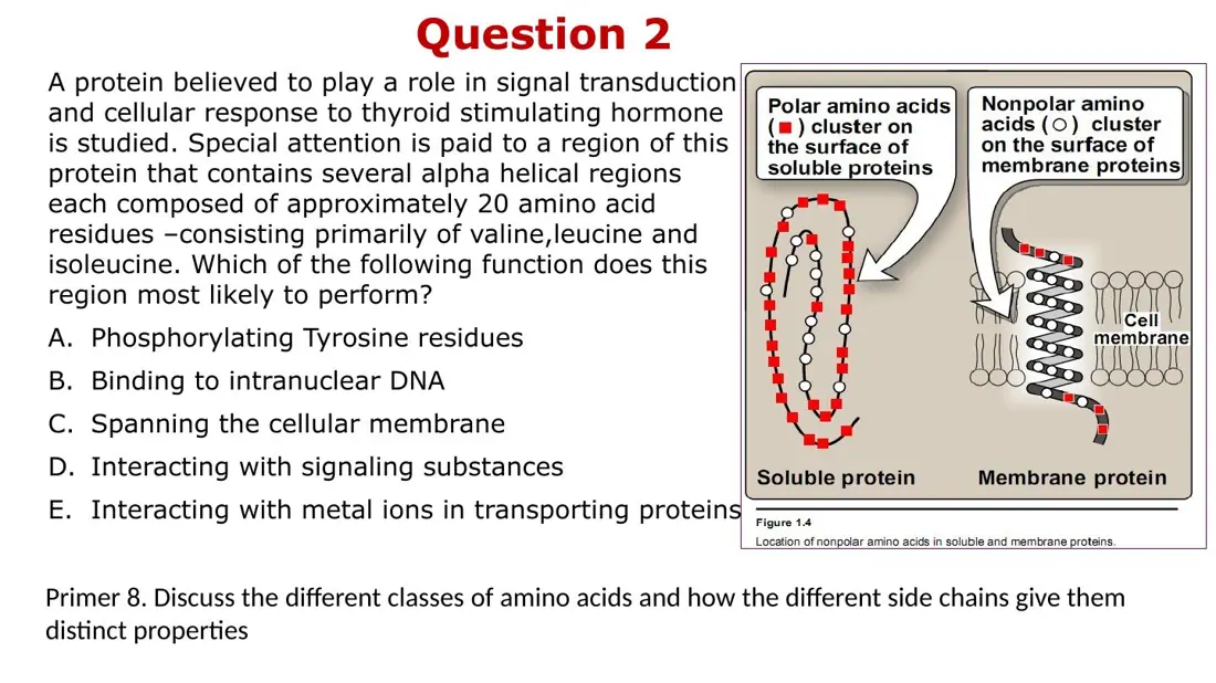
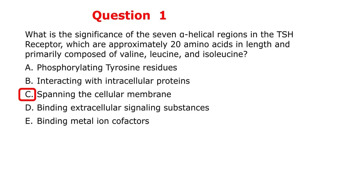
A receptor for thyroid-stimulating hormone (TSH) has seven α-helical segments, each ~20 residues, rich in valine, leucine and isoleucine. What do these segments do?
A. Bind TSH on the outside of the cell
B. Span the cell membrane
C. Phosphorylate tyrosine residues
AnswerB. Span the cell membrane
Why~20-residue helices of Val, Leu, Ile are transmembrane segments: the TSH receptor is a 7-helix GPCR.
Explanation
About 20 residues in an α-helix (1.5 Å rise per residue) give ~30 Å, the thickness of the bilayer's hydrophobic core. Val, Leu and Ile are hydrophobic aliphatic, so a run of them is happiest inside the lipid. Seven such helices is the GPCR signature.
Binding TSH is the job of the extracellular N-terminal domain, which is polar and faces water.
Phosphorylating tyrosines is kinase activity (receptor tyrosine kinases), not a property of hydrophobic helices.
Slides (2)
Bridge: ~20 Val/Leu/Ile residues span the bilayer; structure is the most conservedMade for this pageLecture: structure = function; GPCR's seven hydrophobic helices span the membraneBiochemistry Lecture 22 · Globular proteins (haemoglobin) · slide 5
L26LearningHigh yield
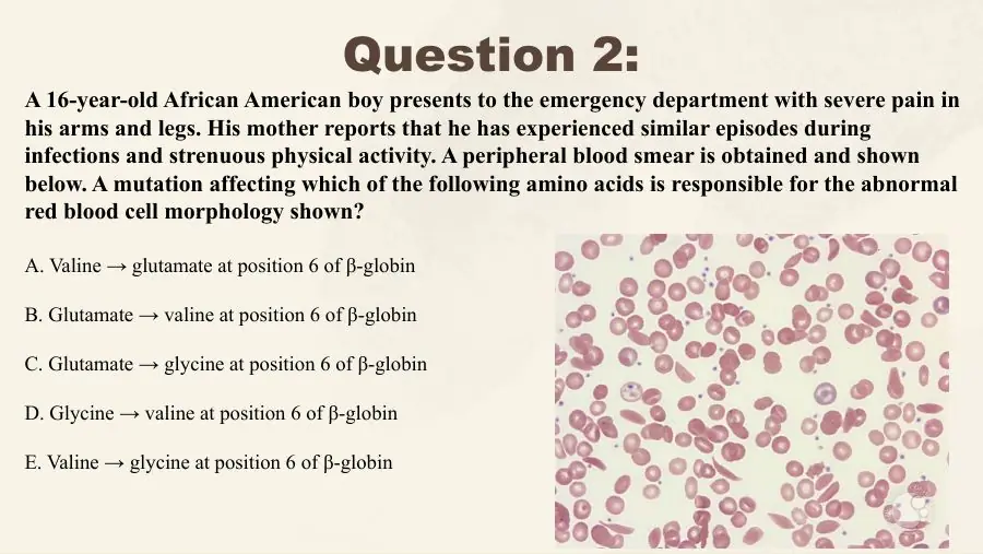
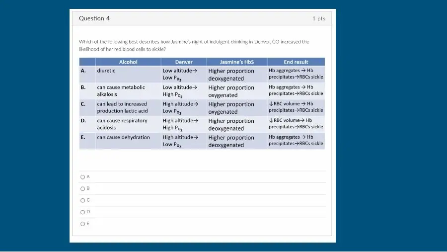
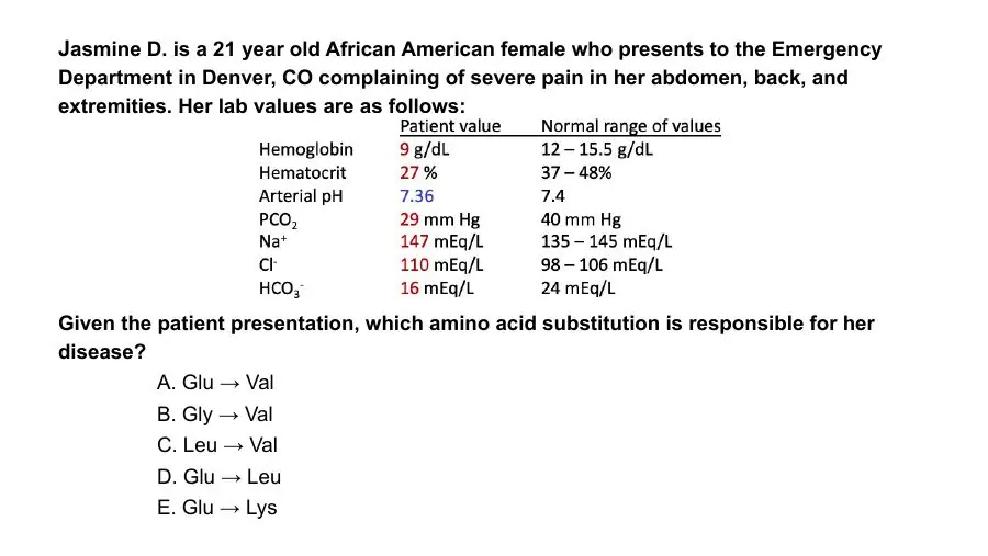
In sickle cell disease, glutamate 6 of the β-globin chain is replaced by valine. How does the side chain change?
A. Basic → hydrophobic
B. Acidic → hydrophobic
C. Acidic → polar uncharged
AnswerB. Acidic → hydrophobic
WhyGlutamate (acidic, negative) → valine (hydrophobic aliphatic): a charged surface residue becomes a sticky nonpolar patch.
Explanation
Glu6 sits on the surface of the β chain. Valine's nonpolar side chain there creates a hydrophobic patch that, in deoxy-HbS, fits into a pocket on a neighbouring tetramer, so HbS polymerises into long fibres and the red cells sickle.
Basic → hydrophobic: glutamate is acidic; the basic residues are Lys, Arg and His. (HbC is Glu6 → Lys: acidic → basic.)
Acidic → polar uncharged: valine has no polar group; Glu → Gln would be that change.
Slides (1)
Bridge: classifying substitutions: Glu→Val, Thr→Leu, Gln→SerMade for this page
L27Learning
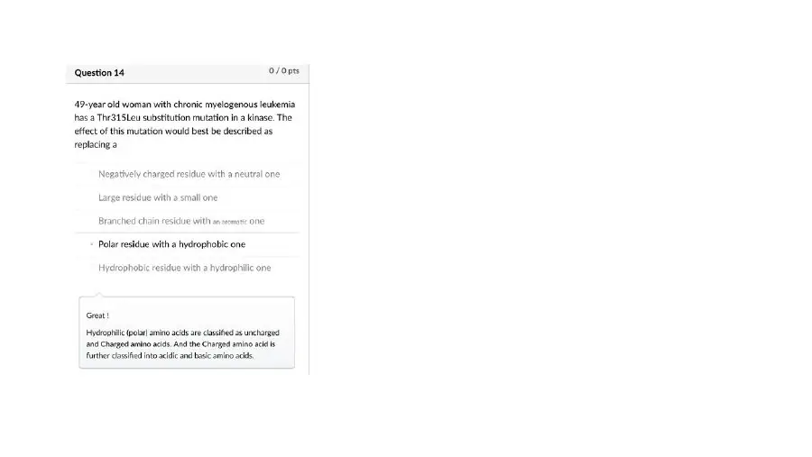
A kinase mutation replaces threonine with leucine (Thr315Leu). How is the side chain changed?
A. Polar residue → hydrophobic residue
B. Hydrophobic residue → polar residue
C. Negatively charged residue → neutral residue
AnswerA. Polar residue → hydrophobic residue
WhyThreonine (–OH, polar uncharged) → leucine (hydrophobic aliphatic): a polar residue is replaced by a hydrophobic one.
Explanation
Threonine's hydroxyl hydrogen-bonds with water, substrates or drugs; leucine's branched hydrocarbon chain cannot, so the site becomes more hydrophobic.
Hydrophobic → polar is the reverse direction.
Negative → neutral: threonine carries no charge at pH 7.4; only Asp and Glu are negative.
Slides (1)
Bridge: classifying substitutions: Glu→Val, Thr→Leu, Gln→SerMade for this page
L28Learning
Glutamine in a designed protein must be replaced by an amino acid of the same side-chain class. Which is the best choice?
A. Leucine
B. Aspartate
C. Serine
AnswerC. Serine
WhySerine, like glutamine, is polar and uncharged, so the substitution keeps the side-chain chemistry.
Explanation
A substitution within the same class (polar uncharged → polar uncharged) usually preserves folding and surface properties. Glutamine (amide) and serine (hydroxyl) both hydrogen-bond and carry no charge at pH 7.4.
Leucine is hydrophobic: it would put a nonpolar group where a polar one was.
Aspartate is close in shape (it is the acid form of asparagine) but adds a negative charge.
Slides (1)
Bridge: classifying substitutions: Glu→Val, Thr→Leu, Gln→SerMade for this page
Charge vs pH: zwitterion and pI
L29Learning
What are the approximate pKas of an amino acid's α-carboxyl and α-amino groups?
A. α-Carboxyl ~2.2 α-Amino ~9.4
B. α-Carboxyl ~4.2 α-Amino ~7.4
C. α-Carboxyl ~9.4 α-Amino ~2.2
AnswerA. α-Carboxyl ~2.2 α-Amino ~9.4
Whyα-COOH pKa~2.2; α-NH3+ pKa~9.4. Every amino acid has at least these two ionisable groups.
Explanation
The pKa is the pH at which a group is half protonated. Below it the protonated form dominates; above it the deprotonated form does. With one pKa far below 7.4 and the other far above, both groups are charged at physiological pH.
~4.2 / ~7.4: ~4 is the side-chain carboxyl of Asp/Glu; 7.4 is blood pH, not a pKa of these groups.
Swapped values would make the carboxyl the weaker acid, which it is not.
At pH 7.4, in what form are an amino acid's α-carboxyl and α-amino groups?
A. –COOH and –NH3+
B. –COOH and –NH2
C. –COO− and –NH3+
AnswerC. –COO− and –NH3+
WhyAt pH 7.4 the α-carboxyl is deprotonated (COO−) and the α-amino is protonated (NH3+).
Explanation
pH 7.4 is far above the carboxyl pKa (~2.2), so it has lost its proton; it is far below the amino pKa (~9.4), so the amino group keeps an extra proton. One + and one − give a net charge of zero for an amino acid with an uncharged side chain: a zwitterion.
–COOH and –NH3+ is the form below pH ~2 (net +1).
–COOH and –NH2 never predominates: it would need the carboxyl protonated and the amine deprotonated at the same time.
Slides (2)
Preface: below pKa a group keeps its proton; the two α-groups at 7.4Made for this pageLecture: α-COOH pKa ~2.2, α-NH2 ~9.4; cation, anion, zwitterionBiochemistry Primer 7 · Amino acids · slide 14
L31LearningHigh yield
What is a zwitterion?
A. A neutral molecule with equal numbers of positive and negative charges
B. A molecule with more positive than negative charges, net positive
C. A molecule with no ionised groups at all, net zero
AnswerA. A neutral molecule with equal numbers of positive and negative charges
WhyA zwitterion carries both + and − charges in equal numbers, so its net charge is zero.
Explanation
The lecture defines three terms: cation = net (+) charge, anion = net (−) charge, zwitterion = neutral molecule with equal (+) and (−) charges. An amino acid with an uncharged side chain is a zwitterion at pH 7.4 (–NH3+ and –COO−).
Net positive is a cation.
No ionised groups is simply uncharged; a zwitterion is neutral because its charges cancel, not because it has none.
Slides (2)
Lecture: α-COOH pKa ~2.2, α-NH2 ~9.4; cation, anion, zwitterionBiochemistry Primer 7 · Amino acids · slide 14Osmosis notes: charge at low, high and neutral pH; zwitterion at the pIOsmosis notes · alternative to slide 2
L32LearningHigh yield
What is the isoelectric point (pI)?
A. The pH at which the net charge is greatest
B. The pH at which the net charge is zero
C. The pH at which the side chain first becomes charged
AnswerB. The pH at which the net charge is zero
WhyThe pI is the pH at which an amino acid (or protein) exists as a zwitterion: net charge zero.
Explanation
At the pI the positive and negative charges balance exactly. Below it the molecule carries extra protons (net +); above it it has lost them (net −). Because the net charge is zero, a molecule at its pI does not move in an electric field and is least soluble.
Greatest net charge is reached at the extremes of pH, far from the pI.
Side chain becomes charged describes the side-chain pKR, not the pI.
Slides (2)
Osmosis notes: charge at low, high and neutral pH; zwitterion at the pIOsmosis notes · alternative to slide 2Lecture: alanine forms I–III, titration curve, buffering regions, pI = 5.7Biochemistry Primer 7 · Amino acids · slide 15
L33LearningHigh yield
What is the net charge of an amino acid or protein below its pI, and above it?
A. Below pI: net positive Above pI: net negative
B. Below pI: net negative Above pI: net positive
C. Below pI: net zero Above pI: net negative
AnswerA. Below pI: net positive Above pI: net negative
WhyBelow the pI (more H+) the net charge is positive; above the pI it is negative.
Explanation
Lowering the pH adds protons: carboxylates become neutral –COOH while amino groups stay –NH3+, so + wins. Raising the pH strips protons: amino groups become neutral –NH2 while carboxylates stay –COO−, so − wins.
The reversed pairing is the common slip.
Net zero below pI: net zero happens only at the pI itself.
The lecture slide calls the negatively charged form 'cationic'; the correct word is anionic (an anion is negative). Hook: 'below the pI, protons pile on → positive'.
Slides (2)
Osmosis notes: charge at low, high and neutral pH; zwitterion at the pIOsmosis notes · alternative to slide 2Bridge: alanine's net charge at pH 1, 2.3, 5.7, 9.1, >10; electrophoresisMade for this page
L34Learning
A protein is placed in an electric field in a buffer whose pH equals the protein's pI. What does it do?
A. Migrates toward the anode (+)
B. Migrates toward the cathode (−)
C. Does not migrate
AnswerC. Does not migrate
WhyAt its pI a protein has no net charge, so it does not migrate toward either electrode.
Explanation
Electrophoresis separates molecules by net charge: net-negative molecules move to the anode (+), net-positive ones to the cathode (−). At the pI the charges cancel. Isoelectric focusing uses this: each protein stops where the pH gradient equals its pI.
Toward the anode happens when the buffer pH is above the pI (net −).
Toward the cathode happens when the buffer pH is below the pI (net +).
Slides (2)
Bridge: alanine's net charge at pH 1, 2.3, 5.7, 9.1, >10; electrophoresisMade for this pageOsmosis notes: charge at low, high and neutral pH; zwitterion at the pIOsmosis notes · alternative to slide 2
L35Learning
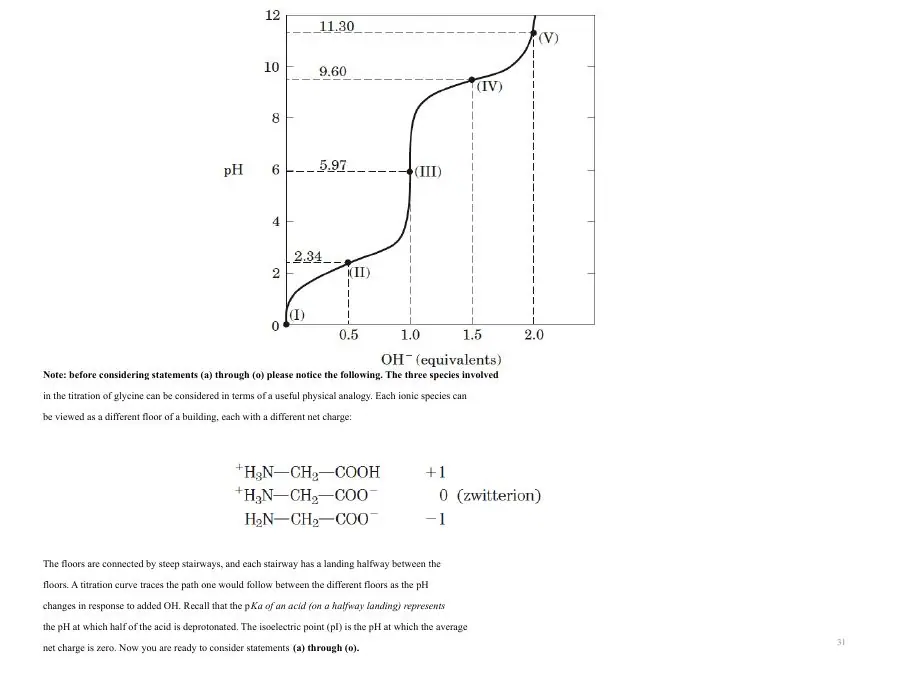
Alanine (pK1 2.3, pK2 9.1) is dissolved at pH 1. Which form predominates?
A. –COO− and –NH3+, net charge 0
B. –COOH and –NH3+, net charge +1
C. –COO− and –NH2, net charge −1
AnswerB. –COOH and –NH3+, net charge +1
WhyBelow pK1 both groups are protonated: –COOH and –NH3+, net +1 (form I, the cation).
Explanation
The titration slide shows three forms of alanine:
Form I (pH < 2): –COOH, –NH3+, net +1.
Form II (pH ~6, the pI 5.7): –COO−, –NH3+, net 0, the zwitterion.
Form III (pH > 10): –COO−, –NH2, net −1.
pH 1 is below pK1 (2.3), so even the carboxyl keeps its proton: form I.
Slides (2)
Bridge: alanine's net charge at pH 1, 2.3, 5.7, 9.1, >10; electrophoresisMade for this pageLecture: alanine forms I–III, titration curve, buffering regions, pI = 5.7Biochemistry Primer 7 · Amino acids · slide 15
L36Learning
On alanine's titration curve, what is the average net charge at pH 2.3 (= pK1)?
A. +1
B. 0
C. +½
AnswerC. +½
WhyAt pH = pK1, half the molecules are form I (+1) and half form II (0): the average net charge is +½.
Explanation
At any pH equal to a group's pKa, that group is exactly half protonated. At 2.3, half of the alanine carboxyls are –COOH (net +1 molecules) and half are –COO− (net 0 zwitterions), so [I] = [II] and the average is +½. Likewise at pK2 (9.1), [II] = [III] and the average is −½.
+1 holds well below pH 2.3.
0 holds at the pI, 5.7.
Slides (2)
Bridge: alanine's net charge at pH 1, 2.3, 5.7, 9.1, >10; electrophoresisMade for this pageLecture: alanine forms I–III, titration curve, buffering regions, pI = 5.7Biochemistry Primer 7 · Amino acids · slide 15
L37Learning
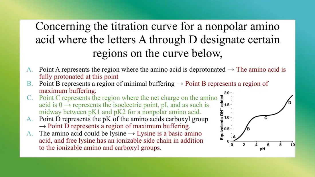
Where on alanine's titration curve is its buffering capacity greatest?
A. Near pH 5.7, its pI
B. Near pH 2.3 and 9.1, its pKas
C. Near pH 7.4, physiological pH
AnswerB. Near pH 2.3 and 9.1, its pKas
WhyAn amino acid buffers best near each pKa, where protonated and deprotonated forms are equal.
Explanation
A buffer resists pH change when it holds plenty of both the proton donor and the proton acceptor. That happens at pH = pKa (± ~1 unit), the flat 'regions of buffering' shaded on the alanine curve at 2.3 and 9.1. At the pI only one form (the zwitterion) dominates, so the curve is steep and buffering is poorest there.
pI (5.7) is the worst buffering region.
pH 7.4 is far from both of alanine's pKas; only histidine's side chain (pKa ~6) buffers near there.
Slides (2)
Lecture: alanine forms I–III, titration curve, buffering regions, pI = 5.7Biochemistry Primer 7 · Amino acids · slide 15Bridge: buffering works near the pKa, so histidine buffers at pH 7.4Made for this page
L38LearningHigh yield
A protein buffers best at physiological pH when it is rich in which amino acid?
A. Histidine
B. Lysine
C. Aspartate
AnswerA. Histidine
WhyHistidine's imidazole has a pKa (~6) closest to pH 7.4, so it can take up or release H+ in the physiological range.
Explanation
A group buffers well within about one pH unit of its pKa. Histidine is the only side chain with a pKa near neutral, which is why histidine-rich proteins (haemoglobin, albumin) are important blood buffers and why histidine is common in enzyme active sites.
Lysine (pKa ~10.5) is fully protonated at 7.4 and cannot absorb more H+.
Aspartate (pKa ~4) is fully deprotonated at 7.4.
Slides (1)
Bridge: buffering works near the pKa, so histidine buffers at pH 7.4Made for this page
Calculating pI and peptide charge
L39Learning
How do you calculate the pI of an amino acid with only two ionisable groups?
A. Take the higher of the two pKas
B. Add the two pKas
C. Average the two pKas
AnswerC. Average the two pKas
WhyWith two ionisable groups, pI = (pK1 + pK2) / 2.
Explanation
The zwitterion dominates between the two pKas, and the point of exact charge balance lies midway between them. For an amino acid with an uncharged side chain, pK1 is the α-carboxyl and pK2 the α-amino group.
The higher pKa is where half the amino groups have lost their proton (net −½), not where net charge is zero.
The sum gives a number above any possible pH (e.g. 11.4 for alanine).
Slides (2)
Lecture: two ionisable groups: alanine pI = (2.3 + 9.1)/2 = 5.7Biochemistry Primer 7 · Amino acids · slide 15Preface: the pI recipe, worked for alanine, aspartate and arginineMade for this page
L40Learning
What is the pI of alanine (pK1 2.3, pK2 9.1)?
A. 6.8
B. 11.4
C. 5.7
AnswerC. 5.7
WhypI = (2.3 + 9.1) / 2 = 5.7.
Explanation
Alanine's side chain (–CH3) does not ionise, so only the α-carboxyl and α-amino groups count. Their average is 5.7: at that pH alanine is the zwitterion (form II) with net charge 0.
11.4 is the sum without dividing by 2.
6.8 is simply 9.1 − 2.3, the difference of the two pKas.
Slides (1)
Lecture: two ionisable groups: alanine pI = (2.3 + 9.1)/2 = 5.7Biochemistry Primer 7 · Amino acids · slide 15
L41LearningHigh yield
How do you calculate the pI of an amino acid with an ionisable side chain?
A. Average the two nearest pKas
B. Use the side-chain pKa alone
C. Average all three pKas
AnswerA. Average the two nearest pKas
WhyWith three ionisable groups, the pI is the average of the two nearest pKas: the two that flank the neutral form.
Explanation
Write out the forms from low to high pH and find the one with net charge 0; the pI is the average of the pKas on either side of it.
Acidic amino acids (Asp, Glu): the neutral form lies between the two low pKas → average pK1 and pKR.
Basic amino acids (Lys, Arg): the neutral form lies between the two high pKas → average pK2 and pKR.
Averaging all three, or using pKR alone, does not land at the point of zero charge.
Slides (2)
Lecture: three ionisable groups: pI of Asp 3.0, Arg 10.75Biochemistry Primer 7 · Amino acids · slide 16Preface: the pI recipe, worked for alanine, aspartate and arginineMade for this page
L42Learning
What is the pI of aspartate (pKas 2.0, 4.0, 9.5)?
A. 6.75 = (4.0 + 9.5) / 2
B. 5.75 = (2.0 + 9.5) / 2
C. 3.0 = (2.0 + 4.0) / 2
AnswerC. 3.0 = (2.0 + 4.0) / 2
WhyAspartate is acidic, so its neutral form sits between the two low pKas: pI = (2.0 + 4.0) / 2 = 3.0.
Explanation
Going up in pH, aspartate passes through net +1 (all protonated), 0 (α-carboxyl lost its proton at 2.0), −1 (side-chain carboxyl lost its proton at 4.0) and −2 (amino group deprotonated at 9.5). The zero-charge form lies between 2.0 and 4.0, so the pI is 3.0. An acidic amino acid always has a low pI.
5.75 averages the two α-groups and ignores the side chain.
6.75 averages the wrong pair: between 4.0 and 9.5 aspartate is already net −1.
Slides (2)
Lecture: three ionisable groups: pI of Asp 3.0, Arg 10.75Biochemistry Primer 7 · Amino acids · slide 16Preface: the pI recipe, worked for alanine, aspartate and arginineMade for this page
L43Learning
What is the pI of arginine (pKas 2.1, 9.0, 12.5)?
A. 7.30 = (2.1 + 12.5) / 2
B. 5.55 = (2.1 + 9.0) / 2
C. 10.75 = (9.0 + 12.5) / 2
AnswerC. 10.75 = (9.0 + 12.5) / 2
WhyArginine is basic, so its neutral form sits between the two high pKas: pI = (9.0 + 12.5) / 2 = 10.75.
Explanation
Going up in pH, arginine passes through +2 (all protonated), +1 (carboxyl deprotonated at 2.1), 0 (α-amino deprotonated at 9.0) and −1 (guanidinium deprotonated at 12.5). Zero charge lies between 9.0 and 12.5. A basic amino acid always has a high pI.
5.55 averages the two low pKas; at that pH arginine is still +1.
7.30 averages the outermost pKas, which is never the rule.
Slides (2)
Lecture: three ionisable groups: pI of Asp 3.0, Arg 10.75Biochemistry Primer 7 · Amino acids · slide 16Preface: the pI recipe, worked for alanine, aspartate and arginineMade for this page
L44LearningHigh yield
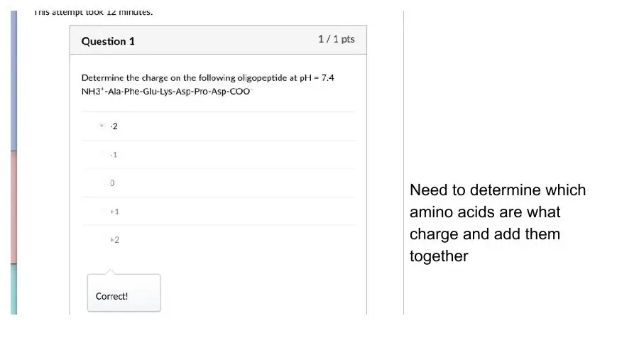
What is the net charge at pH 7.4 of the peptide NH3+-Ala-Phe-Glu-Lys-Asp-Pro-Asp-COO−?
A. −1
B. −2
C. +1
AnswerB. −2
WhyTermini cancel (+1, −1); Glu, Asp, Asp give −3, Lys gives +1: net −2.
His: the course lists it with the positive (basic) residues and review keys count it +1, although with a pKa ~6 most His side chains are uncharged at 7.4. Cys and Tyr are uncharged.
All other residues 0.
Here: −1 (Glu) +1 (Lys) −1 (Asp) −1 (Asp) = −2. −1 forgets one acidic residue; +1 reverses the signs.
Slides (1)
Bridge: counting a peptide's net charge at pH 7.4, four worked examplesMade for this page
L45Learning
What is the net charge at pH 7.4 of the peptide Glu-Asp-Arg-Lys-Lys (free termini)?
A. +1
B. −1
C. +3
AnswerA. +1
WhyTermini cancel; Glu and Asp give −2, Arg and two Lys give +3: net +1.
Explanation
Count only the side chains once the N-terminal +1 and C-terminal −1 cancel:
Acidic: Glu (−1), Asp (−1) → −2.
Basic: Arg (+1), Lys (+1), Lys (+1) → +3.
Sum = +1. +3 forgets the two acidic residues; −1 swaps the signs of the acidic and basic groups.
Slides (1)
Bridge: counting a peptide's net charge at pH 7.4, four worked examplesMade for this page
L46LearningHigh yield
Which peptide carries a net positive charge at pH 7.4?
A. Glu-Pro-Lys-Val-Leu
B. Met-Thr-Asn-Asp-Gly
C. Ile-Val-Gln-Ser-Arg
AnswerC. Ile-Val-Gln-Ser-Arg
WhyIn Ile-Val-Gln-Ser-Arg the termini cancel and the only charged side chain is arginine (+1): net +1.
Explanation
After the termini cancel:
Glu-Pro-Lys-Val-Leu: Glu −1 + Lys +1 = 0.
Met-Thr-Asn-Asp-Gly: Asp −1 = −1 (Asn and Thr are polar but uncharged).
Ile-Val-Gln-Ser-Arg: Arg +1 = +1.
Trap: Gln and Asn look like Glu and Asp but carry no charge.
Slides (1)
Bridge: counting a peptide's net charge at pH 7.4, four worked examplesMade for this page
L47LearningHigh yield
Which peptide is the most acidic (most negative) at pH 7.4?
A. Gly-Tyr-Asp-Glu-Phe-Cys
B. Leu-Met-Ser-Asn-Glu-Val
C. Lys-Arg-Pro-Tyr-Phe-Cys
AnswerA. Gly-Tyr-Asp-Glu-Phe-Cys
WhyGly-Tyr-Asp-Glu-Phe-Cys has two acidic side chains: net −2, the most negative.
Explanation
With the termini cancelling:
Gly-Tyr-Asp-Glu-Phe-Cys: Asp −1, Glu −1 → −2.
Leu-Met-Ser-Asn-Glu-Val: Glu −1 → −1 (Asn is uncharged).
Tyr (pKa ~10) and Cys (~8.3) are protonated and uncharged at 7.4.
Slides (1)
Bridge: counting a peptide's net charge at pH 7.4, four worked examplesMade for this page
The peptide bond and reading a sequence
L48LearningHigh yield
What is a peptide bond?
A. An amide link between the α-amino group of one amino acid and the side chain of the next
B. An ester link between the α-carboxyl of one amino acid and the side-chain hydroxyl of the next
C. An amide link between the α-carboxyl of one amino acid and the α-amino group of the next
AnswerC. An amide link between the α-carboxyl of one amino acid and the α-amino group of the next
WhyA peptide bond is a covalent amide linkage joining the α-carboxyl of one amino acid to the α-amino group of the next.
Explanation
The carbonyl carbon of residue 1 bonds to the nitrogen of residue 2 (–CO–NH–). Repeating this gives the polypeptide backbone, with the side chains hanging off the α-carbons. Peptide bonds are what hold the primary structure together.
α-Amino + side chain: side chains are not part of the backbone link (a side-chain amide bond is an isopeptide, e.g. ubiquitin conjugation).
Ester link: esters join carboxyl and hydroxyl groups (as in triacylglycerols), not amino acids in a chain.
Slides (2)
Lecture: peptide bond forms by condensation (water out), breaks by hydrolysisBiochemistry Primer 8 · Protein structure · slide 6Osmosis: amide (peptide) bond formed by a condensation reactionOsmosis figure · alternative to slide 2
L49LearningHigh yield
What is released when a peptide bond forms, and what is needed to break it?
A. Forming releases ATP (condensation) Breaking needs ATP (phosphorolysis)
B. Forming releases water (condensation) Breaking needs water (hydrolysis)
C. Forming releases CO2 (decarboxylation) Breaking needs water (hydrolysis)
AnswerB. Forming releases water (condensation) Breaking needs water (hydrolysis)
WhyPeptide bonds form by condensation (H2O released) and break by hydrolysis (H2O added).
Explanation
The –OH of one carboxyl and an –H of the next amino group leave as a water molecule, joining the two residues. Putting the water back cleaves the bond, which is what proteases and gastric/pancreatic digestion do. Hydrolysis is thermodynamically favourable but very slow without an enzyme, so proteins are kinetically stable.
ATP: ATP pays for amino acid activation (tRNA charging) but is not a product of the bond itself.
CO2: no carbon is lost; decarboxylation makes amines (e.g. histamine), not peptides.
Slides (2)
Lecture: peptide bond forms by condensation (water out), breaks by hydrolysisBiochemistry Primer 8 · Protein structure · slide 6Osmosis: amide (peptide) bond formed by a condensation reactionOsmosis figure · alternative to slide 2
L50LearningHigh yield
Why can't the peptide bond rotate freely?
A. It has partial double-bond character from resonance
B. Neighbouring side chains clash across it
C. It is a full C=N double bond
AnswerA. It has partial double-bond character from resonance
WhyResonance gives the C–N peptide bond partial double-bond character, so it is rigid and planar.
Explanation
The lone pair on the amide nitrogen delocalises toward the carbonyl oxygen. The C–N bond is therefore shorter than a single bond and behaves like ~40% double bond: it cannot twist, and the six atoms around it lie in one plane (the peptide unit).
Side-chain clash explains why the R groups prefer trans, not why the bond cannot rotate.
Full double bond overstates it: the bond is intermediate between single and double, and the C=O keeps partial double-bond character too.
Slides (2)
Osmosis notes: peptide bond and resonance giving partial double-bond characterOsmosis notes · alternative to slide 2Lecture: partial double bond; peptide unit's six atoms lie in one planeBiochemistry Primer 8 · Protein structure · slide 7
L51LearningHigh yield
Which atoms make up a peptide unit?
A. Cα and R group of one residue + N–H and R group of the next, lying in one plane
B. Cα and C=O of one residue + N–H and Cα of the next, rotating freely
C. Cα and C=O of one residue + N–H and Cα of the next, lying flat in one plane
AnswerC. Cα and C=O of one residue + N–H and Cα of the next, lying flat in one plane
WhyA peptide unit = the Cα and C=O of one amino acid plus the N–H and Cα of the next, all in one plane.
Explanation
Because the C–N bond cannot rotate, these six atoms (Cα, C, O, N, H, Cα) are locked in a flat plate. A protein backbone is a chain of these rigid plates hinged at each α-carbon.
Including R groups: the side chains attach to the α-carbons and project out of the plane; they are not part of the unit.
Rotating freely: the unit is rigid; rotation happens only at the Cα hinges (Φ and Ψ).
Slides (1)
Lecture: partial double bond; peptide unit's six atoms lie in one planeBiochemistry Primer 8 · Protein structure · slide 7
L52Learning
Are the R groups on either side of a peptide bond usually cis or trans?
A. Trans
B. Randomly cis or trans
C. Cis
AnswerA. Trans
WhyThe R groups (and α-carbons) across a peptide bond are almost always trans, on opposite sides, which avoids steric clash.
Explanation
In the trans arrangement the two α-carbons, and so their side chains, sit on opposite sides of the rigid C–N bond, as far apart as possible. Cis would bump the side chains together.
Cis is rare; it occurs mainly in bonds preceding proline, whose ring reduces the energy difference.
Random: the preference for trans is strong (>99.9% for non-proline bonds).
Slides (1)
Lecture: R groups trans; rotation only about N–Cα (Φ) and Cα–C (Ψ)Biochemistry Primer 8 · Protein structure · slide 8
L53LearningHigh yield
Around which bonds can a polypeptide backbone rotate, and what are the angles called?
A. N–Cα (Φ, phi) Cα–C (Ψ, psi)
B. N–Cα (Ψ, psi) Cα–C (Φ, phi)
C. C–N peptide bond (Φ, phi) Cα–C (Ψ, psi)
AnswerA. N–Cα (Φ, phi) Cα–C (Ψ, psi)
WhyRotation occurs at each α-carbon: around N–Cα (Φ, phi) and Cα–C (Ψ, psi). This gives the chain its flexibility.
Explanation
Each peptide unit is a rigid plane; the planes meet at the α-carbons, which act as swivels. The pair of angles (Φ, Ψ) at every residue determines the local backbone shape, and particular pairs give the α-helix or β-sheet. Proline's ring fixes its Φ, which is part of why it breaks helices.
Φ and Ψ swapped mislabels the bonds: Φ is the N–Cα bond.
C–N peptide bond cannot rotate (partial double bond).
Slides (2)
Lecture: R groups trans; rotation only about N–Cα (Φ) and Cα–C (Ψ)Biochemistry Primer 8 · Protein structure · slide 8Preface: the backbone: peptide bond, planar peptide unit, Φ and Ψ, trans R groupsMade for this page
L54LearningHigh yield
In which direction is a peptide sequence written and read?
A. C-terminus (left) → N-terminus (right)
B. N-terminus (left) → C-terminus (right)
C. N-terminus (right) → C-terminus (left)
AnswerB. N-terminus (left) → C-terminus (right)
WhySequences are written and read from the N-terminal (free amino end, left) to the C-terminal (free carboxyl end, right).
Explanation
This matches the direction of synthesis on the ribosome, which adds each new amino acid to the free carboxyl end. The name follows the same order: all residues but the last end in '-yl', so Tyr-Gly-Gly-Phe-Leu (Leu-enkephalin) is tyrosylglycylglycylphenylalanylleucine.
C → N is backwards.
N on the right puts the ends on the wrong sides.
Slides (1)
Lecture: sequences read N-terminal (left) to C-terminal (right)Biochemistry Primer 8 · Protein structure · slide 9
L55Learning
In the peptide tyrosylglycylglycylphenylalanylleucine (Tyr-Gly-Gly-Phe-Leu), which residue carries the free α-amino group?
A. Leucine
B. Tyrosine
C. Phenylalanine
AnswerB. Tyrosine
WhyThe first-named residue, tyrosine, is the N-terminus and keeps the free α-amino group.
Explanation
By convention the residue on the left (first in the name, with the '-yl' ending) is the N-terminal residue with a free –NH3+. The last residue, leucine, keeps its full name and is the C-terminal residue with a free –COO−.
Leucine carries the free α-carboxyl, not the free amino group.
Phenylalanine is internal: both its α-groups are tied up in peptide bonds.
Slides (2)
Bridge: naming Tyr-Gly-Gly-Phe-Leu; which residue holds each free endMade for this pageLecture: sequences read N-terminal (left) to C-terminal (right)Biochemistry Primer 8 · Protein structure · slide 9
Four levels of structure
L56LearningHigh yield
What is primary structure, and what holds it together?
A. The amino acid sequence, held by hydrogen bonds
B. The amino acid sequence, held by disulfide bonds
C. The amino acid sequence, held by peptide bonds
AnswerC. The amino acid sequence, held by peptide bonds
WhyPrimary structure is the linear amino acid sequence, held by covalent peptide bonds.
Explanation
The sequence (read N → C) is the level encoded by the gene, and it determines every higher level. A single change in it can alter the whole protein (Glu → Val in sickle haemoglobin).
Hydrogen bonds hold secondary structure (backbone C=O···H–N).
Disulfide bonds are side-chain cross-links that stabilise tertiary structure.
Slides (3)
Lecture: four levels and the bonds holding eachBiochemistry Primer 8 · Protein structure · slide 4Bridge: each level with the bond that holds itMade for this pageOsmosis: primary to quaternary structure drawn level by levelOsmosis figure · alternative to slide 1
L57LearningHigh yield
What is secondary structure, and what holds it together?
A. Local folding of the backbone, held by peptide bonds between neighbouring residues
B. Local folding of the backbone, held by hydrogen bonds between side chains
C. Local folding of the backbone, held by hydrogen bonds between backbone C=O and N–H
AnswerC. Local folding of the backbone, held by hydrogen bonds between backbone C=O and N–H
WhySecondary structure (α-helix, β-sheet) is local backbone folding held by hydrogen bonds between main-chain C=O and N–H groups.
Explanation
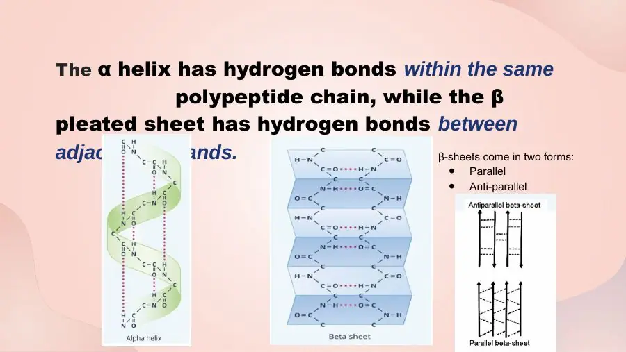
The carbonyl oxygen of one peptide unit accepts a hydrogen bond from the amide N–H of another. In an α-helix these bonds lie within one stretch of chain; in a β-sheet they link neighbouring strands. Both the helix and the sheet are held by hydrogen bonds.
Peptide bonds make the chain itself (primary).
Side-chain hydrogen bonds are a tertiary interaction; secondary structure uses backbone atoms only.
Slides (3)
Lecture: four levels and the bonds holding eachBiochemistry Primer 8 · Protein structure · slide 4Bridge: each level with the bond that holds itMade for this pageCourse lecture — hydrogen bonds in a β-sheet and an α-helixCardio-Pulm 1 flashcards
L58LearningHigh yield
What is tertiary structure, and what holds it together?
A. The 3-D fold of one chain, held by side-chain interactions (non-covalent and covalent)
B. The local folding of the backbone, held by side-chain interactions (covalent only)
C. The arrangement of several chains, held by side-chain interactions (non-covalent and covalent)
AnswerA. The 3-D fold of one chain, held by side-chain interactions (non-covalent and covalent)
WhyTertiary structure is the overall 3-D fold of a single chain, held by R-group interactions, both non-covalent and covalent (disulfide).
Explanation
Secondary elements pack together into motifs and domains. The fold is stabilised by hydrophobic interactions, hydrogen bonds, ionic bonds, metal ions and covalent disulfide bonds between side chains.
Covalent only is wrong twice: that is the local (secondary) level, and most tertiary forces are non-covalent.
Several chains is quaternary structure.
Common trap: 'tertiary structure involves only non-covalent interactions' is false.
Slides (3)
Lecture: four levels and the bonds holding eachBiochemistry Primer 8 · Protein structure · slide 4Bridge: each level with the bond that holds itMade for this pageCourse lecture — side-chain interactions that hold a folded chainCardio-Pulm 1 flashcards
L59LearningHigh yield
What is quaternary structure, and what holds it together?
A. The arrangement of several polypeptide chains, held by non-covalent interactions
B. The 3-D fold of one chain, held by non-covalent interactions
C. The arrangement of several polypeptide chains, held by peptide bonds
AnswerA. The arrangement of several polypeptide chains, held by non-covalent interactions
WhyQuaternary structure is the assembly of two or more subunits, held (per the lecture) by non-covalent forces.
Explanation
The subunits are separate folded chains that associate through hydrogen bonds, ionic bonds, hydrophobic contacts and metal ions. Haemoglobin (2α + 2β) is the standard example; myoglobin, a single chain, has none.
One chain is tertiary.
Peptide bonds join residues within a chain, never separate subunits.
Textbook note: a few multi-chain proteins (e.g. antibodies) also have inter-chain disulfide bonds; for this exam answer 'non-covalent'.
Slides (3)
Lecture: four levels and the bonds holding eachBiochemistry Primer 8 · Protein structure · slide 4Bridge: each level with the bond that holds itMade for this pageOsmosis: primary to quaternary structure drawn level by levelOsmosis figure · alternative to slide 1
α-helix, β-sheet and random coil
L60Learning
What handedness and how many residues per turn does an α-helix have?
A. Right-handed; 3.6 residues per turn
B. Left-handed; 3.6 residues per turn
C. Right-handed; 5.0 residues per turn
AnswerA. Right-handed; 3.6 residues per turn
WhyThe α-helix is a right-handed spiral with 3.6 residues per turn.
Explanation
It is a rigid, rod-like structure that forms spontaneously and looks like a spiral staircase turning clockwise as it moves away from the viewer. 3.6 residues per turn means residue n sits almost directly below residue n + 4, the partner it hydrogen-bonds with.
Left-handed helices are strained for L amino acids and almost never occur in proteins (collagen's individual chains are a special left-handed polyproline-type helix).
5.0 per turn does not match the α-helix geometry.
Slides (1)
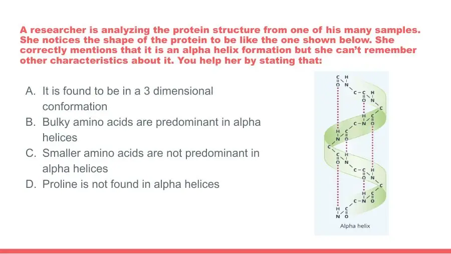
Lecture: α-helix: right-handed, 3.6 residues/turn, H-bond to 4th residue, R outwardBiochemistry Primer 8 · Protein structure · slide 13
L61Learning
How long is an average α-helix?
A. ~12 residues (range 5–45)
B. ~4 residues (range 2–10)
C. ~40 residues (range 20–100)
AnswerA. ~12 residues (range 5–45)
WhyAn average α-helix is about 12 residues long, with a range of 5–45.
Explanation
Twelve residues is just over three turns (3.6 per turn). Transmembrane helices are at the longer end (~20 residues) because they must span the ~30 Å hydrophobic core of the bilayer.
~4 residues is barely one turn, the size of a β-turn.
~40 residues overstates the average, though single helices can reach 45.
Slides (2)
Lecture: α-helix: right-handed, 3.6 residues/turn, H-bond to 4th residue, R outwardBiochemistry Primer 8 · Protein structure · slide 13Bridge: helix, sheet and random coil compared side by sideMade for this page
L62LearningHigh yield
How are the hydrogen bonds arranged in an α-helix?
A. Each side chain bonds the backbone four residues along; bonds run inside, parallel to the axis
B. Each C=O bonds an N–H on a neighbouring chain; bonds run outside, perpendicular to the axis
C. Each C=O bonds the N–H four residues along the same chain; bonds run inside, parallel to the axis
AnswerC. Each C=O bonds the N–H four residues along the same chain; bonds run inside, parallel to the axis
WhyIn an α-helix the C=O of residue n bonds the N–H of residue n + 4 in the same chain (intrachain); the bonds lie inside, parallel to the axis.
Explanation
Every backbone C=O and N–H inside the helix is paired, which is what makes the helix stable. Because each bond links residues one turn apart, the bonds point along the helix axis, and the peptide-unit planes are parallel to that axis.
Side chain to backbone: helix hydrogen bonds are backbone-to-backbone only.
Neighbouring chain, perpendicular: that describes a β-sheet, where bonds run between strands.
Slides (2)
Lecture: α-helix: right-handed, 3.6 residues/turn, H-bond to 4th residue, R outwardBiochemistry Primer 8 · Protein structure · slide 13Osmosis (middle panel): helix H-bonds link residues four apart; sheet bonds join strandsOsmosis figure · alternative to slide 2
L63Learning
Which way do the R groups point in an α-helix?
A. Outward from the helix
B. Along the helix axis
C. Inward to the helix core
AnswerA. Outward from the helix
WhyIn an α-helix the R groups point outward, while the hydrogen bonds are inside.
Explanation
The tightly wound backbone fills the centre of the helix, leaving no room inside; the side chains project out like the steps of a spiral staircase. Their chemistry then decides how the helix packs: a membrane helix shows hydrophobic R groups to the lipid; an amphipathic helix has a polar face and a nonpolar face.
Inward is the classic trap; the core is occupied by backbone atoms.
Along the axis describes the hydrogen bonds, not the side chains.
Slides (1)
Lecture: α-helix: right-handed, 3.6 residues/turn, H-bond to 4th residue, R outwardBiochemistry Primer 8 · Protein structure · slide 13
L64LearningHigh yield
Where do the hydrogen bonds run in a β-sheet?
A. Between residues four apart in the same strand
B. Between side chains on the same strand
C. Between backbone C=O and N–H of adjacent strands
AnswerC. Between backbone C=O and N–H of adjacent strands
WhyA β-sheet is held by hydrogen bonds between adjacent strands (backbone C=O to N–H), laterally packing the strands.
Explanation
Each β-strand is an almost fully extended stretch of chain. Strands line up side by side, and the C=O of one strand bonds the N–H of the neighbour, so the bonds run roughly perpendicular to the strands. The strands can come from distant parts of one chain or from different chains.
Four apart, same strand is the α-helix pattern (intrachain).
Side chains: like the helix, the sheet uses backbone atoms, not R groups.
Summary: α-helix = within the same chain; β-sheet = between adjacent strands.
Slides (2)
Lecture: β-sheet: H-bonds between strands; parallel or antiparallel; R above and belowBiochemistry Primer 8 · Protein structure · slide 14Osmosis (middle panel): helix H-bonds link residues four apart; sheet bonds join strandsOsmosis figure · alternative to slide 2
L65Learning
What two orientations can the strands of a β-sheet have?
A. Coiled (wound together) Antiparallel (opposite directions)
B. Parallel (same direction) Antiparallel (opposite directions)
C. Left-handed (same direction) Right-handed (opposite directions)
Whyβ-sheets are parallel (neighbouring strands run the same N → C direction) or antiparallel (opposite directions).
Explanation
Direction is judged from N- to C-terminus of each strand. Antiparallel sheets, often formed by a chain folding back on itself through a β-turn, have straighter, slightly stronger hydrogen bonds; parallel sheets need a longer crossover connection between strands.
Coiled describes coiled coils of α-helices, not sheet strands.
Left/right-handed describes helices; sheet strands are classed by direction only.
Slides (1)
Lecture: β-sheet: H-bonds between strands; parallel or antiparallel; R above and belowBiochemistry Primer 8 · Protein structure · slide 14
L66Learning
Where do the R groups sit in a β-sheet?
A. All on one face of the sheet
B. Above and below the plane of the sheet
C. Between the strands, in the plane of the sheet
AnswerB. Above and below the plane of the sheet
WhyIn a β-sheet the R groups point above and below the plane of the pleated sheet, alternating from residue to residue.
Explanation
The pleats (zigzag) of the extended chain make successive side chains alternate sides. So one face of a sheet can be hydrophobic and the other polar if the sequence alternates nonpolar and polar residues, as when one face of the sheet packs against the protein core.
All on one face ignores the alternation.
Between the strands is where the hydrogen bonds run; the R groups stick out of the plane.
Slides (1)
Lecture: β-sheet: H-bonds between strands; parallel or antiparallel; R above and belowBiochemistry Primer 8 · Protein structure · slide 14
L67Learning
What is the flexible, irregular structure found between helices and sheets called?
A. β-turn
B. Random coil
C. Domain
AnswerB. Random coil
WhyIrregular, flexible stretches of polypeptide that are neither helix nor sheet are called random coil.
Explanation
'Random' does not mean disordered or useless: these loops have a defined structure in a given protein, often form the active or binding site, and link the regular elements. They simply lack a repeating hydrogen-bond pattern.
β-turn is a regular, tight four-residue U-turn with a specific hydrogen bond (C=O of residue 1 to N–H of residue 4).
Domain is a large, independently folded tertiary unit, not a local backbone shape.
Slides (2)
Lecture: secondary structure: α-helix, β-sheet, random coil, all held by H-bondsBiochemistry Primer 8 · Protein structure · slide 11Bridge: helix, sheet and random coil compared side by sideMade for this page
Which residues build helices, sheets and turns
L68LearningHigh yield
Which small amino acid is most often found in α-helices?
A. Glycine
B. Alanine
C. Proline
AnswerB. Alanine
WhyAlanine, a small amino acid, has the highest helix propensity (0.00 kcal/mol on the helix-propensity scale).
Explanation
Alanine's methyl side chain is small enough to fit the tight helix without clashing, yet it restricts the backbone enough to favour the helical Φ/Ψ angles. On the lecture's helix-propensity table (energy penalty for placing a residue in a helix) alanine is the reference at 0.
Glycine is smaller still, but its flexibility makes it lose entropy when held in a helix (penalty 1.00 kcal/mol).
Proline is the worst (3.16 kcal/mol): the helix breaker.
Slides (2)
Lecture: proline the helix breaker; helix-propensity table; aromatics in β-sheetsBiochemistry Primer 8 · Protein structure · slide 12Preface: which residues favour helices, break them, build sheets and turnsMade for this page
L69LearningHigh yield
Which amino acid is called the 'helix breaker', and why?
A. Glycine, because its bulky ring blocks hydrogen bonding
B. Proline, because its rigid ring kinks the chain
C. Proline, because its charged side chain repels the backbone
AnswerB. Proline, because its rigid ring kinks the chain
WhyProline is the helix breaker: its rigid ring locks the N–Cα bond, kinks the chain and leaves no N–H for the helix hydrogen bond.
Explanation
In a helix every residue's N–H donates a hydrogen bond to the C=O four back. Proline's nitrogen is bonded to its own side chain, so it has no hydrogen to donate, and its ring fixes Φ at an angle that does not fit the helix. Proline is therefore never found inside an α-helix, only at its ends and in bends.
Glycine has no ring at all (R = H); it is a weak helix former because it is too flexible.
Charged: proline's side chain is nonpolar and uncharged.
Slides (2)
Lecture: proline the helix breaker; helix-propensity table; aromatics in β-sheetsBiochemistry Primer 8 · Protein structure · slide 12Preface: which residues favour helices, break them, build sheets and turnsMade for this page
L70Learning
On the lecture's helix-propensity scale, which two amino acids are the least compatible with an α-helix?
A. Alanine and leucine
B. Tryptophan and tyrosine
C. Glycine and proline
AnswerC. Glycine and proline
WhyGlycine (1.00 kcal/mol) and proline (3.16 kcal/mol) carry the largest penalties for being in an α-helix.
Explanation
The scale lists the energy cost of placing each residue in a helix, relative to alanine (0). Proline cannot fit (rigid ring, no N–H); glycine is so flexible that pinning it into a helix costs entropy. The same two residues are what build β-turns.
Alanine and leucine are at the top of the table (0.00 and 0.21): strong helix formers.
Tryptophan and tyrosine sit in the middle (~0.5); the slide notes they are more often found in β-sheets.
Slides (2)
Lecture: proline the helix breaker; helix-propensity table; aromatics in β-sheetsBiochemistry Primer 8 · Protein structure · slide 12Bridge: helix penalties read off the table; Leu→Pro kinks a globin helixMade for this page
L71LearningHigh yield
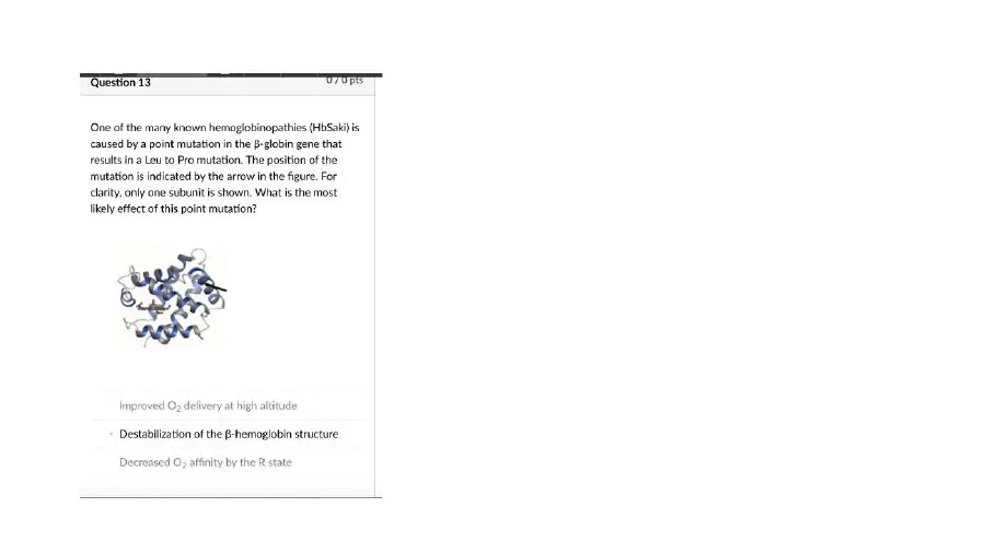
In a haemoglobin variant, a leucine inside an α-helix of the β-globin chain is replaced by proline. What is the most likely effect?
A. The helix is disrupted, destabilising the β-globin fold
B. A new disulfide bond rigidifies the helix
C. Oxygen delivery improves at high altitude
AnswerA. The helix is disrupted, destabilising the β-globin fold
WhyProline is a helix breaker, so Leu → Pro inside a helix disrupts the helix and destabilises the globin.
Explanation
Haemoglobin and myoglobin chains are all-α proteins, so their fold depends on intact helices. Proline's ring cannot adopt helical angles and lacks the N–H the helix needs, so it kinks the helix at the mutation. Unstable haemoglobins of this kind tend to denature and precipitate in red cells.
Disulfide bond: only cysteine forms disulfides; proline cannot.
Better O2 delivery would need a specific shift in O2 affinity, not a broken helix.
Slides (2)
Bridge: helix penalties read off the table; Leu→Pro kinks a globin helixMade for this pageLecture: proline the helix breaker; helix-propensity table; aromatics in β-sheetsBiochemistry Primer 8 · Protein structure · slide 12
L72Learning
Why are tryptophan, tyrosine and phenylalanine often found in β-sheets rather than α-helices?
A. Their rings form the hydrogen bonds between strands
B. The extended sheet gives their large ring side chains room
C. Their rings form covalent cross-links between strands
AnswerB. The extended sheet gives their large ring side chains room
WhyLarge-ring residues (Trp, Tyr, Phe) fit β-sheets because the extended sheet, with side chains above and below, gives them space.
Explanation
In a helix the side chains crowd together around the tight spiral; bulky rings clash. In a β-sheet the chain is extended and successive side chains alternate above and below the sheet, so there is more room.
Rings form the inter-strand hydrogen bonds: sheet hydrogen bonds are backbone C=O to N–H.
Covalent cross-links: only cysteine disulfides cross-link chains covalently.
Note: the same lecture also says 'small amino acids are common, large amino acids destabilise β-sheets' (true for very tightly stacked sheets such as silk). The point tested is the slide's: aromatic residues favour sheets over helices.
Slides (2)
Lecture: proline the helix breaker; helix-propensity table; aromatics in β-sheetsBiochemistry Primer 8 · Protein structure · slide 12Preface: which residues favour helices, break them, build sheets and turnsMade for this page
L73LearningHigh yield
What is a β-turn, and which amino acids usually build it?
A. A tight U-turn built mainly from cysteine and methionine
B. A tight U-turn built mainly from tryptophan and tyrosine
C. A tight U-turn built mainly from glycine and proline
AnswerC. A tight U-turn built mainly from glycine and proline
WhyA β-turn (bend) lets the chain reverse direction in a tight U-turn; it is usually built from glycine (smallest R group) and proline (kink).
Explanation
A β-turn spans four residues and lets the chain fold back on itself, connecting antiparallel β-strands and making the protein compact. Glycine's tiny R group allows the sharp angles, and proline's ring naturally kinks the chain.
Cys and Met: cysteine makes disulfide bonds and methionine is the start residue; neither is a turn-former.
Trp and Tyr: bulky aromatic rings are too big for a tight turn.
Slides (1)
Lecture: β-turn, a tight U-turn built from glycine and prolineBiochemistry Primer 8 · Protein structure · slide 15
L74LearningHigh yield
An Alzheimer-disease protein that is mostly β-sheet is treated with a drug and becomes mostly α-helix. Which bonds have been reorganised?
A. Backbone hydrogen bonds
B. Peptide bonds
C. Disulfide bonds
AnswerA. Backbone hydrogen bonds
WhyChanging β-sheet → α-helix rearranges backbone hydrogen bonds (inter-strand → intrachain n to n + 4); the sequence and peptide bonds are unchanged.
Explanation
Both helix and sheet are secondary structures held by C=O···H–N hydrogen bonds. In a sheet they link adjacent strands; in a helix they link residues four apart in the same chain. Converting one to the other breaks one set and forms the other, without touching covalent bonds. The reverse switch (helix → sheet) is what happens in amyloid and prion diseases.
Peptide bonds make the primary sequence; breaking them would cut the protein.
Disulfide bonds are tertiary side-chain cross-links, not part of secondary structure.
Slides (1)
Bridge: helix-to-sheet switches rearrange only backbone hydrogen bondsMade for this page
Tertiary and quaternary interactions
L75Learning
What are domains? Give an all-α and a β-barrel example.
A. Packed secondary elements within a tertiary fold All-α: myoglobin and haemoglobin chains β-barrel: green fluorescent protein
B. Separate polypeptide chains within a quaternary assembly All-α: myoglobin and haemoglobin chains β-barrel: green fluorescent protein
C. Packed secondary elements within a tertiary fold All-α: green fluorescent protein β-barrel: myoglobin and haemoglobin chains
AnswerA. Packed secondary elements within a tertiary fold All-α: myoglobin and haemoglobin chains β-barrel: green fluorescent protein
WhyDomains are combinations of secondary elements (motifs) packed into compact units of the tertiary fold: α domain = myoglobin/haemoglobin chains; β-can = green fluorescent protein.
Explanation
Tertiary structure is the overall fold of a chain. Within it, helices and strands assemble into motifs, and motifs into domains that often fold independently and carry one function. The globin fold is eight α-helices; green fluorescent protein is eleven β-strands forming a barrel ('β-can') around its chromophore.
Separate chains are subunits (quaternary), not domains.
Swapped examples: globins contain no β-sheet.
Slides (1)
Lecture: tertiary fold; α domain (myoglobin, haemoglobin), β-can (green fluorescent protein)Biochemistry Primer 8 · Protein structure · slide 17
L76LearningHigh yield
Which five interactions stabilise tertiary structure?
A. Hydrophobic interactions Phosphodiester bonds Ionic bonds Disulfide bonds Metal ions
WhyTertiary structure is held by hydrophobic interactions, hydrogen bonds, ionic bonds, disulfide bonds and metal-ion complexes, all between R groups.
Explanation
Four are non-covalent; the disulfide bond is the one covalent cross-link. Mnemonic 'HHIDM': Hydrophobic, Hydrogen, Ionic, Disulfide, Metal.
Phosphodiester bonds join nucleotides in DNA/RNA.
Peptide and glycosidic bonds: peptide bonds make primary structure; glycosidic bonds join sugars.
Quaternary structure uses the same forces minus the disulfide bond (non-covalent only).
Slides (2)
Lecture: tertiary structure stabilised by five side-chain interactionsBiochemistry Primer 8 · Protein structure · slide 18Preface: the five tertiary interactions with the lecture's example for eachMade for this page
L77LearningHigh yield
Which bond does NOT stabilise a protein's tertiary structure?
A. Peptide bonds
B. Ionic bonds
C. Metal-ion complexes
AnswerA. Peptide bonds
WhyPeptide bonds form the primary sequence; tertiary structure is held by interactions between R groups.
Explanation
Tertiary forces act between side chains that may be far apart in the sequence but close in space: hydrophobic interactions, hydrogen bonds, ionic bonds (salt bridges), disulfide bonds and metal-ion coordination. Peptide bonds link neighbouring residues along the backbone and are the same whether the protein is folded or unfolded; denaturing a protein breaks the tertiary forces but leaves the peptide bonds intact.
Ionic bonds, e.g. Asp–Lys, are a tertiary force.
Metal ions, e.g. zinc in zinc fingers, are a tertiary force.
Slides (2)
Lecture: tertiary structure stabilised by five side-chain interactionsBiochemistry Primer 8 · Protein structure · slide 18Preface: the five tertiary interactions with the lecture's example for eachMade for this page
L78LearningHigh yield
Which pair of side chains would most likely form a hydrophobic interaction inside a folded protein?
A. Serine and threonine
B. Leucine and isoleucine
C. Aspartate and lysine
AnswerB. Leucine and isoleucine
WhyLeucine and isoleucine are nonpolar aliphatic side chains that cluster together, excluding water, in the protein core.
Explanation
Hydrophobic interactions are not true bonds: nonpolar side chains gather because this releases the ordered water that would otherwise surround them (the hydrophobic effect). This is the main driving force of folding. The lecture's example shows isoleucine and leucine side chains packed against each other.
Serine and threonine have –OH groups: they form hydrogen bonds.
Aspartate and lysine carry opposite charges: they form an ionic bond.
Slides (1)
Lecture: hydrophobic interaction between leucine and isoleucine side chainsBiochemistry Primer 8 · Protein structure · slide 19
L79Learning
In a folded protein, a serine hydroxyl interacts with a glutamate side-chain carboxylate. What type of interaction is this?
A. Ionic bond
B. Disulfide bond
C. Hydrogen bond
AnswerC. Hydrogen bond
WhySerine's –OH donates a hydrogen bond to glutamate's carboxylate oxygen.
Explanation
A hydrogen bond forms when an H on an electronegative atom (O or N) is attracted to a lone pair on another O or N. Serine is polar uncharged, so it cannot form a salt bridge, but its hydroxyl readily hydrogen-bonds with charged or polar partners.
Ionic bond needs two opposite full charges, e.g. aspartate with lysine; serine is uncharged.
Disulfide bond needs two cysteines.
Slides (1)
Lecture: serine–glutamate hydrogen bond; aspartate–lysine ionic bondBiochemistry Primer 8 · Protein structure · slide 20
L80Learning
Which pair of residues would form an ionic bond (salt bridge) in a folded protein?
A. Aspartate and lysine
B. Leucine and valine
C. Lysine and arginine
AnswerA. Aspartate and lysine
WhyAn ionic (electrostatic) bond forms between oppositely charged side chains, e.g. aspartate (−) with lysine (+).
Explanation
At pH 7.4 Asp and Glu carboxylates are −1 and Lys and Arg are +1; when a negative and a positive side chain come close, they attract. Ionic bonds are also called electrostatic interactions or salt bridges, and they are sensitive to pH because pH changes the charges.
Leucine and valine are nonpolar: hydrophobic interaction.
Lysine and arginine are both positive: they repel.
Slides (1)
Lecture: serine–glutamate hydrogen bond; aspartate–lysine ionic bondBiochemistry Primer 8 · Protein structure · slide 20
L81LearningHigh yield
Which tertiary interaction is covalent, which residues form it, and which protein is the lecture example?
A. Disulfide bond, between methionines; e.g. insulin
B. Disulfide bond, between cysteines; e.g. insulin
C. Disulfide bond, between serines; e.g. myoglobin
AnswerB. Disulfide bond, between cysteines; e.g. insulin
WhyThe disulfide bond (–S–S–) is the covalent tertiary link, formed by oxidising two cysteine –SH groups; insulin's A and B chains are joined this way.
Explanation
Two cysteine sulfhydryls are oxidised to a disulfide (the paired residue is sometimes called cystine). Disulfides are common in secreted and extracellular proteins (insulin, immunoglobulins), where they lock the fold against denaturation. Insulin has two interchain disulfides between its A and B chains and one within the A chain.
Methionine contains sulfur, but as a thioether with no free –SH, so it cannot form disulfides.
Serine has –OH, not –SH; myoglobin has no disulfide bonds.
Slides (1)
Lecture: disulfide bonds in insulin, the covalent cross-linkBiochemistry Primer 8 · Protein structure · slide 21
L82LearningHigh yield
Which two metal-ion examples stabilise protein folds in the lecture?
A. Zinc in the zinc fingers of steroid-hormone receptors Copper in plastocyanin
B. Magnesium in the zinc fingers of steroid-hormone receptors Iron in plastocyanin
C. Iron in the zinc fingers of steroid-hormone receptors Copper in haemoglobin
AnswerA. Zinc in the zinc fingers of steroid-hormone receptors Copper in plastocyanin
WhyZinc holds the zinc-finger domain of steroid-hormone receptors; copper sits in plastocyanin's binding domain.
Explanation
A metal ion coordinates side chains from different parts of the chain (in zinc fingers, cysteine and often histidine side chains), pulling them together and fixing the fold. Steroid-hormone receptors (glucocorticoid, vitamin D, oestrogen receptors) use zinc fingers to bind DNA. This is a tertiary-structure interaction.
Magnesium or iron in a zinc finger: the name gives the metal away.
Copper in haemoglobin: haemoglobin's metal is iron in haem, and it carries O2; plastocyanin is the copper protein.
Slides (1)
Lecture: metal ions: zinc fingers of steroid receptors, copper in plastocyaninBiochemistry Primer 8 · Protein structure · slide 22
L83LearningHigh yield
Which two proteins does the lecture use as examples of quaternary structure?
A. Myoglobin: 2α + 2β Insulin: hexamer around zinc
B. Haemoglobin: 2α + 2β Insulin: hexamer around zinc
C. Haemoglobin: 2α + 2γ Insulin: dimer around copper
AnswerB. Haemoglobin: 2α + 2β Insulin: hexamer around zinc
WhyHaemoglobin (2α + 2β tetramer) and the insulin hexamer (six insulin molecules around zinc ions) have quaternary structure.
Explanation
Quaternary structure needs more than one polypeptide subunit; the subunits can be identical (insulin hexamer) or different (haemoglobin's α and β). Insulin is stored in pancreatic β-cell granules as a zinc-bound hexamer and acts as a monomer.
Myoglobin is a single chain (monomer): no quaternary structure.
2α + 2γ is fetal haemoglobin, and insulin's metal is zinc, not copper.
Slides (1)
Lecture: quaternary structure: haemoglobin 2α2β, insulin hexamer around zincBiochemistry Primer 8 · Protein structure · slide 23
L84Learning
How can the subunits of a quaternary protein behave relative to one another?
A. Only independently, never cooperatively
B. Independently, or cooperatively as in haemoglobin
C. Independently, or cooperatively as in myoglobin
AnswerB. Independently, or cooperatively as in haemoglobin
WhySubunits may act independently or cooperatively; haemoglobin's four subunits bind O2cooperatively.
Explanation
In cooperative binding, O2 binding to one haemoglobin subunit shifts the tetramer from the T (tense) to the R (relaxed) state and raises the affinity of the remaining subunits. That produces the sigmoidal O2-dissociation curve, which lets haemoglobin load O2 in the lungs and unload it in tissues.
Only independently ignores cooperativity, the main reason haemoglobin is a tetramer.
Myoglobin is a monomer; its O2 curve is hyperbolic, not cooperative.
Slides (1)
Lecture: subunits held non-covalently; act independently or cooperativelyBiochemistry Primer 8 · Protein structure · slide 24
Folding, chaperones and misfolding
L85Learning
According to the lecture, what determines a protein's tertiary structure?
A. Its primary sequence
B. The chaperones that fold it
C. The order in which its disulfide bonds form
AnswerA. Its primary sequence
WhyThe tertiary structure is determined by the primary sequence: the side chains' chemistry decides how the chain folds.
Explanation
The amino acid sequence encodes the fold: which residues are hydrophobic (to be buried), which are charged or polar (to face water), where prolines and glycines put turns. A denatured protein can often refold on its own when the denaturant is removed (Anfinsen's experiment).
Chaperones help a chain reach its native fold faster and avoid aggregation, but they do not encode the final shape.
Disulfide bonds stabilise the fold once it forms; which cysteines pair is itself set by the sequence.
Slides (1)
Lecture: sequence sets the fold; hydrophobic effect drives it to minimal free energyBiochemistry Lecture 22 · Globular proteins (haemoglobin) · slide 6
L86LearningHigh yield
What is the main driving force of protein folding?
A. Hydrogen bonding, gathering polar side chains in the core away from water
B. The hydrophobic effect, gathering nonpolar side chains on the surface toward water
C. The hydrophobic effect, gathering nonpolar side chains in the core away from water
AnswerC. The hydrophobic effect, gathering nonpolar side chains in the core away from water
WhyThe hydrophobic effect drives folding: nonpolar side chains collapse into the core, away from water, an entropy-driven process.
Explanation
Water forms ordered 'cages' around exposed nonpolar groups. Burying those groups releases the water, raising the entropy of the solvent; that gain outweighs the loss of chain flexibility, so folding is favourable. The folded protein settles at its lowest free energy. Hydrogen bonds, ionic interactions and van der Waals forces then fine-tune the fold.
Hydrogen bonding stabilises the fold but is not the main driver; polar side chains mostly stay on the surface.
Nonpolar on the surface is the reverse of what happens in a soluble protein.
Slides (1)
Lecture: sequence sets the fold; hydrophobic effect drives it to minimal free energyBiochemistry Lecture 22 · Globular proteins (haemoglobin) · slide 6
L87Learning
At what energy state does a correctly folded protein settle?
A. Its highest free energy
B. Its lowest free energy
C. Its highest chain entropy
AnswerB. Its lowest free energy
WhyProteins fold to minimise free energy; the native fold is the most stable (lowest-energy) arrangement.
Explanation
Folding is spontaneous because the native state has lower free energy (ΔG < 0) than the unfolded chain, mainly thanks to the hydrophobic effect. Folding pathways can get trapped in local energy minima, and chaperones use ATP to help chains escape them.
Highest free energy would be the least stable state.
Highest chain entropy describes the unfolded chain; folding lowers the chain's own entropy (offset by the water's gain).
Slides (1)
Lecture: sequence sets the fold; hydrophobic effect drives it to minimal free energyBiochemistry Lecture 22 · Globular proteins (haemoglobin) · slide 6
L88Learning
What are chaperones, and what energy source do they use?
A. Ubiquitin ligases that tag misfolded proteins; they use ATP
B. Heat shock proteins (Hsp) that assist folding; they use ATP
C. Heat shock proteins (Hsp) that assist folding; they use GTP
AnswerB. Heat shock proteins (Hsp) that assist folding; they use ATP
WhyChaperones are heat shock proteins (Hsp) that help proteins fold correctly; they hydrolyse ATP.
Explanation
Their name comes from their induction by heat and other stresses that make proteins unfold. ATP hydrolysis drives repeated cycles of binding and release, which helps a folding chain escape local energy minima and avoid aggregation.
Ubiquitin ligases tag proteins for proteasomal degradation; they are the disposal system, not the folding helpers.
GTP is used by G proteins and in translation, not by Hsp70/Hsp60 folding cycles.
Slides (2)
Lecture: chaperones: Hsp70 binds hydrophobic regions, Hsp60 encapsulates; ATPBiochemistry Lecture 22 · Globular proteins (haemoglobin) · slide 7Bridge: Hsp70 holds, Hsp60 is a chamber; both heat shock proteins using ATPMade for this page
L89Learning
What does Hsp70 do?
A. Cleaves exposed hydrophobic stretches that would aggregate
B. Encloses the folding chain in a chamber and acts as a template
C. Binds and stabilises exposed hydrophobic stretches that would aggregate
AnswerC. Binds and stabilises exposed hydrophobic stretches that would aggregate
WhyHsp70binds and stabilises hydrophobic regions of a new or unfolded chain that would otherwise aggregate or misfold.
Explanation
A newly made chain leaving the ribosome exposes hydrophobic stretches before the rest of the protein exists to bury them. Hsp70 clamps onto those stretches (with Hsp40 helping), using ATP, and releases them in cycles so the chain can fold.
Cleaves: chaperones do not cut proteins; proteases do.
Encloses as a template is Hsp60 (the barrel-shaped chaperonin).
Hook: Hsp70 = holds the chain (binds); Hsp60 = a 'chamber' around it.
Slides (2)
Lecture: chaperones: Hsp70 binds hydrophobic regions, Hsp60 encapsulates; ATPBiochemistry Lecture 22 · Globular proteins (haemoglobin) · slide 7Bridge: Hsp70 holds, Hsp60 is a chamber; both heat shock proteins using ATPMade for this page
L90Learning
What does Hsp60 do?
A. Encapsulates the folding chain and acts as a template
B. Encapsulates the misfolded chain and degrades it
C. Binds exposed hydrophobic stretches as the chain leaves the ribosome
AnswerA. Encapsulates the folding chain and acts as a template
WhyHsp60 (a chaperonin) encapsulates a folding peptide and acts as a template, repeatedly binding, releasing and stabilising parts of it.
Explanation
Hsp60 forms a barrel-shaped chamber. The chain folds inside this protected space, away from other proteins it might stick to, with ATP-driven cycles of opening and closing.
Degrades: degradation is the job of the proteasome, not chaperonins.
Binds hydrophobic stretches at the ribosome is Hsp70.
Slides (2)
Lecture: chaperones: Hsp70 binds hydrophobic regions, Hsp60 encapsulates; ATPBiochemistry Lecture 22 · Globular proteins (haemoglobin) · slide 7Bridge: Hsp70 holds, Hsp60 is a chamber; both heat shock proteins using ATPMade for this page
L91Learning
What two causes can make a protein misfold?
A. Glycosylation, or a fault in the folding process
B. Phosphorylation, or ubiquitination
C. A mutation in the sequence, or a fault in the folding process
AnswerC. A mutation in the sequence, or a fault in the folding process
WhyProteins misfold because of a mutation in the amino acid sequence or a disruption of the normal folding process.
Explanation
A mutation can change the sequence so the native fold is no longer the most stable state (e.g. familial prion disease, where the mutant protein folds into PrPSc more easily). Even a normal sequence can misfold if folding is disrupted (stress, chaperone overload, ageing).
Glycosylation and phosphorylation are normal post-translational modifications.
Ubiquitination marks misfolded proteins for destruction; it is a consequence, not a cause.
Slides (1)
Lecture: causes of misfolding, usual degradation, amyloid in Alzheimer and ParkinsonBiochemistry Lecture 22 · Globular proteins (haemoglobin) · slide 8
L92Learning
What usually happens to a misfolded protein?
A. It is secreted from the cell
B. It is stored as amyloid plaque
C. It is degraded
AnswerC. It is degraded
WhyMisfolded proteins are usually degraded (ubiquitin–proteasome or autophagy–lysosome); only some escape to form aggregates.
Explanation
Chaperones first try to refold a misfolded chain. If they fail, it is tagged with ubiquitin and destroyed by the proteasome, or aggregates are removed by autophagy. Disease arises when misfolded proteins resist this clearance and form insoluble aggregates or toxic fibrils.
Secreted: quality control in the endoplasmic reticulum holds back misfolded secretory proteins rather than exporting them.
Amyloid plaque is the pathological exception, not the usual fate.
Slides (1)
Lecture: causes of misfolding, usual degradation, amyloid in Alzheimer and ParkinsonBiochemistry Lecture 22 · Globular proteins (haemoglobin) · slide 8
L93LearningHigh yield
What secondary structure do amyloid-forming proteins adopt, and what do they form?
WhyMisfolded amyloid proteins adopt mainly β-sheet structure, oligomerise and form insoluble amyloid fibrils.
Explanation
In amyloid, β-strands from many molecules stack into long, protease-resistant fibrils, held by inter-strand hydrogen bonds. Amyloid β, α-synuclein, TDP-43, polyglutamine proteins and prions all follow this pattern.
Soluble monomers only: fibrils are insoluble aggregates; that is why they deposit as plaques.
α-Helix: the disease change is helix → sheet; a drug that pushes the protein back to α-helix is the therapeutic idea.
Aβ is a 40- or 42-residue fragment cut from amyloid precursor protein (APP). It spontaneously adopts β-sheet structure and aggregates into fibrils that accumulate as plaques between neurons.
Disease
Protein
Alzheimer
Amyloid β
Parkinson
α-Synuclein
ALS
TDP-43
Huntington
Polyglutamine (huntingtin)
Slides (3)
Review: amyloid diseases: Aβ, α-synuclein, TDP-43, polyglutamine (Huntington)Biochemistry interactive session week 10 (pre, 2026) · slide 7Bridge: each misfolding disease with its aggregating proteinMade for this pageLecture: causes of misfolding, usual degradation, amyloid in Alzheimer and ParkinsonBiochemistry Lecture 22 · Globular proteins (haemoglobin) · slide 8
L95Learning
Which protein forms the amyloid in Parkinson disease?
A. Huntingtin
B. α-Synuclein
C. Amyloid β (Aβ)
AnswerB. α-Synuclein
WhyParkinson disease → α-synuclein aggregates (seen as Lewy bodies).
Explanation
α-Synuclein is a neuronal protein that misfolds into β-sheet-rich aggregates inside dopaminergic neurons of the substantia nigra.
Huntingtin with an expanded polyglutamine tract causes Huntington disease.
Amyloid β is the Alzheimer protein.
Slides (3)
Review: amyloid diseases: Aβ, α-synuclein, TDP-43, polyglutamine (Huntington)Biochemistry interactive session week 10 (pre, 2026) · slide 7Bridge: each misfolding disease with its aggregating proteinMade for this pageLecture: causes of misfolding, usual degradation, amyloid in Alzheimer and ParkinsonBiochemistry Lecture 22 · Globular proteins (haemoglobin) · slide 8
L96Learning
Which protein forms the aggregates in amyotrophic lateral sclerosis (ALS)?
A. α-Synuclein
B. TDP-43
C. Amyloid β (Aβ)
AnswerB. TDP-43
WhyALS → TDP-43 aggregates.
Explanation
TDP-43 is an RNA/DNA-binding protein that normally lives in the nucleus; in ALS it misfolds and aggregates in the cytoplasm of motor neurons.
α-Synuclein: Parkinson disease.
Amyloid β: Alzheimer disease.
Slides (2)
Review: amyloid diseases: Aβ, α-synuclein, TDP-43, polyglutamine (Huntington)Biochemistry interactive session week 10 (pre, 2026) · slide 7Bridge: each misfolding disease with its aggregating proteinMade for this page
L97Learning
Huntington disease belongs to which class of protein-misfolding disease?
A. Polyglutamine (polyQ) diseases
B. Prion diseases
C. Synucleinopathies
AnswerA. Polyglutamine (polyQ) diseases
WhyHuntington disease is a polyglutamine (polyQ) disease: an expanded run of glutamines makes huntingtin aggregate.
Explanation
An expanded CAG repeat in the gene adds a long run of glutamine residues to huntingtin. The polyglutamine tract promotes β-sheet aggregation in neurons. ('Q' is the one-letter code for glutamine.)
Prion diseases are transmissible spongiform encephalopathies caused by PrPSc.
Review: amyloid diseases: Aβ, α-synuclein, TDP-43, polyglutamine (Huntington)Biochemistry interactive session week 10 (pre, 2026) · slide 7Bridge: each misfolding disease with its aggregating proteinMade for this page
L98Learning
Huntington disease is called a 'polyQ' disease. What does Q stand for?
A. Glutamate
B. Asparagine
C. Glutamine
AnswerC. Glutamine
WhyQ is the one-letter code for glutamine; huntingtin carries an expanded run of glutamine residues.
Explanation
The expanded CAG repeat in the gene codes for glutamine (CAG = Gln), producing a long polyglutamine tract that makes the protein aggregate.
Glutamate is E (the acid; glutamine is its amide).
Asparagine is N.
Slides (2)
Review: amyloid diseases: Aβ, α-synuclein, TDP-43, polyglutamine (Huntington)Biochemistry interactive session week 10 (pre, 2026) · slide 7Bridge: each misfolding disease with its aggregating proteinMade for this page
L99Learning
How is Aβ produced in Alzheimer disease?
A. Amyloid precursor protein (APP) cut by α- and β-secretase → Aβ 40/42 → neurofibrillary tangles
B. Amyloid precursor protein (APP) cut by α- and γ-secretase → Aβ 40/42 → amyloid plaques
C. Amyloid precursor protein (APP) cut by β- and γ-secretase → Aβ 40/42 → amyloid plaques
AnswerC. Amyloid precursor protein (APP) cut by β- and γ-secretase → Aβ 40/42 → amyloid plaques
Whyβ- then γ-secretase cut APP to release Aβ 40/42, a neurotoxic peptide that aggregates into plaques.
Explanation
APP is a membrane protein. In the amyloidogenic pathway β-secretase cuts outside the membrane and γ-secretase cuts within it, releasing Aβ; the 42-residue form is the most aggregation-prone. Aβ adopts β-sheet structure and forms extracellular fibrils and plaques.
α + β, ending in tangles: tangles are intracellular tau, not Aβ.
α + γ: α-secretase cuts inside the Aβ region, so it prevents Aβ forming.
Slides (2)
Review: APP cut by β- and γ-secretase releases Aβ, which forms plaquesBiochemistry interactive session week 10 (pre, 2026) · slide 6Review: Alzheimer: α-secretase prevents Aβ; β/γ cut gives Aβ40/42; ApoE sporadicBiochemistry interactive session week 10 (pre, 2026) · slide 5
L100Learning
What does α-secretase do to amyloid precursor protein?
A. Makes the pathological cut that releases Aβ
B. Makes the usual cut that releases Aβ
C. Makes the usual cut that prevents Aβ forming
AnswerC. Makes the usual cut that prevents Aβ forming
Whyα-Secretase handles ~90% of APP and cuts inside the Aβ sequence, so it prevents Aβ from forming.
Explanation
Cutting through the middle of the Aβ region destroys the sequence that would become the toxic peptide, so the α-secretase (non-amyloidogenic) route is protective. Only when β- and γ-secretase act instead is intact Aβ released.
Pathological cut: that is β- plus γ-secretase.
Usual cut that releases Aβ contradicts itself: the usual (α) cut destroys Aβ.
Which gene is linked to sporadic Alzheimer disease?
A. SNCA
B. APP
C. APOE
AnswerC. APOE
WhySporadic (late-onset) Alzheimer disease is linked to the APOE gene (apolipoprotein E, ε4 allele).
Explanation
The review slide splits Alzheimer disease into sporadic (ApoE gene) and familial (the 'amyloid hypothesis': APP and its processing). The ε4 allele of apolipoprotein E raises risk and lowers age of onset.
APP mutations (with presenilins) cause familial early-onset disease.
How does the disease-causing prion protein PrPSc differ from normal PrPC?
A. Same amino acid sequence, extra disulfide bonds
B. Different amino acid sequence, same conformation
C. Same amino acid sequence, different conformation
AnswerC. Same amino acid sequence, different conformation
WhyPrPSc has the same amino acid sequence as normal PrPC but a different 3-D conformation (β-sheet-rich).
Explanation
The prion protein gene is a normal part of the genome. Disease comes from a change of shape alone: α-helical PrPC refolds into β-sheet-rich PrPSc. Prions are proteinaceous infectious agents that cause the transmissible spongiform encephalopathies.
Extra disulfide bonds: the difference is conformation, not covalent change.
Different sequence: sporadic and acquired cases have the normal sequence.
Slides (2)
Lecture: prions: PrPSc has PrPC's sequence, new shape, resists proteases, templatesBiochemistry Lecture 22 · Globular proteins (haemoglobin) · slide 9Bridge: prion conformation, origins and host species in one slideMade for this page
L103Learning
What two properties make PrPSc pathogenic?
A. It forms protease-resistant fibrils It carries its own nucleic acid genome
B. It forms protease-resistant fibrils It templates PrPC into more PrPSc
C. It forms soluble α-helical monomers It templates PrPSc back into PrPC
AnswerB. It forms protease-resistant fibrils It templates PrPC into more PrPSc
WhyPrPSc forms insoluble, protease-resistant fibrils and acts as a template that converts normal PrPC into PrPSc.
Explanation
Each PrPSc molecule recruits PrPC and refolds it, so the abnormal form multiplies exponentially without any genetic material. The aggregates resist proteolysis (like other amyloid fibrils), accumulate and destroy neurons, giving the spongy brain.
Own nucleic acid: prions have none; that is what makes them unique among infectious agents.
Soluble α-helical, templating back to PrPC reverses the direction of the disease.
Slides (1)
Lecture: prions: PrPSc has PrPC's sequence, new shape, resists proteases, templatesBiochemistry Lecture 22 · Globular proteins (haemoglobin) · slide 9
L104Learning
What are the three origins of prion disease, with examples?
A. Acquired (most cases) Inherited: familial Creutzfeldt–Jakob disease (X-linked) Sporadic: mad cow disease, cadaveric growth hormone, kuru
B. Sporadic (most cases) Inherited: familial Creutzfeldt–Jakob disease (autosomal dominant) Acquired: mad cow disease, cadaveric growth hormone, kuru
C. Inherited (most cases) Sporadic: familial Creutzfeldt–Jakob disease (autosomal recessive) Acquired: mad cow disease, cadaveric growth hormone, kuru
WhyPrion disease is mostly sporadic; it can be inherited (familial CJD, autosomal dominant) or acquired by infection (mad cow, cadaveric growth hormone, iatrogenic CJD, kuru).
Explanation
Sporadic: most cases; PrPC converts spontaneously.
Inherited: a point mutation lowers the energy barrier for folding into PrPSc; autosomal dominant.
Acquired: eating infected beef (variant CJD from bovine spongiform encephalopathy), cadaver-derived human growth hormone, contaminated instruments or grafts, ritual cannibalism (kuru).
The wrong options misplace 'most cases' and give the wrong inheritance pattern.
Slides (2)
Lecture: prion origins (sporadic, inherited, infection) and the four host speciesBiochemistry Lecture 22 · Globular proteins (haemoglobin) · slide 10Bridge: prion conformation, origins and host species in one slideMade for this page
L105Learning
Name the prion disease of humans, sheep, cattle and deer.
WhyCJD (humans), scrapie (sheep), bovine spongiform encephalopathy (cattle, 'mad cow'), chronic wasting disease (deer and other cervids).
Explanation
All are transmissible spongiform encephalopathies caused by PrPSc. Hook: sheep scrape themselves against fences; bovine = cattle; deer waste away.
The wrong options rotate scrapie, chronic wasting disease and bovine spongiform encephalopathy among the animals.
Slides (2)
Lecture: prion origins (sporadic, inherited, infection) and the four host speciesBiochemistry Lecture 22 · Globular proteins (haemoglobin) · slide 10Bridge: prion conformation, origins and host species in one slideMade for this page
Fibrous vs globular proteins
L106Learning
How do fibrous and globular proteins differ in shape and role?
A. Fibrous: long and narrow, structural Globular: rounded, functional (catalysis, transport)
B. Fibrous: rounded, functional (catalysis, transport) Globular: long and narrow, structural
C. Fibrous: long and narrow, functional (catalysis, transport) Globular: rounded, structural
AnswerA. Fibrous: long and narrow, structural Globular: rounded, functional (catalysis, transport)
WhyFibrous proteins are long, narrow and structural (strength, support); globular proteins are rounded and functional (catalysis, transport).
Explanation
Fibrous
Globular
Shape
Long, narrow
Rounded, spherical
Role
Structural
Functional
In water
Generally insoluble
Generally soluble
Sequence
Repetitive
Irregular
Heat, pH
Less sensitive
More sensitive
Examples
Collagen, keratin, elastin, myosin, fibrin, actin
Haemoglobin, insulin, catalase, immunoglobulins
Slides (2)
Case session: fibrous vs globular: shape, role, solubility, sequence, stability, examplesBiochemistry Week 10 CV · Haemoglobinopathies · slide 6Bridge: sorting the examples, and why each class behaves as it doesMade for this page
L107Learning
How do fibrous and globular proteins differ in water solubility?
A. Fibrous: generally soluble Globular: generally soluble
B. Fibrous: generally soluble Globular: generally insoluble
C. Fibrous: generally insoluble Globular: generally soluble
AnswerC. Fibrous: generally insoluble Globular: generally soluble
WhyFibrous proteins are generally insoluble in water; globular proteins are generally soluble.
Explanation
Globular proteins bury hydrophobic residues and display polar ones, so they dissolve in plasma and cytosol, where they work. Fibrous proteins assemble into long cables and sheets (collagen fibrils, keratin filaments) that must stay solid to bear load.
The wrong options swap the pairing or make both the same.
Slides (2)
Case session: fibrous vs globular: shape, role, solubility, sequence, stability, examplesBiochemistry Week 10 CV · Haemoglobinopathies · slide 6Bridge: sorting the examples, and why each class behaves as it doesMade for this page
L108Learning
How do fibrous and globular proteins differ in amino acid sequence?
A. Fibrous: repetitive Globular: repetitive
B. Fibrous: irregular Globular: repetitive
C. Fibrous: repetitive Globular: irregular
AnswerC. Fibrous: repetitive Globular: irregular
WhyFibrous proteins have a repetitive sequence; globular proteins have an irregular one.
Explanation
A repeating sequence builds a repeating structure, ideal for a long cable (collagen's Gly-X-Y repeat makes the triple helix). A globular protein needs a unique, irregular sequence to fold into a specific shape with a binding or active site.
The wrong options swap the pairing or make both repetitive.
Slides (2)
Case session: fibrous vs globular: shape, role, solubility, sequence, stability, examplesBiochemistry Week 10 CV · Haemoglobinopathies · slide 6Bridge: sorting the examples, and why each class behaves as it doesMade for this page
L109Learning
How do fibrous and globular proteins differ in sensitivity to heat and pH?
A. Fibrous: equally sensitive Globular: equally sensitive
B. Fibrous: more sensitive Globular: less sensitive
C. Fibrous: less sensitive Globular: more sensitive
AnswerC. Fibrous: less sensitive Globular: more sensitive
WhyFibrous proteins are less sensitive to heat and pH; globular proteins are more sensitive (they denature more easily).
Explanation
A globular protein's function depends on a delicate tertiary fold held by many weak interactions; heat or a pH change that alters side-chain charges can unfold it (denaturation) and destroy its activity. Fibrous proteins are built for mechanical stability and keep their structure better.
The wrong options reverse the pairing or make both equal.
Slides (2)
Case session: fibrous vs globular: shape, role, solubility, sequence, stability, examplesBiochemistry Week 10 CV · Haemoglobinopathies · slide 6Bridge: sorting the examples, and why each class behaves as it doesMade for this page
L110Learning
Which list contains only fibrous proteins?
A. Collagen, insulin, elastin, actin, myosin, catalase
B. Collagen, keratin, elastin, actin, myosin, fibrin
C. Myoglobin, keratin, albumin, actin, myosin, fibrin
These form structural fibres: connective tissue (collagen, elastin), hair and skin (keratin), muscle filaments (actin, myosin) and clots (fibrin).
Insulin and catalase are globular.
Myoglobin and albumin are globular.
Textbook note: actin monomers (G-actin) are globular and polymerise into fibrous F-actin; the lecture table lists actin as fibrous.
Slides (2)
Case session: fibrous vs globular: shape, role, solubility, sequence, stability, examplesBiochemistry Week 10 CV · Haemoglobinopathies · slide 6Bridge: sorting the examples, and why each class behaves as it doesMade for this page
L111Learning
Which list contains only globular proteins?
A. Haemoglobin, insulin, catalase, immunoglobulins
B. Keratin, insulin, catalase, immunoglobulins
C. Haemoglobin, insulin, collagen, immunoglobulins
Each is a compact, soluble protein with a specific function: O2 transport, hormone signalling, enzyme (breaks down H2O2), antigen binding.
Keratin is fibrous.
Collagen is fibrous.
Slides (2)
Case session: fibrous vs globular: shape, role, solubility, sequence, stability, examplesBiochemistry Week 10 CV · Haemoglobinopathies · slide 6Bridge: sorting the examples, and why each class behaves as it doesMade for this page
1High-yield5 sources
A group of researchers is studying the pathogenesis of Alzheimer’s disease. A protein isolated from brain tissue of an affected patient has mostly beta pleated sheet structure. A sample of new medication is applied to the protein, and the prevailing structure changes to alpha helical structure. This conformational change is the result of reorganization of which of the following:
A. Ionic interaction
B. Hydrophobic interaction
C. Peptide bond
D. Hydrogen bond
E. Disulfide bonds
AnswerD. Hydrogen bond
Correct key: D. Hydrogen bond. Both α-helix and β-sheet are secondary structures held by backbone C=O···H-N hydrogen bonds, so a β → α change reorganizes the H-bonds; all sources key D.
WhyBoth α-helix and β-sheet are secondary structures held by backbone C=O···H–N hydrogen bonds, so converting β-sheet to α-helix means rearranging those H-bonds, not breaking peptide or disulfide bonds.
2High-yield4 sources
A protein believed to play a role in signal transduction and cellular response to thyroid stimulating hormone is studied. Special attention is paid to a region of this protein that contains several alpha helical regions each composed of approximately 20 amino acid residues –consisting primarily of valine,leucine and isoleucine. Which of the following function does this region most likely to perform?
Show figure
A. Phosphorylating Tyrosine residues
B. Binding to intranuclear DNA
C. Spanning the cellular membrane
D. Interacting with signaling substances
E. Interacting with metal ions in transporting proteins
AnswerC. Spanning the cellular membrane
Correct key: C. Spanning the cellular membrane. Helices of about 20 hydrophobic residues (Val, Leu, Ile) are the classic transmembrane segments, as in the 7-TM TSH receptor (a GPCR); all sources key C.
WhyRepeated α-helices of about 20 hydrophobic residues (Val, Leu, Ile) are classic transmembrane segments, as in the seven-pass TSH receptor (a GPCR) that spans the lipid bilayer.
3High-yield
What is the net charge of the following oligopeptide at neutral pH?
Glu - Asp - Arg - Lys - Lys
A. +1
B. -2
C. -1
D. 0
E. +2
Answer supplied — the source gives no keyA. +1 — At pH 7: N-terminal NH3+ (+1), C-terminal COO− (−1), Glu (−1), Asp (−1), Arg (+1), Lys (+1), Lys (+1) → net +1.
WhyAt pH 7 the termini cancel (N-terminal NH₃⁺ +1, C-terminal COO⁻ −1); Glu and Asp give −2 and Arg, Lys, Lys give +3, so the net charge is +1.
4High-yield
Which of the following peptide will have net positive electrical charge at physiological pH of 7.4?
A. Glu-Pro-Lys-Val-Leu
B. Ile-Val-Gln-Ser-Arg
C. Ala-Gly-Pro-Gln-Met
D. Met-Thr-Asn-Asp-Gly
E. Phe-Ala-Thr-Tyr-Trp
Answer supplied — the source gives no keyB. Ile-Val-Gln-Ser-Arg — The termini cancel (+1, −1); Arg is the only charged side chain (+1) → net +1. Glu-Pro-Lys-Val-Leu = 0 (Glu −1, Lys +1); Ala-Gly-Pro-Gln-Met and Phe-Ala-Thr-Tyr-Trp = 0; Met-Thr-Asn-Asp-Gly = −1.
WhyThe free termini cancel, and Arg is the only charged side chain (+1), so Ile-Val-Gln-Ser-Arg is net +1; the others are 0 or −1 (Met-Thr-Asn-Asp-Gly).
5High-yield
Which of the following peptide will have net negative electrical charge at physiological pH of 7.4?
A. Met-Gly-Thr-Asn-Asp
B. Ala-Gly-Pro-Gln-met
C. Glu-Pro-Lys-Val-Leu
D. Asp-Glu-Lys-Arg-His
E. Glu -Val-Leu-Pro-Lys
Answer supplied — the source gives no keyA. Met-Gly-Thr-Asn-Asp — The termini cancel; Asp is the only charged side chain (−1) → net −1. Ala-Gly-Pro-Gln-met = 0; Glu-Pro-Lys-Val-Leu and Glu-Val-Leu-Pro-Lys = 0 (Glu −1, Lys +1); Asp-Glu-Lys-Arg-His ≈ 0 (−2 +2; His essentially uncharged at 7.4).
WhyThe termini cancel, and Asp is the only charged side chain (−1), so Met-Gly-Thr-Asn-Asp is net −1; peptides pairing Glu with Lys (or Asp-Glu with Lys-Arg) sum to about 0.
6High-yield
Which of the following amino acids would most likely be found on the surface of a protein molecule?
A. Isoleucine
B. Phenylalanine
C. Arginine
D. Leucine
E. Alanine
Answer supplied — the source gives no keyC. Arginine — Arginine is charged and hydrophilic, so it faces water on the protein surface; Ile, Phe, Leu and Ala are hydrophobic and are buried in the core.
WhyArginine has a charged, hydrophilic guanidinium side chain that faces water on the protein surface; Ile, Phe, Leu and Ala are hydrophobic and buried in the core.
7High-yield
One of the many known hemoglobinopathies (HbSaki) is caused by a point mutation in the β-globin gene that results in a Leu to Pro mutation. The position of the mutation is indicated by the arrow in the figure. For clarity, only one subunit is shown. What is the most likely effect of this point mutation?
A. Decreased O2 affinity by the R state
B. Production of a surface hydrophobic patch
C. Increased number of salt bridges (ion pairs) in the T state
D. Improved O2 delivery at high altitude
E. Destabilization of the β-hemoglobin structure
Answer supplied — the source gives no keyE. Destabilization of the β-hemoglobin structure — The mutated residue lies in an α-helix; proline is a helix breaker (rigid ring, no N–H for the helix H-bond), so Leu→Pro distorts the helix → unstable hemoglobin (Hb Saki, β14 Leu→Pro, is an unstable variant).
WhyThe mutation lies in an α-helix, and proline is a helix breaker (rigid ring, no backbone N–H for H-bonding), so Leu→Pro kinks the helix and gives an unstable hemoglobin.
8High-yield
49-year old woman with chronic myelogenous leukemia has a Thr315Leu substitution mutation in a kinase. The effect of this mutation would best be described as replacing a
A. Polar residue with a hydrophobic one
B. Branched chain residue with an aromatic one
C. Large residue with a small one
D. Hydrophobic residue with a hydrophilic one
E. Negatively charged residue with a neutral one
Answer supplied — the source gives no keyA. Polar residue with a hydrophobic one — Threonine has a polar, uncharged hydroxyl side chain; leucine is a nonpolar, hydrophobic branched-chain residue.
WhyThreonine has a polar, uncharged hydroxyl side chain, while leucine is a nonpolar branched-chain residue, so Thr315Leu swaps a polar residue for a hydrophobic one.
9High-yieldBorderline
In human cells, DNA wraps around and binds to histone proteins to form nucleosomes. What property of the histone proteins allows them to bind tightly to DNA?
A. Spherical tertiary structure
B. Endonuclease activity and covalent bonding with end of a DNA strand
C. Intercalation of aromatic amino acids into the DNA helix
D. Methyl groups that create hydrophobic interactions
E. Positive charges on amino acids
AnswerE. Positive charges on amino acids
WhyHistones are rich in lysine and arginine, which are positively charged at physiological pH and form ionic bonds with the negative phosphates of the DNA backbone.
Explanation from the source
Explanation: Histones contain lysine and arginine amino acids that are positively charged at physiological pH. These amino acids form ionic bonds with the negative phosphates in the phosphodiester backbone of DNA. Histones do not have endonuclease activity.
Needs histone composition/DNA packaging (MSK 1, OUT) plus the Primer fact that Lys/Arg side chains are positively charged at physiological pH (Primers 7/8, IN).
10Borderline2 sources
Collagen is made up of peptide chains containing Glycine-X-Y sequence repeats. These peptides are coiled around one another by which of the following bonds to make up the collagen fiber?
Show figures
A. Hydrophobic interaction
B. Ionic bond
C. Hydrogen bond
D. Peptide bond
E. Disulfide bond
AnswerC. Hydrogen bond
WhyThe three collagen α-chains are held in the triple helix by interchain hydrogen bonds (backbone N–H of glycine to C=O on a neighboring chain); peptide bonds only link residues within each chain.
Collagen triple-helix structure is the MSK 2 collagen lecture (OUT); the tested idea (which bond type holds coiled chains / secondary structure = hydrogen bonds) is Primer 7/8 protein-structure content (IN).
11Borderline
A researcher examines collagen isolated from skin. Each collagen molecule consists of three polypeptide chains wound around one another to form a triple helix. Which amino acid is found at every third position in the collagen polypeptide chain and is essential for tight packing of the triple helix?
A. Alanine
B. Glycine
C. Hydroxylysine
D. Proline
E. Valine
AnswerB. Glycine
WhyCollagen is a repeating Gly-X-Y sequence: every third residue must be glycine, the only residue small enough (side chain = H) to fit in the crowded center of the triple helix. X is often proline, Y hydroxyproline/hydroxylysine.
Explanation from the source
The basic form of collagen usually has repeating motifs of Gly-X-Y, with glycine being most abundant and found approximately in every third position. X is often proline, Y is often hydroxyproline or hydroxylysine. Proline content is unusually high but is not specifically mentioned to be every third position like glycine.
Collagen Gly-X-Y composition is the MSK 2 collagen lecture (OUT); glycine as the smallest amino acid / 'Glycine: collagen' appears on the CP1 amino-acid slide of the Hb CV (IN).
12Borderline
Bonus question: Why is the high glycine content helpful in the tight packing of the collagen fibrils into the triple helix? What is it about the structure of glycine specifically that make it useful?
AnswerBonus Answer: Remember that the main function of fibrous proteins is to have structural - mechanical, and tensile strength - so the tight packing is essential. Glycine is the smallest amino acid, which allows for close association with the surrounding amino acids both within its pro-ɑ-collagen chain and in the formation of the triple helix. Placing glycine at every third position of the aa sequence allows the strands to twist into a tight, stable rope-like structure. Glycines small size comes from its side chain being made up of a single hydrogen atom, which also serves as a means for forming the strong intERchain hydrogen bonds for stabilizing the triple helix. TLDR: glycine is the smallest aa and allows for hydrogen bonding → tight packed and strong, stable fibers.
WhyGlycine's side chain is a single hydrogen atom, so it is the smallest amino acid and fits where the three chains meet in the helix core, letting them pack tightly; its backbone N–H forms the interchain hydrogen bonds that stabilize the triple helix.
Glycine side chain = single H, smallest amino acid (Primer 8, IN), applied to collagen triple-helix packing (MSK 2, OUT).
13Borderline
A point mutation in a gene results in substitution of one amino acid for another. In one patient, alanine is replaced by glycine, whereas in a second patient, serine is replaced by proline. Which of the following correctly classifies these mutations?
A. Both are conservative missense mutations
B. Both are nonconservative missense mutations
C. Alanine → glycine is conservative; serine → proline is nonconservative
D. Alanine → glycine is nonconservative; serine → proline is conservative
E. The first is silent; the second is missense
AnswerC. Alanine → glycine is conservative; serine → proline is nonconservative
WhyAla → Gly swaps one small nonpolar residue for another (conservative); Ser → Pro replaces a polar hydroxyl residue with a rigid imino acid that kinks the backbone (nonconservative). Both change the amino acid, so neither is silent.
Conservative vs nonconservative missense is the MSK 2 genetic-code lecture (OUT); deciding it needs the side-chain classes (Ala/Gly small nonpolar; Ser polar vs Pro cyclic imino) from Primer 8 (IN).
14High-yieldBorderline
A missense mutation replaces multiple glycine residues within the repeating sequence of a collagen polypeptide. The mutation severely disrupts fibril formation. Which property of glycine normally makes it particularly important for collagen structure?
A. Its side chain participates in disulfide bonding
B. Its hydroxyl group stabilizes interchain bonds
C. Its small size allows close packing within the triple helix
D. Its positive charge promotes ionic interactions
E. Its cyclic structure introduces bends into the collagen chain
AnswerC. Its small size allows close packing within the triple helix
WhyCollagen repeats Gly-X-Y, and every third residue must be glycine because only its tiny H side chain fits in the crowded center of the triple helix.
Explanation from the source
Collagen follows Gly-X-Y, and glycine occurs about every third residue. Because glycine is the smallest amino acid, it can fit into the tightly packed center of the triple helix.
Collagen Gly-X-Y packing is the MSK 2 collagen lecture (OUT); the options test glycine's side-chain properties (single H, smallest, not charged/cyclic) from Primer 8 (IN).
15Borderline
A 45-year-old woman undergoes a skin biopsy during an evaluation for tissue fragility. Biochemical analysis reveals a triple-helical protein composed of three levorotatory α-chains wound into a dextrorotatory helix. Amino acid sequencing demonstrates that every third residue is glycine. Which structural feature best explains why glycine is required at every third position?
A. Unique ring structure providing rigid directional bends along the chain
B. High hydrogen bonding capacity across neighboring peptide backbones
C. Smallest steric size allowing tight steric packing in the helix center
D. Positively charged side chain driving electrostatic interchain attraction
AnswerC. Smallest steric size allowing tight steric packing in the helix center
WhyGlycine has the smallest side chain (H), the only one that fits in the cramped center of the collagen triple helix where the three chains touch. Proline gives the ring bends; glycine is not charged.
Collagen triple helix (Gly every third residue) is the MSK 2 collagen lecture (OUT); the options test glycine's side-chain properties (smallest, no ring, uncharged) from Primer 8 (IN).
16High-yield
1. A biochemistry researcher analyzing human structural proteins isolated from a tissue biopsy observes that the constituent amino acid residues share specific stereochemical and structural features. Which of the following statements correctly describes a general structural property of the standard amino acids found in human proteins?
A. All standard amino acids contain an asymmetric α-carbon attached to four different chemical groups
B. All amino acids incorporated into mammalian proteins exist exclusively in the D-stereoisomeric configuration
C. Human proteins are synthesized exclusively from L-amino acids
D. There are over 300 different amino acids commonly incorporated into human protein structures
E. The central α-carbon is covalently linked to two amino groups and one carboxyl group
AnswerC. Human proteins are synthesized exclusively from L-amino acids
WhyAll amino acids in mammalian proteins are L-isomers. The trap is A: glycine (R = H) has no asymmetric α-carbon, so not every standard amino acid is chiral.
Explanation from the source
Choice C is correct: All amino acids that occur in mammalian proteins are L-isomers. Choice A is incorrect: While most standard amino acids have an asymmetric α-carbon, Glycine contains two hydrogen atoms attached to its α-carbon (R = H), making it achiral and non-asymmetric. Choice B is incorrect: Mammalian proteins incorporate L-isomers rather than D-isomers. Choice D is incorrect: Although over 300 amino acids exist in nature, only 20 are commonly found in protein structures. Choice E is incorrect: The basic structural formula of an α-amino acid consists of an α-carbon attached to one α-carboxyl group (COO-), one α-amino group (NH3+), one hydrogen atom, and one variable side chain (R group).
17
2. A medical student is studying the basic chemical anatomy of amino acids to understand how mutations alter protein folding. Which of the following correctly identifies the fundamental chemical components attached to the central α-carbon of a standard amino acid?
A. An α-carboxyl group, an α-amino group, a hydrogen atom, and a side chain (R group)
B. Two α-carboxyl groups, an α-amino group, and a hydroxyl group
C. An α-carboxyl group, two variable R groups, and a phosphate group
D. An α-amino group, a hydrogen atom, an aldehyde group, and a side chain
E. Two α-amino groups, an α-carboxyl group, and a hydrogen atom
AnswerA. An α-carboxyl group, an α-amino group, a hydrogen atom, and a side chain (R group)
WhyEvery standard amino acid has a central α-carbon bonded to four groups: an α-amino group, an α-carboxyl group, a hydrogen atom and a variable R group that gives each amino acid its identity.
Explanation from the source
Choice A is correct: The central α-carbon of a standard amino acid is bonded to an α-carboxyl group, an α-amino group, a hydrogen atom, and a variable side chain (R group). Choice B is incorrect: Amino acids contain one α-carboxyl group and one α-amino group attached to the central α-carbon, not two carboxyl groups or a hydroxyl group. Choice C is incorrect: Standard amino acids possess a single R group attached to the α-carbon, not two R groups or a phosphate group. Choice D is incorrect: The functional groups on the α-carbon include a carboxyl group and an amino group, not an aldehyde group. Choice E is incorrect: A standard amino acid has one α-carboxyl group and one α-amino group, not two amino groups.
18
3. A patient with a point mutation replacing a hydrophobic aromatic residue in an enzyme active site exhibits altered substrate binding. When reviewing aromatic amino acids, which of the following statements regarding Tyrosine (Tyr) and Tryptophan (Trp) is correct?
A. Tyrosine is classified as hydrophilic because its side chain cannot engage in hydrophobic interactions
B. Tyrosine has a polar hydroxyl group that can form hydrogen bonds, but it is classified as mainly hydrophobic
C. The indole nitrogen atom of Tryptophan readily forms interstrand hydrogen bonds in α-helices
D. Tryptophan is a hydrophilic charged basic amino acid due to its indole ring nitrogen
E. Phenylalanine contains a hydroxyl group that makes it more polar than Tyrosine
AnswerB. Tyrosine has a polar hydroxyl group that can form hydrogen bonds, but it is classified as mainly hydrophobic
WhyTyrosine is phenylalanine plus a ring –OH; the hydroxyl can hydrogen bond (and be phosphorylated), but the large aromatic ring dominates, so Tyr is classed as mainly hydrophobic aromatic. Tryptophan is also hydrophobic aromatic, not basic.
Explanation from the source
Choice B is correct: Although Tyrosine contains a polar hydroxyl group capable of hydrogen bonding, it is categorized as mainly hydrophobic. Choice A is incorrect: Tyrosine is classified under hydrophobic aromatic amino acids despite having a polar hydroxyl group. Choice C is incorrect: The indole nitrogen of Tryptophan does not form hydrogen bonds. Choice D is incorrect: Tryptophan is classified as a nonpolar hydrophobic aromatic amino acid, not a hydrophilic basic amino acid. Choice E is incorrect: Tyrosine possesses a hydroxyl group, whereas Phenylalanine lacks one and is purely nonpolar aromatic.
19
4. A clinical geneticist reviews a mutation that introduces a Proline (Pro) residue into a tightly structured globular protein segment. Which structural feature of Proline explains its unique impact on protein conformation?
A. Proline contains a basic guanidinium side chain that carries a net positive charge at physiological pH
B. Proline is a cyclic α-imino acid whose rigid ring structure makes it conformationally restricted
C. Proline possesses a sulfhydryl group that forms covalent disulfide bonds
D. Proline has the smallest R group among all amino acids, allowing maximum conformational flexibility
E. Proline is an acidic amino acid that carries a net negative charge at physiological pH
AnswerB. Proline is a cyclic α-imino acid whose rigid ring structure makes it conformationally restricted
WhyProline's side chain loops back to bond its own α-nitrogen, making it a cyclic α-imino acid. The rigid ring locks backbone rotation and removes the N–H donor, so proline kinks chains and sits in bends.
Explanation from the source
Choice B is correct: Proline is a cyclic α-imino acid whose ring structure restricts its backbone conformation, often placing it in bends of folded protein chains. Choice A is incorrect: Arginine contains a guanidinium side chain, whereas Proline is an imino acid. Choice C is incorrect: Cysteine contains a sulfhydryl (-SH) group that forms disulfide bridges, not Proline. Choice D is incorrect: Glycine has the smallest R group (a single hydrogen atom), granting high flexibility, whereas Proline is conformationally restricted. Choice E is incorrect: Aspartate and Glutamate are acidic amino acids with net negative charges at physiological pH; Proline is nonpolar/imino.
20
5. A medical student analyzes the charge states of amino acid side chains at physiological pH (7.4) to understand serum protein electrophoresis. Which of the following amino acids carries a net negative charge at physiological pH?
A. Lysine
B. Histidine
C. Aspartate
D. Arginine
E. Serine
AnswerC. Aspartate
WhyAspartate (like glutamate) has a side-chain carboxyl with pKa ≈ 4, far below 7.4, so it is deprotonated and negatively charged at physiological pH. Lys, Arg and His are basic; Ser is uncharged polar.
Explanation from the source
Choice C is correct: Aspartate (and Glutamate) are hydrophilic acidic amino acids that carry a net negative charge at physiological pH. Choice A is incorrect: Lysine is a basic amino acid with a net positive charge at physiological pH. Choice B is incorrect: Histidine is a basic amino acid whose imidazole nitrogen is weakly basic at physiological pH, carrying a net positive/neutral charge rather than a net negative charge. Choice D is incorrect: Arginine is a basic amino acid containing a guanidinium group with a net positive charge at physiological pH. Choice E is incorrect: Serine is a hydrophilic uncharged amino acid with an uncharged polar hydroxyl side chain.
21High-yield
6. At physiological pH (7.4), free Alanine exists predominantly in a form where its α-carboxyl group is deprotonated (COO-) and its α-amino group is protonated (NH3+), yielding a net charge of zero. What term defines a neutral molecule containing an equal number of positive and negative charges?
A. Cation
B. Anion
C. Isomer
D. Electrophile
E. Zwitterion
AnswerE. Zwitterion
WhyA zwitterion is a neutral molecule carrying equal numbers of positive and negative charges, like alanine at pH 7.4 with COO⁻ and NH₃⁺.
Explanation from the source
Choice E is correct: A zwitterion is defined as a neutral molecule containing an equal number of positive (+) and negative (-) charges. Choice A is incorrect: A cation is defined as a molecule with a net positive charge. Choice B is incorrect: An anion is defined as a molecule with a net negative charge. Choice C is incorrect: An isomer refers to molecules sharing the same chemical formula but distinct structural arrangements. Choice D is incorrect: An electrophile is an electron-deficient species, which is not the definition of a dipolar neutral ion.
22High-yield
7. A laboratory technician performs a titration curve analysis for Alanine, which possesses two ionizable groups: an α-carboxyl group (pK1 = 2.3) and an α-amino group (pK2 = 9.1). What is the calculated isoelectric point (pI) for Alanine?
A. 2.3
B. 5.7
C. 6.8
D. 9.1
E. 11.4
AnswerB. 5.7
WhyWith two ionizable groups, pI = (pK₁ + pK₂)/2 = (2.3 + 9.1)/2 = 5.7.
Explanation from the source
Choice B is correct: For an amino acid with two ionizable groups, pI = (pK1 + pK2) / 2. For Alanine, pI = (2.3 + 9.1) / 2 = 5.7. Choice A is incorrect: 2.3 is pK1 (the pKa of the α-carboxyl group), not the pI. Choice C is incorrect: 6.8 represents the difference of pK1 and pK2 (2.3 and 9.1). Choice D is incorrect: 9.1 is pK2 (the pKa of the α-amino group), not the pI. Choice E is incorrect: 11.4 represents the simple sum of pK1 and pK2 (2.3 + 9.1), without dividing by 2.
23High-yield
8. A researcher is determining the pI of amino acids with three ionizable groups. For Aspartate (an acidic amino acid), the pKas are pK1 = 2.0 (α-COOH), pK2 = 4.0 (β-COOH), and pK3 = 9.5(-NH3+). According to the rules outlined in the source, how is the pI calculated for Aspartate?
A. 5.17
B. 6.75
C. 3.0
D. 5.75
E. Math ;(
AnswerC. 3.0
WhyFor an acidic amino acid, the pI is the average of the two pKas flanking the neutral zwitterion, both carboxyls: pI = (pK₁ + pK₂)/2 = (2.0 + 4.0)/2 = 3.0.
Explanation from the source
Choice C is correct: For amino acids with three ionizable groups, pI is the average of the two nearest pKas surrounding the zwitterion species; for Aspartate, pI = (pK1 + pK2) / 2 = (2.0 + 4.0) / 2 = 3.0. Choice A is incorrect: pI is calculated from the average of nearest pKas, not by averaging all three pKa values. Choice B is incorrect: pI is calculated from the average of nearest pKas, not by averaging pK2 and pK3. Choice D is incorrect: pI is calculated from the average of nearest pKas, not averaging pK1 and pK3 ignores pK2, which defines the transition into the neutral zwitterionic form for an acidic amino acid. Choice E is incorrect: :)
24
1. A medical student is reviewing higher-order protein organization. Which level of protein structure is defined specifically as local folded structures (such as α-helices and β-pleated sheets) held in shape by hydrogen bonds between main-chain peptide backbone atoms?
A. Primary structure
B. Secondary structure
C. Tertiary structure
D. Quaternary structure
E. Super quaternary structure
AnswerB. Secondary structure
WhySecondary structure is local folding (α-helix, β-sheet, β-turn) stabilized by hydrogen bonds between backbone C=O and N–H groups, not by side chains. Tertiary depends on R-group interactions.
Explanation from the source
Choice B is correct: Secondary structure refers to local folded structures (α-helix, β-sheet, random coil) formed within a polypeptide due to hydrogen bonding between backbone main-chain C=O and N-H groups. Choice A is incorrect: Primary structure is the linear sequence of amino acids linked by covalent peptide bonds. Choice C is incorrect: Tertiary structure refers to the overall 3D folding of a single polypeptide chain involving side-chain R-group interactions. Choice D is incorrect: Quaternary structure involves the arrangement and interaction between multiple distinct polypeptide subunits. Choice E is incorrect: Super quaternary structure is not a standard level of protein structure.
25High-yield
2. Hemoglobin is a heterotetramer composed of two α and two β subunits (2 alpha, 2 beta) that bind oxygen in a cooperative fashion. This arrangement of multiple polypeptide subunits held together primarily by non-covalent interactions represents which level of protein structure?
A. Primary structure
B. Secondary structure
C. Tertiary structure
D. Quaternary structure
E. Domain structure
AnswerD. Quaternary structure
WhyMultiple polypeptide subunits (α₂β₂) held together mainly by non-covalent interactions define quaternary structure.
Explanation from the source
Choice D is correct: Quaternary structure involves multiple polypeptide chains (subunits) held together by non-covalent interactions (such as hydrogen bonds, electrostatic interactions, and hydrophobic interactions). Choice A is incorrect: Primary structure refers to the linear amino acid sequence of a single chain. Choice B is incorrect: Secondary structure describes localized backbone folding patterns like α-helices. Choice C is incorrect: Tertiary structure is the 3D conformation of a single polypeptide chain. Choice E is incorrect: A domain is a structural/functional unit within a single tertiary polypeptide chain, not the multi-subunit assembly.
26
3. An investigator isolates a globular monomeric enzyme and determines that its overall 3-dimensional folding involves combinations of secondary structure motifs folded into distinct domains. Which forces primarily stabilize this tertiary structure?
A. Exclusively peptide bonds between α-amino and α-carboxyl groups
B. Interactions between side chain R groups, including hydrophobic interactions, hydrogen bonds, ionic bonds, and disulfide bonds
C. Exclusively interstrand hydrogen bonds between backbone carbonyl and amide groups
D. Covalent peptide links between separate polypeptide subunits
E. Phosphodiester linkages between amino acid side chains
AnswerB. Interactions between side chain R groups, including hydrophobic interactions, hydrogen bonds, ionic bonds, and disulfide bonds
WhyTertiary structure is the 3-D fold of one chain, held by side-chain (R group) interactions: hydrophobic packing, hydrogen bonds, ionic bonds (salt bridges) and disulfide bonds. Peptide bonds set primary structure only.
Explanation from the source
Choice B is correct: Tertiary structure is stabilized by side-chain (R group) interactions, including hydrophobic interactions, hydrogen bonds, ionic bonds, disulfide bridges, and metal ion complexes. Choice A is incorrect: Covalent peptide bonds form the primary structure sequence, not the 3D tertiary stabilization. Choice C is incorrect: Backbone hydrogen bonding stabilizes secondary structures, whereas tertiary structure relies on side-chain interactions. Choice D is incorrect: Peptide bonds link sequential residues in a single chain; quaternary subunits are linked by non-covalent interactions. Choice E is incorrect: Phosphodiester bonds occur in nucleic acids, not in protein tertiary structure stabilization.
27High-yield
4. During protein translation, ribosomes catalyze the formation of peptide bonds between incoming amino acids. Which of the following statements accurately describes the chemical nature and formation of a peptide bond?
A. It is an amide linkage formed by condensation between an α-carboxyl group and an α-amino group with the liberation of water
B. It is an ester bond formed by hydrolysis requiring the consumption of water
C. It is a flexible single bond that allows free rotation around the C-N axis
D. It links the side chain R group of one amino acid to the side chain of the next
E. It is a non-covalent ionic interaction between oppositely charged amino acid side chains
AnswerA. It is an amide linkage formed by condensation between an α-carboxyl group and an α-amino group with the liberation of water
WhyA peptide bond is a covalent amide linkage formed by condensation of one α-carboxyl with the next α-amino group, releasing water; hydrolysis is what breaks it.
Explanation from the source
Choice A is correct: A peptide bond is a covalent amide linkage formed by condensation between the α-carboxyl group of one amino acid and the α-amino group of the next, releasing water. Choice B is incorrect: Breaking a peptide bond requires water (hydrolysis), whereas formation liberates water (condensation). Choice C is incorrect: Resonance gives the peptide bond partial double-bond character, making it rigid and planar rather than freely rotatable. Choice D is incorrect: Peptide bonds link the α-carboxyl and α-amino backbone groups, not side chains. Choice E is incorrect: The peptide bond is a covalent bond, not a non-covalent ionic interaction.
28High-yield
5. A structural biologist examines a diagram of a peptide unit in a protein backbone. Which of the following correctly describes the geometry and conformational flexibility of a peptide unit?
A. The Cɑ and C=O group of one amino acid and the NH and Cɑ of the next amino acid lie in the same plane, with R groups usually in the trans configuration
B. The peptide unit is fully flexible and allows unrestricted rotation around the C-N amide bond
C. Side chains (R groups) across the peptide bond are predominantly in the cis configuration to maximize steric overlap
D. Rotation within the polypeptide backbone occurs exclusively around the peptide C-N bond
E. The peptide unit consists entirely of non-planar atoms that freely rotate around all single and double bonds
AnswerA. The Cɑ and C=O group of one amino acid and the NH and Cɑ of the next amino acid lie in the same plane, with R groups usually in the trans configuration
WhyThe peptide bond has partial double-bond character, so Cα, C=O, N–H and the next Cα are coplanar, with R groups usually trans; rotation occurs only at the φ and ψ bonds.
Explanation from the source
Choice A is correct: A peptide unit consists of the Cɑ and C=O group of one amino acid and the NH and Cɑ of the next amino acid in the same amide plane, with R groups typically in the trans configuration. Choice B is incorrect: The peptide bond has partial double bond character, preventing rotation around the C-N bond. Choice C is incorrect: R groups are in the trans configuration across the peptide bond; cis configuration is rare due to steric hindrance. Choice D is incorrect: Rotation occurs around the N-Cɑ (ɸ) and Cɑ-C (Ѱ) bonds, NOT around the rigid peptide C-N bond. Choice E is incorrect: The peptide unit atoms lie in a rigid plane (amide plane) due to resonance.
29High-yield
6. A clinical lab receives a peptide sequence snippet: Tyrosylglycylglycylphenylalanylleucine. How should this amino acid sequence be read and oriented according to biochemical convention?
A. From the C-terminal (free carboxyl group) on the left to the N-terminal (free amino group) on the right
B. From the N-terminal (free amino group) on the left to the C-terminal (free carboxyl group) on the right
C. From the C-terminal on the left to the N-terminal on the left
D. Bi-directionally, starting from whichever terminal has the largest hydrophobic side chain
E. Randomly, because sequence directionality does not affect peptide function
AnswerB. From the N-terminal (free amino group) on the left to the C-terminal (free carboxyl group) on the right
WhyBy convention, sequences are written and read from the N-terminus (free amino group, left) to the C-terminus (free carboxyl group, right).
Explanation from the source
Choice B is correct: All amino acid sequences are read from the N-terminal (free amino end on the left) to the C-terminal (free carboxyl end on the right). Choice A is incorrect: Reading from C-terminal to N-terminal reverses standard convention. Choice C is incorrect: The N-terminal is on the left and the C-terminal is on the right. Choice D is incorrect: Sequence direction is strictly defined from N-terminal to C-terminal, regardless of side-chain bulk. Choice E is incorrect: Sequence directionality defines primary structure and polarity; it is strictly regulated and standardized.
30High-yield
7. An examination of α-helical secondary structures in proteins reveals specific structural parameters. Which of the following features correctly characterizes an α-helix?
A. It contains 3.6 amino acid residues per turn, with intrachain hydrogen bonds parallel to the helix axis
B. Hydrogen bonds form between the carbonyl O of one amino acid and the amino H of the adjacent (1st) amino acid
C. The side chain R groups project inwardly into the central core of the helix
D. It is a left-handed flexible coil with 5.0 residues per turn
E. Peptide unit planes lie perpendicular to the long axis of the helix
AnswerA. It contains 3.6 amino acid residues per turn, with intrachain hydrogen bonds parallel to the helix axis
WhyThe α-helix has 3.6 residues per turn, and each C=O bonds to the N–H four residues ahead, so the intrachain hydrogen bonds run roughly parallel to the helix axis; side chains point outward.
31High-yield
8. A mutation in a transmembrane protein replaces an Alanine residue with a Proline residue inside an α-helical domain, leading to protein misfolding. Why is Proline designated as a "helix breaker"?
A. Its small size causes excessive flexibility that unwinds the helix
B. Its rigid cyclic ring structure creates a kink and lacks an N-H donor compatible with α-helical hydrogen bonding
C. It forms interchain covalent disulfide bonds that pull adjacent strands apart
D. It carries a net negative charge that repels backbone carbonyl oxygen atoms
E. It favors β-sheet formation because of its large aromatic ring
AnswerB. Its rigid cyclic ring structure creates a kink and lacks an N-H donor compatible with α-helical hydrogen bonding
WhyProline's rigid cyclic side chain kinks the backbone, and its ring nitrogen has no N–H to donate to the helix's hydrogen bonding, so it breaks the α-helix.
Explanation from the source
Choice B is correct: Proline is called a "helix breaker" because its unusual cyclic R group creates a bend/kink in the peptide backbone and lacks a backbone N-H hydrogen for helix hydrogen bonding. Choice A is incorrect: Alanine is small and highly favored in α-helices; Glycine (not Proline) has the smallest R group granting flexibility. Choice C is incorrect: Disulfide bonds are formed by Cysteine, not Proline. Choice D is incorrect: Proline is a nonpolar cyclic imino acid, not a negatively charged residue. Choice E is incorrect: Amino acids with large aromatic rings (Trp, Tyr, Phe) are favored in β-sheets, whereas Proline forms turns/bends.
32High-yield
9. Globular proteins require sharp turns in their polypeptide chains to adopt compact 3-dimensional shape. Which pair of amino acids is most commonly found in β-bends (β-turns) to enable these tight direction reversals?
A. Tryptophan and Phenylalanine
B. Alanine and Leucine
C. Glycine and Proline
D. Histidine and Lysine
E. Aspartate and Glutamate
AnswerC. Glycine and Proline
Whyβ-turns rely on glycine (smallest R group, most flexible) and proline (built-in kink) to reverse the chain direction sharply.
Explanation from the source
Choice C is correct: β-bends (turns) allow tight U-turns and are generally composed of Glycine (smallest R group, minimal steric hindrance) and Proline (creates a kink). Choice A is incorrect: Tryptophan and Phenylalanine have large bulky aromatic rings favored in β-pleated sheets, not tight turns. Choice B is incorrect: Alanine and Leucine have high propensity for α-helices. Choice D is incorrect: Histidine and Lysine are basic charged amino acids, not the characteristic residues of β-turns. Choice E is incorrect: Aspartate and Glutamate are acidic amino acids, not the primary constituents of β-bends.
33High-yield
10. In an aqueous cytosolic environment, a non-membrane protein folds spontaneously into a globular tertiary structure. Which type of non-covalent interaction serves as the primary driving force by clustering nonpolar side chains (such as Leucine and Isoleucine) in the protein interior away from water?
A. Disulfide bonds
B. Hydrophobic interactions
C. Hydrogen bonds
D. Electrostatic (ionic) bonds
E. Metal ion coordination
AnswerB. Hydrophobic interactions
WhyHydrophobic interactions drive folding: nonpolar side chains such as Leu and Ile cluster in the interior, away from water.
Explanation from the source
Choice B is correct: Hydrophobic interactions between nonpolar aliphatic/aromatic side chains (e.g., Leucine, Isoleucine) drive them into the interior of the folded protein away from water. Choice A is incorrect: Disulfide bonds are covalent linkages between Cysteine residues, not nonpolar hydrophobic interactions. Choice C is incorrect: Hydrogen bonds occur between polar uncharged or charged groups, usually on the water-exposed surface or specific internal pairs. Choice D is incorrect: Electrostatic (ionic) bonds form between positively and negatively charged side chains. Choice E is incorrect: Metal ion coordination involves specific metalloproteins binding ions like zinc or copper.
34High-yield
11. Secreted extracellular proteins, such as immunoglobulins and insulin, often contain strong covalent cross-links that stabilize their tertiary and quaternary structures against denaturation. Which amino acid side chains form these covalent bonds?
A. The hydroxyl groups of Serine and Threonine forming ether bonds
B. The sulfhydryl (-SH) groups of two Cysteine residues forming a disulfide bond
C. The carboxyl groups of Aspartate and Glutamate forming anhydride links
D. The guanidinium groups of two Arginine residues
E. The imidazole rings of two Histidine residues
AnswerB. The sulfhydryl (-SH) groups of two Cysteine residues forming a disulfide bond
WhyDisulfide bonds are covalent cross-links formed by oxidation of the –SH groups of two cysteine residues; they are common in secreted proteins such as insulin and immunoglobulins.
Explanation from the source
Choice B is correct: Disulfide bonds are covalent linkages formed by the oxidation of sulfhydryl (-SH) groups on two Cysteine residues. Choice A is incorrect: Serine and Threonine contain hydroxyl groups that participate in non-covalent hydrogen bonding, not disulfide bonds. Choice C is incorrect: Aspartate and Glutamate form non-covalent ionic bonds (salt bridges) with basic residues. Choice D is incorrect: Arginine side chains are positively charged basic groups that form ionic or hydrogen bonds. Choice E is incorrect: Histidine side chains coordinate metal ions or participate in hydrogen/ionic bonding, not covalent cross-links.
35High-yield
12. Steroid hormone receptors contain specific DNA-binding domains known as "Zinc finger" motifs. Which type of tertiary structure stabilizing interaction is exemplified by the binding of a Zinc ion (Zn^2+) within these protein domains?
A. Hydrophobic interaction between nonpolar aliphatic chains
B. Covalent disulfide bridging
C. Metal ion complex / coordination
D. Interchain peptide bond condensation
E. Random coil destabilization
AnswerC. Metal ion complex / coordination
WhyIn zinc fingers, Zn²⁺ is held by Cys and His side chains. This is a metal ion coordination complex that stabilizes the tertiary fold.
Explanation from the source
Choice C is correct: Metalloproteins like steroid hormone receptors utilize metal ion complexes (such as Zn^2+ coordinated by Histidine and Cysteine residues in Zinc finger domains) to stabilize tertiary structure. Choice A is incorrect: Hydrophobic interactions involve nonpolar aliphatic/aromatic side chains, not metal cations. Choice B is incorrect: Disulfide bridges involve direct covalent Cys-S-S-Cys linkages without metal ion coordination. Choice D is incorrect: Interchain peptide bonds do not form tertiary structural coordination complexes with metals. Choice E is incorrect: Zinc fingers adopt defined stable tertiary domains rather than unstructured random coils.
36Borderline
The Glucocorticoid Receptor (GR) binds to its glucocorticoid response element (GRE) to regulate gene transcription. Which DNA-binding motif is characteristic of this receptor?
A. Leucine zipper
B. Helix-turn-helix
C. Zinc finger
D. Beta-sheet recognition motif
E. Hairpin loop
Answer supplied — the source gives no keyC. Zinc finger
WhyThe glucocorticoid receptor is a nuclear (steroid) receptor whose DNA-binding domain uses zinc fingers: Zn²⁺ coordinated by cysteines holds each finger so its helix reads the GRE in the major groove.
Steroid-hormone-receptor DNA-binding motif is Genetics gene-regulation content (OUT); Primer 7 cites the zinc finger of steroid hormone receptors as a metal complex stabilizing tertiary structure (IN).
37Borderline
Which of the following DNA binding motifs is used by the Glucocorticoid Receptor (GR) when binding to its element (GRE)?
Show figure
Answer supplied — the source gives no keyZinc finger (the glucocorticoid receptor binds its GRE through zinc-finger motifs)
WhySteroid-hormone receptors, including the glucocorticoid receptor, bind their response elements through zinc-finger motifs, in which a Zn²⁺ ion coordinated by cysteine (± histidine) residues stabilizes the DNA-reading helix.
Steroid-receptor DNA-binding motif is Genetics gene-regulation content (OUT); Primer 7 cites the zinc finger of steroid hormone receptors as a metal complex stabilizing tertiary structure (IN).
38Borderline
In a Vitamin D receptor, what is stabilizing the structure of the transcription factor motif?
A. Cys and Leu residues are stabilized by a zinc ion
B. Cys and His residues are stabilized by a zinc ion
C. Leu and Leu residues are stabilized by an iron ion
D. Cys and Leu residues are stabilized by an iron ion
AnswerB. Cys and His residues are stabilized by a zinc ion
WhyThe vitamin D receptor is a nuclear receptor with a zinc finger DNA-binding domain, classically modeled as a Zn²⁺ ion coordinated by Cys and His residues that holds the motif's shape; leucine zippers use no metal. Note: nuclear-receptor fingers are actually the all-Cys (Cys₄) type.
Explanation from the source
Remember what types of receptors fall into what categories and be able to work backwards or forwards with the information you’re given. So in this case, we have to remember that the Vitamin D receptor is a type of zinc finger and from there we remember that the zinc ion in that structure stabilizes the Cys and His residues. It’s a lot of rote memorization but these you gotta know pat.
Nuclear-receptor zinc-finger motif is Genetics gene-regulation content (OUT); Primer 7 cites zinc fingers of steroid hormone receptors as metal complexes stabilizing tertiary structure (IN).
39Borderline
Which of the following amino acid residues is most commonly phosphorylated during reversible covalent modification of enzymes?
A. Lysine
B. Proline
C. Serine
D. Alanine
E. Glycine
AnswerC. Serine
WhyKinases add phosphate to the hydroxyl groups of serine, threonine or tyrosine, with serine the most common. Lys, Pro, Ala and Gly have no –OH to phosphorylate.
Reversible covalent modification of enzymes is Foundations metabolic-regulation content (OUT); recognising Ser as the residue with a phosphorylatable side-chain hydroxyl uses the Primer amino-acid side-chain classes (IN).
402 sources
Which of the following is true about amino acids found in human proteins?
A. All amino acids in human proteins are D-isomers
B. Only glycine is chiral
C. All amino acids in mammalian proteins are L-isomers
D. Amino acids in proteins exist only in the D-form and are achiral
E. There are more than 300 amino acids found in human proteins
AnswerC. All amino acids in mammalian proteins are L-isomers
WhyAmino acids in mammalian proteins are all L-isomers, because ribosomes use only L-amino acids. Every amino acid except glycine (R = H) has a chiral α-carbon, and only 20 are genetically encoded.
413 sources
What is the basic chemical structure shared by all amino acids?
A. A central carbon attached to a phosphate group, hydroxyl group, and amino group
B. A central (α) carbon attached to an amino group, carboxyl group, hydrogen, and an R side chain
C. A beta carbon attached to two amino groups and a carboxylic acid group
D. A carbonyl carbon attached to three R groups and one hydrogen
E. A peptide carbon connected to two oxygen atoms
AnswerB. A central (α) carbon attached to an amino group, carboxyl group, hydrogen, and an R side chain
WhyAll amino acids share a central α-carbon carrying an amino group, a carboxyl group, a hydrogen and a variable R side chain; only the R group differs between them.
42High-yield2 sources
Which of the following amino acids contains an aromatic ring and a polar hydroxyl group, allowing it to form hydrogen bonds but still remain largely hydrophobic?
A. Phenylalanine
B. Tryptophan
C. Tyrosine
D. Serine
E. Threonine
AnswerC. Tyrosine
WhyTyrosine is phenylalanine with a para-hydroxyl: the aromatic ring keeps it largely hydrophobic, while the –OH lets it form hydrogen bonds.
433 sources
Why does proline have a significant effect on the folding and structure of proteins?
A. It forms strong disulfide bonds
B. Its side chain forms a covalent bond with the amino group, creating a rigid ring structure
C. It contains an imidazole ring that acts as a weak base
D. It has a polar hydroxyl group that forms hydrogen bonds
E. It is the only amino acid with two carboxyl groups
AnswerB. Its side chain forms a covalent bond with the amino group, creating a rigid ring structure
WhyProline's side chain bonds covalently back to its own α-amino nitrogen, forming a rigid ring (an imino acid). The ring blocks rotation about N–Cα and removes the N–H donor, so proline kinks chains and breaks helices.
44High-yield3 sources
Which of the following amino acids has an imidazole side chain that can act as a weak base and can gain or lose a proton near physiological pH, making it important in enzyme active sites?
A. Histidine
B. Arginine
C. Glutamate
D. Aspartate
E. Lysine
AnswerA. Histidine
WhyHistidine's imidazole has a pKa of about 6, close to physiological pH, so it can gain or lose a proton. That makes it a key acid–base catalyst and buffer in active sites.
452 sources
At physiological pH (7.4), which of the following statements about a simple amino acid (like alanine) is true?
A. The amino group is deprotonated and the carboxyl group is protonated
B. Both amino and carboxyl groups are protonated
C. The amino group is protonated and the carboxyl group is deprotonated
D. Both groups are deprotonated
E. Both groups are neutral
AnswerC. The amino group is protonated and the carboxyl group is deprotonated
WhyAt pH 7.4 the α-carboxyl (pKa ≈ 2) is well above its pKa and deprotonated (COO⁻), while the α-amino group (pKa ≈ 9–10) is below its pKa and protonated (NH₃⁺), giving a zwitterion.
464 sources
What is a zwitterion?
A. A molecule that carries only a positive charge
B. A molecule that carries only a negative charge
C. A molecule with no ionizable groups
D. A molecule with multiple peptide bonds
E. A molecule with both positive and negative charges but overall neutral
AnswerE. A molecule with both positive and negative charges but overall neutral
WhyA zwitterion carries both a positive and a negative charge (for an amino acid, NH₃⁺ and COO⁻) that cancel, so the molecule has no net charge. This is the form that dominates at the pI.
474 sources
How is the isoelectric point (pI) determined for an amino acid with three ionizable groups?
A. By averaging all three pKa values
B. By adding the pKa values and dividing by three
C. By averaging the two nearest pKa values
D. By subtracting the highest from the lowest pKa value
E. By taking the mean of the first and last pKa values
AnswerC. By averaging the two nearest pKa values
WhyFor three ionizable groups, the pI is the average of the two pKa values that flank the neutral (zwitterion) form; for acidic and basic amino acids these are the two nearest. For aspartate, pI = (2.0 + 3.9)/2 ≈ 3.0.
483 sources
Which type of interactions hold amino acids together in order to form proteins?
A. Ionic bonds
B. Hydrogen bonds
C. Peptide bonds
D. Disulfide bonds
AnswerC. Peptide bonds
WhyAmino acids are joined into a chain by covalent peptide (amide) bonds between the α-carboxyl of one and the α-amino group of the next. Hydrogen, ionic and disulfide bonds fold the chain but do not build it.
Explanation from the source
Peptide bond
493 sources
Secondary protein structures rely on which interactions for stability and between which atoms do they occur?
A. Hydrogen bonds between the carbonyl oxygen and the amide hydrogen of the peptide backbone
B. Disulfide bonds between cysteine side chains
C. Ionic bonds between R groups
D. Hydrophobic interactions between nonpolar side chains
AnswerA. Hydrogen bonds between the carbonyl oxygen and the amide hydrogen of the peptide backbone
WhySecondary structure is held by hydrogen bonds between the backbone carbonyl oxygen of one peptide bond and the amide hydrogen (N–H) of another. Side-chain interactions belong to tertiary structure.
503 sources
How is a peptide bond formed?
A. By oxidation of sulfur-containing amino acids
B. By hydrogen bonding between side chains
C. By a condensation (dehydration) reaction between the amino and carboxyl groups of amino acids
D. By breaking a water molecule between two amino acids
AnswerC. By a condensation (dehydration) reaction between the amino and carboxyl groups of amino acids
WhyA peptide bond forms by condensation (dehydration): the α-carboxyl of one amino acid joins the α-amino group of the next and releases water. Breaking it requires hydrolysis.
513 sources
Which of the following best describes a peptide unit and how it affects the flexibility of protein chains?
A. The peptide unit consists of the R groups of adjacent amino acids and allows free rotation around the peptide bond, increasing flexibility
B. The peptide unit consists of the C-N peptide bond and its adjacent atoms, which are planar and restrict rotation, while rotation occurs around the alpha-carbon bonds to provide flexibility
C. The peptide unit only includes the carbonyl oxygen and amide hydrogen, allowing extensive rotation and flexibility
D. The peptide unit is a repeating structure of disulfide bonds that make the chain highly flexible
AnswerB. The peptide unit consists of the C-N peptide bond and its adjacent atoms, which are planar and restrict rotation, while rotation occurs around the alpha-carbon bonds to provide flexibility
WhyThe peptide bond has partial double-bond character, so the C–N bond and its neighboring atoms form a rigid, planar peptide unit. Flexibility comes only from rotation about the bonds to the α-carbon (φ and ψ).
523 sources
The most common types of secondary protein structures are the α helix and the β pleated sheet. Which amino acid is commonly found in the α helix?
A. Glycine
B. Proline
C. Alanine
D. Tryptophan
AnswerC. Alanine
WhyAlanine is a strong helix former: its small, uncharged methyl side chain fits the helix without strain. Proline (rigid ring, no N–H) and glycine (too flexible) break helices; bulky Trp disfavors them.
533 sources
Which amino acid is considered a “helix breaker” and why?
A. Glycine - has a small side chain that allows too much flexibility
B. Alanine - has a small side chain that cannot stabilize hydrogen bonds
C. Valine - has a bulky side chain which causes steric hindrance in the helix
D. Proline - has a rigid ring structure which prevents rotation around the N-Cα bond and disrupts hydrogen bonding
AnswerD. Proline - has a rigid ring structure which prevents rotation around the N-Cα bond and disrupts hydrogen bonding
WhyProline is the classic helix breaker: its ring fixes the N–Cα bond angle and leaves its nitrogen with no hydrogen to donate, so it cannot join the helix's hydrogen-bond pattern and puts a kink in it.
54
The α helix has hydrogen bonds _____ polypeptide chain, while the β pleated sheet has hydrogen bonds _____ .
Answerwithin the same; between adjacent β strands
WhyIn the α-helix, each backbone C=O hydrogen bonds to the N–H four residues later in the same chain; in the β-sheet, hydrogen bonds link adjacent strands (parallel or antiparallel), from separate chains or distant regions of one chain.
Explanation from the source
The α helix has hydrogen bonds within the same polypeptide chain, while the β pleated sheet has hydrogen bonds between adjacent β strands. β-sheets come in two forms: Parallel Anti-parallel
552 sources
A beta bend or turn is generally composed of glycine and proline in order to serve what purpose?
A. To form disulfide bonds that stabilize the β-pleated sheet
B. To allow the polypeptide chain to reverse direction and form a more compact structure
C. To extend the protein chain into a rigid linear structure
D. To promote hydrogen bonding between parallel β strands
AnswerB. To allow the polypeptide chain to reverse direction and form a more compact structure
Whyβ-turns let the chain reverse direction in about four residues, giving a compact globular fold and linking antiparallel β-strands. Glycine (small, flexible) and proline (built-in kink) favor these tight bends.
563 sources
What types of interactions stabilize tertiary and quaternary structures of proteins?
A. Only peptide bonds between amino acids
B. Hydrogen bonds and ionic bonds only
C. Hydrophobic interactions, hydrogen bonds, ionic bonds, disulfide bonds, and metal ions
D. Covalent bonds only
AnswerC. Hydrophobic interactions, hydrogen bonds, ionic bonds, disulfide bonds, and metal ions
WhyTertiary and quaternary structure rely on side-chain interactions: hydrophobic interactions (the main folding force), hydrogen bonds, ionic bonds, disulfide bonds and metal-ion coordination, not peptide bonds alone.
574 sources
Which of the following is a common example of a protein with a quaternary structure?
A. Hemoglobin
B. Myoglobin
C. Insulin (hexamer)
D. A and C
AnswerD. A and C
WhyQuaternary structure needs more than one polypeptide. Hemoglobin (α₂β₂) and the zinc-stabilized insulin hexamer both qualify; myoglobin is a single chain with only tertiary structure.
58
Match Primary structure Secondary structure Tertiary structure Quaternary structure
WhyPrimary = covalent peptide bonds linking the sequence; secondary = backbone C=O···H–N hydrogen bonds (helix, sheet); tertiary = side-chain interactions including cysteine disulfide bonds; quaternary = interactions between separate subunits.
Explanation from the source
Primary structure: peptide bonds between AA → partial double bond nature Secondary structure: H bonds between main chain CO and NH groups → alpha helix and beta-pleated sheets Tertiary structures: involves both non–covalent and covalent interactions → disulfide bonds with cysteine Quaternary structures: non-covalent interactions, NO disulfide bonds
59High-yield5 sources
Which of the following interactions is not typically involved in stabilizing the tertiary structure of a protein?
A) Hydrophobic interactions
B) Hydrogen bonds
C) Ionic bonds
D) Disulfide bonds
E) Peptide bonds
AnswerE. Peptide bonds
WhyPeptide bonds define the primary structure (the sequence); tertiary structure is stabilized by hydrophobic, hydrogen, ionic and disulfide interactions.
Explanation from the source
A) Hydrophobic interactions → Hydrophobic interactions stabilize tertiary structure.B) Hydrogen bonds → Hydrogen bonds contribute to stability. C) Ionic bonds → ionic bonds are stabilizing interactions. D) Disulfide bonds → Disulfide bonds provide stabilization.E) Peptide bonds Which of the following interactions is not typically involved in stabilizing the tertiary structure of a protein?
60
Question 1: Which of the following amino acids is considered hydrophobic and aliphatic?
A. Methionine
B. Lysine
C. Glutamine
D. Tryptophan
E. Aspartate
AnswerA. Methionine
WhyMethionine has a nonpolar, sulfur-containing chain with no ring, so it is hydrophobic aliphatic. Trp is hydrophobic but aromatic; Lys is basic, Asp acidic and Gln polar uncharged.
Explanation from the source
Methionine - hydrophobic and aliphatic Lysine - hydrophilic and (+) charged/basic Glutamine - hydrophilic and uncharged Tryptophan - hydrophobic and aromatic Aspartate - hydrophilic and (-) charged/acidic
61High-yield4 sources
Question 1: Which of the following statements about the structure of proteins is false?
A. The primary structure consists of amino acids linked by peptide bonds
B. The secondary structure consists of hydrogen bonds between main chain CO and NH groups
C. The tertiary structure involves only non-covalent interactions
D. The quaternary structure involves only non-covalent interactions
AnswerC. The tertiary structure involves only non-covalent interactions
WhyTertiary structure involves non-covalent interactions and covalent disulfide bonds, so saying it involves only non-covalent interactions is false.
Explanation from the source
The primary structure consists of amino acids linked by peptide bonds The secondary structure consists of hydrogen bonds between main chain CO and NH groups The tertiary structure involves only non-covalent interactions - involves both covalent and non-covalent interactions The quaternary structure involves only non-covalent interactions
62High-yield3 sources
Which of the following amino acids is non-chiral because its α-carbon is not attached to four distinct substituents?
A. Glycine
B. Alanine
C. Serine
D. Cysteine
AnswerA. Glycine
WhyGlycine's side chain is H, so its α-carbon carries two identical hydrogens and is achiral; every other standard amino acid is chiral.
Explanation from the source
Glycine’s side chain is a hydrogen atom, meaning the α-carbon is attached to two hydrogens and thus lacks chirality. All other standard amino acids are chiral (L-configuration in proteins) except glycine.
63High-yield5 sources
At a pH below its isoelectric point (pI), a protein will carry which of the following net charges?
A. Negative
B. Positive
C. Neutral
D. Cannot be determined
AnswerB. Positive
WhyAt a pH below the pI, the higher [H⁺] protonates ionizable groups (more NH₃⁺, fewer COO⁻), so the protein carries a net positive charge.
Explanation from the source
When pH < pI, the environment is acidic and amino groups are protonated, giving the molecule a net positive charge. When pH > pI, the environment is basic so there’s a lot of OH- so the amino groups get deprotonated, giving the molecule carries a net negative charge.
645 sources
When a protein is in a solution at its isoelectric point (pI), which of the following best describes its behavior in an electric field?
A. It will migrate toward the positive electrode (anode).
B. It will migrate toward the negative electrode (cathode).
C. It will not migrate because it has no net charge.
D. It will denature due to charge neutralization.
AnswerC. It will not migrate because it has no net charge.
WhyAt its isoelectric point a protein's positive and negative charges balance, so it has no net charge and does not move in an electric field. Below the pI it is positive (moves to cathode); above it, negative (moves to anode).
Explanation from the source
At the isoelectric point (pI), a protein’s positive and negative charges are balanced, giving it no net charge. Because migration in an electric field (like in electrophoresis) depends on charge, the protein remains stationary at its pI. Below the pI → protein is positively charged → moves toward the cathode (–). Above the pI → protein is negatively charged → moves toward the anode (+)
653 sources
Which of the following is true?
A. Peptides don’t exist
B. The primary structure of a peptide has a 3 dimensional shape
C. Peptides are made of fats
D. Breaking of a peptide bond requires hydrolysis
AnswerD. Breaking of a peptide bond requires hydrolysis
WhyA peptide bond is cleaved by hydrolysis, adding water back across the amide bond (the reverse of the condensation that formed it). Primary structure is just a linear sequence, not a 3-D shape.
66
A researcher is analyzing the protein structure from one of his many samples. She notices the shape of the protein to be like the one shown below. She correctly mentions that it is an alpha helix formation but she can’t remember other characteristics about it. You help her by stating that:
A. It is found to be in a 3 dimensional conformation
B. Bulky amino acids are predominant in alpha helices
C. Smaller amino acids are not predominant in alpha helices
D. Proline is not found in alpha helices
AnswerD. Proline is not found in alpha helices
WhyProline is excluded from α-helices because its ring has no N–H for the helix's hydrogen bonds and locks the backbone angle. Helices favor small residues like alanine; bulky residues disfavor them.
67
Larger amino acids are considered “helix breakers” because they have larger R groups that are not compatible with the alpha helix. They are more often found in beta pleated sheets. An example of a helix breaker is with a large R group is
A. Glycine
B. Tryptophan
C. Valine
D. Alanine
AnswerB. Tryptophan
WhyOf the options, tryptophan has the bulkiest R group (an indole ring), which clashes sterically in the tight α-helix and favors β-sheets. Glycine also breaks helices, but through excess flexibility, not size.
Explanation from the source
Be aware that glycine is also a helix breaker but it’s not because of its size
68
What are some things that you can find in a tertiary peptide structure?
A. Disulfide bonds
B. Ionic bonds
C. Hydrogen bonds
D. Metal ion complexes
E. All of the above
AnswerE. All of the above
WhyTertiary structure is held by every kind of side-chain interaction: covalent disulfide bonds, ionic bonds (salt bridges), hydrogen bonds, hydrophobic interactions and metal-ion complexes (e.g., zinc fingers).
69
The α helix has hydrogen bonds ________ polypeptide chain, while the β pleated sheet has hydrogen bonds ________.
Answerwithin the same; between adjacent β strands.
WhyIn the α-helix, each backbone C=O hydrogen bonds to the N–H four residues along the same chain; in the β-sheet, hydrogen bonds run between adjacent strands, which may be separate chains or distant parts of one chain.
70
What is the significance of the seven α-helical regions in the TSH Receptor, which are approximately 20 amino acids in length and primarily composed of valine, leucine, and isoleucine?
Answer supplied — the source gives no keyThey are hydrophobic transmembrane α-helices (about 20 Val/Leu/Ile residues span the membrane).
WhyA run of about 20 hydrophobic residues (Val, Leu, Ile) folded as an α-helix is just long enough (≈ 30 Å) to span the lipid bilayer. Seven such helices mark the TSH receptor as a G protein-coupled receptor.
71
A research team investigating Alzheimer's disease pathogenesis isolates a protein from the brain tissue of a patient with the condition. This protein predominantly exhibits a beta-pleated sheet structure. Upon application of a novel drug candidate, the protein's structure shifts to primarily alpha-helical conformation. Which of the following interactions between backbone atoms is most significantly altered during this structural transformation?
Answer supplied — the source gives no keyBackbone hydrogen bonds (a β-sheet to α-helix change).
WhyBoth β-sheets and α-helices are held by backbone hydrogen bonds between C=O and N–H. A β → α shift moves these bonds from between strands to within one chain (residue n to n + 4); peptide bonds are unchanged.
72
Determine the charge on the following oligopeptide at pH = 7.4 NH3+-Ala-Phe-Glu-Lys-Asp-Pro-Asp-COO−
A. -2
B. -1
C. 0
D. +1
E. +2
AnswerA. -2
WhyCount charges at pH 7.4: N-terminus +1, C-terminus −1, Glu −1, Lys +1, two Asp −2; Ala, Phe and Pro are neutral. Net = +1 − 1 − 1 + 1 − 2 = −2.
Explanation from the source
Need to determine which amino acids are what charge and add them together
73
One of the many known hemoglobinopathies (HbSaki) is caused by a point mutation in the β-globin gene that results in a Leu to Pro mutation. The position of the mutation is indicated by the arrow in the figure. For clarity, only one subunit is shown. What is the most likely effect of this point mutation?
A. Improved O2 delivery at high altitude
B. Destabilization of the β-hemoglobin structure
C. Decreased O2 affinity by the R state
AnswerB. Destabilization of the β-hemoglobin structure
WhyProline inserted into an α-helix breaks it: its ring has no N–H for the helix hydrogen bonds and fixes the backbone angle. Disrupting a globin helix destabilizes the β subunit, producing an unstable hemoglobin.
74
49-year old woman with chronic myelogenous leukemia has a Thr315Leu substitution mutation in a kinase. The effect of this mutation would best be described as replacing a
A. Negatively charged residue with a neutral one
B. Large residue with a small one
C. Branched chain residue with an aromatic one
D. Polar residue with a hydrophobic one
E. Hydrophobic residue with a hydrophilic one
AnswerD. Polar residue with a hydrophobic one
WhyThreonine has a side-chain –OH and is polar uncharged; leucine is a branched hydrophobic aliphatic residue, so Thr → Leu swaps polar for hydrophobic. (The classic BCR-ABL gatekeeper mutation, Thr315Ile, is the same kind of swap.)
Explanation from the source
Hydrophilic (polar) amino acids are classified as uncharged and Charged amino acids. And the Charged amino acid is further classified into acidic and basic amino acids.
75
Amino Acid Fun! What amino acids do the 3 letter and 1 letter codes code for?
A. Gly, G A. Pro, P
B. Ala, A B. Ser, S
C. Cys, C C. Thr, T
D. Val, V D. Asn, N
E. Leu, L E. Gln, Q
F. Ile, I F. Asp, D
G. Met, M G. Glu, E
H. Phe, F H. Lys, K
I. Tyr, Y I. Arg, R
J. Trp, W J. His, H
AnswerA. Gly, G → GLYCINE A. Pro, P → PROLINE B. Ala, A → ALANINE B. Ser, S → SERINE C. Cys, C → CYSTEINE C. Thr, T → THREONINE D. Val, V → VALINE D. Asn, N → ASPARAGINE E. Leu, L → LEUCINE E. Gln, Q → GLUTAMINE F. Ile, I → ISOLEUCINE F. Asp, D → ASPARTATE G. Met, M → METHIONINE G. Glu, E → GLUTAMATE H. Phe, F → PHENYLALANINE H. Lys, K → LYSINE I. Tyr, Y → TYROSINE I. Arg, R → ARGININE J. Trp, W → TRYPTOPHAN J. His, H → HISTIDINE
WhyMost one-letter codes are the first letter (G, A, V, L, I, M, C, P, S, T, H). The exceptions: F = Phe, W = Trp, Y = Tyr, R = Arg, K = Lys, D = Asp, E = Glu, N = Asn, Q = Gln.
76
Positively charged vs. Negatively charged
A. Aspartate
B. Arginine
C. Histidine
D. Glutamate
E. Lysine
AnswerA. Aspartate → net (-) charge at physiological pH B. Arginine → net (+) charge at physiological pH C. Histidine → net (+) charge at physiological pH D. Glutamate → net (-) charge at physiological pH E. Lysine → net (+) charge at physiological pH
WhyAcidic Asp and Glu have side-chain carboxyls (pKa ≈ 4) that are COO⁻ at pH 7.4 → negative. Basic Lys and Arg are protonated → positive; His (pKa ≈ 6) is counted as basic/positive by the course, though only partly protonated at 7.4.
77
An amino acid has a secondary amino group that is geometrically incompatible with a right-handed spiral of an alpha helix. It is observed to insert a kink in the amino acid chain and to interfere with the normally smooth, helical structure of the alpha helix, and is found in high concentration in collagen. The amino acid described is:
A. Ala
B. Cys
C. Gly
D. Pro
E. Ser
AnswerD. Pro
WhyProline is the only residue with a secondary amino (imino) group: its ring locks the backbone and lacks an N–H, so it kinks and disrupts α-helices. It is abundant in collagen (Gly-Pro-Hyp repeats).
Explanation from the source
Proline differs from other amino acids in that its side A. Ala chain and α-amino nitrogen form a rigid, B. Cys 5-membered ring structure and therefore contains a C. Gly secondary amino group. It interrupts α helices in D. Pro globular proteins, contributes to the structure of
78
At physiological pH, which protein is the most positively charged?
A. Lys-Lys-Asp-Phe-His-Cys-Asp
B. Lys-Ala-His-Gln-Pro-Asp-Lys
C. Lys-Arg-His-Tyr-Glu-Lys-Pro
D. Lys-Arg-Asp-Gln-Cys-Tyr-Leu
E. Lys-Gln–Arg-Arg-Phe-Val-Asn
AnswerC. Lys-Arg-His-Tyr-Glu-Lys-Pro
Key check: Answer slide rewrites option E as Lys-Gln-Arg-Pro-Phe-Val-Asn (+2). The question’s option E is Lys-Gln–Arg-Arg-Phe-Val-Asn, which is also net +3 if histidine is counted positive (the same slide set calls histidine net positive at physiologic pH). C and that E then tie. The source still presents C as the answer. — Re-check could not settle this.
WhyWith termini cancelling, count side chains: C = Lys + Arg + His + Lys (+4) − Glu (−1) = +3, versus A +1, B +2, D +1. Note: option E (Lys, Arg, Arg) is also +3, and if His is treated as mostly neutral at 7.4, E outranks C.
Explanation from the source
positively charged? A. Lys-Lys-Asp-Phe-His-Cys-Asp (+1) B. Lys-Ala-His-Gln-Pro-Asp-Lys (+2) C. Lys-Arg-His-Tyr-Glu-Lys-Pro (+3) D. Lys-Arg-Asp-Gln-Cys-Tyr-Leu (+1)
79
Concerning the titration curve for a nonpolar amino acid where the letters A through D designate certain regions on the curve below,
A. Point A represents the region where the amino acid is deprotonated
B. Point B represents a region of minimal buffering
C. Point C represents the region where the net charge on the amino acid is 0
A. Point D represents the pK of the amino acids carboxyl group
A. The amino acid could be lysine
AnswerC. Point C represents the region where the net charge on the amino acid is 0
WhyFor a nonpolar amino acid, point C lies midway between the carboxyl and amino pKa values: the isoelectric point, where the zwitterion dominates and net charge is 0. A is fully protonated, B and D are buffering pKa regions, and lysine has a third ionizable group.
Explanation from the source
B. Point B represents a region of minimal buffering → Point B represents a region of maximum buffering. C. Point C represents the region where the net charge on the amino
80
What are the types of bonds within a protein in primary structure?
a. Peptide bonds between amino acids; Hydrogen bonds between CO and NH
b. Non covalent bonds between amino acids; Peptide bonds between CO and NH
c. Disulfide bonds between amino acids; Hydrogen bonds between CO and NH
d. Hydrophobic interactions between amino acids; disulfide bonds between CO and NH
e. Ionic bonds between amino acids; hydrophobic interactions between CO and NH
AnswerA. a. Peptide bonds between amino acids; Hydrogen bonds between CO and NH
WhyPrimary structure is the sequence joined by covalent peptide bonds; the hydrogen bonds between backbone C=O and N–H that this option pairs with it are what build the next level (secondary structure). Disulfide, ionic and hydrophobic interactions are tertiary.
81
What holds β sheets together?
a. Ionic bonds
b. Disulfide bonds
c. Hydrogen bonds
d. Peptide bonds
e. Van der Waals interactions
AnswerC. c. Hydrogen bonds
Whyβ-sheets are held by hydrogen bonds between the backbone C=O of one strand and the N–H of the adjacent strand, whether the strands run parallel or antiparallel.
82
What holds quaternary structure together?
a. Covalent interactions
b. Hydrogen interactions
c. Disulfide interactions
d. Non-covalent interactions
e. Peptide bonds
AnswerD. d. Non-covalent interactions
WhySubunits are assembled mainly by non-covalent interactions (hydrophobic contacts, hydrogen bonds and ionic bonds) between their surfaces, which is why, for example, hemoglobin's α and β chains can dissociate and shift between T and R states.
Explanation from the source
d. Non-covalent interactions: this includes hydrogen bonds, ionic bonds, hydrophobic interactions, and
83
What amino acids are involved in β bends?
a. Glycine and proline
b. Leucine and methionine
c. Cystine and isoleucine
d. Serine and histidine
e. Lysine and arginine
AnswerA. a. Glycine and proline
Whyβ-bends are tight four-residue reverse turns rich in glycine (small and flexible) and proline (its ring builds in a kink), letting the chain fold back on itself.
84
What are the free groups at the end of an amino acid sequence?
a. Free carboxyl on the right (N-terminal) and free amino on the left (C-terminal)
b. Free carboxyl on the right (C-terminal) and free amino on the left (N-terminal)
c. Free amino on the left (C-terminal) and free carboxyl on the right (N-terminal)
d. Free amino on the left (N-terminal) and free carboxyl on the right (C-terminal)
e. Free amino on the left and right
AnswerD. d. Free amino on the left (N-terminal) and free carboxyl on the right (C-terminal)
WhyPeptides are written N → C: the free α-amino end (N-terminus) on the left and the free α-carboxyl end (C-terminus) on the right.
85
A 100 mL solution of 0.1 M glycine at pH 1.72 was titrated with 2 M NaOH solution. The pH was monitored and the results were plotted as shown in the graph in the next slide. The key points in the titration are designated I to V.For each of the statements (a) Glycine is present predominantly as the species H3NOCH2OCOOH.
Answer supplied — the source gives no keyI — Point I is before any base is added; glycine is still +H3N–CH2–COOH.
WhyPoint I (pH 1.72, before any base) is below the carboxyl pKa of 2.34, so both groups are protonated and glycine is mostly the cation ⁺H₃N–CH₂–COOH.
86
A 100 mL solution of 0.1 M glycine at pH 1.72 was titrated with 2 M NaOH solution. The pH was monitored and the results were plotted as shown in the graph in the next slide. The key points in the titration are designated I to V.For each of the statements (b) The average net charge of glycine is + 1/2 .
Answer supplied — the source gives no keyII — Point II (pH 2.34, 0.5 equivalent) is the midpoint between charge +1 and 0.
WhyAt point II (0.5 equivalent OH⁻, pH 2.34 = carboxyl pKa), half the molecules are ⁺H₃N–CH₂–COOH (+1) and half are the zwitterion (0), so the average net charge is +½.
87
A 100 mL solution of 0.1 M glycine at pH 1.72 was titrated with 2 M NaOH solution. The pH was monitored and the results were plotted as shown in the graph in the next slide. The key points in the titration are designated I to V.For each of the statements (c) Half of the amino groups are ionized.
Answer supplied — the source gives no keyIV — Point IV (pH 9.60, 1.5 equivalents) is the pKa of the ammonium group, so half of those groups are deprotonated.
WhyPoint IV (1.5 equivalents, pH 9.60) is the pKa of the amino group; when pH = pKa, half of the –NH₃⁺ groups have lost their proton to become –NH₂.
88
A 100 mL solution of 0.1 M glycine at pH 1.72 was titrated with 2 M NaOH solution. The pH was monitored and the results were plotted as shown in the graph in the next slide. The key points in the titration are designated I to V.For each of the statements (d) The pH is equal to the pKa of the carboxyl group.
Answer supplied — the source gives no keyII — Point II is labeled pH 2.34, the carboxyl pKa.
WhyWhen pH = pKa the acid and its conjugate base are equal. Point II, halfway to the first equivalence point (0.5 equivalent OH⁻), sits at pH 2.34, the carboxyl pKa.
89
A 100 mL solution of 0.1 M glycine at pH 1.72 was titrated with 2 M NaOH solution. The pH was monitored and the results were plotted as shown in the graph in the next slide. The key points in the titration are designated I to V.For each of the statements (e) The pH is equal to the pKa of the protonated amino group.
Answer supplied — the source gives no keyIV — Point IV is labeled pH 9.60, the pKa of the protonated amino group.
WhyPoint IV, halfway between the first and second equivalence points (1.5 equivalents OH⁻), sits at pH 9.60, the pKa of the –NH₃⁺ group, where it is half deprotonated.
90
A 100 mL solution of 0.1 M glycine at pH 1.72 was titrated with 2 M NaOH solution. The pH was monitored and the results were plotted as shown in the graph in the next slide. The key points in the titration are designated I to V.For each of the statements (f) Glycine has its maximum buffering capacity.
Answer supplied — the source gives no keyII and IV — Buffering is maximal at the two pKa landings, points II and IV.
WhyBuffering is greatest where pH = pKa, since equal acid and conjugate base absorb added H⁺ or OH⁻ with little pH change. That is the flat part of the curve at II (pH 2.34) and IV (pH 9.60).
91
A 100 mL solution of 0.1 M glycine at pH 1.72 was titrated with 2 M NaOH solution. The pH was monitored and the results were plotted as shown in the graph in the next slide. The key points in the titration are designated I to V.For each of the statements (g) The average net charge of glycine is zero.
Answer supplied — the source gives no keyIII — Point III is labeled pH 5.97, the zwitterion, net charge 0.
WhyPoint III (1.0 equivalent, pH 5.97) is where nearly all glycine is the zwitterion ⁺H₃N–CH₂–COO⁻, so its average net charge is 0: the isoelectric point, pI = (2.34 + 9.60)/2 = 5.97.
92
A 100 mL solution of 0.1 M glycine at pH 1.72 was titrated with 2 M NaOH solution. The pH was monitored and the results were plotted as shown in the graph in the next slide. The key points in the titration are designated I to V.For each of the statements (h) The carboxyl group has been completely titrated (first equivalence point).
Answer supplied — the source gives no keyIII — Point III is at 1.0 equivalent, the first equivalence point.
WhyOne equivalent of OH⁻ removes the carboxyl proton from every glycine; point III (1.0 equivalent) is therefore the first equivalence point, where the curve jumps steeply.
93
A 100 mL solution of 0.1 M glycine at pH 1.72 was titrated with 2 M NaOH solution. The pH was monitored and the results were plotted as shown in the graph in the next slide. The key points in the titration are designated I to V.For each of the statements (i) Glycine is completely titrated (second equivalence point).
Answer supplied — the source gives no keyV — Point V is at 2.0 equivalents, the second equivalence point.
WhyTwo equivalents of OH⁻ remove both protons (carboxyl and ammonium), so point V (2.0 equivalents) is the second equivalence point, with glycine fully converted to H₂N–CH₂–COO⁻.
94
A 100 mL solution of 0.1 M glycine at pH 1.72 was titrated with 2 M NaOH solution. The pH was monitored and the results were plotted as shown in the graph in the next slide. The key points in the titration are designated I to V.For each of the statements (j) The predominant species is H3NOCH2OCOO.
Answer supplied — the source gives no keyIII — At point III the only species drawn is the zwitterion +H3N–CH2–COO−.
WhyThe species ⁺H₃N–CH₂–COO⁻ is the zwitterion, which predominates at point III (pH 5.97), after the carboxyl is fully titrated and before the amino group begins to lose its proton.
95
A 100 mL solution of 0.1 M glycine at pH 1.72 was titrated with 2 M NaOH solution. The pH was monitored and the results were plotted as shown in the graph in the next slide. The key points in the titration are designated I to V.For each of the statements (k) The average net charge of glycine is 1.
Answer supplied — the source gives no keyI — Point I is the fully protonated form, net charge +1.
WhyAt point I (pH 1.72, below the carboxyl pKa of 2.34) glycine is mostly the fully protonated cation ⁺H₃N–CH₂–COOH: +1 on the amino group, 0 on the carboxyl, net +1.
96
A 100 mL solution of 0.1 M glycine at pH 1.72 was titrated with 2 M NaOH solution. The pH was monitored and the results were plotted as shown in the graph in the next slide. The key points in the titration are designated I to V.For each of the statements (l) Glycine is present predominantly as a 50:50 mixture of H3NOCH2OCOOH andH3NOCH2OCOO.
Answer supplied — the source gives no keyII — Point II is the carboxyl pKa, a 50:50 mix of the +1 acid and the zwitterion.
WhyWhen pH equals a group's pKa, the protonated and deprotonated forms are 50:50. At point II, pH = 2.34 = carboxyl pKa, so ⁺H₃N–CH₂–COOH and ⁺H₃N–CH₂–COO⁻ are equal.
97
A 100 mL solution of 0.1 M glycine at pH 1.72 was titrated with 2 M NaOH solution. The pH was monitored and the results were plotted as shown in the graph in the next slide. The key points in the titration are designated I to V.For each of the statements (m) This is the isoelectric point.
Answer supplied — the source gives no keyIII — Point III is labeled 5.97 and is the isoelectric point on the figure.
WhyPoint III (pH 5.97) is the isoelectric point, the average of the two pKa values flanking the zwitterion: pI = (2.34 + 9.60)/2 = 5.97, where net charge is 0.
98
A 100 mL solution of 0.1 M glycine at pH 1.72 was titrated with 2 M NaOH solution. The pH was monitored and the results were plotted as shown in the graph in the next slide. The key points in the titration are designated I to V.For each of the statements (n) This is the end of the titration.
Answer supplied — the source gives no keyV — Point V is the last labeled point, 2.0 equivalents of OH−.
WhyThe titration ends at point V, after 2.0 equivalents of OH⁻ have removed both of glycine's protons (one from –COOH, one from –NH₃⁺).
99
A 100 mL solution of 0.1 M glycine at pH 1.72 was titrated with 2 M NaOH solution. The pH was monitored and the results were plotted as shown in the graph in the next slide. The key points in the titration are designated I to V.For each of the statements (o) These are the worst pH regions for buffering power.
Answer supplied — the source gives no keyIII and V — The curve is steepest at the equivalence points III and V, where buffering is worst.
Supplied answer not certain: III and V (equivalence points, steepest curve) are certainly worst. The Lehninger key this problem comes from, as best recalled, also lists point I, where the plotted curve is steep. Exact recall of that key is not certain.
WhyBuffering is poorest where the curve is steepest, at the equivalence points: III (pH 5.97) and V. There almost all molecules are one species, so there is no acid/base pair to absorb added OH⁻.
100
The citrulline isolated from watermelons has the structureshown below. Is it a D- or L-amino acid? Explain.
Answer supplied — the source gives no keyL-amino acid. In the drawing the carboxylate is down and the side chain is up, with H on the left and NH3+ on the right. Turned 180° in the plane so the carboxylate is on top, the α-amino group is on the left.
WhyCitrulline is an L-amino acid: redrawn as a Fischer projection with the carboxylate on top and the side chain below, the α-amino group points left, the L configuration (like all amino acids made in humans).
1015 sources
What is the significance of the seven α-helical regions in the TSH Receptor, which are approximately 20 amino acids in length and primarily composed of valine, leucine, and isoleucine?
A. Phosphorylating Tyrosine residues
B. Interacting with intracellular proteins
C. Spanning the cellular membrane
D. Binding extracellular signaling substances
E. Binding metal ion cofactors
AnswerC. Spanning the cellular membrane
WhyEach α-helix of about 20 hydrophobic residues (Val, Leu, Ile) is just long enough to span the lipid bilayer; seven of them make the TSH receptor a seven-transmembrane G protein-coupled receptor.
Explanation from the source
TSH receptor is a GPCR Explanation of Each Choice Spanning the Cellular Membrane The seven α-helical regions of the TSH receptor are crucial for spanning the cellular membrane. These helices are hydrophobic and composed primarily of valine, leucine, and isoleucine, which are amino acids that favor the lipid environment of the cell membrane. This structural arrangement allows the receptor to traverse the lipid bilayer, making it an integral membrane protein. This is the correct function of these α-helical regions. Phosphorylating Tyrosine Residues
1025 sources
A research team investigating Alzheimer’s disease pathogenesis isolates a protein from the brain tissue of a patient with the condition. This protein predominantly exhibits a beta-pleated sheet structure. Upon application of a novel drug candidate, the protein’s structure shifts to primarily alpha-helical conformation. Which of the following interactions between backbone atoms is most significantly altered during this structural transformation?
Show figure
A. Ionic interactions
B. Hydrophobic interactions
C. Peptide bonds
D. Hydrogen bonds
E. Disulfide bonds
AnswerD. Hydrogen bonds
Whyβ-sheets and α-helices are both held by backbone hydrogen bonds (C=O to N–H). A β → α change moves them from between strands to within one strand (residue n to n + 4); peptide bonds and side-chain bonds are not the defining change.
Explanation from the source
alpha is intra strand and beta is inter strand
103
Which amino acid is hydrophobic AND aliphatic?
A. Cys
B. Phe
C. Asn
D. Arg
E. Pro
AnswerA. Cys
WhyIn the course's grouping, cysteine is a sulfur-containing hydrophobic aliphatic residue (with Met). Phe is hydrophobic but aromatic, Asn polar uncharged, Arg basic, and Pro a cyclic imino acid.
Explanation from the source
Phe - hydrophobic & aromatic Asn - hydrophilic & uncharged Arg - hydrophilic & charged Pro - hydrophobic & cyclic a-imino acid Please make sure to know the three letter abbreviation for amino acids!
104
Which amino acid sequence is the most basic at physiological pH?
A. Asp - Glu - Pro - Cys - Asp
B. Lys - Lys- Phe - Tyr - Trp
C. Gly - Ala - Leu - Arg - Met
D. Asp - His - Glu - Pro - Thr
AnswerB. Lys - Lys- Phe - Tyr - Trp
WhyWith termini cancelling, count charged side chains: B has two Lys (+2), the most positive; C has one Arg (+1), D has Asp + His + Glu (−1 if His counted +1), and A has two Asp + Glu (−3).
Explanation from the source
Asp - Glu - Pro - Cys - Asp (-3) Lys - Lys- Phe - Tyr - Trp (+2) Gly - Ala - Leu - Arg - Met (+1) Asp - His - Glu - Pro - Thr (-1)
WhyPrimary = peptide bonds linking the sequence; secondary = backbone hydrogen bonds (helix, sheet); tertiary = side-chain interactions such as cysteine disulfide bonds; quaternary = subunit interactions between separate chains.
Explanation from the source
Primary structure: peptide bonds between AA → partial double bond nature Secondary structure: H bonds between main chain CO and NH groups → alpha helix and beta-pleated sheets Tertiary structures: involves both non–covalent and covalent interactions → disulfide bonds with cysteine Quaternary structures: non-covalent interactions, NO disulfide bonds
106High-yield4 sources
Which statement is false?
A. Alanine is a small AA and is more often found in alpha-helices
B. Beta-pleated sheets are held together by H-bonds but alpha-helices are not
C. Proline is a helix breaker
D. Tryptophan tyrosine and phenylalanine are often found in beta-pleated sheets
AnswerB. Beta-pleated sheets are held together by H-bonds but alpha-helices are not
WhyThis is false because both α-helices (intrachain) and β-sheets (between strands) are held together by backbone hydrogen bonds.
Explanation from the source
Alanine is a small AA and is more often found in alpha-helices → TRUE Beta-pleated sheets are held together by H-bonds but alpha-helices are not → FALSE, they are both held together by H-bonds Proline is a helix breaker→ TRUE Tryptophan tyrosine and phenylalanine are often found in beta-pleated sheets→ TRUE
WhyPrimary = peptide bonds (the sequence); secondary = backbone C=O···H–N hydrogen bonds; tertiary = side-chain interactions such as disulfide bonds; quaternary = subunit interactions between separate chains.
Explanation from the source
Primary structure = 2 Secondary structure =1 Tertiary structure = 4 Quaternary structure = 3 Primary structure: peptide bonds between AA → partial double bond nature Secondary structure: H bonds between main chain CO and NH groups → alpha helix and beta-pleated sheets Tertiary structures: involves both non–covalent and covalent interactions → disulfide bonds with cysteine Quaternary structures: non-covalent interactions, NO disulfide bonds
1082 sources
At physiological pH, which protein is most acidic?
A. Gly-Tyr-Asp-Phe-Cys-Ala
B. Leu-Met-Ser-Asn-Glu-Val
C. Trp-Pro-Cys-Gly-Lys-Tyr
D. Ala-Gly-Asp-Glu-Leu-Ile
E. Lys-Arg-Pro-Tyr-Phe-Cys
AnswerD. Ala-Gly-Asp-Glu-Leu-Ile
WhyOnly Asp and Glu carry negative side chains at pH 7.4. D has both (net −2); A and B have one each (−1), C has Lys (+1), and E has Lys + Arg (+2).
A college student just learned about a dipeptide called anserine. Anserine is found in the skeletal muscle and brains of mammals and birds. He states that it has a hydrophobic aliphatic group on the N terminus and a basic group on the C terminus. The dipeptide is composed of which pair of amino acids?
A. Ala and Val
B. Ser and Met
C. Pro and Glu
D. Arg and Gln
E. Val and His
AnswerE. Val and His
WhyThe pair must be an aliphatic hydrophobic N-terminal residue and a basic C-terminal one; only Val–His fits. (Real anserine is β-alanyl-methylhistidine, also aliphatic then basic histidine.)
1102 sources
Which amino acid is NOT hydrophobic?
A. Gly
B. Val
C. Glu
D. Trp
E. Phe
AnswerC. Glu
WhyGlutamate has a side-chain carboxyl that is negatively charged at pH 7.4, making it hydrophilic and acidic. Gly and Val are hydrophobic aliphatic; Trp and Phe are hydrophobic aromatic.
111
At physiological pH, which pH would have a net positive charge?
A. Gly – Cys – Arg – Glu – Glu – Ile
B. Lys – Met – Asp – Ala – Val – Gly
C. Asp – Gly – Cys – Val – His – Leu
D. Cys – Ala – Val – Met – Gly – Gly
E. Met – Arg – Arg – Val – Glu – Cys
AnswerE. Met – Arg – Arg – Val – Glu – Cys
WhyCount charged side chains (termini cancel): E has two Arg (+2) and one Glu (−1) = +1. A is −1 (Arg, 2 Glu), B is 0 (Lys, Asp), C is −1 or 0 (Asp, His), D is 0.
112
A scientist is on the brink of creating a new protein! She realizes that adding Gln into the protein causes an unexpected toxicity. Which amino acid could be used instead with minimal side effects?
A. Pro
B. Leu
C. Ser
D. Asp
E. His
AnswerC. Ser
WhyGln is a polar, uncharged residue, so the most conservative swap is another polar uncharged one: serine. Pro kinks chains, Leu is hydrophobic, Asp is acidic and His basic.
1132 sources
Which amino acid is least likely to be present in an alpha helix?
A. His
B. Pro
C. Gly
D. Arg
E. Ile
AnswerB. Pro
WhyProline is least compatible with an α-helix: its ring fixes the N–Cα angle and its nitrogen has no hydrogen to donate to the helix's backbone hydrogen bonds, so it kinks the helix.
1142 sources
Which type of bond is not present in formation of the hemoglobin (2 alpha, 2 beta) but, is present in folding of alpha subunits of hemoglobin?
A. Hydrogen bonds
B. Metal ion complexes
C. Hydrophobic interactions
D. Ionic bonds
E. Di-sulfide bonds
AnswerE. Di-sulfide bonds
Key check: The slide keys disulfide bonds as present in folding of the hemoglobin α subunit and absent from quaternary assembly. Hemoglobin subunits are not disulfide-linked; disulfide bonds are a tertiary interaction in other proteins (insulin). The keyed choice is the one the slide marks. — Re-check could not settle this.
WhyDisulfide bonds are taught as a tertiary (within-chain) interaction, while quaternary assembly is non-covalent, so they fit subunit folding but not α₂β₂ assembly. Note: hemoglobin itself has no disulfide bonds, within or between its subunits.
Explanation from the source
- This question is asking what bond is a tertiary structure but, is not in a quaternary structure. The answer is disulfide bond!
1152 sources
Which amino acids are commonly found in the Beta bends?
A) Glycine and leucine
B) Arginine and proline
C) Alanine and glycine
D) Arginine and leucine
E) Glycine and proline
AnswerE. Glycine and proline
Whyβ-bends (reverse turns) are rich in glycine, whose tiny side chain allows the sharp turn, and proline, whose ring builds in a kink.
1162 sources
Which one of the following bonds is covalent?
A. Hydrophobic
B. Hydrogen
C. Electrostatic
D. Disulfide
E. Van der Waals
AnswerD. Disulfide
WhyA disulfide bond is a covalent S–S bond formed by oxidation of two cysteine –SH groups. Hydrophobic, hydrogen, electrostatic and van der Waals interactions are all non-covalent.
1172 sources
Determine the charge on the following oligopeptide at pH = 7.4 NH3+-Ala-Phe-Glu-Lys-Asp-Pro-Asp-COO-
A. +2
B. +1
C. 0
D. -1
E. -2
AnswerE. -2
WhyCount charges at pH 7.4: N-terminus +1, C-terminus −1, Glu −1, Lys +1, two Asp −2; Ala, Phe and Pro are neutral. Net = +1 − 1 − 1 + 1 − 2 = −2.
1183 sources
Which level of a protein structure is mainly held together by zinc finger motifs?
A. Primary structure
B. Secondary structure
C. Tertiary structure
D. Quaternary structure
AnswerB. Secondary structure
Key check: Primer 7/8 places the zinc finger of steroid-hormone receptors with the metal complexes that stabilize tertiary structure. The answer slide marks secondary structure. Correct option is C. Tertiary structure. Key left as printed. — Confirmed on re-check; correct answer: C. Tertiary structure
WhyA zinc finger keeps its fold through metal-ion coordination: Zn²⁺ bound by Cys/His side chains pins a small α-helix and β-hairpin together. Side-chain and metal-ion interactions like this stabilize tertiary structure.
Explanation from the source
Zn finger motifs hold together alpha helix and beta sheets which are secondary structures
1192 sources
Which of the following amino acids has a hydrophilic and acidic R group?
A. Glutamate
B. Lysine
C. Serine
D. Threonine
E. Valine
AnswerA. Glutamate
WhyGlutamate has a side-chain carboxyl (pKa ≈ 4) that is negatively charged at pH 7.4, so it is hydrophilic and acidic. Lys is basic, Ser and Thr polar uncharged, Val hydrophobic.
120High-yield2 sources
In terms of property of the R group present, which of the following would be the expected consequence of a Glu —> Val substitution?
A. Acidic to basic
B. Basic to acidic
C. Acidic to hydrophobic
D. Hydrophobic to acidic
E. Basic to hydrophobic
AnswerC. Acidic to hydrophobic
WhyGlutamate has an acidic, negatively charged side chain; valine is nonpolar and hydrophobic (the HbS change).
121
At physiological pH, which protein is most acidic?
A. Gly-Tyr-Asp-Glu-Phe-Cys
B. Leu-Met-Ser-Asn-Glu-Val
C. Trp-Pro-Cys-Gly-Lys-Tyr
D. Ala-Gly-Asp-Phe-Leu-Ile
E. Lys-Arg-Pro-Tyr-Phe-Cys
AnswerA. Gly-Tyr-Asp-Glu-Phe-Cys
WhyOnly Asp and Glu are negatively charged at pH 7.4. A has both (net −2); B and D have one (−1), C has Lys (+1), E has Lys + Arg (+2).
Explanation from the source
Aspartate and glutamate are acidic amino acids making the charge on answer choice A -2
122
Which amino acid is NOT hydrophobic?
A. Glycine
B. Valine
C. Histidine
D. Tryptophan
E. Isoleucine
AnswerC. Histidine
WhyHistidine has an imidazole side chain (pKa ≈ 6) that can be protonated, so it is hydrophilic and basic. Gly, Val and Ile are hydrophobic aliphatic; Trp is hydrophobic aromatic.
Explanation from the source
Histidine is a hydrophilic, charged, basic amino acid
1234 sources
Which of the following types of interaction found in proteins is most appropriately matched with the structural feature?
WhyHydrophobic interactions, which bury nonpolar side chains in the core, are the main force shaping tertiary structure. Disulfide bonds and salt bridges are also tertiary (or interchain), not primary; primary is just peptide bonds.
Explanation from the source
Honestly just understand that interactions with structural features Make sure to know primary-amino acid sequence , Secondary folding with beta pleated sheet, and hydrophobic interactions Tertiary structure-3 dimensional 4-interaction between subunits
1244 sources
What causes a kink in secondary structure?
A. Glutamate
B. Proline
C. Serine
D. Aspartate
E. Glycine
AnswerB. Proline
WhyProline's rigid ring and missing backbone N–H stop it from joining the α-helix hydrogen-bond pattern and lock its backbone angle, putting a kink in the chain.
125
The greatest buffering capacity at physiological pH would be provided by a protein rich in which of the following amino acids?
A. Lysine
B. Histidine
C. Aspartic acid
D. Leucine
AnswerB. Histidine
WhyA buffer works best within about 1 unit of its pKa. Histidine's imidazole pKa (≈ 6.0) is the only side-chain pKa near physiological pH; Lys (≈ 10.5), Asp (≈ 3.9) and Leu (no ionizable side chain) are far off.
126
The ability of a polypeptide to fold into its 3D shape depends on:
A. The amino acid sequence
B. Interactions between the side chains of amino acids
C. Peptide bonds between carboxyl and amino groups
D. Random interactions that occur within a cell
AnswerB. Interactions between the side chains of amino acids
WhyFolding into a 3-D shape (tertiary structure) is driven by side-chain (R group) interactions: hydrophobic burial, hydrogen bonds, ionic bonds and disulfides. The sequence encodes which side chains are present, but peptide bonds only set primary structure.
Explanation from the source
3D shape refers to the tertiary structure of a protein. This occurs through interactions between the R groups/side chains of the amino acids that make up the primary sequence.
127
The protein albumin is a major buffer of the pH in the blood, which is normally kept between 7.2 and 7.4. Which of the following is an amino acid side chain of albumin that participates in this buffering range?
A. Histidine
B. Aspartate
C. Glutamate
D. Lysine
E. Arginine
AnswerA. Histidine
WhyBuffering requires a side-chain pKa near blood pH. Histidine's imidazole (pKa ≈ 6–7 in proteins) is the only one close to 7.2–7.4; Asp and Glu (≈ 4) and Lys and Arg (≈ 10.5–12.5) are fully ionized.
128
Consider the following structure: This structure is best described as which of the following?
A. An amino acid
B. A tripeptide
C. A tetrapeptide
D. A lipid
E. A carbohydrate
AnswerC. A tetrapeptide
WhyA tetrapeptide has four amino acid residues joined by three peptide bonds, with a free amino group at the N-terminus and a free carboxyl at the C-terminus.
129
The activated form of the drug omeprazole (used to treat peptic ulcer disease) prevents acid secretion by forming a covalent bond with the H+, K+-ATPase, thereby inhibiting the enzyme’s transport capabilities. Analysis of the drug-treated protein demonstrated that an internal cysteine residue was involved in the covalent interaction with the drug. Further analysis indicated that the bond was not susceptible to acid or base catalyzed hydrolysis. Based on this information, one would expect the drug to contain which of the following functional groups that would be critical for its inhibitory action?
A. A carboxylic acid
B. A free primary amino group
C. An imidazole group
D. A reactive sulfhydryl group
E. A phosphate group
AnswerD. A reactive sulfhydryl group
WhyA covalent bond to a cysteine that resists acid/base hydrolysis is a disulfide (S–S) bond, which needs a reactive sulfur on the drug. Activated omeprazole reacts with the pump's Cys –SH to form this disulfide.
130Borderline
A critical histidine side chain in an enzyme’s active site displays a pKa value of 8.2. Which of the following best describes the local environment in which this histidine residue resides?
A. A surface-associated domain
B. A very hydrophobic environment
C. A very polar environment
D. Buried deep within the core of this globular protein
E. Surrounded by phenylalanine, valine, and leucine residues
AnswerC. A very polar environment
WhyHistidine's normal pKa is ≈ 6.0. A raised pKa of 8.2 means the protonated, positively charged form is stabilized, which happens in a polar environment (e.g., near negative charges). A hydrophobic core would favor the neutral form and lower the pKa.
Side-chain pKa is primer material. A shifted histidine pKa from the local environment goes past the primer table.
131
Shown below is a section of a protein which forms a typical α-helix. In the form of an α-helix, a hydrogen bond would be formed between which two of the labeled atoms?
A. A and C
B. B and D
C. B and E
D. B and F
E. D and F
AnswerC. B and E
WhyIn an α-helix, the backbone C=O of residue n hydrogen bonds to the N–H of residue n + 4. Of the labeled atoms, B and E are the carbonyl oxygen and amide hydrogen spaced one turn apart.
132
Why is Huntington disease referred to as a polyQ disease?
A. Expanded CAG repeats cause resulting protein to curl like Q shape
B. CAG is the codon for glutamic acid
C. Q is the single letter designation for glutamine
D. Abnormal huntingtin protein has additional glutamic acid residues
E. Abnormal huntingtin protein form aggregates called Q plaques
AnswerC. Q is the single letter designation for glutamine
WhyHuntington disease has an expanded CAG repeat in exon 1; CAG codes for glutamine, whose one-letter code is Q, so the protein carries a long polyglutamine (polyQ) tract. Glutamic acid is E.
133Borderline
While investigating structure-function studies in a membrane transport protein, a researcher discovered a single nucleotide mutation that led to the loss of a key alpha-helical segment of the protein. The mutation that led to this finding is most likely which of the following?
A. CUC to CCC
B. GUU to GCU
C. UUG to CUG
D. AUC to GUC
E. GUG to UUG
Answer supplied — the source gives no keyA. CUC to CCC
WhyCUC (Leu) → CCC (Pro) introduces proline, which breaks α-helices (no N–H to donate, rigid ring). The others are conservative hydrophobic swaps: Val → Ala, Leu → Leu, Ile → Val, Val → Leu.
Explanation from the source
Proline's side chain loops back onto its backbone nitrogen, so it has no N-H to donate for the helix's hydrogen bonds, and its ring blocks the needed backbone angle. Putting a proline in the middle of a helix therefore breaks the alpha helix. CUC (Leu) to CCC (Pro) does this, while the other options are conservative swaps among hydrophobic residues (Val to Ala, Leu to Leu, Ile to Val, Val to Leu).
Picking the codon needs the genetic-code table (MSK II). The reason that codon breaks the helix is that proline is a helix breaker (Primer 7/8).
134
Which one of the following statements concerning the titration curve for a nonpolar amino acid is correct? The letters A through D designate certain regions on the curve below.
A. Point A represents the region where the amino acid is deprotonated.
B. Point B represents a region of minimal buffering.
C. Point C represents the region where the net charge on the amino acid is zero.
D. Point D represents the pK of the amino acid’s carboxyl group.
E. The amino acid could be lysine.
AnswerC. Point C represents the region where the net charge on the amino acid is zero.
WhyFor a nonpolar amino acid, point C lies midway between pK₁ (carboxyl) and pK₂ (amino): the isoelectric point, where the zwitterion predominates and net charge is 0. B and D are maximum-buffering pK regions.
Explanation from the source
Correct answer = C. C represents the isoelectric point, or pI, and as such is midway between pK1 and pK2 for a nonpolar amino acid. The amino acid is fully protonated at Point A. Point B represents a region of maximum buffering, as does Point D. Lysine is a basic amino acid, and has an ionizable side chain.
135
Which one of the following statements concerning the peptide shown below is correct? Val-Cys-Glu-Ser-Asp-Arg-Cys
A. The peptide contains asparagine.
B. The peptide contains a side chain with a secondary amino group.
C. The peptide contains a side chain that can be phosphorylated.
D. The peptide cannot form an internal disulfide bond.
E. The peptide would move to the cathode (negative electrode) during electrophoresis at pH 5.
AnswerC. The peptide contains a side chain that can be phosphorylated.
WhySerine's side-chain hydroxyl can be phosphorylated by kinases. The peptide has Asp, not Asn; no proline; two Cys that can form a disulfide; and at pH 5 its net charge is negative, so it moves to the anode.
Explanation from the source
Correct answer = C. The hydroxyl group of serine can accept a phosphate group. Asp is aspartate. Proline contains a secondary amino group. The two cysteine residues can, under oxidizing conditions, form a disulfide (covalent) bond. The net charge on the peptide at pH 5 is negative, and it would move to the anode.
136
Which one of the following statements concerning protein structure is correct?
A. Proteins consisting of one polypeptide have quaternary structure that is stabilized by covalent bonds.
B. The peptide bonds that link amino acids in a protein most commonly occur in the cis configuration.
C. The formation of a disulfide bond in a protein requires the participating cysteine residues to be adjacent in the primary structure.
D. The denaturation of proteins leads to irreversible loss of secondary structural elements such as the α-helix.
E. The primary driving force for protein folding is the hydrophobic effect.
AnswerE. The primary driving force for protein folding is the hydrophobic effect.
WhyThe hydrophobic effect (nonpolar side chains clustering away from water) is the main driving force of folding. Peptide bonds are almost always trans, disulfide cysteines can be far apart, and quaternary structure needs more than one chain.
Explanation from the source
Correct answer = E. The hydrophobic effect, or the tendency of nonpolar entities to associate in a polar environment, is the driving force of protein folding. Quaternary structure requires more than one polypeptide, and, when present, it is stabilized primarily by noncovalent bonds. The peptide bond is almost always trans. The two cysteine residues participating in disulfide bond formation may be a great distance apart in the amino acid sequence of a polypeptide (or on two separate polypeptides) but are brought into close proximity by the three-dimensional folding of the polypeptide. Denaturation may be reversible or irreversible.
137High-yield
A particular point mutation results in disruption of the α-helical structure in a segment of the mutant protein. The most likely change in the primary structure of the mutant protein is:
A. glutamate to aspartate.
B. lysine to arginine.
C. methionine to proline.
D. valine to alanine.
AnswerC. methionine to proline.
WhyProline has a secondary amino (imino) group with no N–H and a rigid ring, so it is incompatible with the α-helix. That makes Met→Pro the substitution most likely to disrupt it.
Explanation from the source
Correct answer = C. Proline, because of its secondary amino group, is incompatible with an α-helix. Glutamate, aspartate, lysine, and arginine are charged amino acids, and valine is a branched amino acid. Charged and branched (bulky) amino acids may disrupt an α-helix.
138
In comparing the α-helix to the β-sheet, which statement is correct only for the β-sheet?
A. Extensive hydrogen bonds between the carbonyl oxygen (C=O) and the amide hydrogen (N-H) of the peptide bond are formed.
B. It may be found in typical globular proteins.
C. It is stabilized by interchain hydrogen bonds.
D. it is an example of secondary structure.
E. It may be found in supersecondary structures.
AnswerC. It is stabilized by interchain hydrogen bonds.
WhyOnly the β-sheet can be stabilized by interchain hydrogen bonds between separate polypeptide strands. The α-helix's hydrogen bonds are always intrachain (n to n + 4); the other statements fit both.
Explanation from the source
Correct answer = C. The β-sheet is stabilized by interchain hydrogen bonds formed between separate polypeptide chains and by intrachain hydrogen bonds formed between regions of a single polypeptide. The α-helix, however, is stabilized only by intrachain hydrogen bonds. Statements A, B, D, and E are true for both of these secondary structural elements.
BIO-P9 Primer: acids & bases
51 questions · 12 high-yield
Learning questions 54
pH, acids and bases
L1Learning
What is the definition of pH?
A. pH = −log[H+]
B. pH = log[H+]
C. pH = −ln[H+]
AnswerA. pH = −log[H+]
WhypH is the negative base-10 logarithm of the hydrogen ion concentration: pH = −log[H+].
Explanation
pH expresses [H+] (in mol/L) on a compressed scale: take the base-10 logarithm and change its sign. Blood [H+] is about 4 × 10⁻⁸ M, which becomes the handier number 7.4.
log[H+] without the minus sign would give negative numbers (about −7.4 for blood); the minus sign is what makes pH positive and makes it fall as acidity rises.
−ln[H+] uses the natural logarithm; pH is defined with log10, which is why one pH unit equals a tenfold change.
Slides (2)
pH = −log[H+]: one unit is tenfold; lower pH means more H+.Biochemistry Primer 9 · Acids and bases · slide 4Osmosis notes: pH = −log[H+], ideal blood [H+] 40 nEq/L, logarithmic relationship.Osmosis notes · alternative to slide 2
L2LearningHigh yield
How much does [H+] change for a one-unit change in pH, and which way?
A. Tenfold; lower pH = less H+
B. Twofold; lower pH = more H+
C. Tenfold; lower pH = more H+
AnswerC. Tenfold; lower pH = more H+
WhypH is a log scale: each unit is a tenfold change in [H+], and because of the minus sign a lower pH means more H+.
Explanation
Because pH = −log[H+], moving one pH unit multiplies or divides [H+] by 10, two units by 100, three by 1000. The minus sign reverses the direction: high [H+] = low pH = more acidic; low [H+] = high pH = more basic.
Lower pH = less H+ has the direction backwards; this is the commonest trap (the school asks "which is NOT true: a low pH means a low [H+]").
Twofold is the change for about 0.3 pH units (log 2 ≈ 0.3), not for one whole unit.
Slides (2)
pH = −log[H+]: one unit is tenfold; lower pH means more H+.Biochemistry Primer 9 · Acids and bases · slide 4Counting pH units: ×10 per unit, ×100 per two units, ×2 per 0.3.Made for this page
L3LearningHigh yield
The pH of a solution falls from 7.0 to 5.0. What has happened to [H+]?
A. It has risen 100-fold
B. It has risen 2-fold
C. It has fallen 100-fold
AnswerA. It has risen 100-fold
WhyTwo pH units = 10 × 10 = 100-fold; a fall in pH means [H+] went up, from 10−7 to 10−5 M.
Explanation
At pH 7.0, [H+] = 10−7 M; at pH 5.0, [H+] = 10−5 M. 10−5 / 10−7 = 102 = 100.
Risen 2-fold treats the scale as linear ("two units = double"); pH is logarithmic.
Fallen 100-fold has the right size but the wrong direction: a lower pH is more acidic, so [H+] rises.
Same logic the other way: pH 7.4 → 8.4 is a tenfold fall in [H+].
Slides (2)
Counting pH units: ×10 per unit, ×100 per two units, ×2 per 0.3.Made for this pagepH = −log[H+]: one unit is tenfold; lower pH means more H+.Biochemistry Primer 9 · Acids and bases · slide 4
L4Learning
Why is the pH of pure water 7?
A. In pure water [H+] = [OH−] = 10−14 M
B. In pure water [H+] = [OH−] = 107 M
C. In pure water [H+] = [OH−] = 10−7 M
AnswerC. In pure water [H+] = [OH−] = 10−7 M
WhyWater dissociates so that [H+][OH−] = 10−14; with equal amounts each is 10−7 M, and −log 10−7 = 7.
Explanation
H2O ⇌ H+ + OH−. The product [H+][OH−] is fixed at 1 × 10⁻¹⁴ (the ion product of water). In pure water the two ions are made in equal amounts, so each is the square root, 1 × 10⁻⁷ M, and pH = −log(10−7) = 7, neutral.
10−14 M is the product of the two concentrations, not each one.
107 M drops the minus sign of the exponent: that would be an impossibly huge concentration.
Slides (1)
Pure water: [H+][OH−] = 10⁻¹⁴, each ion 10⁻⁷ M, so pH 7.Biochemistry Primer 9 · Acids and bases · slide 5
L5Learning
How does [H+] at blood pH 7.1 compare with [H+] at the normal pH of 7.4?
A. About half as much
B. About ten times as much
C. About twice as much
AnswerC. About twice as much
WhyA fall of 0.3 pH units is a factor of 100.3 ≈ 2: [H+] at 7.1 is about double normal (+99%).
Explanation
The primer’s "Goldilocks zone" table: normal [H+] at pH 7.4 is 4.0 × 10⁻⁸ M; at 7.1 it is 7.9 × 10⁻⁸ M (+99%); at 7.0 it is 1.0 × 10⁻⁷ M (+151%); at 7.7 it is −50%. So the narrow-looking blood pH window hides large swings in [H+].
About half is the direction for a pH rise of 0.3 (pH 7.7).
Ten times would need a full pH unit (7.4 → 6.4).
Slides (2)
“Goldilocks” table: pH 7.1 carries about twice the [H+] of pH 7.4.Biochemistry Primer 9 · Acids and bases · slide 18Counting pH units: ×10 per unit, ×100 per two units, ×2 per 0.3.Made for this page
L6Learning
What is an acid, by the proton (Brønsted) definition?
A. A proton acceptor: HA + H+ → H2A+
B. A proton donor: HA → H+ + A−
C. An electron donor: HA → H + A
AnswerB. A proton donor: HA → H+ + A−
WhyAn acid donates a proton (H+); what is left, A−, is its conjugate base.
Explanation
HA ⇌ H+ + A−. The acid (protonated form) gives up H+; the deprotonated form A− can take a proton back, so it is a base, the conjugate base of HA (acetic acid → acetate, lactic acid → lactate, carbonic acid → bicarbonate).
Proton acceptor is the definition of a base.
Electron donor belongs to redox chemistry (and the Lewis definition of a base), not to the proton definition used here.
Slides (2)
Acid donates a proton (→ conjugate base); base accepts one (→ conjugate acid).Biochemistry Primer 9 · Acids and bases · slide 6Exam-review recap: proton donor/acceptor, Ka, pKa, weak acid and weak base forms.Biochemistry CP1 exam review (2026) · slide 4
L7Learning
What is a base, by the proton (Brønsted) definition?
A. A proton donor: BH+ → B + H+
B. A proton acceptor: B + H+ → BH+
C. An electron acceptor: B + e− → B−
AnswerB. A proton acceptor: B + H+ → BH+
WhyA base accepts a proton; after accepting it, BH+ is the base’s conjugate acid.
Explanation
B + H+ ⇌ BH+. Example from the primer: ammonia, NH3 + H+ ⇌ NH4+. The protonated form BH+ can give the proton back, so it is an acid, the conjugate acid of B.
Proton donor describes an acid (BH+ giving up its proton is acting as an acid).
Electron acceptor is redox (oxidising agent) language, not the proton definition.
Slides (2)
Acid donates a proton (→ conjugate base); base accepts one (→ conjugate acid).Biochemistry Primer 9 · Acids and bases · slide 6Exam-review recap: proton donor/acceptor, Ka, pKa, weak acid and weak base forms.Biochemistry CP1 exam review (2026) · slide 4
L8Learning
Ammonia (NH3) accepts a proton to become ammonium (NH4+). In this pair, what is NH4+?
A. The conjugate acid of ammonia
B. The conjugate base of ammonia
C. A strong acid
AnswerA. The conjugate acid of ammonia
WhyNH3 is a weak base; once it has accepted H+, NH4+ can donate it back, so it is ammonia’s conjugate acid.
Explanation
Every weak acid/base reaction is a pair: weak acid ⇌ H+ + weak base. For ammonia: NH4+ (proton donor, acid) ⇌ H+ + NH3 (proton acceptor, base). The primer stresses that a molecule can be a weak acid or base regardless of its charge: NH4+ is a positively charged weak acid.
Conjugate base is what is left after an acid loses a proton; NH4+ has gained one.
Strong acid: NH4+ dissociates only partly (it is the weak conjugate acid of a weak base), unlike HCl.
This is the form in which the kidney excretes H+ in urine.
Slides (2)
pKa ladder: lowest pKa = strongest acid; NH4+ is ammonia’s conjugate acid.Made for this pageAcid donates a proton (→ conjugate base); base accepts one (→ conjugate acid).Biochemistry Primer 9 · Acids and bases · slide 6
L9LearningHigh yield
What makes an acid or base strong?
A. It dissociates only partly in water (e.g., HCl, NaOH)
B. It dissociates completely in water (e.g., HCl, NaOH)
C. It is present at a high concentration (e.g., concentrated acetic acid)
AnswerB. It dissociates completely in water (e.g., HCl, NaOH)
WhyA strong acid or base dissociates completely in water (HA → H+ + A−, one-way arrow): HCl and NaOH.
Explanation
"Strong" describes the extent of dissociation, not the concentration. HCl is essentially 100% H+ + Cl− in water, so there is no meaningful HA left and no equilibrium to describe.
Dissociates only partly is the definition of a weak acid or base.
High concentration is a different property: concentrated acetic acid is still a weak acid (pKa 4.8) because only a small fraction of its molecules dissociate.
A. It dissociates completely, in one direction (HA → H+ + A−)
B. It dissociates only partly, reaching an equilibrium (HA ⇌ H+ + A−)
C. It does not dissociate in water at all
AnswerB. It dissociates only partly, reaching an equilibrium (HA ⇌ H+ + A−)
WhyA weak acid or base dissociates incompletely: it sets up a dynamic equilibrium between HA and A−, whose position is set by its pKa.
Explanation
Weak acids (acetic, lactic, carbonic acid) and weak bases (NH3) exist as a mixture of protonated and deprotonated forms. Because both forms are present, the ratio between them can be described by the Henderson–Hasselbalch equation, and the pair can act as a buffer.
Complete, one-way dissociation defines a strong acid (HCl).
No dissociation at all would not make it an acid; a weak acid does release some H+.
Slides (2)
Strong acids/bases dissociate completely (HCl, NaOH); weak ones partly (acetic, lactic, NH3).Biochemistry Primer 9 · Acids and bases · slide 7Weak acids form equilibria with their conjugate bases; primer pKa table.Biochemistry Primer 9 · Acids and bases · slide 8
L11Learning
Which list contains only weak acids or bases?
A. Acetic acid, lactic acid, ammonia (NH3)
B. Hydrochloric acid, acetic acid, ammonia (NH3)
C. Hydrochloric acid, sodium hydroxide, lactic acid
AnswerA. Acetic acid, lactic acid, ammonia (NH3)
WhyAcetic acid and lactic acid are weak acids and ammonia is a weak base; HCl and NaOH are the primer’s strong acid and strong base.
Explanation
The primer’s examples: strong = HCl (acid), NaOH (base), which dissociate completely; weak = acetic acid, lactic acid (acids) and NH3 (base), which dissociate incompletely. The school asks this as "acetic acid dissociates completely in water: true or false?" (false).
The list with hydrochloric acid, acetic acid, ammonia slips in one strong acid.
The list with HCl, NaOH, lactic acid has two strong electrolytes.
A. It is a weaker acid (holds its proton more tightly)
B. It is a stronger base
C. It is a stronger acid (dissociates more readily)
AnswerC. It is a stronger acid (dissociates more readily)
WhypKa = −log Ka, so a lower pKa means a larger dissociation constant: the acid gives up its proton more readily, a stronger acid.
Explanation
Ka is the dissociation constant: high Ka = more H+ released = stronger acid. Taking −log flips the scale, so high Ka = low pKa. The pKa is also the pH at which the acid is exactly half dissociated: an acid with a low pKa is already half dissociated at a very acidic pH.
Weaker acid goes with a high pKa (low Ka).
Stronger base: a low pKa describes the acid; its conjugate base is actually a weaker base.
Slides (3)
pKa ladder: lowest pKa = strongest acid; NH4+ is ammonia’s conjugate acid.Made for this pageWeak acids form equilibria with their conjugate bases; primer pKa table.Biochemistry Primer 9 · Acids and bases · slide 8Exam-review recap: proton donor/acceptor, Ka, pKa, weak acid and weak base forms.Biochemistry CP1 exam review (2026) · slide 4
L13Learning
What are the pKa values of lactic acid and acetic acid?
A. Lactic acid 3.9; acetic acid 4.8
B. Lactic acid 4.8; acetic acid 3.9
C. Lactic acid 6.1; acetic acid 6.8
AnswerA. Lactic acid 3.9; acetic acid 4.8
WhyFrom the primer table: lactic acid pKa 3.9, acetic acid pKa 4.8; lactic acid, with the lower pKa, is the stronger acid.
Explanation
The primer’s pKa table: pyruvic acid 2.4, α-COOH of amino acids 2.2, aspartate R-group 3.7, formic acid 3.8, lactic acid 3.9, acetic acid 4.8, carbonic acid 6.4, α-NH3+ 9.4. At pH 7.4 lactic acid is therefore almost entirely lactate, which is why lactic acidosis adds a free unmeasured anion to blood.
Lactic 4.8, acetic 3.9 swaps the two values.
6.1 and 6.8 are the pKa values of the bicarbonate and phosphate buffers.
Slides (1)
Weak acids form equilibria with their conjugate bases; primer pKa table.Biochemistry Primer 9 · Acids and bases · slide 8
L14Learning
Using the primer table, which of these is the strongest acid?
A. Acetic acid (pKa 4.8)
B. Pyruvic acid (pKa 2.4)
C. Carbonic acid (pKa 6.4)
AnswerB. Pyruvic acid (pKa 2.4)
WhyThe lowest pKa marks the strongest acid: pyruvic acid (2.4) gives up its proton most readily.
Explanation
Lower pKa = higher Ka = more dissociated at any given pH. Pyruvic acid at 2.4 is a much stronger (though still weak) acid than acetic acid at 4.8.
Acetic acid has a pKa 2.4 units higher, so it is roughly 250 times weaker (102.4).
Carbonic acid has the highest pKa of the three, so it is the weakest acid here.
Slides (2)
Weak acids form equilibria with their conjugate bases; primer pKa table.Biochemistry Primer 9 · Acids and bases · slide 8pKa ladder: lowest pKa = strongest acid; NH4+ is ammonia’s conjugate acid.Made for this page
Henderson–Hasselbalch and buffers
L15Learning
For a weak acid, HA ⇌ H+ + A−, what is the acid dissociation constant (Ka) from which the Henderson–Hasselbalch equation is derived?
A. Ka = [HA] / ([H+][A−])
B. Ka = [H+][A−] / [HA]
C. Ka = [H+][HA] / [A−]
AnswerB. Ka = [H+][A−] / [HA]
WhyKa is products over reactant: [H+][A−]/[HA]. Taking −log and rearranging gives pH = pKa + log([A−]/[HA]).
Explanation
Ka is the equilibrium constant for deprotonation of a weak acid, written like any Keq: product concentrations over reactant concentration (the same logic as the ion product of water). Taking −log of both sides gives pKa = pH − log([A−]/[HA]), which rearranges to the Henderson–Hasselbalch equation. A large Ka means more dissociation, so a small pKa means a stronger acid. For a weak base the constant is written for its conjugate acid, BH+ ⇌ H+ + B: Ka = [H+][B]/[BH+], giving pH = pKa + log([B]/[BH+]).
[HA]/([H+][A−]) is upside down: it is the constant for the reverse (association) reaction, H+ + A− → HA.
[H+][HA]/[A−] swaps the acid and its conjugate base; it does not follow from the reaction as written.
Slides (2)
Ka for a weak acid and a weak base, and the HH equation for each.Biochemistry Primer 9 · Acids and bases · slide 10Osmosis notes: HH derived from the weak-acid equilibrium constant; pH = pK when equal.Osmosis notes · alternative to slide 2
L16Learning
State the Henderson–Hasselbalch equation for a weak acid.
A. pH = pKa + log([HA]/[A−])
B. pH = pKa + log([A−]/[HA])
C. pKa = pH + log([A−]/[HA])
AnswerB. pH = pKa + log([A−]/[HA])
WhypH = pKa + log([A−]/[HA]): deprotonated (base) form on top, protonated (acid) form below.
Explanation
The Henderson–Hasselbalch (HH) equation links the pH of a solution to the ratio of the two forms of a weak acid. For a weak base it reads pH = pKa + log([B]/[BH+]): again the deprotonated form on top.
log([HA]/[A−]) inverts the ratio; it would make pH fall as the base form rises.
pKa = pH + log([A−]/[HA]) puts the log term on the wrong side: rearranged correctly, pKa = pH − log([A−]/[HA]).
Hook: "base over acid", as in A over HA.
Slides (2)
Ka for a weak acid and a weak base, and the HH equation for each.Biochemistry Primer 9 · Acids and bases · slide 10Osmosis notes: HH derived from the weak-acid equilibrium constant; pH = pK when equal.Osmosis notes · alternative to slide 2
L17LearningHigh yield
When pH = pKa, how do [A−] and [HA] compare?
A. They are equal
B. [A−] is ten times [HA]
C. [A−] is twice [HA]
AnswerA. They are equal
WhyIf pH = pKa, log([A−]/[HA]) = 0, so the ratio is 1: [A−] = [HA], the acid is half dissociated.
Explanation
Put pH = pKa into the HH equation: 0 = log(ratio), and log 1 = 0. This is the midpoint of the titration curve (0.5 equivalents of OH− added), the point of maximum buffering.
Ten times is the ratio when pH is one unit above the pKa.
Twice corresponds to pH about 0.3 above the pKa (log 2 ≈ 0.3).
Slides (2)
Osmosis notes: HH derived from the weak-acid equilibrium constant; pH = pK when equal.Osmosis notes · alternative to slide 2pH above, at or below pKa decides the main form; weak acids only.Biochemistry Primer 9 · Acids and bases · slide 11
L18Learning
When the pH is one unit above the pKa of a weak acid, what is the ratio [A−]/[HA]?
A. About 1 : 10
B. About 2 : 1
C. About 10 : 1
AnswerC. About 10 : 1
WhypH − pKa = log([A−]/[HA]) = 1, so the ratio is 101 = 10 : 1; above the pKa the base form dominates.
Explanation
Every unit of pH above the pKa multiplies [A−]/[HA] by 10 (pH = pKa + 2 → 100 : 1); every unit below divides it by 10.
1 : 10 is the ratio one unit below the pKa.
2 : 1 treats the log scale as linear; it is the ratio at about 0.3 units above the pKa.
Slides (2)
Each pH unit from pKa shifts the base:acid ratio tenfold; two worked examples.Made for this pagepH above, at or below pKa decides the main form; weak acids only.Biochemistry Primer 9 · Acids and bases · slide 11
L19Learning
A weak acid has pKa 3. The solution contains [HA] = 10 mM and [A−] = 100 mM. What is the pH?
A. 2
B. 4
C. 13
AnswerB. 4
WhypH = 3 + log(100/10) = 3 + log 10 = 3 + 1 = 4.
Explanation
Plug into pH = pKa + log([A−]/[HA]). The base form is ten times the acid form, log 10 = 1, so the pH sits one unit above the pKa.
2 comes from inverting the ratio (log 0.1 = −1): acid on top instead of base.
13 comes from adding the ratio itself (10) instead of its logarithm.
Slides (2)
Each pH unit from pKa shifts the base:acid ratio tenfold; two worked examples.Made for this pageKa for a weak acid and a weak base, and the HH equation for each.Biochemistry Primer 9 · Acids and bases · slide 10
L20Learning
A solute with pKa 9 is dissolved at pH 7. Which form predominates?
A. [A−] is about 100 times [HA]
B. [HA] equals [A−]
C. [HA] is about 100 times [A−]
AnswerC. [HA] is about 100 times [A−]
WhypH is 2 units below the pKa: log([A−]/[HA]) = −2, so HA is ~100 times A−, the protonated form wins.
Explanation
Rule of thumb from the primer: pH < pKa → more HA than A−; pH > pKa → more A−; pH = pKa → equal. Here 7 − 9 = −2, ratio 10−2.
A− 100 times HA is the answer for pH two units above the pKa (pH 11).
Equal needs pH = pKa.
Slides (2)
Each pH unit from pKa shifts the base:acid ratio tenfold; two worked examples.Made for this pagepH above, at or below pKa decides the main form; weak acids only.Biochemistry Primer 9 · Acids and bases · slide 11
L21Learning
When the pH is above a weak acid’s pKa, which form dominates, and how does it cross membranes?
A. Charged A−; crosses membranes poorly
B. Uncharged HA; crosses membranes easily
C. Charged A−; crosses membranes easily
AnswerA. Charged A−; crosses membranes poorly
WhyAbove the pKa the acid is mostly deprotonated, charged A−; lipid membranes are hydrophobic, so the charged form crosses poorly.
Explanation
The membrane’s lipid core lets uncharged molecules diffuse through but blocks charged ones. A weak acid in a compartment whose pH is above its pKa is mostly A−, so it tends to stay there ("ion trapping"). This is why alkalinising the urine traps salicylate in the urine and speeds its excretion.
Uncharged HA dominates below the pKa, not above it.
Charged, crosses easily pairs the right form with the wrong behaviour: charge is what keeps it out of the membrane.
Slides (3)
HH with charge: above pKa charged, crosses poorly; below pKa uncharged, crosses better.Biochemistry interactive session week 10 (pre, 2026) · slide 27pH above, at or below pKa decides the main form; weak acids only.Biochemistry Primer 9 · Acids and bases · slide 11Weak acid vs weak base: which form is charged above and below pKa.Made for this page
L22Learning
When the pH is below a weak acid’s pKa, which form dominates, and how does it cross membranes?
A. Uncharged HA; crosses membranes poorly
B. Uncharged HA; crosses membranes easily
C. Charged A−; crosses membranes poorly
AnswerB. Uncharged HA; crosses membranes easily
WhyBelow the pKa the acid is mostly protonated, uncharged HA, which diffuses through lipid membranes easily.
Explanation
Absorption of a weak-acid drug depends on two factors: its pKa and the pH of the fluid around it. In an acidic compartment (pH < pKa) the drug holds on to its proton, carries no charge and can cross the hydrophobic membrane.
Uncharged but crosses poorly: being uncharged is exactly what allows easy crossing.
Charged A− dominates above the pKa.
Slides (3)
HH with charge: above pKa charged, crosses poorly; below pKa uncharged, crosses better.Biochemistry interactive session week 10 (pre, 2026) · slide 27pH above, at or below pKa decides the main form; weak acids only.Biochemistry Primer 9 · Acids and bases · slide 11Weak acid vs weak base: which form is charged above and below pKa.Made for this page
L23Learning
To which compounds does the Henderson–Hasselbalch equation apply?
A. Weak acids and weak bases only
B. Strong acids and strong bases only
C. Strong acids only
AnswerA. Weak acids and weak bases only
WhyThe HH equation describes an equilibrium between two forms; only weak acids and bases have one. Strong acids dissociate completely.
Explanation
For a strong acid such as HCl there is essentially no HA left at any physiological pH, so there is no meaningful [A−]/[HA] ratio and no pKa-centred buffering. Weak acids (and weak bases, written with [B]/[BH+]) exist as both forms in proportions the equation predicts.
Strong acids and bases only is the exact reverse.
Strong acids only is also reversed and leaves out bases.
Slides (1)
pH above, at or below pKa decides the main form; weak acids only.Biochemistry Primer 9 · Acids and bases · slide 11
L24Learning
A drug is a weak base with pKa 7.0. At pH 9.0, which form predominates?
A. Charged BH+ (about 99%), which is trapped
B. Uncharged B (about 99%), which crosses membranes
C. Equal amounts of B and BH+
AnswerB. Uncharged B (about 99%), which crosses membranes
WhyFor a base, pH = pKa + log([B]/[BH+]); at pH 9 the ratio is 102, so ~99% is uncharged B and can cross membranes.
Explanation
A weak base is protonated (charged, BH+) in acid and deprotonated (uncharged, B) in alkali: the mirror of a weak acid. Above its pKa a base is mostly uncharged and lipid-soluble; below its pKa it is mostly charged.
Charged BH+ dominates below the pKa (for example in the acidic stomach).
Equal amounts occurs only at pH = pKa = 7.0.
Tip: for an acid, charged = above pKa; for a base, charged = below pKa.
Slides (2)
Weak acid vs weak base: which form is charged above and below pKa.Made for this pageDrug absorption: membranes pass uncharged forms; ionisation depends on pKa and pH.Biochemistry CP1 exam review (2026) · slide 6
L25Learning
What kind of compound is aspirin (acetylsalicylic acid), and what is its pKa as given in the course?
A. A weak base, pKa 3.5
B. A weak acid, pKa 3.5
C. A weak acid, pKa 6.1
AnswerB. A weak acid, pKa 3.5
WhyAspirin is a weak acid (it has a carboxyl group) with pKa 3.5, the value used in the school’s absorption question.
Explanation
Aspirin carries a –COOH group, which can donate a proton (–COOH ⇌ –COO− + H+). With pKa 3.5 it is uncharged in the stomach (pH ~2) and charged in the duodenum (pH ~6) and blood (pH 7.4).
Weak base: aspirin donates rather than accepts a proton.
pKa 6.1 is the bicarbonate buffer of blood.
Slides (1)
Aspirin (pKa 3.5) is uncharged in the stomach, so it moves stomach → blood.Made for this page
L26LearningHigh yield
Aspirin is a weak acid (pKa 3.5). Stomach pH ≈ 2, duodenum pH ≈ 6, blood pH ≈ 7.4. Which best describes its initial transport?
A. Stomach → blood
B. Duodenum → blood
C. Blood → stomach
AnswerA. Stomach → blood
WhyIn the stomach pH 2 < pKa 3.5, so aspirin is mostly uncharged HA and diffuses across the gastric lining into the blood.
Explanation
Uncharged drugs cross membranes; charged ones do not. Compare pH with pKa in each place:
Stomach (pH 2, below 3.5): mostly HA, uncharged → absorbed. This is the first site the drug meets, so it is the initial transport.
Duodenum → blood: at pH 6 (2.5 units above the pKa) aspirin is >99% charged A−, so passive absorption there is poor.
Blood → stomach: in blood (pH 7.4) aspirin is charged and trapped; it does not diffuse back into the stomach.
A weak acid with a higher pKa still follows the same rule: compare stomach pH with the pKa.
Slides (2)
Aspirin (pKa 3.5) is uncharged in the stomach, so it moves stomach → blood.Made for this pageDrug absorption: membranes pass uncharged forms; ionisation depends on pKa and pH.Biochemistry CP1 exam review (2026) · slide 6
L27Learning
What is a buffer?
A. A strong acid mixed with its conjugate base
B. A strong acid mixed with a strong base
C. A weak acid mixed with its conjugate base
AnswerC. A weak acid mixed with its conjugate base
WhyA buffer is a solution that resists pH change: a weak acid (protonated) plus its conjugate base (deprotonated).
Explanation
Both halves must be present in useful amounts: the acid half neutralises added base and the base half neutralises added acid. Examples: acetic acid/acetate, carbonic acid (CO2)/bicarbonate, H2PO4−/HPO42−.
A strong acid dissociates completely, so its "conjugate base" (for example Cl−) cannot take protons back: no buffering.
Strong acid + strong base simply neutralise each other to salt and water.
Slides (2)
Buffer = weak acid plus conjugate base; capacity highest within ±1 of pKa.Biochemistry Primer 9 · Acids and bases · slide 12Osmosis notes: buffer types and how each half resists added acid or base.Osmosis notes · alternative to slide 10
L28Learning
In an acetic acid/acetate buffer, what neutralises an added strong base (OH−)?
A. Acetate takes up the OH−: OH− + CH3COO− → CH3COOH
B. Water takes up the OH−, so the buffer is not involved
C. Acetic acid gives up its proton: OH− + CH3COOH → CH3COO− + H2O
AnswerC. Acetic acid gives up its proton: OH− + CH3COOH → CH3COO− + H2O
WhyThe acid half of a buffer neutralises added base: acetic acid donates H+, which turns OH− into water and leaves acetate.
Explanation
This is the reaction in the primer’s titration of acetic acid with NaOH. Each OH− added converts one CH3COOH into CH3COO−, so the ratio shifts only slightly and the pH barely moves while both forms remain.
Acetate taking up OH−: acetate is a base; it neutralises added acid, not base.
Water alone does not buffer; that is why unbuffered water changes pH sharply when base is added.
Slides (2)
Acid half neutralises added OH−; base half takes up added H+.Made for this pageOsmosis notes: buffer types and how each half resists added acid or base.Osmosis notes · alternative to slide 10
L29Learning
A small amount of strong acid (H+) is added to a buffer. What limits the fall in pH?
A. The conjugate base binds the H+, forming more weak acid
B. The weak acid dissociates completely, releasing more H+
C. The added H+ is removed by being converted to OH−
AnswerA. The conjugate base binds the H+, forming more weak acid
WhyThe base half of the buffer (A−) mops up added H+ (A− + H+ → HA), so free [H+] rises only a little.
Explanation
The added protons are not free for long: they are taken up by the conjugate base and stored as the weak acid. The [A−]/[HA] ratio falls slightly and so does the pH, but far less than in unbuffered water. In blood, HCO3− does this job: HCO3− + H+ → H2CO3 → CO2 + H2O, and the lungs exhale the CO2.
The weak acid dissociating would add more H+, the opposite of buffering; a weak acid does not dissociate completely in any case.
Converted to OH− is chemically impossible; H+ combines with OH−, it does not turn into it.
Slides (2)
Acid half neutralises added OH−; base half takes up added H+.Made for this pageOsmosis notes: buffer types and how each half resists added acid or base.Osmosis notes · alternative to slide 10
L30Learning
At what pH does a buffer resist pH change best, and why?
A. At pH = 7, where H+ and OH− are equal
B. At pH = pKa + 1, where the base form is ten times greater
C. At pH = pKa, where acid and base forms are equal
AnswerC. At pH = pKa, where acid and base forms are equal
WhyBuffering capacity is highest at pH = pKa: there are equal amounts of acid and base, ready for either added base or added acid.
Explanation
On the titration curve of acetic acid (pKa 4.8), the flattest part, where adding OH− changes pH least, is centred on pH 4.8, where [HA] = [A−]. Moving away from the pKa, one form runs short and the curve steepens.
pH 7 is the neutrality of pure water; it has nothing to do with a buffer’s best pH unless its pKa happens to be 7.
pKa + 1 is the edge of the useful range: only one-eleventh of the buffer is left as acid to neutralise added base.
Slides (2)
Acetic acid titration: flattest at pH = pKa 4.8, at 0.5 equivalents OH−.Biochemistry Primer 9 · Acids and bases · slide 13Buffer = weak acid plus conjugate base; capacity highest within ±1 of pKa.Biochemistry Primer 9 · Acids and bases · slide 12
L31Learning
On the titration curve of acetic acid with NaOH, what is the pH when 0.5 equivalents of OH− have been added?
A. 7.0, the neutral point
B. 4.8, the pKa of acetic acid
C. 3.9, the pKa of lactic acid
AnswerB. 4.8, the pKa of acetic acid
WhyHalf-way through the titration half the acetic acid has become acetate, so [HA] = [A−] and pH = pKa = 4.8.
Explanation
The curve plots equivalents of OH− added (y-axis) against pH (x-axis). At 0.5 equivalents, half of CH3COOH (form I) has been converted to CH3COO− (form II), the two are equal, and HH gives pH = pKa. The shaded buffer region spans about 3.8 to 5.8.
7.0: a weak acid titrated with a strong base reaches pH 7 only near the end of the titration, not at the midpoint.
3.9 is lactic acid’s pKa; the curve is for acetic acid.
Slides (2)
Acetic acid titration: flattest at pH = pKa 4.8, at 0.5 equivalents OH−.Biochemistry Primer 9 · Acids and bases · slide 13Course lecture — titration of acetic acid and its buffer regionCardio-Pulm 1 flashcards
L32LearningHigh yield
Over what range of pH is a buffer useful?
A. Within about ±0.1 pH unit of its pKa
B. Within about ±3 pH units of its pKa
C. Within about ±1 pH unit of its pKa
AnswerC. Within about ±1 pH unit of its pKa
WhyBuffering capacity is high within ±1 pH unit of the pKa, where both forms are present at ratios between 1 : 10 and 10 : 1.
Explanation
Outside that window one form makes up more than about 90% of the pair, so there is too little of the other form to neutralise further additions and pH changes quickly. For acetic acid (pKa 4.8) the buffer region is about 3.8–5.8.
±0.1 is far too narrow; buffering is still good half a unit away from the pKa.
±3 units is far too wide: three units away the ratio is 1000 : 1 and one form is essentially gone.
Slides (2)
Buffer = weak acid plus conjugate base; capacity highest within ±1 of pKa.Biochemistry Primer 9 · Acids and bases · slide 12Choose the buffer whose pKa lies within one unit of the target pH.Made for this page
L33Learning
Phosphate buffer has pKa 6.8. At which pH would it buffer least well?
A. 5.5
B. 6.3
C. 7.5
AnswerA. 5.5
WhyThe useful range is pKa ±1, about 5.8–7.8; pH 5.5 lies outside it, so buffering there is weakest.
Explanation
Distance from the pKa decides buffering power: 6.3 is 0.5 below, 7.5 is 0.7 above, but 5.5 is 1.3 below, beyond the ±1 window, where almost all of the phosphate is in the acid form (H2PO4−) and little base form is left to absorb more H+.
6.3 is within half a unit of the pKa: good buffering.
7.5 is within one unit (and is close to the intracellular-to-plasma pH range where phosphate works).
Slides (2)
Choose the buffer whose pKa lies within one unit of the target pH.Made for this pageBuffer = weak acid plus conjugate base; capacity highest within ±1 of pKa.Biochemistry Primer 9 · Acids and bases · slide 12
L34Learning
You need a buffer to hold a solution at pH 5.0. Which weak acid would serve best?
A. Lactic acid (pKa 3.9)
B. Carbonic acid (pKa 6.4)
C. Acetic acid (pKa 4.8)
AnswerC. Acetic acid (pKa 4.8)
WhyA buffer works best when its pKa is close to the pH to be held: acetic acid (4.8) is only 0.2 units from 5.0.
Explanation
Choose the buffer whose pKa sits nearest the target pH, and at least within one unit of it. At pH 5.0 the acetate/acetic acid ratio is about 1.6 : 1, both forms are plentiful.
Lactic acid (pKa 3.9) is 1.1 units away: at pH 5.0 it is >90% lactate, outside its useful range.
Carbonic acid (pKa 6.4) is 1.4 units away: at pH 5.0 it is mostly undissociated.
Slides (1)
Choose the buffer whose pKa lies within one unit of the target pH.Made for this page
The body's buffers
L35LearningHigh yield
What is the main buffer in blood, and what is its pKa?
A. Bicarbonate/CO2, pKa 6.8
B. Bicarbonate/CO2, pKa 6.1
C. Haemoglobin, pKa 7.8
AnswerB. Bicarbonate/CO2, pKa 6.1
WhyThe most important blood buffer is bicarbonate/CO2, pKa6.1; phosphate (6.8) works inside cells and haemoglobin (7.8) also buffers in blood.
Explanation
Buffer
Where
pKa
Bicarbonate/CO2
Blood
6.1
Phosphate
Inside cells
6.8
Haemoglobin
Blood (red cells)
7.8
Plasma pH 7.4; intracellular pH 6.9.
6.8 is phosphate’s pKa, not bicarbonate’s.
Haemoglobin buffers in blood too, but it is not the main blood buffer system the lecture names.
Slides (2)
Main buffers: bicarbonate 6.1 (blood), phosphate 6.8 (cells), haemoglobin 7.8; plasma 7.4, cells 6.9.Biochemistry Lecture 23 · Acid–base balance · slide 4Same buffer table, adding: buffers work best when pKa is near the surrounding pH.Biochemistry interactive session week 10 (pre, 2026) · slide 30 · alternative to slide 2
L36LearningHigh yield
What is the main buffer inside cells, and what is its pKa?
A. Phosphate, pKa 6.8
B. Phosphate, pKa 6.1
C. Bicarbonate, pKa 6.1
AnswerA. Phosphate, pKa 6.8
WhyInside cells the main buffer is phosphate (H2PO4−/HPO42−), pKa6.8, close to the intracellular pH of 6.9.
Explanation
Phosphate is concentrated inside cells (organic phosphates, ATP, inorganic phosphate), and its pKa of 6.8 lies right beside the intracellular pH of 6.9, so it buffers there almost at maximum capacity.
Phosphate, 6.1 borrows bicarbonate’s pKa.
Bicarbonate is the main blood (extracellular) buffer, the classic swap tested by the school.
Slides (2)
Main buffers: bicarbonate 6.1 (blood), phosphate 6.8 (cells), haemoglobin 7.8; plasma 7.4, cells 6.9.Biochemistry Lecture 23 · Acid–base balance · slide 4Same buffer table, adding: buffers work best when pKa is near the surrounding pH.Biochemistry interactive session week 10 (pre, 2026) · slide 30 · alternative to slide 2
L37Learning
What is the pKa of haemoglobin as a blood buffer?
A. 6.8
B. 6.1
C. 7.8
AnswerC. 7.8
WhyHaemoglobin buffers in blood with a pKa of 7.8 (its histidine imidazole groups).
Explanation
The three numbers to know: bicarbonate 6.1, phosphate 6.8, haemoglobin 7.8. Haemoglobin is the only one of the three with a pKaabove plasma pH. Deoxyhaemoglobin binds H+ (HbH) in tissue capillaries, which buffers the H+ made when CO2 is converted to HCO3−.
What are the normal pH values of blood plasma and of the inside of cells?
A. Plasma 7.4; intracellular 6.9
B. Plasma 6.9; intracellular 7.4
C. Plasma 7.4; intracellular 7.4
AnswerA. Plasma 7.4; intracellular 6.9
WhyThe lecture gives plasma pH 7.4 and intracellular pH 6.9: the inside of cells is slightly more acidic.
Explanation
These two values explain the pairing of buffers with compartments: phosphate (pKa 6.8) sits beside intracellular pH 6.9; bicarbonate works in plasma at 7.4 because the lungs and kidneys keep resetting it.
Plasma 6.9, intracellular 7.4 swaps the two.
Both 7.4 ignores that cells, which generate acid by metabolism, run more acidic than plasma.
Why is bicarbonate the main blood buffer even though its pKa (6.1) is more than one unit below blood pH (7.4)?
A. It is the only buffer present in plasma
B. Carbonic anhydrase raises its effective pKa to 7.4
C. The lungs and the kidneys keep resetting both of its halves
AnswerC. The lungs and the kidneys keep resetting both of its halves
WhyBicarbonate is an open system: the lungs adjust CO2 and the kidneys adjust HCO3−, so it never runs out; it is also plentiful (24 mEq/L).
Explanation
A closed buffer at pH 7.4 with pKa 6.1 would be poor (ratio 20 : 1). But the acid half, CO2, is a gas that the lungs can blow off or retain within minutes, and the base half, HCO3−, is reclaimed and regenerated by the kidneys. So both sides of the ratio are actively controlled.
Only buffer in plasma: plasma also contains phosphate and proteins, and red cells contain haemoglobin.
Carbonic anhydrase only speeds up CO2 + H2O ⇌ H2CO3; an enzyme does not change a pKa or an equilibrium.
Slides (2)
Bicarbonate is the main buffer despite pKa 6.1: lungs and kidneys reset both halves.Made for this pageUnique: pH regulated partly through a gas; lungs control CO2, kidneys bicarbonate.Biochemistry Lecture 23 · Acid–base balance · slide 6
L40Learning
Write the bicarbonate buffer reaction.
A. CO2 + H2O ⇌ HCO3− ⇌ H+ + H2CO3
B. CO2 + OH− ⇌ H2CO3 ⇌ H+ + CO32−
C. CO2 + H2O ⇌ H2CO3 ⇌ H+ + HCO3−
AnswerC. CO2 + H2O ⇌ H2CO3 ⇌ H+ + HCO3−
WhyCarbon dioxide + water ⇌ carbonic acid ⇌ H+ + bicarbonate; carbonic anhydrase speeds the first step.
Explanation
CO2 + H2O ⇌ H2CO3 ⇌ H+ + HCO3−. Carbonic acid is the weak acid and bicarbonate its conjugate base. Because [H2CO3] is proportional to dissolved CO2 (and so to PCO2), the system is usually written CO2 + H2O ⇌ H+ + HCO3−.
Putting HCO3− in the middle and carbonic acid at the end reverses the acid and its conjugate base.
OH− and carbonate (CO32−): the physiological pair is carbonic acid/bicarbonate, not bicarbonate/carbonate.
Slides (2)
CO2 + H2O ⇌ H2CO3 ⇌ H+ + HCO3−; carbonic acid tracks PCO2.Biochemistry Primer 9 · Acids and bases · slide 15Same points, with carbonic anhydrase labelled on the reaction at the alveoli.Biochemistry Week 10 CV · Acid–base disorder · slide 4 · alternative to slide 6
L41Learning
In CO2 + H2O ⇌ H+ + HCO3−, what happens when the reaction is pushed to the right (for example by adding CO2)?
A. H+ is consumed, so pH rises
B. More H+ is generated, so pH falls
C. No change in pH, because bicarbonate also rises
AnswerB. More H+ is generated, so pH falls
WhyPushing right turns CO2 into H+ + HCO3−: acid is generated and pH falls; pulling left consumes H+.
Explanation
This is the core of acid–base physiology: retain CO2 (hypoventilation) → reaction driven right → more H+ → respiratory acidosis. Blow off CO2 (hyperventilation) → reaction pulled left → H+ used up → respiratory alkalosis. Adding acid from elsewhere drives it left, using up HCO3− and producing CO2 for the lungs to exhale.
H+ consumed is what happens when the reaction runs left.
No change: HCO3− does rise slightly, but [H+] rises too; the ratio HCO3−/CO2 falls, so pH falls.
Slides (2)
Retaining CO2 pushes the reaction right (pH falls); blowing it off pulls left.Made for this pageSame equation with arrows: more CO2 → more H+ → lower pH.Biochemistry Week 10 CV · Acid–base disorder · slide 5 · alternative to slide 9
L42LearningHigh yield
Why is the bicarbonate buffer called an open buffer system?
A. Both halves are controlled: kidneys → CO2; lungs → HCO3−
B. Both halves are controlled: lungs → CO2; kidneys → HCO3−
C. Its pKa equals blood pH, so it is never saturated
AnswerB. Both halves are controlled: lungs → CO2; kidneys → HCO3−
WhyCO2 levels are controlled by the lungs and bicarbonate levels by the kidneys, so neither half is fixed: the system is open to the outside.
Explanation
In a closed beaker, buffering uses up the base form for good. In the body the acid half (CO2) is exhaled or retained by the lungs, and the base half (HCO3−) is reabsorbed, regenerated or excreted by the kidneys. This is the framework for naming disorders: CO2 problems are respiratory, HCO3− problems are metabolic.
Kidneys → CO2, lungs → HCO3− swaps the organs (the school’s favourite distractor: "flip the organ systems").
pKa equals blood pH is false: 6.1 is 1.3 units below 7.4.
Slides (3)
Unique: pH regulated partly through a gas; lungs control CO2, kidneys bicarbonate.Biochemistry Lecture 23 · Acid–base balance · slide 6Bicarbonate is the main buffer despite pKa 6.1: lungs and kidneys reset both halves.Made for this pageOsmosis notes: bicarbonate system, lung and kidney outlets, intracellular buffers.Osmosis notes · alternative to slide 2
L43Learning
What makes the CO2/bicarbonate buffer unique among the body’s buffers?
A. pH is regulated partly through the release and uptake of a gas
B. It is the only buffer whose pKa lies above blood pH
C. It works only inside cells, where carbonic anhydrase is found
AnswerA. pH is regulated partly through the release and uptake of a gas
WhyOne half of the pair, CO2, is a gas: the lungs can exhale or retain it, so pH is regulated partly through gas release and uptake.
Explanation
CO2 (the volatile acid) moves freely between blood and alveolar air. Changing ventilation therefore changes [CO2] and with it the pH, something no other buffer pair allows.
pKa above blood pH describes haemoglobin (7.8); bicarbonate’s pKa of 6.1 lies below it.
Only inside cells: bicarbonate is the main extracellular (blood) buffer; carbonic anhydrase sits in red cells and renal tubule cells, but the buffer works in plasma.
Slides (2)
Unique: pH regulated partly through a gas; lungs control CO2, kidneys bicarbonate.Biochemistry Lecture 23 · Acid–base balance · slide 6Same points, with carbonic anhydrase labelled on the reaction at the alveoli.Biochemistry Week 10 CV · Acid–base disorder · slide 4 · alternative to slide 6
L44Learning
Which half of the bicarbonate system can be adjusted faster, and why?
A. HCO3−: the kidneys act within minutes; the lungs need hours to days for CO2
B. Both equally fast: lungs and kidneys respond within seconds
C. CO2: the lungs change ventilation within minutes; the kidneys need hours to days for HCO3−
AnswerC. CO2: the lungs change ventilation within minutes; the kidneys need hours to days for HCO3−
WhyBreathing changes PCO2 within minutes; the kidneys need hours to days to reabsorb or make enough HCO3−.
Explanation
This timing explains the pattern of compensation: metabolic disorders are compensated by the lungs (fast), respiratory disorders by the kidneys (slow), which is why a respiratory disorder has an acute (uncompensated) and a chronic (renally compensated) phase.
Kidneys within minutes reverses the speeds.
Both within seconds: renal acid–base adjustment requires changes in tubular transport and ammonium production that take hours to days.
Slides (2)
Unique: pH regulated partly through a gas; lungs control CO2, kidneys bicarbonate.Biochemistry Lecture 23 · Acid–base balance · slide 6Bicarbonate is the main buffer despite pKa 6.1: lungs and kidneys reset both halves.Made for this page
L45LearningHigh yield
What does breathing faster (hyperventilation) do to blood pH?
A. Blows off CO2 → reaction pulled left → pH rises
B. Blows off CO2 → reaction pushed right → pH falls
C. Retains CO2 → reaction pushed right → pH falls
AnswerA. Blows off CO2 → reaction pulled left → pH rises
WhyHyperventilation lowers PCO2, pulling the bicarbonate reaction left: H+ is used up and pH rises.
Explanation
PCO2 sits in the denominator of the blood HH equation: lower PCO2 → larger [HCO3−]/(0.03 × PCO2) ratio → higher pH (respiratory alkalosis). Slow breathing does the reverse: CO2 retained, reaction pushed right, pH falls (respiratory acidosis).
Blows off CO2 → pushed right: losing a reactant pulls a reaction left, not right.
Retains CO2 describes hypoventilation, the opposite of breathing faster.
Slides (2)
Retaining CO2 pushes the reaction right (pH falls); blowing it off pulls left.Made for this pageSame equation with arrows: more CO2 → more H+ → lower pH.Biochemistry Week 10 CV · Acid–base disorder · slide 5 · alternative to slide 9
L46LearningHigh yield
Write the Henderson–Hasselbalch equation for blood (bicarbonate system).
A. pH = 6.8 + log([HCO3−] / (0.03 × PCO2))
B. pH = 6.1 + log((0.03 × PCO2) / [HCO3−])
C. pH = 6.1 + log([HCO3−] / (0.03 × PCO2))
AnswerC. pH = 6.1 + log([HCO3−] / (0.03 × PCO2))
WhypH = 6.1 + log([HCO3−] / (0.03 × PCO2)): bicarbonate (base) on top, dissolved CO2 (acid) below, pKa 6.1.
Explanation
This is the general HH equation (pH = pKa + log[base]/[acid]) applied to blood. Dissolved CO2 stands in for carbonic acid and is calculated from the partial pressure: [CO2] = 0.03 × PCO2 (mm Hg) in mmol/L. The equation shows at a glance why HCO3− (kidneys, metabolic) and PCO2 (lungs, respiratory) push pH in opposite directions.
6.8 is the pKa of phosphate.
PCO2 on top inverts the ratio: acid must go in the denominator.
WhyDissolved CO2 (mmol/L) = 0.03 × PCO2 (mm Hg); at PCO2 40 that is 1.2 mmol/L.
Explanation
Clinically CO2 is measured as a partial pressure (PCO2 or PaCO2, in mm Hg), the pressure of CO2 in gas in contact with the blood. The solubility factor 0.03 converts it into a concentration so it can be compared with [HCO3−] in the HH equation.
Divide by 0.03 would give 1333 mmol/L at PCO2 40, an impossible value.
0.3 is ten times too large; it would make the normal ratio 2 instead of 20.
Why0.03 × 40 = 1.2; 24/1.2 = 20; log 20 = 1.3; 6.1 + 1.3 = 7.4. A 20 : 1 ratio always means pH 7.4.
Explanation
Memorise the ratio: whenever [HCO3−]/(0.03 × PCO2) is 20, pH is 7.4, whatever the absolute values. A disorder changes one term; compensation moves the other term in the same direction to bring the ratio back towards 20. The acid–base map (figure) plots PCO2 against HCO3−; each line is one pH, and the orange ellipse marks the normal range around 24 and 40.
6.8 + 1.3 uses phosphate’s pKa.
(0.03 × 40)/24 inverts the ratio (acid over base).
Slides (2)
Normal blood: 24 / (0.03 × 40) = 20; log 20 = 1.3; pH 7.4.Biochemistry Primer 9 · Acids and bases · slide 16Blood HH forwards (pH 7.4) and backwards (HCO3− 7.9); use 6.1, not 6.4.Made for this page
L49Learning
Arterial pH is 7.22 and PCO2 is 20 mm Hg. What is the HCO3− concentration?
Low pH, very low HCO3− and low PCO2: a metabolic acidosis with respiratory compensation (hyperventilation).
13.2 is the ratio itself: it forgets to multiply by the dissolved CO2.
24 is the normal value; with pH 7.22 and PCO2 20 the ratio cannot be 20 : 1 at HCO3− 24 (that would give 24/0.6 = 40, pH ≈ 7.7).
Slides (2)
Blood HH forwards (pH 7.4) and backwards (HCO3− 7.9); use 6.1, not 6.4.Made for this pageBlood HH: pH = 6.1 + log [HCO3−]/[CO2], with [CO2] = 0.03 × PCO2.Biochemistry Lecture 23 · Acid–base balance · slide 5
L50Learning
Which pKa does this course use for the bicarbonate system in blood?
A. 6.1
B. 6.4
C. 6.8
AnswerA. 6.1
WhyUse 6.1 for plasma CO2/HCO3−. The primer’s acid table lists 6.4 for carbonic acid/bicarbonate; L23 and every blood-gas calculation use 6.1.
Explanation
The two numbers differ because 6.4 is the apparent pKa of CO2/bicarbonate in water at room temperature, whereas 6.1 is the value for plasma at 37 °C, written against dissolved CO2 = 0.03 × PCO2. Standard physiology texts (Costanzo) also use 6.1. On the exam, use 6.1 with HCO3− and 0.03 × PCO2.
6.4 is the primer table’s chemical value, not the one for blood calculations.
6.8 is phosphate.
Slides (2)
Blood HH forwards (pH 7.4) and backwards (HCO3− 7.9); use 6.1, not 6.4.Made for this pageBlood HH: pH = 6.1 + log [HCO3−]/[CO2], with [CO2] = 0.03 × PCO2.Biochemistry Lecture 23 · Acid–base balance · slide 5
L51LearningHigh yield
In which three ways do the kidneys contribute to acid–base balance?
A. Reabsorb filtered HCO3− Produce new HCO3− Excrete H+ as ammonium (NH4+)
B. Reabsorb filtered H+ Produce new HCO3− Excrete HCO3− as ammonium (NH4+)
C. Reabsorb filtered HCO3− Produce new H+ Excrete H+ as glutamine
AnswerA. Reabsorb filtered HCO3− Produce new HCO3− Excrete H+ as ammonium (NH4+)
WhyKidneys reabsorb filtered HCO3−, make new HCO3− and excrete H+ as NH4+.
Explanation
Bicarbonate is freely filtered; the tubules reclaim almost all of it (otherwise it would be lost in urine) and also generate new HCO3− to replace what was used up buffering metabolic acids. The H+ secreted to do this leaves in the urine, largely bound to NH3 as NH4+ (the NH3 comes from glutamine).
Reabsorb H+: the kidney secretes and excretes H+; it does not reclaim it. Bicarbonate is not excreted as ammonium.
Produce new H+ and excrete H+ as glutamine: glutamine is the source of NH3, not the carrier excreted.
Slides (2)
Kidneys: reabsorb HCO3−, produce new HCO3−, excrete H+ as NH4+.Biochemistry Lecture 23 · Acid–base balance · slide 7Kidney step by step: reclaim filtered HCO3−; excrete H+ as NH4+, adding new HCO3−.Made for this page
L52Learning
Which enzyme in renal tubule cells is central to reabsorbing filtered bicarbonate and making new bicarbonate?
A. Glutaminase
B. Carbonic anhydrase
C. Na+/K+-ATPase
AnswerB. Carbonic anhydrase
WhyCarbonic anhydrase interconverts CO2 + H2O and H2CO3, letting the tubule reclaim filtered HCO3− and generate new HCO3−.
Explanation
In the proximal tubule diagram: secreted H+ meets filtered HCO3− in the lumen → H2CO3 → (luminal carbonic anhydrase) CO2 + H2O; CO2 enters the cell, where intracellular carbonic anhydrase re-forms H2CO3 → H+ + HCO3−; the HCO3− goes to blood and the H+ is secreted again.
Glutaminase frees NH3 from glutamine for NH4+ excretion; it is not the bicarbonate-handling enzyme the lecture names.
Na+/K+-ATPase powers sodium gradients on the blood side; it does not convert CO2 and bicarbonate.
Slides (2)
Proximal tubule: carbonic anhydrase reclaims filtered HCO3−; NH3 from glutamine carries H+.Biochemistry Lecture 23 · Acid–base balance · slide 8Bootcamp, proximal tubule: HCO3− reclaimed as CO2 + H2O; carbonic anhydrase key.Bootcamp outline (Nephrology) · alternative to slide 15
L53Learning
In the proximal tubule diagram, how is secreted H+ carried into the urine?
A. Bound to filtered HCO3− as bicarbonate in the urine
B. Bound to Cl− as free hydrochloric acid
C. Bound to NH3 (from glutamine) as ammonium, NH4+
AnswerC. Bound to NH3 (from glutamine) as ammonium, NH4+
WhyTubule cells make NH3 from glutamine; it picks up H+ to form NH4+, which is excreted, removing acid in buffered form.
Explanation
NH3 + H+ ⇌ NH4+: ammonia acts as a weak base that buffers the secreted protons, so large amounts of acid can leave the body in a buffered form. Each H+ excreted this way is matched by a new HCO3− returned to the blood.
Bound to filtered HCO3−: that H+ + HCO3− reaction reclaims bicarbonate (the CO2 re-enters the cell); it does not excrete acid.
Free hydrochloric acid: urine acid is not excreted as free HCl; unbuffered H+ would be limited by the minimum urine pH.
Slides (2)
Proximal tubule: carbonic anhydrase reclaims filtered HCO3−; NH3 from glutamine carries H+.Biochemistry Lecture 23 · Acid–base balance · slide 8Kidney step by step: reclaim filtered HCO3−; excrete H+ as NH4+, adding new HCO3−.Made for this page
L54Learning
In red cells in tissue capillaries, CO2 from metabolism is converted to HCO3−. What happens next?
A. Cl− leaves the red cell in exchange for HCO3−; H+ is exhaled
B. HCO3− leaves the red cell in exchange for Cl− (chloride shift); H+ binds haemoglobin
C. HCO3− stays inside the red cell; H+ is released into plasma
AnswerB. HCO3− leaves the red cell in exchange for Cl− (chloride shift); H+ binds haemoglobin
WhyHCO3− leaves the red cell in exchange for Cl− (chloride shift), while the H+ formed with it is buffered by deoxyhaemoglobin (HbH).
Explanation
Tissue capillary: CO2 + H2O → (carbonic anhydrase) H2CO3 → H+ + HCO3−. The H+ binds haemoglobin, which helps it unload O2 (Bohr effect). HCO3− is carried to the lungs in plasma. In the lungs everything reverses (reverse chloride shift): HCO3− re-enters the red cell, combines with H+ released from Hb as it binds O2, and the CO2 formed is exhaled.
Cl− leaving the cell is the reverse chloride shift, which happens in the lungs.
HCO3− staying in, H+ released: the H+ is held by haemoglobin, otherwise venous blood would become strongly acidic.
Slides (3)
Red cells: CO2 → HCO3− out (chloride shift), H+ on haemoglobin; reversed in lungs.Biochemistry CP1 exam review (2026) · slide 11Osmosis: carbonic anhydrase in red cells; HCO3− leaves in exchange for Cl−.Osmosis figure · alternative to slide 19Osmosis notes: H+ buffered by deoxyhaemoglobin; band 3 chloride shift; lungs reverse it.Osmosis notes · alternative to slide 19
139
Q1. An experimental drug that is absorbed into the stomach drug X has a pKa of 4.4. With the stomach having a pH of 2.4 and the blood having a pH of 7.4 the drug creators want to know what percentage of the drug gets into the blood.
Show figure
A) 99%
B) 90%
C) 50%
D) 0%
AnswerA. 99%
WhyWeak acid pKa 4.4: stomach (pH 2.4) A⁻/HA = 10⁻² → total 1.01; blood (pH 7.4) A⁻/HA = 10³ → total 1001. The unionized form equilibrates, and ion trapping in blood gives 1001/1002.01 ≈ 99.9%, i.e. 99%.
Explanation from the source
Ionized blood + unionized stomach / total = % in blood. (1000 + 1) / 1002.01 ≈ .99 = 99% in blood
140
Q2. Sally took way to much Acetylsalicylic acid (pka 4) one day and needed a trip to the hospital. The doctor need to alkalize the urine to what pH to only get rid of 50% of aspirin from the blood (pH 7)?
A. pH 7 if both pH’s are the same the drug will equally distribute into both compartments.
B. pH 5
C. pH 8
D. pH 9
AnswerA. pH 7 if both pH’s are the same the drug will equally distribute into both compartments.
WhyOnly the unionized form crosses membranes, so it equalizes; each compartment's total depends on its pH. If urine pH = blood pH = 7, ionization is the same on both sides, giving an equal 50:50 split.
141
Q3. A pt comes in complaining of peeing acid after taking a mystery pill (pKa 6) on the street. This mystery pill made his urine surprisingly acidic (pH 4) What percent of the mystery pill is in the urine compartment and about to be excreted? (blood pH 7.) Side note: never take mystery pills
Show figures
A. 99%
B. 91%
C. 15%
D. 8 % explanation on next slide
E. 50%
AnswerD. 8 %
WhypKa 6: urine pH 4 → A⁻/HA = 10⁻² → total 1.01; blood pH 7 → A⁻/HA = 10 → total 11. Fraction in urine = 1.01/12.01 ≈ 8%. Acidic urine keeps a weak acid unionized, so it is reabsorbed, not trapped.
Explanation from the source
Ionized urine + unionized blood / total = % in urine. (.01 + 1) / 12.01 ≈ .084 = 8% in urine
142
A buffer pair (HA/A−) has a pK of 5.4. At a blood pH of 7.4, the concentration of HA is
A. 1/l00 that of A−
B. 1/10 that of A−
C. equal to that of A−
D. 10 times that of A−
E. 100 times that of A−
AnswerA. 1/l00 that of A−
WhyHenderson–Hasselbalch: 7.4 = 5.4 + log(A⁻/HA), so log(A⁻/HA) = 2 and A⁻/HA = 100. Therefore [HA] is 1/100 of [A⁻].
Explanation from the source
The answer is A [IX B 3]. The Henderson-Hasselbalch equation can be used to calculate the ratio of HA/A−: [equation printed as a picture on p.364, transcribed: pH = pK + log A− / HA; 7.4 = 5.4 + log A− / HA; 2.0 = log A− / HA; 100 = A− / HA or HA / A− is 1/100]
143High-yield
A 5-year-old boy swallows a bottle of aspirin (salicylic acid) and is treated in the emergency room. The treatment produces a change in urine pH that increases the excretion of salicylic acid. What was the change in urine pH, and what is the mechanism of increased salicylic acid excretion?
A. Acidification, which converts salicylic acid to its HA form
B. Alkalinization, which converts salicylic acid to its A− form
C. Acidification, which converts salicylic acid to its A− form
D. Alkalinization, which converts salicylic acid to its HA form
AnswerB. Alkalinization, which converts salicylic acid to its A− form
WhyAlkalinizing the urine converts salicylic acid to its charged A⁻ form, which cannot diffuse back across the tubule and is trapped and excreted (ion trapping).
Explanation from the source
The answer is B [III E]. Alkalinization of the urine converts more salicylic acid to its A− form. The A− form is charged and cannot back-diffuse from urine to blood. Therefore, it is trapped in the urine and excreted.
144High-yield3 sources
Which is NOT true about pH?
A. The lower the pH, the higher the [H+] ion concentration
B. pH is the negative logarithm of hydrogen ion concentration
C. Every 1 unit change in pH represents a ten-fold change in [H+]
D. A low pH is indicative of a low [H+] concentration
E. The pH of water is 7
AnswerD. A low pH is indicative of a low [H+] concentration
WhypH = −log[H⁺], so a low pH means a high [H⁺], not a low one.
145High-yield2 sources
A weak acid dissociates __________, and a strong base dissociates __________.
A. completely, completely
B. completely, incompletely
C. incompletely, completely
D. incompletely, incompletely
AnswerC. incompletely, completely
WhyA weak acid dissociates only partially (an equilibrium governed by Ka), whereas a strong base dissociates completely in water.
146High-yield4 sources
What is the pH of a weak acid when the pKa is 3, acid concentration is 10, and base concentration is 100?
C. The physiological process responsible for acidemia
D. The physiological process responsible for alkalemia
AnswerC. The physiological process responsible for acidemia
WhyAcidosis is the process (metabolic or respiratory) that tends to lower pH; acidemia is the resulting blood pH below 7.35–7.4.
Explanation from the source
A. An increase in blood pH above 7.4 - alkalemia | B. A decrease in blood pH below 7.4 - acidemia | D. The physiological process responsible for alkalemia - alkalosis
1482 sources
Acetic acid dissociates completely in water. True or false?
A. True
B. False
AnswerB. False
WhyAcetic acid is a weak acid (pKa ≈ 4.76) that only partially dissociates in water; strong acids such as HCl dissociate completely.
Explanation from the source
Acetic acid is a WEAK acid. Weak acids and bases dissociate incompletely in water.
1497 sources
Buffering capacity is highest at pH values within 1 unit from the pKa. True or false?
A. True
B. False
AnswerA. True
WhyA buffer works best within about ±1 pH unit of its pKa, where substantial amounts of both the weak acid and its conjugate base are present to absorb added H⁺ or OH⁻.
Explanation from the source
A buffer is a mixture of a weak acid and its conjugate base. It resists changes in pH, so it functions most optimally at a pH close in value to the pKa.
1508 sources
Aspirin is a weak acid with a pKa of 3.5. Which of the following best represents the initial transport of aspirin in the digestive tract? Stomach pH ~2, duodenum pH ~6, blood pH ~7.4
A. Stomach → blood
B. Blood → stomach
C. Duodenum → blood
D. Blood → duodenum
AnswerA. Stomach → blood
WhyAspirin (pKa 3.5) in the stomach (pH 2) is mostly unionized (HA), so it is lipid-soluble and diffuses into blood (pH 7.4), where it ionizes and is trapped. In the duodenum (pH 6) it is mostly A⁻ and crosses poorly.
Explanation from the source
pH > pKa → BASIC pKa > pH → ACIDIC In the duodenum, pH 6 > pKa 3.5, which is a basic environment. In a basic environment, weak acids, uch as aspirin, are deprotonated and carry a negative charge. In the stomach, pH 2 < pKa 3.5, which we know is an acidic environment. The COOH R-group would be protonated and neutral. Unionized (neutral) compounds are lipid-soluble and readily cross lipid membranes. Therefore, since aspirin is neutral still at a pH of 2, it will be absorbed into the bloodstream from the stomach.
1515 sources
According to the Henderson–Hasselbalch equation, what happens to the ratio [A⁻]/[HA] when the pH is one unit higher than the pKa?
A. The ratio [A⁻]/[HA] ≈ 1 : 1
B. The ratio [A⁻]/[HA] ≈ 10 : 1
C. The ratio [A⁻]/[HA] ≈ 1 : 10
D. The ratio [A⁻]/[HA] cannot be predicted
AnswerB. The ratio [A⁻]/[HA] ≈ 10 : 1
WhyHenderson–Hasselbalch: if pH = pKa + 1, then log([A⁻]/[HA]) = 1, so [A⁻]/[HA] = 10 : 1; the deprotonated form predominates.
Explanation from the source
From the equation pH = pKa + log([A⁻]/[HA]): If pH = pKa + 1, then log([A⁻]/[HA]) = 1 → [A⁻]/[HA] = 10. That means the deprotonated form predominates when pH is one unit above pKa.
1523 sources
Which of the following best explains how a buffer resists changes in pH when a small amount of acid is added to a solution?
A. The weak acid in the buffer completely dissociates to neutralize the added acid.
B. The conjugate base in the buffer combines with added H⁺ to form more weak acid, limiting pH change.
C. The added H⁺ immediately lowers the pH because buffers cannot absorb protons.
D. The weak acid increases dissociation of water to counteract H⁺ addition.
AnswerB. The conjugate base in the buffer combines with added H⁺ to form more weak acid, limiting pH change.
WhyA buffer's conjugate base (A⁻) takes up added H⁺ to form more weak acid (HA). Free H⁺ rises only slightly, so pH changes little as long as A⁻ remains.
Explanation from the source
Buffers contain both a weak acid (HA) and its conjugate base (A⁻). When extra H⁺ is added, A⁻ binds it, forming more HA.
153High-yield5 sources
A phosphate buffer has a pKa ≈ 6.8. At which of the following pH values would its ability to resist pH change be least effective?
A. 5.8
B. 6.0
C. 6.8
D. 7.0
AnswerA. 5.8
WhyA buffer works best at pH = pKa and only within about ±1 unit. Of these choices, 5.8 is farthest from 6.8 (a full unit away, at the edge of the range), so buffering is weakest there.
Explanation from the source
Buffering capacity is strongest within ±1 pH unit of pKa.
154
Aspirin is a weak acid with a pKa of 3.5. Based on the pH of different regions of the digestive tract, which of the following best describes the most likely direction of its initial passive absorption into the bloodstream? Stomach pH ~2 ; Duodenum pH~6 ; Blood pH~7.4 Aspirin molecule [IMAGE]
AnswerStomach → Blood
WhyAspirin (pKa 3.5) at stomach pH 2 is mostly unionized (HA/A⁻ ≈ 30:1), so it is lipid-soluble and diffuses into blood (pH 7.4), where it ionizes and is trapped. At duodenal pH 6 it is mostly charged.
155
Aspirin is a weak acid with a pKa of 3.5. Based on the pH of different regions of the digestive tract, which of the following best describes the most likely direction of its initial passive absorption into the bloodstream? Stomach pH ~2 ; Duodenum pH~6 ; Blood pH~7.4
Show figure
A. Stomach → Blood
B. Blood → Stomach
C. Duodenum → Blood
D. Blood → Duodenum
AnswerA. Stomach → Blood
WhyAspirin (pKa 3.5) at stomach pH 2 is mostly unionized, so it crosses the mucosa into blood (pH 7.4), where it becomes ionized and trapped. At duodenal pH 6 it is about 300:1 ionized and crosses poorly.
156
8. When a weak acid drug (pKa 6.4) is administered orally, what approximate percentage (+/- 5%) of the total drug do you expect to reach the circulation when gut pH is 4.4 and blood pH is 7.4, assuming a bioavailability of 1?
A. 100%
B. 99%
C. 90%
D. 50%
E. 10%
AnswerC. 90%
WhypKa 6.4: gut pH 4.4 → A⁻/HA = 10⁻² → total 1.01; blood pH 7.4 → A⁻/HA = 10 → total 11. Fraction in blood = 11/12.01 ≈ 92%, i.e., ≈ 90% by ion trapping.
157Borderline
9. During a drug overdose toxicity with a weak acid sleeping aid (pKa 5.4), the emergency medicine team has added a drug to alkalinize the urine (pH 7.4) in order to improve drug elimination via the urine. Plasma pH remains 7.4. Under these conditions, what percentage of the total drug which is filtered to tubular fluid will remain in urine and be eliminated?
A. 100%
B. 99%
C. 90%
D. 50%
E. 20%
AnswerD. 50%
WhyOnly the unionized drug crosses and equalizes. With urine pH = plasma pH = 7.4, ionization is identical on both sides (A⁻/HA = 100 each), so the drug splits 50:50; alkalinizing urine helps only when urine pH exceeds plasma pH.
Same Henderson–Hasselbalch ionization rule as Primer 9 absorption, but the stem is renal elimination of a sleeping aid. L23 names urine alkalinization for salicylate, not a generic weak acid.
158
Drug Absorption – Distribution. pH = 6.5 pKa = 4.5 What % of drug will distribute?
Answer1% can cross membranes, 99% is trapped.
WhyFor a weak acid, pH − pKa = 6.5 − 4.5 = 2, so A⁻/HA = 100: 1 part unionized to 100 ionized. Only the unionized form crosses membranes: 1/101 ≈ 1%; 99% is trapped.
Explanation from the source
6. Drug Absorption – Distribution. pH = 6.5 pKa = 4.5 What % of drug will distribute? Answer: pH – pKa = 6.5 – 4.5 = 2. Unionized drug = 1, Ionized drug = 100, total = 101 Therefore, 1% can cross membranes, 99% is trapped.
159
Drug Absorption – Distribution. pH = 6.5 pKa = 6.5 What % of drug will distribute?
Answer1/2 of the total drug will distribute, 50%.
WhypH − pKa = 0, so log(A⁻/HA) = 0 and A⁻/HA = 1: equal ionized and unionized drug. Half the drug (1/2 = 50%) is the lipid-soluble form that can distribute.
Explanation from the source
pH = 6.5 pKa = 6.5 What % of drug will distribute? Answer: pH – pKa = 6.5 - 6.5 = 0 = log (1/1) Unionized drug = 1, Ionized drug = 1, total = 1+1=2 Therefore, 1/2 of the total drug will distribute, 50%.
160
Drug Absorption – Distribution. pH = 6.5 pKa = 7.5 What % of drug will distribute?
Answer1 will move, 90%; 0.1 will stay
WhyFor a weak acid, pH − pKa = 6.5 − 7.5 = −1, so A⁻/HA = 0.1: 1 part unionized to 0.1 ionized. Unionized fraction = 1/1.1 ≈ 90% can cross; about 10% is trapped.
Explanation from the source
pH = 6.5 pKa = 7.5 What % of drug will distribute? Answer: pH – pKa = 6.5 – 7.5 = - 1 = log (0.1/1) Unionized drug is always 1, Ionized is 0.1 Total = 0.1 + 1 = 1.1 1 will move, 90%; 0.1 will stay
161
Drug Absorption – Distribution. Drug pKa = 6.4 Compartment A pH = 4.4 Compartment B pH = 7.4 What % of total drug is found in A and B?
AnswerA = 8%, B = 92%
WhyUnionized HA = 1 in each compartment. A: 4.4 − 6.4 = −2 → A⁻ = 0.01, total 1.01. B: 7.4 − 6.4 = 1 → A⁻ = 10, total 11. Sum 12.01: A = 1.01/12.01 ≈ 8%, B ≈ 92% (ion trapping in the more basic side).
Explanation from the source
Drug pKa = 6.4 Compartment A pH = 4.4 Compartment B pH = 7.4 What % of total drug is found in A and B? Answer: A 4.4 – 6.4 = - 2 = log (0.01/1), tot = 1.01 B 7.4 – 6.4 = 1 = log (10/1), tot = 11 Total drug = 12.01; A = 8%, B = 92%
162
Drug Absorption – Distribution. Drug pKa = 4.4 Compartment A pH = 4.4 Compartment B pH = 7.4 What % of total drug is found in A and B?
AnswerB 100%
WhyUnionized HA = 1 in each. A: pH = pKa → A⁻ = 1, total 2. B: 7.4 − 4.4 = 3 → A⁻ = 1000, total 1001. B holds 1001/1003 ≈ 99.8%, essentially 100%: a weak acid is trapped on the basic side.
Explanation from the source
Drug pKa = 4.4 Compartment A pH = 4.4 Compartment B pH = 7.4 What % of total drug is found in A and B? Answer: A 4.4 – 4.4 = 0 = log (1/1), Tot = 2 B 7.4 – 4.4 = 3 = log (1000/1), tot = 1,001 B 100%
163
Drug Absorption – Distribution. Drug pKa = 7.4 Compartment A pH = 4.4 Compartment B pH = 7.4 What % of total drug is found in A and B?
Answer~33% (A), ~67% (B)
WhyUnionized HA = 1 in each. A: 4.4 − 7.4 = −3 → A⁻ = 0.001, total 1.001. B: pH = pKa → A⁻ = 1, total 2. Sum 3.001: A ≈ 33%, B ≈ 67%; a high pKa leaves little to trap.
Explanation from the source
Drug pKa = 7.4 Answer: A 4.4 - 7.4 = - 3 = log (.001/1) tot 1.001 Compartment A pH = 4.4 B 7.4 - 7.4 = 0 = log (1/1) tot 2 Compartment B pH = 7.4 Total drug = 3.001 What % of total drug is found in A and B? ~33% (A), ~67% (B)
164
A decrease in blood pH from 7.5 to 6.5 would be accompanied by which of the following changes in ion concentration?
A. A 10-fold increase in hydrogen ion concentration
B. A 10-fold increase in hydroxyl ion concentration
C. An increase in hydrogen ion concentration by a factor of 7.5/6.5
D. A shift in concentration of buffer anions with no change in hydrogen ion concentration
E. A decrease in hydrogen ion concentration by a factor of 6.5/7.5
AnswerA 10-fold increase in hydrogen ion concentration
WhypH = −log[H⁺], so a 1-unit fall (7.5 → 6.5) means a 10-fold increase in [H⁺] (≈ 32 → 316 nmol/L). The scale is logarithmic, not a ratio of the pH values.
165High-yield
Sulfamethoxazole is an antibiotic with a pKa of 5.6. Which of the following best represents the initial transport of orally ingested sulfamethoxazole? Stomach pH ~2 ; Duodenum pH~6 ; Blood pH~7.4
A. Blood → Duodenum
B. Duodenum → Blood
C. Stomach → Blood
D. Blood → Stomach
AnswerB. Duodenum → Blood
WhyKeyed treating the drug as a weak base: at stomach pH 2 it is protonated and charged, but at duodenal pH 6 (above pKa 5.6) it is mostly uncharged, so it is first absorbed duodenum → blood. Note: sulfamethoxazole's 5.6 pKa actually belongs to its acidic sulfonamide N–H.
166
Match the following terms with their definitions:
1. Acidemia 1. physiological process responsible for 2. Alkalemia acidemia 3. Acidosis 2. physiological process responsible for 4. Alkalosis acidemia 3. a decrease in blood pH below 7.4 4. an increase in blood pH above 7.4
WhyAcidemia and alkalemia describe the blood pH itself (below or above 7.4); acidosis and alkalosis are the physiological processes that push pH toward acidemia or alkalemia.
Explanation from the source
1. Acidemia 3 1. physiological process responsible for 2. Alkalemia 4 acidemia 3. Acidosis 1 2. physiological process responsible for 4. Alkalosis 2 acidemia 3. a decrease in blood pH below 7.4 4. an increase in blood pH above 7.4
167
What would be the buffering capacity of the buffer with a pka of 6.8?
a. 7.2
b. 2.1
c. 6.0
d. 8.0
e. A and B
f. B and C
g. C and D
h. A and C
i. B and D
AnswerH. h. A and C
WhyEffective buffering spans about pKa ± 1, here 5.8–7.8. Both 7.2 and 6.0 fall in that range; 2.1 and 8.0 do not.
Explanation from the source
e. A and B f. B and C g. C and D
168
Strong vs weak acids/bases?
a. strong acids/ bases: completely dissociate in water
b. weak acids/bases: completely dissociate in water
c. strong acids/ bases: incompletely dissociate in water
d. weak acids/bases: incompletely dissociate in water
AnswerA, D. a. strong acids/ bases: completely dissociate in water ; d. weak acids/bases: incompletely dissociate in water
WhyStrong acids and bases (HCl, NaOH) dissociate completely in water; weak acids and bases (acetic acid, ammonia) dissociate incompletely, leaving an equilibrium between the acid and its conjugate base.
169
If the pH of a solution changes from 5 to 3, how does the concentration of hydrogen ions (H⁺) change?
A) It doubles.
B) It becomes 10 times greater.
C) It becomes 100 times greater.
D) It stays the same.
AnswerC) It becomes 100 times greater.
WhypH is a log scale: each unit = 10-fold change in [H⁺]. Dropping from pH 5 to 3 (2 units) raises [H⁺] from 10⁻⁵ to 10⁻³ M, a 100-fold increase.
Explanation from the source
A change of 1 pH unit represents a 10-fold change in H⁺ concentration. Therefore, a change of 2 pH units (from 5 to 3) results in a 10 × 10 =
170
Using the Henderson-Hasselbalch equation, what is the pH of a buffer solution when the concentration of the conjugate base is equal to the concentration of the weak acid?
A) pH = pKa + 1
B) pH = pKa
C) pH = pKa - 1
D) pH = 0
AnswerB) pH = pKa
WhyHenderson–Hasselbalch: pH = pKa + log([A⁻]/[HA]). When [A⁻] = [HA], log 1 = 0, so pH = pKa, the point of maximal buffering.
Explanation from the source
When the concentrations of the conjugate base and acid are equal, log(1)=0log(1)=0, so
171High-yield15 sources
Aspirin (shown below) is a weak acid drug with a pKa of 3.5. Which of the following best represents the initial transport of aspirin in the digestive tract? Stomach pH ~2 ; Duodenum pH~6 ; Blood pH~7.4
Show figure
A. Stomach → Blood
B. Blood → Stomach
C. Duodenum → Blood
D. Blood → Duodenum
AnswerA. Stomach → Blood
Correct key: A. Stomach → Blood. At stomach pH 2, below the pKa of 3.5, aspirin is mostly protonated and uncharged, so it crosses into the blood (pH 7.4), where it ionizes and is trapped; all sources key A.
WhyAt stomach pH 2, below its pKa of 3.5, aspirin is mostly protonated (HA) and uncharged, so it crosses into the blood, where pH 7.4 ionizes it and traps it.
Explanation from the source
if it get protonated it becomes more hydrophobic
172High-yield3 sources
What is the definition of a buffer?
A. Mixture of a strong acid and its conjugate base
B. Mixture of a strong base and its conjugate acid
C. Mixture of a weak acid and its conjugate base
D. Mixture of a weak base and its conjugate acid
AnswerC. Mixture of a weak acid and its conjugate base
WhyA buffer is a weak acid plus its conjugate base (e.g. H₂CO₃/HCO₃⁻), which resists pH change by absorbing added H⁺ or OH⁻.
Explanation from the source
Mixture of a weak acid and its conjugate base – a buffer is a mixture that resists changes in pH → examples: H2CO3 and HCO3-, NH4+ and NH3 Mixture of a weak base and its conjugate acid Reminder to tie in acid-base/metabolic conditions: CO2 is acidic, while HCO3- is basic (this is not the same as your buffer combination) Note: the conjugate base must be deprotonated to constitute a buffer. But deprotonated does not necessarily mean negatively charged! It depends on the charge of the acid (look at examples of buffers above)
1734 sources
Please calculate the pH of a solution containing [HCO3-] = 30 mEq/L and [CO2] = 20 mEq/L
AnswerpH = 8.7
Key check: Source computes pH = 7 + log(30/(0.03×20)) = 8.7. The stem gives [CO2] = 20 mEq/L, which is already a concentration, so the 0.03 solubility factor (used to turn PCO2 in mm Hg into mEq/L) does not belong. With the stated pKa of 7, pH = 7 + log(30/20) ≈ 7.18. 8.7 would be right only if 20 were a PCO2 in mm Hg. — Confirmed on re-check; correct answer: pH = pKa + log(30/20) ≈ pKa + 0.18 → 7.18 with the answer slide's pKa 7 (6.28 with the bicarbonate pKa 6.1)
WhyHenderson–Hasselbalch with the given concentrations: pH = pKa + log(30/20) = pKa + 0.18, so 7.18 with pKa 7 (6.28 with bicarbonate's pKa 6.1). The 0.03 factor converts only a PCO₂ in mmHg, not [CO₂] already in mEq/L.
Explanation from the source
Please calculate the pH of a solution containing [HCO3-] = 30 mEq/L, [CO2] = 20 mEq/L, and pKa of 7: Using the Henderson-Hasselbach (HH) equation: pH = pKa + log [A-] / [HA] pH = 7 + log [30/0.03 x 20] pH = 7 + 1.7 pH = 8.7 Note: if you are assuming NORMAL values for HCO3- (24), CO2 (40), and pKa of bicarb (6.1), you will always get a value of 7.4 (blood pH). You are expected to memorize these values!!!!! However, in this question, the values were changed in which case you ended up calculating a pH more basic than blood pH. Do not assume the answer is 7.4 without reading the question first!
1743 sources
An increase in blood pH from 7.4 to 8.4 is associated with which of the following changes in ion concentration?
A. A 10-fold decrease in hydroxyl ion concentration
B. A decrease in hydrogen ion concentration by a factor of 7.4/8.4
C. A decrease in hydrogen ion concentration by a factor of 8.4/7.4
D. A 10-fold decrease in hydrogen ion concentration
E. A 10-fold increase in hydrogen ion concentration
AnswerD. A 10-fold decrease in hydrogen ion concentration
WhypH = −log[H⁺], so a rise of 1 unit (7.4 → 8.4) is a 10-fold decrease in [H⁺] (≈ 40 → 4 nmol/L). Changes scale logarithmically, not by the ratio of pH values.
1753 sources
A decrease in blood pH below 7.4 is…
A. Acidosis
B. Acidemia
C. Alkalosis
D. Alkalemia
AnswerB. Acidemia
WhyAcidemia is the measured state of blood pH below 7.4; acidosis is the underlying process (acid gain or HCO₃⁻ loss) that tends to cause it, and may be compensated.
Explanation from the source
May seem as a straightforward question; however, you need to understand the difference in definitions for Acidosis vs Acidemia. Acidemia vs Acidosis: Acidemia is a decrease in blood pH below 7.40, whereas Acidosis is a process responsible for Acidemia taking place
176High-yield2 sources
A decrease in blood pH from 7.4 to 6.4 would be accompanied by which of the following changes in ion concentration?
A. A 10-fold increase in hydrogen ion concentration
B. A decrease in hydrogen ion concentration by a factor of 6.4/7.4
C. An increase in hydrogen ion concentration by a factor of 7.4/6.4
D. A 10-fold increase in hydroxyl ion concentration
E. A 10-fold decrease in hydrogen ion concentration
AnswerA 10-fold increase in hydrogen ion concentration
WhypH is a log scale: a 1-unit drop (7.4 → 6.4) means a 10-fold increase in [H⁺].
1772 sources
A decrease in blood pH from 7.5 to 6.5 would be accompanied by which of the following changes in ion concentration?
A. A 10-fold increase in hydrogen ion concentration
B. A 10-fold increase in hydroxyl ion concentration
C. An increase in hydrogen ion concentration by a factor of 7.5/6.5
D. A decrease in hydrogen ion concentration by a factor of 6.5/7.5
E. A shift in concentration of buffer anions with no change in hydrogen ion concentration
AnswerA 10-fold increase in hydrogen ion concentration
WhypH = −log[H⁺], so a 1-unit fall (7.5 → 6.5) means a 10-fold increase in [H⁺]. The pH scale is logarithmic, so the change is not 7.5/6.5.
1782 sources
Sulfamethoxazole is an antibiotic with a pKa of 4.6. Which of the following best represents the initial transport of orally ingested sulfamethoxazole? Stomach pH ~2 ; Duodenum pH~6 ; Blood pH~7.4
A. Stomach → Blood
B. Blood → Stomach
C. Duodenum → Blood
D. Blood → Duodenum
AnswerC. Duodenum → Blood
WhyTreated as a weak base with pKa 4.6, sulfamethoxazole is protonated and charged at stomach pH 2 but mostly uncharged at duodenal pH 6 (B/BH⁺ = 101.4 ≈ 25:1), so it is absorbed from the duodenum. Note: the real drug is an amphoteric sulfonamide; as a weak acid it would favor the stomach.
179
A 34-year-old man recently diagnosed with grand mal epilepsy started a treatment with valproic acid, an antiseizure drug with a pKa of 5. What percentage of the drug was most likely lipid soluble in the patient’s duodenal lumen (assuming pH=7 in the lumen)?
Show figure
A. 1%
B. 24%
C. 50%
D. 76%
E. 99%
AnswerA. 1%
WhyWeak acid: pH − pKa = 7 − 5 = 2, so A⁻/HA = 100. Only the unionized HA is lipid soluble: 1/101 ≈ 1%; 99% is ionized and trapped in the lumen.
Explanation from the source
pKa= 5 pH=7 7-5=2 Unionized drug= 1 ionized drug=101 Therefore 1% can cross (lipid soluble), and 99% is trapped
180
At pH 9.0, morphine (a weak base with a pKa of 7.0) would exist predominantly in the charged form. True or false?
Show figure
A. True
B. False
AnswerB. False
WhyFor a weak base, pH above the pKa favors the uncharged B form: at pH 9 vs pKa 7, B/BH⁺ = 10² = 100, so about 99% of morphine is uncharged. The statement is false.
Explanation from the source
pH-pKA = 9-7 = 2 unionized= 1 ionized= 100 total = 101 Therefore, 99% is charged
1814 sources
The pH of a solution rises from pH = 7 to pH = 9. What has happened to the hydrogen ion concentration in this solution?
A. The concentration of H+ has doubled
B. The concentration of H+ has is now two times less
C. The concentration of H+ is now 100 time greater
D. The concentration of H+ is now 100 times less
E. The concentration of H+ is the same as before
AnswerD. The concentration of H+ is now 100 times less
WhypH = −log[H⁺], so each 1-unit rise is a 10-fold fall in [H⁺]. Going from pH 7 to 9 drops [H⁺] from 10⁻⁷ to 10⁻⁹ M, 100 times less.
Explanation from the source
Every increase of 1 in pH represents a 10 times decrease in the hydrogen ion concentration in a solution. Increasing the pH by 2 therefore represents a 100 times decrease in the hydrogen ion concentration.
182High-yield4 sources
The pH of a solution made from a dilution of a single unknown molecule is 7 while the pKa of the solute is 9. Applying the Henderson-Hasselbach equation, which of the following best describes the protonation of the solution?
A. [HA] = [A-]
B. [HA] > [A-]
C. [A-] > [HA]
D. More information must be provided
AnswerB. [HA] > [A-]
WhyWhen the pH is below the pKa, the protonated form predominates: log([A⁻]/[HA]) = 7 − 9 = −2, so [A⁻]/[HA] = 1/100 and [HA] exceeds [A⁻].
Explanation from the source
A - When pH is equal to the pKa, 50% of the molecules will exist in the protonated form while 50% of the molecules will exist in the protonated form. Since the pH is not equal to the pKa, answer choice A is wrong B- pH < pKa, so more hydrogen ions are available, so the hydrogenated form of the molecule (HA) prevails over the deprotonated form (A-). Alternatively, if you really wanted to, you could have inputted the pH and pKa into the Henderson Hasselbach equation and solved for the ratio of A/HA, which in this case comes out to (1/100). I don’t think that your ability to solve for this equation will be tested outright in this course, however it is still very important to understand the relationships between A- and HA. C - See explanations above.
183
Weak acid “A” has a pK of 5.5 and weak acid “B” has a pK of 7.5. At pH 7, which weak acid is predominantly in its A− form?
AnswerWeak acid “A”
WhyA weak acid is mostly A⁻ when pH is above its pK. At pH 7, acid A (pK 5.5) is 1.5 units above → A⁻/HA ≈ 30; acid B (pK 7.5) is still mostly protonated HA.
184High-yield
If a person’s arterial blood has a pH of 7.22 and PCO2 of 20 mm Hg, what is the HCO3− concentration?
What is the significance of the linear portion of the Titration curve?
AnswerA: the linear portion is where there is there is the most effective buffering happens and there are only small changes in pH when acid is added/removed
WhyThe flat, nearly linear middle of a titration curve lies around the pKa, where weak acid and conjugate base are both abundant. Added acid or base causes only small pH changes: the region of maximal buffering.
186
Which of the following characteristics is true of weak acids?
A. High K and high pK
B. High K and low pK
C. Low K and high pK
D. Low K and low pK
AnswerC. Low K and high pK
WhyA weak acid dissociates little, so its dissociation constant Ka is low; since pKa = −log Ka, a low K gives a high pKa.
187
The pH of a solution falls from pH = 7 to pH = 5. What has happened to the hydrogen ion concentration in this solution?
A. Because there was a two point decrease in pH, the concentration of H+ has doubled
B. Because there was a two point decrease in pH, the concentration of H+ has is now two times less
C. Because there was a two point decrease in pH, the concentration of H+ has decreased from 10-7 to 10-5
D. Because there was a two point decrease in pH, the concentration of H+ has increased from 10-7 to 10-5
AnswerD. Because there was a two point decrease in pH, the concentration of H+ has increased from 10-7 to 10-5
WhypH = −log[H⁺], so pH 7 means [H⁺] = 10⁻⁷ M and pH 5 means 10⁻⁵ M. A 2-unit fall in pH is a 100-fold increase in [H⁺].
188
A drug contains one ionizable group, a weak base with a pKa of 9.0. The drug enters cells via free diffusion through the membrane in its uncharged form. This will occur most readily at which of the following pH values?
A. 3.5
B. 5.5
C. 7.0
D. 7.6
E. 9.2
AnswerE. 9.2
WhyA weak base is mostly uncharged (B) when pH is above its pKa. Of the options, only pH 9.2 exceeds the pKa of 9.0; at the lower pH values the drug is mostly protonated (BH⁺) and cannot diffuse.
189
A 2-year-old child presents with metabolic acidosis after ingesting an unknown number of flavored aspirin tablets. At presentation, her blood pH was 7.0. Given that the pKa of aspirin (salicylic acid) is 3, calculate the ratio of its ionized to un-ionized forms at pH 7.0.
Answer10,000 to 1
WhyHenderson–Hasselbalch: 7.0 = 3 + log(A⁻/HA), so log(A⁻/HA) = 4 and ionized : un-ionized = 10,000 : 1.
Explanation from the source
Correct answer = 10,000 to 1. pH = pKa + log [A–]/[HA]. Therefore, 7 = 3 + × and × = 4. The ratio of A– (ionized) to HA (un-ionized), then, is 10,000 to 1 because the log of 10,000 is 4.
BIO-P10 Primer: lipid structures
42 questions · 10 high-yield
Learning questions 69
What a lipid is: polar, nonpolar, storage, membrane
L1LearningHigh yield
What property defines lipids as a group?
A. Insoluble in water; soluble in nonpolar solvents (chloroform, ether)
B. Soluble in water; insoluble in nonpolar solvents (chloroform, ether)
C. Insoluble in both water and nonpolar solvents (chloroform, ether)
AnswerA. Insoluble in water; soluble in nonpolar solvents (chloroform, ether)
WhyLipids are a chemically heterogeneous group united by solubility: relatively insoluble in water, soluble in nonpolar solvents such as chloroform and ether.
Explanation
Fatty acids, triacylglycerols, phospholipids, sphingolipids and sterols share no single chemical skeleton. What they share is that they are mostly hydrocarbon: C–H bonds cannot hydrogen-bond with water, so lipids stay out of water and dissolve instead in nonpolar organic solvents (like dissolves like). That is why lipids are extracted from tissue with chloroform or ether.
Soluble in water, insoluble in nonpolar solvents describes sugars, amino acids and salts: the opposite of a lipid.
Insoluble in both is wrong: solubility in nonpolar solvents is the very definition of the group.
Slides (2)
Lipids: insoluble in water, soluble in nonpolar solvents; four main functions.Biochemistry Primer 10 · Lipid structures · slide 4Preface: lipids defined by solubility; storage (neutral) versus membrane (amphipathic) lipids.Made for this page
L2Learning
Which four main functions does the lecture give for lipids?
A. Store energy Build membranes Catalyse reactions Insulate
B. Store energy Build membranes Carry signals Insulate
C. Store energy Build membranes Carry signals Carry oxygen
AnswerB. Store energy Build membranes Carry signals Insulate
WhyLipids serve energy storage, membrane structure and function, cell signalling and insulation.
Explanation
Each function belongs to a lipid class you meet in this lesson:
Energy storage: triacylglycerol in adipose tissue.
Membranes: phospholipids, cholesterol and glycolipids.
Signalling: e.g. phosphatidylinositol (source of second messengers) and platelet-activating factor.
Insulation: subcutaneous fat against heat loss; the sphingomyelin-rich myelin sheath around axons.
Catalysing reactions is the job of enzymes (proteins) and carrying oxygen is haemoglobin's; lipids do neither.
Slides (1)
Lipids: insoluble in water, soluble in nonpolar solvents; four main functions.Biochemistry Primer 10 · Lipid structures · slide 4
L3Learning
What is the difference between a nonpolar and a polar molecule?
A. Nonpolar: dissolves in water Polar: dissolves only in chloroform
B. Nonpolar: electrons shared unequally Polar: electrons shared equally, no charged poles
C. Nonpolar: electrons shared equally, no charged poles Polar: electrons shared unequally
AnswerC. Nonpolar: electrons shared equally, no charged poles Polar: electrons shared unequally
WhyIn a nonpolar molecule electrons are shared equally (no poles, symmetrical); in a polar molecule they are shared unequally, giving partial charges and an asymmetrical shape.
Explanation
Polarity depends on how evenly bonding electrons are shared and on shape. In water, oxygen pulls electrons away from the hydrogens and the bent shape leaves a negative oxygen end and positive hydrogen ends: polar. CO2 has polar bonds, but its straight, symmetrical shape cancels them: nonpolar. C–H bonds share electrons almost equally, so hydrocarbon chains are nonpolar.
The swapped option reverses the two definitions.
The solubility option is backwards: polar substances dissolve in water, nonpolar ones in chloroform.
Hook: storage lipids are nonpolar; membrane lipids have a polar part as well.
Slides (2)
Polar versus nonpolar molecules: bent water is polar, symmetrical CO2 is nonpolar.Biochemistry Primer 10 · Lipid structures · slide 5Polarity: electron sharing, shape, solubility, and which parts of lipids are polar.Made for this page
L4LearningHigh yield
Which lipids are storage lipids, and what are their properties?
A. Triacylglycerols and cholesteryl esters; neutral, kept in intracellular lipid droplets (above all adipose)
B. Phospholipids and cholesterol; amphipathic, kept in cell membranes (above all myelin)
C. Triacylglycerols and cholesteryl esters; amphipathic, kept in cell membranes (above all adipose)
AnswerA. Triacylglycerols and cholesteryl esters; neutral, kept in intracellular lipid droplets (above all adipose)
WhyStorage lipids (triacylglycerols, cholesteryl esters) are neutral and stored inside cells in lipid droplets, above all in adipose tissue.
Explanation
A storage lipid has every polar group masked: triacylglycerol is glycerol with all three hydroxyls esterified to fatty acids, and a cholesteryl ester is cholesterol with its single hydroxyl esterified. With nothing polar left, they cannot sit in a membrane; they coalesce into droplets (in a white adipocyte one droplet fills most of the cell).
Phospholipids and cholesterol are membrane lipids, not storage lipids.
Amphipathic, in membranes is the description of membrane lipids; TAG and cholesteryl esters are not amphipathic.
Slides (2)
Storage lipids (neutral, droplets) versus membrane lipids (amphipathic), with structures.Biochemistry Primer 10 · Lipid structures · slide 6Amphipathic lipids form a bilayer; neutral TAG and cholesteryl esters form droplets.Made for this page
L5LearningHigh yield
Which lipids are membrane lipids, and what property do they share?
A. Phospholipids, cholesterol, triacylglycerols; fully nonpolar
B. Triacylglycerols, cholesteryl esters, glycolipids; neutral
C. Phospholipids, cholesterol, glycolipids; amphipathic
WhyMembrane lipids (phospholipids, cholesterol, glycolipids) are amphipathic: each has a polar part and a nonpolar part.
Explanation
To build a membrane a lipid must face water on one side and the oily interior on the other. Phospholipids have a charged phosphate head; glycolipids have a sugar head; cholesterol has a single hydroxyl group. All three also carry nonpolar hydrocarbon tails or rings.
Triacylglycerols are neutral storage lipids; they are not membrane components, and membrane lipids are not fully nonpolar.
Triacylglycerols and cholesteryl esters are the neutral storage lipids; glycolipids belong in the membrane list.
Slides (2)
Storage lipids (neutral, droplets) versus membrane lipids (amphipathic), with structures.Biochemistry Primer 10 · Lipid structures · slide 6Amphipathic lipids form a bilayer; neutral TAG and cholesteryl esters form droplets.Made for this page
L6Learning
What does amphipathic mean, and why does it matter for membranes?
A. Both polar and nonpolar parts → the molecules can form a bilayer
B. Only nonpolar parts → the molecules gather into lipid droplets
C. Both acidic and basic parts → the molecules buffer pH
AnswerA. Both polar and nonpolar parts → the molecules can form a bilayer
WhyAmphipathic molecules have both polar and nonpolar parts: heads face water, tails hide from it, so they self-assemble into a bilayer.
Explanation
In water, amphipathic lipids turn their polar heads towards the water and bury their nonpolar tails away from it. Two sheets placed tail-to-tail give the lipid bilayer of every cell membrane, with water on both faces and a hydrophobic core in the middle.
Only nonpolar parts describes storage lipids (triacylglycerols, cholesteryl esters); they form droplets, not membranes.
Acidic and basic parts describes an amphoteric molecule such as an amino acid; that is about proton exchange, not membrane formation.
Slides (2)
Amphipathic lipids form a bilayer; neutral TAG and cholesteryl esters form droplets.Made for this pageStorage lipids (neutral, droplets) versus membrane lipids (amphipathic), with structures.Biochemistry Primer 10 · Lipid structures · slide 6
Fatty acid structure and naming
L7Learning
What is the structure of a fatty acid?
A. Long hydrocarbon chain + alcohol (hydroxyl) group at each end
B. Short hydrocarbon chain + phosphate group at one end
C. Long hydrocarbon chain + acid (carboxyl) group at one end
AnswerC. Long hydrocarbon chain + acid (carboxyl) group at one end
WhyA fatty acid is a carboxylic acid: a hydrocarbon chain ending in a carboxyl (–COOH) group at one end and a methyl group at the other.
Explanation
The general formula is CH3–(CH2)n–COOH. The chain is nonpolar; the carboxyl end is polar and, at body pH, mostly ionised to carboxylate (–COO–), so a free fatty acid is itself amphipathic. Almost every lipid in this lesson is built from fatty acids attached to a backbone (glycerol or sphingosine).
A hydroxyl at each end would make a diol (an alcohol), not an acid.
A phosphate group is what a phospholipid adds through its glycerol backbone; fatty acids themselves carry none.
Slides (2)
Fatty acid: carboxylic acid on a hydrocarbon chain; 4–36 carbons, even, unbranched.Made for this pageFatty acid structure: palmitate numbered from carboxyl C1; α, β, ω; 16:0 shorthand.Biochemistry Primer 10 · Lipid structures · slide 8
L8Learning
How long are natural fatty acid chains, and what two features do almost all share?
A. 4–36 carbons; unbranched, even number of carbons
B. 12–24 carbons; branched, even number of carbons
C. 2–10 carbons; unbranched, odd number of carbons
AnswerA. 4–36 carbons; unbranched, even number of carbons
WhyNatural fatty acids have 4–36 carbons; almost all have an even number of carbons and are unbranched.
Explanation
The even number follows from how fatty acids are made and broken down: two carbons at a time (acetyl-CoA units in synthesis, acetyl-CoA released in β-oxidation). The straight, unbranched chain lets saturated fatty acids pack tightly.
12–24, branched: the range is too narrow and branching is the exception (e.g. phytanic acid), not the rule.
2–10, odd: too short, and odd-chain fatty acids are rare (their breakdown leaves propionyl-CoA).
Slides (1)
Fatty acid: carboxylic acid on a hydrocarbon chain; 4–36 carbons, even, unbranched.Made for this page
L9Learning
How are the carbons of a fatty acid numbered and named?
A. C1 = carboxyl carbon C2 = β-carbon C3 = α-carbon Far methyl carbon = ω-carbon
B. C1 = methyl carbon C2 = α-carbon C3 = β-carbon Far carboxyl carbon = ω-carbon
C. C1 = carboxyl carbon C2 = α-carbon C3 = β-carbon Far methyl carbon = ω-carbon
WhyNumbering starts at the carboxyl carbon (C1); C2 is the α-carbon, C3 the β-carbon, and the terminal methyl carbon is the ω-carbon.
Explanation
The carboxyl end is where most reactions of storage and energy release start, so it is C1. The Greek letters label the carbons next to it: α right beside the carboxyl, β after that; ω (the last Greek letter) is the methyl carbon at the far end. In palmitic acid the ω-carbon is C16.
β at C2, α at C3 swaps the two letters.
C1 = methyl carbon numbers from the wrong end; the methyl end is the ω end.
Hook: β-oxidation is named for the β-carbon (C3), the one oxidised in each cycle.
Slides (3)
Fatty acid structure: palmitate numbered from carboxyl C1; α, β, ω; 16:0 shorthand.Biochemistry Primer 10 · Lipid structures · slide 8Carbon numbering (α, β, ω) and the 16:0, Δ and ω naming rules.Made for this pageCourse lecture — numbering a fatty acid: palmitic acid, 16:0Cardio-Pulm 1 flashcards
L10Learning
What does fatty acid shorthand such as 16:0 mean?
A. Number of carbons : number of double bonds
B. Number of double bonds : number of carbons
C. Number of carbons : number of branches
AnswerA. Number of carbons : number of double bonds
WhyIn 16:0 the first number is the carbon count and the second the number of double bonds: palmitic acid has 16 carbons and none.
Explanation
Examples from the lecture: palmitic 16:0 and stearic 18:0 (saturated), palmitoleic 16:1 and oleic 18:1 (one double bond), linoleic 18:2, linolenic 18:3, arachidonic 20:4 (polyunsaturated).
Double bonds : carbons reverses the order; 0:16 is never written.
Branches: natural fatty acids are unbranched, so the shorthand does not count branches.
Slides (3)
Fatty acid structure: palmitate numbered from carboxyl C1; α, β, ω; 16:0 shorthand.Biochemistry Primer 10 · Lipid structures · slide 8Carbon numbering (α, β, ω) and the 16:0, Δ and ω naming rules.Made for this pageCourse lecture — numbering a fatty acid: palmitic acid, 16:0Cardio-Pulm 1 flashcards
L11Learning
What does the name 18:2Δ9,12 (linoleic acid) mean?
A. 18 carbons; 2 double bonds, at C9 and C12 counted from the methyl (ω) carbon
B. 18 carbons; 2 double bonds, at C9 and C12 counted from the carboxyl carbon
C. 18 carbons; 2 branches, at C9 and C12 counted from the carboxyl carbon
AnswerB. 18 carbons; 2 double bonds, at C9 and C12 counted from the carboxyl carbon
WhyΔ numbers give where each double bond starts, counted from the carboxyl carbon (C1): linoleic acid has double bonds at C9–C10 and C12–C13.
Explanation
Δ (delta) notation follows the normal numbering, which starts at the carboxyl carbon. So 18:2Δ9,12 means 18 carbons, 2 double bonds, beginning at C9 and C12. Oleic acid is 18:1Δ9.
Counted from the methyl end is the ω (omega) system. From that end linoleic acid's first double bond is at ω-6 (18 − 12 = 6), which is why it is an omega-6 fatty acid; ω-9 would be wrong for it.
Branches: the number after the colon counts double bonds, and natural fatty acids are unbranched.
Slides (1)
Carbon numbering (α, β, ω) and the 16:0, Δ and ω naming rules.Made for this page
L12LearningHigh yield
How are fatty acids classified by chain length?
A. Short <4 C Medium 4–8 Long 10–16 Very long >16
B. Short <6 C Medium 6–12 Long 14–22 Very long >36
C. Short <6 C Medium 6–12 Long 14–22 Very long >22
AnswerC. Short <6 C Medium 6–12 Long 14–22 Very long >22
WhyShort <6 carbons, medium 6–12, long 14–22, very long >22.
Explanation
The classes matter later: medium-chain fatty acids are handled differently from long-chain ones in digestion and in mitochondrial uptake, and very-long-chain ones start their breakdown in peroxisomes. Dietary fat and stored fat are mostly long-chain (palmitic 16, stearic 18, oleic 18).
The <4 / 4–8 / 10–16 scheme shifts every boundary down.
Very long >36 is impossible: 36 carbons is the upper limit of natural chains.
Some texts put the very-long cut-off at more than 20 carbons; this lecture uses more than 22.
Slides (3)
Chain-length classes; medium chains made for infants; very long chains in myelin.Biochemistry Primer 10 · Lipid structures · slide 9Chain-length classes with this lecture's cut-offs and where each class matters.Made for this pageOsmosis notes: fatty acids are long hydrocarbon chains, classed short to very long.Osmosis notes
L13Learning
Which tissue makes medium-chain fatty acids, and why does it matter?
A. Liver; adults store them easily in adipose
B. Lactating mammary gland; infants digest them easily
C. Lactating mammary gland; infants cannot absorb them
AnswerB. Lactating mammary gland; infants digest them easily
WhyThe lactating mammary gland makes medium-chain fatty acids (6–12 C) because infants digest them easily.
Explanation
Medium-chain fatty acids are more water-soluble than long-chain ones. They need less help from bile-salt micelles, are absorbed readily and can pass straight into the portal blood instead of being packaged into chylomicrons, a useful shortcut for an infant whose fat digestion is still immature.
Liver, for adult storage: the lecture names the breast, and the point is easy digestion by the infant.
Infants cannot absorb them is the opposite of the truth.
Slides (2)
Chain-length classes; medium chains made for infants; very long chains in myelin.Biochemistry Primer 10 · Lipid structures · slide 9Chain-length classes with this lecture's cut-offs and where each class matters.Made for this page
L14Learning
Where have fatty acid chains as long as 34 carbons been found?
A. Central nervous system, especially myelin
B. Skeletal muscle, especially cell membranes
C. Adipose tissue, especially brown fat
AnswerA. Central nervous system, especially myelin
WhyVery-long-chain fatty acids (>22 C, up to 34) occur in the CNS, particularly in myelin.
Explanation
Myelin is unusually rich in sphingolipids (sphingomyelin, cerebrosides) whose fatty acids are often very long; the long chains help make a thick, stable insulating membrane.
Muscle membranes and adipose (white or brown) use mainly long-chain fatty acids (16–18 carbons).
Hook: very-long-chain fatty acids are first shortened in peroxisomes; when that fails (X-linked adrenoleukodystrophy) they build up and damage myelin.
Slides (2)
Chain-length classes; medium chains made for infants; very long chains in myelin.Biochemistry Primer 10 · Lipid structures · slide 9Chain-length classes with this lecture's cut-offs and where each class matters.Made for this page
Saturation, kinks and melting point
L15Learning
What is an unsaturated fatty acid?
A. One or more double bonds in the chain
B. One or more branches in the chain
C. One or more triple bonds in the chain
AnswerA. One or more double bonds in the chain
WhyAn unsaturated fatty acid has one or more C=C double bonds; a saturated one has none (every carbon carries its full load of hydrogens).
Explanation
“Saturated” means saturated with hydrogen: only single bonds, so each chain carbon carries two hydrogens. Each double bond removes two hydrogens. One double bond = monounsaturated; two or more = polyunsaturated (PUFA).
Branches do not change saturation, and natural fatty acids are unbranched.
Triple bonds do not occur in human fatty acids.
Slides (2)
Saturated, mono- and polyunsaturated examples; melting-point rules; unsaturated fats are liquid.Biochemistry Primer 10 · Lipid structures · slide 10Preface: double bonds set chain shape, packing and melting point.Made for this page
L16Learning
What is a saturated fatty acid, and how does it behave?
A. No double bonds → straight, packs loosely, liquid at room temperature (butter)
B. No double bonds → kinked, packs loosely, liquid at room temperature (olive oil)
C. No double bonds → straight, packs tightly, solid at room temperature (butter)
AnswerC. No double bonds → straight, packs tightly, solid at room temperature (butter)
WhyWith no double bonds the chain is straight, neighbouring chains pack tightly, and the fat is solid at room temperature.
Explanation
Straight chains line up side by side, maximising van der Waals contacts; more energy (a higher temperature) is needed to pull them apart, so saturated fats such as butter and lard are solid at room temperature.
Packs loosely, liquid describes unsaturated fats.
Kinked chains need a cis double bond, which a saturated chain lacks; olive oil is mostly unsaturated.
Slides (2)
Saturated, mono- and polyunsaturated examples; melting-point rules; unsaturated fats are liquid.Biochemistry Primer 10 · Lipid structures · slide 10A cis double bond bends the chain; trans keeps it nearly straight.Made for this page
L17Learning
Which two fatty acids are saturated?
A. Palmitic and stearic acid
B. Palmitoleic and oleic acid
C. Oleic and linoleic acid
AnswerA. Palmitic and stearic acid
WhyPalmitic (16:0) and stearic (18:0) acid have no double bonds: they are saturated.
Explanation
Palmitic acid (16 carbons) is the main product of human fatty acid synthesis; stearic acid (18 carbons) is its two-carbon-longer relative.
Palmitoleic (16:1) and oleic (18:1) each have one double bond: monounsaturated. Note the trap: palmitoleic is the unsaturated twin of palmitic.
Linoleic (18:2) is polyunsaturated.
Slides (2)
The seven lecture fatty acids: shorthand, number of double bonds and class.Made for this pageSaturated, mono- and polyunsaturated examples; melting-point rules; unsaturated fats are liquid.Biochemistry Primer 10 · Lipid structures · slide 10
L18Learning
Which two fatty acids are monounsaturated?
A. Palmitic and stearic acid
B. Linoleic and linolenic acid
C. Palmitoleic and oleic acid
AnswerC. Palmitoleic and oleic acid
WhyMonounsaturated = one double bond: palmitoleic (16:1) and oleic (18:1) acid.
Explanation
Oleic acid (18:1Δ9) is the most abundant fatty acid in olive oil and in human adipose tissue; palmitoleic acid (16:1) is the 16-carbon version. Each has a single cis double bond and so one kink.
Palmitic (16:0) and stearic (18:0) have no double bonds: saturated.
Linoleic (18:2) and linolenic (18:3) have two and three: polyunsaturated.
Trap: palmitoleic and oleic share the “ole” of the monounsaturated pair.
Slides (2)
The seven lecture fatty acids: shorthand, number of double bonds and class.Made for this pageSaturated, mono- and polyunsaturated examples; melting-point rules; unsaturated fats are liquid.Biochemistry Primer 10 · Lipid structures · slide 10
L19Learning
Which three fatty acids are polyunsaturated (PUFA)?
A. Oleic, linoleic and arachidonic acid
B. Linoleic, linolenic and arachidonic acid
C. Palmitoleic, oleic and linolenic acid
AnswerB. Linoleic, linolenic and arachidonic acid
WhyPolyunsaturated = two or more double bonds: linoleic (18:2), linolenic (18:3) and arachidonic (20:4) acid.
Explanation
Type
Double bonds
Examples
Saturated
none
palmitic 16:0, stearic 18:0
Monounsaturated
one
palmitoleic 16:1, oleic 18:1
PUFA
two or more
linoleic 18:2, linolenic 18:3, arachidonic 20:4
Both wrong options slip in a monounsaturated acid (oleic, palmitoleic). Linoleic and linolenic acid are the essential fatty acids; arachidonic acid (20:4) is made from linoleic acid.
Slides (2)
The seven lecture fatty acids: shorthand, number of double bonds and class.Made for this pageSaturated, mono- and polyunsaturated examples; melting-point rules; unsaturated fats are liquid.Biochemistry Primer 10 · Lipid structures · slide 10
L20LearningHigh yield
How are stearic, oleic and linoleic acid classified, in that order?
WhyStearic 18:0 is saturated, oleic 18:1 monounsaturated and linoleic 18:2 polyunsaturated: three 18-carbon acids with 0, 1 and 2 double bonds.
Explanation
This trio is the cleanest way to remember the classes, because the chain length stays at 18 and only the number of double bonds changes: stearic (0), oleic (1), linoleic (2), and linolenic (3).
Stearic monounsaturated is wrong: the “:0” means no double bonds.
Oleic polyunsaturated, linoleic monounsaturated swaps the two: oleic has one double bond, linoleic two.
Hook: o-LE-ic → one; lino-LE-ic adds a second.
Slides (1)
The seven lecture fatty acids: shorthand, number of double bonds and class.Made for this page
L21Learning
Which form of double bond is natural in fatty acids, and what does it do to the chain?
A. Cis; keeps the chain straight
B. Cis; puts a kink in the chain
C. Trans; puts a kink in the chain
AnswerB. Cis; puts a kink in the chain
WhyNatural unsaturated fatty acids have cis double bonds, each of which puts a kink (about 30°) in the chain; more cis bonds = more kinks.
Explanation
In a cis double bond both chain segments sit on the same side of the C=C, so the chain bends there. Oleic acid (cis-18:1) has one bend; linoleic acid two; arachidonic acid four, making it strongly curved.
Cis, straight: a cis bond always bends the chain.
Trans, kinked: in a trans bond the segments sit on opposite sides, so the chain stays nearly straight.
Slides (2)
Cis bonds kink chains; trans chains stay straight; saturated and trans fats raise coronary risk.Biochemistry Primer 10 · Lipid structures · slide 11A cis double bond bends the chain; trans keeps it nearly straight.Made for this page
L22Learning
What does a trans double bond do to a fatty acid chain?
A. Puts a sharp kink in it, like a cis bond
B. Makes the chain branch at the double bond
C. Leaves it almost straight, like a saturated chain
AnswerC. Leaves it almost straight, like a saturated chain
WhyIn a trans double bond the chain continues on opposite sides of the C=C, so the chain stays nearly straight and packs like a saturated fat.
Explanation
The lecture's models show it: stearic acid (saturated) and elaidic acid (trans-18:1) are both almost straight, while oleic acid (cis-18:1) bends. Trans fats arise mainly from industrial partial hydrogenation of vegetable oils. Because they pack tightly, they behave like saturated fats, which is why both are listed as coronary risk factors.
Sharp kink is what a cis bond does.
Branching never results from a double bond; a branch needs an extra carbon attached to the chain.
Slides (2)
Cis bonds kink chains; trans chains stay straight; saturated and trans fats raise coronary risk.Biochemistry Primer 10 · Lipid structures · slide 11A cis double bond bends the chain; trans keeps it nearly straight.Made for this page
L23LearningHigh yield
How does a fatty acid's melting point change with chain length and with unsaturation?
A. Rises with chain length; rises with unsaturation
B. Falls with chain length; rises with unsaturation
C. Rises with chain length; falls with unsaturation
AnswerC. Rises with chain length; falls with unsaturation
WhyMelting point rises with chain length (more van der Waals contact) and falls with unsaturation (cis kinks stop tight packing).
Explanation
Melting means pulling chains apart. Longer chains touch over a greater length, so they need more heat. Each cis double bond adds a kink that keeps chains apart, so less heat is needed. That is why fats rich in unsaturated fatty acids (vegetable oils) are liquid at room temperature and saturated animal fats are solid.
Rises with unsaturation is backwards: unsaturation lowers the melting point.
Falls with chain length is the classic true/false trap; it rises.
Slides (2)
Melting point rises with chain length and falls with each cis double bond.Made for this pageSaturated, mono- and polyunsaturated examples; melting-point rules; unsaturated fats are liquid.Biochemistry Primer 10 · Lipid structures · slide 10
L24Learning
Why are unsaturated fats (e.g. olive oil) liquid at room temperature?
A. Trans double bonds → straight chains → tight packing → liquid
WhyCis double bonds kink the chains, so they cannot pack tightly; weaker attractions mean a lower melting point and a liquid fat.
Explanation
Solid fats are chains stacked closely side by side. Kinked chains leave gaps, have fewer van der Waals contacts and melt below room temperature.
Trans bonds keep chains straight; straight chains pack tightly and make a solid (margarine-like) fat.
Cis bonds giving straight chains is a contradiction: a cis bond is what makes the kink.
Tight packing gives a solid, never a liquid.
Slides (3)
Saturated, mono- and polyunsaturated examples; melting-point rules; unsaturated fats are liquid.Biochemistry Primer 10 · Lipid structures · slide 10Cis bonds kink chains; trans chains stay straight; saturated and trans fats raise coronary risk.Biochemistry Primer 10 · Lipid structures · slide 11Melting point rises with chain length and falls with each cis double bond.Made for this page
L25LearningHigh yield
Which fatty acid has the highest melting point?
A. Stearic acid (18:0)
B. Palmitic acid (16:0)
C. Oleic acid (18:1)
AnswerA. Stearic acid (18:0)
WhyStearic acid is both the longest and fully saturated, so it has the highest melting point (about 70 °C).
Explanation
Apply both rules: melting point rises with chain length and falls with unsaturation.
Palmitic acid (16:0) is saturated too but two carbons shorter: melts at about 63 °C.
Oleic acid (18:1) is as long as stearic acid, but its one cis double bond kinks the chain: it melts at about 13–16 °C, so it is liquid at room temperature.
Comparing stearic with oleic isolates the effect of one double bond; comparing stearic with palmitic isolates chain length.
Slides (2)
Melting point rises with chain length and falls with each cis double bond.Made for this pageThe seven lecture fatty acids: shorthand, number of double bonds and class.Made for this page
L26Learning
Which fatty acid has the lowest melting point?
A. Oleic acid (18:1)
B. Linoleic acid (18:2)
C. Stearic acid (18:0)
AnswerB. Linoleic acid (18:2)
WhyAt equal chain length, more cis double bonds mean more kinks and a lower melting point: linoleic acid (18:2) melts lowest, below 0 °C.
Explanation
All three have 18 carbons, so only unsaturation differs:
Stearic (18:0), no double bonds: about 70 °C.
Oleic (18:1), one kink: about 13–16 °C.
Linoleic (18:2), two kinks: about −5 °C.
Rule: each extra cis double bond lowers the melting point further.
Slides (2)
Melting point rises with chain length and falls with each cis double bond.Made for this pageThe seven lecture fatty acids: shorthand, number of double bonds and class.Made for this page
L27Learning
How does replacing saturated fatty acid tails with cis-unsaturated ones change a membrane?
A. Kinked tails pack loosely → membrane more fluid
B. Kinked tails pack tightly → membrane more rigid
C. Straight tails pack loosely → membrane more fluid
AnswerA. Kinked tails pack loosely → membrane more fluid
WhyCis double bonds kink the phospholipid tails, which cannot pack tightly, so the membrane becomes more fluid.
Explanation
The same packing logic that sets a fat's melting point sets a membrane's fluidity. Saturated tails are straight, pack tightly and make the bilayer more ordered and less fluid. Kinked unsaturated tails leave space between them, so lipids move more freely. This is also why the unsaturated fatty acid usually sits at C2 of membrane glycerophospholipids.
Kinked tails pack tightly is a contradiction: kinks prevent tight packing.
Straight tails belong to saturated fatty acids, and straight tails pack tightly.
Slides (2)
A cis double bond bends the chain; trans keeps it nearly straight.Made for this pageCis bonds kink chains; trans chains stay straight; saturated and trans fats raise coronary risk.Biochemistry Primer 10 · Lipid structures · slide 11
L28Learning
Which fats does the lecture list as risk factors for coronary artery disease?
A. Monounsaturated and cis fats
B. Omega-3 and omega-6 fats
C. Saturated and trans fats
AnswerC. Saturated and trans fats
WhySaturated and trans fats are risk factors for coronary artery disease; both have straight chains that pack tightly.
Explanation
Diets high in saturated and trans fat raise blood LDL cholesterol and promote atherosclerosis. Trans fats are the worse of the two (they also lower HDL), which is why they have been largely removed from processed foods.
Monounsaturated cis fats (oleic acid, olive oil) are associated with lower coronary risk.
Omega-3 and omega-6 fats are polyunsaturated cis fats; they are not on the risk list.
Slides (2)
Cis bonds kink chains; trans chains stay straight; saturated and trans fats raise coronary risk.Biochemistry Primer 10 · Lipid structures · slide 11Saturated and trans fats raise coronary risk; oleic acid (olive oil) lowers it.Made for this page
L29Learning
Which dietary fatty acid, the main fat of olive oil, is associated with a lower risk of coronary heart disease?
A. Palmitic acid (16:0)
B. Oleic acid (18:1)
C. Stearic acid (18:0)
AnswerB. Oleic acid (18:1)
WhyOleic acid, a monounsaturatedcis fatty acid that dominates olive oil, is linked to lower coronary heart disease risk.
Explanation
Monounsaturated fats (olive oil and many vegetable and fish oils; the Mediterranean diet) are associated with lower LDL cholesterol, higher HDL and less coronary disease. Oleic acid has one cis double bond, so olive oil is liquid at room temperature.
Palmitic acid (abundant in palm oil) and stearic acid (cocoa butter, animal fat) are saturated, the class the lecture lists as a coronary risk factor.
Slides (1)
Saturated and trans fats raise coronary risk; oleic acid (olive oil) lowers it.Made for this page
Triacylglycerol: the storage fat
L30LearningHigh yield
What is a triacylglycerol (TAG)?
A. Three fatty acids on a sphingosine backbone
B. Three fatty acids on a glycerol backbone
C. Two fatty acids on a glycerol backbone
AnswerB. Three fatty acids on a glycerol backbone
WhyA triacylglycerol (triglyceride) is three fatty acids ester-linked to the three hydroxyl groups of glycerol.
Explanation
Glycerol is a three-carbon alcohol with one hydroxyl on each carbon; each hydroxyl forms an ester with one fatty acid. The three fatty acids are usually different (often an unsaturated one in the middle position). TAG is fat as you know it: the lipid of adipose tissue, butter and vegetable oil.
Sphingosine is the backbone of sphingolipids, which carry only one amide-linked fatty acid.
Two fatty acids on glycerol is a diacylglycerol, or with a phosphate head on C3, a glycerophospholipid.
Slides (2)
TAG: three fatty acids ester-linked to glycerol; lipases; neutral; less dense than water.Biochemistry Primer 10 · Lipid structures · slide 12Glycerol carrying one, two or three fatty acids: mono-, di- and triglyceride.Osmosis figure · alternative to slide 2
L31LearningHigh yield
What bond joins each fatty acid to glycerol in TAG, and how is it broken?
A. Ester bond; hydrolysis by a lipase
B. Amide bond; hydrolysis by a lipase
C. Ester bond; hydrolysis by a protease
AnswerA. Ester bond; hydrolysis by a lipase
WhyEach fatty acid is joined to glycerol by an ester bond; removing it needs hydrolysis by a lipase.
Explanation
An ester forms when an acid's –COOH condenses with an alcohol's –OH, releasing water; hydrolysis adds the water back. Lipases do this for TAG: pancreatic lipase in the gut, lipoprotein lipase on capillary walls, and hormone-sensitive lipase inside adipocytes.
Amide bond (acid + amine) is how a fatty acid joins sphingosine in ceramide, not glycerol.
Proteases hydrolyse peptide bonds in proteins; they do not act on fat.
Slides (3)
TAG: three fatty acids ester-linked to glycerol; lipases; neutral; less dense than water.Biochemistry Primer 10 · Lipid structures · slide 12The ester bond forms by condensation and is cut by lipases; amide and protease contrasts.Made for this pageOsmosis notes: pancreatic lipase hydrolyses triglyceride to monoglyceride plus two fatty acids.Osmosis notes
L32Learning
In what form are most of the body's fatty acids found?
A. Phospholipids in cell membranes
B. Triacylglycerols in fat stores
C. Cholesteryl esters in lipid droplets
AnswerB. Triacylglycerols in fat stores
WhyThe majority of fatty acids in biological systems are found in triacylglycerols, the body's storage fat.
Explanation
Free fatty acids are detergent-like and potentially toxic in quantity, so cells store them safely by esterifying them to glycerol. Adipose TAG holds many kilograms of fatty acid; membranes hold only a small fraction.
Phospholipids contain fatty acids too, but membranes are thin and their total mass is small compared with fat stores.
Cholesteryl esters are a storage lipid, but each carries only one fatty acid and their total amount is small.
Slides (2)
TAG: three fatty acids ester-linked to glycerol; lipases; neutral; less dense than water.Biochemistry Primer 10 · Lipid structures · slide 12Preface: TAG structure, ester bonds, neutrality, storage role and density.Made for this page
L33LearningHigh yield
What is the body's main store of fuel?
A. Glycogen in the liver
B. Protein in skeletal muscle
C. Triacylglycerol in adipose tissue
AnswerC. Triacylglycerol in adipose tissue
WhyTriacylglycerol in adipose tissue is the largest fuel reserve: roughly 15 kg in a lean adult, against only about 100 g of liver glycogen.
Explanation
TAG is the most important form of lipid storage in both white and brown adipocytes, and it holds by far the most energy (well over 80% of stored fuel energy in a typical adult).
Liver glycogen (about 100 g) supports blood glucose for less than a day of fasting; it is a buffer, not the main store.
Muscle protein is structural and functional; breaking it down for fuel is a last resort in prolonged starvation.
Slides (1)
TAG versus liver glycogen: amount, energy per gram, water and role.Made for this page
L34Learning
Why is fat a better energy store than glycogen?
A. Same energy per gram as carbohydrate; stored dry, whereas glycogen carries water
B. More than twice the energy per gram; stored with water, whereas glycogen is stored dry
C. More than twice the energy per gram; stored dry, whereas glycogen carries water
AnswerC. More than twice the energy per gram; stored dry, whereas glycogen carries water
WhyFat gives about 9 kcal/g versus about 4 kcal/g for carbohydrate or protein, and it is stored without water, whereas glycogen is heavily hydrated.
Explanation
Fatty acid carbons are highly reduced (mostly CH2), so their oxidation releases more energy per gram. Being hydrophobic, TAG packs into droplets with almost no water, while each gram of glycogen holds roughly two grams of water. Gram for gram of stored tissue, fat therefore carries several times more usable energy.
Same energy per gram is wrong: fat gives more than twice as much.
Fat stored with water, glycogen dry reverses the hydration: glycogen is the hydrated one.
Slides (1)
TAG versus liver glycogen: amount, energy per gram, water and role.Made for this page
L35Learning
Why is TAG even less water-soluble than free fatty acids?
A. Charged phosphate groups are added through the ester bonds
B. Charged carboxylate groups are used up in the ester bonds
C. Charged amino groups are used up in the amide bonds
AnswerB. Charged carboxylate groups are used up in the ester bonds
WhyForming the three ester bonds uses up each fatty acid's charged carboxylate group, leaving TAG with nothing polar: it is fully neutral.
Explanation
A free fatty acid has a nonpolar tail but an ionised –COO– head, so it is slightly water-soluble and amphipathic. In TAG every carboxyl is tied up in an ester with a glycerol hydroxyl, so the molecule loses its last charge and becomes a neutral storage lipid that separates into droplets.
Phosphate groups are added in glycerophospholipids, not TAG, and they would make it more water-soluble.
Amino groups / amide bonds belong to sphingosine and ceramide, not to TAG.
Slides (2)
TAG: three fatty acids ester-linked to glycerol; lipases; neutral; less dense than water.Biochemistry Primer 10 · Lipid structures · slide 12Preface: TAG structure, ester bonds, neutrality, storage role and density.Made for this page
L36Learning
Why do fats and oils float on water?
A. TAG is denser than water
B. TAG traps air bubbles between its chains
C. TAG is less dense than water
AnswerC. TAG is less dense than water
WhyTAG is less dense than water (about 0.9 g/mL), so fats and oils float.
Explanation
Hydrocarbon chains are bulky and light compared with tightly hydrogen-bonded water, so fat has a lower density than water. Because TAG is also insoluble, it separates out as a layer on top, as oil does on soup.
Denser than water would make fat sink.
Trapped air is not the reason; pure oil with no air still floats.
Slides (1)
TAG: three fatty acids ester-linked to glycerol; lipases; neutral; less dense than water.Biochemistry Primer 10 · Lipid structures · slide 12
Glycerophospholipids
L37Learning
How does a glycerophospholipid differ from a TAG?
A. All three fatty acids are replaced by phosphate groups
B. One fatty acid is replaced by a sugar head group
C. One fatty acid is replaced by a phosphate-containing head group
AnswerC. One fatty acid is replaced by a phosphate-containing head group
WhyA glycerophospholipid keeps fatty acids on C1 and C2 of glycerol but carries a phosphate-linked polar head group on C3 instead of a third fatty acid.
Explanation
Swapping one fatty acid for a charged phosphate head turns a neutral storage lipid into an amphipathic membrane lipid: two nonpolar tails plus one polar head. That single change is why glycerophospholipids are the primary constituents of cell membranes.
All three replaced would leave no nonpolar tails, so the molecule could not form a membrane.
A sugar head group without phosphate describes a glycolipid.
Slides (2)
Preface: a glycerophospholipid is TAG with the C3 fatty acid swapped for a phosphate head.Made for this pageGlycerophospholipid structure, head-group examples, charged head, unsaturated fatty acid at C2.Biochemistry Primer 10 · Lipid structures · slide 14
L38Learning
What do phospholipids form in water, and how are they arranged?
A. A micelle: tails outward, heads inward
B. A bilayer: tails inward, heads outward
C. A bilayer: heads inward, tails outward
AnswerB. A bilayer: tails inward, heads outward
WhyPhospholipids form a bilayer: polar heads face the water on both surfaces and nonpolar tails meet in the middle.
Explanation
Two tails per molecule make a phospholipid roughly cylindrical, which suits a flat double layer rather than a sphere. The heads line the extracellular and cytoplasmic faces; the tails form the hydrophobic core that blocks ions and polar molecules.
Micelle with tails outward: in water, tails always point away from water, and single-tailed lipids (fatty acids, bile salts) are the ones that form micelles.
Bilayer with heads inward, tails outward would expose the tails to water, which is unstable.
Slides (1)
Two-tailed phospholipids form a bilayer; single-tailed lipids form micelles.Made for this page
L39Learning
What is the structure of a glycerophospholipid?
A. Two fatty acids amide-linked to C1 and C2 of sphingosine; C3 phosphate carries a polar head group
B. Two fatty acids ester-linked to C2 and C3 of L-glycerol 1-phosphate; C1 phosphate carries a polar head group
C. Two fatty acids ester-linked to C1 and C2 of L-glycerol 3-phosphate; C3 phosphate carries a polar head group
AnswerC. Two fatty acids ester-linked to C1 and C2 of L-glycerol 3-phosphate; C3 phosphate carries a polar head group
WhyTwo fatty acids form ester bonds with the first and second hydroxyls of L-glycerol 3-phosphate; the C3 phosphate links the polar head group.
Explanation
Read it as glycerol carbons 1–3: C1 and C2 carry fatty acids (R1, R2) as esters; C3 carries a phosphate, joined to the head group (R3) by a phosphodiester bridge. All glycerophospholipids are derivatives of phosphatidic acid, in which R3 is just hydrogen.
Amide-linked to sphingosine describes a sphingolipid, which has only one fatty acid.
C2 and C3 of glycerol 1-phosphate puts the phosphate on the wrong end.
Slides (1)
Glycerophospholipid structure, head-group examples, charged head, unsaturated fatty acid at C2.Biochemistry Primer 10 · Lipid structures · slide 14
L40Learning
In a glycerophospholipid, which carbon usually holds the unsaturated fatty acid?
A. C1
B. C2
C. C3
AnswerB. C2
WhyUnsaturated fatty acids are commonly found on C2 of the glycerol backbone; C1 usually carries a saturated one.
Explanation
A typical membrane phospholipid pairs a saturated fatty acid at C1 with an unsaturated one at C2. The cis kink of the C2 chain keeps the membrane fluid. Arachidonic acid is often esterified at C2 (for example in phosphatidylinositol), where phospholipase A2 can release it.
C1 usually carries a saturated fatty acid.
C3 carries the phosphate and head group, not a fatty acid.
Slides (1)
Glycerophospholipid structure, head-group examples, charged head, unsaturated fatty acid at C2.Biochemistry Primer 10 · Lipid structures · slide 14
L41Learning
At physiological pH, what is true of a glycerophospholipid's polar head group?
A. Uncharged and nonpolar, like the fatty acid tails
B. Charged: it carries ionised groups
C. Uncharged, and buried in the core of the bilayer
AnswerB. Charged: it carries ionised groups
WhyThe head group is charged at physiological pH: the phosphate is negative, and many head groups add a positive or negative group of their own.
Explanation
The phosphodiester carries a negative charge at pH 7.4. Choline and ethanolamine add a positive charge, so phosphatidylcholine and phosphatidylethanolamine are zwitterions (net charge about zero, but still charged groups). Serine adds a carboxylate as well as an amino group, so phosphatidylserine carries a net negative charge; phosphatidylinositol is also negative.
Uncharged and nonpolar describes the tails, not the head.
Buried in the core is wrong: the charged heads face the water on the membrane surfaces.
Slides (2)
Glycerophospholipid structure, head-group examples, charged head, unsaturated fatty acid at C2.Biochemistry Primer 10 · Lipid structures · slide 14Head groups on phosphatidic acid: lipid names, charges at pH 7.4, key notes.Made for this page
L42Learning
What is phosphatidic acid?
A. The simplest glycerophospholipid: phosphate with no further head group; precursor of the others
B. A glycerophospholipid with a choline head group; precursor of the others
C. A sphingolipid with no head group; precursor of the others
AnswerA. The simplest glycerophospholipid: phosphate with no further head group; precursor of the others
WhyPhosphatidic acid is diacylglycerol 3-phosphate with nothing attached to the phosphate: the precursor from which all glycerophospholipids derive.
Explanation
Attach a head group to its phosphate and you get the familiar phospholipids: serine → phosphatidylserine, ethanolamine → phosphatidylethanolamine, choline → phosphatidylcholine, inositol → phosphatidylinositol. Cardiolipin is two phosphatidic acids joined through a glycerol.
Choline head group describes phosphatidylcholine (lecithin), a derivative of phosphatidic acid, not the parent.
A sphingolipid: the parent of the sphingolipids is ceramide, built on sphingosine.
Slides (2)
PS, PE and PC are most abundant; PC is lecithin; phosphatidic acid is the precursor.Biochemistry Primer 10 · Lipid structures · slide 15Head groups on phosphatidic acid: lipid names, charges at pH 7.4, key notes.Made for this page
L43Learning
What are the three most abundant phospholipids in humans?
A. Phosphatidylcholine (PC) Phosphatidylethanolamine (PE) Phosphatidylserine (PS)
B. Phosphatidylglycerol (PG) Phosphatidylserine (PS) Platelet-activating factor (PAF)
C. Phosphatidylinositol (PI) Phosphatidylethanolamine (PE) Phosphatidic acid (PA)
WhyThe three most abundant human phospholipids are PC, PE and PS, each built on phosphatidic acid.
Explanation
PC (lecithin) is the most abundant; PE and PS follow. In the plasma membrane PC sits mainly in the outer leaflet, while PE and PS sit mainly in the inner leaflet.
PAF is a potent signalling lipid present in tiny amounts, and phosphatidylglycerol is a minor lipid (best known in lung surfactant).
PI is a minor membrane lipid valued for signalling, and phosphatidic acid is the precursor, kept at low levels.
Slides (2)
PS, PE and PC are most abundant; PC is lecithin; phosphatidic acid is the precursor.Biochemistry Primer 10 · Lipid structures · slide 15Course lecture — PS, PE and PC, each built on phosphatidic acidCardio-Pulm 1 flashcards
Lecithin is the most abundant phospholipid of human membranes and the main lipid of lung surfactant (as dipalmitoylphosphatidylcholine). The lecithin/sphingomyelin ratio in amniotic fluid is used to judge fetal lung maturity.
PE (ethanolamine head) and PS (serine head) are the other two of the big three, but neither is lecithin.
Hook: leCithin = Choline.
Slides (1)
PS, PE and PC are most abundant; PC is lecithin; phosphatidic acid is the precursor.Biochemistry Primer 10 · Lipid structures · slide 15
L45Learning
Which glycerophospholipid carries an amino acid as its head group?
A. Phosphatidylethanolamine (PE)
B. Phosphatidylcholine (PC)
C. Phosphatidylserine (PS)
AnswerC. Phosphatidylserine (PS)
WhyPhosphatidylserine carries the amino acid serine, with both its amino and carboxylate groups, giving PS a net negative charge.
Explanation
The head groups on the lecture's list: ethanolamine, choline, serine, glycerol and myo-inositol. Only serine is an amino acid, so only PS has a free carboxylate (COO–) on its head.
Ethanolamine (PE) is an amino alcohol: serine without its carboxyl group.
Choline (PC) is ethanolamine with three methyl groups on the nitrogen.
PS normally faces the cytoplasm; when it appears on the outer surface, it marks a cell (for example an old or damaged red cell) for removal.
Slides (2)
Head groups on phosphatidic acid: lipid names, charges at pH 7.4, key notes.Made for this pageCourse lecture — PS, PE and PC, each built on phosphatidic acidCardio-Pulm 1 flashcards
L46Learning
Which of these lipids is built on a glycerol backbone?
A. Phosphatidylcholine
B. Sphingomyelin
C. Ceramide
AnswerA. Phosphatidylcholine
WhyPhosphatidylcholine is a glycerophospholipid (glycerol backbone); sphingomyelin and ceramide are built on sphingosine.
Explanation
Glycerol-based lipids: triacylglycerols and all glycerophospholipids (PC, PE, PS, PI, cardiolipin, PAF). Sphingosine-based lipids: ceramide and everything made from it (sphingomyelin, cerebrosides, gangliosides).
Sphingomyelin shares PC's phosphocholine head group, which makes it the classic trap; its backbone is ceramide.
Ceramide is sphingosine + an amide-linked fatty acid: the parent of all sphingolipids.
Slides (1)
Lipids sorted by backbone: glycerol (ester) versus sphingosine (amide).Made for this page
Cardiolipin, phosphatidylinositol and PAF
L47Learning
What is the structure of cardiolipin?
A. Two phosphatidylcholines joined through a sphingosine
B. Two phosphatidic acids joined through a glycerol
C. One phosphatidic acid joined to an inositol
AnswerB. Two phosphatidic acids joined through a glycerol
WhyCardiolipin is two molecules of phosphatidic acid linked covalently through a molecule of glycerol.
Explanation
The central glycerol bridges the two phosphates, so cardiolipin has four fatty acid tails and two phosphate groups. The two negative phosphates make it an acidic phospholipid.
Phosphatidylcholines through sphingosine mixes the glycerol and sphingosine families; no such lipid exists.
Phosphatidic acid + inositol is phosphatidylinositol.
Slides (1)
Cardiolipin: two phosphatidic acids joined by glycerol; inner mitochondrial membrane; antibodies.Biochemistry Primer 10 · Lipid structures · slide 16
L48Learning
Where is cardiolipin found?
A. Inner mitochondrial membrane (and bacteria)
B. Lysosomal membrane (and peroxisomes)
C. Outer leaflet of the plasma membrane (and red cells)
WhyCardiolipin is an acidic phospholipid of the inner mitochondrial membrane, and is also found in bacteria.
Explanation
Cardiolipin's presence in both mitochondria and bacteria fits the bacterial (endosymbiotic) origin of mitochondria. In the inner membrane it supports the electron transport chain complexes. Its name comes from its discovery in heart muscle, which is packed with mitochondria.
Lysosomes and peroxisomes are not where cardiolipin is found.
The plasma membrane outer leaflet is rich in PC and sphingomyelin, not cardiolipin.
Slides (2)
Cardiolipin: two phosphatidic acids joined by glycerol; inner mitochondrial membrane; antibodies.Biochemistry Primer 10 · Lipid structures · slide 16Cardiolipin's features; anti-cardiolipin antibodies in antiphospholipid syndrome, syphilis and lupus.Made for this page
L49LearningHigh yield
Which three conditions are classically associated with anti-cardiolipin antibodies?
A. Antiphospholipid syndrome Syphilis Systemic lupus erythematosus
B. Antiphospholipid syndrome Tuberculosis Rheumatoid arthritis
C. Graves' disease Lyme disease Systemic lupus erythematosus
WhyAnti-cardiolipin antibodies occur in antiphospholipid syndrome, syphilis and systemic lupus erythematosus.
Explanation
The non-treponemal syphilis screening tests (VDRL, RPR) use cardiolipin as their antigen, which is why syphilis is the classic association. In antiphospholipid syndrome, often with lupus, these antibodies go with venous and arterial thrombosis and recurrent miscarriage; they can also give a false-positive syphilis test.
Tuberculosis and rheumatoid arthritis are not on the list.
Graves' disease (anti-TSH receptor) and Lyme disease have different antibodies.
Hook: cardiolipin → APS, Syphilis, SLE.
Slides (2)
Cardiolipin: two phosphatidic acids joined by glycerol; inner mitochondrial membrane; antibodies.Biochemistry Primer 10 · Lipid structures · slide 16Cardiolipin's features; anti-cardiolipin antibodies in antiphospholipid syndrome, syphilis and lupus.Made for this page
L50Learning
A 30-year-old woman has had two deep-vein thromboses and three miscarriages. Her serum contains antiphospholipid antibodies against the lipid also used as the antigen in syphilis screening. Which lipid is it?
A. Phosphatidylinositol
B. Sphingomyelin
C. Cardiolipin
AnswerC. Cardiolipin
WhyThrombosis + recurrent miscarriage = antiphospholipid syndrome; the classic target is cardiolipin, the inner-mitochondrial-membrane lipid also used in syphilis tests.
Explanation
Cardiolipin (two phosphatidic acids joined through a glycerol) is the antigen of anti-cardiolipin antibodies, found in antiphospholipid syndrome, syphilis and lupus. Antiphospholipid antibodies make the blood prone to clot, causing venous and arterial thromboses and placental clotting that leads to pregnancy loss.
Phosphatidylinositol is a signalling membrane lipid; the lecture links no antibody disease to it.
Sphingomyelin is the myelin sphingophospholipid; it is not the antigen in these tests.
Slides (2)
Cardiolipin's features; anti-cardiolipin antibodies in antiphospholipid syndrome, syphilis and lupus.Made for this pageCardiolipin: two phosphatidic acids joined by glycerol; inner mitochondrial membrane; antibodies.Biochemistry Primer 10 · Lipid structures · slide 16
L51Learning
What is phosphatidylinositol, and what does it do?
A. Choline head group; a structural lipid with no signalling role
B. Inositol in place of glycerol as backbone; a storage lipid in adipose
C. Inositol head group; a membrane lipid that also works in cell signalling
AnswerC. Inositol head group; a membrane lipid that also works in cell signalling
WhyPhosphatidylinositol is a glycerophospholipid with myo-inositol as its head group; besides being a membrane lipid it has roles in cell signalling.
Explanation
Its backbone is still glycerol, with fatty acids at C1 and C2 and phosphate-inositol at C3. The inositol ring's free hydroxyls can be phosphorylated, which is what makes PI a signalling lipid (see the next question).
Choline head group describes phosphatidylcholine (lecithin).
Inositol as the backbone is wrong: inositol is the head group, and PI is a membrane lipid, not a storage lipid.
Slides (1)
Phosphatidylinositol: inositol head group; a membrane lipid with signalling roles.Biochemistry Primer 10 · Lipid structures · slide 17
L52LearningHigh yield
How does phosphatidylinositol take part in cell signalling?
A. Its phosphorylated form, PIP2, is split by phospholipase C into IP3 and DAG (Gq pathway)
B. Its phosphorylated form, PIP2, is split by adenylyl cyclase into cAMP and DAG (Gs pathway)
C. Its phosphorylated form, PIP2, is split by phospholipase C into cAMP and ceramide (Gi pathway)
AnswerA. Its phosphorylated form, PIP2, is split by phospholipase C into IP3 and DAG (Gq pathway)
WhyPI is phosphorylated to PIP2, which Gq-activated phospholipase C splits into the second messengers IP3 and diacylglycerol (DAG).
Explanation
Phosphatidylinositol 4,5-bisphosphate (PIP2) sits in the inner leaflet. When a Gq-coupled receptor fires, phospholipase C cleaves it: IP3 diffuses to the endoplasmic reticulum and releases Ca2+, while DAG stays in the membrane and activates protein kinase C. PI's C2 is often esterified with arachidonic acid, linking it to eicosanoid production.
Adenylyl cyclase makes cAMP from ATP (Gs), not from PIP2.
Ceramide comes from sphingolipids, and phospholipase C does not make cAMP.
Slides (1)
PI signalling: PIP2 split by phospholipase C into IP3 and DAG.Made for this page
L53Learning
What is the structure of platelet-activating factor (PAF)?
A. C1: ether-linked saturated chain C2: acetyl ester C3: phosphoinositol
B. C1: ether-linked saturated chain C2: acetyl ester C3: phosphocholine
C. C1: ester-linked saturated chain C2: acetyl ester C3: phosphocholine
WhyPAF = glycerol with an ether-linked saturated chain at C1, an acetyl ester at C2 and phosphocholine at C3.
Explanation
PAF is an unusual glycerophospholipid. At C1 the chain is attached through an ether bond (C–O–C), not the usual ester. At C2 there is only a two-carbon acetyl group instead of a long fatty acid. Its head group is the same phosphocholine found in PC.
Phosphoinositol at C3 is the head of phosphatidylinositol.
Ester-linked at C1 misses PAF's defining ether bond.
Hook: PAF is the ether lipid of the lesson; sphingolipids are the amide lipids.
Slides (2)
PAF: ether chain at C1, acetyl at C2, phosphocholine at C3; its actions.Biochemistry Primer 10 · Lipid structures · slide 18PAF compared with phosphatidylcholine carbon by carbon; what PAF does.Made for this page
L54Learning
What does platelet-activating factor (PAF) do?
A. Activates inflammatory cells Mediates surfactant secretion and lung expansion Platelets aggregate and release serotonin
B. Inhibits inflammatory cells Mediates wound healing and tolerance Platelets disaggregate and take up serotonin
C. Activates inflammatory cells Mediates hypersensitivity, acute inflammation, anaphylaxis Platelets aggregate and release serotonin
WhyPAF activates inflammatory cells, mediates hypersensitivity, acute inflammatory and anaphylactic reactions, and makes platelets aggregate and release serotonin.
Explanation
PAF is a potent lipid mediator made by activated leucocytes, platelets and endothelium at very low concentrations. It recruits and activates neutrophils and other inflammatory cells, increases vascular permeability, constricts bronchi, and drives the hypotension and shock of anaphylaxis. It was named for its first-found action: aggregating platelets and making them release serotonin.
Surfactant secretion and lung expansion are the job of type II pneumocytes and their phosphatidylcholine-rich surfactant, not PAF.
Inhibits inflammation, platelets disaggregate is the opposite of what PAF does.
Slides (2)
PAF: ether chain at C1, acetyl at C2, phosphocholine at C3; its actions.Biochemistry Primer 10 · Lipid structures · slide 18PAF compared with phosphatidylcholine carbon by carbon; what PAF does.Made for this page
Sphingolipids and sphingomyelin
L55LearningHigh yield
What is the main feature that distinguishes glycerolipids from sphingolipids?
A. The type of head group attached
B. The backbone molecule
C. The type of fatty acid present
AnswerB. The backbone molecule
WhyThe backbone decides the class: glycerol for glycerolipids, the amino alcohol sphingosine for sphingolipids.
Explanation
Everything else follows from the backbone. Glycerol carries fatty acids as esters on C1 and C2; sphingosine already has its own long hydrocarbon chain and takes one more fatty acid through an amide bond, forming ceramide.
Head group: both families can carry the same head (PC and sphingomyelin both have phosphocholine).
Fatty acid type: both can carry saturated or unsaturated fatty acids; it is not the distinguishing feature.
Slides (2)
Preface: sphingosine backbone, amide-linked fatty acid, ceramide and head-group links.Made for this pageSphingosine backbone and ceramide. The heading wrongly says ester: the link is an amide.Biochemistry Primer 10 · Lipid structures · slide 19
L56LearningHigh yield
What backbone do sphingolipids use instead of glycerol?
A. Sphingosine, a long-chain amino alcohol
B. Ceramide, a long-chain fatty alcohol
C. Sphingosine, a short-chain amino acid
AnswerA. Sphingosine, a long-chain amino alcohol
WhyThe backbone of sphingolipids is not glycerol but sphingosine, a long-chain amino alcohol.
Explanation
Sphingosine is an 18-carbon chain carrying two hydroxyl groups and one amino group near one end, plus a trans double bond. Its own long tail acts as one of the two hydrophobic tails, so a sphingolipid needs only one extra fatty acid.
Ceramide is sphingosine plus a fatty acid: it is the parent compound, not the bare backbone.
Short-chain amino acid: sphingosine is a long chain, and an alcohol with an amino group, not an amino acid.
Slides (1)
Sphingosine backbone and ceramide. The heading wrongly says ester: the link is an amide.Biochemistry Primer 10 · Lipid structures · slide 19
L57LearningHigh yield
What bond joins a fatty acid to sphingosine, and what does it form?
A. Amide bond → ceramide
B. Ester bond → ceramide
C. Ether bond → cerebroside
AnswerA. Amide bond → ceramide
WhyA fatty acid is joined to sphingosine's amino group by an amide bond (not the usual ester), forming ceramide.
Explanation
The fatty acid's carboxyl condenses with sphingosine's –NH2 on C2, giving –NH–CO–: an amide. The product, ceramide, is the parent of every sphingolipid.
Ester bond: esters join fatty acids to glycerol (TAG, glycerophospholipids). The lecture's slide heading calls ceramide an ester, but its own bullet and structure show an amide; the amide is correct.
Ether bond is PAF's C1 link; a cerebroside is ceramide plus a sugar.
Slides (2)
Sphingosine backbone and ceramide. The heading wrongly says ester: the link is an amide.Biochemistry Primer 10 · Lipid structures · slide 19Correction: ceramide's fatty acid is amide-linked; ester, amide and ether bonds compared.Made for this page
L58Learning
What is ceramide?
A. Sphingosine + phosphocholine; the parent of every sphingolipid
B. Sphingosine + a fatty acid (amide); the parent of every sphingolipid
C. Glycerol + a fatty acid (amide); the parent of every glycerophospholipid
AnswerB. Sphingosine + a fatty acid (amide); the parent of every sphingolipid
WhyCeramide = sphingosine + one amide-linked fatty acid; all sphingolipids are derivatives of ceramide.
Explanation
Ceramide is the sphingolipid equivalent of diacylglycerol: two hydrophobic tails (sphingosine's own chain plus the fatty acid) and a free C1 hydroxyl waiting for a head group. Add phosphocholine → sphingomyelin; add a sugar → cerebroside; add an oligosaccharide with sialic acid → ganglioside.
Sphingosine + phosphocholine lacks the fatty acid (sphingosylphosphocholine); it is not ceramide.
Glycerol + fatty acid: glycerol takes fatty acids as esters, and the parent of glycerophospholipids is phosphatidic acid.
Slides (2)
Sphingosine backbone and ceramide. The heading wrongly says ester: the link is an amide.Biochemistry Primer 10 · Lipid structures · slide 19Preface: sphingosine backbone, amide-linked fatty acid, ceramide and head-group links.Made for this page
L59Learning
How is a polar head group attached to sphingosine (ceramide)?
A. By a glycosidic or amide bond
B. By a glycosidic or phosphodiester bond
C. By an amide or peptide bond
AnswerB. By a glycosidic or phosphodiester bond
WhyThe head group joins ceramide's C1 hydroxyl by a glycosidic bond (sugar heads: glycosphingolipids) or a phosphodiester bond (phosphate heads: sphingomyelin).
The amide bond is used once, to attach the fatty acid, not the head group. Peptide bonds join amino acids in proteins.
Slides (2)
Preface: sphingosine backbone, amide-linked fatty acid, ceramide and head-group links.Made for this pageCeramide plus head groups: sphingomyelin, cerebroside, globoside, ganglioside; which carry phosphate.Made for this page
L60LearningHigh yield
Which bond type marks each lipid family: glycerol lipids, sphingolipids and PAF's C1?
A. Ester → glycerol (TAG, glycerophospholipids) Ether → sphingosine (ceramide, sphingolipids) Amide → C1 of PAF
B. Ester → glycerol (TAG, glycerophospholipids) Amide → sphingosine (ceramide, sphingolipids) Ether → C1 of PAF
C. Amide → glycerol (TAG, glycerophospholipids) Ester → sphingosine (ceramide, sphingolipids) Ether → C1 of PAF
The wrong options swap the amide with the ether or with the ester. Hook: sphingosine has the amino group, so it makes the amide.
Slides (1)
Correction: ceramide's fatty acid is amide-linked; ester, amide and ether bonds compared.Made for this page
L61Learning
Which of these lipids is NOT a phospholipid?
A. Sphingomyelin
B. Cerebroside
C. Phosphatidylinositol
AnswerB. Cerebroside
WhyA cerebroside is ceramide + one sugar joined by a glycosidic bond: a glycosphingolipid with no phosphate.
Explanation
A phospholipid must contain a phosphate group. Sphingolipids split by how the head attaches: a phosphodiester gives a sphingophospholipid (sphingomyelin); a glycosidic bond gives a glycosphingolipid (cerebrosides with one glucose or galactose; gangliosides with sugar chains). Galactocerebroside is abundant in myelin.
Sphingomyelin has a phosphocholine head: the only significant sphingophospholipid in humans.
Phosphatidylinositol is a glycerophospholipid with a phospho-inositol head.
Slides (1)
Ceramide plus head groups: sphingomyelin, cerebroside, globoside, ganglioside; which carry phosphate.Made for this page
L62Learning
What is sphingomyelin?
A. Ceramide + one sugar head (glycosidic link); a glycosphingolipid with no phosphate
B. Glycerol + phosphocholine head; the main glycerophospholipid in humans
C. Ceramide + phosphocholine head; the only significant sphingophospholipid in humans
AnswerC. Ceramide + phosphocholine head; the only significant sphingophospholipid in humans
WhySphingomyelin = ceramide + phosphocholine head (phosphodiester link): the only significant sphingophospholipid in humans.
Explanation
It is a phospholipid (it has phosphate) but not a glycerophospholipid (its backbone is sphingosine). It sits in the plasma membrane, above all in the myelin sheath around axons.
Ceramide + one sugar is a cerebroside, a glycosphingolipid: no phosphate, so not a phospholipid at all.
Glycerol + phosphocholine describes phosphatidylcholine (lecithin), the main glycerophospholipid.
Slides (2)
Sphingomyelin: ceramide plus phosphocholine; myelin is 76% lipid, 18% protein; pairs with cholesterol.Biochemistry Primer 10 · Lipid structures · slide 20Ceramide plus head groups: sphingomyelin, cerebroside, globoside, ganglioside; which carry phosphate.Made for this page
L63Learning
What do sphingomyelin and lecithin (phosphatidylcholine) have in common, and how do they differ?
A. Same phosphocholine head; sphingomyelin is built on ceramide (sphingosine), lecithin on glycerol
B. Same glycerol backbone; sphingomyelin carries a phosphocholine head, lecithin a phosphoserine head
C. Same phosphocholine head; sphingomyelin is built on glycerol, lecithin on ceramide (sphingosine)
AnswerA. Same phosphocholine head; sphingomyelin is built on ceramide (sphingosine), lecithin on glycerol
WhyBoth carry a phosphocholine head group (both are phospholipids), but sphingomyelin sits on ceramide while lecithin sits on glycerol.
Explanation
Same head, different backbone: lecithin is a glycerophospholipid (two ester-linked fatty acids on glycerol); sphingomyelin is a sphingophospholipid (sphingosine's own chain + one amide-linked fatty acid). Both are abundant in the outer leaflet of the plasma membrane. The two are compared in the lecithin/sphingomyelin ratio of amniotic fluid, used to judge fetal lung maturity.
Same glycerol backbone is wrong for sphingomyelin, and a phosphoserine head belongs to PS, not lecithin.
The third option swaps the backbones, the classic mix-up.
Slides (1)
Sphingomyelin versus lecithin; myelin composition; where related lipids sit.Made for this page
L64Learning
Where is sphingomyelin especially important, and with which lipid is it usually associated?
A. Myelin sheath; alongside cholesterol in the membrane
B. Lung surfactant; alongside phosphatidylglycerol
C. Inner mitochondrial membrane; alongside cardiolipin
AnswerA. Myelin sheath; alongside cholesterol in the membrane
WhySphingomyelin is an important constituent of the myelin sheath, and in membranes it is usually associated with cholesterol.
Explanation
Myelin wraps axons in many layers of membrane that insulate and protect the fibres and speed conduction. Sphingomyelin's long, mostly saturated chains pack tightly with cholesterol into ordered membrane patches.
Lung surfactant is mainly phosphatidylcholine (lecithin) with phosphatidylglycerol; sphingomyelin is only a minor component.
The inner mitochondrial membrane is the home of cardiolipin, not sphingomyelin.
Slides (2)
Sphingomyelin: ceramide plus phosphocholine; myelin is 76% lipid, 18% protein; pairs with cholesterol.Biochemistry Primer 10 · Lipid structures · slide 20Sphingomyelin versus lecithin; myelin composition; where related lipids sit.Made for this page
L65Learning
What is the composition of myelin by weight?
A. About 76% lipid and 18% protein
B. About 50% lipid and 50% protein
C. About 18% lipid and 76% protein
AnswerA. About 76% lipid and 18% protein
WhyMyelin is about 76% lipid and 18% protein: an unusually lipid-rich membrane, which suits its job as an insulator.
Explanation
Most membranes are roughly half lipid and half protein. Myelin's job is passive insulation, not transport or signalling, so it needs few proteins and a lot of lipid (sphingomyelin, cholesterol, galactocerebroside).
50/50 is the typical plasma-membrane ratio, not myelin.
18% lipid, 76% protein swaps the two numbers; a protein-rich membrane such as the inner mitochondrial membrane is the opposite extreme.
Slides (1)
Sphingomyelin: ceramide plus phosphocholine; myelin is 76% lipid, 18% protein; pairs with cholesterol.Biochemistry Primer 10 · Lipid structures · slide 20
Cholesterol in membranes
L66Learning
What is the basic shape of cholesterol?
A. A flexible three-ring structure with a fatty acid tail
B. A rigid four-ring structure with one hydroxyl group
C. A rigid six-ring structure with a phosphate head
AnswerB. A rigid four-ring structure with one hydroxyl group
WhyCholesterol is a steroid: a rigid, flat four-ring nucleus with a short hydrocarbon side chain and a single hydroxyl (–OH) group.
Explanation
The four fused rings (three six-membered, one five-membered) form a stiff plate rather than a flexible chain. The one –OH on ring A is the molecule's only polar group; the short branched chain at the other end is nonpolar.
Three flexible rings with a fatty acid tail: cholesterol has four rigid rings, and a fatty acid is attached only in a cholesteryl ester.
Six rings with a phosphate: there is no phosphate in cholesterol.
Slides (1)
Cholesterol: rigid four-ring steroid with one –OH, which sits by the phospholipid heads.Made for this page
L67LearningHigh yield
How does free cholesterol sit in a phospholipid bilayer?
A. Fully nonpolar; buried entirely among the fatty acid tails
B. Amphipathic; its hydroxyl by the polar heads, its rings among the fatty acid tails
C. Fully polar; lying flat on the membrane surface in the water
AnswerB. Amphipathic; its hydroxyl by the polar heads, its rings among the fatty acid tails
WhyFree cholesterol is amphipathic: its single –OH sits next to the phospholipid heads while its rigid rings line up with the fatty acid tails.
Explanation
That one hydroxyl is enough to make cholesterol a membrane lipid (the lecture lists it with phospholipids and glycolipids). It anchors the molecule at the water interface while the ring system slots in between the acyl chains.
Fully nonpolar describes cholesteryl esters and TAG, not free cholesterol.
Fully polar is wrong: the ring system and side chain are hydrocarbon.
Of TAG, cholesteryl esters and cholesterol, only cholesterol is amphipathic.
Slides (2)
Cholesterol: rigid four-ring steroid with one –OH, which sits by the phospholipid heads.Made for this pageOsmosis notes: cholesterol keeps membranes fluid when cold and pulls lipids together when warm.Osmosis notes
L68Learning
What does cholesterol do in membranes?
A. Forms the bilayer core on its own; has no effect on fluidity
B. Wedges between phospholipids; buffers membrane fluidity
C. Spans the membrane as a channel; regulates ion flow
AnswerB. Wedges between phospholipids; buffers membrane fluidity
WhyCholesterol wedges between phospholipids and acts as a fluidity buffer: less fluid when warm, stops the membrane stiffening when cold.
Explanation
The rigid rings restrain the movement of the neighbouring fatty acid chains, so at body temperature cholesterol reduces fluidity. At low temperatures it keeps the chains from packing into a rigid gel, so it prevents stiffening. It also clusters with sphingomyelin into ordered membrane patches.
Forms the core alone: cholesterol cannot form a bilayer by itself; it needs phospholipids.
Channel: cholesterol is a small lipid, not a pore-forming protein.
Slides (2)
Osmosis notes: cholesterol keeps membranes fluid when cold and pulls lipids together when warm.Osmosis notesCholesterol as a membrane fluidity buffer, and what it cannot do.Made for this page
L69Learning
Why is a cholesteryl ester a storage lipid, whereas free cholesterol is a membrane lipid?
A. Its hydroxyl is esterified to a fatty acid → fully nonpolar → stored in droplets
B. Its hydroxyl is replaced by a phosphate → amphipathic → stored in membranes
C. Its ring system is opened by a fatty acid → fully nonpolar → stored in droplets
AnswerA. Its hydroxyl is esterified to a fatty acid → fully nonpolar → stored in droplets
WhyIn a cholesteryl ester the single –OH is esterified to a fatty acid, removing cholesterol's only polar group, so it becomes neutral and goes into lipid droplets.
Explanation
This is the same trick as TAG: esterifying the polar groups turns an amphipathic molecule into a neutral one. Cholesteryl esters are therefore listed with triacylglycerols as storage lipids and are kept in intracellular lipid droplets.
Phosphate in place of the hydroxyl: no such step occurs, and it would make the molecule more polar, not a storage lipid.
Opening the rings: the steroid nucleus stays intact; only the C3 hydroxyl is modified.
Slides (2)
Free cholesterol versus cholesteryl ester versus TAG: polar groups left and destination.Made for this pageSteroid among membrane lipids; cholesterol esters among neutral storage lipids.Biochemistry Primer 10 · Lipid structures · slide 6
190High-yieldBorderline2 sources
Which of the following is associated with lower plasma cholesterol and LDL, higher HDL and decreased risk of coronary heart disease?
A. Butyric acid (contained in butter)
B. Lauric acid (contained in coconut oil)
C. Palmitic acid (contained in palm oil)
D. Stearic acid (contained in cocoa butter)
E. Oleic acid (contained in olive oil)
AnswerE. Oleic acid (contained in olive oil)
WhyOleic acid (18:1, in olive oil) is a monounsaturated fat linked to lower total and LDL cholesterol, higher HDL and less CHD; the other choices are saturated fats.
Explanation from the source
Butyric acid with 4 Lauric acid with 12 Palmitic acid with 16 Stearic acid with 18 Oleic acid with 18 | Monounsaturated Fats Associated with lower plasma cholesterol and LDL, higher HDL and decreased risk of CHD Sources: most vegetable and fish oils Mediterranean diet: high in monounsaturated fats (olive and fish oil)
Fatty-acid names/chain length/saturation (palmitic 16:0, stearic 18:0, oleic 18:1) and saturated/trans fats as coronary risk are Primer 10 (IN); diet–lipoprotein (LDL/HDL) effects are Foundations nutrition/CP2 lipoproteins (OUT).
1912 sources
What is the main storage of fuel in the body?
Show figure
A. Liver glycogen
B. Subcutaneous fat
C. Muscle glycogen
D. Proteins in muscle
AnswerA. Liver glycogen
Key check: Source key (A. Liver glycogen) is WRONG. The body's main fuel store is fat - triacylglycerol in adipose tissue (option B, Subcutaneous fat): ~84% of stored energy per L24; liver glycogen is only ~100 g. — Confirmed on re-check; correct answer: B. Subcutaneous fat
WhyThe body's main fuel store is triacylglycerol in adipose tissue (subcutaneous fat): about 80–85% of stored energy, at 9 kcal/g and stored without water. Liver glycogen is only about 100 g and lasts roughly a day of fasting.
1922 sources
Lipids are specifically soluble in which solvent?
A. Water
B. Alcoholic
C. Non-polar
D. Ketones
AnswerC. Non-polar
WhyLipids are mostly hydrocarbon and therefore hydrophobic: insoluble in water but soluble in nonpolar organic solvents (e.g., chloroform, ether). This solubility is what defines the class.
Explanation from the source
Water (INSOLUBLE)
1932 sources
Classify the following as either unsaturated, saturated or polyunsaturated lipids.
Answerlinoleic acid: polyunsaturated (2 or more double bonds present); oleic acid: unsaturated (1 double bond); stearic acid: saturated (No double bonds)
WhyClassify by number of C=C double bonds: stearic (18:0) has none → saturated; oleic (18:1) has one → monounsaturated; linoleic (18:2) has two → polyunsaturated.
1942 sources
What is the main distinguishing feature between glycerolipids and sphingolipids?
A. Type of head group attached
B. Type of fatty acid present
C. Location in the cell membrane
D. Type of backbone molecule
AnswerD. Type of backbone molecule
WhyGlycerolipids are built on a glycerol backbone, sphingolipids on the amino alcohol sphingosine. Both can carry the same fatty acids or head groups, so the backbone is the defining difference.
1955 sources
What are the main types of lipids and how do they differ?
WhyLipid classes differ by backbone and head group: triglycerides (glycerol + 3 fatty acids) store energy; phospholipids (glycerol + 2 fatty acids + phosphate head) build membranes; cholesterol (steroid rings) tunes fluidity and makes hormones; glycolipids carry sugars for recognition.
1962 sources
How does saturation of fatty acid tails affect fluidity?
Answer• Saturated fatty acids: – No double bonds – Straight chains → pack tightly – Decrease membrane fluidity • Unsaturated fatty acids: – One or more cis double bonds – Kinked chains → prevent tight packing – Increase membrane fluidity • Cholesterol’s role: – Acts as a fluidity buffer – Increases fluidity at low temps – Decreases fluidity at high temps
WhySaturated chains are straight and pack tightly, lowering fluidity; cis double bonds kink chains, prevent packing and raise fluidity. Cholesterol buffers fluidity, increasing it in cold and decreasing it in heat.
197High-yield9 sources
Which type of lipid is primarily involved in energy storage?
A) Phospholipids
B) Cholesterol
C) Triacylglycerols
D) Glycolipids
AnswerC. Triacylglycerols
WhyTriacylglycerols are neutral, anhydrous, highly reduced fuel stored in adipocytes, making them the body's main energy store.
1982 sources
Match the type of fatty acid with its corresponding carbon chain length:
Short Chain Fatty Acid (SCFA)
Medium Chain Fatty Acid (MCFA)
Long Chain Fatty Acid (LCFA)
Very Long Chain Fatty Acid (VLCFA)
A) 14-22 carbons
B) < 6 carbons
C) 6-12 carbons
D) > 22 carbons
AnswerShort Chain Fatty Acid (SCFA) = B) < 6 carbons; Medium Chain Fatty Acid (MCFA) = C) 6-12 carbons; Long Chain Fatty Acid (LCFA) = A) 14-22 carbons; Very Long Chain Fatty Acid (VLCFA) = D) > 22 carbons
Correct key: SCFA = B) < 6 carbons; MCFA = C) 6-12 carbons; LCFA = A) 14-22 carbons; VLCFA = D) > 22 carbons. Both TA review sources give the same matching, which matches the Primer 10 cut-offs.
WhyChain length classes: short fewer than 6 carbons, medium 6–12, long 14–22, very long more than 22. Medium chains enter mitochondria without carnitine; very long chains start oxidation in peroxisomes.
199High-yield3 sources
Which of the following statements about sphingolipids is true?
A) The backbone of sphingolipids is glycerol.
B) Sphingolipids are formed by an ester linkage between sphingosine and a fatty acid.
C) The backbone of sphingolipids is sphingosine, and a fatty acid is joined via an amide linkage.
D) Sphingolipids do not contain any polar head groups.
AnswerC. The backbone of sphingolipids is sphingosine, and a fatty acid is joined via an amide linkage.
WhySphingolipids are built on sphingosine (not glycerol), with a fatty acid attached by an amide linkage to form ceramide; they also carry polar head groups.
200High-yield
Lecithin is also known as:
A. Phosphatidylserine
B. Phosphatidylethanolamine
C. Phosphatidylcholine
D. Phosphatidic acid
AnswerC. Phosphatidylcholine
WhyLecithin is phosphatidylcholine (PC), the main phospholipid of membranes and lung surfactant (as dipalmitoyl-PC).
Explanation from the source
Phosphatidylserine → PS Phosphatidylethanolamine → PE Phosphatidylcholine → PC = Lecithin Phosphatidic acid → precursor molecule to these phospholipids
201Borderline5 sources
A lipoprotein's spherical structure consists of a hydrophobic core surrounded by a hydrophilic shell. What is found in the hydrophobic core?
A. Free cholesterol and phospholipids
B. Triglycerides and cholesterol esters
C. Apolipoproteins
D. Water and salt ions
AnswerB. Triglycerides and cholesterol esters
WhyA lipoprotein's core holds the most nonpolar lipids, triglycerides and cholesteryl esters; the surface monolayer holds the amphipathic phospholipids, free cholesterol and apolipoproteins that face the plasma.
Explanation from the source
Structure Outer Shell: Hydrophilic lipids on surface •Phospholipids •Unesterified cholesterol •OH near outside •Apoproteins: Class A-E •Provides recognition sites for Cell-surface receptors •Activates enzyme activity Core: Hydrophobic (Non-polar), Neutral lipids, Triglycerides, Cholesterol esters Note that the enclosing membrane is a monolayer of lipids
Lipoprotein structure is CP2 (Primer 13 / lipoprotein lecture); answerable from Primer 10 (TAG and cholesteryl esters = neutral storage lipids vs amphipathic membrane lipids).
202Borderline
Which of the following statements best describes how cholesterol interacts with the phospholipid bilayer of cell membranes?
A. Cholesterol is completely hydrophobic and embeds fully within the fatty acid tails, avoiding contact with the aqueous environment.
B. Cholesterol is amphipathic, with its hydroxyl group near the polar head groups and its hydrophobic rings aligned with the fatty acid chains.
C. Cholesterol is fully polar and orients its entire structure toward the aqueous environment.
D. Cholesterol forms ionic bonds with phospholipid head groups to stabilize the membrane structure.
AnswerB. Cholesterol is amphipathic, with its hydroxyl group near the polar head groups and its hydrophobic rings aligned with the fatty acid chains.
WhyCholesterol is amphipathic: its single 3-OH sits among the polar phospholipid head groups while the rigid sterol rings and hydrocarbon tail lie alongside the fatty acid chains in the bilayer.
Explanation from the source
A. Cholesterol is completely hydrophobic (has one polar group; hydroxyl) and embeds fully within the fatty acid tails, avoiding contact with the aqueous environment. B. Cholesterol is amphipathic, with its hydroxyl group near the polar head groups and its hydrophobic rings adjacent with the fatty acid chains. C. Cholesterol is fully polar and orients its entire structure toward the aqueous environment (hydroxyl group and hydrophobic ring system are oriented in different directions) D. Cholesterol forms hydrogen bonds with phospholipid head groups to stabilize the membrane structure. **Watch out for extremes, like “completely” in answer choice A
Cholesterol orientation in the bilayer is Primer 13 (CP2); Primer 10 teaches cholesterol as an amphipathic membrane lipid.
203Borderline
What is the difference between sphingomyelin and lecithin?
a. Sphingomyelin is made from lecithin + ceramide
b. Sphingomyelin is present in the liver, whereas lecithin is present in the lungs
c. Sphingomyelin is a phospholipid, whereas lecithin is a sphingophospholipid
d. Lecithin and sphingomyelin do not share any structural similarities
AnswerA. Sphingomyelin is made from lecithin + ceramide
WhySphingomyelin synthase transfers phosphocholine from lecithin (phosphatidylcholine) onto ceramide, giving sphingomyelin plus DAG. Both share a phosphocholine head; lecithin is glycerol-based, sphingomyelin sphingosine-based.
Keyed fact (sphingomyelin made from ceramide + phosphocholine of lecithin) is CP2 phospholipid metabolism; the structural contrasts in the distractors are Primer 10 (lecithin = PC glycerophospholipid; sphingomyelin = sphingophospholipid with phosphocholine head).
204Borderline
Which of the following statements about free cholesterol is TRUE?
A. It is completely hydrophobic and cannot interact with phospholipid head groups.
B. It is amphipathic due to its hydroxyl group, allowing it to align in cell membranes.
C. It is mainly stored inside lipoproteins in the bloodstream.
D. It cannot be found in cell membranes.
AnswerB. It is amphipathic due to its hydroxyl group, allowing it to align in cell membranes.
WhyFree cholesterol is amphipathic: the 3-OH faces phospholipid heads and the rings and tail align with acyl chains, letting it sit in membranes. Cholesteryl esters, not free cholesterol, are the main form carried in lipoprotein cores.
Explanation from the source
Free cholesterol is amphipathic → it has a hydrophobic ring and tail, and a hydrophilic hydroxyl group that interacts with polar phospholipid heads in membranes.
Cholesterol structure is Primer 13 (CP2); Primer 10 lists cholesterol as an amphipathic membrane lipid.
205Borderline2 sources
Q2: Which of the following correctly distinguishes free cholesterol from cholesteryl ester?
A) Free cholesterol contains a hydroxyl group (-OH), while cholesteryl ester contains a fatty acid attached via an ester bond.
B) Free cholesterol contains a fatty acid tail, while cholesteryl ester contains a phosphate group.
C) Free cholesterol is more hydrophobic than cholesteryl ester.
D) Free cholesterol is stored inside lipid droplets, while cholesteryl ester is found in cell membranes.
AnswerA. Free cholesterol contains a hydroxyl group (-OH), while cholesteryl ester contains a fatty acid attached via an ester bond.
WhyFree cholesterol has a polar 3-OH; in a cholesteryl ester that OH is joined to a fatty acid by an ester bond, removing the polar group and making it more hydrophobic, so it is stored in lipid droplets and lipoprotein cores.
Explanation from the source
Explanation: Free cholesterol has a hydroxyl (-OH) group at the C-3 position of the sterol ring, while cholesteryl ester has that hydroxyl group replaced by a fatty acid via an ester bond, making it more hydrophobic. B. Incorrect: Free cholesterol does not have a fatty acid tail—its side chain is a hydrocarbon, and cholesteryl ester does not have a phosphate group. C. Incorrect: Cholesteryl ester (not free cholesterol) is more hydrophobic because the polar -OH group is replaced by a nonpolar fatty acid. D. Incorrect: Free cholesterol is found in cell membranes (amphipathic), while cholesteryl ester is stored in lipid droplets (neutral and hydrophobic).
Free cholesterol vs cholesteryl ester structure is old Primer 13 (= this year's Primer 11, CP2); Primer 10 only contrasts cholesteryl esters (storage, neutral) with cholesterol (membrane, amphipathic) and L24 covers cholesterol esterase/ACAT. In by the storage-vs-membrane-lipid contrast only.
206High-yield5 sources
Match the type of fatty acid with its corresponding carbon chain length: Short Chain Fatty Acid (SCFA) Medium Chain Fatty Acid (MCFA) Long Chain Fatty Acid (LCFA) Very Long Chain Fatty Acid (VLCFA)
A) 14-22 carbons
B) < 6 carbons
C) 6-12 carbons
D) > 22 carbons
AnswerB, C, A, D. Short Chain Fatty Acid (SCFA) → B) < 6 carbons; Medium Chain Fatty Acid (MCFA) → C) 6-12 carbons; Long Chain Fatty Acid (LCFA) → A) 14-22 carbons; Very Long Chain Fatty Acid (VLCFA) → D) > 22 carbons
WhySCFA: fewer than 6 carbons; MCFA: 6–12; LCFA: 14–22; VLCFA: more than 22 (B, C, A, D).
207Borderline
20. A patient on chronic NSAIDs has GI mucosal injury. Which phosphoinositide feature links membrane lipids to eicosanoid production and second-messenger signaling?
A. PI’s C2 acyl chain is saturated
B. C2 of PI is esterified with arachidonic acid; PI yields PIP₂ → DAG+IP₃
C. PI is the major lung surfactant
D. PI has no role in insulin signaling
E. PI is degraded only by PLA2
AnswerB. C2 of PI is esterified with arachidonic acid; PI yields PIP₂ → DAG+IP₃
WhyPhosphatidylinositol usually carries arachidonic acid at C2, which PLA₂ releases for eicosanoid synthesis; PI is also phosphorylated to PIP₂, which phospholipase C cleaves into the second messengers DAG and IP₃.
Explanation from the source
PI is enriched in arachidonate, fueling eicosanoids; PIP₂ generates DAG/IP₃ in Gq pathways and participates in PI3K signaling.
Primer 10: phosphatidylinositol structure (unsaturated/arachidonate at C2) and signaling role; the eicosanoid framing (NSAIDs) is CP2.
208High-yield
What is the backbone of Triglycerides and how are fatty acids linked to it
A. Glycerol; Hydrogen bond
B. Glucose; Ester linkage
C. Glycogen; Hydrogen bond
D. Glycerol; Ester linkage
AnswerD. Glycerol; Ester linkage
WhyTriglycerides are a glycerol backbone with three fatty acids attached by ester linkages.
209
What disease are anti-cardiolipin associated with?
A. Hunter
B. Hurler
C. Syphilis
D. G6PD Def
AnswerC. Syphilis
WhyCardiolipin is the antigen in the nontreponemal (VDRL/RPR) tests: syphilis causes anti-cardiolipin antibodies (they also occur in antiphospholipid syndrome/SLE, giving false-positive VDRL).
210High-yield2 sources
Which of the following correctly describes the difference between storage lipids and membrane lipids?
Storage lipids are amphipathic; membrane lipids are primarily nonpolar
Storage lipids are found in cell membranes; membrane lipids are found intracellularly
Storage lipids, like triacylglycerol, are neutral & stored intracellularly, while membrane lipids, like phospholipids, are amphipathic and form cell membranes.
An example of a storage lipid is cholesterol, while an example of a membrane lipid is cholesterol ester.
AnswerC. Storage lipids, like triacylglycerol, are neutral & stored intracellularly, while membrane lipids, like phospholipids, are amphipathic and form cell membranes.
WhyStorage lipids (triacylglycerols) are neutral and stored in cells, while membrane lipids (phospholipids) are amphipathic and form bilayers.
Explanation from the source
Storage lipids, like triacylglycerol, are neutral & stored intracellularly, while membrane lipids, like phospholipids, are amphipathic and form cell membranes.
211High-yield2 sources
Which of the following is false regarding fatty acids?
Long chain fatty acids are 14-22 carbons
Medium chain fatty acids are 6-12 carbons & are found in mammary glands
16:0 represents palmitic acid, which has 16 carbons & is saturated.
Melting points of fatty acids decrease with chain length & increase with unsaturation.
Monounsaturated fatty acids are palmitoleic acid & oleic acid. Polyunsaturated fatty acids are linoleic, linolenic, and arachidonic acid.
AnswerD. Melting points of fatty acids decrease with chain length & increase with unsaturation.
WhyMelting point increases with chain length and decreases with unsaturation (cis kinks prevent packing), which is why unsaturated fats are liquid at room temperature. The statement reverses both trends.
Explanation from the source
Melting points of fatty acids decrease with chain length & increase with unsaturation. → False. Melting point increases with chain length & decreases with unsaturation. This is why fats with unsaturated fatty acids are liquids at room temperature.
2122 sources
Which of the following correctly describes triacylglycerols?
Triacylglycerols are three fatty acids attached by ether linkage to a backbone of glycerol
Removal of fatty acids from triacylglycerols requires hydrolysis by proteases
Triacylglycerols are two fatty acids attached by ester linkage to a backbone of glycerol
Triacylglycerols are less soluble in water than fatty acids due to a lack of a charged carboxylate group
AnswerD. Triacylglycerols are less soluble in water than fatty acids due to a lack of a charged carboxylate group
WhyEsterifying all three glycerol hydroxyls with fatty acids removes the ionizable carboxylate, so triacylglycerols are neutral and more hydrophobic than free fatty acids. The links are ester bonds (not ether), there are three fatty acids, and lipases (not proteases) hydrolyze them.
Explanation from the source
Triacylglycerols are less soluble in water than fatty acids due to a lack of a charged carboxylate group → true
213
Which statement accurately contrasts glycerophospholipids and sphingolipids?
Glycerophospholipids have a sphingosine backbone; sphingolipids have a glycerol backbone
Glycerophospholipids have a glycerol backbone with fatty acids linked by ester bonds; sphingolipids have a sphingosine backbone with a fatty acid linked by an amide bond.
The only significant glycerophospholipid in humans in sphingomyelin; while the three most abundant sphingolipids in humans are phosphatidylserine, phosphatidylethanolamine, and lecithin (phosphatidylcholine).
Glycerophospholipids are derivatives of ceramide; sphingolipids are derivatives of phosphatidic acid.
AnswerB. Glycerophospholipids have a glycerol backbone with fatty acids linked by ester bonds; sphingolipids have a sphingosine backbone with a fatty acid linked by an amide bond.
WhyGlycerophospholipids are built on glycerol (phosphatidic acid) with fatty acids in ester bonds; sphingolipids are built on sphingosine, whose amino group carries one fatty acid in an amide bond (ceramide). Sphingomyelin is the sphingolipid, and PC, PE, PS are glycerophospholipids.
Explanation from the source
Glycerophospholipids have a glycerol backbone with fatty acids linked by ester bonds; sphingolipids have a sphingosine backbone with a fatty acid linked by an amide bond. → true
214
Which statement accurately distinguishes the features of cardiolipin, phosphatidylinositol, & platelet activating factor (PAF)?
Cardiolipin & phosphatidylinositol are sphingolipids; PAF is a glycerophospholipid.
Cardiolipin consists of 2 phosphatidic acids linked by 1 glycerol; phosphatidylinositol has inositol as a polar head group; PAF has an ether linked saturated fatty acid at C1, acetyl ester at C2, and phosphocholine at C3.
Cardiolipin activates inflammatory cells; phosphatidylinositol has antibodies against it in antiphospholipid syndrome, syphilis, and SLE (lupus); PAF is a membrane lipid with roles in cell signaling
AnswerB. Cardiolipin consists of 2 phosphatidic acids linked by 1 glycerol; phosphatidylinositol has inositol as a polar head group; PAF has an ether linked saturated fatty acid at C1, acetyl ester at C2, and phosphocholine at C3.
WhyCardiolipin (diphosphatidylglycerol) is two phosphatidic acids joined through a glycerol; PI carries inositol as its head group; PAF has an ether-linked alkyl chain at C1, an acetyl at C2 and phosphocholine at C3. Antibodies in syphilis, APS and SLE target cardiolipin, and PAF activates inflammatory cells.
Explanation from the source
Cardiolipin consists of 2 phosphatidic acids linked by 1 glycerol; phosphatidylinositol has inositol as a polar head group; PAF has an ether linked saturated fatty acid at C1, acetyl ester at C2, and phosphocholine at C3. → true
215
What are some characteristics of lipids? (Choose all that apply)
A. Insoluble in water
B. Soluble in non-polar solvents
C. Storage form of energy
D. Membrane structure
E. Cell signaling
F. Insulation
AnswerAll of them: Insoluble in water; Soluble in non-polar solvents; Storage form of energy; Membrane structure; Cell signaling; Insulation.
WhyLipids are defined by hydrophobicity: insoluble in water but soluble in non-polar solvents. Functionally they store energy (TAG), build membranes (phospholipids, cholesterol), signal (eicosanoids, steroids, PIP₂) and insulate (subcutaneous fat, myelin), so every choice applies.
Explanation from the source
YES
216High-yield
What phospholipid is targeted in syphilis infection?
A. Cardiolipin
B. Glycerophospholipids
C. Phosphatidylinositol
D. Platelet-aggregating factor
E. Sphingolipids
AnswerA. Cardiolipin
WhySyphilis triggers antibodies to cardiolipin (diphosphatidylglycerol of the inner mitochondrial membrane), which nontreponemal tests (VDRL/RPR) detect.
2176 sources
Which one of the following is an amphiphilic acid?
Show figure
A. Triglycerides
B. Cholesteryl esters
C. Cholesterol
D. Apolipoproteins
AnswerC. Cholesterol
WhyCholesterol is amphipathic: its small polar 3-OH group faces water while the rigid sterol rings and side chain are hydrophobic, so it orients within the membrane. Triglycerides and cholesteryl esters have no free polar group and are fully hydrophobic; apolipoproteins are proteins, not lipids.
218Borderline
Which of the following is an important biological role of phosphoinositides (PI)?
A. Cell Surface Receptor
B. Participate in β-amyloid formation
C. Mediate allergic response
D. Participate in Gq signalling
AnswerD. Participate in Gq signalling
WhyMembrane PIP₂ is the substrate of phospholipase C-β in Gq signaling, which cleaves it into IP₃ (releases ER Ca²⁺) and DAG (activates protein kinase C). Phosphoinositides are not receptors, amyloid precursors or allergy mediators.
Primer 10 lists phosphatidylinositol as a membrane lipid used in cell signaling. Naming the Gq pathway specifically is signal transduction (Foundations).
219
As the chain of a fatty acid increases, the melting point decreases
A)TRUE
B)FALSE
AnswerB. FALSE
WhyLonger fatty acid chains have more surface for van der Waals contacts, so melting point rises with chain length. Melting point falls with more cis double bonds, whose kinks prevent tight packing.
220
What best describes a sphingolipid?
A)Two fatty acids form ester linkages with the first and second hydroxyl groups oF L-glycerol-3-phosphate
B)Derivatives of ceramide an ester between a fatty acid and sphingosine
C)Three fatty acids attached by ester linkage to a backbone of glycerol
AnswerB. Derivatives of ceramide an ester between a fatty acid and sphingosine
WhySphingolipids are derivatives of ceramide, which is sphingosine with a fatty acid on its amino group; the backbone is sphingosine, not glycerol. The other options describe a glycerophospholipid (phosphatidic acid) and a triacylglycerol. Note: the fatty acid–sphingosine link in ceramide is an amide, not an ester.
221
What are majority fatty acids in the form of?
A)Sphingolipids
B)Ketone Bodies
C)Cholesteryl esters
D)Triacylglycerols
AnswerD. Triacylglycerols
WhyMost of the body's fatty acids are stored esterified to glycerol as triacylglycerols in adipose tissue, the main energy reserve. Only small amounts circulate free (bound to albumin) or sit in phospholipids, sphingolipids and cholesteryl esters.
222
What is the most important form of lipid storage in whte and brown adipocytes
Glucose
B. Cholesterol
C. Triglycerides
D. Glycerol
E. Fatty Acids
AnswerC. Triglycerides
WhyBoth white and brown adipocytes store lipid as triglycerides (TAG) in cytoplasmic droplets: one large droplet in white fat, many small ones in brown fat. Free fatty acids and glycerol are TAG's breakdown products, not its storage form.
223High-yield
Which of the following is not considered a phospholipid?
A. Sphingomyelin
B. Phosphatidylinositol
C. Phosphatidylserine
D. Cerebroside
AnswerD. Cerebroside
WhyA cerebroside is a glycosphingolipid, ceramide plus a single sugar, with no phosphate. Sphingomyelin is a sphingophospholipid.
2244 sources
What is the correct composition of a Triacylglyercol?
A. 3 Acetyl CoA + glycerol
B. 3 Acetyl CoA + glucose
C. 3 FA + glycerol
AnswerC. 3 FA + glycerol
WhyA triacylglycerol is three fatty acids esterified to the three hydroxyls of glycerol (in synthesis, three fatty acyl-CoAs added to glycerol 3-phosphate). Acetyl-CoA is the building block of the fatty acids, not the units attached to glycerol.
225
True or False? As the chain length of a FA increases, the melting point decreases
A. True
B. False
AnswerB. False
WhyFalse: a longer chain gives more van der Waals contacts, so the melting point increases with chain length. It is more cis double bonds that lower the melting point by kinking the chain.
Explanation from the source
As the chain increases, melting point increases! As the number of double bonds increases, the melting point decreases!
2262 sources
What is the most important form of lipid storage in white and brown adipocytes?
A. Free fatty acids
B. Cholesterol
C. Chylomicron
D. Glycerol
E. Triglycerides
AnswerE. Triglycerides
WhyWhite and brown adipocytes both store lipid as triglycerides in lipid droplets; lipolysis releases free fatty acids and glycerol on demand. Chylomicrons are intestinal lipoproteins that deliver dietary TAG, not a storage form.
227
True or False: the backbone of sphingolipid is glycerol
A. True
B. False
AnswerB. False
WhyFalse: the backbone of a sphingolipid is sphingosine, an amino alcohol, to which a fatty acid is amide-linked to make ceramide. Glycerol is the backbone of triacylglycerols and glycerophospholipids.
228
Your patient has a mechanical heart valve and is chronically anemic due to damage to red blood cells as they pass through this valve. One of the signals that target damaged red blood cells for removal from the circulation is the presence of phosphatidylserine in the outer leaflet of the red cell membrane. Phosphatidylserine is an integral part of cell membranes and is normally found in the inner leaflet of the red cell membrane. This flip-flop of phosphatidylserine between membrane leaflets exposes which part of the phosphatidylserine to the environment?
A. The head group
B. Fatty acids
C. Sphingosine
D. Glycerol
E. Ceramide
AnswerA. The head group
WhyIn the bilayer the fatty acid tails stay buried; flip-flop moves the whole molecule to the outer leaflet so its polar head group (phosphoserine) faces the outside. Exposed phosphatidylserine is an "eat me" signal for splenic macrophages. Sphingosine and ceramide are not part of PS.
229
Coconut palm tress cannot survive growing outdoors in Kansas. Which of the following is the best explanation for this finding?
A. Coconut/palm oil is a saturated fat
B. Coconut/palm oil is a monounsaturated fat
C. Coconut/palm oil is a polyunsaturated fat
D. Kansas soil is not sandy enough to support growth
E. Kansas soil is too rocky to support growth
AnswerA. Coconut/palm oil is a saturated fat
WhySaturated fatty acids pack tightly and have high melting points, which suits plants in hot tropical climates. In cold Kansas winters, coconut palm lipids would solidify and lose membrane fluidity; cold-climate plants use more unsaturated oils.
230
What is the most important form of lipid storage in both white and brown adipocytes?
Show figure
A. Free fatty acids
B. Cholesterol
C. Chylomicrons
D. Triglycerides
AnswerD. Triglycerides
WhyBoth white and brown adipocytes store lipid as triglycerides in lipid droplets, the body's main fuel reserve. Free fatty acids are released by lipolysis, and chylomicrons are dietary lipoproteins, not storage.
231
CASE 1: CHEST PAIN Patient Presentation: A 35-year-old man with severe substernal chest pain of about 2 hours duration is brought by ambulance to his local hospital at 5 a.m. The pain is accompanied by dyspnea (shortness of breath), diaphoresis (sweating), and nausea. Focused History: The patient reports episodes of exertional chest pain in the last few months, but they were less severe and of short duration. He smokes (2–3 packs per day), drinks alcohol only rarely, eats a “typical” diet, and walks with his wife most weekends. His blood pressure has been normal. Family history reveals that his father and paternal aunt died of heart disease at age 45 years and 39 years, respectively. His mother and younger (age 31 years) brother are said to be in good health. Physical Examination (Pertinent Findings): The patient is pale and clammy and is in distress due to chest pain. Blood pressure and respiratory rate are elevated. Lipid deposits are noted on the periphery of his corneas (corneal arcus; see left Image) and under the skin on and around his eyelids (xanthelasmas; see right Image). No deposits on his tendons (xanthomas) are detected. Pertinent Test Results: The patient’s electrocardiogram is consistent with a myocardial infarction (MI). Angiography reveals areas of severe stenosis (narrowing) of several coronary arteries. Initial results from the clinical laboratory include: Diagnosis: MI, the irreversible necrosis (death) of heart muscle secondary to ischemia (decreased blood supply) is caused by the occlusion (blockage) of a blood vessel most commonly by a blood clot (thrombus). The patient subsequently is determined to have heterozygous familial hypercholesterolemia (FH), also known as type IIa hyperlipidemia. Immediate Treatment: The patient is given O2, a vasodilator, pain medication, and drugs to dissolve blood clots (thrombolytics) and reduce clotting (antithrombotics). Long-term Treatment: Lipid-lowering drugs (for example, high-potency statins, bile acid [BA] sequestrants, and niacin), daily aspirin; β-blockers; and counseling on nutrition, exercise, and smoking cessation would be part of the long-term treatment plan. Prognosis: Patients with heterozygous FH have ~50% of the normal number of functional LDL receptors and a hypercholesterolemia (two to three times normal) that puts them at high risk (>50% risk) for premature coronary heart disease (CHD). However, less than 5% of patients with hypercholesterolemia have FH. Triacylglycerols are glycerol-based lipids. Which of the following is also a glycerol-based lipid?
A. Ganglioside GM2
B. Phosphatidylcholine
C. Prostaglandin PGI2
D. Sphingomyelin
E. Vitamin D
AnswerB. Phosphatidylcholine
WhyPhosphatidylcholine is a glycerophospholipid made from phosphatidic acid/DAG plus CDP-choline, so it has a glycerol backbone. GM2 and sphingomyelin are ceramide (sphingosine) lipids, PGI₂ comes from arachidonic acid, and vitamin D from the cholesterol pathway.
Explanation from the source
Answer = B. Phosphatidylcholine is a glycerol-based phospholipid derived from diacylglycerol phosphate (phosphatidic acid) and cytidine diphosphate-choline. Gangliosides are derived from ceramides, lipids with a sphingosine backbone. Prostaglandins of the 2 series are derived from the 20-carbon polyunsaturated fatty acid arachadonic acid. Sphingomyelin is a sphingophospholipid derived from ceramide. Vitamin D is derived from an intermediate in the biosynthetic pathway for the sterol cholesterol.
Myoglobin and haemoglobin differ at which level of protein structure?
A. Tertiary
B. Quaternary
C. Secondary
AnswerB. Quaternary
WhyBoth fold the same way around a haem; they differ in quaternary structure: myoglobin is one chain, haemoglobin four.
Explanation
Quaternary structure is the arrangement of several separate polypeptide chains (subunits) into one molecule. Myoglobin is a single chain, so it has none; haemoglobin is a tetramer of four chains. That one difference is what lets haemoglobin's subunits signal to each other (cooperativity, allosteric control by H+, CO2 and 2,3-BPG).
Tertiary: both chains have the same 'globin fold', about eight α-helices wrapped around a haem pocket.
Secondary: both are mostly α-helix.
The lecture also calls their primary structures similar; strictly, their amino-acid sequences are only distantly related, but they fold the same way.
Slides (3)
Mb and Hb share primary to tertiary structure; differ in quaternary: monomer vs tetramer.Biochemistry Lecture 22 · Globular proteins (haemoglobin) · slide 12Four levels of protein structure; quaternary means several chains assembled into one molecule.Osmosis figureSide by side: chains, haems, mass, location and job of myoglobin vs haemoglobin.Made for this page
L2LearningHigh yield
What is myoglobin's structure?
A. Monomer: one chain, one haem, one O2
B. Dimer: two chains, two haems, two O2
C. Monomer: one chain, four haems, four O2
AnswerA. Monomer: one chain, one haem, one O2
WhyMyoglobin is a monomer: one polypeptide carrying one haem, so it binds one O2.
Explanation
In every globin, one chain holds one haem and one haem binds one O2. Myoglobin is a single chain of about 17 kDa, so it binds a single O2 and has no partner subunits to communicate with.
Dimer: no globin in this lecture works as a dimer (haemoglobin's two αβ halves are only half of the tetramer).
One chain with four haems: four haems means four chains; that is haemoglobin.
Slides (2)
Myoglobin: ~17 kDa monomer, one haem, muscle O2 store; proximal His F8, distal His E7.Biochemistry Lecture 22 · Globular proteins (haemoglobin) · slide 13Side by side: chains, haems, mass, location and job of myoglobin vs haemoglobin.Made for this page
L3LearningHigh yield
What is adult haemoglobin's structure?
A. Tetramer: 2α + 2γ chains, four haems, four O2
B. Tetramer: 4α chains, four haems, four O2
C. Tetramer: 2α + 2β chains, four haems, four O2
AnswerC. Tetramer: 2α + 2β chains, four haems, four O2
WhyAdult haemoglobin (HbA) is α2β2: four chains, each with a haem, so it carries four O2.
Explanation
Every haemoglobin is built from two α-like and two β-like chains. In adults the main form, HbA, is α2β2. Each chain holds one haem, so one tetramer carries up to four O2.
2α + 2γ is fetal haemoglobin (HbF), the main form before birth.
Four α chains never forms a working haemoglobin; it needs β-like partners.
Slides (3)
Haemoglobin: ~64 kDa tetramer, 2 α-like + 2 β-like chains, ~96% of red-cell dry weight.Biochemistry Lecture 22 · Globular proteins (haemoglobin) · slide 14Osmosis notes: adult Hb α2β2, each chain one Fe2+ haem, four O2 per molecule.Osmosis notes · alternative to slide 5Haemoglobin types: adult HbA is α2β2 (the majority); fetal HbF is α2γ2.Bootcamp outline (Hematology & Oncology)
L4Learning
What are the approximate molecular masses of myoglobin and haemoglobin?
A. Myoglobin ~64 kDa Haemoglobin ~17 kDa
B. Myoglobin ~17 kDa Haemoglobin ~64 kDa
C. Myoglobin ~17 kDa Haemoglobin ~34 kDa
AnswerB. Myoglobin ~17 kDa Haemoglobin ~64 kDa
WhyOne globin chain is about 17 kDa (myoglobin); four chains make haemoglobin about 64 kDa.
Explanation
A single globin chain with its haem weighs roughly 16–17 kDa. Myoglobin is one such chain; haemoglobin is four of them, about 64 kDa.
Swapped values: the single-chain protein has to be the lighter one.
34 kDa is the mass of an αβ dimer, half a haemoglobin.
Slides (3)
Side by side: chains, haems, mass, location and job of myoglobin vs haemoglobin.Made for this pageMyoglobin: ~17 kDa monomer, one haem, muscle O2 store; proximal His F8, distal His E7.Biochemistry Lecture 22 · Globular proteins (haemoglobin) · slide 13Haemoglobin: ~64 kDa tetramer, 2 α-like + 2 β-like chains, ~96% of red-cell dry weight.Biochemistry Lecture 22 · Globular proteins (haemoglobin) · slide 14
L5LearningHigh yield
Where is myoglobin found, and what does it do?
A. Red blood cells: carries O2 from the lungs to the contracting muscles
B. Cardiac and skeletal muscle: stores O2 for the muscle's own ATP production during contraction
C. Cardiac and skeletal muscle: carries CO2 out of the muscle to the capillaries
AnswerB. Cardiac and skeletal muscle: stores O2 for the muscle's own ATP production during contraction
WhyMyoglobin sits in cardiac and skeletal muscle and stores O2 for the muscle's own oxidative ATP production during contraction.
Explanation
Myoglobin is the muscle's private O2 reservoir. Its very high affinity lets it take O2 from haemoglobin in the muscle capillaries and hold it until the muscle cell's PO2 drops during heavy contraction; then it releases O2 to the mitochondria. It is also why red, slow-twitch muscle and heart muscle look dark red.
Red blood cells, lungs to muscle: that is haemoglobin's job.
Carrying CO2 out of muscle: CO2 leaves as bicarbonate, dissolved CO2 and carbamino-haemoglobin, not on myoglobin.
Slides (2)
Myoglobin: ~17 kDa monomer, one haem, muscle O2 store; proximal His F8, distal His E7.Biochemistry Lecture 22 · Globular proteins (haemoglobin) · slide 13Side by side: chains, haems, mass, location and job of myoglobin vs haemoglobin.Made for this page
L6Learning
What is haemoglobin's job?
A. Carry O2 from the lungs to the tissues
B. Carry O2 from the tissues to the lungs
C. Carry CO2 from the lungs to the tissues
AnswerA. Carry O2 from the lungs to the tissues
WhyHaemoglobin loads O2 in the lungs, carries it in red cells and releases it in the tissues.
Explanation
Haemoglobin is the blood's O2 transporter: it picks up O2 where PO2 is high (lungs) and gives it up where PO2 is low (tissues). On the way back it carries some CO2 (as carbamino-haemoglobin) and H+ from the tissues to the lungs.
Tissues to lungs reverses the direction of O2 transport.
CO2 from lungs to tissues reverses the direction of CO2 transport; CO2 is made in the tissues and breathed out in the lungs.
Slides (2)
Side by side: chains, haems, mass, location and job of myoglobin vs haemoglobin.Made for this pageEach of haemoglobin's four subunits holds a haem that binds one O2.Osmosis figure · alternative to slide 5
L7Learning
What share of a red cell's dry weight is haemoglobin?
A. About 33%
B. About 66%
C. About 96%
AnswerC. About 96%
WhyHaemoglobin makes up about 96% of a red cell's dry weight: the mature red cell is essentially a bag of haemoglobin.
Explanation
A mature red cell has no nucleus, mitochondria or ribosomes, so almost all its solid matter is haemoglobin.
About 33% is the haemoglobin concentration of the red cell's whole (wet) contents, about 33 g/dL (the MCHC), because most of the cell is water. It is not the dry-weight share.
A. Ferrous iron (Fe2+) held in a protoporphyrin ring
B. Magnesium held in a chlorin ring
C. Cobalt held in a corrin ring
AnswerA. Ferrous iron (Fe2+) held in a protoporphyrin ring
WhyHaem is a ferrous iron (Fe2+) atom held at the centre of a protoporphyrin ring; the globin chain wraps around it.
Explanation
Haem is the prosthetic group of haemoglobin and myoglobin: a flat protoporphyrin ring (four linked pyrrole rings) whose four nitrogens grip one iron atom. The globin plus its haem is the working unit; the iron is where O2 binds.
Magnesium in a chlorin ring is chlorophyll.
Cobalt in a corrin ring is vitamin B12 (cobalamin).
Slides (1)
Haem: Fe2+ in protoporphyrin; His F8 bonds below, O2 above; only Fe2+ binds O2.Made for this page
L9LearningHigh yield
Which form of haem iron binds O2?
A. Ferric (Fe3+)
B. Ferrous (Fe2+)
C. Either form: the charge does not matter
AnswerB. Ferrous (Fe2+)
WhyOnly ferrous (Fe2+) haem iron binds O2; oxidised ferric (Fe3+) iron makes methaemoglobin, which cannot.
Explanation
O2 binds reversibly to Fe2+ at the iron's sixth coordination position. The globin pocket protects the iron from being oxidised by the O2 it carries. If the iron is oxidised to Fe3+, the haem can no longer carry O2 (methaemoglobin).
Ferric (Fe3+) is the oxidised, non-functional state.
Either form: the charge decides everything; only Fe2+ binds O2 (ferrous = 2+, ferric = 3+).
Hook: '-ous' is the lower charge, '-ic' the higher (as in sulphurous/sulphuric).
Slides (1)
Haem: Fe2+ in protoporphyrin; His F8 bonds below, O2 above; only Fe2+ binds O2.Made for this page
L10Learning
Which amino acid bonds the haem iron from below in myoglobin and in each haemoglobin chain?
A. Proximal histidine (F8)
B. Distal histidine (E7)
C. Proximal cysteine (F8)
AnswerA. Proximal histidine (F8)
WhyThe proximal histidine (F8) bonds the haem iron (fifth position); O2 binds on the other face (sixth position), next to the distal histidine (E7).
Explanation
Ferrous iron makes six bonds: four to the protoporphyrin nitrogens, one to the proximal histidine (the 8th residue of helix F) and one, on the opposite face, to O2. When O2 binds, the iron moves into the ring plane and pulls the proximal histidine and helix F with it; in haemoglobin this movement is what passes the T → R change to the other subunits.
Distal histidine (E7) sits on the O2 side without bonding the iron; it hydrogen-bonds the bound O2 and crowds out CO, which is why CO's advantage in haemoglobin is ~220× rather than the thousands-fold seen with free haem.
Cysteine is the iron ligand in cytochrome P450, not in the globins.
Slides (2)
Haem: Fe2+ in protoporphyrin; His F8 bonds below, O2 above; only Fe2+ binds O2.Made for this pageMyoglobin: ~17 kDa monomer, one haem, muscle O2 store; proximal His F8, distal His E7.Biochemistry Lecture 22 · Globular proteins (haemoglobin) · slide 13
L11Learning
Besides haemoglobin and myoglobin, which proteins also carry haem?
A. Ferritin and transferrin
B. Cytochromes and carbonic anhydrase
C. Cytochromes and catalase
AnswerC. Cytochromes and catalase
WhyCytochromes (electron transport) and catalase (breaks down H2O2) are haem proteins, like haemoglobin and myoglobin.
Explanation
Haem is a shared cofactor: in haemoglobin and myoglobin it binds O2 reversibly; in cytochromes its iron shuttles electrons between Fe2+ and Fe3+; in catalase it sits in the active site that splits hydrogen peroxide.
Ferritin and transferrin store and carry iron, but as non-haem iron.
Carbonic anhydrase is a zinc enzyme, not a haem protein.
Slides (2)
Cytochromes and catalase carry haem; ferritin, transferrin and carbonic anhydrase do not.Made for this pageHaem proteins: Hb, cytochromes, catalase. Slide errors: γ4 is α-thalassaemia; lecture HbA2 2–3%, A1c 4–6%.Biochemistry Week 10 CV · Haemoglobinopathies · slide 7
Cooperativity, the oxygen curve and P50
L12LearningHigh yield
What is cooperativity in haemoglobin?
A. One subunit binds O2 → changes shape → the other subunits bind O2 more easily
B. One subunit binds O2 → changes shape → the other subunits bind O2 less easily
C. One subunit binds CO2 → changes shape → the other subunits bind O2 more easily
AnswerA. One subunit binds O2 → changes shape → the other subunits bind O2 more easily
WhyPositive cooperativity: O2 binding to one subunit changes the shape of the others and raises their O2 affinity.
Explanation
When the first O2 binds, its subunit shifts shape and loosens the bonds that hold the whole tetramer in the low-affinity T state. The remaining subunits flip toward the high-affinity R state, so each further O2 binds more easily. This positive cooperativity produces the sigmoid curve.
'Less easily' would be negative cooperativity; haemoglobin does the opposite.
CO2 binding stabilises the T state, so it lowers, not raises, O2 affinity.
Cooperativity needs several subunits that touch each other, which is why the single-chain myoglobin cannot show it.
Slides (3)
Positive cooperativity: O2 on one subunit raises the others' affinity, giving a sigmoid curve.Biochemistry Lecture 22 · Globular proteins (haemoglobin) · slide 15How one O2 binding tips the tetramer toward R; why myoglobin cannot cooperate.Made for this pageOsmosis notes: positive cooperativity makes the curve sigmoid; rapid unloading at low PO2.Osmosis notes · alternative to slide 2
L13Learning
As haemoglobin's O2 saturation rises, what happens to its affinity for more O2?
A. It falls, because fewer free binding sites are left
B. It rises, because each bound O2 tips the molecule toward the R state
C. It stays constant, because each haem binds O2 independently
AnswerB. It rises, because each bound O2 tips the molecule toward the R state
WhyBecause of cooperativity, haemoglobin's O2affinity increases as its saturation increases: each bound O2 tips the tetramer toward the high-affinity R state.
Explanation
This is cooperativity seen from the curve: at low saturation haemoglobin is mostly T (low affinity) and the curve is flat; as O2 binds, more molecules switch to R and the curve climbs steeply.
'Fewer free sites, so affinity falls' confuses the number of empty sites (which does fall) with how tightly each empty site binds (which rises).
Independent haems with constant affinity describes myoglobin, which gives a hyperbola.
Slides (2)
How one O2 binding tips the tetramer toward R; why myoglobin cannot cooperate.Made for this pagePositive cooperativity: O2 on one subunit raises the others' affinity, giving a sigmoid curve.Biochemistry Lecture 22 · Globular proteins (haemoglobin) · slide 15
L14LearningHigh yield
Which property of haemoglobin does myoglobin lack?
A. Reversible O2 binding by Fe2+ haem
B. A haem pocket formed by α-helices
C. Cooperative O2 binding
AnswerC. Cooperative O2 binding
WhyWith a single subunit, myoglobin has no neighbours to signal to, so it shows no cooperativity and is not allosteric.
Explanation
The lecture's comparison table: haemoglobin is a tetramer that binds 4 O2, is allosteric and cooperative, gives a sigmoid curve and is a carrier; myoglobin is a monomer that binds 1 O2, is non-allosteric, shows no cooperativity, gives a hyperbola and is a store.
Reversible binding by Fe2+ haem is shared: both proteins bind O2 the same way at the iron.
An α-helical haem pocket is shared too: both have the same globin fold.
Slides (2)
How one O2 binding tips the tetramer toward R; why myoglobin cannot cooperate.Made for this pageHb vs Mb table; alveolar PO2 falling 104→60 mm Hg drops saturation only 97%→89%.Biochemistry Week 10 CV · Haemoglobinopathies · slide 10
L15LearningHigh yield
What shape is each oxygen-binding curve?
A. Haemoglobin: sigmoid (S-shaped) Myoglobin: sigmoid (S-shaped)
B. Haemoglobin: hyperbola Myoglobin: sigmoid (S-shaped)
C. Haemoglobin: sigmoid (S-shaped) Myoglobin: hyperbola
WhyHaemoglobin (cooperative) gives a sigmoid curve; myoglobin (one site) gives a hyperbola that rises steeply and flattens near 100%.
Explanation
On the graph, myoglobin's hyperbola lies above and to the left of haemoglobin's S-curve at every PO2 below the lungs: it is nearly saturated even at very low PO2 (P50 about 1 mm Hg), while haemoglobin is half-saturated at about 26 mm Hg.
Both sigmoid: a sigmoid curve needs cooperativity, which a single-site protein cannot have.
Swapped shapes is the classic trap: the four-site, cooperative protein is the S-shaped one.
Slides (3)
Mb hyperbola vs Hb sigmoid; P50 1 vs 26 mm Hg; O2 passes Hb→Mb in tissues.Biochemistry Lecture 22 · Globular proteins (haemoglobin) · slide 17Both curves drawn to the lecture P50s, with tissue and lung PO2 ranges marked.Made for this pageCourse lecture — oxygen-binding curves of myoglobin and haemoglobinCardio-Pulm 1 flashcards
L16LearningHigh yield
Why does haemoglobin's curve need to be sigmoid?
A. It loads fully in the lungs (high PO2) and unloads readily in the tissues (lower PO2)
B. It holds O2 tightly at all PO2 values so none is lost before the tissues
C. It loads fully in the tissues (high PO2) and unloads readily in the lungs (lower PO2)
AnswerA. It loads fully in the lungs (high PO2) and unloads readily in the tissues (lower PO2)
WhyThe S-shape lets haemoglobin load almost fully in the lungs yet release a large share of its O2 over the lower PO2 of the tissues.
Explanation
The top of the S is flat across lung PO2 (about 100 mm Hg), so haemoglobin fills almost completely. The middle of the S is steep across tissue PO2 (about 20–40 mm Hg), so it gives up much of its load there. A transporter needs both properties.
Holding O2 tightly everywhere describes a high-affinity store such as myoglobin; it would deliver little.
Loading in the tissues and unloading in the lungs reverses where PO2 is high and low.
Slides (3)
Store vs carrier: why a hyperbolic carrier would fail and the S-curve delivers.Made for this pageOsmosis notes: positive cooperativity makes the curve sigmoid; rapid unloading at low PO2.Osmosis notes · alternative to slide 2Both curves drawn to the lecture P50s, with tissue and lung PO2 ranges marked.Made for this page
L17Learning
Why would a hyperbolic, myoglobin-like carrier be a poor O2 transporter?
A. It binds O2 only above 100 mm Hg, so it stays empty in the blood
B. It stays nearly saturated at tissue PO2, so it releases little: right for a store, wrong for a carrier
C. It releases O2 too readily in the lungs, so it never fills
AnswerB. It stays nearly saturated at tissue PO2, so it releases little: right for a store, wrong for a carrier
WhyA hyperbolic, high-affinity carrier is still nearly full at tissue PO2, so it would release very little O2 where it is needed.
Explanation
Myoglobin's curve is already near the top at 20–40 mm Hg. Blood carrying it would leave the tissues almost as full as it arrived. That suits myoglobin's job (hold O2 inside the muscle) but would make it useless as the blood's carrier.
Binding only above 100 mm Hg: myoglobin binds O2 at very low PO2 (P50 about 1 mm Hg).
Releasing too readily in the lungs: the problem is the opposite; it binds too tightly.
Slides (1)
Store vs carrier: why a hyperbolic carrier would fail and the S-curve delivers.Made for this page
L18Learning
A mutation stops haemoglobin's subunits from communicating with each other. What happens to its O2 curve and to O2 delivery?
A. Hyperbolic like myoglobin → O2 held at tissue PO2, delivery falls
B. Still sigmoid but shifted right → more O2 unloaded in the tissues
C. Hyperbolic with lower affinity → more O2 unloaded in the tissues
AnswerA. Hyperbolic like myoglobin → O2 held at tissue PO2, delivery falls
WhyWithout cooperativity haemoglobin behaves like myoglobin: a hyperbolic curve that stays high at tissue PO2, so less O2 is released.
Explanation
The sigmoid shape comes only from subunit-to-subunit signalling. Remove it and each haem binds independently, giving a hyperbola. Such mutant tetramers tend to stay in the high-affinity form, so they load in the lungs but release little in the tissues.
Still sigmoid, shifted right: a sigmoid curve requires the cooperativity the mutation removed.
Hyperbolic with better unloading: losing the steep middle of the curve removes exactly the part that releases O2 over the tissue range.
Slides (3)
Store vs carrier: why a hyperbolic carrier would fail and the S-curve delivers.Made for this pageHow one O2 binding tips the tetramer toward R; why myoglobin cannot cooperate.Made for this pageMb hyperbola vs Hb sigmoid; P50 1 vs 26 mm Hg; O2 passes Hb→Mb in tissues.Biochemistry Lecture 22 · Globular proteins (haemoglobin) · slide 17
L19LearningHigh yield
What is the P50?
A. The PO2 at which half the haem iron is oxidised
B. The PO2 at which half the binding sites are occupied by O2
C. The O2 saturation reached at a PO2 of 50 mm Hg
AnswerB. The PO2 at which half the binding sites are occupied by O2
WhyP50 is the PO2 at which haemoglobin is 50% saturated; a higher P50 means lower affinity.
Explanation
P50 is a single number for affinity: read across from 50% saturation to the curve, then down to the PO2. A protein that half-fills at a low PO2 grips O2 tightly (low P50); one that needs a high PO2 grips it loosely (high P50).
Half the iron oxidised describes methaemoglobin formation, not O2 binding.
Saturation at 50 mm Hg swaps the axes: P50 is a pressure read off at 50% saturation, not a saturation read off at 50 mm Hg.
Slides (2)
Osmosis O2–Hb curve: P50 is the PO2 at which haemoglobin is 50% saturated.Osmosis figure · alternative to slide 7Osmosis notes: P50 measures affinity; left shift lowers it, right shift raises it.Osmosis notes
L20LearningHigh yield
Which protein binds O2 with the higher affinity, and how does that suit its job?
A. Haemoglobin, which suits its job of transporting O2
B. Myoglobin, which suits its job of transporting O2
C. Myoglobin, which suits its job of storing O2
AnswerC. Myoglobin, which suits its job of storing O2
WhyMyoglobin has the higher O2 affinity (lower P50), which suits a store that must hold O2 until the muscle runs short.
Explanation
Myoglobin is half-saturated at about 1 mm Hg, haemoglobin at about 26 mm Hg, so myoglobin's affinity is far higher. High affinity is right for storage inside muscle; a transporter needs lower, adjustable affinity so it can let go in the tissues.
Haemoglobin has the higher affinity: its curve lies to the right of myoglobin's.
Myoglobin as the transporter: myoglobin stays in muscle cells; haemoglobin in red cells does the transporting.
Slides (2)
Mb hyperbola vs Hb sigmoid; P50 1 vs 26 mm Hg; O2 passes Hb→Mb in tissues.Biochemistry Lecture 22 · Globular proteins (haemoglobin) · slide 17Store vs carrier: why a hyperbolic carrier would fail and the S-curve delivers.Made for this page
L21Learning
What are the P50s of myoglobin and haemoglobin?
A. Myoglobin ~1 mm Hg Haemoglobin ~50 mm Hg
B. Myoglobin ~26 mm Hg Haemoglobin ~1 mm Hg
C. Myoglobin ~1 mm Hg Haemoglobin ~26 mm Hg
AnswerC. Myoglobin ~1 mm Hg Haemoglobin ~26 mm Hg
WhyMyoglobin is half-saturated at about 1 mm Hg, haemoglobin at about 26 mm Hg.
Explanation
Both values are marked under the lecture's curve. Haemoglobin's P50 of 26 mm Hg sits inside the tissue PO2 range, which is why it unloads there; myoglobin's 1 mm Hg means it stays loaded until the muscle is almost anoxic. (One lecture graph labels myoglobin's P50 as 2.8 mm Hg instead of 1; either way it is a few mm Hg, far below haemoglobin's.)
Haemoglobin ~50 mm Hg would describe a strongly right-shifted curve, not normal adult blood.
Swapped values: the storage protein has the lower P50.
Slides (2)
Mb hyperbola vs Hb sigmoid; P50 1 vs 26 mm Hg; O2 passes Hb→Mb in tissues.Biochemistry Lecture 22 · Globular proteins (haemoglobin) · slide 17Both curves drawn to the lecture P50s, with tissue and lung PO2 ranges marked.Made for this page
L22Learning
Where is haemoglobin's curve steepest, and why does that matter?
A. Across tissue PO2 → a small fall in PO2 releases a lot of O2
B. At very low PO2 near zero → only severe hypoxia releases O2
C. Across alveolar PO2 → a small fall in PO2 loads much more O2
AnswerA. Across tissue PO2 → a small fall in PO2 releases a lot of O2
WhyHaemoglobin's curve is steepest over the PO2 range of the tissues, so a small fall in tissue PO2 releases a large amount of O2.
Explanation
Between roughly 20 and 40 mm Hg each mm Hg drop releases a lot of O2, so delivery automatically rises when a tissue works harder and its PO2 falls. In the lungs haemoglobin is already almost fully saturated.
Near zero: the curve's foot is shallow (the low-affinity T state), not steep.
Across alveolar PO2: that is the flat top; a change there barely alters saturation.
Slides (2)
Both curves drawn to the lecture P50s, with tissue and lung PO2 ranges marked.Made for this pageOsmosis notes: positive cooperativity makes the curve sigmoid; rapid unloading at low PO2.Osmosis notes · alternative to slide 2
L23LearningHigh yield
Alveolar PO2 is normally 104 mm Hg (saturation ~97%). While climbing a mountain it falls to 60 mm Hg. What happens to saturation?
A. Falls to about 50%
B. Falls only to about 89%
C. Falls to about 75%
AnswerB. Falls only to about 89%
WhyOn the flat top of the curve, alveolar PO2 can fall from 104 to 60 mm Hg while saturation falls only from ~97% to ~89%.
Explanation
The plateau of the S-curve is a safety margin: a big drop in alveolar PO2 (altitude, mild lung disease) costs only a few percent of saturation. Below about 60 mm Hg the curve turns steep and saturation falls quickly.
About 50% happens only near the P50, about 26 mm Hg.
About 75% is the saturation of mixed venous blood at a PO2 of about 40 mm Hg.
Slides (1)
Hb vs Mb table; alveolar PO2 falling 104→60 mm Hg drops saturation only 97%→89%.Biochemistry Week 10 CV · Haemoglobinopathies · slide 10
L24Learning
In working muscle, which way does O2 pass between haemoglobin and myoglobin, and why?
A. Haemoglobin → myoglobin: myoglobin has a sigmoid curve and a higher P50
B. Myoglobin → haemoglobin: haemoglobin has the higher affinity at tissue PO2
C. Haemoglobin → myoglobin: myoglobin stays almost fully saturated at tissue PO2
AnswerC. Haemoglobin → myoglobin: myoglobin stays almost fully saturated at tissue PO2
WhyAt tissue PO2 haemoglobin is releasing O2 while myoglobin is still nearly full, so O2 moves from haemoglobin to myoglobin; this suits myoglobin's storage role.
Explanation
At about 30 mm Hg haemoglobin is roughly half-saturated and unloading; myoglobin at the same PO2 is above 90% saturated. O2 therefore flows down the affinity gradient into the muscle's myoglobin store.
Sigmoid, higher P50 describes haemoglobin, not myoglobin.
Myoglobin → haemoglobin reverses the flow; haemoglobin has the lower affinity at tissue PO2.
Slides (2)
Mb hyperbola vs Hb sigmoid; P50 1 vs 26 mm Hg; O2 passes Hb→Mb in tissues.Biochemistry Lecture 22 · Globular proteins (haemoglobin) · slide 17Both curves drawn to the lecture P50s, with tissue and lung PO2 ranges marked.Made for this page
T and R states: shifting the curve
L25LearningHigh yield
Which state is deoxyhaemoglobin in, and what is its O2 affinity?
A. T (taut): low affinity
B. T (taut): high affinity
C. R (relaxed): low affinity
AnswerA. T (taut): low affinity
WhyDeoxyhaemoglobin is the T (taut) state, held by extra bonds between its subunits, with low O2 affinity.
Explanation
In the T state the subunits are tied together by extra salt bridges and hydrogen bonds; that constraint ('taut') makes the haems harder to load. T = deoxy = low affinity is the anchor for every shift question: anything that stabilises T lowers affinity and shifts the curve right.
T with high affinity pairs the right name with the wrong property.
R (relaxed) is the oxygenated, high-affinity form.
Slides (2)
T (taut, deoxy) = low affinity; R (relaxed, oxy) = high affinity; the equilibrium is modulated.Biochemistry Lecture 22 · Globular proteins (haemoglobin) · slide 16T and R states; effectors H+, CO2, 2,3-BPG and O2 tell Hb to bind or release.Biochemistry Week 10 CV · Haemoglobinopathies · slide 9 · alternative to slide 2
L26LearningHigh yield
Which state is oxyhaemoglobin in, and what is its O2 affinity?
A. T (taut): high affinity
B. R (relaxed): high affinity
C. R (relaxed): low affinity
AnswerB. R (relaxed): high affinity
WhyOxyhaemoglobin is the R (relaxed) state, with high O2 affinity.
Explanation
When O2 binds, the inter-subunit bonds of the T state break and the tetramer relaxes into R, in which the remaining haems bind O2 easily. Hook: R = Relaxed = Ready to bind.
T is the deoxy, low-affinity form.
R with low affinity pairs the right name with the wrong property.
Slides (2)
T (taut, deoxy) = low affinity; R (relaxed, oxy) = high affinity; the equilibrium is modulated.Biochemistry Lecture 22 · Globular proteins (haemoglobin) · slide 16T and R states; effectors H+, CO2, 2,3-BPG and O2 tell Hb to bind or release.Biochemistry Week 10 CV · Haemoglobinopathies · slide 9 · alternative to slide 2
L27Learning
How do the effectors H+, CO2, 2,3-bisphosphoglycerate (2,3-BPG), O2 and CO change haemoglobin's grip on O2?
A. By changing the oxidation state of the haem iron
B. By shifting the T ⇌ R equilibrium
C. By changing the number of globin chains
AnswerB. By shifting the T ⇌ R equilibrium
WhyEffectors bind haemoglobin and shift the T ⇌ R equilibrium: T-stabilisers lower O2 affinity, R-stabilisers raise it.
Explanation
Haemoglobin is always a mix of T and R molecules. An effector that binds T better (H+, CO2, 2,3-BPG) pulls the mix toward T: lower affinity, right shift, more O2 released. One that binds R better (O2, CO) pulls it toward R: higher affinity, left shift.
Oxidising the iron makes methaemoglobin, which cannot bind O2 at all; it is not how normal regulation works.
Changing the number of chains: the tetramer stays a tetramer in both states.
Slides (3)
T and R states; effectors H+, CO2, 2,3-BPG and O2 tell Hb to bind or release.Biochemistry Week 10 CV · Haemoglobinopathies · slide 9 · alternative to slide 2T (taut, deoxy) = low affinity; R (relaxed, oxy) = high affinity; the equilibrium is modulated.Biochemistry Lecture 22 · Globular proteins (haemoglobin) · slide 16BPG, H+, CO2 favour T (right); O2, CO favour R (left); acid raises P50 20→30.Biochemistry interactive session week 10 (pre, 2026) · slide 19
L28LearningHigh yield
If the O2–haemoglobin curve shifts to the right, what happens to P50 and to O2 affinity?
A. P50 falls; affinity rises; less O2 is released to the tissues
B. P50 rises; affinity rises; less O2 is released to the tissues
C. P50 rises; affinity falls; more O2 is released to the tissues
AnswerC. P50 rises; affinity falls; more O2 is released to the tissues
WhyA right shift means a higher PO2 is needed for 50% saturation: P50 rises, affinity falls and more O2 is unloaded.
Explanation
Moving the whole curve right means that at any given PO2 haemoglobin is less saturated, so it has given up more O2. In the lecture's example, lowering pH from 7.6 to 7.2 moves P50 from about 20 to about 30 mm Hg. A left shift is the reverse: lower P50, higher affinity, less unloading.
P50 falls, affinity rises describes a left shift.
P50 rises with affinity rising is impossible: a higher P50 is lower affinity by definition.
Hook: Right = Release.
Slides (3)
Osmosis: left- and right-shift factors; a right shift raises P50 and lowers affinity.Osmosis figure · alternative to slide 5Osmosis notes: right shift = lower affinity, unloading; from ↑PCO2, ↓pH, ↑temperature.Osmosis notesBPG, H+, CO2 favour T (right); O2, CO favour R (left); acid raises P50 20→30.Biochemistry interactive session week 10 (pre, 2026) · slide 19
L29LearningHigh yield
Which two molecules favour the R state, and which way do they shift the curve?
A. O2 and carbon monoxide → shift left (lower P50)
B. O2 and 2,3-BPG → shift left (lower P50)
C. O2 and carbon monoxide → shift right (higher P50)
AnswerA. O2 and carbon monoxide → shift left (lower P50)
WhyO2 and carbon monoxide bind the haem iron and stabilise R, so they shift the curve left (higher affinity).
Explanation
O2 favouring R is cooperativity itself. CO binds the same iron the same way and also pushes the tetramer into R, so the haems it has not occupied hold their O2 tightly.
2,3-BPG binds the T state and shifts the curve right.
Shift right is the direction for the T-stabilisers (H+, CO2, 2,3-BPG), not for O2 and CO.
Slides (2)
BPG, H+, CO2 favour T (right); O2, CO favour R (left); acid raises P50 20→30.Biochemistry interactive session week 10 (pre, 2026) · slide 19T-stabilisers shift right, R-stabilisers shift left; a fall in 2,3-BPG and Hb Kansas worked out.Made for this page
L30LearningHigh yield
Which three molecules favour the T state, and which way do they shift the curve?
A. H+, CO, 2,3-BPG → shift right (higher P50)
B. H+, CO2, 2,3-BPG → shift right (higher P50)
C. OH−, O2, 2,3-BPG → shift right (higher P50)
AnswerB. H+, CO2, 2,3-BPG → shift right (higher P50)
WhyH+, CO2 and 2,3-BPG stabilise deoxyhaemoglobin (T), lower its O2 affinity and shift the curve right.
Explanation
All three are signals of a working tissue (acid, CO2) or of chronic O2 shortage (2,3-BPG), and all three bind the T state preferentially, so they help haemoglobin unload. Their opposites (alkalosis, low CO2, low 2,3-BPG) shift it left.
CO (carbon monoxide) is easily confused with CO2, but it stabilises R and shifts the curve left.
OH− and O2: alkalinity and O2 both favour R.
Slides (3)
BPG, H+, CO2 favour T (right); O2, CO favour R (left); acid raises P50 20→30.Biochemistry interactive session week 10 (pre, 2026) · slide 19T-stabilisers shift right, R-stabilisers shift left; a fall in 2,3-BPG and Hb Kansas worked out.Made for this pageOsmosis notes: right shift = lower affinity, unloading; from ↑PCO2, ↓pH, ↑temperature.Osmosis notes
L31LearningHigh yield
Which change promotes the R (oxygenated) state of haemoglobin?
A. A fall in pH
B. A rise in PCO2
C. A fall in 2,3-BPG
AnswerC. A fall in 2,3-BPG
WhyRemoving a T-state stabiliser such as 2,3-BPG lets haemoglobin slip into the R state: affinity rises and the curve shifts left.
Explanation
Work through each change with one rule: more of a T-stabiliser (H+, CO2, 2,3-BPG) → T; less of it → R.
A fall in pH means more H+, which stabilises T (Bohr effect).
A rise in PCO2 stabilises T, both through carbonic acid and through carbamino groups.
Slides (2)
T-stabilisers shift right, R-stabilisers shift left; a fall in 2,3-BPG and Hb Kansas worked out.Made for this pageOsmosis notes: left shift = higher affinity, impaired unloading; ↓PCO2, ↑pH, ↓2,3-BPG.Osmosis notes
L32Learning
In haemoglobin Kansas, a β-chain asparagine that forms a bond stabilising the R state is replaced by threonine. What is the expected effect?
A. Lower O2 affinity than HbA (curve shifted right)
B. Higher O2 affinity than HbA (curve shifted left)
C. Sickling of the red cells when deoxygenated
AnswerA. Lower O2 affinity than HbA (curve shifted right)
WhyLosing a bond that holds the R state tips the equilibrium toward T, so Hb Kansas has lower O2 affinity (right shift).
Explanation
Use the T ⇌ R rule for any variant: weaken R and the molecule spends more time in T (low affinity, right shift); weaken T or its 2,3-BPG binding and it spends more time in R (high affinity, left shift; e.g. Hb Helsinki, or HbF). Hb Kansas is a real low-affinity variant that causes mild cyanosis.
Higher affinity would follow from destabilising T, not R.
Sickling is specific to HbS, whose β6 valine makes deoxyhaemoglobin polymerise.
Slides (1)
T-stabilisers shift right, R-stabilisers shift left; a fall in 2,3-BPG and Hb Kansas worked out.Made for this page
The Bohr effect and CO2
L33LearningHigh yield
What is the Bohr effect?
A. Acid and CO2 raising haemoglobin's O2 affinity
B. Acid and CO2 lowering haemoglobin's O2 affinity
C. O2 binding lowering haemoglobin's affinity for CO2
AnswerB. Acid and CO2 lowering haemoglobin's O2 affinity
WhyThe Bohr effect: H+ (low pH) and CO2 lower haemoglobin's O2 affinity, shifting the curve right and promoting O2 release.
Explanation
A fall in pH pushes the T ⇌ R equilibrium toward T, so the curve shifts right (in the lecture figure, P50 rises from about 20 at pH 7.6 to about 30 mm Hg at pH 7.2); a rise in pH shifts it left. CO2 acts mostly by producing H+ and partly by forming carbamino groups.
Raising affinity reverses the direction.
O2 lowering the affinity for CO2 is the reverse relationship, known as the Haldane effect; it is not the Bohr effect.
Slides (3)
Bohr effect: ↓pH shifts Hb toward T and right (O2 release); ↑pH toward R and left.Biochemistry Lecture 22 · Globular proteins (haemoglobin) · slide 19Bohr effect: O2 binding depends on pH and CO2; release is enhanced when pH falls.Biochemistry Week 10 CV · Haemoglobinopathies · slide 11 · alternative to slide 2Bootcamp mnemonic: Bohr = body, Hb affinity for O2; Haldane = lungs, affinity for CO2.Bootcamp outline (Pulmonology)
L34Learning
Which features of hard-working tissue make haemoglobin release O2 more readily?
A. Low pH High CO2 Low PO2
B. High pH High CO2 Low PO2
C. Low pH Low CO2 Low PO2
AnswerA. Low pH High CO2 Low PO2
WhyMetabolically active tissue is acidic, CO2-rich and O2-poor: the first two shift the curve right, and the low PO2 puts haemoglobin on the steep part of its curve.
Explanation
Busy cells burn fuel, make CO2 and acid, and use up O2. All three push haemoglobin toward the T state and toward release, exactly where O2 is needed. Heat from working muscle also shifts the curve right (a physiology point, not on this lecture's slides).
High pH (alkaline) would shift the curve left and hold O2.
Low CO2 would weaken the Bohr effect and also hold O2.
Slides (3)
Bohr effect: ↓pH shifts Hb toward T and right (O2 release); ↑pH toward R and left.Biochemistry Lecture 22 · Globular proteins (haemoglobin) · slide 19Three cases: busy muscle releases more, the lungs reverse it, hyperventilation releases less.Made for this pageCostanzo, Physiology 6e — fig 5.22Cardio-Pulm 1 flashcards
L35Learning
Why does the Bohr effect deliver O2 where it is needed most?
A. The kidneys release erythropoietin when tissue O2 is low
B. The busiest tissue makes the most CO2 and acid, which unload O2 right there
C. Active tissue releases 2,3-BPG into the plasma for red cells to take up
AnswerB. The busiest tissue makes the most CO2 and acid, which unload O2 right there
WhyA tissue signals its own need with its own waste: the CO2 and acid it makes lower haemoglobin's affinity locally, so O2 is released right there.
Explanation
No hormone or nerve is needed: the more a tissue works, the more CO2 and H+ it produces, and the more O2 haemoglobin drops off in that tissue's capillaries.
Erythropoietin raises red-cell production over days to weeks; it does not steer O2 to one tissue.
2,3-BPG is made inside red cells from glycolysis, not released by tissues, and it changes over hours to days, not capillary by capillary.
Slides (2)
Three cases: busy muscle releases more, the lungs reverse it, hyperventilation releases less.Made for this pageIn active tissue carbonic anhydrase makes carbonic acid; its H+ binds Hb side chains, stabilising T.Biochemistry Lecture 22 · Globular proteins (haemoglobin) · slide 20
L36LearningHigh yield
How does carbonic anhydrase drive the Bohr effect in the tissues?
A. CO2 → carbonic acid → H+ binds the haem iron → stabilises R
B. CO2 → carbonic acid → H+ binds ionisable side chains on the globin → stabilises R
C. CO2 → carbonic acid → H+ binds ionisable side chains on the globin → stabilises T
AnswerC. CO2 → carbonic acid → H+ binds ionisable side chains on the globin → stabilises T
WhyCarbonic anhydrase turns tissue CO2 into carbonic acid; its H+ binds ionisable amino-acid side chains on haemoglobin and stabilises the deoxy T state.
Explanation
In red cells: CO2 + H2O → H2CO3 → H+ + HCO3− (carbonic anhydrase speeds the first step). The H+ protonates side chains (chiefly histidines) that then form T-state salt bridges, so O2 is released (HbO2 + H+ → HbH + O2). The bicarbonate leaves the cell for the plasma.
H+ on the haem iron: H+ binds the protein, not the iron.
Stabilises R reverses the effect; protonated haemoglobin is the T form.
Slides (3)
In active tissue carbonic anhydrase makes carbonic acid; its H+ binds Hb side chains, stabilising T.Biochemistry Lecture 22 · Globular proteins (haemoglobin) · slide 20Red-cell chemistry of the Bohr effect, and why histidine takes up the H+.Made for this pageCO2 + H2O → carbonic acid → H+ + HCO3− (carbonic anhydrase); bicarbonate exchanged for chloride.Osmosis figure
L37Learning
Which globin side chain takes up most of the H+ in the Bohr effect?
A. Lysine
B. Histidine
C. Aspartate
AnswerB. Histidine
WhyHistidine's imidazole side chain has a pKa near blood pH, so it gains and loses H+ as pH changes; protonated histidines help hold haemoglobin in T.
Explanation
A side chain can only respond to small pH changes if its pKa is close to the surrounding pH. Histidine (about 6–8 in haemoglobin, higher in the deoxy form) fits; that is also why haemoglobin is an important blood buffer.
Lysine (pKa about 10.5) is already protonated at pH 7.4, so a small fall in pH changes nothing.
Aspartate (pKa about 3.9) stays deprotonated at blood pH; it would take up H+ only at a far lower pH, so a small fall in blood pH changes nothing.
Slides (1)
Red-cell chemistry of the Bohr effect, and why histidine takes up the H+.Made for this page
L38Learning
What is carbaminohaemoglobin?
A. CO2 bound in the 2,3-BPG pocket between the β chains: stabilises R
B. CO2 bound to the terminal amino groups of the globin chains: stabilises T
C. CO bound to the terminal amino groups of the globin chains: stabilises R
AnswerB. CO2 bound to the terminal amino groups of the globin chains: stabilises T
WhyCarbaminohaemoglobin forms when CO2 reacts with the terminal amino groups of the globin chains; this stabilises T and promotes O2 release.
Explanation
Hb–NH2 + CO2 ⇌ Hb–NH–COO− + H+. So CO2 lowers O2 affinity in two ways: as the source of carbonic acid (H+), and directly by forming these carbamino groups, which also release more H+. It is also one of the ways CO2 is carried back to the lungs.
The 2,3-BPG pocket binds 2,3-BPG, not CO2.
CO (carbon monoxide) binds the haem iron, not amino groups, and makes carboxyhaemoglobin.
Slides (2)
CO2 binds Hb terminal amino groups (carbamino-Hb), stabilising T and promoting O2 offloading.Biochemistry Lecture 22 · Globular proteins (haemoglobin) · slide 21Osmosis notes: carbaminohaemoglobin forms at terminal amino groups and lowers O2 affinity.Osmosis notes · alternative to slide 9
L39Learning
What happens to the Bohr-effect chemistry in the lungs?
A. It runs backwards: CO2 breathed out → H+ leaves haemoglobin → O2 loads
B. It runs backwards: CO2 breathed out → H+ binds haemoglobin → O2 unloads
C. It runs the same way: CO2 taken up → H+ binds haemoglobin → O2 unloads
AnswerA. It runs backwards: CO2 breathed out → H+ leaves haemoglobin → O2 loads
WhyIn the lungs the high PO2 drives haemoglobin to R, which releases its H+; H+ + HCO3− re-form CO2, which is breathed out, and haemoglobin loads O2.
Explanation
Binding O2 and binding H+ oppose each other. In the tissues H+ wins and O2 leaves; in the lungs O2 wins and H+ leaves (HbH + O2 → HbO2 + H+). Carbonic anhydrase then converts H+ + HCO3− back to CO2 for exhalation. So oxygenation lowers haemoglobin's affinity for H+.
H+ binding and O2 unloading in the lungs would stop haemoglobin loading where it must.
CO2 taken up in the lungs: CO2 is lost in the lungs, not gained.
Slides (3)
Tissue red cell: H+ binds Hb, O2 released; lung red cell: reversed, CO2 expired.Biochemistry CP1 exam review (2026) · slide 11Bootcamp red-cell cycle: Bohr unloading in tissues, Haldane CO2 unloading in lungs, chloride shift.Bootcamp outline (Pulmonology) · alternative to slide 11Three cases: busy muscle releases more, the lungs reverse it, hyperventilation releases less.Made for this page
L40LearningHigh yield
A sprinter has just finished a 400 m race. In his contracting leg muscles, how does haemoglobin's O2 binding change?
A. Release falls: CO2 and H+ lock haemoglobin in the R state
B. Release falls: glycolysis uses up the red cells' 2,3-BPG
C. Release rises: CO2 and H+ shift haemoglobin toward the T state
AnswerC. Release rises: CO2 and H+ shift haemoglobin toward the T state
WhyWorking muscle is acidic and CO2-rich; H+ and CO2 stabilise T, shift the curve right and increase O2 release.
Explanation
This is the Bohr effect at work: the lactic acid and CO2 from intense exercise lower local pH and raise PCO2, so haemoglobin unloads more O2 in exactly the muscles that need it. Exercise is also on the lecture's list of states with more 2,3-BPG.
CO2 and H+ lock R: they stabilise T, not R.
Glycolysis using up 2,3-BPG: red-cell glycolysis is where 2,3-BPG comes from; exercise raises it rather than draining it.
Slides (2)
Three cases: busy muscle releases more, the lungs reverse it, hyperventilation releases less.Made for this pageBohr effect: ↓pH shifts Hb toward T and right (O2 release); ↑pH toward R and left.Biochemistry Lecture 22 · Globular proteins (haemoglobin) · slide 19
L41LearningHigh yield
A diver hyperventilates before a dive and blows off CO2. How does this change O2 unloading in his tissues?
A. Less unloading: low CO2 and H+ weaken the Bohr effect and shift the curve left
B. More unloading: low CO2 strengthens the Bohr effect and shifts the curve right
C. No change: the Bohr effect only operates in the lungs
AnswerA. Less unloading: low CO2 and H+ weaken the Bohr effect and shift the curve left
WhyBlowing off CO2 raises pH, weakening the Bohr effect: the curve shifts left and haemoglobin holds O2 more tightly.
Explanation
Hyperventilation lowers PCO2 and H+ (respiratory alkalosis). With less CO2 and acid, haemoglobin stays nearer R (P50 falls) and gives up less O2 in the tissues. The low CO2 also delays the urge to breathe, so the diver can black out from hypoxia before he feels he must surface.
Low CO2 strengthening the Bohr effect reverses it: the Bohr effect is driven by more CO2 and acid.
Only in the lungs: the Bohr effect matters most in the tissues, where it promotes unloading.
Slides (2)
Three cases: busy muscle releases more, the lungs reverse it, hyperventilation releases less.Made for this pageBohr effect: ↓pH shifts Hb toward T and right (O2 release); ↑pH toward R and left.Biochemistry Lecture 22 · Globular proteins (haemoglobin) · slide 19
2,3-BPG and altitude
L42LearningHigh yield
Where does 2,3-BPG come from?
A. The liver makes it from a glycolytic intermediate and releases it into the plasma
B. Red cells make it from a glycolytic intermediate, and can change how fast they make or break it down
C. Red cells make it from a Krebs-cycle intermediate, at a fixed rate they cannot change
AnswerB. Red cells make it from a glycolytic intermediate, and can change how fast they make or break it down
WhyRed cells make 2,3-BPG from the glycolytic intermediate 1,3-bisphosphoglycerate and tune its level by changing its synthesis and breakdown.
Explanation
A side-branch of red-cell glycolysis converts 1,3-bisphosphoglycerate into 2,3-BPG. By speeding or slowing this branch and the enzyme that breaks 2,3-BPG down, red cells adjust their own O2 affinity. Like the Bohr effect it shifts the curve right, but it works over hours to days rather than capillary by capillary.
The liver: 2,3-BPG is made and kept inside red cells.
Krebs cycle, fixed rate: mature red cells have no mitochondria, so no Krebs cycle, and the whole point is that the level can change.
Slides (2)
2,3-BPG is made from the glycolytic intermediate 1,3-bisphosphoglycerate; red cells tune its level.Biochemistry Lecture 22 · Globular proteins (haemoglobin) · slide 22The glycolytic side branch making 2,3-BPG; pyruvate kinase deficiency raises it, shifting right.Made for this page
L43LearningHigh yield
Where does 2,3-BPG bind haemoglobin, and with what effect?
A. Positively charged pocket between the two β chains of deoxyhaemoglobin → stabilises T, lowers affinity
B. Positively charged pocket between the two α chains of deoxyhaemoglobin → stabilises T, lowers affinity
C. Negatively charged pocket between the two β chains of oxyhaemoglobin → stabilises R, raises affinity
AnswerA. Positively charged pocket between the two β chains of deoxyhaemoglobin → stabilises T, lowers affinity
WhyNegatively charged 2,3-BPG fits a positively charged pocket between the two β chains of deoxyhaemoglobin, stabilising T and lowering O2 affinity.
Explanation
One 2,3-BPG binds per tetramer, in the central cavity between the β chains, held by positively charged residues (such as β-lysine 82 and β-histidine 143). In the R state the cavity narrows and 2,3-BPG is squeezed out, so it can only bind T. Result: right shift, more O2 released at tissue PO2.
Between the α chains: the pocket is formed by the β chains.
Negative pocket in oxyhaemoglobin: a negatively charged molecule needs a positive pocket, and it binds the deoxy form.
Slides (2)
One 2,3-BPG binds the positive pocket between deoxy-Hb's β chains, stabilising T.Biochemistry Lecture 22 · Globular proteins (haemoglobin) · slide 23Osmosis notes: 2,3-BPG binds the β chains, lowers affinity; rises with hypoxia such as altitude.Osmosis notes
L44Learning
Haemoglobin is stripped of all its 2,3-BPG. What happens to its O2-binding curve and to O2 unloading in the tissues?
A. Far right shift → loads poorly in the lungs but unloads freely in the tissues
B. Far left shift → loads fully in the lungs but releases little O2 at tissue PO2
C. Far left shift → loads poorly in the lungs and releases little O2 in the tissues
AnswerB. Far left shift → loads fully in the lungs but releases little O2 at tissue PO2
WhyWithout 2,3-BPG nothing holds haemoglobin in the T state, so its affinity is very high: the curve moves far left and little O2 is released at tissue PO2.
Explanation
2,3-BPG is the main T-state stabiliser inside the red cell. On the lecture's graph, haemoglobin stripped of 2,3-BPG (0 mmol/L) has a curve well to the left of normal blood (5 mmol/L): it is still highly saturated at the PO2 of the tissues, so it holds on to its O2. This is why stored blood that has lost its 2,3-BPG unloads O2 poorly until the red cells rebuild it.
Far right shift is what more 2,3-BPG does, not less.
Loads poorly in the lungs: a high-affinity haemoglobin loads easily; its problem is unloading.
Slides (2)
Pocket residues, Hb Helsinki and HbF, and haemoglobin stripped of 2,3-BPG.Made for this page2,3-BPG shifts right by stabilising T; curves for 0, 5 (normal) and 8 mmol/L (altitude).Biochemistry interactive session week 10 (pre, 2026) · slide 20 · alternative to slide 5
L45LearningHigh yield
β-lysine 82 helps form the 2,3-BPG pocket. In haemoglobin Helsinki it is replaced by methionine. What is the expected effect?
A. Lower O2 affinity than HbA (curve shifted right)
B. Higher O2 affinity than HbA (curve shifted left)
C. Sickling of the red cells when deoxygenated
AnswerB. Higher O2 affinity than HbA (curve shifted left)
WhyReplacing a positive lysine with uncharged methionine weakens 2,3-BPG binding; less T-state stabilisation means higher O2 affinity and poorer unloading.
Explanation
2,3-BPG is held by positive charges in the β-chain pocket. Remove one and 2,3-BPG binds less well, so the molecule spends more time in R: left shift, lower P50. Fetal haemoglobin uses the same trick: its γ chains have serine instead of histidine 143, so HbF binds 2,3-BPG weakly and has higher affinity than HbA.
Lower affinity would need more T stabilisation, the opposite of losing a binding residue.
Sickling is caused by the β6 glutamate → valine change of HbS.
Slides (2)
Pocket residues, Hb Helsinki and HbF, and haemoglobin stripped of 2,3-BPG.Made for this pageOne 2,3-BPG binds the positive pocket between deoxy-Hb's β chains, stabilising T.Biochemistry Lecture 22 · Globular proteins (haemoglobin) · slide 23
L46LearningHigh yield
When does the red-cell 2,3-BPG level rise?
A. Acidosis Hypothermia
B. Long storage of donated blood Breathing 100% O2
C. Chronic hypoxia (high altitude, anaemia, chronic lung disease) Exercise
Why2,3-BPG rises when O2 supply is low or demand is high: chronic hypoxia (altitude, anaemia, chronic lung disease) and exercise.
Explanation
The lecture lists chronic hypoxia, high altitude and exercise. Anaemia and chronic lung disease (e.g. COPD) are chronic hypoxia too. The extra 2,3-BPG shifts the curve right so a larger share of the available O2 is released in the tissues.
Acidosis slows 2,3-BPG production (this offsets the Bohr effect over hours); hypothermia does not raise it.
Stored blood loses 2,3-BPG, and extra O2 removes the hypoxic stimulus.
Slides (2)
2,3-BPG shifts right; rises in chronic hypoxia, altitude, exercise; saturations at sea level and altitude.Biochemistry Lecture 22 · Globular proteins (haemoglobin) · slide 24Osmosis notes: 2,3-BPG binds the β chains, lowers affinity; rises with hypoxia such as altitude.Osmosis notes
L47LearningHigh yield
A 3-month-old girl has red-cell pyruvate kinase deficiency. What does her haemoglobin's oxygen-binding curve look like?
A. Sigmoid, shifted to the left
B. Sigmoid, shifted to the right
C. Hyperbolic, shifted to the right
AnswerB. Sigmoid, shifted to the right
WhyThe block at pyruvate kinase backs up glycolytic intermediates, so red cells make more 2,3-BPG: the curve stays sigmoid but shifts right.
Explanation
Pyruvate kinase is the last step of glycolysis, and red cells depend entirely on glycolysis. When it is deficient, the intermediates above it, including 1,3-bisphosphoglycerate, pile up and more is diverted to 2,3-BPG. More 2,3-BPG stabilises T: right shift, more O2 unloaded (partly offsetting the anaemia caused by haemolysis).
Shifted left would need less 2,3-BPG, as in a block above the 2,3-BPG branch (hexokinase deficiency).
Hyperbolic: her haemoglobin is normal, so cooperativity and the S-shape are intact.
Slides (2)
The glycolytic side branch making 2,3-BPG; pyruvate kinase deficiency raises it, shifting right.Made for this pageOsmosis notes: PK deficiency blocks glycolysis → 2,3-BPG accumulates → right shift.Osmosis notes
L48LearningHigh yield
A woman moves from sea level to Leadville, Colorado (about 3,100 m). After some days there, what has happened to her O2–haemoglobin curve, and why?
A. Shifted right, because her red cells make more 2,3-BPG
B. Shifted left, because her red cells make more 2,3-BPG
C. Unchanged, because the curve is fixed by haemoglobin's amino-acid sequence
AnswerA. Shifted right, because her red cells make more 2,3-BPG
WhyChronic hypoxia at altitude raises red-cell 2,3-BPG, which stabilises T and shifts the curve right so more O2 is unloaded.
Explanation
At altitude less O2 reaches the blood. Red cells respond within days by making more 2,3-BPG, which lowers affinity and lets haemoglobin release a larger share of what it carries. (Over weeks, erythropoietin also raises haemoglobin concentration.) The hyperventilation of altitude causes an alkalosis that pulls the other way, but the exam answer is the 2,3-BPG right shift.
Shifted left: more 2,3-BPG always shifts the curve right.
Unchanged: the sequence is fixed, but the curve's position depends on effectors (H+, CO2, 2,3-BPG).
Slides (3)
2,3-BPG shifts right; rises in chronic hypoxia, altitude, exercise; saturations at sea level and altitude.Biochemistry Lecture 22 · Globular proteins (haemoglobin) · slide 24Altitude: 40% → 30% → back to 40% delivered once 2,3-BPG rises.Made for this page2,3-BPG shifts right by stabilising T; curves for 0, 5 (normal) and 8 mmol/L (altitude).Biochemistry interactive session week 10 (pre, 2026) · slide 20 · alternative to slide 5
L49Learning
On the lecture's graph, what is the red-cell 2,3-BPG concentration in normal blood at sea level, and in blood adapted to high altitude?
A. Normal ~8 mmol/L Altitude-adapted ~5 mmol/L
B. Normal ~0.5 mmol/L Altitude-adapted ~0.8 mmol/L
C. Normal ~5 mmol/L Altitude-adapted ~8 mmol/L
AnswerC. Normal ~5 mmol/L Altitude-adapted ~8 mmol/L
WhyRed cells hold about 5 mmol/L of 2,3-BPG at sea level; adaptation to altitude raises it to about 8 mmol/L, which shifts the curve right.
Explanation
The rise from about 5 to about 8 mmol/L builds up over hours to days of hypoxia, as red cells speed up the glycolytic side-branch that makes 2,3-BPG. On the graph the 8 mmol/L curve lies to the right of the 5 mmol/L curve; this is the shift that brings O2 delivery at altitude from 30% back to 40% of capacity. The 0 mmol/L curve (haemoglobin stripped of 2,3-BPG) lies far to the left.
8 → 5 reverses the direction: chronic hypoxia raises 2,3-BPG.
0.5 → 0.8 is ten times too low; in the red cell 2,3-BPG is present at roughly the same molar concentration as haemoglobin.
Slides (1)
2,3-BPG shifts right by stabilising T; curves for 0, 5 (normal) and 8 mmol/L (altitude).Biochemistry interactive session week 10 (pre, 2026) · slide 20 · alternative to slide 5
L50Learning
In the lecture's example, normal blood at sea level: what saturation does haemoglobin reach in the lungs and in the tissues, and what share of its O2 capacity is delivered?
A. Lungs 100% → tissues 80% → 20% delivered
B. Lungs 100% → tissues 60% → 40% delivered
C. Lungs 100% → tissues 40% → 60% delivered
AnswerB. Lungs 100% → tissues 60% → 40% delivered
WhyLecture model: at sea level haemoglobin is 100% saturated in the lungs and 60% in the tissues, so it delivers 40% of its O2 capacity.
Explanation
This is the baseline for the altitude comparison on the same slide. Delivered share = saturation in the lungs minus saturation leaving the tissues. Physiology texts quote a resting mixed-venous saturation nearer 75% (about 25% extraction); the slide's 60% is its model figure, and the exam follows the slide.
80% → 20% and 40% → 60% are not the slide's numbers.
Slides (2)
Altitude: 40% → 30% → back to 40% delivered once 2,3-BPG rises.Made for this page2,3-BPG shifts right; rises in chronic hypoxia, altitude, exercise; saturations at sea level and altitude.Biochemistry Lecture 22 · Globular proteins (haemoglobin) · slide 24
L51Learning
Normal blood at high altitude, before any adaptation: what saturations, and what share of O2 capacity is delivered?
A. Lungs 90% → tissues 60% → 30% delivered
B. Lungs 90% → tissues 40% → 50% delivered
C. Lungs 80% → tissues 60% → 20% delivered
AnswerA. Lungs 90% → tissues 60% → 30% delivered
WhyUnadapted blood at altitude loads only to 90% and still leaves the tissues at 60%, so delivery falls to 30% of capacity.
Explanation
The lower alveolar PO2 at altitude costs some loading (100 → 90%), while tissue saturation stays at 60%, so each pass delivers less (40 → 30%).
90 → 40 → 50% mixes the unadapted loading figure with the adapted unloading figure.
80 → 60 → 20% is not on the slide.
Slides (2)
Altitude: 40% → 30% → back to 40% delivered once 2,3-BPG rises.Made for this page2,3-BPG shifts right; rises in chronic hypoxia, altitude, exercise; saturations at sea level and altitude.Biochemistry Lecture 22 · Globular proteins (haemoglobin) · slide 24
L52Learning
After adaptation to altitude (more 2,3-BPG), what saturations, and what share of O2 capacity is delivered?
A. Lungs 80% → tissues 60% → 20% delivered
B. Lungs 90% → tissues 40% → 50% delivered
C. Lungs 80% → tissues 40% → 40% delivered
AnswerC. Lungs 80% → tissues 40% → 40% delivered
WhyAdapted blood loads a little less (80%) but unloads much further (40%), restoring delivery to 40% of capacity, the same share as at sea level.
Explanation
Extra 2,3-BPG shifts the curve right. On the flat top this costs only a little loading (90 → 80%); on the steep tissue part it gains a lot of unloading (60 → 40%).
80 → 60 → 20% takes the cost of the right shift without its benefit.
90 → 40 → 50% takes the benefit without the cost; a right shift lowers loading too.
Slides (2)
Altitude: 40% → 30% → back to 40% delivered once 2,3-BPG rises.Made for this page2,3-BPG shifts right; rises in chronic hypoxia, altitude, exercise; saturations at sea level and altitude.Biochemistry Lecture 22 · Globular proteins (haemoglobin) · slide 24
L53LearningHigh yield
Why does a right shift help at high altitude?
A. A large loss in loading is offset by better unloading
B. A small gain in loading buys a bigger gain in unloading
C. A small loss in loading buys a bigger gain in unloading
AnswerC. A small loss in loading buys a bigger gain in unloading
WhyBecause the curve's top is flat and its middle steep, a right shift costs little loading in the lungs but gains much unloading in the tissues.
Explanation
On the lecture's numbers: loading falls by 10 points (90 → 80%) while unloading improves by 20 points (60 → 40%), so delivery rises from 30% to 40% of capacity. A patient who cannot make 2,3-BPG would therefore adapt poorly to altitude.
A large loss in loading: the loss is small, because it happens on the flat part of the curve.
A gain in loading: a right shift lowers affinity, so loading falls slightly; it never rises.
Slides (2)
Altitude: 40% → 30% → back to 40% delivered once 2,3-BPG rises.Made for this page2,3-BPG shifts right; rises in chronic hypoxia, altitude, exercise; saturations at sea level and altitude.Biochemistry Lecture 22 · Globular proteins (haemoglobin) · slide 24
Carbon monoxide; PO2, saturation and O2 content
L54LearningHigh yield
What do partial pressure of O2, O2 saturation and O2 content each measure?
A. PO2: total O2 per unit of blood Saturation: fraction of Hb sites occupied Content: pressure of dissolved O2
B. PO2: pressure of dissolved O2 Saturation: total O2 per unit of blood Content: fraction of Hb sites occupied
C. PO2: pressure of dissolved O2 Saturation: fraction of Hb sites occupied Content: total O2 per unit of blood
AnswerC. PO2: pressure of dissolved O2 Saturation: fraction of Hb sites occupied Content: total O2 per unit of blood
WhyPO2 is the pressure of O2dissolved in plasma; saturation is the share of haem sites holding O2; content is the total O2 per volume of blood (bound + dissolved).
Explanation
PO2 is the 'push' that loads haemoglobin; it depends on the air and the lungs, not on haemoglobin. Saturation is read off the curve at that PO2. Content (about 20 mL O2 per 100 mL blood normally) is mostly haemoglobin-bound O2: roughly 1.34 × haemoglobin (g/dL) × saturation, plus a tiny dissolved part. Keeping the three apart is the key to the CO, methaemoglobin and anaemia questions.
'PO2 = total O2, content = pressure' swaps the definitions of PO2 and content.
'Saturation = total O2, content = fraction of sites' swaps the definitions of saturation and content.
Slides (2)
Section map: PO2, saturation and content defined; content ≈ Hb amount × saturation.Made for this pageOsmosis notes: binding capacity, the O2 content formula, dissolved vs Hb-bound O2, delivery.Osmosis notes · alternative to slide 2
L55LearningHigh yield
Which two things mainly set the O2 content of arterial blood?
A. Saturation Amount of haemoglobin
B. Partial pressure Cardiac output
C. Saturation 2,3-BPG concentration
AnswerA. Saturation Amount of haemoglobin
WhyO2 content is set mainly by how much haemoglobin there is and how saturated it is; dissolved O2 adds very little.
Explanation
Nearly all blood O2 rides on haemoglobin, so content ≈ haemoglobin amount × saturation. That is why content can be low with a normal PO2 (anaemia, CO, methaemoglobin).
Partial pressure and cardiac output: PO2 affects content only through saturation, and cardiac output sets O2delivery (content × flow), not content.
2,3-BPG moves the curve and so changes unloading in the tissues; it is not one of the two terms of content.
Slides (3)
Section map: PO2, saturation and content defined; content ≈ Hb amount × saturation.Made for this pageBootcamp: O2 content = 1.34×Hb×SaO2 + 0.003×PaO2, about 20 mL/100 mL.Bootcamp outline (Pulmonology)Osmosis notes: binding capacity, the O2 content formula, dissolved vs Hb-bound O2, delivery.Osmosis notes · alternative to slide 2
L56LearningHigh yield
How much more tightly than O2 does carbon monoxide (CO) bind haemoglobin?
A. About 22×
B. About 220×
C. About 2,200×
AnswerB. About 220×
WhyHaemoglobin binds CO about 220 times more tightly than O2, although the binding is still reversible.
Explanation
CO binds the same ferrous iron as O2. With a ~220-fold advantage, even a small amount of CO in inspired air occupies a large share of haem sites over time (e.g. faulty heaters, car exhaust in a closed garage, propane equipment indoors). Other sources quote 200–250×.
Why does carbon monoxide cut O2 delivery in two ways at once?
A. Takes haem sites from O2 Pushes Hb into the R state (left shift) → the O2 still bound is not released
B. Oxidises the haem iron to Fe3+ Pushes Hb into the R state (left shift) → the O2 still bound is not released
C. Takes haem sites from O2 Pushes Hb into the T state (right shift) → O2 released too early, in the lungs
AnswerA. Takes haem sites from O2 Pushes Hb into the R state (left shift) → the O2 still bound is not released
WhyCO occupies haem sites (less O2 carried) and stabilises R (left shift), so the O2 on the remaining sites is held too tightly to be released.
Explanation
Hit one: each CO-bound haem is a site lost to O2, so content falls. Hit two: CO binding, like O2 binding, pushes the tetramer into the high-affinity R state; the remaining haems bind O2 more tightly, the curve shifts left and becomes more hyperbolic, and little O2 comes off at tissue PO2.
Oxidising the iron to Fe3+ is methaemoglobin, a different poisoning; CO binds Fe2+.
T state, right shift reverses CO's effect on the remaining sites; the classic exam trap is 'remaining sites bind with lower affinity'.
Slides (2)
CO binds Hb 220× tighter (reversible), pushes R, inhibits release; 50% CO-Hb halves content.Biochemistry Lecture 22 · Globular proteins (haemoglobin) · slide 25The two hits of CO on a content graph, compared with an anaemia of equal size.Made for this page
L58LearningHigh yield
In carbon monoxide poisoning, what happens to arterial PO2, true O2 saturation and O2 content?
A. PO2 low Saturation low Content low
B. PO2 normal Saturation normal Content low
C. PO2 normal Saturation low Content low
AnswerC. PO2 normal Saturation low Content low
WhyCO does not affect the O2dissolved in plasma, so PO2 is normal; CO occupies haem sites, so O2saturation and content fall.
Explanation
The lungs and the air are normal, so dissolved O2 and therefore PO2 are normal (e.g. PaO2 100 mm Hg with a measured O2 saturation of 60%). Haem sites taken by CO lower both the true saturation and the content. A standard pulse oximeter reads carboxyhaemoglobin as if it were oxyhaemoglobin and can show a falsely normal value; a co-oximeter on a blood sample shows the real fall.
PO2 low: CO does not block O2 from entering the blood.
Saturation normal: the share of haem sites holding O2 is what CO steals.
Slides (1)
PO2, saturation and content in anaemia, CO and methaemoglobin; treating CO with O2.Made for this page
L59LearningHigh yield
In anaemia, what happens to arterial PO2, O2 saturation and O2 content?
A. PO2 normal Saturation normal Content low
B. PO2 low Saturation normal Content normal
C. PO2 normal Saturation low Content low
AnswerA. PO2 normal Saturation normal Content low
WhyIn anaemia the lungs and the haemoglobin present are normal, so PO2 and saturation are normal, but there is less haemoglobin, so O2 content is low.
Explanation
Anaemia lowers the amount of haemoglobin, not its function. Each haemoglobin molecule is as saturated as usual at the normal PO2; there are just fewer of them, so each 100 mL of blood carries less O2.
PO2 low, content normal: anaemia does not affect gas exchange, and less haemoglobin must lower content.
Saturation low: the haemoglobin that is present loads normally; low saturation with normal PO2 points to CO or methaemoglobin instead.
Slides (2)
PO2, saturation and content in anaemia, CO and methaemoglobin; treating CO with O2.Made for this pageThe two hits of CO on a content graph, compared with an anaemia of equal size.Made for this page
L60LearningHigh yield
Losing half of haemoglobin's O2 capacity to CO (50% carboxyhaemoglobin) can kill, while an anaemia that halves haemoglobin usually does not. Why?
A. CO also lowers arterial PO2, so less O2 enters the blood
B. With CO the remaining haemoglobin holds its O2 tightly and releases little in the tissues
C. In anaemia the remaining haemoglobin shifts left and so releases more O2
AnswerB. With CO the remaining haemoglobin holds its O2 tightly and releases little in the tissues
WhyAt 50% carboxyhaemoglobin the curve is shifted left, so the remaining haemoglobin will not release its O2 at tissue PO2; in anaemia the remaining haemoglobin unloads normally.
Explanation
The lecture graph shows it: with 50% CO-Hb the maximum O2 content is halved (about 10 instead of 20 mL/100 mL), like a severe anaemia, but the curve also bends left. At tissue PO2 (about 40 mm Hg) that blood is still nearly full, so it gives up only a small amount, far less than the halved but normal curve of anaemia.
CO lowers arterial PO2: PO2 is normal in CO poisoning.
Anaemia shifts the curve left: chronic anaemia raises 2,3-BPG and shifts it right; and a left shift would release less, not more.
Slides (2)
The two hits of CO on a content graph, compared with an anaemia of equal size.Made for this pageCO binds Hb 220× tighter (reversible), pushes R, inhibits release; 50% CO-Hb halves content.Biochemistry Lecture 22 · Globular proteins (haemoglobin) · slide 25
L61LearningHigh yield
Why does 100% O2 (hyperbaric if severe) treat carbon monoxide poisoning?
A. CO binding is irreversible, so extra O2 only raises the O2 dissolved in plasma
B. O2 oxidises the bound CO to CO2, which is then breathed out
C. CO binding is reversible and competitive, so a very high PO2 displaces CO from haem
AnswerC. CO binding is reversible and competitive, so a very high PO2 displaces CO from haem
WhyCO and O2compete reversibly for the same haem iron; flooding the blood with O2displaces CO and speeds its removal.
Explanation
Because the binding is reversible, the share of haem holding CO depends on the ratio of CO to O2. Raising inspired O2 to 100% shortens the half-life of carboxyhaemoglobin from roughly 4–6 hours on room air to about 1–1.5 hours; hyperbaric O2 shortens it further, to under half an hour. This is the immediate treatment in the clinic cases (closed tent with a propane lantern, forklift in a closed room).
Irreversible: the lecture stresses that CO binding is still reversible, which is why O2 works.
Oxidising CO to CO2 does not happen in blood; CO leaves unchanged through the lungs.
Slides (2)
PO2, saturation and content in anaemia, CO and methaemoglobin; treating CO with O2.Made for this pageCO binds Hb 220× tighter (reversible), pushes R, inhibits release; 50% CO-Hb halves content.Biochemistry Lecture 22 · Globular proteins (haemoglobin) · slide 25
Methaemoglobin
L62LearningHigh yield
What is methaemoglobin?
A. Haemoglobin whose iron is oxidised to Fe3+: it cannot carry O2
B. Haemoglobin with CO bound to its Fe2+: it cannot carry O2
C. Haemoglobin whose iron is reduced to Fe+: it cannot carry O2
AnswerA. Haemoglobin whose iron is oxidised to Fe3+: it cannot carry O2
WhyMethaemoglobin is haemoglobin with its haem iron oxidised from Fe2+ to Fe3+; ferric haem cannot bind O2.
Explanation
A little methaemoglobin forms every day as the iron is oxidised by the O2 it carries and by reactive oxygen species; red-cell enzymes reduce it back. When oxidation outpaces repair, methaemoglobin builds up (methaemoglobinaemia) and the blood carries less O2. Partly oxidised tetramers also hold the O2 they still carry more tightly (left shift).
CO on Fe2+ is carboxyhaemoglobin.
Reduced to Fe+: methaemoglobin is an oxidation, not a reduction.
Slides (2)
Haem iron must be Fe2+ to bind O2; oxidation to Fe3+ gives non-functional methaemoglobin.Biochemistry Lecture 22 · Globular proteins (haemoglobin) · slide 25Section map: Fe3+ haem, its causes, signs, normal PO2, and methylene blue.Made for this page
L63LearningHigh yield
Which group of agents oxidises haemoglobin to methaemoglobin?
A. Ascorbic acid Glutathione NADH
B. Nitrates and nitrites Local anaesthetics (benzocaine) Sodium nitroprusside NO2
C. Carbon monoxide Carbon dioxide 2,3-BPG
AnswerB. Nitrates and nitrites Local anaesthetics (benzocaine) Sodium nitroprusside NO2
WhyOxidising drugs and chemicals, such as nitrates and nitrites, benzocaine, sodium nitroprusside and NO2, turn Fe2+ into Fe3+; the cell's own reactive oxygen species can too.
Explanation
Typical scenarios: nitrite exposure at work; nitrate drugs for angina; well water contaminated with nitrate from farm run-off; benzocaine teething gels or throat sprays.
Ascorbic acid, glutathione and NADH are reducing agents: they keep the iron as Fe2+ or return it there.
CO, CO2 and 2,3-BPG bind haemoglobin and change its O2 binding, but do not oxidise the iron.
Slides (3)
Methaemoglobinaemia: oxidising drugs, reductase deficiency, cyanosis, chocolate-brown blood; methylene blue treats.Biochemistry Lecture 22 · Globular proteins (haemoglobin) · slide 41Causes, signs and lab pattern of methaemoglobinaemia: normal PO2, low saturation and content.Made for this pageNitrite oxidises Fe2+ → Fe3+; reductase reverses it. (Lecture: HbA2 2–3%; γ4 is α-thalassaemia.)Biochemistry interactive session week 10 (pre, 2026) · slide 14 · alternative to slide 3
L64Learning
Which enzyme normally reduces methaemoglobin back to haemoglobin, and what happens when it is deficient?
A. Glucose-6-phosphate dehydrogenase (G6PD) → carboxyhaemoglobin builds up
B. NADH–cytochrome b5 reductase (NADH–methaemoglobin reductase) → carboxyhaemoglobin builds up
C. NADH–cytochrome b5 reductase (NADH–methaemoglobin reductase) → methaemoglobin builds up
AnswerC. NADH–cytochrome b5 reductase (NADH–methaemoglobin reductase) → methaemoglobin builds up
WhyNADH–cytochrome b5 reductase uses NADH from glycolysis to return Fe3+ to Fe2+; when it is deficient, methaemoglobin builds up.
Explanation
This NADH-dependent enzyme does most of the daily repair of oxidised haem in red cells. An inherited deficiency causes lifelong cyanosis from birth (the blue-skinned patient whose relative had the same 'blood problem').
G6PD makes NADPH; its deficiency causes oxidative haemolysis (Heinz bodies), and carboxyhaemoglobin comes only from CO.
Right enzyme, carboxyhaemoglobin: the reductase acts on oxidised iron; it has nothing to do with CO.
Slides (3)
Methaemoglobinaemia: oxidising drugs, reductase deficiency, cyanosis, chocolate-brown blood; methylene blue treats.Biochemistry Lecture 22 · Globular proteins (haemoglobin) · slide 41Nitrite oxidises Fe2+ → Fe3+; reductase reverses it. (Lecture: HbA2 2–3%; γ4 is α-thalassaemia.)Biochemistry interactive session week 10 (pre, 2026) · slide 14 · alternative to slide 3Oxidation and repair cycle; methylene blue vs sodium nitrite vs hyperbaric O2.Made for this page
L65Learning
Which inherited haemoglobin variant causes methaemoglobinaemia?
A. HbC
B. HbS
C. HbM
AnswerC. HbM
WhyHbM variants carry a globin mutation near the haem (typically a histidine replaced by tyrosine) that keeps the iron in the Fe3+ state.
Explanation
HbM is one of the qualitative (structural) haemoglobinopathies, alongside HbS and HbC. Its abnormal residue stabilises ferric iron, so the affected chains cannot carry O2 and the patient is cyanotic.
HbC (β6 glutamate → lysine) forms crystals and a mild haemolytic anaemia.
HbS (β6 glutamate → valine) polymerises when deoxygenated and causes sickling.
Slides (2)
Qualitative haemoglobinopathies include HbS, HbC and HbM, the inherited methaemoglobin variant.Biochemistry Week 10 CV · Haemoglobinopathies · slide 11Methaemoglobinaemia: oxidising drugs, reductase deficiency, cyanosis, chocolate-brown blood; methylene blue treats.Biochemistry Lecture 22 · Globular proteins (haemoglobin) · slide 41
L66LearningHigh yield
What are the signs of methaemoglobinaemia?
A. Cyanosis with chocolate-brown blood Anxiety, headache, dyspnoea (in proportion to the hypoxia)
B. Pallor with cherry-red blood Anxiety, headache, dyspnoea (in proportion to the hypoxia)
C. Jaundice with dark-red blood Itch, fatigue, dark urine (in proportion to the haemolysis)
AnswerA. Cyanosis with chocolate-brown blood Anxiety, headache, dyspnoea (in proportion to the hypoxia)
WhyCyanosis with chocolate-brown blood, plus anxiety, headache and dyspnoea whose severity follows the degree of hypoxia.
Explanation
Methaemoglobin is dark brown, so blood looks chocolate-coloured and skin and lips look blue-grey, and the cyanosis does not improve with supplemental O2. The nickname 'blue baby syndrome' comes from infants given formula made with nitrate-contaminated well water: their reductase activity is still low.
Cherry-red blood is the classic (if unreliable) sign of CO poisoning.
Jaundice and dark urine are signs of haemolysis, not of oxidised haem.
Slides (2)
Methaemoglobinaemia: oxidising drugs, reductase deficiency, cyanosis, chocolate-brown blood; methylene blue treats.Biochemistry Lecture 22 · Globular proteins (haemoglobin) · slide 41Causes, signs and lab pattern of methaemoglobinaemia: normal PO2, low saturation and content.Made for this page
L67LearningHigh yield
A worker exposed to nitrites has cyanosis that does not improve with supplemental O2. Which arterial value is most likely normal?
A. Arterial PO2
B. Arterial O2 content
C. Arterial O2 saturation
AnswerA. Arterial PO2
WhyMethaemoglobin does not change the O2dissolved in plasma, so arterial PO2 is normal; saturation and content fall because Fe3+ haem cannot carry O2.
Explanation
The lungs work and the air is normal, so PO2 is normal. But every oxidised haem is a site that cannot hold O2, so saturation and content fall, and the left shift of the partly oxidised haemoglobin cuts unloading further. Extra O2 raises PO2 (dissolved O2) but cannot load Fe3+ haem, which is why the cyanosis persists.
O2 content falls with fewer working haem sites.
O2 saturation (the share of haem holding O2) falls too.
The same pattern (normal PO2, low saturation and content) is seen in CO poisoning.
Slides (1)
Causes, signs and lab pattern of methaemoglobinaemia: normal PO2, low saturation and content.Made for this page
L68Learning
How is methaemoglobinaemia treated?
A. Sodium nitrite: drives Fe3+ back to Fe2+
B. Methylene blue: drives Fe3+ back to Fe2+
C. Hyperbaric O2: displaces CO from the haem iron
AnswerB. Methylene blue: drives Fe3+ back to Fe2+
WhyMethylene blue acts as an electron carrier that lets red-cell methaemoglobin reductase reduce Fe3+ back to Fe2+.
Explanation
The lecture says methylene blue works via methaemoglobin reductase. Strictly, it uses the red cell's NADPH-dependent reductase, so the NADPH supply from G6PD must be intact: in G6PD deficiency methylene blue works poorly and can trigger haemolysis.
Sodium nitrite does the opposite: it oxidises haemoglobin and is given deliberately to make methaemoglobin in cyanide poisoning.
Hyperbaric O2 treats CO poisoning; it cannot reduce Fe3+.
Slides (2)
Methaemoglobinaemia: oxidising drugs, reductase deficiency, cyanosis, chocolate-brown blood; methylene blue treats.Biochemistry Lecture 22 · Globular proteins (haemoglobin) · slide 41Oxidation and repair cycle; methylene blue vs sodium nitrite vs hyperbaric O2.Made for this page
Globin genes and switching by age
L69Learning
Which chromosome carries the α-globin gene family, and which genes does it hold?
A. Chromosome 16: ζ (zeta), γ (gamma) and δ (delta)
B. Chromosome 11: ζ (zeta), α1 and α2
C. Chromosome 16: ζ (zeta), α1 and α2
AnswerC. Chromosome 16: ζ (zeta), α1 and α2
WhyThe α-globin family sits on chromosome 16: the embryonic ζ gene plus the two adult genes α1 and α2.
Explanation
Humans have two globin gene clusters. The α-like cluster on chromosome 16 holds ζ (used only in the embryo) and two nearly identical α genes, α1 and α2. Because each chromosome 16 carries two α genes, a person has four α genes in all, which is why α-thalassaemia comes in four grades.
γ and δ are β-like genes; they live on chromosome 11.
Chromosome 11 holds the β-like cluster (ε, Gγ, Aγ, δ, β), not α1/α2.
Hook: α → chromosome 16, β → chromosome 11.
Slides (2)
Lecture: α-globin family (ζ, α1, α2) and β-globin family (ε, Gγ, Aγ, δ, β)Biochemistry Lecture 22 · Globular proteins (haemoglobin) · slide 27α-like cluster on chromosome 16, β-like on 11; 4 α genes, 2 β genesMade for this page
L70Learning
Which chromosome carries the β-globin gene family, and which genes does it hold?
A. Chromosome 11: ε (epsilon), α1 and α2, δ (delta), β
B. Chromosome 16: ε (epsilon), Gγ and Aγ (gamma), δ (delta), β
C. Chromosome 11: ε (epsilon), Gγ and Aγ (gamma), δ (delta), β
WhyThe β-globin family on chromosome 11 holds, in order of use, ε (embryo), Gγ and Aγ (fetus), and δ and β (adult).
Explanation
The β-like cluster on chromosome 11 contains five genes arranged roughly in the order they are switched on during development: ε (embryonic), the two γ genes Gγ and Aγ (fetal, differing by one amino acid), then δ and β (adult). There is only one β gene per chromosome 11, so a person has two β genes, which is why β-thalassaemia genotypes are written as pairs (β+/β, β0/β0…).
α1 and α2 belong to the α family on chromosome 16.
Chromosome 16 is the α-family chromosome; the γ, δ and β genes are not there.
Slides (2)
Lecture: α-globin family (ζ, α1, α2) and β-globin family (ε, Gγ, Aγ, δ, β)Biochemistry Lecture 22 · Globular proteins (haemoglobin) · slide 27α-like cluster on chromosome 16, β-like on 11; 4 α genes, 2 β genesMade for this page
L71Learning
What chain make-up does every normal human haemoglobin share, from embryo to adult?
A. Two α-like chains (α or ζ) + two β-like chains (ε, γ, δ or β)
B. Two true α chains + two true β chains in every haemoglobin
C. Four identical chains, all from one globin gene family
AnswerA. Two α-like chains (α or ζ) + two β-like chains (ε, γ, δ or β)
WhyA mature haemoglobin is always a tetramer of two α-like and two β-like chains; which members are used changes with age.
Explanation
The rule on the slides is "2 α-like + 2 β-like". The α-like slot is filled by ζ in the embryo and by α from about week 5 onwards; the β-like slot is filled by ε, then γ, then β (with a little δ). So Hb Gower 1 is ζ2ε2, HbF is α2γ2, HbA is α2β2 and HbA2 is α2δ2.
Two true α + two true β describes only HbA; HbF and the embryonic haemoglobins break it.
Four identical chains (β4 = HbH, γ4 = Hb Bart) appear only when α chains are missing in α-thalassaemia; they are abnormal, not normal haemoglobins.
Slides (2)
Preface: two globin families, one 2 α-like + 2 β-like rule, three age switchesMade for this pageStage by stage: site, α-like and β-like chains, and the haemoglobin madeMade for this page
L72Learning
Which haemoglobin does the embryo make in the first month of development, and where?
A. Hb Gower, made by the embryonic yolk sac
B. Hb Gower, made by the fetal liver
C. HbF, made by the embryonic yolk sac
AnswerA. Hb Gower, made by the embryonic yolk sac
WhyIn the first month the yolk sac makes embryonic Hb Gower (Gower 1 = ζ2ε2), built from the two embryonic chains ζ and ε.
Explanation
Blood formation starts in the yolk sac, and its red cells carry the embryonic haemoglobins. Hb Gower 1 (ζ2ε2) is the main one; on the graph ζ and ε are at their peak at −9 months and fall away by about −6 months as α and γ take over.
The fetal liver becomes the main site only from about week 5, when production switches to HbF.
HbF (α2γ2) is the product of the liver/marrow phase, not of the yolk sac.
Sequence to remember: yolk sac → Gower; liver/marrow → HbF; around birth → HbA.
Slides (2)
Lecture: Hb Gower (yolk sac) → HbF from week 5 → HbA from ~8 monthsBiochemistry Lecture 22 · Globular proteins (haemoglobin) · slide 28Stage by stage: site, α-like and β-like chains, and the haemoglobin madeMade for this page
L73Learning
From about week 5 of development, which haemoglobin becomes the main product, and where is it made?
A. HbA (α2β2), made in the liver and then the marrow
B. HbF (α2γ2), made in the liver and then the marrow
C. HbF (α2γ2), made in the embryonic yolk sac
AnswerB. HbF (α2γ2), made in the liver and then the marrow
WhyAround week 5 globin synthesis moves to the liver/marrow, and HbF (α2γ2) becomes the main haemoglobin for the rest of fetal life.
Explanation
As ζ and ε production stops, α and γ chains rise to their plateau (the graph shows each at about 50% of its family's chains), so HbF dominates from the end of the embryonic period until around birth. Production moves first to the fetal liver and later to the bone marrow.
HbA stays a small fraction until about the 8th month of pregnancy; it does not take over at week 5.
The yolk sac phase ends with the embryonic haemoglobins (Gower); it does not make HbF as its main product.
Slides (3)
Lecture: Hb Gower (yolk sac) → HbF from week 5 → HbA from ~8 monthsBiochemistry Lecture 22 · Globular proteins (haemoglobin) · slide 28Stage by stage: site, α-like and β-like chains, and the haemoglobin madeMade for this pageCourse lecture — globin chains made before and after birthCardio-Pulm 1 flashcards
L74Learning
When does HbA (α2β2) synthesis rise, and when does it become the main haemoglobin?
A. From about the 3rd month of pregnancy; main haemoglobin well before birth
B. From about the 8th month of pregnancy; gradually becomes the main haemoglobin after birth
C. From about the 8th month of pregnancy; becomes the main haemoglobin only at about age 5 years
AnswerB. From about the 8th month of pregnancy; gradually becomes the main haemoglobin after birth
Whyβ-chain synthesis climbs from about the 8th month of pregnancy, and HbA gradually overtakes HbF after birth, within the first months of life.
Explanation
On the graph the red β curve is low through most of gestation, rises steeply from about −1 to −2 months, crosses the falling γ curve just after birth and plateaus by about 6 months of age. δ chains (for HbA2) appear at the same time in small amounts.
At 3 months of pregnancy γ, not β, is near its peak; HbF is the fetal haemoglobin.
Age 5 years is far too late: the γ → β switch is essentially complete within the first 6 months or so.
Slides (3)
Lecture: Hb Gower (yolk sac) → HbF from week 5 → HbA from ~8 monthsBiochemistry Lecture 22 · Globular proteins (haemoglobin) · slide 28Course lecture — globin chains made before and after birthCardio-Pulm 1 flashcardsStage by stage: site, α-like and β-like chains, and the haemoglobin madeMade for this page
L75Learning
Why do β-chain disorders such as sickle cell disease and β-thalassaemia major usually cause no symptoms until a few months after birth?
A. Maternal HbA crossing the placenta protects the infant for the first months
B. HbF (α2γ2), which has no β chains, carries most O2 until the γ → β switch
C. α-chain synthesis does not begin until a few months after birth
AnswerB. HbF (α2γ2), which has no β chains, carries most O2 until the γ → β switch
WhyNewborns still carry mostly HbF (α2γ2); a faulty β gene only matters once β chains replace γ chains over the first months of life.
Explanation
A β-globin mutation can only harm haemoglobins that contain β chains. At birth most haemoglobin is still HbF, whose β-like chains are γ, so the mutant β chain is a minor product. As γ falls and β rises (the switch is largely complete by about 6 months), HbS or the β-chain shortfall becomes the dominant problem and symptoms appear.
Maternal haemoglobin stays inside maternal red cells; it does not cross the placenta.
α chains are made from about week 5 of development, so α-chain timing does not explain the delay (and α-thalassaemia, unlike β disease, can harm the fetus).
The same logic is why raising HbF (hydroxyurea, Casgevy) treats both diseases.
Slides (2)
Why β-chain diseases appear months after birth: HbF carries no β chainsMade for this pageCourse lecture — globin chains made before and after birthCardio-Pulm 1 flashcards
Fetal haemoglobin and the adult haemoglobins
L76LearningHigh yield
Why does HbF bind 2,3-BPG more weakly than HbA?
A. Its γ chains carry extra positive residues that repel 2,3-BPG
B. Its γ chains lack some of the positive residues lining the 2,3-BPG pocket
C. Its γ chains lack the negative residues lining the 2,3-BPG pocket
AnswerB. Its γ chains lack some of the positive residues lining the 2,3-BPG pocket
Why2,3-BPG is negatively charged and sits in a positively charged pocket between the two β-like chains; HbF's γ chains lack some of those positive residues.
Explanation
In HbA, 2,3-BPG binds in the central cavity between the two β chains of deoxy-haemoglobin, held by positively charged side chains (including His143). Binding stabilises the T state and lowers O2 affinity. The γ chains of HbF have serine in place of His143, so the pocket holds 2,3-BPG less well.
Extra positive residues would make 2,3-BPG bind more tightly, not repel it: opposite charges attract.
The pocket is lined by positive, not negative, residues; 2,3-BPG itself is the negative partner.
Slides (3)
Lecture: γ chains bind 2,3-BPG weakly, so HbF has higher O2 affinityBiochemistry Lecture 22 · Globular proteins (haemoglobin) · slide 292,3-BPG pocket: γ chains have serine for His143, so fewer positive chargesMade for this pageOsmosis notes: HbF left-shifts the curve; 2,3-DPG binds γ chains weaklyOsmosis notes · alternative to slide 2
L77LearningHigh yield
In the γ chain of HbF, the residue that is histidine 143 in the β chain (the 2,3-BPG pocket) is serine. What is the expected consequence?
A. HbF is stabilised more in the taut (T) than in the relaxed (R) state
B. HbF has a right-shifted O2 curve compared with HbA
C. HbF has a higher O2 affinity than HbA
AnswerC. HbF has a higher O2 affinity than HbA
WhyLosing positively charged His143 weakens 2,3-BPG binding, so HbF is held less in the T state and has higher O2 affinity than HbA.
Explanation
Histidine is positively charged at red-cell pH and helps hold negatively charged 2,3-BPG in the β-chain pocket. Serine is polar but uncharged, so in HbF that anchor is missing. Less bound 2,3-BPG means less stabilisation of the T (deoxy) state, a shift towards the R state, a left-shifted curve and a lower P50.
More T-state stabilisation is what more 2,3-BPG would do; HbF gets less.
A right shift means lower affinity, the opposite of HbF.
The same reasoning answers "β-Lys82 → Met" (Hb Helsinki): any loss of a positive pocket residue raises O2 affinity. Position 143 is also written H21 (helix H, residue 21).
Slides (2)
2,3-BPG pocket: γ chains have serine for His143, so fewer positive chargesMade for this pageLecture: γ chains bind 2,3-BPG weakly, so HbF has higher O2 affinityBiochemistry Lecture 22 · Globular proteins (haemoglobin) · slide 29
L78LearningHigh yield
What does weak 2,3-BPG binding do to HbF's O2 affinity, and why does the fetus need it?
A. Lower O2 affinity, so O2 is pushed back across the placenta to the mother
B. Higher O2 affinity, so CO2 cannot cross the placenta to the mother
C. Higher O2 affinity, so O2 is pulled across the placenta from maternal blood
AnswerC. Higher O2 affinity, so O2 is pulled across the placenta from maternal blood
WhyHbF's higher O2 affinity (left-shifted curve, lower P50) lets fetal blood take O2 from maternal HbA at the low PO2 of the placenta.
Explanation
In the placenta maternal and fetal blood exchange gases at the same low PO2. At that PO2 the left-shifted fetal curve sits above the maternal curve: HbF is more saturated, so O2 moves from maternal HbA to fetal HbF. HbF is therefore one of the classic causes of a left shift.
Lower affinity is the opposite of what weak 2,3-BPG binding produces, and would move O2 the wrong way.
HbF's affinity has nothing to do with blocking CO2; fetal CO2 must cross to the mother to be exhaled.
Slides (3)
Lecture: γ chains bind 2,3-BPG weakly, so HbF has higher O2 affinityBiochemistry Lecture 22 · Globular proteins (haemoglobin) · slide 29Osmosis notes: fetal blood needs higher O2 affinity; HbF is α2γ2Osmosis notes · alternative to slide 2P50 HbF ~19 vs HbA ~27 mm Hg; O2 moves mother to fetusMade for this page
L79Learning
On the lecture's fetal and adult curves, at what PO2 is each haemoglobin 50% saturated (its P50)?
A. HbF ~19 mm Hg HbA ~27 mm Hg
B. HbF ~27 mm Hg HbA ~19 mm Hg
C. HbF ~1 mm Hg HbA ~27 mm Hg
AnswerA. HbF ~19 mm Hg HbA ~27 mm Hg
WhyHbF is half-saturated at about 19 mm Hg and HbA at about 27 mm Hg; HbF's lower P50 is its higher O2 affinity.
Explanation
A lower P50 puts the curve further left: at any PO2 below the plateau, HbF is more saturated than HbA. At the low PO2 of the placenta, maternal HbA is giving up O2 while fetal HbF can still take it on. Both curves reach the same plateau (about 96% by 80 mm Hg), so the difference matters in the steep, low-PO2 range.
Swapped values would give HbA the higher affinity and move O2 from fetus to mother.
~1 mm Hg is myoglobin's P50, not HbF's.
The graph gives HbA's P50 as 26.8 mm Hg; elsewhere the lecture rounds it to 26 mm Hg.
Slides (1)
P50 HbF ~19 vs HbA ~27 mm Hg; O2 moves mother to fetusMade for this page
L80LearningHigh yield
What are HbA's chains, and what share of normal adult haemoglobin is it?
A. α2γ2, about 90%
B. α2β2, about 90%
C. α2β2, about 70%
AnswerB. α2β2, about 90%
WhyHbA = α2β2 and makes up about 90% of adult haemoglobin; the rest is HbA2, HbF and glycated HbA1c.
Explanation
Form
Chains
Share (adult)
HbA
α2β2
~90%
HbA2
α2δ2
2–3%
HbF
α2γ2
<2%
HbA1c
α2β2 + glucose
4–6%
α2γ2 is HbF, the fetal form.
70% is too low: HbA is by far the dominant adult haemoglobin.
The case-session table prints HbA2 as 2–5% and HbA1c as 3–9%; the lecture's values are the ones above.
What are HbA2's chains, and what share of normal adult haemoglobin is it?
A. α2δ2, 2–3%
B. α2γ2, 2–3%
C. β2δ2, 2–3%
AnswerA. α2δ2, 2–3%
WhyHbA2 = α2δ2, a minor adult haemoglobin at 2–3%; it rises in β-thalassaemia because δ chains pair with the spare α chains.
Explanation
δ is a β-like chain made in small amounts from around birth, so HbA2 pairs two α chains with two δ chains. A raised HbA2 (above about 3.5%) is the classic laboratory sign of β-thalassaemia trait.
α2γ2 is HbF.
β2δ2 breaks the rule of two α-like + two β-like chains: β and δ are both β-like, so this tetramer does not exist.
What are HbF's chains, and what share of normal adult haemoglobin is it?
A. α2δ2, under 2%
B. ζ2γ2, under 2%
C. α2γ2, under 2%
AnswerC. α2γ2, under 2%
WhyHbF = α2γ2: the main fetal haemoglobin, but under 2% of the total in a normal adult.
Explanation
After the γ → β switch, a small amount of γ-chain synthesis continues, so adults keep a trace of HbF. It rises when β chains are scarce (β-thalassaemia) and is deliberately raised by hydroxyurea in sickle cell disease.
α2δ2 is HbA2.
ζ2γ2 pairs the embryonic ζ chain with γ (Hb Portland); it is an embryonic haemoglobin, not HbF, and is absent in adults.
What is HbA1c, and what share of normal adult haemoglobin is it?
A. HbA (α2β2) with glucose attached; 4–6%
B. HbA (α2β2) with 2,3-BPG bound; 4–6%
C. HbF (α2γ2) with glucose attached; 4–6%
AnswerA. HbA (α2β2) with glucose attached; 4–6%
WhyHbA1c is ordinary HbA with glucose attached (glycated HbA); normally about 4–6% of total haemoglobin.
Explanation
HbA1c is not a separate gene product: it is HbA whose β-chain N-terminal valine has picked up a glucose molecule. The higher the blood glucose over the red cell's life, the larger this fraction.
2,3-BPG binds reversibly in the β-chain pocket of deoxy-HbA; it is not what defines HbA1c.
HbA1c is formed from HbA, not HbF.
The case-session table gives the range as 3–9%; the lecture gives 4–6%.
Slides (2)
Lecture: HbA 90%, HbA2 2–3%, HbF <2%, HbA1c 4–6%; glycation reflects 2–3 monthsBiochemistry Lecture 22 · Globular proteins (haemoglobin) · slide 30HbA1c: non-enzymatic glycation of HbA, a 2–3-month glucose recordMade for this page
L84LearningHigh yield
How does HbA1c form, and what does its level reflect?
A. Enzymatic glycosylation of HbF; the fasting blood glucose on the day of testing
B. Spontaneous glycation of HbA; average blood glucose over the previous 2–3 weeks
C. Spontaneous glycation of HbA; average blood glucose over the previous 2–3 months
AnswerC. Spontaneous glycation of HbA; average blood glucose over the previous 2–3 months
WhyGlucose attaches to HbA without an enzyme (glycation), and red cells live about 120 days, so HbA1c reflects average glucose over the past 2–3 months.
Explanation
Glycation is a slow, non-enzymatic reaction between glucose and a free amino group (the N-terminal valine of the β chain). It is effectively irreversible for the life of the red cell. Because red cells survive about 120 days, the HbA1c fraction integrates glucose over the previous 8–12 weeks, which is why it is used to follow diabetic control.
Enzymatic glycosylation of HbF is wrong on both counts, and a single day's fasting glucose is what a fasting glucose test measures.
2–3 weeks is too short: the window matches red-cell lifespan, measured in months.
Slides (2)
HbA1c: non-enzymatic glycation of HbA, a 2–3-month glucose recordMade for this pageLecture: HbA 90%, HbA2 2–3%, HbF <2%, HbA1c 4–6%; glycation reflects 2–3 monthsBiochemistry Lecture 22 · Globular proteins (haemoglobin) · slide 30
Disease types and β-thalassaemia
L85LearningHigh yield
What is a qualitative haemoglobinopathy, and what is the lecture's example?
A. A normal amount of a structurally abnormal chain; sickle cell disease
B. Excess production of a structurally abnormal chain; α-thalassaemia
C. A structurally normal chain made in too small an amount; sickle cell disease
AnswerA. A normal amount of a structurally abnormal chain; sickle cell disease
WhyQualitative = the chain is made in normal amount but its amino-acid sequence is changed (a structural variant): HbS (sickle cell) and HbC.
Explanation
Qualitative haemoglobinopathies are point mutations that alter globin structure and function. The lecture's examples are HbS (β E6V) and HbC (β E6K); the case session also lists HbM. The protein is present, it just behaves abnormally (HbS polymerises, HbC crystallises).
α-thalassaemia is quantitative: too little α chain, not too much of an abnormal one.
"Structurally normal chain, too little made" is the definition of a quantitative disorder (the thalassaemias), not sickle cell.
Slides (3)
Preface: qualitative (abnormal chain) vs quantitative (too little chain) haemoglobinopathiesMade for this pageCase session: qualitative HbS, HbC, HbM vs quantitative β- and α-thalassaemiaBiochemistry Week 10 CV · Haemoglobinopathies · slide 11Lecture: qualitative variants HbS (E6V) and HbC; slide's 'E6L' should read E6K (Glu→Lys)Biochemistry Lecture 22 · Globular proteins (haemoglobin) · slide 35
L86LearningHigh yield
What is a quantitative haemoglobinopathy, and what is the lecture's example?
A. A normal amount of a structurally abnormal chain; the thalassaemias
B. A structurally normal chain made in too small an amount; the thalassaemias
C. A structurally normal chain made in too small an amount; sickle cell disease
AnswerB. A structurally normal chain made in too small an amount; the thalassaemias
WhyQuantitative = the globin chain is normal in structure but too little is made: the thalassaemias (α and β).
Explanation
In thalassaemia, deletions or mutations reduce or abolish synthesis of one chain type (β+ = reduced, β0 = none). What is made is normal; the damage comes from the imbalance, because the partner chain is left over and precipitates.
"Normal amount, abnormal structure" is a qualitative disorder.
Sickle cell disease is qualitative: HbS is made in normal amounts but has valine at β6.
Slides (2)
Preface: qualitative (abnormal chain) vs quantitative (too little chain) haemoglobinopathiesMade for this pageCase session: qualitative HbS, HbC, HbM vs quantitative β- and α-thalassaemiaBiochemistry Week 10 CV · Haemoglobinopathies · slide 11
L87Learning
Which of these haemoglobin disorders is a quantitative haemoglobinopathy?
A. β-thalassaemia
B. Haemoglobin C disease
C. Haemoglobin M
AnswerA. β-thalassaemia
Whyβ-thalassaemia is quantitative (reduced β-chain synthesis); HbC and HbM are structural (qualitative) variants.
Explanation
The case session sorts the haemoglobinopathies into two branches:
Qualitative (structural variants)
Quantitative (less chain made)
HbS (novel property: polymerises) HbC HbM
β-thalassaemia α-thalassaemia
HbC has a single amino-acid change at β6 (glutamate → lysine); the amount of β chain is normal.
HbM is a structural variant whose haem iron stays oxidised (Fe3+), a form of methaemoglobin; again the chain is abnormal, not scarce.
Slides (2)
Case session: qualitative HbS, HbC, HbM vs quantitative β- and α-thalassaemiaBiochemistry Week 10 CV · Haemoglobinopathies · slide 11Preface: qualitative (abnormal chain) vs quantitative (too little chain) haemoglobinopathiesMade for this page
L88Learning
What are the thalassaemias, in terms of inheritance and cause?
A. Autosomal recessive haemolytic diseases caused by an imbalance in globin-chain synthesis
B. Autosomal recessive haemolytic diseases caused by oxidation of haem iron to Fe3+
C. Autosomal dominant haemolytic diseases caused by a single amino-acid substitution in globin
AnswerA. Autosomal recessive haemolytic diseases caused by an imbalance in globin-chain synthesis
WhyThalassaemias are autosomal recessive, hereditary haemolytic anaemias caused by an imbalance in α- versus β-chain synthesis.
Explanation
Normally α and β chains are made in equal amounts so none are left over when HbA (α2β2) assembles. When one chain is under-produced, the partner chain accumulates, aggregates and damages the red cell and its precursors, shortening their life (haemolysis). Carriers (one faulty allele, or one or two α genes lost) have mild or no disease, which is why inheritance is described as recessive.
Oxidation of haem iron to Fe3+ produces methaemoglobin; that is methaemoglobinaemia, not thalassaemia.
A single amino-acid substitution describes the qualitative disorders (HbS, HbC).
Slides (2)
Lecture: thalassaemias are autosomal recessive, chain-imbalance haemolytic diseases; most common monogenicBiochemistry Lecture 22 · Globular proteins (haemoglobin) · slide 32Osmosis notes: thalassaemia causes, autosomal recessive inheritance, chain imbalance and precipitationOsmosis notes · alternative to slide 6
L89Learning
According to the lecture, which group of disorders is the most common single-gene (monogenic) disorder in humans?
A. Cystic fibrosis
B. The thalassaemias
C. Haemophilia A
AnswerB. The thalassaemias
WhyThe lecture calls the thalassaemias the most common monogenic disorders in humans.
Explanation
Thalassaemia alleles are very common across the Mediterranean, the Middle East, Africa and Asia, largely because carriers are partly protected against malaria. Mild α-thalassaemia in particular is extremely frequent in some populations.
Cystic fibrosis is the most common lethal autosomal recessive disease in people of Northern European descent, but far rarer worldwide.
Haemophilia A is an X-linked clotting-factor VIII deficiency, much less common.
Slides (1)
Lecture: thalassaemias are autosomal recessive, chain-imbalance haemolytic diseases; most common monogenicBiochemistry Lecture 22 · Globular proteins (haemoglobin) · slide 32
L90Learning
Why does under-producing one globin chain damage red cells?
A. α and β are normally made 1:1, so the partner chain is left in excess
B. α and β are normally made 2:1, so the partner chain is left in excess
C. α and β are normally made 1:1, so the partner chain is degraded as well
AnswerA. α and β are normally made 1:1, so the partner chain is left in excess
Whyα and β chains are normally made in equal (1:1) amounts; a shortfall of one leaves the other in excess, and the spare chains precipitate.
Explanation
HbA needs two α and two β chains, so balanced 1:1 synthesis leaves no extras. If β synthesis falls, unpaired α chains aggregate and precipitate inside erythroid precursors and red cells, causing ineffective erythropoiesis and haemolysis; if α synthesis falls, spare β (or γ) chains form β4 (HbH) or γ4 (Hb Bart). The shortfall can come from point mutations, gene deletions or other defects.
The ratio is 1:1, not 2:1; each tetramer has two of each.
The partner chain is not efficiently removed; its accumulation is the problem.
Slides (2)
α:β made 1:1; spare α in β-thal, spare β or γ in α-thalMade for this pageOsmosis notes: thalassaemia causes, autosomal recessive inheritance, chain imbalance and precipitationOsmosis notes · alternative to slide 6
L91Learning
How many β-globin genes does a person have, and what do β+ and β0 alleles make?
A. Four (two on each chromosome 11); β+ makes a reduced amount, β0 makes none
B. Two (one on each chromosome 11); β+ makes none, β0 makes a reduced amount
C. Two (one on each chromosome 11); β+ makes a reduced amount, β0 makes none
AnswerC. Two (one on each chromosome 11); β+ makes a reduced amount, β0 makes none
WhyEach chromosome 11 has one β gene (two in all). β+ = reduced β-chain synthesis; β0 = no functional β chain.
Explanation
The β-thalassaemia types depend on which two alleles a person has: β (normal), β+ (some β chain) or β0 (none). One abnormal allele gives minor, two mildly abnormal give intermedia, and β0/β0 gives major (Cooley anaemia).
Four genes, two per chromosome describes the α genes on chromosome 16.
The superscripts are easy to swap: think of 0 as "zero β made" and + as "some made".
In β-thalassaemia, what kills red cells and their precursors early?
A. Excess β chains aggregate and form Heinz bodies
B. Excess α chains aggregate and precipitate
C. Excess γ chains form Hb Bart (γ4)
AnswerB. Excess α chains aggregate and precipitate
WhyWith too little β chain, the unpaired α chains aggregate and precipitate, damaging red cells and their precursors (premature cell death).
Explanation
Free α chains cannot form a stable tetramer on their own, so they precipitate inside erythroblasts and red cells. The precipitates damage membranes, causing ineffective erythropoiesis in the marrow and haemolysis in the circulation. Some of the spare α chains are rescued by pairing with δ and γ chains, which is why HbA2 and HbF rise.
Excess β chains forming Heinz bodies (β4, HbH) is the picture in α-thalassaemia with three genes lost.
Hb Bart (γ4) forms when α chains are absent, i.e. in α-thalassaemia with four genes lost.
One case-session diagram also writes "↑γ4" under β-thalassaemia; γ4 belongs to α-thalassaemia.
WhyIn β-thalassaemia the spare α chains pair with the other β-like chains, so HbA2 (α2δ2) and HbF (α2γ2) rise.
Explanation
HbA2 and HbF contain no β chain, so their synthesis is untouched by a β-gene defect, and the excess α chains are available to pair with δ and γ. A raised HbA2 (above about 3.5%) is the diagnostic marker of β-thalassaemia trait; in β-thalassaemia major HbF is the main haemoglobin and HbA may be absent.
Hb Bart (γ4) and HbH (β4) form only when α chains are lacking: α-thalassaemia.
HbA falls, not rises, when β chains are scarce.
Slides (2)
Lecture: β, β+, β0 alleles; excess α precipitates; ↑HbA2, ↑HbF; genotype tableBiochemistry Lecture 22 · Globular proteins (haemoglobin) · slide 33Electrophoresis patterns: ↑HbA2 + ↑HbF means β shortfall; no HbA means β0/β0Made for this page
L94Learning
In which populations is β-thalassaemia most common?
A. Asian, Mediterranean, Middle Eastern, Hispanic and West Indian
B. Ashkenazi Jewish, French Canadian, Cajun and Amish
C. Northern European, Scandinavian, Native American and Inuit
AnswerA. Asian, Mediterranean, Middle Eastern, Hispanic and West Indian
Whyβ-thalassaemia is most common in Asian, Mediterranean, Middle Eastern, Hispanic and West Indian populations.
Explanation
Like other haemoglobin disorders, β-thalassaemia is common where malaria was common, because carriers are partly protected. A Greek, Italian or Middle Eastern patient with microcytic anaemia and normal iron should prompt thought of β-thalassaemia trait.
Ashkenazi Jewish, French Canadian, Cajun, Amish are the classic high-risk groups for Tay-Sachs disease.
Northern European/Scandinavian populations have low thalassaemia frequencies.
Compare α-thalassaemia: Southeast Asian, West Indian, Mediterranean and African.
Slides (1)
β-thalassaemia minor, intermedia, major: genotypes and presentation; high-risk populationsMade for this page
L95LearningHigh yield
β-thalassaemia minor: what is the genotype, and how does it present?
A. β0/β0; mild microcytic anaemia
B. β+/β or β0/β; mild microcytic anaemia
C. β+/β+; no anaemia (silent carrier)
AnswerB. β+/β or β0/β; mild microcytic anaemia
WhyMinor = one abnormal allele with one normal (β+/β or β0/β); mild microcytic anaemia.
Explanation
One normal β gene makes enough β chain for near-normal haemoglobin, but the cells are small (microcytic) and HbA2 is raised. Patients are usually well; iron does not correct the anaemia, and the main importance is genetic counselling.
Type
Genotype
Picture
Minor
β+/β, β0/β
mild microcytic anaemia
Intermedia
β+/β+, β+/β0
microcytic anaemia, occasional transfusions
Major (Cooley)
β0/β0
severe anaemia, splenomegaly, bone deformities, regular transfusions
β0/β0 is major; β+/β+ is intermedia, which is anaemic.
Slides (3)
β-thalassaemia minor, intermedia, major: genotypes and presentation; high-risk populationsMade for this pageLecture: β, β+, β0 alleles; excess α precipitates; ↑HbA2, ↑HbF; genotype tableBiochemistry Lecture 22 · Globular proteins (haemoglobin) · slide 33Osmosis notes: one β mutation = minor; two with reduced β = intermediaOsmosis notes · alternative to slide 9
L96Learning
β-thalassaemia intermedia: what is the genotype, and how does it present?
A. β+/β+ or β+/β0; macrocytic anaemia, never transfused
B. β+/β+ or β+/β0; microcytic anaemia, occasional transfusions
C. β+/β or β0/β; microcytic anaemia, occasional transfusions
AnswerB. β+/β+ or β+/β0; microcytic anaemia, occasional transfusions
WhyIntermedia = both alleles abnormal but at least one still makes some β (β+/β+ or β+/β0): microcytic anaemia, sometimes needing transfusion.
Explanation
With no normal β allele but some β chain still made, the anaemia sits between minor and major. Patients may need occasional transfusions, for example during infection or pregnancy, but are not transfusion-dependent.
Thalassaemias cause microcytic, not macrocytic, anaemia: less haemoglobin is made per cell.
β+/β or β0/β (one normal allele) is minor.
Slides (3)
β-thalassaemia minor, intermedia, major: genotypes and presentation; high-risk populationsMade for this pageLecture: β, β+, β0 alleles; excess α precipitates; ↑HbA2, ↑HbF; genotype tableBiochemistry Lecture 22 · Globular proteins (haemoglobin) · slide 33Osmosis notes: one β mutation = minor; two with reduced β = intermediaOsmosis notes · alternative to slide 9
L97LearningHigh yield
β-thalassaemia major (Cooley anaemia): what is the genotype, and how does it present?
A. β0/β0; severe anaemia, splenomegaly, bone deformities; regular transfusions
B. β+/β0; severe anaemia, splenomegaly, bone deformities; regular transfusions
C. β0/β; severe anaemia, splenomegaly, bone deformities; occasional transfusions
AnswerA. β0/β0; severe anaemia, splenomegaly, bone deformities; regular transfusions
WhyMajor (Cooley) = β0/β0, no β chain at all: severe anaemia, splenomegaly, bone deformities and regular transfusions.
Explanation
With no HbA, the child depends on HbF and becomes severely anaemic as γ synthesis falls in the first year. The marrow expands massively to compensate, deforming the skull and facial bones, and the spleen enlarges from red-cell destruction and blood formation outside the marrow. Lifelong transfusion is needed, which leads to iron overload.
β+/β0 is listed under intermedia on the slide (some β is still made).
β0/β has one normal allele: that is minor.
Slides (3)
β-thalassaemia minor, intermedia, major: genotypes and presentation; high-risk populationsMade for this pageOsmosis notes: no β chains = β-thalassaemia major, transfusion dependentOsmosis notes · alternative to slide 9Osmosis notes: complications — marrow expansion bone deformities, haemolysis, iron overloadOsmosis notes
L98LearningHigh yield
A 25-year-old Greek woman has lifelong mild anaemia with raised HbA2 and HbF. Her brother had severe anaemia, needed regular transfusions and died at 10. Which chain is under-produced?
A. The α chain
B. The γ chain
C. The β chain
AnswerC. The β chain
WhyRaised HbA2 and HbF with mild anaemia is β-thalassaemia minor; her brother had β-thalassaemia major. The deficient chain is β.
Explanation
The clues: Mediterranean origin, mild lifelong anaemia, and an increase in the two haemoglobins that need no β chain (HbA2 α2δ2, HbF α2γ2). The brother's transfusion-dependent anaemia fits β0/β0 (Cooley anaemia), consistent with autosomal recessive inheritance in the family.
α-chain deficiency would lower all normal haemoglobins (HbA, HbA2 and HbF all contain α) and can produce HbH (β4).
A γ-chain shortfall would lower HbF, not raise it, and matters mainly in fetal life.
Slides (2)
Electrophoresis patterns: ↑HbA2 + ↑HbF means β shortfall; no HbA means β0/β0Made for this pageβ-thalassaemia minor, intermedia, major: genotypes and presentation; high-risk populationsMade for this page
L99LearningHigh yield
A well 30-year-old man has haemoglobin electrophoresis showing HbA2 5% and HbF 2%. Most likely diagnosis?
A. β-thalassaemia minor
B. α-thalassaemia trait
C. β-thalassaemia major
AnswerA. β-thalassaemia minor
WhyA raised HbA2 (5%, normal 2–3%) with slightly raised HbF in a well adult is β-thalassaemia minor (trait).
Explanation
In β-thalassaemia trait one β allele is faulty, so spare α chains pair with δ chains and HbA2 rises above about 3.5%. The person is usually well, with at most mild microcytic anaemia.
α-thalassaemia trait lowers α-chain supply for all haemoglobins; HbA2 is normal or low, not raised.
β-thalassaemia major causes severe transfusion-dependent anaemia from early childhood, with HbF as the main haemoglobin; a well adult does not fit.
Slides (2)
Electrophoresis patterns: ↑HbA2 + ↑HbF means β shortfall; no HbA means β0/β0Made for this pageβ-thalassaemia minor, intermedia, major: genotypes and presentation; high-risk populationsMade for this page
L100LearningHigh yield
A 5-year-old boy of Mediterranean descent has severe anaemia, hepatosplenomegaly and facial bone deformities. Electrophoresis shows no HbA, raised HbF and raised HbA2. Most likely genotype?
A. β+/β (β-thalassaemia minor)
B. β0/β0 (β-thalassaemia major)
C. Three α genes deleted (HbH disease)
AnswerB. β0/β0 (β-thalassaemia major)
WhyNo HbA at all means no β chain is made: β0/β0, β-thalassaemia major (Cooley anaemia), with HbF and HbA2 taking over.
Explanation
Absent HbA is the key: only a person with two β0 alleles makes no β chain. Severe anaemia drives marrow expansion (bone deformities, "chipmunk" face) and extramedullary blood formation and haemolysis enlarge the liver and spleen.
β-thalassaemia minor has one normal β allele, so HbA is present and the anaemia is mild.
HbH disease is α-thalassaemia: HbA is reduced but present, HbA2 is not raised, and the clue would be HbH (β4) and Heinz bodies.
Slides (3)
Electrophoresis patterns: ↑HbA2 + ↑HbF means β shortfall; no HbA means β0/β0Made for this pageβ-thalassaemia minor, intermedia, major: genotypes and presentation; high-risk populationsMade for this pageOsmosis notes: complications — marrow expansion bone deformities, haemolysis, iron overloadOsmosis notes
α-thalassaemia
L101Learning
How many α-globin genes does a person have, and what sets the severity of α-thalassaemia?
A. Four (two on each chromosome 11); severity tracks how many are lost
B. Two (one on each chromosome 16); severity tracks which one is lost
C. Four (two on each chromosome 16); severity tracks how many are lost
AnswerC. Four (two on each chromosome 16); severity tracks how many are lost
WhyThere are four α genes (α1 and α2 on each chromosome 16); α-thalassaemia severity rises with the number of genes lost, from 1 to 4.
Explanation
Because α genes are duplicated, losing one or two leaves enough α chain for mild or no disease; losing three or four causes serious disease. Most α-thalassaemia is due to gene deletions (the HBA1 and HBA2 genes).
Genes lost
Type
Picture
1
silent carrier
no symptoms
2
α-thalassaemia trait
mild anaemia
3
HbH disease
β4, Heinz bodies, variable anaemia
4
Hb Bart (γ4)
hydrops fetalis, fetal death
α genes are on chromosome 16; chromosome 11 holds the β family.
Slides (2)
Lecture: 4 α genes; table of 1–4 defective genes and presentationsBiochemistry Lecture 22 · Globular proteins (haemoglobin) · slide 34α-thalassaemia: four α genes; 1–4 lost gives silent, trait, HbH, Hb BartMade for this page
L102Learning
α-thalassaemia with one α gene lost: what is it called, and how does it present?
A. HbH disease; variable anaemia
B. α-thalassaemia trait; mild anaemia
C. Silent carrier; no symptoms
AnswerC. Silent carrier; no symptoms
WhyLosing one of four α genes is the silent carrier state: three genes make enough α chain, so there are no symptoms.
Explanation
Three working α genes keep α and β synthesis close enough to balance that blood counts are normal or near-normal. The importance is genetic: two silent carriers can have a child with two or more genes missing.
HbH disease is three genes lost.
α-thalassaemia trait (mild anaemia) is two genes lost.
Slides (2)
Lecture: 4 α genes; table of 1–4 defective genes and presentationsBiochemistry Lecture 22 · Globular proteins (haemoglobin) · slide 34α-thalassaemia: four α genes; 1–4 lost gives silent, trait, HbH, Hb BartMade for this page
L103Learning
α-thalassaemia with two α genes lost: what is it called, and how does it present?
A. HbH disease; variable anaemia
B. α-thalassaemia trait; mild anaemia
C. Hb Bart disease; hydrops fetalis
AnswerB. α-thalassaemia trait; mild anaemia
WhyLosing two α genes gives α-thalassaemia trait: mild (microcytic) anaemia.
Explanation
With half the α genes working, α-chain supply falls enough to make red cells small, but not enough to leave many spare β chains. The two missing genes can be on the same chromosome (cis, common in Southeast Asia) or one on each (trans, common in people of African descent); cis carriers are the ones at risk of having a child with four genes missing.
HbH disease needs three genes lost.
Hb Bart / hydrops fetalis needs all four lost.
Slides (2)
Lecture: 4 α genes; table of 1–4 defective genes and presentationsBiochemistry Lecture 22 · Globular proteins (haemoglobin) · slide 34α-thalassaemia: four α genes; 1–4 lost gives silent, trait, HbH, Hb BartMade for this page
L104Learning
α-thalassaemia with three α genes lost: what is it called, and what forms?
A. Hb Bart disease: spare γ chains form γ4, causing hydrops fetalis
B. HbH disease: spare β chains form β4 (HbH), which precipitates as Heinz bodies; variable anaemia
C. HbH disease: spare α chains form α4 (HbH), which precipitates as Heinz bodies; variable anaemia
AnswerB. HbH disease: spare β chains form β4 (HbH), which precipitates as Heinz bodies; variable anaemia
WhyWith three α genes lost, spare β chains form β4 tetramers (HbH), which precipitate as Heinz bodies; anaemia is variable.
Explanation
Only one α gene is left, so β chains greatly outnumber α chains. The unpaired β chains assemble into β4 (HbH). HbH has a very high O2 affinity and no cooperativity, so it is useless for O2 delivery, and it is unstable: it precipitates inside red cells as Heinz bodies, and the damaged cells are removed by the spleen (haemolytic anaemia, splenomegaly).
γ4 (Hb Bart) dominates when all four genes are lost, causing hydrops fetalis.
α4 cannot be the problem: α chains are the ones in short supply.
Slides (2)
Lecture: 4 α genes; table of 1–4 defective genes and presentationsBiochemistry Lecture 22 · Globular proteins (haemoglobin) · slide 34Why β4 (HbH) and γ4 (Hb Bart) fail; Hb Bart means α-chain deficiencyMade for this page
L105LearningHigh yield
α-thalassaemia with all four α genes lost: what forms, and with what result?
A. HbH (β4); hydrops fetalis and fetal death
B. Hb Bart (γ4); hydrops fetalis and fetal death
C. Hb Bart (α4); hydrops fetalis and fetal death
AnswerB. Hb Bart (γ4); hydrops fetalis and fetal death
WhyWith no α genes, fetal γ chains form γ4 (Hb Bart), which cannot release O2 to tissues: hydrops fetalis and fetal death.
Explanation
The fetus normally relies on HbF (α2γ2). Without α chains, the γ chains pair with each other. γ4 binds O2 extremely tightly and without cooperativity, so tissues are starved of O2. Severe anaemia and hypoxia cause heart failure with massive oedema (hydrops fetalis), usually fatal before or soon after birth. Hb Bart syndrome is the most severe form of α-thalassaemia.
HbH (β4) is the three-gene picture, compatible with life.
Hb Bart is γ4, not α4: there are no α chains to form it.
Slides (2)
Lecture: 4 α genes; table of 1–4 defective genes and presentationsBiochemistry Lecture 22 · Globular proteins (haemoglobin) · slide 34Why β4 (HbH) and γ4 (Hb Bart) fail; Hb Bart means α-chain deficiencyMade for this page
L106Learning
A baby delivered at 30 weeks has a distended abdomen with ascites, an enlarged liver and spleen, anaemia, and Hb Bart on the newborn screen. Synthesis of which globin chain is deficient?
A. The α chain
B. The β chain
C. The γ chain
AnswerA. The α chain
WhyHb Bart (γ4) forms only when there are no α chains for γ to pair with: this is α-thalassaemia with hydrops fetalis.
Explanation
The picture of hydrops fetalis (generalised oedema, ascites, hepatosplenomegaly from extramedullary blood formation, severe anaemia) plus Hb Bart on the screen points to deletion of all four α genes. γ chains, normally paired with α in HbF, are left to form γ4, which holds O2 too tightly to deliver it.
β-chain deficiency would not harm the fetus, because fetal haemoglobin (α2γ2) has no β chain.
Hb Bart is made of γ chains, so γ chains are plentiful, not deficient.
Slides (2)
Why β4 (HbH) and γ4 (Hb Bart) fail; Hb Bart means α-chain deficiencyMade for this pageLecture: 4 α genes; table of 1–4 defective genes and presentationsBiochemistry Lecture 22 · Globular proteins (haemoglobin) · slide 34
L107Learning
In which populations is α-thalassaemia most common?
A. Southeast Asian, West Indian, Mediterranean and African
B. Ashkenazi Jewish, French Canadian, Cajun and Amish
C. Northern European, Scandinavian, Native American and Inuit
AnswerA. Southeast Asian, West Indian, Mediterranean and African
Whyα-thalassaemia is most common in Southeast Asian, West Indian, Mediterranean and African populations.
Explanation
As with β-thalassaemia and sickle cell, carriers have some protection against malaria, so α-thalassaemia alleles are frequent in tropical and subtropical regions. Two-gene deletions on the same chromosome are typical of Southeast Asia, which is why Hb Bart hydrops fetalis is mostly seen there.
Ashkenazi Jewish, French Canadian, Cajun, Amish are classic Tay-Sachs groups.
Northern European/Scandinavian populations have low frequencies.
Compare β-thalassaemia: Asian, Mediterranean, Middle Eastern, Hispanic and West Indian.
Slides (2)
Lecture: 4 α genes; table of 1–4 defective genes and presentationsBiochemistry Lecture 22 · Globular proteins (haemoglobin) · slide 34α-thalassaemia: four α genes; 1–4 lost gives silent, trait, HbH, Hb BartMade for this page
Sickle cell disease: mutation and mechanism
L108LearningHigh yield
What is the sickle cell (HbS) mutation?
A. Glutamate → lysine at position 6 of the β chain (E6K)
B. Valine → glutamate at position 6 of the β chain (V6E)
C. Glutamate → valine at position 6 of the β chain (E6V)
AnswerC. Glutamate → valine at position 6 of the β chain (E6V)
WhyHbS = E6V: the glutamate at position 6 of β-globin is replaced by valine, in both β chains of HbSS.
Explanation
A single base change in codon 6 of the β-globin gene (GAG → GTG) swaps negatively charged glutamate for nonpolar valine. Every HbS molecule carries the change on both of its β chains; α chains are normal.
Glutamate → lysine (E6K) is HbC, at the same position.
Valine → glutamate reverses the direction: in HbS valine is the new residue, glutamate the normal one.
C. A nonsense point mutation creating an early stop codon in β-globin
AnswerB. A missense point mutation in the β-globin gene
WhySickle cell disease comes from a missense point mutation: one base change (GAG → GTG) swaps one amino acid (Glu → Val) in a full-length β chain.
Explanation
A point mutation alters a single nucleotide. When the new codon codes for a different amino acid it is a missense mutation. In HbS the mRNA and protein are normal in length and amount, but one residue is changed, which is why sickle cell is a qualitative haemoglobinopathy.
A frameshift (insertion or deletion not divisible by three) scrambles every downstream codon and usually destroys the protein.
A nonsense mutation truncates the chain; such mutations cause β0-thalassaemia (no β chain), not sickle cell.
Slides (2)
Missense vs nonsense vs frameshift: HbS and HbC are missense mutationsMade for this pagePreface: GAG→GTG, Glu→Val, sticky patch, deoxy-HbS polymerises, two consequencesMade for this page
L110LearningHigh yield
A 6-year-old African American boy tires and gets breathless quickly when playing, has been admitted with a chest infection and with abdominal pain, and says his "bones hurt". Which change in β-globin most likely explains his condition?
A. Valine substituted for lysine
B. Early termination of β-globin synthesis
C. Valine substituted for glutamic acid
AnswerC. Valine substituted for glutamic acid
WhyRecurrent pain (bones, abdomen), infections and exercise intolerance in an African American child point to sickle cell disease: valine replaces glutamic acid at β6.
Explanation
Each clue is a sickle cell feature: bone and abdominal pain are vaso-occlusive crises; the chest infection matters because the spleen is damaged by repeated infarction, raising infection risk; breathlessness on play reflects chronic haemolytic anaemia.
Valine for lysine is the wrong normal residue: position 6 is glutamate, and lysine is the substitute in HbC.
Early termination (a nonsense mutation) gives β0-thalassaemia, a quantitative disease without vaso-occlusive pain.
Slides (2)
Vaso-occlusion and haemolysis side by side; decoding the 6-year-old's caseMade for this pageBootcamp: HbS β-gene missense mutation Glu→Val; HbC Glu→Lys; sickling and haemolysisBootcamp outline (Hematology & Oncology)
L111LearningHigh yield
Why does the glutamate → valine change make HbS molecules stick to each other?
A. It adds a hydrophilic charged group that bonds ionically to a neighbouring molecule
B. It removes a hydrophobic residue, so neighbouring molecules no longer repel
C. It creates a hydrophobic protrusion that fits a hydrophobic pocket on a neighbouring molecule
AnswerC. It creates a hydrophobic protrusion that fits a hydrophobic pocket on a neighbouring molecule
WhyReplacing charged glutamate with nonpolar valine puts a hydrophobic patch on the β-chain surface that docks into a hydrophobic pocket of another HbS molecule.
Explanation
Glutamate is negatively charged and sits happily in water on the protein surface. Valine is nonpolar, so at β6 it forms a sticky hydrophobic knob. In deoxy-HbS a complementary hydrophobic pocket on the β chain of a neighbouring molecule is exposed, the knob fits into it, and molecules link into long chains.
Valine is nonpolar and uncharged; the change removes a charge rather than adding one.
The change adds a hydrophobic residue; it does not remove one.
In which state does HbS polymerise, and with what result?
A. Deoxy (T) state: soluble tetramers that swell the red cell into a sphere
B. Oxy (R) state: long insoluble fibres that bend the red cell into a sickle
C. Deoxy (T) state: long insoluble fibres that bend the red cell into a sickle
AnswerC. Deoxy (T) state: long insoluble fibres that bend the red cell into a sickle
WhyDeoxy-HbS (T state) polymerises into long, rope-like insoluble fibres that distort the red cell into a rigid sickle shape.
Explanation
Only in the deoxy conformation is the hydrophobic pocket available, so polymerisation happens when HbS gives up its O2, in capillaries and veins. The fibres stretch and stiffen the cell. Early on, sickling reverses when the cell is reoxygenated; repeated cycles damage the membrane until cells stay irreversibly sickled.
Polymerisation produces insoluble fibres, not soluble tetramers; spherical cells are a different disease (hereditary spherocytosis).
Oxy (R) HbS behaves almost like HbA and does not polymerise; that is why O2 protects against sickling.
Slides (3)
Lecture: E6V creates a hydrophobic protrusion; deoxy-HbS polymerises into fibresBiochemistry Lecture 22 · Globular proteins (haemoglobin) · slide 36Case session: point mutation, deoxy-HbS polymerises into rope-like fibres, sicklingBiochemistry Week 10 CV · Haemoglobinopathies · slide 15 · alternative to slide 2Osmosis: electron micrograph of a sickled red cellOsmosis figure
L113LearningHigh yield
What general rule follows from HbS polymerising only in the T (deoxy) state?
A. Anything that favours the R (oxy) state favours sickling
B. Anything that favours the T (deoxy) state favours sickling
C. Anything that lowers 2,3-BPG favours sickling
AnswerB. Anything that favours the T (deoxy) state favours sickling
WhyBecause only deoxy-HbS polymerises, anything that favours the T state (hypoxia, acidosis, ↑CO2, ↑2,3-BPG, ↑temperature) promotes sickling.
Explanation
This one rule explains most crisis triggers. Low PO2 (altitude, exercise, lung disease) leaves more HbS deoxygenated; acidosis and CO2 stabilise the T state through the Bohr effect; 2,3-BPG binds and stabilises deoxy-HbS.
Favouring the R state keeps HbS oxygenated and soluble: it reduces sickling (this was the logic of voxelotor).
Lowering 2,3-BPG shifts haemoglobin towards R, so it would reduce sickling, not favour it.
Slides (2)
T-state rule: factors favouring deoxy-HbS increase sickling; O2 reduces itMade for this pageOsmosis: right-shift factors (↑CO2, temperature, 2,3-DPG, ↓pH) favour the T stateOsmosis figure
L114Learning
Red cells from a patient homozygous for β E6V are incubated in solutions. Raising the concentration of which substance reduces sickling?
A. O2
B. 2,3-BPG
C. H+
AnswerA. O2
WhyO2 converts HbS to the R (oxy) state, which does not polymerise, so more O2 means less sickling.
Explanation
The hydrophobic pocket that the β6 valine binds is only exposed in deoxy-HbS. Each O2 that binds shifts the molecule towards R, lowering the chance of polymerisation; that is why even the first O2 bound to HbS reduces sickling, and why patients in crisis receive O2 when saturation is low.
2,3-BPG binds and stabilises the T (deoxy) state, so raising it increases sickling.
H+ (acidosis) also stabilises the T state through the Bohr effect, so it increases sickling.
Slides (1)
T-state rule: factors favouring deoxy-HbS increase sickling; O2 reduces itMade for this page
L115LearningHigh yield
How can acidosis precipitate a crisis in sickle cell disease?
A. It raises haemoglobin's O2 affinity, shifting the curve to the left
B. It favours the T (deoxy) state, lowering the solubility of HbS
C. It favours the R (oxy) state of HbS, which polymerises
AnswerB. It favours the T (deoxy) state, lowering the solubility of HbS
WhyThrough the Bohr effect, H+ stabilises the T (deoxy) state; more deoxy-HbS means lower HbS solubility and more polymerisation.
Explanation
Protons bind ionisable groups (mainly histidines) on deoxy-haemoglobin and stabilise the T state, so a lower pH releases O2 and right-shifts the curve. For HbS this means a larger deoxy fraction, which is the insoluble, polymerising form. In the Jasmine D. case, alcohol-driven lactic acidosis acts this way.
Acidosis lowers O2 affinity and shifts the curve to the right, not the left.
The R (oxy) state of HbS does not polymerise.
Slides (2)
T-state rule: factors favouring deoxy-HbS increase sickling; O2 reduces itMade for this pageOsmosis: right-shift factors (↑CO2, temperature, 2,3-DPG, ↓pH) favour the T stateOsmosis figure
L116LearningHigh yield
What is a vaso-occlusive crisis in sickle cell disease?
A. Rigid sickled cells block capillaries, causing ischaemia, microinfarction, tissue anoxia and severe pain
B. Rigid sickled cells block large arteries, causing painless gangrene of whole limbs
C. Sickled cells pool and haemolyse in the spleen, causing a sudden fall in haemoglobin and shock
AnswerA. Rigid sickled cells block capillaries, causing ischaemia, microinfarction, tissue anoxia and severe pain
WhyIn a vaso-occlusive crisis rigid sickled cells plug small vessels (capillaries), causing ischaemia, microinfarction and tissue anoxia, felt as severe pain.
Explanation
Sickled cells are stiff and sticky; they lodge in capillaries and small venules, slow the flow, deoxygenate further and recruit more sickling (a vicious cycle). The ischaemic tissue becomes inflamed and painful. Typical sites are bones, back, chest and abdomen, as in Jasmine D.'s back, abdominal and limb pain.
The occlusion is in the microcirculation, and it is intensely painful, not painless.
Sudden splenic pooling (splenic sequestration) is a different crisis, mostly in young children, defined by a falling haemoglobin and an enlarging spleen rather than pain.
Slides (2)
Lecture: vaso-occlusive crises, chronic haemolytic anaemia, jaundice; autosomal recessive, 1 in 500Biochemistry Lecture 22 · Globular proteins (haemoglobin) · slide 37Vaso-occlusion and haemolysis side by side; decoding the 6-year-old's caseMade for this page
L117LearningHigh yield
Why do patients with sickle cell disease have chronic anaemia and sometimes jaundice?
A. Sickled cells are fragile, haemolyse and are removed by the spleen
B. Sickled cells are rigid and are filtered by the kidney into the urine
C. Sickled cells cannot leave the marrow, so too few are released
AnswerA. Sickled cells are fragile, haemolyse and are removed by the spleen
WhySickled cells are fragile: they haemolyse and are removed by the spleen, shortening red-cell life (chronic haemolytic anaemia); the bilirubin released causes jaundice.
Explanation
Red-cell survival in sickle cell disease is a fraction of the normal 120 days. Haemoglobin from destroyed cells is broken down to unconjugated bilirubin, which can exceed the liver's capacity and show as jaundice and scleral icterus. Jasmine D.'s Hb 9 g/dL, haematocrit 27% and pale nail beds show this chronic anaemia.
The kidney does not filter whole red cells; renal damage in sickle cell is from medullary infarction.
Marrow output is increased, not blocked; the problem is destruction, not production.
Slides (3)
Lecture: vaso-occlusive crises, chronic haemolytic anaemia, jaundice; autosomal recessive, 1 in 500Biochemistry Lecture 22 · Globular proteins (haemoglobin) · slide 37Bootcamp: haemolysis — splenic destruction, bilirubin, jaundice and scleral icterusBootcamp outline (Hematology & Oncology)Vaso-occlusion and haemolysis side by side; decoding the 6-year-old's caseMade for this page
L118LearningHigh yield
How is sickle cell disease inherited, and how common is it among African Americans (per the lecture)?
A. Autosomal dominant; about 1 in 500
B. Autosomal recessive; about 1 in 500
C. Autosomal recessive; about 1 in 50
AnswerB. Autosomal recessive; about 1 in 500
WhySickle cell disease is autosomal recessive (HbSS, both β alleles mutant) and affects about 1 in 500 African Americans (lecture figure).
Explanation
Disease requires two HbS alleles (or HbS plus another β variant such as HbC). Carriers (HbAS, sickle cell trait) are usually well. Two carrier parents have a 1 in 4 chance of an affected child with each pregnancy. Current US screening data put the birth rate nearer 1 in 365 Black births; the lecture uses 1 in 500.
It is not dominant: one HbS allele gives only the trait.
1 in 50 is far too high for the disease; carrier frequency, by contrast, is roughly 1 in 13 African Americans.
Besides favouring the T (deoxy) state, by what other routes can triggers provoke sickle crises?
A. They raise the HbS concentration in the cell, blood viscosity or oxidative stress
B. They lower the HbS concentration in the cell, blood viscosity or oxidative stress
C. They raise blood O2 content, red-cell volume or 2,3-BPG breakdown
AnswerA. They raise the HbS concentration in the cell, blood viscosity or oxidative stress
WhyBeyond pushing HbS into the T state, triggers act by ↑[HbS] inside the cell (dehydration), ↑blood viscosity and ↑oxidative stress.
Explanation
Polymerisation depends strongly on HbS concentration: when a red cell loses water, HbS molecules crowd together and polymerise faster. Dehydration also thickens the blood and slows flow, giving sickled cells time to stick and occlude vessels. Oxidative stress damages the red-cell membrane (the target of L-glutamine therapy).
Lowering HbS concentration or viscosity is protective; that is the point of hydration.
Raising O2 content or cell volume, or removing 2,3-BPG, would all reduce sickling.
Slides (2)
Lecture: T-state triggers (hypoxia, acidosis) and ↑[HbS], viscosity, oxidative-stress triggersBiochemistry Lecture 22 · Globular proteins (haemoglobin) · slide 38How each trigger promotes sickling, one line eachMade for this page
L120LearningHigh yield
Which list gives recognised triggers of sickle cell pain crises?
Hypoxia and acidosis favour the deoxy T state. Dehydration (including from alcohol) concentrates HbS and thickens the blood. Infection brings fever, dehydration and inflammation, which is why Jasmine D.'s earlier crisis came with influenza. Smoking adds hypoxaemia and oxidative stress; cold causes vasoconstriction and slows flow.
Alkalosis favours the R state and overhydration dilutes HbS; neither triggers sickling.
High-concentration O2 keeps HbS oxygenated; it is part of treatment when saturation is low, not a trigger.
Slides (2)
Case session: common triggers of acute painful episodesBiochemistry Week 10 CV · Haemoglobinopathies · slide 19 · alternative to slide 2Lecture: T-state triggers (hypoxia, acidosis) and ↑[HbS], viscosity, oxidative-stress triggersBiochemistry Lecture 22 · Globular proteins (haemoglobin) · slide 38
L121LearningHigh yield
A 21-year-old woman with known sickle cell disease flew to Denver 3 days ago, drank heavily last night, and now has severe back, abdominal and limb pain. She is dehydrated and tachypnoeic with pale nail beds; Hb 9 g/dL. Most likely diagnosis?
Jasmine D.'s triggers stack up: Denver's lower PO2 raises the deoxy-HbS fraction, alcohol dehydrates her (↑[HbS], ↑viscosity) and adds lactic acid (↓pH, more T state). Sickled cells occlude small vessels, causing ischaemia, inflammation and pain in many sites at once. Her low Hb and pale nail beds are her chronic haemolytic anaemia; she had a similar crisis with influenza.
Pancreatitis could explain epigastric/back pain after drinking but not diffusely tender limbs.
Drug-related haemolytic anaemia causes anaemia and jaundice, not multisite ischaemic pain; her Hb fits her baseline sickle anaemia.
Slides (3)
Case flowchart: altitude, dehydration, acidosis → sickling → acute vaso-occlusive pain episodeBiochemistry Week 10 CV · Haemoglobinopathies · slide 22Case summary with the differential diagnosis of her painBiochemistry Week 10 CV · Haemoglobinopathies · slide 5The case: Jasmine D., known sickle cell disease, Denver, a night of drinkingBiochemistry Week 10 CV · Haemoglobinopathies · slide 3
L122LearningHigh yield
How does being in Denver (about 5,300 ft) help provoke a sickle crisis?
A. PO2 is about 50–60% lower, so essentially all HbS is deoxygenated
B. PCO2 is about 17–20% higher, so more HbS is in the oxy state
C. PO2 is about 17–20% lower, so more HbS is in the deoxy (T) state
AnswerC. PO2 is about 17–20% lower, so more HbS is in the deoxy (T) state
WhyAt Denver's altitude the PO2 is about 17–20% lower than at sea level, raising the proportion of HbS in the deoxy (T) state, which polymerises.
Explanation
Barometric pressure, and with it inspired and alveolar PO2, falls with altitude. A modest fall in arterial PO2 lowers haemoglobin saturation and leaves more deoxy-HbS in the capillaries and veins, so polymerisation and sickling increase. Air travel adds to this (cabins are pressurised to a similar altitude).
50–60% overstates the change at 5,300 ft; Denver is high, not Everest.
Altitude lowers PO2, and hyperventilation lowers PCO2; neither puts HbS into the oxy state.
How does a night of heavy drinking help provoke a sickle crisis?
A. Alcohol is an antidiuretic: fluid overload swells red cells, dilutes HbS and thins the blood, so cells lyse
B. Alcohol is a diuretic: dehydration shrinks red cells (concentrating HbS) and thickens and slows the blood, so sickled cells stick and block capillaries
C. Alcohol is a diuretic: dehydration swells red cells (diluting HbS) and thins and speeds the blood, so sickled cells stick and block capillaries
AnswerB. Alcohol is a diuretic: dehydration shrinks red cells (concentrating HbS) and thickens and slows the blood, so sickled cells stick and block capillaries
WhyAlcohol is a diuretic (it suppresses ADH): dehydration concentrates HbS inside the cells and makes blood viscous and sluggish, so sickled cells stick and occlude capillaries.
Explanation
The case answer chains it: alcohol → diuresis → dehydration → (1) ↓intracellular water, so deoxy-HbS polymerises more readily; (2) ↓blood volume → sluggish flow and ↑viscosity → ↑red-cell adhesiveness → sickled cells block capillaries → ischaemia and inflammation → pain.
Alcohol inhibits antidiuretic hormone; it causes fluid loss, not overload.
Dehydration shrinks cells and thickens blood; swelling and thinning would protect against sickling.
Slides (2)
Altitude lowers PO2 17–20%; alcohol dehydrates and raises lactic acidBiochemistry Week 10 CV · Haemoglobinopathies · slide 18How each trigger promotes sickling, one line eachMade for this page
L124LearningHigh yield
Apart from dehydration, how does alcohol metabolism favour sickling in a patient like Jasmine D.?
A. It raises lactic acid; the lower pH stabilises the deoxy (T) state of HbS
B. It raises lactic acid; the lower pH stabilises the oxy (R) state of HbS
C. It raises bicarbonate; the higher pH stabilises the deoxy (T) state of HbS
AnswerA. It raises lactic acid; the lower pH stabilises the deoxy (T) state of HbS
WhyAlcohol metabolism raises lactic acid; the fall in pH (Bohr effect) stabilises deoxy (T) HbS, which polymerises.
Explanation
Ethanol oxidation generates large amounts of NADH, which drives pyruvate to lactate. The resulting metabolic acidosis (Jasmine's high anion gap) lowers pH, and H+ stabilises the T state, increasing the deoxy-HbS fraction and its tendency to polymerise.
Lower pH favours the T, not the R, state.
Alcohol produces acid, not bicarbonate; and a higher pH would favour the R state anyway.
Jasmine D.'s labs: Na+ 147, Cl− 110, HCO3− 16 mEq/L; pH 7.36; PCO2 29 mmHg. What is her anion gap, and what acid–base disorder is it?
A. 21; normal-anion-gap (hyperchloraemic) metabolic acidosis
B. 37; high-anion-gap metabolic acidosis
C. 21; high-anion-gap metabolic acidosis
AnswerC. 21; high-anion-gap metabolic acidosis
WhyAnion gap = Na+ − (Cl− + HCO3−) = 147 − (110 + 16) = 21 (normal 8–16): a high-anion-gap metabolic acidosis, from alcohol-driven lactic acid.
Explanation
pH 7.36 with HCO3− 16 (below 24) is a metabolic acidosis. The anion gap estimates unmeasured anions: when an acid such as lactic acid is added, its anion replaces the HCO3− it used up, so the gap widens. When HCO3− is simply lost (diarrhoea), Cl− replaces it and the gap stays normal.
21 is above 8–16, so this is not a normal-gap (hyperchloraemic) acidosis.
37 comes from forgetting to subtract HCO3− (147 − 110).
Slides (2)
Acid–base step by step: anion gap 147 − (110 + 16) = 21Made for this pageHer labs: high-anion-gap (21) metabolic acidosis; tachypnoea is compensationBiochemistry Week 10 CV · Haemoglobinopathies · slide 21
L126LearningHigh yield
Why was Jasmine D. breathing fast (respiratory rate 24, PCO2 29 mmHg)?
A. Respiratory compensation for a metabolic alkalosis
B. Respiratory compensation for her metabolic acidosis
C. A primary respiratory alkalosis from pain and anxiety
AnswerB. Respiratory compensation for her metabolic acidosis
WhyHer tachypnoea is respiratory compensation: hyperventilation blows off CO2 (PCO2 29) to limit the fall in pH from her metabolic acidosis.
Explanation
A metabolic acidosis is compensated quickly by the lungs: the fall in pH stimulates breathing, PCO2 drops, and pH is pulled back towards 7.4 (here 7.36). Compensation limits but does not fully correct the change. Her HCO3− fall of 8 predicts a PCO2 fall of about 10 (to ~30), close to the 29 measured.
A metabolic alkalosis would raise HCO3− and be compensated by hypoventilation.
A primary respiratory alkalosis would raise pH above 7.4; her pH is 7.36 with a low HCO3−, so the primary process is acidosis.
Slides (2)
Her labs: high-anion-gap (21) metabolic acidosis; tachypnoea is compensationBiochemistry Week 10 CV · Haemoglobinopathies · slide 21Acid–base step by step: anion gap 147 − (110 + 16) = 21Made for this page
L127LearningHigh yield
What is the appropriate short-term treatment for an acute sickle cell vaso-occlusive pain episode like Jasmine D.'s?
A. IV opioids · IV normal saline · O2 if saturation <95% · ambulation · heparin prophylaxis if admitted
B. IV opioids · IV diuretics · O2 if saturation <95% · bed rest · warfarin to dissolve clots if admitted
C. IV opioids · IV normal saline · hyperbaric O2 for every patient · bed rest · thrombolysis if admitted
AnswerA. IV opioids · IV normal saline · O2 if saturation <95% · ambulation · heparin prophylaxis if admitted
WhyAcute care: IV opioids (morphine, hydromorphone), rehydration with IV normal saline, O2 if saturation <95%, encourage ambulation, and heparin thromboprophylaxis if admitted.
Explanation
Each step reverses part of the cascade: opioids treat severe ischaemic pain; IV saline (then IV plus oral fluids) corrects dehydration, dilutes HbS and lowers viscosity; O2 when saturation is low reduces the deoxy-HbS fraction; walking and low-molecular-weight or unfractionated heparin lower the risk of venous thromboembolism in a hospitalised patient.
Diuretics would worsen dehydration; warfarin does not dissolve clots.
Hyperbaric O2 is for CO poisoning, and thrombolysis has no role: the occlusion is by sickled cells, not a fibrin clot.
Slides (2)
Short-term care: IV opioids, IV saline, O2 if <95%, ambulation, heparinBiochemistry Week 10 CV · Haemoglobinopathies · slide 23Why each acute treatment step helps, and what is not usedMade for this page
L128LearningHigh yield
What long-term lifestyle advice helps prevent sickle cell crises?
A. Avoid alcohol and smoking · drink plenty of water · avoid vigorous exercise · take care at high altitude
B. Avoid alcohol and smoking · limit water so the blood is not diluted · avoid vigorous exercise · take care at high altitude
C. Avoid alcohol and smoking · drink plenty of water · train with vigorous exercise to build tolerance · take care at high altitude
AnswerA. Avoid alcohol and smoking · drink plenty of water · avoid vigorous exercise · take care at high altitude
WhyAdvice: limit alcohol, don't smoke, keep hydrated (at least eight 8-oz glasses of water a day), avoid vigorous exercise, and be careful at altitude (hydrate, ascend slowly, rest).
Explanation
Each item removes a trigger: alcohol (dehydration, lactic acid), smoking (hypoxaemia, oxidative stress), dehydration (↑[HbS], viscosity), strenuous exercise (hypoxia, acidosis, heat, fluid loss) and altitude or high-altitude flights (low PO2).
Limiting water is backwards: dilution of HbS and of the blood is protective.
Vigorous exercise is itself a trigger; the advice is to avoid it, not train through it.
What are the basic management measures for sickle cell disease?
A. Hydration · analgesics · transfusion of packed red cells
B. Fluid restriction · analgesics · transfusion of packed red cells
C. Hydration · analgesics · iron supplements
AnswerA. Hydration · analgesics · transfusion of packed red cells
WhyBasic management is hydration, analgesics for pain, and packed red-cell transfusion when needed.
Explanation
Hydration keeps HbS dilute and blood flowing; analgesics treat crises; transfused normal red cells correct anaemia and dilute HbS-containing cells. Disease-modifying drugs (hydroxyurea, crizanlizumab, L-glutamine) and curative or gene therapies are added on top.
Fluid restriction would concentrate HbS and raise viscosity.
Iron supplements do not help: the anaemia is haemolytic, not iron-deficient, and transfused patients tend towards iron overload.
B. It raises fetal haemoglobin (HbF, γ-chain) production
C. It raises 2,3-BPG so HbS unloads O2 faster
AnswerB. It raises fetal haemoglobin (HbF, γ-chain) production
WhyHydroxyurea (a chemotherapy drug) raises HbF (α2γ2) production; HbF dilutes HbS and does not join the polymer, so sickling and painful crises fall.
Explanation
Raising γ-chain synthesis means each red cell holds more HbF and less HbS. HbF molecules cannot fit into the HbS fibre, so they interrupt polymerisation; this is also why people who inherit persistently high HbF with HbS have milder disease. Hydroxyurea reduces the frequency of acute painful episodes.
No drug reverses the DNA mutation; gene therapy adds or edits genes rather than converting the residue.
More 2,3-BPG stabilises the deoxy T state and would increase sickling.
Slides (3)
Drug table: hydroxyurea, crizanlizumab, L-glutamine; voxelotor withdrawn 2024Made for this pageOsmosis notes: hydroxyurea increases HbF and deformability, reduces sicklingOsmosis notes · alternative to slide 2Lecture: management, hydroxyurea, Casgevy, Lyfgenia ('Lifgenia'), crizanlizumab, L-glutamineBiochemistry Lecture 22 · Globular proteins (haemoglobin) · slide 39
L131Learning
What is crizanlizumab, and what does it do in sickle cell disease?
A. A monoclonal antibody against E-selectin (2019); reduces vaso-occlusive crises
B. A monoclonal antibody against P-selectin (2019); reduces vaso-occlusive crises
C. A small molecule that blocks HbS polymerisation (2019); reduces haemolysis
AnswerB. A monoclonal antibody against P-selectin (2019); reduces vaso-occlusive crises
WhyCrizanlizumab (approved 2019) is a monoclonal antibody against P-selectin that reduces the frequency of vaso-occlusive crises.
Explanation
P-selectin is an adhesion molecule on activated endothelial cells and platelets. It helps sickled red cells, white cells and platelets stick to the vessel wall, which seeds vaso-occlusion. Blocking it lowers the number of painful crises.
E-selectin is a related endothelial adhesion molecule, but it is not crizanlizumab's target.
A small-molecule polymerisation inhibitor describes voxelotor, also first approved in 2019 and later withdrawn.
Slides (2)
Drug table: hydroxyurea, crizanlizumab, L-glutamine; voxelotor withdrawn 2024Made for this pageLecture: management, hydroxyurea, Casgevy, Lyfgenia ('Lifgenia'), crizanlizumab, L-glutamineBiochemistry Lecture 22 · Globular proteins (haemoglobin) · slide 39
L132Learning
What does L-glutamine (Endari, approved 2017) do in sickle cell disease?
A. Blocks HbS polymerisation
B. Blocks P-selectin-mediated adhesion
C. Reduces oxidative (ROS) stress in red cells and the frequency of crises
AnswerC. Reduces oxidative (ROS) stress in red cells and the frequency of crises
WhyL-glutamine (Endari, 2017) reduces oxidative stress from reactive oxygen species in sickle red cells and reduces the frequency of crises.
Explanation
Sickle red cells are under heavy oxidative stress, which damages their membranes. Glutamine is a precursor for NAD, which supports the red cell's antioxidant defences. The lecture slide credits it with fewer haemolytic crises; the case session lists it among drugs that reduce acute pain episodes.
Blocking polymerisation was voxelotor's mechanism.
Blocking P-selectin is crizanlizumab.
Slides (2)
Drug table: hydroxyurea, crizanlizumab, L-glutamine; voxelotor withdrawn 2024Made for this pageLecture: management, hydroxyurea, Casgevy, Lyfgenia ('Lifgenia'), crizanlizumab, L-glutamineBiochemistry Lecture 22 · Globular proteins (haemoglobin) · slide 39
L133Learning
How does Casgevy (approved 2023) treat sickle cell disease?
A. Lentiviral vector adding an anti-sickling β-globin gene, with a stem-cell transplant
B. CRISPR/Cas9 gene editing that raises HbF, with a stem-cell transplant
C. CRISPR/Cas9 editing that corrects the E6V mutation directly, with no transplant needed
AnswerB. CRISPR/Cas9 gene editing that raises HbF, with a stem-cell transplant
WhyCasgevy uses CRISPR/Cas9 gene editing of the patient's own blood stem cells to raise HbF; the cells are given back by haematopoietic stem cell transplantation.
Explanation
The edit disrupts BCL11A, a repressor that normally switches γ-globin off after birth. With BCL11A down, the edited cells keep making γ chains and therefore HbF, which does not sickle. The patient's marrow is cleared with chemotherapy before the edited cells are reinfused (autologous transplant).
A lentiviral anti-sickling gene is Lyfgenia.
Casgevy does not correct the E6V base itself, and it does require a stem-cell transplant.
Slides (2)
Transfusion (HbS <50%, iron overload), stem cell transplant, Casgevy, LyfgeniaMade for this pageLecture: management, hydroxyurea, Casgevy, Lyfgenia ('Lifgenia'), crizanlizumab, L-glutamineBiochemistry Lecture 22 · Globular proteins (haemoglobin) · slide 39
L134Learning
How does Lyfgenia (approved 2023) treat sickle cell disease?
A. CRISPR/Cas9 editing of BCL11A to raise HbF, with a stem-cell transplant
B. Lentiviral vector adding a gene for an anti-sickling HbA (HbAT87Q), with a stem-cell transplant
C. Adenoviral vector adding a gene for extra HbA2 (α2δ2), with a stem-cell transplant
AnswerB. Lentiviral vector adding a gene for an anti-sickling HbA (HbAT87Q), with a stem-cell transplant
WhyLyfgenia is lentiviral gene therapy: the patient's stem cells get a gene for HbAT87Q, a modified HbA that resists sickling, then are transplanted back.
Explanation
The added β-globin gene carries a threonine-to-glutamine change at position 87 that makes the resulting haemoglobin interfere with HbS polymerisation, like HbF does. As with Casgevy, the patient's marrow is cleared before the modified cells are returned (haematopoietic stem cell transplantation). The lecture slide spells it "Lifgenia".
CRISPR editing of BCL11A is Casgevy.
No approved therapy uses an adenoviral vector to raise HbA2.
Slides (2)
Transfusion (HbS <50%, iron overload), stem cell transplant, Casgevy, LyfgeniaMade for this pageLecture: management, hydroxyurea, Casgevy, Lyfgenia ('Lifgenia'), crizanlizumab, L-glutamineBiochemistry Lecture 22 · Globular proteins (haemoglobin) · slide 39
L135Learning
What was voxelotor, and what happened to it?
A. It raised HbF production; approved, then withdrawn in 2024
B. It inhibited HbS polymerisation; approved, then withdrawn in 2024
C. It inhibited HbS polymerisation; never approved, trials stopped in 2019
AnswerB. It inhibited HbS polymerisation; approved, then withdrawn in 2024
WhyVoxelotor was an HbS polymerisation inhibitor (approved 2019) that was withdrawn from the market in 2024.
Explanation
Voxelotor bound haemoglobin and held it in the high-affinity oxy (R) state, the form of HbS that does not polymerise, applying the T-state rule in reverse. It raised haemoglobin levels, but it was withdrawn worldwide in 2024 after later studies showed more vaso-occlusive crises and deaths. The case slide still lists it among four FDA-approved drugs; that list predates the withdrawal.
Raising HbF is how hydroxyurea and Casgevy work.
Voxelotor was approved (2019) before being withdrawn.
Slides (2)
Drug table: hydroxyurea, crizanlizumab, L-glutamine; voxelotor withdrawn 2024Made for this pageLong-term options; voxelotor, listed here as approved, was withdrawn in 2024Biochemistry Week 10 CV · Haemoglobinopathies · slide 24
L136Learning
In sickle cell disease, what does chronic red-cell transfusion aim for, and what is its main long-term cost?
A. Keeping HbS below about 50% (for example to prevent stroke); cost: iron overload
B. Keeping HbS below about 50% (for example to prevent stroke); cost: iron deficiency
C. Keeping HbF above about 50% (for example to prevent pain crises); cost: iron overload
AnswerA. Keeping HbS below about 50% (for example to prevent stroke); cost: iron overload
WhyTransfusion dilutes the patient's own cells to keep HbS below about 50% (to treat anaemia, prepare for surgery, prevent stroke); the cost is transfusion reactions and iron overload.
Explanation
Transfused red cells contain HbA, so the HbS fraction falls and sickling decreases. Each unit also brings about 200–250 mg of iron that the body cannot excrete, so repeated transfusion causes iron overload (treated with iron chelation). For primary stroke prevention, many guidelines aim even lower, at HbS below 30%; the case slide gives 50% as its example.
Transfusion causes iron excess, not deficiency.
Transfusion lowers HbS; it does not raise HbF (that is hydroxyurea).
Slides (1)
Transfusion (HbS <50%, iron overload), stem cell transplant, Casgevy, LyfgeniaMade for this page
L137Learning
Which long-term option can cure sickle cell disease by replacing the patient's blood-forming cells with a donor's?
A. Chronic red-cell transfusion
B. Hydroxyurea
C. Haematopoietic stem cell transplantation
AnswerC. Haematopoietic stem cell transplantation
WhyHaematopoietic stem cell transplantation replaces the patient's marrow with donor stem cells that make normal haemoglobin, which can cure the disease.
Explanation
The case session lists four long-term approaches: drugs (hydroxyurea and others), red-cell transfusions, haematopoietic stem cell transplantation, and gene therapy/editing (not routine). A matched-donor transplant removes the source of HbS entirely, but needs a donor and carries the risks of marrow conditioning and graft-versus-host disease.
Transfusion only dilutes HbS temporarily; the marrow keeps making sickle cells.
Hydroxyurea raises HbF and reduces crises but does not remove HbS production.
Slides (1)
Transfusion (HbS <50%, iron overload), stem cell transplant, Casgevy, LyfgeniaMade for this page
L138LearningHigh yield
What is the HbC mutation?
A. Glutamate → valine at position 6 of the β chain (E6V)
B. Glutamate → lysine at position 6 of the β chain (E6K)
C. Glutamate → lysine at position 6 of the α chain (E6K)
AnswerB. Glutamate → lysine at position 6 of the β chain (E6K)
WhyHbC = glutamate → lysine at position 6 of β-globin (E6K), the same position as the sickle mutation.
Explanation
The lecture's figure shows lysine (Lys) at β6 in HbC; the slide text prints "E6L", but L is the code for leucine, so the correct shorthand is E6K (K = lysine). Lysine is positively charged, so HbC swaps a negative charge for a positive one rather than adding a hydrophobic residue.
E6V (valine) is HbS.
The change is in the β chain, not the α chain; HbC disease is a β-globin disorder.
Slides (2)
Lecture: HbC crystals, mild haemolytic anaemia, HbSC; slide's 'E6L' is E6KBiochemistry Lecture 22 · Globular proteins (haemoglobin) · slide 40HbS vs HbC compared: residue, behaviour, homozygotes, carriers, HbSCMade for this page
L139LearningHigh yield
Why does HbC not polymerise into fibres the way HbS does?
A. Its β6 lysine is charged and polar, so it creates no hydrophobic sticky patch
B. Its β6 lysine is nonpolar, but the β chain lacks the matching hydrophobic pocket
C. HbC carries its mutation on the α chain, away from the polymer contact site
AnswerA. Its β6 lysine is charged and polar, so it creates no hydrophobic sticky patch
WhyIn HbC, glutamate is replaced by lysine, which is charged and polar: no hydrophobic patch forms, so HbC does not polymerise or sickle.
Explanation
Sickling depends on a hydrophobic residue (valine) docking into a hydrophobic pocket on a neighbouring deoxy-HbS. Lysine stays charged and water-loving on the protein surface, so that contact never forms. HbC is less soluble than HbA, however, and tends to form crystals instead, which stiffen the red cell.
Lysine is not nonpolar; it carries a positive charge. The hydrophobic pocket exists in HbC as in HbA.
The HbC mutation is on the β chain, at the same position 6 as HbS.
Slides (1)
HbS vs HbC compared: residue, behaviour, homozygotes, carriers, HbSCMade for this page
L140Learning
What does HbC do inside red cells?
A. Lowers haemoglobin solubility, forming crystals that make red cells less deformable
B. Raises haemoglobin solubility, so red cells become more deformable
C. Lowers haemoglobin solubility, so HbC polymerises into fibres and the cells sickle
AnswerA. Lowers haemoglobin solubility, forming crystals that make red cells less deformable
WhyHbC is less soluble and forms HbC crystals, which reduce red-cell plasticity (deformability) and shorten red-cell survival.
Explanation
Stiff, crystal-containing cells pass poorly through the spleen and are removed early, giving a mild chronic haemolytic anaemia. Because HbC crystallises rather than forming long polymer fibres, it does not cause the vaso-occlusive pain crises of sickle cell disease.
HbC lowers, not raises, solubility, and makes cells less deformable.
Fibres and sickling are the behaviour of deoxy-HbS, not HbC.
Slides (2)
Lecture: HbC crystals, mild haemolytic anaemia, HbSC; slide's 'E6L' is E6KBiochemistry Lecture 22 · Globular proteins (haemoglobin) · slide 40HbS vs HbC compared: residue, behaviour, homozygotes, carriers, HbSCMade for this page
L141Learning
What disease do HbC homozygotes and HbC carriers have?
A. Homozygotes: mild chronic haemolytic anaemia without pain crises; carriers: silent
B. Homozygotes: severe haemolytic anaemia with frequent pain crises; carriers: mild anaemia
C. Homozygotes: no disease; carriers: mild chronic haemolytic anaemia
WhyHbC homozygotes have a mild, chronic haemolytic anaemia without the severe pain crises of sickle cell; carriers are "silent".
Explanation
Two HbC alleles mean all β chains are HbC, enough crystal formation to shorten red-cell life modestly. One HbC allele plus one normal β allele leaves mostly HbA, with no clinical disease.
Severe anaemia with frequent pain crises describes sickle cell disease (HbSS).
The pattern is not reversed: disease needs two HbC alleles (or HbC with HbS); one allele is silent.
Slides (2)
Lecture: HbC crystals, mild haemolytic anaemia, HbSC; slide's 'E6L' is E6KBiochemistry Lecture 22 · Globular proteins (haemoglobin) · slide 40HbS vs HbC compared: residue, behaviour, homozygotes, carriers, HbSCMade for this page
L142Learning
What is HbSC disease?
A. A compound heterozygote (one HbS + one HbC allele) with a silent carrier state and no disease
B. A compound heterozygote (one HbS + one HbC allele) with sickle-type disease that is usually milder
C. A compound heterozygote (one HbS + one β-thalassaemia allele) with sickle-type disease that is usually milder
AnswerB. A compound heterozygote (one HbS + one HbC allele) with sickle-type disease that is usually milder
WhyHbSC disease = one HbS and one HbC allele (both β alleles abnormal); it causes the same kind of disease as sickle cell, usually milder.
Explanation
A person with HbSC makes no HbA. Their cells contain roughly equal HbS and HbC; HbC does not polymerise itself, but it does not dilute HbS the way HbA does, so sickling, vaso-occlusion and haemolysis still happen, generally less often and less severely than in HbSS.
It is not silent: with no normal β allele, sickling disease occurs.
HbS plus a β-thalassaemia allele is HbS/β-thalassaemia, a different compound state.
Slides (3)
HbS vs HbC compared: residue, behaviour, homozygotes, carriers, HbSCMade for this pageLecture: HbC crystals, mild haemolytic anaemia, HbSC; slide's 'E6L' is E6KBiochemistry Lecture 22 · Globular proteins (haemoglobin) · slide 40Osmosis notes: HbSS chronic haemolytic anaemia; HbSC is milder sickle-cell diseaseOsmosis notes · alternative to slide 7
232High-yield22 sources
The normal P of alveoli is 104 mm Hg. Hb satn = 97% If alveolar Po2 drops to 60 mmHg while climbing a mountain What happens?
Show figure
AnswerHb saturation drops to 89%
WhyHb saturation falls only to about 89%: PO₂ 60 mm Hg is still on the flat upper plateau of the sigmoid curve, so loading in the lungs is largely preserved.
2336 sources
What is the primary structural consequence of the Glu → Val substitution in β-globin chain of individuals with sickle cell disease?
WhyGlu → Val at β6 places a hydrophobic valine on the surface. In the deoxy (T) form it fits a hydrophobic pocket on a neighboring HbS, so deoxyHbS polymerizes into fibers that sickle and stiffen RBCs, causing hemolysis (chronic anemia) and vaso-occlusion (pain crises).
2346 sources
Jasmine D. is a 21-year-old African-American female with known sickle cell disease who was brought to the emergency room in Denver, CO for severe pain in her back, abdomen and extremities. Her symptoms began about 3 days ago soon after landing in Denver to spend Christmas break with her best friend and celebrate her 21st birthday. The pain became unbearable this morning after a night of indulgent drinking. Jasmine relates she had a similar crisis two years ago, during an episode of influenza. Physical examination reveals a dehydrated individual writhing in pain with tachypnea (RR = 24 breaths/min.) and pale nail beds/conjunctiva and with diffusely tender extremities on limb palpation. Her lab values are as follows: Patient value Reference values/point Hemoglobin 9 g/dL 12 – 15.5 g/dL Hematocrit 27 % 37 – 48% Arterial pH 7.36 7.4 PCO2 29 mm Hg 40 mm Hg Na+ 147 mEq/L 135 – 145 mEq/L Cl- 110 mEq/L 98 – 106 mEq/L HCO3- 16 mEq/L 24 mEq/L What might have triggered Jasmine’s acute pain episode?
Answer• High altitude→ Denver, CO (5,300 ft) → decrease in PO2 (17 – 20 % less than at sea level) → increase in the proportion of HbS in the deoxy (T) state→ deoxyHbS more likely to polymerize and precipitate → sickling of RBCs • Alcohol →diuretic → may cause dehydration → trigger a pain crisis in Jasmine • Dehydration → relative decrease in intracellular volume → deoxyHbS more likely to polymerize and precipitate → sickling of RBCs • Alcohol metab → Increase in lactic acid → deoxyHbS • Dehydration → decreased blood volume → sluggish blood flow → ↑blood viscosity → ↑ tendency of sickled RBCs to stick together → ↑RBC adhesivity → sickled RBCs block capillaries (vaso-occlusion) → vaso-occlusion→ tissue ischemia and inflammation → → tissue damage → PAIN EPISODE
WhyTwo triggers stack. Denver's altitude lowers PO₂, raising the share of deoxyHbS (T state) that polymerizes; alcohol's diuresis dehydrates her, concentrating HbS inside RBCs and raising blood viscosity, so sickled cells stick and cause vaso-occlusion, ischemia and pain.
Explanation from the source
Alcohol is a diuretic and may cause dehydration and trigger a pain crisis in individuals like Jasmine who has sickle cell disease. Jasmine is in Denver, CO which is at high altitude (5,300 ft). With the decrease in PO2 (17 – 20 % less than at sea level) there may be an increase in the proportion of HbS in the deoxygenated state and relative decrease in intracellular volume as a result of dehydration makes HbS more likely to polymerize and precipitate causing sickling of RBCs. Alcohol-induced dehydration leads to decreased blood volume, sluggish blood flow (increased blood viscosity) and increased tendency of sickled red blood cells to stick together (RBC adhesivity) and block capillaries (small caliber blood vessels). Pain crisis usually secondary to tissue damage, caused by vaso-occlusion and resulting tissue ischemia and inflammation
2356 sources
Jasmine D. is a 21-year-old African-American female with known sickle cell disease who was brought to the emergency room in Denver, CO for severe pain in her back, abdomen and extremities. Her symptoms began about 3 days ago soon after landing in Denver to spend Christmas break with her best friend and celebrate her 21st birthday. The pain became unbearable this morning after a night of indulgent drinking. Jasmine relates she had a similar crisis two years ago, during an episode of influenza. Physical examination reveals a dehydrated individual writhing in pain with tachypnea (RR = 24 breaths/min.) and pale nail beds/conjunctiva and with diffusely tender extremities on limb palpation. Her lab values are as follows: Patient value Reference values/point Hemoglobin 9 g/dL 12 – 15.5 g/dL Hematocrit 27 % 37 – 48% Arterial pH 7.36 7.4 PCO2 29 mm Hg 40 mm Hg Na+ 147 mEq/L 135 – 145 mEq/L Cl- 110 mEq/L 98 – 106 mEq/L HCO3- 16 mEq/L 24 mEq/L Which of the following best explains how Jasmine’s “night of indulgent drinking” in Denver might have triggered the acute painful episode?
Answer• High altitude→ Denver, CO (5,300 ft) → decrease in PO2 (17 – 20 % less than at sea level) → increase in the proportion of HbS in the deoxy (T) state→ deoxyHbS more likely to polymerize and precipitate → sickling of RBCs • Alcohol →diuretic → may cause dehydration → trigger a pain crisis in Jasmine • Dehydration → relative decrease in intracellular volume → deoxyHbS more likely to polymerize and precipitate → sickling of RBCs • Alcohol metab → Increase in lactic acid → deoxyHbS • Dehydration → decreased blood volume → sluggish blood flow → ↑blood viscosity → ↑ tendency of sickled RBCs to stick together → ↑RBC adhesivity → sickled RBCs block capillaries (vaso-occlusion) → vaso-occlusion→ tissue ischemia and inflammation → → tissue damage → PAIN EPISODE
WhyAlcohol is a diuretic: dehydration concentrates HbS inside RBCs (faster polymerization) and raises blood viscosity, slowing flow so sickled cells lodge in capillaries. At Denver's altitude low PO₂ already increases deoxyHbS, and alcohol metabolism adds lactic acid (Bohr shift to T state).
Explanation from the source
Alcohol is a diuretic and may cause dehydration and trigger a pain crisis in individuals like Jasmine who has sickle cell disease. Jasmine is in Denver, CO which is at high altitude (5,300 ft). With the decrease in PO2 (17 – 20 % less than at sea level) there may be an increase in the proportion of HbS in the deoxygenated state and relative decrease in intracellular volume as a result of dehydration makes HbS more likely to polymerize and precipitate causing sickling of RBCs. Alcohol-induced dehydration leads to decreased blood volume, sluggish blood flow (increased blood viscosity) and increased tendency of sickled red blood cells to stick together (RBC adhesivity) and block capillaries (small caliber blood vessels). Pain crisis usually secondary to tissue damage, caused by vaso-occlusion and resulting tissue ischemia and inflammation
236High-yield4 sources
In Fetal hemoglobin (Hb F), γ chain– H21 residue is serine instead of histidine. What would be an expected consequence of the amino change in fetal hemoglobin ?
A. Hb F would be more stabilized in the taut rather than relaxed form
B. Hb F would have an increased affinity for oxygen compared to HbA
C. Hb F would have a more right-shifted the oxygen dissociation relative to HbA
D. Hb F would tend to aggregate and sickle when it is deoxygenated
E. No consequence since it does not affect oxygen binding
AnswerB. Hb F would have an increased affinity for oxygen compared to HbA
Correct key: B. Hb F would have an increased affinity for oxygen compared to HbA. γ H21 (residue 143) His → Ser removes a positive charge in the 2,3-BPG pocket, so HbF binds 2,3-BPG less, has a left-shifted curve and higher O2 affinity; all sources key B.
Whyγ H21 (residue 143) His→Ser removes a positive charge from the 2,3-BPG pocket, so HbF binds 2,3-BPG less, its curve shifts left, and it has higher O₂ affinity than HbA.
237High-yield4 sources
Q. A 6–year–old African American male is brought to your office for a routine check up. His mother remarks that he often seems uninterested in playing with his peers and appears to “run out of breath quickly". His medical records reveal that he has missed several pediatric vaccinations and has been hospitalized twice ,once with a “chest infection” and once with abdominal pain. The patient mentions to you that occasionally his “bones hurt”. Which of the following changes most likely accounts for this patient’s condition?
A. Phenylalanine deletion
B. Valine substitution for glutamic acid
C. Phenylalanine substitution for proline
D. Valine substitution for lysine
E. Early termination of polypeptide synthesis
AnswerB. Valine substitution for glutamic acid
Correct key: B. Valine substitution for glutamic acid. Sickle cell disease (dyspnea, infections, abdominal and bone pain crises) comes from β6 Glu → Val in HbS; all sources key B.
WhyDyspnea, infections, and abdominal and bone pain crises point to sickle cell disease, caused by β6 Glu → Val in HbS.
238
Jasmine D. is a 21-year-old African American female with known sickle cell disease who was brought to the emergency room in Denver, CO for severe pain in her back, abdomen and extremities. Her symptoms began about 3 days ago soon after landing in Denver to spend Christmas break with her best friend and celebrate her 21st birthday. The pain became unbearable this morning after a night of indulgent drinking. Jasmine relates she had a similar crisis two years ago, during an episode of influenza. Physical examination reveals a dehydrated individual writhing in pain with tachypnea (RR = 24 breaths/min) and pale nail beds/conjunctiva and with diffusely tender extremities on limb palpation. Her lab values are as follows: Jasmine’s Results Normal Range of Values Hemoglobin 9 g/dL 12 – 15.5 g/dL Hematocrit 27% 37 – 48% Arterial pH 7.36 7.4 PCO2 29 mm Hg 40 mm Hg Na+ 147 mEq/L 135 – 145 mEq/L Cl- 110 mEq/L 98 – 106 mEq/L HCO3- 16 mEq/L 24 mEq/L How and why would a “night of indulgent drinking” in Denver, CO trigger an acute painful episode in Jasmine?
AnswerHigh altitude→ Denver, CO (5,300 ft) → decrease in PO2 (17 – 20 % less than at sea level) → increase in the proportion of HbS in the deoxy (T) state→ deoxyHbS more likely to polymerize and precipitate → sickling of RBCs Alcohol →diuretic → may cause dehydration → trigger a pain crisis in Jasmine Dehydration → relative decrease in intracellular volume → deoxyHbS more likely to polymerize and precipitate → sickling of RBCs Alcohol metab → Increase in lactic acid → deoxyHbS Dehydration → decreased blood volume → sluggish blood flow → ↑blood viscosity → ↑ tendency of sickled RBCs to stick together → ↑RBC adhesivity → sickled RBCs block capillaries (vaso-occlusion) → vaso-occlusion→ tissue ischemia and inflammation → → tissue damage → PAIN EPISODE
WhyAltitude ↓PO₂ → more deoxyHbS (T state) → polymerization; alcohol → diuresis → dehydration → ↑intracellular HbS concentration and ↑blood viscosity; alcohol metabolism → lactic acid → acidosis favors the T state. Together they promote sickling, vaso-occlusion, ischemia and pain.
239
Jasmine D. is a 21-year-old African American female with known sickle cell disease who was brought to the emergency room in Denver, CO for severe pain in her back, abdomen and extremities. Her symptoms began about 3 days ago soon after landing in Denver to spend Christmas break with her best friend and celebrate her 21st birthday. The pain became unbearable this morning after a night of indulgent drinking. Jasmine relates she had a similar crisis two years ago, during an episode of influenza. Physical examination reveals a dehydrated individual writhing in pain with tachypnea (RR = 24 breaths/min) and pale nail beds/conjunctiva and with diffusely tender extremities on limb palpation. Her lab values are as follows: Jasmine’s Results Normal Range of Values Hemoglobin 9 g/dL 12 – 15.5 g/dL Hematocrit 27% 37 – 48% Arterial pH 7.36 7.4 PCO2 29 mm Hg 40 mm Hg Na+ 147 mEq/L 135 – 145 mEq/L Cl- 110 mEq/L 98 – 106 mEq/L HCO3- 16 mEq/L 24 mEq/L What short-term treatment is appropriate for Jasmine?
AnswerAcute pain management – Severe pain, initiate IV opioids like morphine or hydromorphone B. Supportive Care: Rehydration/maintain fluid balance – IV normal saline then combination IV and oral fluids Administration of oxygen (esp. if O2 satn. <95%) Encourage ambulation and activity C. If admitted, may need to give thromboembolism prophylaxis (thromboprophylaxis)– heparins (low molecular weight or unfractionated)
WhyA vaso-occlusive crisis is treated with fast analgesia (IV opioids for severe pain), IV fluids to reverse dehydration and lower viscosity, O₂ if saturation is below 95%, early ambulation, and thromboprophylaxis (heparin) if admitted.
240
Jasmine D. is a 21-year-old African American female with known sickle cell disease who was brought to the emergency room in Denver, CO for severe pain in her back, abdomen and extremities. Her symptoms began about 3 days ago soon after landing in Denver to spend Christmas break with her best friend and celebrate her 21st birthday. The pain became unbearable this morning after a night of indulgent drinking. Jasmine relates she had a similar crisis two years ago, during an episode of influenza. Physical examination reveals a dehydrated individual writhing in pain with tachypnea (RR = 24 breaths/min) and pale nail beds/conjunctiva and with diffusely tender extremities on limb palpation. Her lab values are as follows: Jasmine’s Results Normal Range of Values Hemoglobin 9 g/dL 12 – 15.5 g/dL Hematocrit 27% 37 – 48% Arterial pH 7.36 7.4 PCO2 29 mm Hg 40 mm Hg Na+ 147 mEq/L 135 – 145 mEq/L Cl- 110 mEq/L 98 – 106 mEq/L HCO3- 16 mEq/L 24 mEq/L What lifestyle changes would you recommend to Jasmine?
AnswerA. Limit/avoid alcohol intake and refrain from smoking tobacco (quit smoking/don’t start smoking) B. Drink at least 8, 8-oz. glasses of water per day (keep hydrated) C. Avoid vigorous exercise D. Exercise caution when visiting places with high altitude or flying at a high altitude (can induce hypoxemia) 1) keep hydrated 2) slow ascent (acclimatize, if possible) 3) rest and sleep, take it slow
WhyEach recommendation removes a sickling trigger: avoiding alcohol and smoking and keeping well hydrated prevent dehydration and high viscosity, while avoiding vigorous exercise and taking care at altitude (slow ascent, fluids, rest) prevent hypoxemia and acidosis that favor deoxyHbS.
241High-yield
Carbon dioxide facilitates the release of oxygen from hemoglobin by:
A. By inhibiting carbonic anhydrase
B. By shifting the oxygen binding curve of hemoglobin to left
C. By potentiating the Bohr effect
D. Binding to the iron atom in heme and displacing the oxygen
E. By causing the dissociation of the subunits of hemoglobin
Answer supplied — the source gives no keyC. By potentiating the Bohr effect — CO2 is hydrated by carbonic anhydrase to H+ + HCO3−; the H+ (and carbamino-Hb formation) stabilizes the T (deoxy) state → right shift and O2 release = the Bohr effect.
WhyCO₂ is converted by carbonic anhydrase to H⁺ + HCO₃⁻; the H⁺ (and carbamino-Hb) stabilize the T state, shifting the curve right and releasing O₂ (the Bohr effect).
242High-yield
A 30 year-old male is found to have 2% HbF, and 5% HbA2 on hemoglobin electrophoresis. Which of the following is the most likely diagnosis?
A. Sickle cell trait
B. Alpha thalassemia major
C. Marfan syndrome
D. Beta-thalassemia minor
E. Diabetes mellitus
Answer supplied — the source gives no keyD. Beta-thalassemia minor — Raised HbA2 (5%; normal 2–3%) with a slightly raised HbF in a well adult = β-thalassemia minor (trait).
WhyRaised HbA₂ (5%; normal about 2–3%) with slightly raised HbF in a well adult is the signature of β-thalassemia minor (trait).
243High-yield
Which of the following amino acids is bound to heme in hemoglobin?
A. Histidine
B. Glutamic acid
C. Aspartic acid
D. Arginine
E. Lysine
Answer supplied — the source gives no keyA. Histidine — The proximal histidine (F8) binds the heme iron (Fe2+); the distal histidine (E7) lies on the O2-binding side.
WhyThe proximal histidine (F8) coordinates the heme Fe²⁺; the distal histidine (E7) sits on the O₂-binding side.
244High-yield
A researcher was studying red blood cells from a patient homozygous for the sickle cell mutation in the β-globin gene (βE6V). She found that increasing the concentration of one chemical decreased the frequency of sickling by these cells. What chemical is this most likely to be?
A. 2,3-BPG
B. O2
C. Glucose
D. Histidine
E. Fe2+
Answer supplied — the source gives no keyB. O2 — Only deoxygenated (T-state) HbS polymerizes; more O2 keeps HbS in the oxy (R) state and lowers sickling. 2,3-BPG stabilizes the T state and would increase sickling.
WhyOnly deoxygenated (T-state) HbS polymerizes, so more O₂ keeps HbS in the R state and reduces sickling; 2,3-BPG would stabilize the T state and worsen it.
245High-yield
A 9-month old girl presents with a hemolytic anemia. Shown above is the oxygen binding curve for hemoglobin in her red blood cells compared to a curve for normal red cells. She is found to have a deficiency in pyruvate kinase, an enzyme in glycolysis. Which of the following is the most likely explanation for the right shift in the O2 saturation curve in the patient?
Show figure
A. Increased 2,3 bisphosphoglycerate
B. Decreased 2,3 bisphosphoglycerate
C. Metabolic alkalosis
D. Decreased blood glucose
Answer supplied — the source gives no keyA. Increased 2,3 bisphosphoglycerate — Pyruvate kinase block → upstream glycolytic intermediates (1,3-BPG) accumulate → more 2,3-BPG → stabilizes the T state → right shift. Decreased 2,3-BPG and alkalosis would shift the curve left.
WhyA pyruvate kinase block backs up upstream glycolytic intermediates, raising 2,3-BPG, which stabilizes the T state and right-shifts the curve.
246Borderline
16. A 69-year-old former bank president cannot tell you the name of the current president and has difficulty identifying the woman sitting next to him (his wife). He began having memory problems 3 years ago. The postmortem brain biopsy or this patient is most likely to show
WhyGradual memory loss over years, progressing to failure to recognize family (prosopagnosia), is Alzheimer disease. Its pathology is extracellular β-amyloid plaques (misfolded Aβ from APP) plus intracellular tau neurofibrillary tangles. HVA and MHPG are dopamine and norepinephrine metabolites.
Alzheimer disease -> beta-amyloid deposition (misfolded Abeta, L22); recognising Alzheimer dementia from the history is behavioral science/neurology.
247Borderline
2. Although he has no prior psychiatric history, a 45-year-old man reports that over the past few weeks he has become very anxious. His wife tells the doctor that the patient also has recently started to show memory loss and is behaving differently than he ever did before. The man’s memory and behavior decline precipitously over the next year and he then dies. The autopsy reveals neurofibrillary tangles in the brain. The most likely diagnosis or this patient is major neurocognitive disorder (NCD) due to
Show figure
(A) Frontotemporal disease
(B) Alzheimer’s disease
(C) prion disease
(D) Parkinson’s disease
(E) Huntington’s disease
AnswerC. prion disease
WhyA rapidly progressive dementia with anxiety and personality change ending in death within about a year fits prion disease (CJD), whereas Alzheimer disease progresses over many years. Note: neurofibrillary tangles are the classic Alzheimer finding; CJD typically shows spongiform change.
Prion disease (CJD) is on the L22 misfolding slides; the question tests recognising it from the rapid clinical course (behavioral science/neurology).
2482 sources
CLINICAL VIGNETTE Richard F. is a 50-year-old male who was brought to the ER by paramedics after suffering from a ‘grand mal’ seizure at his workplace. Richard works as a material handler and forklift truck operator in a large cold storage room of a food company. His duties alternated between manually moving the supplies and driving a propane-powered forklift truck. A day prior to the seizure he felt unusually tired after his shift and was extremely irritable but felt well enough this morning to go to work. While at work, Richard suffered from a headache that intensified even after taking an analgesic. While on a break, his supervisor noted that Richard was having some difficulty breathing and advised Richard to go home. While waiting for his wife to pick him up, Richard’s condition worsened, he lost consciousness and his muscles suddenly went into rhythmic contractions (convulsions, grand mal seizure). Their shift supervisor promptly called the paramedics. Richard is a non-smoker with no known history of seizures. On PE, his heart rate was 120 beats/minute and his breathing rate was 30 breaths/minute. He was feeling dizzy and confused. Oxygen saturation of hemoglobin as measured by standard pulse oximetry was 99%. It appears that Richard had some kind of a toxic exposure. 1. What is the most likely source of Richard’s toxic exposure? What is the mechanism of the toxicity?
Answer supplied — the source gives no keyCarbon monoxide in propane-forklift exhaust. CO binds hemoglobin about 220 times as tightly as oxygen, favors the relaxed state, and blocks oxygen release. A standard pulse oximeter still reads near normal because it does not separate carboxyhemoglobin from oxyhemoglobin.
WhyA propane forklift in an enclosed cold room produces carbon monoxide by incomplete combustion. CO binds heme Fe²⁺ about 220× more tightly than O₂, locks Hb in the R state (left shift) so bound O₂ is not released, and standard pulse oximetry reads COHb as oxyHb, giving a falsely normal 99%.
2492 sources
CLINICAL VIGNETTE Richard F. is a 50-year-old male who was brought to the ER by paramedics after suffering from a ‘grand mal’ seizure at his workplace. Richard works as a material handler and forklift truck operator in a large cold storage room of a food company. His duties alternated between manually moving the supplies and driving a propane-powered forklift truck. A day prior to the seizure he felt unusually tired after his shift and was extremely irritable but felt well enough this morning to go to work. While at work, Richard suffered from a headache that intensified even after taking an analgesic. While on a break, his supervisor noted that Richard was having some difficulty breathing and advised Richard to go home. While waiting for his wife to pick him up, Richard’s condition worsened, he lost consciousness and his muscles suddenly went into rhythmic contractions (convulsions, grand mal seizure). Their shift supervisor promptly called the paramedics. Richard is a non-smoker with no known history of seizures. On PE, his heart rate was 120 beats/minute and his breathing rate was 30 breaths/minute. He was feeling dizzy and confused. Oxygen saturation of hemoglobin as measured by standard pulse oximetry was 99%. It appears that Richard had some kind of a toxic exposure. 2. What systems are affected by this toxin? What are the common symptoms of exposure to this toxin?
Answer supplied — the source gives no keyBrain and heart are hit hardest: headache, irritability, confusion, seizure, dyspnea, tachycardia, and a pulse-oximetry reading that can look normal.
WhyThe organs with the highest O₂ demand fail first: the brain (headache, irritability, confusion, loss of consciousness, seizure) and the heart (tachycardia, ischemia). Dyspnea and tachypnea reflect tissue hypoxia even though pulse oximetry looks normal.
250Borderline2 sources
CLINICAL VIGNETTE Richard F. is a 50-year-old male who was brought to the ER by paramedics after suffering from a ‘grand mal’ seizure at his workplace. Richard works as a material handler and forklift truck operator in a large cold storage room of a food company. His duties alternated between manually moving the supplies and driving a propane-powered forklift truck. A day prior to the seizure he felt unusually tired after his shift and was extremely irritable but felt well enough this morning to go to work. While at work, Richard suffered from a headache that intensified even after taking an analgesic. While on a break, his supervisor noted that Richard was having some difficulty breathing and advised Richard to go home. While waiting for his wife to pick him up, Richard’s condition worsened, he lost consciousness and his muscles suddenly went into rhythmic contractions (convulsions, grand mal seizure). Their shift supervisor promptly called the paramedics. Richard is a non-smoker with no known history of seizures. On PE, his heart rate was 120 beats/minute and his breathing rate was 30 breaths/minute. He was feeling dizzy and confused. Oxygen saturation of hemoglobin as measured by standard pulse oximetry was 99%. It appears that Richard had some kind of a toxic exposure. 4. Is Richard’s condition the result of toxic exposure to a competitive or a noncompetitive inhibitor? Is this inhibition reversible? How?
Answer supplied — the source gives no keyCompetitive and reversible: carbon monoxide and oxygen compete for hemoglobin, and a high inspired oxygen displaces carbon monoxide.
WhyCO and O₂ compete for the same heme Fe²⁺ site, so the inhibition is competitive. It is reversible: raising inspired O₂ (100% or hyperbaric) outcompetes CO and speeds its dissociation, shortening COHb half-life.
Reversible carbon monoxide binding to hemoglobin is Lecture 22. Asking whether the toxin is a competitive or noncompetitive inhibitor is enzyme-kinetics language from Foundations.
2512 sources
CLINICAL VIGNETTE Richard F. is a 50-year-old male who was brought to the ER by paramedics after suffering from a ‘grand mal’ seizure at his workplace. Richard works as a material handler and forklift truck operator in a large cold storage room of a food company. His duties alternated between manually moving the supplies and driving a propane-powered forklift truck. A day prior to the seizure he felt unusually tired after his shift and was extremely irritable but felt well enough this morning to go to work. While at work, Richard suffered from a headache that intensified even after taking an analgesic. While on a break, his supervisor noted that Richard was having some difficulty breathing and advised Richard to go home. While waiting for his wife to pick him up, Richard’s condition worsened, he lost consciousness and his muscles suddenly went into rhythmic contractions (convulsions, grand mal seizure). Their shift supervisor promptly called the paramedics. Richard is a non-smoker with no known history of seizures. On PE, his heart rate was 120 beats/minute and his breathing rate was 30 breaths/minute. He was feeling dizzy and confused. Oxygen saturation of hemoglobin as measured by standard pulse oximetry was 99%. It appears that Richard had some kind of a toxic exposure. 5. What is the immediate treatment for Richard’s condition?
Answer supplied — the source gives no keyGive 100% oxygen (hyperbaric oxygen if the exposure is severe) so oxygen displaces carbon monoxide from hemoglobin.
WhyBecause O₂ competes with CO for heme, 100% O₂ displaces CO and cuts the COHb half-life from about 4–6 h on room air to roughly 80 min; hyperbaric O₂ shortens it further (about 20 min) and is used for severe cases such as seizures or coma.
252High-yieldBorderline
Based on the mechanism of toxicity of the toxin Richard was exposed to, which of the following would be the immediate treatment for his condition?
A. Carbon dioxide (breathing in and out of a brown bag)
B. Ferrous sulfate tablets
C. Nitrites
D. 100% oxygen
E. Thiosulfate
AnswerD. 100% oxygen
WhyFor carbon monoxide poisoning, 100% O₂ (hyperbaric if severe) competes CO off heme and shortens the COHb half-life; nitrites and thiosulfate are cyanide antidotes.
Richard F. = CV3 Toxic Exposure (CO poisoning), a Foundations 1 vignette; 100% O2 works because CO binding to Hb is reversible/competitive (L22 CO–Hb); CO-poisoning treatment itself is not on the L22 slides. Stem assumes the CV3 case (not reproduced on the slide).
253High-yieldBorderline3 sources
A 34-year-old male who is accidently exposed to nitrites at work presents to the ER with anxiety, weakness, dyspnoea and headaches. Physical examination reveals cyanosis that is not corrected by oxygen supplementation. Which of the following is most likely to be normal in this patient?
A. Oxygen content of the arterial blood
B. Oxygen carrying capacity of the arterial blood
C. The partial pressure of oxygen in the arterial blood
D. Bound fraction of oxygen in the arterial blood
E. Oxygen delivery to the peripheral tissue
AnswerC. The partial pressure of oxygen in the arterial blood
WhyNitrites oxidize heme to Fe³⁺ (methemoglobin), lowering O₂ content, saturation and delivery, but PaO₂ reflects dissolved O₂ and stays normal, so cyanosis does not respond to O₂.
Explanation from the source
In summary, the partial pressure of oxygen in the arterial blood is the most likely value to remain normal in a patient with methemoglobinemia because it is not dependent on the oxygen-binding capacity of hemoglobin but rather on the amount of oxygen dissolved in the plasma.
Nitrite → methemoglobinemia is L22 (IN); distinguishing PaO2 vs O2 content/capacity/delivery is O2-transport physiology (CP2).
254Borderline2 sources
A 33-year-old man was found dead in a closed tent with a burning propane lantern. Which chemical in the smoke most likely caused his death?
Show figure
A. Thiosulfate
B. Nitrate
C. Carbon dioxide
D. Carbon monoxide
E. Nitrite
AnswerD. Carbon monoxide
WhyA burning propane lantern in a sealed tent runs short of O₂, so incomplete combustion makes carbon monoxide. CO binds Hb about 200× more avidly than O₂ and inhibits complex IV, causing fatal tissue hypoxia. CO₂ is the product of complete combustion and is far less toxic.
Explanation from the source
A) Thiosulfate: Typically associated with industrial processes and not a primary toxicant in smoke inhalation; it does not significantly affect oxygen transport or cellular respiration. B) Nitrate: Present in some types of smoke but not a primary cause of death in cases of propane combustion; does not substantially interfere with respiratory or circulatory function in this context. C) Carbon dioxide: Although a significant product of combustion, carbon dioxide primarily affects the body through increased respiratory drive at high concentrations rather than directly causing fatality in typical smoke exposure. D) Carbon monoxide: A colorless, odorless gas that binds to hemoglobin with a higher affinity than oxygen, leading to reduced oxygen delivery to tissues and potentially causing hypoxic damage and death. E) Nitrite: Found in some combustion products but not a primary component of smoke from propane; its toxic effects are less relevant in the context of typical smoke inhalation.
CO poisoning (Hb affinity ~220x O2, left shift) is L22 (IN); the item was written for the Foundations ETC lecture (CO at complex IV).
255Borderline
A 34-year-old man exposed to nitrites presents with anxiety, weakness, dyspnea, headache, and cyanosis not corrected by oxygen. Which value is most likely to be normal?
A. Oxygen content of arterial blood
B. Oxygen carrying capacity of arterial blood
C. Partial pressure of oxygen in arterial blood
D. Bound fraction of oxygen in arterial blood
E. Oxygen delivery to peripheral tissue
AnswerC. Partial pressure of oxygen in arterial blood
WhyNitrites oxidize heme Fe²⁺ to Fe³⁺ (methemoglobinemia), so content, carrying capacity, saturation and delivery all fall. PaO₂ measures only O₂ dissolved in plasma, which depends on alveolar gas exchange, so it stays normal; that is also why supplemental O₂ does not fix the cyanosis.
Explanation from the source
Key distinction: PaO₂ measures dissolved oxygen; O₂ content depends mostly on hemoglobin-bound oxygen. For Q5: oxygen supplementation may not correct cyanosis because the problem is hemoglobin chemistry, not alveolar oxygen entry.
Nitrite → methemoglobinemia is L22 (IN); PaO2 vs O2 content/capacity/delivery is O2-transport physiology (CP2).
256Borderline4 sources
Richard F. is a 50-year-old male who was brought to the ER by paramedics after suffering from a ‘grand mal’ seizure at his workplace. Richard works as a material handler and forklift truck operator in a large cold storage room of a food company. His duties alternated between manually moving the supplies and driving a propane-powered forklift truck. A day prior to the seizure he felt unusually tired after his shift and was extremely irritable but felt well enough this morning to go to work. While at work, Richard suffered from a headache that intensified even after taking an analgesic. While on a break, his supervisor noted that Richard was having some difficulty breathing and advised Richard to go home. While waiting for his wife to pick him up, Richard’s condition worsened, he lost consciousness and his muscles suddenly went into rhythmic contractions (convulsions, grand mal seizure). Their shift supervisor promptly called the paramedics. Richard is a non-smoker with no known history of seizures. On PE, his heart rate was 120 beats/minute and his breathing rate was 30 breaths/minute. He was feeling dizzy and confused. Oxygen saturation of hemoglobin as measured by standard pulse oximetry was 99%. It appears that Richard had some kind of a toxic exposure. What is the most likely source of Richard’s toxic exposure? What is the mechanism of the toxicity?
AnswerA. What is the most likely source of Richard’s toxic exposure? Fumes emitted from propane-powered forklift truck in confined space → incomplete combustion → ↑ carbon monoxide (CO) B. What is the mechanism of the toxicity? Impaired oxygen delivery CO has approx. 240 times greater affinity for Hb than O2 COHb ↓ ability for release of other 3 O2 bound to Hb Left-shift dissociation curve Impaired oxygen utilization CO binds to Fe2+ in Cytochrome c oxidase (Complex IV) → competitively inhibits ETC (oxidative phosphorylation) → prevents transfer of e- from Cyt c to O2 to form H20 Generation of reactive oxygen species (ROS) Superoxide, oxidative stress Causes lipid peroxidation, may contribute to neurologic injury
Correct key: Source: carbon monoxide from incomplete combustion of the propane-powered forklift in a closed cold room. Mechanism: CO binds Hb with ~220–240× O2 affinity (COHb, left shift, ↓O2 release) and binds cytochrome c oxidase (complex IV) → ↓O2 delivery and utilization, ROS. Both sources agree on CO from the propane forklift; the CV 3 Toxic Exposure faculty answer is the complete one (L22 slide gives ~220×).
WhyIncomplete combustion in the propane forklift in a closed cold room produces carbon monoxide. CO binds Hb about 220–240× more tightly than O₂ and left-shifts the curve (↓O₂ release), and it also binds cytochrome c oxidase (complex IV), blocking O₂ use and generating ROS.
Explanation from the source
Reduced Hb oxygen carrying capacity Results in tissue hypoxia and ischemic damage
CO poisoning: CO–Hb binding (~240×, left shift, ↓O2 release) is L22 (IN); the source (propane combustion) and Complex IV inhibition are Foundations 1 (CV3 Toxic Exposure).
2574 sources
Richard F. is a 50-year-old male who was brought to the ER by paramedics after suffering from a 'grand mal' seizure at his workplace. Richard works as a material handler and forklift truck operator in a large cold storage room of a food company. His duties alternated between manually moving the supplies and driving a propane-powered forklift truck. A day prior to the seizure he felt unusually tired after his shift and was extremely irritable but felt well enough this morning to go to work. While at work, Richard suffered from a headache that intensified even after taking an analgesic. While on a break, his supervisor noted that Richard was having some difficulty breathing and advised Richard to go home. While waiting for his wife to pick him up, Richard's condition worsened, he lost consciousness and his muscles suddenly went into rhythmic contractions (convulsions, grand mal seizure). Their shift supervisor promptly called the paramedics. Richard is a non-smoker with no known history of seizures. On PE, his heart rate was 120 beats/minute and his breathing rate was 30 breaths/minute. He was feeling dizzy and confused. Oxygen saturation of hemoglobin as measured by standard pulse oximetry was 99%. It appears that Richard had some kind of a toxic exposure. What is the immediate treatment for Richard's condition?
Answerimmediate treatment for carbon monoxide exposure is 100% oxygen. because it's competitive inhibition, increasing the availability of O2 (substrate) can overcome the toxin's affects /out-beat carbon monoxide. Most likely diagnosis: carbon monoxide poisoning.
Correct key: 100% oxygen (hyperbaric O2 if severe): O2 competes with CO for Hb, shortening COHb half-life (~4–6 h room air → ~80 min 100% O2 → ~22 min hyperbaric). Both sources say 100% O2; the CV 3 Toxic Exposure faculty slide adds the COHb half-life data.
WhyCO is a competitive inhibitor at the heme site, so flooding the blood with 100% O₂ displaces it and shortens the COHb half-life from about 4–6 h to about 80 min; hyperbaric O₂ (about 22 min) is added for severe exposure.
258
A 37 year old man is rescued from an enclosed house fire after being found unconscious. On arrival, he is confused and tachycardic. He has no burns. Laboratory studies reveal an elevated carboxyhemoglobin level and significant metabolic acidosis. What is happening?
A. Carbon Dioxide Poisoning
B. Carbon Monoxide Poisoning
C. Cyanide Poisoning
D. 2,3-BPG Poisoning
E. EPO Poisoning
AnswerB. Carbon Monoxide Poisoning
WhyAn enclosed fire produces CO by incomplete combustion; an elevated carboxyhemoglobin level confirms carbon monoxide poisoning. Tissue hypoxia (and CO block of complex IV) forces anaerobic glycolysis, giving the lactic metabolic acidosis.
259Borderline
A 37 year old man is rescued from an enclosed house fire after being found unconscious. On arrival, he is confused and tachycardic. He has no burns. Laboratory studies reveal an elevated carboxyhemoglobin level and significant metabolic acidosis. What is happening? Treatment?
Answer100% Hyperbaric Oxygen!
WhyCO competes with O₂ for heme, so treatment is high-flow 100% O₂, which displaces CO and shortens COHb half-life; an unconscious patient like this one is a candidate for hyperbaric oxygen, which shortens it further.
Treatment of CO poisoning rests on reversible CO–Hb binding (L22) but is not on the L22 slides.
260High-yield12 sources
β-Lysine 82 in HbA is important for binding of 2,3-BPG. In Hb Helsinki, this amino acid has been replaced by methionine. What would be an expected consequence of this change in amino acid ?
A. Hb Helsinki would be more stabilized in the taut rather than relaxed form
B. Hb Helsinki would have an increased affinity for oxygen compared to HbA
C. Hb Helsinki would have a more right-shifted the oxygen dissociation relative to HbA
D. Hb Helsinki would tend to aggregate and sickle when it is deoxygenated
E. No consequence since it does not affect oxygen binding
AnswerB. Hb Helsinki would have an increased affinity for oxygen compared to HbA
Whyβ-Lys82 is a positive residue that binds 2,3-BPG; replacing it with uncharged Met weakens 2,3-BPG binding, favoring the R state and increasing O₂ affinity.
Explanation from the source
Key point to notice in this question is that it states that the lysine is important for binding of 2,3 BPG so when you replace the amino acid with something that isnt known for binding you risk the change causing the HB Helsinki to have an increased affinity for oxygen compared to HbA - Adding met decreases affinity for bpg- the role of 2,3 bpg is to release oxygen so when you decrease 2,3 bpg then you increase your affinity for oxygen Met= uncharged lys=+;
261High-yield4 sources
A 24-year old patient was diving and blacked out when he rose to the water surface. His instructor believes that he hyperventilated before diving. Hyperventilating before diving lowers blood CO2 level and thereby reduces the urge to breathe. How will the lowered CO2 concentration affect O2 delivery to the tissues?
A. O2 delivery is decreased because of a reduced Bohr Effect
B. O2 delivery is decreased because of an increased Bohr Effect
C. O2 delivery is increased because of a reduced Bohr Effect
D. O2 delivery is increased because of an increased Bohr Effect
E. O2 delivery is unaffected
AnswerA. O2 delivery is decreased because of a reduced Bohr Effect
WhyHyperventilation lowers CO₂ and H⁺, giving a reduced Bohr effect and a left shift, so Hb holds O₂ more tightly and O₂ delivery to tissues falls.
Explanation from the source
bc the bohr effect describes when you have increased rapid release of O2 when CO2 increases, so if you have decreased CO2 then you will have less o2 being delivered since the bohr effect is not in effect How is the hemoglobin being regulated to release to tissues More co2 the body recognixes shifts curve to the right and the body releases oxygen easily Lowering co2 the body recognizes and shifts curve to the left Bohr affect helps to release oxygen shifts to the right of the curve so bc the release of oxygen is less then the bohr affect Increase in protons or co2 you help releases o2 so if you have a decrease in co2 then not as much oxygen is needed and you
262High-yield4 sources
Which of the following effects is produced when the first molecule of oxygen binds to Sickle cell hemoglobin (HbS)?
A. A conformational change in the primary structure
B. A cooperative effect that is different from normal hemoglobin (HbA)
C. A decrease in the probability of sickling
D. An enhancement in carbaminoHb formation
E. Increased binding of protons
AnswerC. A decrease in the probability of sickling
WhyO₂ binding shifts HbS toward the R (oxy) state, and only deoxy-HbS polymerizes, so the first O₂ bound lowers the probability of sickling.
Explanation from the source
Since the sickling happens when porotion of deoxy hemoglobin is higher then when the cell Is oxygenated it has a decrease in sickling
263High-yield4 sources
Which of the following best describes how acidosis can potentially precipitate a crisis in patients with sickle cell disease?
A. Acidosis increases the oxygen affinity of hemoglobin
B. Acidosis favors the R form of HbS
C. Acidosis decreases the solubility of HbS
D. Acidosis shifts the oxygen-dissociation curve to the left
E. Acidosis decreases the ability of 2,3-BPG to bind to HbS
AnswerC. Acidosis decreases the solubility of HbS
WhyAcidosis (Bohr effect) stabilizes the T/deoxy state, and deoxy-HbS is far less soluble, so it polymerizes and sickles, precipitating a crisis.
2642 sources
In an individual diagnosed with type 2 diabetes mellitus, which of the following tests would allow estimation of the individual’s average blood glucose levels?
A. Fasting blood glucose
B. Random blood glucose
C. Glucose tolerance test
D. HbA1c test
E. Glucose dipstick test
AnswerD. HbA1c test
WhyGlucose binds Hb non-enzymatically (glycation) in proportion to its concentration, and RBCs live about 120 days, so HbA1c reflects average glucose over roughly the past 2–3 months. Fasting, random and dipstick tests give only single-moment snapshots.
265High-yieldBorderline14 sources
A 3-month-old baby girl is diagnosed with pyruvate kinase deficiency. Which of the following best describes the oxygen binding curve for her hemoglobin?
Show figure
A. Hyperbolic, shifted to the left
B. Hyperbolic, shifted to the right
C. Sigmoidal, shifted to the left
D. Sigmoidal, shifted to the right
E. Sigmoidal, normal
AnswerD. Sigmoidal, shifted to the right
WhyHb itself is normal, so the curve stays sigmoidal, but pyruvate kinase deficiency raises 2,3-BPG, shifting it right.
Explanation from the source
inc in 2,3 bpg since youre not making pyruvate from the kinase and shift it to the right D sigmoidal bc there is nothing wrong w her hemoglobin
Curve shape/right shift by 2,3-BPG is L22 (IN); knowing that pyruvate kinase deficiency raises 2,3-BPG is glycolysis (Foundations, OUT).
266Borderline
Which feature is shared by both G6PD deficiency and sickle cell disease?
A. X-linked recessive inheritance
B. Formation of Heinz bodies
C. Polymerization of HbS
D. Shortened RBC survival resulting in hemolytic anemia
E. Deficiency of NADPH
AnswerD. Shortened RBC survival resulting in hemolytic anemia
WhyBoth disorders shorten red cell lifespan, causing hemolytic anemia: oxidant damage in G6PD deficiency, polymer-stiffened cells in sickle cell disease. X-linked inheritance, Heinz bodies and low NADPH belong to G6PD only, and HbS polymerization only to sickle cell.
Explanation from the source
A. X-linked recessive inheritance (G6PD only, sickle cell is autosomal recessive) B. Formation of Heinz bodies (G6PD only) C. Polymerization of HbS (Sicke cell anemia only) D. Shortened RBC survival resulting in hemolytic anemia E. Deficiency of NADPH (G6PD only)
Needs sickle cell disease features (AR, HbS polymerization, chronic hemolytic anemia — L22) and G6PD deficiency features (Foundations 2, OUT).
267High-yield2 sources
A patient comes into your clinic for their annual physical. After ensuring they fasted for at least 8 hours before, your phlebotomist draws some labs and obtains a blood sugar level (BGL). Your phlebotomist then tells you that the patient is hyperglycemic. After reviewing the patient’s chart (from the previous question), you discover that the patient has had elevated fasting BGL values during previous visits. To evaluate their average glycemic control over the preceding 2–3 months, which laboratory test should be ordered next?
A. Fasting insulin level
B. Urine ketone body assay
C. Hemoglobin A1c (HbA1c)
D. Serum C-peptide
E. Postprandial plasma glucagon
AnswerC. Hemoglobin A1c (HbA1c)
WhyHbA1c forms by non-enzymatic glycation of HbA over the roughly 120-day RBC lifespan, so it reflects average glycemia over 2–3 months.
Explanation from the source
Fasting insulin level → Assesses insulin resistance and endogenous secretion Urine ketone body assay → detects diabetic ketoacidosis, not chronic glycemic control Serum C-peptide → measures Beta-cell function to distinguish between Type 1 & 2 diabetes Postprandial plasma glucagon → Measures Alpha-cell hormonal response after meals Increased amounts of HbA1c are found in the RBCs of patients with diabetes because their HbA has contact with higher glucose concentrations during the 120-day lifetime of these cells. Measurement of HbA1c provides info about the patient’s average glycemic control over the past 2-3 months, aiding in the diagnosis and monitoring of diabetes
268High-yieldBorderline
A patient comes into your clinic for their annual physical. After ensuring they fasted for at least 8 hours before, your phlebotomist draws some labs and obtains a blood sugar level (BGL). Your phlebotomist then tells you that the patient is hyperglycemic. After reviewing the patient’s chart (from the previous question), you discover that the patient has had elevated fasting BGL values during previous visits. The HbA1c value returns with a prediabetic warning. Which of the following values below is most likely the value you obtained?
A. 2.3 %
B. 4.5 %
C. 5 %
D. 5.9 %
E. 6.6 %
AnswerD. 5.9 %
WhyPrediabetes is HbA1c 5.7–6.4%, so 5.9% fits; below 5.7% is normal and 6.5% or higher is diabetes.
HbA1c itself is L22, but the deciding fact (prediabetes cut-off 5.7-6.4%) is not on any CP1 slide (the L22 table gives a normal range of 4-6%); diabetes diagnostics belong to Foundations 2.
269High-yield2 sources
A 16-year-old African American boy presents to the emergency department with severe pain in his arms and legs. His mother reports that he has experienced similar episodes during infections and strenuous physical activity. A peripheral blood smear is obtained and shown below. A mutation affecting which of the following amino acids is responsible for the abnormal red blood cell morphology shown?
A. Valine → glutamate at position 6 of β-globin
B. Glutamate → valine at position 6 of β-globin
C. Glutamate → glycine at position 6 of β-globin
D. Glycine → valine at position 6 of β-globin
E. Valine → glycine at position 6 of β-globin
AnswerB. Glutamate → valine at position 6 of β-globin
WhySickle cells come from a Glu → Val missense mutation at position 6 of β-globin (HbS), which lets deoxy-Hb polymerize.
Explanation from the source
Explanation: This is just asking for the mechanism behind sickle cell anemia and the point mutation that causes the characteristic sickle cells we see on blood smear. This is something you will just have to know but for your information and boards here’s a more detailed explanation. Glutamate is a polar, acidic amino acid with a negatively charged side chain which becomes a Valine codon following the point mutation. Valine is a non-polar amino acid with a neutral side chain. The isoelectric point of hemoglobin becomes disrupted as a result and when not bound to oxygen it can polymerize with other hemoglobin molecules creating the sickle shape. This can be treated with hydroxyurea which promotes formation of fetal hemoglobin which does not contain beta chains and thus is an effective treatment for sickle cell anemia. Why the others are wrong: A.) Opposite should be Glu → Val C.) Glycine is not involved in the point mutation D.) Glycine is not involved in the point mutation E.) Glycine is not involved in the point mutation
270High-yieldBorderline2 sources
Two patients are evaluated for inherited disorders that cause erythrocyte instability. Patient 1 experiences episodic hemolytic anemia following exposure to oxidative medications like sulfa drugs. Patient 2 experiences recurrent vaso-occlusive pain during periods of low oxygen availability. Which of the following correctly describes the inheritance patterns of these patients' disorders?
A. Patient 1: autosomal recessive; Patient 2: X-linked recessive
B. Patient 1: X-linked recessive; Patient 2: autosomal recessive
C. Patient 1: autosomal dominant; Patient 2: X-linked recessive
D. Patient 1: X-linked dominant; Patient 2: autosomal dominant
E. Patient 1: autosomal recessive; Patient 2: autosomal dominant
WhyOxidant-triggered hemolysis is G6PD deficiency (X-linked recessive); hypoxia-triggered vaso-occlusion is sickle cell disease (autosomal recessive).
Explanation from the source
Explanation: This question needs 2 things. One we need to diagnose and based on the presentation we can say patient 1 has G6PD deficiency while patient 2 has sickle cell. Now it’s identifying the inheritance pattern. Sickle cell is autosomal recessive and G6PD is X-linked recessive which explains why it is also found primarily in males as we only have 1 X chromosome. Why the others are wrong: A.) G6PD is X-linked recessive and Sickle cell is autosomal recessive C.) G6PD is X-linked recessive and Sickle cell is autosomal recessive D.) G6PD is X-linked recessive and Sickle cell is autosomal recessive E.) G6PD is X-linked recessive and Sickle cell is autosomal recessive, you have now read that 4 times = 4 passes :)
A 20-year-old man with sickle cell disease presents to the emergency department with severe pain in his arms, legs, and lower back after spending the day hiking at a high-altitude location. He reports that the pain began suddenly and has progressively worsened. His physician explains that the low-oxygen environment likely precipitated a vaso-occlusive crisis. Which of the following molecular events most directly contributed to the change in shape of this patient's red blood cells?
A. Decreased NADPH production
B. Failure to maintain glutathione in its reduced state
C. Accumulation of hydrogen peroxide within erythrocytes
D. Clumping and polymerization of deoxygenated HbS
E. Decreased glucose-6-phosphate dehydrogenase activity
AnswerD. Clumping and polymerization of deoxygenated HbS
WhyHypoxia leaves HbS deoxygenated, and the exposed β6 valine drives polymerization into fibers that distort RBCs; the NADPH and glutathione options describe G6PD deficiency.
Explanation from the source
Explanation: In sickle cell anemia, he isoelectric point of hemoglobin becomes disrupted as a result of a point mutation converting glutamic acid to valine. When this hemoglobin is not bound to oxygen as in this patient at high altitudes where oxygen levels are decreased, it can polymerize with other hemoglobin molecules creating the characteristic sickle shape. This can be treated with hydroxyurea which promotes formation of fetal hemoglobin which does not contain beta chains and thus is an effective treatment for sickle cell anemia. Why the others are wrong: A.) This is G6PD, the stem says he has sickle cell anemia and NADPH is not involved in the sickling of the cells in sickle cell B.) Also describing complications of G6PD, the stem says he has sickle cell anemia and reduced glutathione is not involved in the sickling of the cells in sickle cell C.) Also describing complications of G6PD, the stem says he has sickle cell anemia and hydrogen peroxide accumulation is not involved in the sickling of the cells in sickle cell E.) Just read the other answer explanations, this is a form of G6PD
272High-yield2 sources
An 11-year-old boy with sickle cell disease is brought to his pediatrician for follow-up. Over the past year, he has required several emergency department visits for episodes of severe pain due to vaso-occlusive crises. His physician decides to begin treatment with hydroxyurea to reduce the frequency of these episodes. Which of the following changes is expected to contribute to the therapeutic effect of this medication?
A. Increased NADPH production
B. Increased reduced glutathione
C. Increased fetal hemoglobin production
D. Increased glucose-6-phosphate dehydrogenase activity
E. Increased peroxide removal
AnswerC. Increased fetal hemoglobin production
WhyHydroxyurea induces HbF (α₂γ₂), which lacks the β-S chain and inhibits HbS polymerization, reducing sickling crises.
Explanation from the source
Explanation: In sickle cell anemia, he isoelectric point of hemoglobin becomes disrupted as a result of a point mutation converting glutamic acid to valine in the beta-globin gene. When this hemoglobin is not bound to oxygen, it can polymerize with other hemoglobin molecules creating the characteristic sickle shape. By introducing hydroxyurea we can promote the formation of fetal hemoglobin which does not have beta chains but instead has 2 alpha and 2 gamma chains. Without beta chains we will not have to worry about vaso-occlusion and sickling of the cells. Why the others are wrong: A.) Wrong disease, this is G6PD and having fetal hemoglobin will not help you here B.) Wrong disease, this is G6PD and having fetal hemoglobin will not help you here D.) Wrong disease, this is G6PD and having fetal hemoglobin will not help you here E.) That would be useful to treat G6PD but not sickle cell anemia and it’s also not the mechanism of action for hydroxyurea
273Borderline
Which of the following correctly contrasts the primary molecular defects of G6PD Deficiency and Sickle Cell Disease?
A) G6PD Deficiency involves a mutation in the Beta subunit of hemoglobin; Sickle Cell Disease involves a G6PDH deficiency.
B) G6PD Deficiency is characterized by a deficiency of G6PDH in the HMPS; Sickle Cell Disease is caused by a point mutation in the Beta subunit of Hb at the 6th position (Glu to Val).
C) G6PD Deficiency is an autosomal recessive disorder; Sickle Cell Disease is an X-linked recessive disorder.
E) G6PD Deficiency is caused by low O2 conditions; Sickle Cell Disease is caused by oxidative drugs.
AnswerB. G6PD Deficiency is characterized by a deficiency of G6PDH in the HMPS; Sickle Cell Disease is caused by a point mutation in the Beta subunit of Hb at the 6th position (Glu to Val).
WhyG6PD deficiency is a defect in the first enzyme of the HMP shunt (↓NADPH, X-linked), whereas sickle cell disease is a point mutation in β-globin at position 6 (Glu → Val, autosomal recessive). The other options swap the two diseases' features.
Explanation from the source
B is correct: G6PD Deficiency is due to a deficiency of the Glucose 6-Phosphate Dehydrogenase enzyme in the Hexose Monophosphate Shunt. Sickle Cell Disease is caused by a genetic mutation in the Beta subunit of hemoglobin (Hb) at the 6th position, substituting Glutamate with Valine (forming HbS). A is incorrect: The molecular defects are reversed; G6PD Deficiency affects G6PDH in the HMPS, whereas Sickle Cell Disease affects the Beta subunit of hemoglobin. C is incorrect: G6PD Deficiency is an X-linked recessive disorder (presents mostly in males), while Sickle Cell Disease is an autosomal recessive disorder. D is incorrect: HbS clumping into "tactoids" occurs in Sickle Cell Disease due to deoxygenated HbS exposing Valine residues, whereas G6PD Deficiency is associated with an inability to reduce glutathione. E is incorrect: G6PD hemolytic episodes are triggered by oxidative stresses (drugs, fava beans), while Sickle Cell crises are triggered by low oxygen (hypoxic-deoxygenated) conditions.
Needs sickle cell disease = beta-6 Glu->Val (L22, IN) and G6PD deficiency = G6PDH defect in the HMPS (Foundations 2, OUT).
274Borderline
Which of the following clinical presentations and laboratory findings is uniquely characteristic of G6PD Deficiency rather than Sickle Cell Disease?
A) Hemoglobin denaturation presenting as "Heinz Bodies" on blood smears.
B) Severe bone pain and vaso-occlusive crises.
C) Splenic sequestration crisis and high risk of strokes.
D) Autosomal recessive inheritance patterns with a high incidence in African Americans.
E) Therapeutic management using Hydroxyurea to increase HbF levels.
AnswerA. Hemoglobin denaturation presenting as "Heinz Bodies" on blood smears.
WhyWithout NADPH, glutathione can't be regenerated, so oxidant stress denatures Hb into Heinz bodies (and bite cells) in G6PD deficiency. Vaso-occlusive pain, splenic sequestration, stroke, autosomal recessive inheritance and hydroxyurea all belong to sickle cell disease.
Explanation from the source
A is correct: Heinz bodies (oxidized, precipitated hemoglobin) are a hallmark finding on blood smears during a G6PD oxidative crisis. Sickle Cell Disease displays sickled RBCs and vaso-occlusion, but not classic Heinz bodies. B is incorrect: Severe pain and vaso-occlusive crises are classic presentations of Sickle Cell Disease, not G6PD deficiency. C is incorrect: Splenic sequestration crisis and strokes are clinical presentations of Sickle Cell Disease. D is incorrect: Autosomal recessive inheritance and a 1:500 incidence in African Americans are genetic characteristics of Sickle Cell Disease; G6PD deficiency is X-linked. E is incorrect: Hydroxyurea is used to increase fetal hemoglobin (HbF) in Sickle Cell patients to prevent sickling, not as a treatment for G6PD deficiency.
The keyed fact (Heinz bodies) is a G6PD fact (OUT); the distractors are sickle-cell facts from L22 (vaso-occlusive crises, autosomal recessive, hydroxyurea/HbF).
275High-yieldBorderline
In comparing the triggers and prevention of crises in G6PD Deficiency versus Sickle Cell Disease, which of the following is a critical difference?
A) G6PD crises are prevented by avoiding high altitudes, whereas Sickle Cell crises are prevented by avoiding fava beans.
B) G6PD hemolytic episodes can be precipitated by infections, whereas infections have no clinical relevance to Sickle Cell Disease.
C) G6PD crises are triggered by oxidative stresses (such as sulfonamides, antipyretics, and fava beans), whereas Sickle Cell crises are triggered by hypoxic-deoxygenated conditions (such as heavy exercise or high altitudes).
D) Sickle Cell crises require treatment of hyperbilirubinemia with phototherapy in adults, whereas G6PD deficiency uses phototherapy only for infants.
E) There is no overlap in preventative strategies; both diseases require continuous blood transfusions.
AnswerC. G6PD crises are triggered by oxidative stresses (such as sulfonamides, antipyretics, and fava beans), whereas Sickle Cell crises are triggered by hypoxic-deoxygenated conditions (such as heavy exercise or high altitudes).
C is correct: G6PD hemolytic crises are prevented by avoiding oxidative drugs (antimalarials, sulfonamides, some antipyretics), fava beans, and mothballs. Sickle Cell crises are prevented by avoiding conditions that cause low oxygen/hypoxia (heavy exercise, high altitudes, cold exposure) which promote HbS polymerization. A is incorrect: The triggers are reversed; high altitudes trigger Sickle Cell crises due to deoxygenation, while fava beans trigger G6PD hemolytic crises. B is incorrect: Infections can trigger or exacerbate crises in both conditions. Indeed, infection is the most common cause of hemolytic anemia in G6PD deficiency, and infections are also a major clinical concern in Sickle Cell Disease. D is incorrect: Phototherapy is used to treat hyperbilirubinemia in G6PD-deficient infants, not adult Sickle Cell patients. E is incorrect: Continuous blood transfusions are not the standard preventive strategy for both; rather, avoidance of triggers is paramount, with Hydroxyurea used specifically for Sickle Cell Disease.
Sickle-cell crisis triggers (hypoxia: heavy exercise, altitude) are L22 (IN); G6PD oxidant triggers are Foundations 2 (OUT).
276
1. Which of the following best describes the function of Hsp70?
A) Forms a barrel structure that encloses misfolded proteins and refolds them using ATP, primarily in mitochondria
B) Marks misfolded proteins with ubiquitin for proteasomal degradation
C) Synthesizes new proteins during heat stress to replace denatured ones
D) The most abundant chaperone; stabilizes newly translated and translocating proteins in an unfolded state until they reach their destination, then allows spontaneous folding
E) Cleaves disulfide bonds in misfolded proteins in the ER lumen
AnswerD. The most abundant chaperone; stabilizes newly translated and translocating proteins in an unfolded state until they reach their destination, then allows spontaneous folding
WhyHsp70 is the most abundant chaperone: it binds exposed hydrophobic stretches of nascent or translocating proteins, holding them unfolded until they reach their destination, and helps disaggregate. The ATP-driven barrel is Hsp60 (chaperonin); ubiquitin tagging is a separate system.
Explanation from the source
Hsp70 is the most abundant chaperone. It binds hydrophobic regions of newly synthesized or translocating proteins, keeping them unfolded until they reach their final destination. It also helps disaggregate protein aggregates. Hsp60 (not Hsp70) forms the barrel structure.
277
2. A neurologist is seeing a patient with progressive dementia. Autopsy later shows amyloid-β plaques. Which other neurodegenerative disease is caused by misfolding of α-synuclein, and what is the protein involved in ALS?
A) Huntington's; huntingtin / ALS; CFTR
B) Parkinson's; α-synuclein / ALS; SOD1
C) Parkinson's; α-synuclein / ALS; amyloid-β
D) Huntington's; huntingtin / Parkinson's; SOD1
E) ALS; SOD1 / Parkinson's; CFTR
AnswerB. Parkinson's; α-synuclein / ALS; SOD1
WhyParkinson disease is misfolded α-synuclein (Lewy bodies), and familial ALS is linked to misfolded SOD1. Amyloid-β is Alzheimer disease, huntingtin is Huntington disease, and CFTR is cystic fibrosis.
Explanation from the source
Parkinson's disease is associated with misfolding of α-synuclein. ALS involves misfolding of SOD1 (superoxide dismutase 1). Alzheimer's involves amyloid-β, Huntington's involves huntingtin, and cystic fibrosis involves CFTR — all are protein misfolding diseases covered in these slides
278High-yield
A newly synthesized protein contains exposed hydrophobic amino acid residues and has not yet achieved its proper three-dimensional conformation. A family of ATP-dependent proteins recognizes these exposed regions and assists the protein in achieving its correct structure. Which of the following best describes these proteins?
A. Release factors
B. Chaperones
C. Aminoacyl-tRNA synthetases
D. Signal recognition particles
E. Peptidyl transferases
AnswerB. Chaperones
WhyChaperones are ATP-dependent proteins that recognize exposed hydrophobic regions on unfolded or misfolded proteins and help them reach their correct conformation.
Explanation from the source
Explanation: Chaperones recognize incompletely or incorrectly folded proteins, often through exposed hydrophobic residues, and assist them in proper folding. They can also unfold incorrectly folded proteins and allow another attempt at folding.If a protein cannot be successfully repaired, it may instead be targeted for degradation. Why the others are wrong: Release factors terminate translation; aminoacyl-tRNA synthetases charge tRNAs; SRP targets proteins containing signal sequences to the RER; peptidyl transferase forms peptide bonds during translation.
279High-yield
A researcher studies an ATP-dependent protein complex that forms a barrel-shaped structure. Newly synthesized unfolded proteins enter the central cavity of the complex, where they are provided an environment that promotes proper protein folding. This system is particularly important within mitochondria. Which of the following proteins is being described?
A. Hsp10
B. Hsp60
C. Hsp70
D. Ubiquitin
E. Signal recognition particle
AnswerB. Hsp60
WhyHsp60 is a chaperonin: a barrel-shaped complex, capped by Hsp10, whose cavity gives unfolded proteins a protected place to fold, especially in mitochondria.
Explanation from the source
Explanation: Hsp60 is a chaperonin that forms a barrel-like structure. Unfolded proteins enter the barrel, which provides an environment that allows proper folding. The lecture particularly emphasizes its importance in mitochondria and its cooperation with Hsp10. Hsp70, in contrast, is the most abundant chaperone and is particularly important for newly translated protein folding and translocation across organelle membranes.
280High-yield
A patient has a progressive neurodegenerative disorder characterized by abnormal accumulation of a misfolded protein. Analysis of affected neurons demonstrates aggregates containing α-synuclein. Which of the following diseases is most strongly associated with this misfolded protein?
A. Huntington disease
B. Amyotrophic lateral sclerosis
C. Parkinson disease
D. Alzheimer disease
E. Cystic fibrosis
AnswerC. Parkinson disease
WhyParkinson disease is defined by misfolded α-synuclein aggregates (Lewy bodies); huntingtin goes with HD, SOD1 with ALS and amyloid-β with Alzheimer disease.
Explanation from the source
Explanation: The lecture associates Parkinson disease with α-synuclein misfolding/aggregation. Know the lecture’s pairings: Huntington → huntingtin ALS → SOD1 Parkinson → α-synuclein Alzheimer → amyloid-β Cystic fibrosis → CFTR
281High-yield
1. Which statement correctly distinguishes Hsp60 from Hsp70?
A. Hsp60 is primarily involved in translocation; Hsp70 forms a barrel
B. Hsp60 forms a barrel for protein folding; Hsp70 is the most abundant chaperone and assists translocation
C. Hsp60 marks proteins for degradation; Hsp70 cleaves signal peptides
D. Both function as ribosomal release factors
AnswerB. Hsp60 forms a barrel for protein folding; Hsp70 is the most abundant chaperone and assists translocation
WhyHsp60 forms the barrel (with Hsp10, in mitochondria) for folding, whereas Hsp70 is the most abundant chaperone and keeps proteins unfolded for translocation.
Explanation from the source
Hsp60 = barrel + Hsp10 + mitochondria Hsp70 = most abundant + folding + keeps proteins unfolded for translocation
282
2. Misfolded α-synuclein in Parkinson disease is normally handled by:
A. Release factors
B. Heat shock proteins
C. Aminoacyl-tRNA synthetases
D. SRP
AnswerB. Heat shock proteins
WhyHeat shock proteins (chaperones such as Hsp70) bind misfolded proteins like α-synuclein to refold them or route them to the ubiquitin–proteasome system. Release factors, aminoacyl-tRNA synthetases and SRP work in translation and targeting, not refolding.
Explanation from the source
HSPs help refold or degrade misfolded proteins. Parkinson disease is associated with α-synuclein misfolding.
283High-yieldBorderline5 sources
6. A 10-year-old boy presents with episodic pain crises, fatigue, and jaundice. A peripheral blood smear shows numerous sickled red blood cells. Genetic analysis reveals a mutation in the β-globin gene. Which of the following mutations best explains the patient’s condition, given that mRNA levels and size are normal, but the function of the hemoglobin protein is altered?
A) Silent
B) Nonsense
C) Frameshift
D) Missense
AnswerD. Missense
WhyNormal mRNA level and size with altered protein function means a single amino acid change, a missense mutation (β6 Glu → Val, GAG→GTG); nonsense and frameshift mutations would change the mRNA or protein length.
Explanation from the source
Explanation: This patient’s symptoms are consistent with sickle cell disease, which is caused by a missense mutation in the β-globin gene (HBB). The mutation results in a single amino acid substitution: glutamic acid (GAG) is replaced by valine (GTG) at position 6. Since the mRNA levels and size are normal, the mutation does not introduce a premature stop codon (nonsense) or disrupt the reading frame (frameshift).
Sickle cell disease = single amino-acid substitution (β-globin E6V) is L22 (IN); classifying the mutation type (missense vs nonsense/frameshift) is the MSK 2 genetic-code lecture (OUT).
2842 sources
The function of chaperone protein is to _________ and _______ is a type of chaperone that forms a barrel structure and binds via hydrophobic patches between the barrel and the newly formed protein
WhyChaperones help newly translated proteins reach their correct fold without aggregating. Hsp60 (a chaperonin, with Hsp10 as its lid, prominent in mitochondria) forms an ATP-driven barrel that encloses the protein and contacts it through hydrophobic patches.
Explanation from the source
HSP 60 forms a barrel and the newly formed proteins enters the barrel where it interacts with the barrel via hydrophobic patches. This protein is important in mitochondria. Know that HSPs contain ATPase!!!!
285High-yieldBorderline5 sources
A GWAS study of patients with Alzheimer’s Disease found polymorphisms in the apolipoprotein E allele. Which of the following is correct regarding these polymorphisms ?
A.ApoE2 is associated with increased risk of disease
B.ApoE4 is protective against disease
C.ApoB100 is the main component of beta amyloid plaques
D.Increased frequency of ApoE4 leads to greater formation of beta amyloid plaques
AnswerD. Increased frequency of ApoE4 leads to greater formation of beta amyloid plaques
WhyApoE4 is the Alzheimer risk allele and promotes β-amyloid plaque deposition; ApoE2 is protective.
APOE allele risk is Genetics Lecture 13 GWAS content (OUT); L22 names APOE as the sporadic-Alzheimer factor and covers β-amyloid plaques (IN).
286
Question: Which of the following best explains the anemia in this patient?
A. Impaired DNA synthesis in erythrocyte precursors
B. Accelerated destruction of abnormally shaped red blood cells
C. Bone marrow failure to produce erythroblasts
D. Autoimmune destruction of mature RBCs
AnswerB. Accelerated destruction of abnormally shaped red blood cells
WhyIn sickle cell disease, deoxyHbS polymers make RBCs rigid and sickled, so the spleen removes them early and some lyse in vessels: a hemolytic anemia from accelerated destruction. DNA synthesis failure is megaloblastic, marrow failure is aplastic, and autoimmune hemolysis is antibody-mediated.
Explanation from the source
Sickle RBCs undergo hemolysis due to their fragile, abnormal shape. Not due to DNA synthesis failure (that’s megaloblastic anemia). Not bone marrow failure (that’s aplastic anemia). Autoimmune destruction is different (e.g., autoimmune hemolytic anemia).
287
Questions 13 and 14 In the hemoglobin–O2 dissociation curves shown above, the shift from curve A to curve B could be caused by
A. increased pH
B. decreased 2,3-diphosphoglycerate (DPG) concentration
C. strenuous exercise
D. fetal hemoglobin (HbF)
E. carbon monoxide (CO) poisoning
AnswerC. strenuous exercise
WhyCurve B lies to the right of A (lower affinity). Strenuous exercise raises muscle temperature, PCO₂ and H⁺ (lower pH), all of which shift the curve right to unload O₂. Increased pH, less 2,3-DPG, HbF and CO all shift it left.
Explanation from the source
The answer is C [IV C 1; Figure 4.8]. Strenuous exercise increases the temperature and decreases the pH of skeletal muscle; both effects would cause the hemoglobin–O2 dissociation curve to shift to the right, making it easier to unload O2 in the tissues to meet the high demand of the exercising muscle. 2,3-Diphosphoglycerate (DPG) binds to the β chains of adult hemoglobin and reduces its affinity for O2, shifting the curve to the right. In fetal hemoglobin, the β chains are replaced by γ chains, which do not bind 2,3-DPG, so the curve is shifted to the left. Because carbon monoxide (CO) increases the affinity of the remaining binding sites for O2, the curve is shifted to the left.
288
Questions 13 and 14 The shift from curve A to curve B is associated with
A. increased P50
B. increased affinity of hemoglobin for O2
C. impaired ability to unload O2 in the tissues
D. increased O2-carrying capacity of hemoglobin
E. decreased O2-carrying capacity of hemoglobin
AnswerA. increased P50
WhyA right shift means lower Hb affinity for O₂, so the PO₂ needed for 50% saturation, the P50, rises, and O₂ unloading in tissues is easier. O₂-carrying capacity depends on Hb concentration and is unchanged by the shift.
Explanation from the source
The answer is A [IV C 1; Figure 4.8]. A shift to the right of the hemoglobin–O2 dissociation curve represents decreased affinity of hemoglobin for O2. At any given PO2, the percent saturation is decreased, the P50 is increased (read the PO2 from the graph at 50% hemoglobin saturation), and unloading of O2 in the tissues is facilitated. The O2-carrying capacity of hemoglobin is the mL of O2 that can be bound to a gram of hemoglobin at 100% saturation and is unaffected by the shift from curve A to curve B.
289Borderline
In the transport of CO2 from the tissues to the lungs, which of the following occurs in venous blood?
A. Conversion of CO2 and H2O to H+ and HCO3− in the red blood cells (RBCs)
B. Buffering of H+ by oxyhemoglobin
C. Shifting of HCO3− into the RBCs from plasma in exchange for Cl−
D. Binding of HCO3− to hemoglobin
E. Alkalinization of the RBCs
AnswerA. Conversion of CO2 and H2O to H+ and HCO3− in the red blood cells (RBCs)
WhyIn venous RBCs, carbonic anhydrase hydrates CO₂: CO₂ + H₂O → H₂CO₃ → H⁺ + HCO₃⁻. H⁺ is buffered by deoxyHb (not oxyHb), HCO₃⁻ leaves the cell in exchange for Cl⁻ (chloride shift), and CO₂ (not HCO₃⁻) forms carbamino-Hb; the RBC is acidified, not alkalinized.
Explanation from the source
The answer is A [V B; Figure 4.9]. CO2 generated in the tissues is hydrated to form H+ and HCO3− in red blood cells (RBCs). H+ is buffered inside the RBCs by deoxyhemoglobin, which acidifies the RBCs. HCO3− leaves the RBCs in exchange for Cl− and is carried to the lungs in the plasma. A small amount of CO2 (not HCO3−) binds directly to hemoglobin (carbaminohemoglobin).
CO2 transport is CP2 physiology; the keyed step (carbonic anhydrase makes H+ + HCO3- in RBCs, H+ buffered by deoxyHb) is on the BIO-L22 Bohr-effect slides.
290
A 38-year-old woman moves with her family from New York City (sea level) to Leadville, Colorado (10,200 feet above sea level). Which of the following will occur as a result of residing at high altitude?
A. Hypoventilation
B. Arterial PO2 greater than 100 mm Hg
C. Decreased 2,3-diphosphoglycerate (DPG) concentration
D. Shift to the right of the hemoglobin–O2 dissociation curve
E. Pulmonary vasodilation
F. Hypertrophy of the left ventricle
G. Respiratory acidosis
AnswerD. Shift to the right of the hemoglobin–O2 dissociation curve
WhyAt altitude the low alveolar PO₂ causes hypoxemia (PaO₂ less than 100 mm Hg), which drives hyperventilation and respiratory alkalosis. RBC 2,3-DPG rises, shifting the curve right to aid unloading; hypoxic pulmonary vasoconstriction loads the right, not left, ventricle.
Explanation from the source
The answer is D [IX B; Table 4.9]. At high altitudes, the PO2 of alveolar air is decreased because barometric pressure is decreased. As a result, arterial PO2 is decreased (<100 mm Hg), and hypoxemia occurs and causes hyperventilation by an effect on peripheral chemoreceptors. Hyperventilation leads to respiratory alkalosis. 2,3-Diphosphoglycerate (DPG) levels increase adaptively; 2,3-DPG binds to hemoglobin and causes the hemoglobin–O2 dissociation curve to shift to the right to improve unloading of O2 in the tissues. The pulmonary vasculature vasoconstricts in response to alveolar hypoxia, resulting in increased pulmonary arterial pressure and hypertrophy of the right ventricle (not the left ventricle).
291
Questions 5 and 6 On the accompanying graph, the shift from curve A to curve B could be caused by
A. fetal hemoglobin (HbF)
B. carbon monoxide (CO) poisoning
C. decreased pH
D. increased temperature
E. increased 2,3-diphosphoglycerate (DPG)
AnswerA. fetal hemoglobin (HbF)
WhyCurve B lies to the left of A (higher affinity). HbF (α₂γ₂) binds 2,3-DPG poorly, so its affinity is higher and its curve shifts left. Low pH, high temperature and high 2,3-DPG shift right; CO shifts left but also lowers the plateau.
Explanation from the source
The answer is A [Chapter 4, IV A 2, C; Figure 4.7]. Because fetal hemoglobin (HbF) has a greater affinity for O2 than does adult hemoglobin, the O2–hemoglobin dissociation curve would shift to the left. Carbon monoxide poisoning would cause a shift to the left but would also cause a decrease in total O2-carrying capacity (decreased percent saturation) because CO occupies O2-binding sites. Decreased pH, increased temperature, and increased 2,3-diphosphoglycerate (DPG) all would shift the curve to the right.
292
Questions 5 and 6 The shift from curve A to curve B is associated with
A. decreased P50
B. decreased affinity of hemoglobin for O2
C. decreased O2-carrying capacity of hemoglobin
D. increased ability to unload O2 in the tissues
AnswerA. decreased P50
WhyA left shift means higher O₂ affinity, so 50% saturation is reached at a lower PO₂: P50 decreases and unloading in tissues becomes harder. O₂-carrying capacity depends on Hb concentration and does not change.
Explanation from the source
The answer is A [Chapter 4, IV C 2]. A shift to the left of the O2–hemoglobin dissociation curve represents an increased affinity of hemoglobin for O2. Accordingly, at any given level of PO2, the percent saturation is increased, the P50 is decreased (read the PO2 at 50% saturation), and the ability to unload O2 to the tissues is impaired (because of the higher affinity of hemoglobin for O2). The O2-carrying capacity is determined by hemoglobin concentration and is unaffected by the shift from curve A to curve B.
293Borderline
CO2 generated in the tissues is carried in venous blood primarily as
A. CO2 in the plasma
B. H2CO3 in the plasma
C. HCO3− in the plasma
D. CO2 in the red blood cells (RBCs)
E. carboxyhemoglobin in the RBCs
AnswerC. HCO3− in the plasma
WhyIn RBCs, carbonic anhydrase converts CO₂ + H₂O to H₂CO₃ → H⁺ + HCO₃⁻; H⁺ is buffered by deoxyHb and HCO₃⁻ moves into plasma in exchange for Cl⁻. Most CO₂ therefore travels as HCO₃⁻ in plasma, with smaller amounts dissolved or as carbamino-Hb.
Explanation from the source
The answer is C [Chapter 4, V B; Figure 4.9]. CO2 generated in the tissues enters venous blood and, in the red blood cells (RBCs), combines with H2O in the presence of carbonic anhydrase to form H2CO3. H2CO3 dissociates into H+ and HCO3−. The H+ remains in the RBCs to be buffered by deoxyhemoglobin, and the HCO3− moves into plasma in exchange for Cl−. Thus, CO2 is carried in venous blood to the lungs as HCO3−. In the lungs, the reactions occur in reverse: CO2 is regenerated and expired.
Form in which CO2 is carried (HCO3- in plasma) is CP2 O2/CO2-transport physiology; BIO-L22 slides show CO2 -> carbonic anhydrase -> H+ + HCO3- and carbamino-Hb.
294
(#) How do compounds like hydroxyurea and decitabine help in the management of sickle cell disease and beta-thalassemia?
Answer supplied — the source gives no keyThey increase γ-globin synthesis and raise fetal hemoglobin.
WhyBoth drugs reactivate the γ-globin genes to raise HbF (α₂γ₂). HbF does not join HbS polymers, and in β-thalassemia γ chains replace missing β chains and pair with excess α chains. Decitabine hypomethylates the γ-globin promoter; hydroxyurea inhibits ribonucleotide reductase and induces HbF.
295Borderline
(#) For each of the following single gene disorders inherited in an autosomal recessive fashion – Cystic fibrosis, phenylketonuria, sickle cell disease and xeroderma pigmentosum: (#) Know the genes that are usually mutated in these diseases (#) Describe the effect of these mutations on protein function (#) How would a deficiency in the specific protein explain the common clinical presentation of the genetic disorder
Answer supplied — the source gives no keySickle cell disease: HBB Glu6Val; deoxyhemoglobin S polymerizes.
WhyGene → protein → clinic: CFTR (ΔF508 misfolds, Cl⁻ channel lost → thick secretions); PAH (phenylalanine hydroxylase lost → ↑Phe, intellectual disability); HBB Glu6Val (deoxyHbS polymerizes → hemolysis, vaso-occlusion); XP genes (nucleotide excision repair lost → UV skin cancers).
The sickle-cell gene, the Glu6Val substitution, and the sickling phenotype are Lecture 22. Cystic fibrosis, phenylketonuria, and xeroderma pigmentosum in the same bullet are not CP1.
296High-yield9 sources
Why for people having a 50% loss of the Hb when exposed to CO is fatal while people having a 50% loss of functional Hb, in severe anemia, is not fatal?
Show figure
AnswerAffected hemoglobin is unable to release oxygen to the tissues.
WhyCO binding to one heme raises the O₂ affinity of the remaining hemes (a hyperbolic, left-shifted curve), so the O₂ that is bound cannot be unloaded; in anemia the remaining Hb still releases O₂ normally.
Explanation from the source
When CO binds to one heme, it does not allow the other Oxygen to offload = oxygen stays stuck on the heme & cannot be used. CO binds to Fe2+ in Complex 4 Symptom: Cherry red cyanosis Treatment: 100% Hyperbaric oxygen to replace the CO with oxygen instead Know the facts that are in blue text / highlighted in yellow. See how the clinical case presents camping with a burning propane lantern, which hints at CO poisoning.
297Borderline4 sources
A 33-year-old male was found dead on the scene. The tent was tightly closed, and a propane camping lantern had been burning inside. His death was due to smoke inhalation. What chemical within the smoke most was responsible for his death?
Show figure
A. Thiosulfate
B. Nitrate
C. Methanol
D. Carbon monoxide
E. Nitrite
Answer supplied — the source gives no keyD. Carbon monoxide
Correct key: D. Carbon monoxide. A propane lantern burning in a closed tent makes CO by incomplete combustion, and CO binds Hb about 200x more avidly than O2 and inhibits complex IV; all sources key D.
WhyA propane lantern burning in a closed tent depletes O₂, so incomplete combustion releases carbon monoxide. CO binds Hb about 200× more avidly than O₂ and blocks complex IV, causing lethal hypoxia. Nitrite causes methemoglobinemia and thiosulfate is a cyanide antidote.
Tagged to Foundations Lecture 3 (ETC inhibitors); CO poisoning (binds Hb ~220x, blocks O2 delivery) is also on the L22 slides.
298High-yieldBorderline9 sources
A 35 -year -old man presents to the emergency room as a case of cyanide poisoning and was treated with the inhalation of Amyl nitrite. Formation of which one of the following leads to the sequestration of cyanide in this case?
A. Oxy hemoglobin
B. Deoxy Hemoglobin
C. Carbamino Hemoglobin
D. Methemoglobin
E. Glycated Hemoglobin
AnswerD. Methemoglobin
Correct key: D. Methemoglobin. Nitrites oxidize Hb Fe2+ to Fe3+ (methemoglobin), which binds cyanide as cyanmethemoglobin and pulls it off cytochrome a3; all sources key D.
WhyNitrites oxidize Hb Fe²⁺ to Fe³⁺ (methemoglobin), which binds cyanide as cyanmethemoglobin and pulls it off cytochrome a₃ of complex IV.
Explanation from the source
Amyl nitrite and sodium nitrite (NaNO(2)) antagonize cyanide toxicity in part by oxidizing hemoglobin to methemoglobin (mHb), which then scavenges cyanide. mHb generation is thought to be a primary mechanism by which the NO(2)(-) ion antagonizes cyanide. mHb or met Hb, in which the iron in the heme group is in the Fe³⁺ state, not the Fe²⁺ of normal hemoglobin. Sometimes, it is also referred to as ferrihemoglobin. Methemoglobin cannot bind oxygen, which means it cannot carry oxygen to tissues.
Nitrites oxidize Hb to methemoglobin (Fe3+) — L22; its use as a cyanide antidote is Foundations 1 (ETC inhibitors).
299Borderline
A 34-year-old male who is accidently exposed to nitrites at work presents to the ER with anxiety, weakness, dyspnea and headaches. Physical examination reveals cyanosis that is not corrected by oxygen supplementation. Which of the following is most likely to be normal in this patient?
Show figure
A. Oxygen content of the arterial blood
B. Oxygen carrying capacity of the arterial blood
C. The partial pressure of oxygen in the arterial blood
D. Bound fraction of oxygen in the arterial blood
E. Oxygen delivery to the peripheral tissue
AnswerC. The partial pressure of oxygen in the arterial blood
WhyNitrites oxidize heme Fe²⁺ to Fe³⁺, making methemoglobin, which cannot carry O₂, so O₂ content, bound fraction and delivery fall. PaO₂ reflects O₂ dissolved in plasma, set by alveolar gas exchange, so it stays normal; that is also why extra O₂ does not reverse the cyanosis.
Explanation from the source
Short answer: In summary, the partial pressure of oxygen in the arterial blood is the most likely value to remain normal in a patient with methemoglobinemia because it is not dependent on the oxygen-binding capacity of hemoglobin but rather on the amount of oxygen dissolved in the plasma. In the scenario provided, a 34-year-old male exposed to nitrites presents with symptoms of anxiety, weakness, dyspnea, headaches, and cyanosis that is not corrected by oxygen supplementation. This clinical presentation is consistent with methemoglobinemia, a condition caused by nitrite exposure, where nitrites convert hemoglobin into methemoglobin. Methemoglobin cannot effectively carry oxygen, leading to reduced oxygen delivery to tissues despite normal oxygen levels in the blood. Why "Oxygen content of the arterial blood" is not the correct answer: Oxygen Content of the Arterial Blood: This refers to the total amount of oxygen bound to hemoglobin and dissolved in plasma. In methemoglobinemia, the oxygen content is decreased because methemoglobin cannot carry oxygen effectively, leading to reduced total oxygen content despite the presence of normal levels of oxygen in the blood. Correct Answer: Partial pressure of oxygen in the arterial blood Explanation: Partial Pressure of Oxygen in the Arterial Blood: This measures the amount of oxygen dissolved in the blood and is not affected by the presence of methemoglobin. The partial pressure of oxygen remains normal because it reflects the amount of oxygen that is not bound to hemoglobin and is dissolved in the plasma. Here's a brief explanation of why the other options are affected: Oxygen Carrying Capacity of the Arterial Blood: Reduced in methemoglobinemia because methemoglobin cannot carry oxygen efficiently. Bound Fraction of Oxygen in the Arterial Blood: Reduced because a significant portion of hemoglobin is converted to methemoglobin, which does not bind oxygen effectively. Oxygen Delivery to the Peripheral Tissue: Decreased due to the impaired ability of methemoglobin to release oxygen to tissues. In summary, the partial pressure of oxygen in the arterial blood is the most likely value to remain normal in a patient with methemoglobinemia because it is not dependent on the oxygen-binding capacity of hemoglobin but rather on the amount of oxygen dissolved in the plasma.
Nitrite-induced methemoglobinemia is L22 (IN); choosing PaO2 over O2 content/capacity/delivery is O2-transport physiology (Physiology CP2, OUT).
300Borderline
Match the following to the correct disease
A. Caused by low production of NADPH
B. Caused by a point mutation in hemoglobin
C. Can be precipitated by hypoxia
D. Can be precipitated by medications and fava beans
E. Show Heinz bodies on blood smear
F. Autosomal recessive
G. X-linked recessive
H. Can be treated with blood transfusion
I. G6PD Deficiency
J. Sickle Cell Anemia
AnswerG6PD Deficiency: Caused by low production of NADPH; Can be precipitated by medications and fava beans; Show Heinz bodies on blood smear; X-linked recessive | Sickle Cell Anemia: Caused by a point mutation in hemoglobin; Can be precipitated by hypoxia; Autosomal recessive; Can be treated with blood transfusion
WhyG6PD deficiency (X-linked) is an enzyme defect lowering NADPH, so oxidants (drugs, fava beans) denature Hb into Heinz bodies. Sickle cell anemia (autosomal recessive) is a β-globin point mutation; hypoxia triggers polymerization, and transfusion lowers the HbS fraction.
Sickle-cell items (point mutation in hemoglobin, hypoxia trigger, autosomal recessive, transfusion) are L22 (IN); G6PD deficiency/NADPH/Heinz bodies with oxidant drugs are Foundations (OUT; scope: Heinz bodies OUT for G6PD).
301High-yield6 sources
Which of the following is not true about carbon monoxide?
A. It shifts the oxygen binding curve to the left
B. It has a hyperbola shape on the oxygen binding curve
C. It causes the remaining heme sites to bind to oxygen with lower affinity
D. It shifts hemoglobin to a relaxed confirmation
AnswerC. It causes the remaining heme sites to bind to oxygen with lower affinity
WhyThis statement is false because CO makes the remaining hemes bind O₂ with higher affinity (R state, left shift, hyperbolic curve), so O₂ is not released to tissues.
Explanation from the source
Carbon monoxide causes the remaining heme sites to bind oxygen with HIGH affinity. This is dangerous because the affected hemoglobin can’t release oxygen to the tissues. Know and understand this concept well - it’ll keep coming back!
302Borderline
Pyruvate kinase deficiency leads to the accumulation of:
A. Lactate/Hyperbolic, Hb oxygen binding shifts to the right
B. Oxaloacetate/Sigmoidal, Hb oxygen binding shifts to the left
C. 2,3-BPG/Sigmoidal, Hb oxygen binding shifts to the right
D. Pyruvate/Sigmoidal, normal binding curve
E. PEP/Hyperbolic, Hb oxygen binding shifts to the left
AnswerC. 2,3-BPG/Sigmoidal, Hb oxygen binding shifts to the right
WhyPyruvate kinase deficiency backs up glycolytic intermediates in the RBC, including 1,3-BPG, which feeds the Rapoport–Luebering shunt and raises 2,3-BPG. More 2,3-BPG stabilizes the T state, keeping Hb's curve sigmoidal but shifting it right.
Pyruvate kinase deficiency is Foundations glycolysis (OUT); the 2,3-BPG right shift with a sigmoidal Hb curve is L22 (IN). Scope doc: 2,3-BPG effects IN, the glycolytic shunt enzyme borderline.
303Borderline5 sources
Which of the following correctly compares the instability of red blood cells (RBCs) in G6PD deficiency and sickle cell disease?
A. In both G6PD deficiency and sickle cell disease, RBCs become unstable due to oxidative stress, but in G6PD deficiency, this is caused by the absence of hemoglobin polymerization, while in sickle cell disease, hemoglobin polymerizes under low oxygen conditions.
B. In G6PD deficiency, RBC instability occurs due to the accumulation of reactive oxygen species (ROS), while in sickle cell disease, instability results from the abnormal shape of hemoglobin molecules that distort RBCs.
C. RBCs in G6PD deficiency and sickle cell disease share the same mechanisms of instability, involving structural changes in hemoglobin and membrane proteins under oxidative stress.
D. Both conditions cause RBCs to become unstable through the same pathway, involving genetic mutations that prevent proper hemoglobin function and lead to cell lysis under normal physiological conditions.
AnswerB. In G6PD deficiency, RBC instability occurs due to the accumulation of reactive oxygen species (ROS), while in sickle cell disease, instability results from the abnormal shape of hemoglobin molecules that distort RBCs.
WhyIn G6PD deficiency, low NADPH leaves glutathione oxidized, so reactive oxygen species damage Hb and membranes. In sickle cell disease, deoxyHbS polymers distort and stiffen the cell. The mechanisms differ, which rules out the "same pathway" options.
Keyed option needs G6PD oxidative stress from ROS (pentose-phosphate pathway, Foundations, OUT) and the sickle-cell shape change (L22, IN).
3042 sources
A 25-year-old Greek female is consulting for prenatal care with her first pregnancy. She has a history of “mild anemia,” unlike her brother who had severe case of anemia and died at age 10 after receiving multiple periodic transfusions. She had elevated levels of HbA2 and HbF in her blood. The synthesis of which Hb subunit may be deficient in this patient?
Show figure
A. Alpha chain
B. Beta chain
C. Gamma chain
D. Delta chain
E. Epsilon chain
AnswerB. Beta chain
WhyRaised HbA2 (α₂δ₂) and HbF (α₂γ₂) mean δ and γ chains are pairing with α chains in place of scarce β chains. A Greek woman with mild anemia (β-thalassemia minor) and a brother who died transfusion-dependent (β-thalassemia major) fits reduced β-globin synthesis.
305High-yield6 sources
Q1: A 70-year-old man with worsening memory is found to have amyloid plaques and neurofibrillary tangles. Which of the following most accurately distinguishes between the two lesions?
A) Amyloid plaques are intracellular β-sheet aggregates; tangles are extracellular tau aggregates
B) Both are extracellular β-sheet aggregates with different amino acid substitutions
C) Amyloid plaques are extracellular β-amyloid aggregates; tangles are intracellular hyperphosphorylated tau aggregates
D) Amyloid plaques form from abnormal PrPSc; tangles form from amyloid precursor protein
E) Amyloid plaques result from polyglutamine expansion; tangles result from α-synuclein misfolding
AnswerC. Amyloid plaques are extracellular β-amyloid aggregates; tangles are intracellular hyperphosphorylated tau aggregates
WhyIn Alzheimer disease, amyloid plaques are extracellular β-amyloid aggregates, and neurofibrillary tangles are intracellular hyperphosphorylated tau.
Explanation from the source
Explanation: Amyloid plaques are extracellular β-amyloid aggregates, while neurofibrillary tangles are intracellular hyperphosphorylated tau aggregates. This is a hallmark distinction in the neuropathology of Alzheimer's disease.
3064 sources
Q2: A 43-year-old patient presents with progressive movement disorder and family history of neurodegeneration. Genetic testing reveals a polyglutamine (polyQ) expansion in the huntingtin protein. Which of the following best explains the cellular pathology?
A) Loss of heme group incorporation prevents normal protein function
B) Reduced proteasome degradation increases α-synuclein accumulation
C) β-sheet rich amyloid plaques accumulate extracellularly in cerebral cortex
E) Increased hydrophobic interactions cause protein aggregation and misfolding
AnswerE. Increased hydrophobic interactions cause protein aggregation and misfolding
WhyAn expanded CAG repeat adds a long polyglutamine tract to huntingtin, which misfolds and exposes sticky surfaces that drive aggregation into toxic neuronal inclusions. α-synuclein is Parkinson disease and extracellular amyloid plaques are Alzheimer disease.
Explanation from the source
Explanation: Polyglutamine (CAG repeat) expansions cause abnormal protein folding → hydrophobic aggregation → toxic neuronal inclusions (Huntington disease).
307High-yield7 sources
Q3: A researcher is studying heat shock proteins (HSPs). Which of the following describes their physiologic role in preventing protein misfolding?
A) HSPs stabilize α-helical domains by forming disulfide bonds
B) HSPs hydrolyze ATP to assist in correct folding or refolding of denatured proteins
C) HSPs tag misfolded proteins with ubiquitin for proteasome degradation
D) HSPs form β-pleated sheets to stabilize tertiary structure
E) HSPs substitute amino acids to correct abnormal folding
AnswerB. HSPs hydrolyze ATP to assist in correct folding or refolding of denatured proteins
WhyHeat shock proteins (e.g. Hsp70) use ATP hydrolysis to prevent aggregation and fold or refold denatured proteins; ubiquitin tagging is a separate degradation system.
Explanation from the source
Explanation: Chaperones (eg: HSP70) use ATP hydrolysis to prevent aggregation and refold proteins. Ubiquitination (C) involves a different system.
308High-yield6 sources
Q4: Which of the following most distinguishes prion diseases from other protein misfolding disorders?
A) They are caused by expansion of trinucleotide repeats
B) They primarily affect the neuromuscular junction rather than the CNS
C) They produce extracellular β-sheet deposits surrounded by inflammation
D) They are associated with transmissible, infectious misfolded proteins without nucleic acid
E) They can be reversed with chaperone-mediated refolding therapies
AnswerD. They are associated with transmissible, infectious misfolded proteins without nucleic acid
WhyPrions are uniquely infectious proteins without nucleic acid: misfolded PrPSc templates normal PrPC into the abnormal form.
Explanation from the source
Explanation: Prions are unique in being infectious proteins without DNA or RNA. Unlike amyloid-β or α-synuclein, they propagate by templating normal PrPC.
309High-yield6 sources
Q5: A 42-year-old man from New Guinea develops tremors, ataxia, and dementia. Biopsy shows vacuolated neurons and accumulation of PrPSc. Which of the following best explains why this disease is difficult to treat?
A) PrPSc is resistant to protease degradation due to its β-sheet–rich conformation
B) PrPSc has increased α-helical content, making it resistant to proteasome degradation
C) PrPSc is degraded too rapidly by lysosomes, overwhelming neuronal metabolism
D) PrPSc is cleared quickly by chaperones, leaving toxic oligomers
E) PrPSc depends on trinucleotide repeat expansion, making gene therapy ineffective
AnswerA. PrPSc is resistant to protease degradation due to its β-sheet–rich conformation
WhyPrPSc is rich in β-sheet, which makes it protease resistant, so it accumulates and cannot be cleared.
Explanation from the source
Explanation: The β-sheet–rich form of prion protein is protease resistant and accumulates, explaining its persistence and neurotoxicity.
310High-yield4 sources
Q1: A medical student compares hemoglobin and collagen in the lab. Which of the following is the most accurate distinction between globular and fibrous proteins?
A) Globular proteins like hemoglobin are soluble and compact, whereas fibrous proteins like collagen are elongated, insoluble, and provide tensile strength
B) Fibrous proteins are usually intracellular and metabolic; globular proteins are extracellular and structural
C) Both globular and fibrous proteins are soluble but differ in quaternary structure
D) Fibrous proteins undergo Bohr effect; globular proteins do not
E) Globular proteins are composed primarily of Gly-X-Y repeats, whereas fibrous proteins have α-helices and heme groups
AnswerA. Globular proteins like hemoglobin are soluble and compact, whereas fibrous proteins like collagen are elongated, insoluble, and provide tensile strength
WhyGlobular proteins (Hb, enzymes) are compact and soluble, while fibrous proteins (collagen, keratin, elastin) are elongated, insoluble and structural, providing tensile strength.
Q2: Which of the following explains why collagen disorders present with joint laxity and vascular fragility, whereas hemoglobinopathies present with anemia and hypoxia?
A) Both have prosthetic groups but differ in quaternary structure
B) Fibrous proteins form rigid structures; globular proteins function dynamically in solution
C) Collagen undergoes Bohr effect while hemoglobin does not
D) Hemoglobin is synthesized in fibroblasts, collagen in erythrocytes
E) Collagen contains iron while hemoglobin contains glycine repeats
AnswerB. Fibrous proteins form rigid structures; globular proteins function dynamically in solution
WhyFibrous proteins like collagen form rigid, insoluble structural scaffolds, so defects weaken tissues (lax joints, fragile vessels). Globular proteins like Hb are soluble and change shape to do work, so defects impair function (O₂ transport → anemia, hypoxia).
Explanation from the source
Explanation: Fibrous proteins (collagen) form structural scaffolds; globular proteins (Hb) serve dynamic functions like oxygen transport
3124 sources
Q1: A 35-year-old climber develops hypoxemia at high altitude. Which of the following structural/functional differences explains why myoglobin cannot compensate for hemoglobin in oxygen transport under these conditions?
A) Myoglobin binds O₂ with low affinity, preventing O₂ storage in muscle
B) Hemoglobin lacks heme prosthetic groups, while myoglobin requires heme for O₂ binding
C) Myoglobin is tetrameric with four heme groups, while hemoglobin is monomeric with one heme group
D) Hemoglobin exhibits positive cooperativity and a sigmoidal curve, while myoglobin binds O₂ hyperbolically and lacks cooperative release
E) Myoglobin demonstrates the Bohr effect, whereas hemoglobin does not
AnswerD. Hemoglobin exhibits positive cooperativity and a sigmoidal curve, while myoglobin binds O₂ hyperbolically and lacks cooperative release
WhyDelivery needs cooperative release: tetrameric Hb has a sigmoidal curve, so it unloads much of its O₂ over the tissue PO₂ range. Monomeric myoglobin binds with high affinity on a hyperbolic curve and releases O₂ only at very low PO₂, so it stores O₂ but cannot transport it.
Explanation from the source
Explanation: Myoglobin (monomer, hyperbolic binding curve, high O₂ affinity) stores O₂ but cannot release it efficiently. Hemoglobin (tetramer, sigmoidal curve) unloads O₂ cooperatively → critical for tissue delivery.
313High-yield4 sources
Q2: A genetic mutation abolishes cooperative interactions between hemoglobin subunits. Which of the following would best describe the resulting O₂ binding curve and functional consequence?
A) Hyperbolic curve resembling myoglobin, leading to impaired O₂ delivery to tissues
B) Sigmoidal curve shifted right, leading to enhanced O₂ unloading
C) Sigmoidal curve shifted left, leading to impaired O₂ loading in lungs
D) Hyperbolic curve with reduced affinity, improving O₂ delivery
E) Sigmoidal curve unchanged but reduced P₅₀, improving O₂ affinity
AnswerA. Hyperbolic curve resembling myoglobin, leading to impaired O₂ delivery to tissues
WhyWithout cooperativity, Hb behaves like myoglobin, giving a hyperbolic curve with high affinity that binds O₂ but releases little to the tissues.
Explanation from the source
Explanation: Without cooperativity, Hb would behave like Mb → hyperbolic curve, binding tightly but releasing poorly in tissues.
3144 sources
Q3: A patient with CO poisoning presents to the ED. Which of the following most accurately compares the effect of CO on myoglobin vs hemoglobin?
A) CO binds Hb with 230× greater affinity than O₂, stabilizing the R state; myoglobin has low CO affinity and is unaffected
B) CO binding shifts Hb curve right, while Mb curve shifts left
C) CO binds Hb and Mb with high affinity, but cooperative effects in Hb make tissue O₂ delivery particularly impaired
D) CO binding prevents BPG binding to Mb, while Hb remains unaffected
E) Hb can displace CO with high O₂ concentration, while Mb permanently binds CO
AnswerC. CO binds Hb and Mb with high affinity, but cooperative effects in Hb make tissue O₂ delivery particularly impaired
WhyCO binds the heme of both Hb and Mb far more tightly than O₂. In Hb, CO on one subunit also stabilizes the R state (cooperativity), so the remaining sites hold O₂ more tightly and the curve shifts left, which makes tissue O₂ delivery especially impaired.
Explanation from the source
Explanation: Both Hb and Mb bind CO strongly, but in Hb, cooperative binding + stabilization of R state severely limits O₂ release. Myoglobin is also affected but lacks cooperativity, so tissue hypoxia is more profound in Hb dysfunction.
3154 sources
Q4: A physiology experiment shows that myoglobin and hemoglobin both bind O₂ via Fe²⁺ in the heme group. Which of the following best explains why only hemoglobin demonstrates cooperative binding?
A) Hemoglobin contains four heme groups, allowing inter-subunit conformational changes upon O₂ binding
B) Myoglobin lacks heme groups and therefore cannot alter affinity with O₂ binding
C) Hemoglobin has disulfide bonds linking subunits, while myoglobin does not
D) Myoglobin contains multiple heme groups but they bind O₂ independently
E) Hemoglobin has a hyperbolic O₂ curve due to single subunit folding
AnswerA. Hemoglobin contains four heme groups, allowing inter-subunit conformational changes upon O₂ binding
WhyCooperativity requires quaternary structure: O₂ binding to one heme in the Hb tetramer shifts subunit contacts from T toward R, raising the other subunits' affinity. Myoglobin is a monomer with one heme, so it has no subunit interactions and binds hyperbolically.
Q1: Which of the following scenarios best illustrates the physiological significance of the Bohr effect in peripheral tissue oxygen delivery?
A) Increased pH in active muscle shifts the oxyhemoglobin dissociation curve to the left, enhancing oxygen unloading
B) Decreased pH in active muscle shifts the oxyhemoglobin dissociation curve to the right, facilitating oxygen release
C) Increased pCO₂ in the lungs shifts the oxyhemoglobin dissociation curve to the right, promoting oxygen loading
D) Decreased pCO₂ in peripheral tissues shifts the oxyhemoglobin dissociation curve to the left, reducing oxygen release
AnswerB. Decreased pH in active muscle shifts the oxyhemoglobin dissociation curve to the right, facilitating oxygen release
WhyActive muscle produces CO₂ and acid; ↑H⁺ (lower pH) protonates Hb and stabilizes the T state, shifting the curve right so more O₂ is released where it is needed. This is the Bohr effect; in the lungs the reverse favors loading.
Explanation from the source
Explanation: The Bohr effect describes how a decrease in pH (increase in H⁺) and/or increase in pCO₂ in metabolically active tissues reduces hemoglobin’s affinity for oxygen, shifting the oxyhemoglobin dissociation curve to the right and facilitating oxygen release to tissues.
3174 sources
Q2: A mutation replaces β-lysine 82 in HbA with methionine (Hb Helsinki). What is the expected result?
A) Increased O₂ affinity due to impaired 2,3-BPG binding
B) Decreased O₂ affinity due to stabilization of T state
C) No effect since 2,3-BPG is non-essential
D) Increased aggregation and sickling
E) Enhanced carbaminoHb formation
AnswerA. Increased O₂ affinity due to impaired 2,3-BPG binding
Whyβ-Lys82 is a positively charged residue lining the central cavity that binds the negative 2,3-BPG. Replacing it with neutral Met weakens BPG binding, so the T state is less stabilized, the curve shifts left and O₂ affinity increases.
Explanation from the source
Explanation: β-Lysine 82 is critical for 2,3-BPG binding. Mutation decreases BPG binding, stabilizing R state → increased O₂ affinity (less release)
318High-yield4 sources
Q3: A 22-year-old sprinter collapses after a 400 m race. In his actively contracting muscles, which of the following best describes hemoglobin’s oxygen-binding behavior?
A) Oxygen binding is enhanced because lactate decreases 2,3-BPG levels
B) Oxygen release is impaired because increased CO₂ and H⁺ stabilize the R state
C) Oxygen release is enhanced because increased CO₂ and H⁺ shift hemoglobin toward T state
D) Oxygen release is impaired because 2,3-BPG is consumed during glycolysis
E) Oxygen release is unchanged because Bohr effect only occurs in the lungs
AnswerC. Oxygen release is enhanced because increased CO₂ and H⁺ shift hemoglobin toward T state
WhyWorking muscle has ↑CO₂ and ↑H⁺, which stabilize the T state and enhance O₂ release (Bohr effect).
Explanation from the source
Explanation: In metabolically active tissues: ↑CO₂, ↑H⁺ (low pH) → T state favored → O₂ release (classic Bohr effect).
3194 sources
Q4: A diver hyperventilates before submerging. Several minutes later, he loses consciousness. Which best explains the mechanism?
A) Hyperventilation lowers BPG production, shifting O₂ curve right
B Increased O₂ binding affinity causes Hb to stabilize the T state, impairing O₂ delivery
C) Hyperventilation raises CO₂ levels, increasing O₂ release and causing hypoxia
D) Increased CO₂ displaces O₂ directly from Hb, causing hypoxemia
E) Decreased CO₂ leads to reduced H⁺, diminished Bohr effect, and impaired O₂ unloading in tissues
AnswerE. Decreased CO₂ leads to reduced H⁺, diminished Bohr effect, and impaired O₂ unloading in tissues
WhyHyperventilation blows off CO₂, so ↓PCO₂ → ↓H⁺ and the Bohr effect is diminished: Hb stays in the high-affinity R state (left shift) and releases less O₂ to tissues. Low CO₂ also delays the urge to breathe, so the diver becomes hypoxic before he surfaces.
E) Increased Bohr effect in the alveoli to improve loading
AnswerA. Increased 2,3-BPG levels shifting the O₂ dissociation curve right
WhyChronic hypoxemia raises RBC 2,3-BPG, which right-shifts the curve and promotes O₂ unloading in tissues.
Explanation from the source
Explanation: Chronic hypoxemia → ↑2,3-BPG → shifts curve right → promotes O₂ unloading in tissues.
3214 sources
Q6: A soldier at high altitude develops tachypnea and polycythemia. Which combination of changes best explains his improved tissue oxygenation after acclimatization?
A) ↑2,3-BPG, left-shifted curve; ↓Hb concentration, improved unloading
B) ↓2,3-BPG, left-shifted curve; ↑HbF expression, improved O₂ delivery
C) ↑2,3-BPG, right-shifted curve; ↑Hb concentration, improved O₂ unloading
WhyAcclimatization raises 2,3-BPG, shifting the curve right so O₂ unloads more easily, and hypoxia stimulates EPO, increasing Hb concentration (polycythemia) and O₂-carrying capacity. Tachypnea raises alveolar PO₂ for loading.
Explanation from the source
Explanation: Acclimatization → ↑EPO → ↑Hb; ↑2,3-BPG → right shift → better O₂ unloading to tissues.
3224 sources
Q7: A 24-year-old diver hyperventilates before diving. What effect does this have on O₂ delivery?
A) Stabilization of hemoglobin T state
B) Increased binding of BPG to hemoglobin
C) No change in O₂ delivery
D) Increased O₂ release due to increased Bohr effect
E) Decreased O₂ release due to reduced Bohr effect
AnswerE. Decreased O₂ release due to reduced Bohr effect
WhyHyperventilation lowers PCO₂ and therefore H⁺, so the Bohr effect is reduced: Hb holds O₂ more tightly (left shift) and releases less to tissues, which contributes to blackout during the dive.
Explanation from the source
Explanation: Hyperventilation lowers CO₂ and H⁺ levels. Low CO₂ → less H⁺ → less Bohr effect → less O₂ release to tissues, contributing to hypoxia and blackout
3234 sources
Q1: Which of the following explains why the fetus can extract O₂ from maternal blood?
A) HbF binds 2,3-BPG more tightly, shifting curve right
B) HbA has higher affinity for O₂ than HbF
C) HbF binds 2,3-BPG weakly, shifting curve left
D) HbF has no heme groups and relies on myoglobin
E) HbF releases O₂ more readily at higher pO₂
AnswerC. HbF binds 2,3-BPG weakly, shifting curve left
WhyHbF (α₂γ₂) binds 2,3-BPG weakly because γ-chain Ser143 replaces the β-chain His143 that helps bind BPG. Less BPG means a left-shifted curve and higher O₂ affinity than maternal HbA, so O₂ moves from mother to fetus at the placenta.
Explanation from the source
Explanation: HbF binds BPG weakly, shifting O₂ curve left → higher O₂ affinity at low pO₂, allowing fetal uptake from maternal blood.
3244 sources
Q2: In a newborn, which of the following would you expect compared to an adult?
A) Higher P₅₀ for HbF compared to HbA
B) HbF more often stabilized in T state than HbA
C) HbF shows greater Bohr effect than HbA
D) HbF favors R state at low pO₂
E) HbA binds BPG weakly while HbF binds strongly
AnswerD. HbF favors R state at low pO₂
WhyBecause HbF binds 2,3-BPG weakly, it is less held in the T state and has a lower P50 (higher affinity), so at the low PO₂ of the placenta more HbF is in the R state and loaded with O₂. HbF shows less, not more, BPG binding than HbA.
Explanation from the source
Explanation: HbF has higher O₂ affinity, favors R state at low O₂ → efficient uptake from placenta.
325High-yield4 sources
Q3: A 28-year-old pregnant woman attends her prenatal visit. Her physician explains how her fetus is able to efficiently obtain oxygen across the placenta despite the mother’s hemoglobin carrying oxygen in her own circulation. Which of the following best explains the biochemical difference between fetal hemoglobin (HbF) and adult hemoglobin (HbA)?
A) HbF has a higher affinity for 2,3-BPG, allowing it to release oxygen more readily to tissues.
B) HbF has γ-globin chains instead of β-globin chains, which reduces its affinity for 2,3-BPG.
C) HbA has two γ-globin and two α-globin chains, while HbF has two β-globin and two α-globin chains.
D) Both HbA and HbF bind 2,3-BPG with equal affinity, but HbF has a higher affinity for carbon dioxide.
E) HbF and HbA are structurally identical, but HbF is regulated by different transcription factors.
AnswerB. HbF has γ-globin chains instead of β-globin chains, which reduces its affinity for 2,3-BPG.
WhyHbF is α₂γ₂; its γ chains bind 2,3-BPG poorly, so HbF has a higher O₂ affinity and pulls O₂ from maternal HbA across the placenta.
Explanation from the source
Explanation: HbF is composed of 2 α and 2 γ chains (α₂γ₂), whereas HbA is composed of 2 α and 2 β chains (α₂β₂). The γ chains bind 2,3-BPG less effectively than β chains, reducing the right-shifting effect of 2,3-BPG. This results in HbF having a higher oxygen affinity, allowing the fetus to extract oxygen from maternal blood across the placenta.
3264 sources
Q4: A 45-year-old man presents with fatigue, pallor, and shortness of breath. Laboratory studies reveal microcytic anemia with low hemoglobin levels. Genetic testing shows a β-globin gene mutation resulting in defective production of adult hemoglobin (HbA). Which of the following best explains the clinical significance of HbA in adults?
A) HbA is the predominant hemoglobin after infancy, and β-globin mutations impair oxygen delivery and cause diseases such as sickle cell anemia.
B) HbA consists of α₂δ₂ chains, and mutations in δ-globin are responsible for most clinically relevant anemias.
C) HbA is normally expressed only during fetal development and replaced entirely by HbF in adults.
D) HbA binds oxygen with higher affinity than HbF, which is why adults tolerate hypoxia more effectively.
E) HbA has reduced interaction with 2,3-BPG, resulting in poor unloading of oxygen to tissues compared with HbF.
AnswerA. HbA is the predominant hemoglobin after infancy, and β-globin mutations impair oxygen delivery and cause diseases such as sickle cell anemia.
WhyAfter about 6 months of age HbA (α₂β₂) is the predominant hemoglobin, so β-globin mutations matter clinically: reduced β-chain output gives microcytic β-thalassemia, and the β6 substitution gives sickle cell disease. HbA2 (α₂δ₂) is a minor adult form, and HbF has higher affinity than HbA.
Explanation from the source
Explanation: HbA = α₂β₂, and it becomes the main hemoglobin after ~6 months of age. Mutations in β-globin underlie major clinical hemoglobinopathies (e.g., sickle cell disease, β-thalassemia).
3274 sources
Q1: A patient presents with chronic anemia and episodes of severe pain. Genetic testing reveals a β-globin mutation substituting valine for glutamate at position 6. Which of the following best explains the pathology?
A) Increased α-helix stability
B) Hydrophobic aggregation of deoxyHbS
C) Impaired heme group synthesis
D) Loss of BPG binding to HbS
E) Impaired CO₂ binding to hemoglobin
AnswerB. Hydrophobic aggregation of deoxyHbS
WhyValine is hydrophobic, unlike glutamate. At β6 it creates a sticky surface patch that, in the deoxy conformation, binds a complementary pocket on another HbS, so deoxyHbS polymerizes into fibers that sickle the RBC.
Explanation from the source
Explanation: In HbS, Glu→Val creates a hydrophobic patch → aggregation of deoxyHb → sickling
328High-yield4 sources
Q2: A child is diagnosed with hemoglobin C disease. Which of the following best distinguishes it from sickle cell anemia (HbS)?
A) HbC has glutamate → lysine substitution, which destabilizes the Hb structure without causing hydrophobic polymerization
B) HbC involves glutamate → valine substitution, producing hydrophobic interactions and polymerization
C) HbC mutation enhances BPG binding, increasing O₂ release to tissues
D) HbC mutation stabilizes Hb in the R state, leading to increased O₂ affinity
E) HbC mutation decreases α-chain production, similar to α-thalassemia
AnswerA. HbC has glutamate → lysine substitution, which destabilizes the Hb structure without causing hydrophobic polymerization
WhyHbC is β6 Glu → Lys: it is less stable and causes mild hemolysis but has no hydrophobic patch, so it does not polymerize like HbS (Glu → Val).
Explanation from the source
Explanation: HbC = Glu → Lys substitution → less stable, no hydrophobic aggregation → hemolysis but not sickling. HbS = Glu → Val → hydrophobic aggregation.
329High-yield4 sources
Q3: A 5-year-old boy of Mediterranean descent presents with severe anemia, hepatosplenomegaly, and facial bone deformities. Labs show microcytosis and target cells. Hemoglobin electrophoresis demonstrates absent HbA, increased HbF, and increased HbA₂. Which genetic abnormality is most likely?
A) Deletion of both β-globin alleles (β⁰ thalassemia major)
B) Deletion of one β-globin allele (β⁺ thalassemia minor)
C) Deletion of three α-globin alleles (HbH disease)
D) Substitution of valine for glutamate on β-globin (HbS)
E) Deletion of all four α-globin alleles
AnswerA. Deletion of both β-globin alleles (β⁰ thalassemia major)
WhyAbsent HbA with ↑HbF and ↑HbA₂ means no β-chain production at all, so this is β⁰-thalassemia major (Cooley anemia): severe and transfusion-dependent.
Explanation from the source
Explanation: β-thalassemia major (Cooley’s anemia) = absent HbA (no β production), ↑HbF, ↑HbA₂. Severe disease, transfusion dependent.
330High-yield4 sources
Q4: A 12-year-old boy with sickle cell anemia presents with vaso-occlusive pain. Which molecular event initiates sickling?
A) Oxygen binding to HbS stabilizes the T (tense) state, leading to polymerization
B) 2,3-BPG binds excessively to HbS, increasing deoxygenated polymerization
C) Lysine substitution for glutamate increases Hb solubility, destabilizing red cells
D) HbS favors R (relaxed) state at low oxygen, preventing release to tissues
E) Deoxygenated HbS exposes a hydrophobic valine at position 6
AnswerE. Deoxygenated HbS exposes a hydrophobic valine at position 6
Whyβ6 Glu → Val creates a hydrophobic patch; in the deoxy state it binds a complementary hydrophobic pocket on another HbS, initiating polymerization.
Explanation from the source
Explanation: Exposure of a hydrophobic valine at position 6 promotes polymerization of Hb tetramers. HbS = Glu → Val at β6 → hydrophobic patch → in deoxy state, HbS polymerizes → sickling. Hemoglobin S becomes "sticky" and leads to the formation of sickled red blood cells due to a single amino acid substitution (valine for glutamic acid at position 6 of the β-globin chain), which creates a hydrophobic patch on the hemoglobin molecule. When hemoglobin S (HbS) is deoxygenated, these hydrophobic valine residues interact with complementary hydrophobic regions on other HbS molecules, promoting polymerization into long, rigid fibers within the red cell. This polymerization distorts the red blood cell into the characteristic sickle shape and increases its rigidity.
331
Jasmine D. is a 21-year-old African-American female with known sickle cell disease who was brought to the emergency room in Denver, CO for severe pain in her back, abdomen and extremities. Her symptoms began about 3 days ago soon after landing in Denver to spend Christmas break with her best friend and celebrate her 21st birthday. The pain became unbearable this morning after a night of indulgent drinking. Jasmine relates she had a similar crisis two years ago, during an episode of influenza. Physical examination reveals a dehydrated individual writhing in pain with tachypnea (RR = 24 breaths/min) and pale nail beds/conjunctiva and with diffusely tender extremities on limb palpation. Her lab values are as follows: (Jasmine’s Results / Normal Range of Values) Hemoglobin 9 g/dL / 12 – 15.5 g/dL; Hematocrit 27% / 37 – 48%; Arterial pH 7.36 / 7.4; PCO₂ 29 mm Hg / 40 mm Hg; Na⁺ 147 mEq/L / 135 – 145 mEq/L; Cl⁻ 110 mEq/L / 98 – 106 mEq/L; HCO₃⁻ 16 mEq/L / 24 mEq/L Q1: What structural change in hemoglobin (Hb) leads to sickling of RBCs observed in patients with sickle cell disease?
Show figure
A. Decreased affinity of hemoglobin for oxygen
B. Increased glycosylation of hemoglobin chains
C. Deoxyhemoglobin S molecules aggregate due to sticky mutant β chains
D. Oxidation of heme iron from ferrous to ferric state
E. Increased binding of 2,3-BPG to hemoglobin
AnswerC. Deoxyhemoglobin S molecules aggregate due to sticky mutant β chains
WhyThe β6 Glu → Val change creates a hydrophobic patch, and only deoxyHbS exposes the complementary pocket, so deoxy molecules link into long polymers that distort RBCs. Fe²⁺ oxidation is methemoglobinemia, and glycation is HbA1c.
Explanation from the source
A: Low O₂ affinity No — doesn’t cause aggregation B: Glycosylation No — diabetes-related only D: Iron oxidation No — causes methemoglobinemia E: 2,3-BPG binding No — alters O₂ delivery only
332
Jasmine D. is a 21-year-old African-American female with known sickle cell disease who was brought to the emergency room in Denver, CO for severe pain in her back, abdomen and extremities. Her symptoms began about 3 days ago soon after landing in Denver to spend Christmas break with her best friend and celebrate her 21st birthday. The pain became unbearable this morning after a night of indulgent drinking. Jasmine relates she had a similar crisis two years ago, during an episode of influenza. Physical examination reveals a dehydrated individual writhing in pain with tachypnea (RR = 24 breaths/min) and pale nail beds/conjunctiva and with diffusely tender extremities on limb palpation. Her lab values are as follows: (Jasmine’s Results / Normal Range of Values) Hemoglobin 9 g/dL / 12 – 15.5 g/dL; Hematocrit 27% / 37 – 48%; Arterial pH 7.36 / 7.4; PCO₂ 29 mm Hg / 40 mm Hg; Na⁺ 147 mEq/L / 135 – 145 mEq/L; Cl⁻ 110 mEq/L / 98 – 106 mEq/L; HCO₃⁻ 16 mEq/L / 24 mEq/L Q2: How and why would a “night of indulgent drinking” in Denver, CO trigger an acute painful episode in Jasmine?
Show figure
A. High altitude decreases PO₂, increasing deoxyhemoglobin S polymerization, while alcohol-induced dehydration increases blood viscosity and promotes vascular occlusion.
B. High altitude increases PO₂, leading to oxidation of hemoglobin S and intravascular hemolysis.
C. Alcohol acts as a vasodilator, increasing blood flow and trapping sickled red blood cells in tissues.
D. Alcohol increases erythropoietin release, resulting in excessive hemoglobin production and sickling.
E. Decreased temperature at high altitude leads to freezing of red blood cells and microvascular obstruction.
AnswerA. High altitude decreases PO₂, increasing deoxyhemoglobin S polymerization, while alcohol-induced dehydration increases blood viscosity and promotes vascular occlusion.
WhyBoth insults favor sickling: high altitude lowers PO₂, increasing deoxyHbS that polymerizes, and alcohol's diuresis causes dehydration that concentrates HbS and raises blood viscosity, so sickled cells lodge in capillaries (vaso-occlusion → ischemia → pain).
Jasmine D. is a 21-year-old African-American female with known sickle cell disease who was brought to the emergency room in Denver, CO for severe pain in her back, abdomen and extremities. Her symptoms began about 3 days ago soon after landing in Denver to spend Christmas break with her best friend and celebrate her 21st birthday. The pain became unbearable this morning after a night of indulgent drinking. Jasmine relates she had a similar crisis two years ago, during an episode of influenza. Physical examination reveals a dehydrated individual writhing in pain with tachypnea (RR = 24 breaths/min) and pale nail beds/conjunctiva and with diffusely tender extremities on limb palpation. Her lab values are as follows: (Jasmine’s Results / Normal Range of Values) Hemoglobin 9 g/dL / 12 – 15.5 g/dL; Hematocrit 27% / 37 – 48%; Arterial pH 7.36 / 7.4; PCO₂ 29 mm Hg / 40 mm Hg; Na⁺ 147 mEq/L / 135 – 145 mEq/L; Cl⁻ 110 mEq/L / 98 – 106 mEq/L; HCO₃⁻ 16 mEq/L / 24 mEq/L Q4: Which short-term treatment options are appropriate for Jasmine?
Show figure
A. Begin high-dose corticosteroids with acetaminophen, and restrict fluids to prevent pulmonary edema
B. Initiate IV opioids for severe pain control, rehydrate with IV normal saline, administer supplemental oxygen, and encourage ambulation
C. Start hydroxyurea and folic acid supplementation immediately
D. Administer blood transfusion to correct sickling and raise hemoglobin above 12 g/dL
AnswerB. Initiate IV opioids for severe pain control, rehydrate with IV normal saline, administer supplemental oxygen, and encourage ambulation
WhyA vaso-occlusive crisis calls for fast analgesia (IV opioids), IV normal saline to correct dehydration and viscosity, O₂ if saturation is below 95%, and ambulation. Fluid restriction would worsen sickling, and hydroxyurea is long-term, not acute, therapy.
Explanation from the source
This patient is having a vaso-occlusive pain crisis due to sickle cell disease, triggered by hypoxia at high altitude (Denver). Short-term (acute) management includes: IV opioids (morphine/hydromorphone) for severe pain relief, If mild to moderate, NSAIDS unless contraindicated IV normal saline for rehydration, then transition to oral fluids Oxygen if O₂ saturation <95% Encourage ambulation to prevent thrombosis Consider heparin prophylaxis if hospitalized
334
Jasmine D. is a 21-year-old African-American female with known sickle cell disease who was brought to the emergency room in Denver, CO for severe pain in her back, abdomen and extremities. Her symptoms began about 3 days ago soon after landing in Denver to spend Christmas break with her best friend and celebrate her 21st birthday. The pain became unbearable this morning after a night of indulgent drinking. Jasmine relates she had a similar crisis two years ago, during an episode of influenza. Physical examination reveals a dehydrated individual writhing in pain with tachypnea (RR = 24 breaths/min) and pale nail beds/conjunctiva and with diffusely tender extremities on limb palpation. Her lab values are as follows: (Jasmine’s Results / Normal Range of Values) Hemoglobin 9 g/dL / 12 – 15.5 g/dL; Hematocrit 27% / 37 – 48%; Arterial pH 7.36 / 7.4; PCO₂ 29 mm Hg / 40 mm Hg; Na⁺ 147 mEq/L / 135 – 145 mEq/L; Cl⁻ 110 mEq/L / 98 – 106 mEq/L; HCO₃⁻ 16 mEq/L / 24 mEq/L Q5: Which long-term treatment options are most recommended for individuals with sickle cell disease?
Show figure
A) Hematopoietic stem cell transfusion + hydroxyurea
WhyHydroxyurea raises HbF, which dilutes HbS and reduces crises, and hematopoietic stem cell transplantation replaces the mutant stem cells and is the established cure. ACE inhibitors, SGLT-2 inhibitors and thiazides do not treat sickle cell disease.
Explanation from the source
Hydroxyurea stimulates the production of fetal hemoglobin (HbF) HSCT it is the only currently established curative treatment, as it replaces the patient's defective stem cells with healthy ones to produce normal red blood cells RBC transfusions used to treat and prevent complications of SCD, but, ACE I’s are not first-line for long term tx transfusions keep HbS <50%, treat anemia, in preparation for surgery, to prevent stroke Gene therapy is not in routine use for SCD tx
335Borderline3 sources
Which of the following is an example of a poly-Q repeat disease, and what is the main molecular mechanism behind its pathology?
A) Duchenne Muscular Dystrophy – Muscle degeneration due to a mutation in the dystrophin gene.
B) Huntington's Disease – Neurodegeneration caused by an expanded CAG repeat in the HTT gene leading to toxic protein aggregates.
C) Cystic Fibrosis – Lung and digestive tract issues due to mutations in the CFTR gene.
D) Myotonic Dystrophy – Myotonia and muscle weakness due to an expanded CTG repeat in the DMPK gene.
AnswerB. Huntington's Disease – Neurodegeneration caused by an expanded CAG repeat in the HTT gene leading to toxic protein aggregates.
WhyHuntington disease is the classic polyQ disorder: an expanded CAG repeat in HTT encodes a long glutamine tract that misfolds into toxic aggregates. Myotonic dystrophy's CTG expansion lies in the 3′ UTR (no polyQ), and DMD and CF are not repeat diseases.
Explanation from the source
DMD is due to a nonsense mutation in the dystrophin gene (X linked recessive) CF is due to a nonframeshift mutation in the CFTR gene Myotonic dystrophy is due to expanded repeats, but it is an example of a non-polyQ repeat disorder.
Huntington as a polyglutamine misfolding/aggregate disease is L22. Identifying it as a CAG poly-Q repeat, and telling it from myotonic dystrophy, is MSK II genetic code.
3362 sources
A 70-year-old man presents with progressive memory loss, confusion, and difficulty performing daily activities. A brain biopsy reveals intracellular neurofibrillary tangles and extracellular amyloid plaques. Which of the following molecular processes is most directly implicated in the pathogenesis of this patient’s disease?
A) Defective ubiquitination
B) Impaired chaperone-mediated protein folding
C) Impaired N-linked glycosylation
D) Defective ribosomal scanning
AnswerB. Impaired chaperone-mediated protein folding
WhyNeurofibrillary tangles plus amyloid plaques mean Alzheimer disease, a protein-misfolding disorder: Aβ and tau adopt abnormal β-sheet conformations and aggregate. Chaperones (heat shock proteins) normally prevent this, so failed chaperone-mediated folding is most directly implicated.
Explanation from the source
The patient likely has Alzheimer’s disease, which is associated with the accumulation of misfolded amyloid-β proteins. Chaperone proteins such as heat shock proteins (Hsp) play a role in proper protein folding, and their dysfunction can contribute to neurodegenerative diseases.
337High-yield6 sources
Which of the following accurately describes the function of a chaperone protein?
A. Proteins that facilitate proper folding, by recognizing incompletely or incorrectly folded proteins.
B. Chaperone proteins aka “heat shock proteins” are transcriptionally deregulated under cellular stress.
C. Chaperonin (HSP 60) functions by binding via hydrophilic patches between “barrel” and protein
D. HSP 70 stabilizes the unfolded state until proteins reach cellular destination and then folds it in a process of steps that take time.
AnswerA. Proteins that facilitate proper folding, by recognizing incompletely or incorrectly folded proteins.
WhyChaperones recognize incompletely or incorrectly folded proteins (via exposed hydrophobic regions) and help them fold. Heat shock proteins are induced by stress, and chaperonins bind hydrophobic patches, not hydrophilic ones.
338Borderline4 sources
Which organelle contains many barrel shaped proteins that help fold new proteins?
A: Rough ER
B: Golgi Apparatus
C: Mitochondria
D: Nucleus
AnswerC. Mitochondria
WhyThe barrel-shaped chaperonin Hsp60, capped by Hsp10, encloses newly imported proteins and folds them using ATP; it is the major folding machine of the mitochondrial matrix.
Explanation from the source
A: Rough ER B: Golgi Apparatus C: Mitochondria-I just described Hsp60. It is heavily involved in folding de novo proteins in the mitochondria with the help of Hsp10. D: Nucleus
Hsp60 as a barrel/template chaperone is L22. The lecture slide does not say the barrel is in mitochondria; that location is the TA's.
3395 sources
Which Hsp can stabilize unfolded proteins?
A: Hsp60
B: Hsp70
C: Hsp10
AnswerB. Hsp70
WhyHsp70 binds exposed hydrophobic segments of unfolded proteins and keeps them stabilized in the unfolded state during synthesis and transport to their destination. Hsp60 is the barrel that actively folds proteins, and Hsp10 is its co-chaperonin lid.
Explanation from the source
A: Hsp60-Primarily folds new proteins B: Hsp70-Can stabilize hydrophobic residues in unfolded proteins while they are transported to their proper target. Can also fold new proteins in the cytosol/organelles and disaggregate protein aggregates C: Hsp10-Primarily a cofactor for Hsp60
340High-yield4 sources
Which of the following correctly matches the protein folding disease with its associated misfolded protein?
A: ALS-amyloid-β
B: Cystic fibrosis-SOD1
C: Parkinson’s-Huntingtin
D: ALS-SOD1
E: Parkinson’s-amyloid-β
AnswerD. ALS-SOD1
WhyALS is linked to misfolded SOD1; Parkinson disease goes with α-synuclein, Alzheimer disease with amyloid-β, and Huntington disease with huntingtin.
341High-yield7 sources
What is the term used to describe the phenomenon where the binding of the first oxygen molecule to hemoglobin increases the affinity for oxygen of the adjacent subunits, resulting in a sigmoid (S-shaped) binding curve?
A. Positive cooperativity
B. Allosteric inhibition
C. Negative feedback
D. Negative cooperativity
AnswerA. Positive cooperativity
WhyBinding of the first O₂ shifts neighbouring subunits from T toward R, raising their affinity; this positive cooperativity gives hemoglobin its sigmoid curve.
Explanation from the source
The term for this phenomenon is "positive cooperativity." It describes how the binding of the first oxygen molecule to hemoglobin increases the affinity of the remaining subunits for oxygen, which leads to the sigmoid (S-shaped) shape of the oxygen-binding curve. This cooperative effect allows hemoglobin to pick up oxygen more efficiently in areas of high oxygen concentration and release it more readily in areas of low oxygen concentration.
3427 sources
What effect does a decrease in pH have on the oxygen-binding affinity of hemoglobin (Hb)?
A) It increases the oxygen-binding affinity of hemoglobin.
B) It decreases the oxygen-binding affinity of hemoglobin, enhancing the release of oxygen.
C) It has no effect on the oxygen-binding affinity of hemoglobin.
D) It causes hemoglobin to bind more tightly to carbon dioxide instead of oxygen.
AnswerB. It decreases the oxygen-binding affinity of hemoglobin, enhancing the release of oxygen.
WhyLower pH means more H⁺, which protonates histidines in Hb and stabilizes the T (deoxy) state, shifting the curve right and lowering O₂ affinity. This Bohr effect releases more O₂ in acidic, active tissues.
Explanation from the source
A decrease in pH (which indicates an increase in hydrogen ion concentration) lowers the oxygen-binding affinity of hemoglobin through a phenomenon known as the Bohr effect. This reduced affinity facilitates the release of oxygen in tissues where it is needed most, such as in active muscles that produce more acid (lactic acid) during exercise.
3437 sources
What is the effect of increased partial pressure of CO2 (pCO2) on the oxygen-binding affinity of hemoglobin (Hb) in peripheral tissues?
A) It increases the oxygen-binding affinity of hemoglobin, making it harder to release oxygen.
B) It decreases the oxygen-binding affinity of hemoglobin, enhancing the release of oxygen.
C) It has no effect on the oxygen-binding affinity of hemoglobin.
D) It causes hemoglobin to bind more tightly to oxygen in peripheral tissues.
AnswerB. It decreases the oxygen-binding affinity of hemoglobin, enhancing the release of oxygen.
WhyIn tissues, CO₂ lowers pH (via carbonic anhydrase) and forms carbamino groups on Hb's N-termini; both stabilize the T state, so affinity falls and the curve shifts right (Bohr effect), unloading O₂ where metabolism is high.
Explanation from the source
Increased partial pressure of CO2 (pCO2) in peripheral tissues lowers the oxygen-binding affinity of hemoglobin through the Bohr effect. This decrease in affinity promotes the release of oxygen where it is most needed and also results in hemoglobin binding more CO2. This effect helps in delivering oxygen more efficiently to actively metabolizing tissues.
3447 sources
What is the effect of 2,3-Bisphosphoglycerate (BPG) on the oxygen-binding affinity of hemoglobin (Hb)?
A) BPG increases the oxygen-binding affinity of hemoglobin.
B) BPG decreases the oxygen-binding affinity of hemoglobin.
C) BPG has no effect on the oxygen-binding affinity of hemoglobin.
D) BPG causes hemoglobin to bind more tightly to carbon dioxide instead of oxygen.
AnswerB. BPG decreases the oxygen-binding affinity of hemoglobin.
Why2,3-BPG binds the central cavity of deoxyHb (one per tetramer) and stabilizes the T state, lowering O₂ affinity and shifting the curve right. It rises in hypoxia, anemia and at altitude to improve O₂ unloading.
Explanation from the source
2,3-Bisphosphoglycerate (BPG) is an allosteric regulator that binds to hemoglobin and stabilizes the T (tense) state of the molecule, which has a lower affinity for oxygen compared to the R (relaxed) state. By binding to hemoglobin, BPG decreases its affinity for oxygen, promoting the release of oxygen in tissues where it is needed. This mechanism is especially important at high altitudes or in conditions where oxygen delivery needs to be enhanced.
3457 sources
Jasmine is a 21 year old female with known history of sickle cell disease. Which of the following drugs is a chemotherapeutic that is also used in the treatment of sickle cell.
A. Tetracyclin
B. Sulfamethoxazole
C. Methotrexate
D. Hydroxyurea
AnswerD. Hydroxyurea
WhyHydroxyurea is an antineoplastic that inhibits ribonucleotide reductase. In sickle cell disease it raises HbF, which doesn't join HbS polymers, so crises fall. Methotrexate (DHFR inhibitor) and the two antibiotics have no such role.
Explanation from the source
Hydroxyurea is a drug that inhibits the production ribonucleotide reductase and is a useful chemotherapeutic agent. However, it also stimulates the production of HbF, which has a higher affinity to oxygen then HbA.
346High-yield7 sources
Jasmine is a 21 year old female who experiences a sickle cell crisis while in the high altitude of Denver, CO. What is the mechanism by which this occurs?
A. Low Oxygen tension → HbF increases → RBCs sickle
B. High Oxygen tension → HbS decreased → RBCs sickle
WhyLow PO₂ at altitude increases the fraction of deoxy (T-state) HbS, which exposes the β6 valine so HbS polymerizes and RBCs sickle.
Explanation from the source
The low oxygen tension of Denver results in more Hb in a taught confirmation promoting the release of oxygen. The taught confirmation exposes the Valine amino acid, which facilitates clumping of RBCs.
347
Jasmine D. is a 21 year-old, African-American female with sickle cell disease. What amino acid substitution is responsible for her underlying condition?
A. Glu → Gly
B. Glu → Lys
C. Glu → Val
D. Glu → Pro
AnswerC. Glu → Val
WhySickle cell disease is a point mutation in β-globin codon 6 (GAG → GTG) that replaces glutamate with valine (E6V). Glu → Lys at the same position is HbC, which crystallizes rather than polymerizes.
348High-yield4 sources
Question 1: Which of the following statements is true about fibrous proteins?
A. Fibrous proteins are rounded and spherical
B. Fibrous proteins have an irregular amino acid sequence
C. Fibrous proteins are generally soluble in water
D. Fibrous proteins are less sensitive to changes in heat, pH, etc.
AnswerD. Fibrous proteins are less sensitive to changes in heat, pH, etc.
WhyFibrous proteins (collagen, keratin, fibrin) are long, insoluble, repetitive structural proteins, so they are built to be less sensitive to heat and pH changes than globular proteins.
Explanation from the source
Fibrous proteins are rounded and spherical - long and narrow Fibrous proteins have an irregular amino acid sequence - repetitive amino acid sequence Fibrous proteins are generally soluble in water - generally insoluble in water Fibrous proteins are less sensitive to changes in heat, pH, etc. - think about the main role of fibrous proteins (examples including fibrin and collagen) as a structural role for strength and support, therefore these proteins need to be more resistant/less sensitive to such changes
349High-yield4 sources
Question 1: Which of the following statements is false about the structures/oxygen binding properties of myoglobin and hemoglobin?
A. The P50 for the hemoglobin is higher, relative to the P50 for myoglobin
B. Hemoglobin is mainly involved in oxygen transport, while myoglobin is mainly involved in oxygen storage
C. The oxygen (O2) binding curve for hemoglobin is a hyperbolic curve, while the oxygen (O2) binding curve for myoglobin is a sigmoidal curve
D. Myoglobin has a higher O2 binding affinity, relative to hemoglobin
AnswerC. The oxygen (O2) binding curve for hemoglobin is a hyperbolic curve, while the oxygen (O2) binding curve for myoglobin is a sigmoidal curve
WhyIt is reversed: hemoglobin has a sigmoidal curve (cooperative tetramer), while monomeric myoglobin has a hyperbolic curve.
Explanation from the source
The P50 for the hemoglobin binding curve is higher, relative to the P50 for myoglobin binding curve - see next slide for graph Hemoglobin is involved in oxygen transport, while myoglobin is involved in oxygen storage The oxygen (O2) binding curve for hemoglobin is a hyperbolic curve, while the oxygen (O2) binding curve for myoglobin is a sigmoidal curve - the O2 binding curve for Hb is sigmoidal whereas the O2 binding curve for myoglobin is hyperbolic Myoglobin has a higher O2 binding affinity, relative to hemoglobin
350High-yield4 sources
Question 1: Which of the following would promote the R (relaxed) state for hemoglobin in the O2 binding curve? (Hint: R state = oxygenated form of Hb)
A. Decrease in pH
B. Decrease in 2,3-BPG
C. Increase in pCO2
D. Increase in 2,3-BPG
AnswerB. Decrease in 2,3-BPG
WhyLess 2,3-BPG means fewer T-state cross-links, so Hb favours the R state and the curve shifts left; ↓pH, ↑PCO₂ and ↑2,3-BPG all stabilize the T state.
Explanation from the source
Decrease in pH - this promotes the T (taut) state -> shifting the O2 binding curve to the right Decrease in 2,3-BPG - this promotes the R(relaxed) state -> shifting the O2 binding curve to the LEFT Increase in pCO2 - this promotes the T (taut) state -> shifting the O2 binding curve to the right Increase in 2,3-BPG - this promotes the T (taut) state -> shifting the O2 binding curve to the right
3514 sources
In the O2 binding curve for Hb, the T (taut) state is promoted by a _______ shift of the O2 binding curve -> representing an increase/decrease in the O2 binding affinity and an increase/decrease in O2 release.
AnswerRIGHTWARD; decrease; increase
WhyThe T state is deoxyHb's low-affinity conformation. Anything that stabilizes it (H⁺, CO₂, 2,3-BPG, heat) produces a rightward shift: lower O₂ affinity (higher P50) and more O₂ release to tissues.
Explanation from the source
T (taut) state = deoxygenated form of Hb
3524 sources
Question 1: Which of the following statements is true?
A. The major component of adult hemoglobin is HbA2 and the minor component of adult hemoglobin is HbA
B. Since HbF (fetal hemoglobin) binds weakly with 2,3-BPG, there will be a rightward shift of the O2 binding curve -> promoting the R (relaxed) state of HbF
C. HbF has a higher O2 binding affinity, relative to HbA
D. Since more of HbF is in a R state (relative to HbA) at low pO2, oxygen will flow from maternal deoxyhemoglobin to fetal oxyhemoglobin
AnswerC. HbF has a higher O2 binding affinity, relative to HbA
WhyHbF (α₂γ₂) binds 2,3-BPG weakly, so its curve shifts left and it has higher O₂ affinity than HbA. HbA is the major adult Hb (HbA2 is minor), and O₂ moves from maternal oxyhemoglobin to fetal deoxyhemoglobin, not the other way round.
Explanation from the source
The major component of adult hemoglobin is HbA2 and the minor component of adult hemoglobin is HbA - the major component of adult hemoglobin is HbA while the minor component of adult hemoglobin is HbA2 Since HbF (fetal hemoglobin) binds weakly with 2,3-BPG, there will be a rightward shift of the O2 binding curve -> promoting the R (relaxed) state of HbF - there will be a LEFTWARD shift of the O2 binding curve -> promoting the R (relaxed) state of HbF HbF has a higher O2 binding affinity, relative to HbA Since more of HbF is in a R state (relative to HbA) at low pO2, oxygen will flow from maternal deoxyhemoglobin to fetal oxyhemoglobin - oxygen will flow from maternal oxyhemoglobin (O2 saturated) to fetal deoxyhemoglobin (O2 unsaturated)
3534 sources
Question 1: Which of the following best describes how acidosis can potentially precipitate a crisis in patients w/ sickle cell disease? (hint: think about applying how pH can affect the O2 binding curve)
A. Acidosis increases the O2 binding affinity of Hb
B. Acidosis decreases the solubility of HbS
C. Acidosis favors the R form of HbS
D. Acidosis shifts the O2 binding curve to the left
E. Acidosis decreases the ability of 2,3-BPG to bind to HbS
AnswerB. Acidosis decreases the solubility of HbS
WhyAcidosis (Bohr effect) stabilizes the T (deoxy) state and shifts the curve right, so more HbS is deoxygenated. DeoxyHbS is far less soluble than oxyHbS, so its solubility falls and it polymerizes, precipitating sickling. All the other options run the wrong way.
Explanation from the source
Acidosis increases the O2 binding affinity of Hb - would decrease the O2 binding affinity Acidosis decreases the solubility of HbS - decreased solubility would promote sticky aggregation Acidosis favors the R form of HbS - would favor the T form of HbS Acidosis shifts the O2 binding curve to the left - would shift the O2 binding curve to the right Acidosis decreases the ability of 2,3-BPG to bind to HbS - would increase the ability of 2,3-BPG to bind to HbS Approach:** Acidosis = low pH Low pH causes what kind of shift to the O2 binding curve? To the right A shift to the right indicates a preference for the T (taut) form -> decreased O2 binding affinity and increased O2 release A shift to the right would also indicate that there is increased concentration of 2,3-BPG
354High-yield7 sources
A 25-year old Greek female is consulting for prenatal care with her first pregnancy. She has a history of “mild anemia,” unlike her brother who had severe case of anemia and died at age 10 after receiving multiple periodic transfusions. She had elevated levels of HbA2 and HbF in her blood. The synthesis of which Hb subunit may be deficient in this patient?
A. Alpha chain
B. Beta chain
C. Gamma chain
D. Delta chain
E. Epsilon chain
AnswerB. Beta chain
WhyReduced β-chain synthesis (β-thalassemia) is offset by other chains pairing with α, raising HbA₂ (α₂δ₂) and HbF (α₂γ₂); she is a carrier (minor), her brother had β-thalassemia major.
Explanation from the source
One gene for β-chain on each copy of chromosome 11 Loss of one causes β+ (thalassemia minor) Loss of both results in βo (thalassemia major, Cooley’s anemia)
3555 sources
Which of the following diseases results from abnormal protein misfolding and aggregation of β-pleated sheet–rich fibrils in neural tissue?
A. Alzheimer’s disease
B. Sickle cell anemia
C. Parkinson’s Disease
D. Marfan syndrome
AnswerA. Alzheimer’s disease
WhyIn Alzheimer disease, Aβ peptide cleaved from APP misfolds into β-pleated sheet-rich amyloid fibrils that form extracellular plaques in cortex. Sickle cell is HbS polymerization in RBCs, and Marfan syndrome is a fibrillin defect. Note: Parkinson α-synuclein (Lewy bodies) also forms β-sheet fibrils, but Alzheimer amyloid is the classic answer.
Explanation from the source
Alzheimer’s disease involves misfolding and aggregation of β-amyloid peptides into amyloid fibrils rich in β-sheet structure. There are other protein-misfolding diseases like prion diseases (Creutzfeldt-Jakob) and Parkinson’s disease (α-synuclein aggregates), but they don’t form this characteristic structure. Chaperone proteins normally help ensure proper folding; failure leads to aggregation, ER stress, and cell death.
3565 sources
Which of the following best describes fibrous proteins compared to globular proteins?
A. Water-soluble and function as enzymes
B. Insoluble, structural, and composed mainly of repetitive secondary structures
C. Contain prosthetic groups and serve transport functions
D. Highly compact with irregular tertiary structure
AnswerB. Insoluble, structural, and composed mainly of repetitive secondary structures
WhyFibrous proteins (collagen, keratin, elastin) are elongated, water-insoluble and structural, built from repeating secondary structure (triple helix, α-helical coils). Compact, soluble proteins with irregular tertiary structure and transport or enzyme roles are globular.
Explanation from the source
Fibrous proteins such as collagen, keratin, and elastin are elongated, insoluble, and provide structural support. Globular proteins such as enzymes and hemoglobin are compact, water-soluble, and function in transport, catalysis, or regulation.
3575 sources
Which of the following statements correctly distinguishes myoglobin from hemoglobin?
A. Myoglobin displays cooperative oxygen binding.
B. Hemoglobin binds oxygen with higher affinity than myoglobin.
C. Myoglobin serves as an oxygen reservoir in muscle and shows a hyperbolic O₂ dissociation curve.
D. Hemoglobin is a monomeric protein with one heme group.
AnswerC. Myoglobin serves as an oxygen reservoir in muscle and shows a hyperbolic O₂ dissociation curve.
WhyMyoglobin is a monomer with one heme that binds O₂ with high affinity and no cooperativity, giving a hyperbolic curve, so it stores O₂ in muscle. Hemoglobin is the tetramer with a sigmoidal curve and lower affinity than myoglobin.
Explanation from the source
Myoglobin is a single polypeptide that binds oxygen with high affinity and displays a hyperbolic binding curve. Hemoglobin is a tetramer that exhibits cooperative binding, producing a sigmoidal curve. Myoglobin stores oxygen in muscle, while hemoglobin transports oxygen in blood.
3585 sources
Which statement best describes the Bohr effect?
A. Decreased pH and increased CO₂ promote oxygen release from hemoglobin.
B. Increased pH promotes oxygen release from hemoglobin.
C. 2,3-BPG decreases oxygen delivery by increasing hemoglobin’s O₂ affinity.
D. CO₂ binds to the heme iron, preventing O₂ release.
AnswerA. Decreased pH and increased CO₂ promote oxygen release from hemoglobin.
WhyThe Bohr effect: in active tissues ↑CO₂ and ↑H⁺ (lower pH) stabilize the T state, lowering O₂ affinity and shifting the curve right, so O₂ is released. CO₂ binds the globin N-termini (carbamino), not heme iron, and 2,3-BPG lowers affinity.
Explanation from the source
The Bohr effect describes how a decrease in pH or increase in CO₂ stabilizes the T (tense) deoxygenated form of hemoglobin, lowering oxygen affinity. This promotes oxygen release in metabolically active tissues where CO₂ and H⁺ are high.
3595 sources
Which of the following best explains why fetal hemoglobin (HbF) has a higher oxygen affinity than adult hemoglobin (HbA)?
A. HbF contains β-chains instead of γ-chains.
B. HbF binds more strongly to 2,3-BPG.
C. HbF has reduced binding affinity for 2,3-BPG, stabilizing the R (relaxed) form.
D. HbF lacks heme groups, preventing O₂ release.
AnswerC. HbF has reduced binding affinity for 2,3-BPG, stabilizing the R (relaxed) form.
WhyHbF is α₂γ₂: γ chains (Ser143 in place of the β-chain His143) bind 2,3-BPG poorly, so HbF is less held in the T state, favors the R state and has higher O₂ affinity, letting the fetus extract O₂ from maternal blood.
Explanation from the source
Fetal hemoglobin (α₂γ₂) binds 2,3-BPG less strongly than adult hemoglobin (α₂β₂). Reduced 2,3-BPG binding increases oxygen affinity, allowing fetal blood to extract oxygen from maternal circulation across the placenta.
3605 sources
Jasmine D. is a 21-year-old African-American female with known sickle cell disease who was brought to the emergency room in Denver, CO for severe pain in her back, abdomen and extremities. Her symptoms began about 3 days ago soon after landing in Denver to spend Christmas break with her best friend and celebrate her 21st birthday. The pain became unbearable this morning after a night of indulgent drinking. Jasmine relates she had a similar crisis two years ago, during an episode of influenza. Physical examination reveals a dehydrated individual writhing in pain with tachypnea (RR = 24 breaths/min.) and pale nail beds/conjunctiva and with diffusely tender extremities on limb palpation. Her lab values are as follows: Hemoglobin 9 g/dL (12 – 15.5 g/dL); Hematocrit 27% (37 – 48%); Arterial pH 7.36 (7.4); PCO2 29 mm Hg (40 mm Hg); Na+ 147 mEq/L (135 – 145 mEq/L); Cl- 110 mEq/L (98 – 106 mEq/L); HCO3- 16 mEq/L (24 mEq/L). Why does being in Denver precipitate the symptoms Jasmine is experiencing?
A. High altitude → decr O2 available → incr taut Hb conformation → RBCs sickle
B. Low altitude → incr O2 available → incr HbF → RBCs sickle
C. High altitude → incr O2 available → decr HbS → RBCs sickle
D. Denver is a weird city
AnswerA. High altitude → decr O2 available → incr taut Hb conformation → RBCs sickle
WhyDenver's altitude lowers PO₂, so more HbS sits in the deoxygenated taut (T) conformation, which is the form that polymerizes. Polymer fibers distort RBCs into sickles that block small vessels, causing the vaso-occlusive pain crisis.
Explanation from the source
High altitude → decr O2 available → incr HbS in deoxy state → RBCs sickle In high altitudes, there is decr oxygen availability. This causes HbS to polymerize and then RBCs sickle. This can lead to pain due to vaso-occlusive crisis when doing these questions, also relate them to how the oxygen-hemoglobin dissociation curve shifts in different scenarios
3613 sources
Jasmine D. is a 21-year-old African-American female with known sickle cell disease who was brought to the emergency room in Denver, CO for severe pain in her back, abdomen and extremities. Her symptoms began about 3 days ago soon after landing in Denver to spend Christmas break with her best friend and celebrate her 21st birthday. The pain became unbearable this morning after a night of indulgent drinking. Jasmine relates she had a similar crisis two years ago, during an episode of influenza. Physical examination reveals a dehydrated individual writhing in pain with tachypnea (RR = 24 breaths/min.) and pale nail beds/conjunctiva and with diffusely tender extremities on limb palpation. Her lab values are as follows: Hemoglobin 9 g/dL (12 – 15.5 g/dL); Hematocrit 27% (37 – 48%); Arterial pH 7.36 (7.4); PCO2 29 mm Hg (40 mm Hg); Na+ 147 mEq/L (135 – 145 mEq/L); Cl- 110 mEq/L (98 – 106 mEq/L); HCO3- 16 mEq/L (24 mEq/L). Given it was Jasmine’s 21st, how can drinking alcohol contribute to her symptoms?
A. Alcohol prevents vaso-occlusion
B. Alcohol has no effect on her symptoms
C. Alcohol metabolism increases lactic acid, which leads to incr deoxy state of HbS
D. Alcohol metabolism increases hydration, which prevents RBC sickling
AnswerC. Alcohol metabolism increases lactic acid, which leads to incr deoxy state of HbS
WhyEthanol metabolism raises hepatic NADH/NAD⁺, driving pyruvate to lactate. The resulting acidosis (↑H⁺) stabilizes the T state (Bohr effect), increasing deoxyHbS, which polymerizes and sickles. Alcohol also dehydrates rather than hydrates.
Explanation from the source
Alcohol metabolism increases lactic acid production, which leads to incr deoxy state of HbS
3624 sources
Jasmine D. is a 21-year-old African-American female with known sickle cell disease who was brought to the emergency room in Denver, CO for severe pain in her back, abdomen and extremities. Her symptoms began about 3 days ago soon after landing in Denver to spend Christmas break with her best friend and celebrate her 21st birthday. The pain became unbearable this morning after a night of indulgent drinking. Jasmine relates she had a similar crisis two years ago, during an episode of influenza. Physical examination reveals a dehydrated individual writhing in pain with tachypnea (RR = 24 breaths/min.) and pale nail beds/conjunctiva and with diffusely tender extremities on limb palpation. Her lab values are as follows: Hemoglobin 9 g/dL (12 – 15.5 g/dL); Hematocrit 27% (37 – 48%); Arterial pH 7.36 (7.4); PCO2 29 mm Hg (40 mm Hg); Na+ 147 mEq/L (135 – 145 mEq/L); Cl- 110 mEq/L (98 – 106 mEq/L); HCO3- 16 mEq/L (24 mEq/L). Q1: What structural change in hemoglobin (Hb) leads to sickling of RBCs observed in patients with sickle cell disease?
A. Decreased affinity of hemoglobin for oxygen
B. Increased glycosylation of hemoglobin chains
C. Deoxyhemoglobin S molecules aggregate due to sticky mutant β chains
D. Oxidation of heme iron from ferrous to ferric state
E. Increased binding of 2,3-BPG to hemoglobin
AnswerC. Deoxyhemoglobin S molecules aggregate due to sticky mutant β chains
WhyThe β6 Glu → Val substitution creates a hydrophobic patch, and only deoxyHbS exposes the complementary pocket, so deoxy molecules link into long polymers that distort RBCs. Fe²⁺ oxidation is methemoglobinemia, and glycation is HbA1c.
Explanation from the source
C. Deoxyhemoglobin S molecules aggregate due to sticky mutant β chains **see below** A: Low O₂ affinity No — doesn’t cause aggregation B: Glycosylation No — diabetes-related only D: Iron oxidation No — causes methemoglobinemia E: 2,3-BPG binding No — alters O₂ delivery only
3634 sources
Jasmine D. is a 21-year-old African-American female with known sickle cell disease who was brought to the emergency room in Denver, CO for severe pain in her back, abdomen and extremities. Her symptoms began about 3 days ago soon after landing in Denver to spend Christmas break with her best friend and celebrate her 21st birthday. The pain became unbearable this morning after a night of indulgent drinking. Jasmine relates she had a similar crisis two years ago, during an episode of influenza. Physical examination reveals a dehydrated individual writhing in pain with tachypnea (RR = 24 breaths/min.) and pale nail beds/conjunctiva and with diffusely tender extremities on limb palpation. Her lab values are as follows: Hemoglobin 9 g/dL (12 – 15.5 g/dL); Hematocrit 27% (37 – 48%); Arterial pH 7.36 (7.4); PCO2 29 mm Hg (40 mm Hg); Na+ 147 mEq/L (135 – 145 mEq/L); Cl- 110 mEq/L (98 – 106 mEq/L); HCO3- 16 mEq/L (24 mEq/L). Q2: How and why would a “night of indulgent drinking” in Denver, CO trigger an acute painful episode in Jasmine?
A. High altitude decreases PO₂, increasing deoxyhemoglobin S polymerization, while alcohol-induced dehydration increases blood viscosity and promotes vascular occlusion.
B. High altitude increases PO₂, leading to oxidation of hemoglobin S and intravascular hemolysis.
C. Alcohol acts as a vasodilator, increasing blood flow and trapping sickled red blood cells in tissues.
D. Alcohol increases erythropoietin release, resulting in excessive hemoglobin production and sickling.
E. Decreased temperature at high altitude leads to freezing of red blood cells and microvascular obstruction.
AnswerA. High altitude decreases PO₂, increasing deoxyhemoglobin S polymerization, while alcohol-induced dehydration increases blood viscosity and promotes vascular occlusion.
WhyAltitude lowers PO₂, increasing deoxyHbS available to polymerize, and alcohol's diuresis causes dehydration that concentrates HbS and raises blood viscosity, so sickled cells stick and occlude capillaries (ischemia → pain). Altitude does not oxidize HbS, and alcohol does not raise EPO.
Jasmine D. is a 21-year-old African-American female with known sickle cell disease who was brought to the emergency room in Denver, CO for severe pain in her back, abdomen and extremities. Her symptoms began about 3 days ago soon after landing in Denver to spend Christmas break with her best friend and celebrate her 21st birthday. The pain became unbearable this morning after a night of indulgent drinking. Jasmine relates she had a similar crisis two years ago, during an episode of influenza. Physical examination reveals a dehydrated individual writhing in pain with tachypnea (RR = 24 breaths/min.) and pale nail beds/conjunctiva and with diffusely tender extremities on limb palpation. Her lab values are as follows: Hemoglobin 9 g/dL (12 – 15.5 g/dL); Hematocrit 27% (37 – 48%); Arterial pH 7.36 (7.4); PCO2 29 mm Hg (40 mm Hg); Na+ 147 mEq/L (135 – 145 mEq/L); Cl- 110 mEq/L (98 – 106 mEq/L); HCO3- 16 mEq/L (24 mEq/L). Q4: Which short-term treatment options are appropriate for Jasmine?
A. Begin high-dose corticosteroids with acetaminophen, and restrict fluids to prevent pulmonary edema
B. Initiate IV opioids for severe pain control, rehydrate with IV normal saline, administer supplemental oxygen, and encourage ambulation
C. Start hydroxyurea and folic acid supplementation immediately
D. Administer blood transfusion to correct sickling and raise hemoglobin above 12 g/dL
AnswerB. Initiate IV opioids for severe pain control, rehydrate with IV normal saline, administer supplemental oxygen, and encourage ambulation
WhyAcute vaso-occlusive crisis management: IV opioids for severe pain, IV normal saline to correct dehydration and viscosity, O₂ if saturation is below 95%, and ambulation. Fluid restriction would worsen sickling, and hydroxyurea is long-term therapy.
Explanation from the source
B. Initiate IV opioids for severe pain control, rehydrate with IV normal saline, administer supplemental oxygen, and encourage ambulation (**see below) This patient is having a vaso-occlusive pain crisis due to sickle cell disease, triggered by hypoxia at high altitude (Denver). Short-term (acute) management includes: IV opioids (morphine/hydromorphone) for severe pain relief, If mild to moderate, NSAIDS unless contraindicated IV normal saline for rehydration, then transition to oral fluids Oxygen if O₂ saturation <95% Encourage ambulation to prevent thrombosis Consider heparin prophylaxis if hospitalized
3654 sources
Jasmine D. is a 21-year-old African-American female with known sickle cell disease who was brought to the emergency room in Denver, CO for severe pain in her back, abdomen and extremities. Her symptoms began about 3 days ago soon after landing in Denver to spend Christmas break with her best friend and celebrate her 21st birthday. The pain became unbearable this morning after a night of indulgent drinking. Jasmine relates she had a similar crisis two years ago, during an episode of influenza. Physical examination reveals a dehydrated individual writhing in pain with tachypnea (RR = 24 breaths/min.) and pale nail beds/conjunctiva and with diffusely tender extremities on limb palpation. Her lab values are as follows: Hemoglobin 9 g/dL (12 – 15.5 g/dL); Hematocrit 27% (37 – 48%); Arterial pH 7.36 (7.4); PCO2 29 mm Hg (40 mm Hg); Na+ 147 mEq/L (135 – 145 mEq/L); Cl- 110 mEq/L (98 – 106 mEq/L); HCO3- 16 mEq/L (24 mEq/L). Q5: Which long-term treatment options are most recommended for individuals with sickle cell disease?
A) Hematopoietic stem cell transfusion + hydroxyurea
WhyHydroxyurea raises HbF, reducing polymerization and crises, and hematopoietic stem cell transplantation replaces the mutant stem cells and is the established cure. The paired antihypertensive or diabetic drugs do not treat sickle cell disease.
Explanation from the source
A) Hematopoietic stem cell transfusion (HSCT) + hydroxyurea Hydroxyurea stimulates the production of fetal hemoglobin (HbF) HSCT it is the only currently established curative treatment, as it replaces the patient's defective stem cells with healthy ones to produce normal red blood cells RBC transfusions used to treat and prevent complications of SCD, but, ACE I’s are not first-line for long term tx transfusions keep HbS <50%, treat anemia, in preparation for surgery, to prevent stroke Gene therapy is not in routine use for SCD tx
3664 sources
A 6y/o African American male is brought to your office for a routine check up. His mother remarks that he often seems uninterested in playing with his peers and appears to “run out of breath quickly”. Medical records reveal he missed several pediatric vaccinations and was hospitalized with a “chest infection” and again with abdominal pain. The patient mentions that occasionally his “bones hurt”. Which of the following changes to β-globin most likely accounts for this patient’s condition?
A. Phenylalanine deletion
B. Valine substitution for glutamic acid
C. Phenylalanine substitution for proline
D. Valine substitution for lysine
E. Early termination of polypeptide synthesis
AnswerB. Valine substitution for glutamic acid
WhyDyspnea, bone pain (vaso-occlusion), abdominal pain and a chest infection (acute chest syndrome, or infection from functional asplenia) in an African American child point to sickle cell disease: valine replaces glutamate at β6. Phenylalanine deletion is CF's ΔF508; early termination suggests β⁰-thalassemia.
Explanation from the source
A 6-year-old African American boy is brought in for a routine check-up. His mother notes he often seems uninterested in playing and gets out of breath quickly. His medical history includes missed vaccinations and two hospitalizations (one for a chest infection and one for abdominal pain). The boy also mentions occasional bone pain. Which protein change most likely accounts for his condition? A. Phenylalanine deletion B. Valine substitution for glutamic acid C. Phenylalanine substitution for proline D. Valine substitution for lysine E. Early termination of polypeptide synthesis How to Figure It Out from the Stem Information Analyze Symptoms: The boy's symptoms (fatigue, shortness of breath, bone pain) and history of hospitalizations for chest infection and abdominal pain are indicative of a chronic condition affecting multiple systems. Consider Demographic Information: The fact that the patient is African American suggests a higher likelihood of sickle cell disease, which is more prevalent in this population. Link Symptoms to Sickle Cell Disease: Sickle cell disease can cause bone pain (due to vaso-occlusive crises), fatigue, and shortness of breath (due to anemia), and frequent infections. Identify Molecular Basis: Knowing that sickle cell disease involves a substitution of valine for glutamic acid in the hemoglobin beta-chain helps to narrow down the correct answer. By integrating clinical presentation, demographic context, and knowledge of genetic disorders, the reader can deduce that the most likely protein change accounting for the patient's condition is the valine substitution for glutamic acid, pointing towards sickle cell disease.
3674 sources
Q. A 6 y/o African American male is brought to your office for a routine check up. His mother remarks that he often seems uninterested in playing with his peers and appears to “run out of breath quickly”. Medical records reveal he missed several pediatric vaccinations and was hospitalized with a “chest infection” and again with abdominal pain. The patient mentions that occasionally his “bones hurt”. DNA sequencing reveals an E6V mutation in his β-globin chain. What is the likely result of this mutation?
A. Accumulation of amyloids fibrils in CNS tissue
B. Aggregation of hemoglobin into fibrils in RBCs
C. Decreased hemoglobin affinity for oxygen
D. Thalasemia from the loss of one β-chain
AnswerB. Aggregation of hemoglobin into fibrils in RBCs
WhyE6V puts a hydrophobic valine on the β-chain surface; in the deoxy state it docks into a pocket on a neighboring HbS, so Hb polymerizes into fibers inside RBCs and sickles them. Amyloid fibrils are CNS misfolding diseases, and thalassemia is reduced chain synthesis.
368High-yieldBorderline4 sources
Which of the following correctly matches the heme protein with its biological role?
A. Myoglobin – electron transport
B. Catalase – reversible oxygen binding
C. Cytochromes – electron transport
D. Hemoglobin – breakdown of hydrogen peroxide
AnswerC. Cytochromes – electron transport
WhyCytochromes use heme iron cycling Fe²⁺/Fe³⁺ for electron transport; myoglobin and hemoglobin bind O₂ reversibly, and catalase breaks down H₂O₂.
Explanation from the source
A. Myoglobin – electron transport Correct function = reversibly bind oxygen B. Catalase – reversible oxygen binding Correct function = breakdown of H2O2 C. Cytochromes – electron transport D. Hemoglobin – breakdown of hydrogen peroxide Correct function = reversibly bind oxygen
Myoglobin and hemoglobin reversibly binding O2 is L22, and the Week 10 Hemoglobinopathy CV lists hemoglobin, cytochromes and catalase as heme proteins. The functions of cytochromes (electron transport) and catalase (H2O2 breakdown) are taught in GI Lecture 36 (heme).
369
In the hemoglobin beta-chain (HbA), histidine at position 143 forms the 2,3-BPG binding pocket. In fetal hemoglobin (HbF), this position is occupied by serine. What is the expected consequence of this substitution?
Answer supplied — the source gives no keyWeaker 2,3-BPG binding and higher oxygen affinity than HbA, which favors maternal-to-fetal oxygen transfer.
WhyIn HbA, positively charged β-His143 helps bind the negatively charged 2,3-BPG in the central cavity. Neutral Ser in γ chains weakens BPG binding, so HbF is less stabilized in the T state, its curve shifts left, and its O₂ affinity exceeds maternal HbA.
370
A 6y/o African American male is brought to your office for a routine check up. His mother remarks that he often seems uninterested in playing with his peers and appears to “run out of breath quickly”. Medical records reveal he missed several pediatric vaccinations and was hospitalized with a “chest infection” and again with abdominal pain. The patient mentions that occasionally his “bones hurt”. Which of the following changes to β-globin most likely accounts for this patient’s condition?
Answer supplied — the source gives no keyValine replaces glutamic acid at position 6 of the β-globin chain (E6V).
WhyExertional dyspnea, bone pain crises, abdominal pain and serious chest infection (acute chest syndrome, or functional asplenia) in an African American child indicate sickle cell disease, caused by glutamate → valine at position 6 of β-globin (E6V).
371
=== PAGE 1 === WEEK 2 FALL 2023 MEDICAL BIOCHEMISTRY SESSION OBJECTIVES: 1. Explain the structural changes in hemoglobin in individuals with sickle cell disease. 2. Enumerate factors that could potentially trigger a pain crisis in individuals with sickle cell disease and explain why. CLINICAL VIGNETTE #2 Jasmine D. is a 21-year-old African-American female with known sickle cell disease who was brought to the emergency room in Denver, CO for severe pain in her back, abdomen and extremities. Her symptoms began about 3 days ago soon after landing in Denver to spend Christmas break with her best friend and celebrate her 21st birthday. The pain became unbearable this morning after a night of indulgent drinking. Jasmine relates she had a similar crisis two years ago, during an episode of influenza. Physical examination reveals a dehydrated individual writhing in pain with tachypnea (RR = 24 breaths/min) and pale nail beds/conjunctiva and with diffusely tender extremities on limb palpation. Her lab values are as follows: Jasmine’s Results Normal Range of Values Hemoglobin 9 g/dL 12 – 15.5 g/dL Hematocrit 27% 37 – 48% Arterial pH 7.36 7.4 PCO2 29 mm Hg 40 mm Hg Na + 147 mEq/L 135 – 145 mEq/L Cl - 110 mEq/L 98 – 106 mEq/L HCO3- 16 mEq/L 24 mEq/L What structural change in hemoglobin (Hb) leads to sickling of red blood cells observed in patients with sickle cell disease?
Answer supplied — the source gives no keyA point mutation in β-globin replaces glutamate with valine at position 6 (E6V). DeoxyHbS polymerizes into fibers.
WhyGlu → Val at β6 creates a hydrophobic surface patch. In the deoxy (T) state it docks into a complementary hydrophobic pocket on another HbS molecule, so deoxyHbS polymerizes into long fibers that distort and stiffen the RBC into a sickle.
372
=== PAGE 1 === WEEK 2 FALL 2023 MEDICAL BIOCHEMISTRY SESSION OBJECTIVES: 1. Explain the structural changes in hemoglobin in individuals with sickle cell disease. 2. Enumerate factors that could potentially trigger a pain crisis in individuals with sickle cell disease and explain why. CLINICAL VIGNETTE #2 Jasmine D. is a 21-year-old African-American female with known sickle cell disease who was brought to the emergency room in Denver, CO for severe pain in her back, abdomen and extremities. Her symptoms began about 3 days ago soon after landing in Denver to spend Christmas break with her best friend and celebrate her 21st birthday. The pain became unbearable this morning after a night of indulgent drinking. Jasmine relates she had a similar crisis two years ago, during an episode of influenza. Physical examination reveals a dehydrated individual writhing in pain with tachypnea (RR = 24 breaths/min) and pale nail beds/conjunctiva and with diffusely tender extremities on limb palpation. Her lab values are as follows: Jasmine’s Results Normal Range of Values Hemoglobin 9 g/dL 12 – 15.5 g/dL Hematocrit 27% 37 – 48% Arterial pH 7.36 7.4 PCO2 29 mm Hg 40 mm Hg Na + 147 mEq/L 135 – 145 mEq/L Cl - 110 mEq/L 98 – 106 mEq/L HCO3- 16 mEq/L 24 mEq/L How and why would a “night of indulgent drinking” in Denver, CO trigger an acute painful episode in Jasmine?
Answer supplied — the source gives no keyDenver altitude lowers PO2, and alcohol causes a diuresis that dehydrates her and raises HbS concentration and blood viscosity. Both favor the T state and sickling.
WhyAltitude lowers PO₂, raising the deoxy (T) fraction of HbS; alcohol's diuresis dehydrates RBCs, concentrating HbS so it polymerizes faster, and raises blood viscosity, slowing flow. Both favor sickling and capillary occlusion, causing ischemic pain.
373
=== PAGE 1 === WEEK 2 FALL 2023 MEDICAL BIOCHEMISTRY SESSION OBJECTIVES: 1. Explain the structural changes in hemoglobin in individuals with sickle cell disease. 2. Enumerate factors that could potentially trigger a pain crisis in individuals with sickle cell disease and explain why. CLINICAL VIGNETTE #2 Jasmine D. is a 21-year-old African-American female with known sickle cell disease who was brought to the emergency room in Denver, CO for severe pain in her back, abdomen and extremities. Her symptoms began about 3 days ago soon after landing in Denver to spend Christmas break with her best friend and celebrate her 21st birthday. The pain became unbearable this morning after a night of indulgent drinking. Jasmine relates she had a similar crisis two years ago, during an episode of influenza. Physical examination reveals a dehydrated individual writhing in pain with tachypnea (RR = 24 breaths/min) and pale nail beds/conjunctiva and with diffusely tender extremities on limb palpation. Her lab values are as follows: Jasmine’s Results Normal Range of Values Hemoglobin 9 g/dL 12 – 15.5 g/dL Hematocrit 27% 37 – 48% Arterial pH 7.36 7.4 PCO2 29 mm Hg 40 mm Hg Na + 147 mEq/L 135 – 145 mEq/L Cl - 110 mEq/L 98 – 106 mEq/L HCO3- 16 mEq/L 24 mEq/L What short-term treatment is appropriate for Jasmine?
Answer supplied — the source gives no keyIV opioids, IV saline, oxygen if saturation is under 95%, and heparin prophylaxis.
WhyTreat the vaso-occlusive crisis: IV opioids for severe pain, IV normal saline (then oral fluids) for dehydration, O₂ if saturation is below 95%, and heparin thromboprophylaxis if she is admitted, plus early ambulation.
374
=== PAGE 1 === WEEK 2 FALL 2023 MEDICAL BIOCHEMISTRY SESSION OBJECTIVES: 1. Explain the structural changes in hemoglobin in individuals with sickle cell disease. 2. Enumerate factors that could potentially trigger a pain crisis in individuals with sickle cell disease and explain why. CLINICAL VIGNETTE #2 Jasmine D. is a 21-year-old African-American female with known sickle cell disease who was brought to the emergency room in Denver, CO for severe pain in her back, abdomen and extremities. Her symptoms began about 3 days ago soon after landing in Denver to spend Christmas break with her best friend and celebrate her 21st birthday. The pain became unbearable this morning after a night of indulgent drinking. Jasmine relates she had a similar crisis two years ago, during an episode of influenza. Physical examination reveals a dehydrated individual writhing in pain with tachypnea (RR = 24 breaths/min) and pale nail beds/conjunctiva and with diffusely tender extremities on limb palpation. Her lab values are as follows: Jasmine’s Results Normal Range of Values Hemoglobin 9 g/dL 12 – 15.5 g/dL Hematocrit 27% 37 – 48% Arterial pH 7.36 7.4 PCO2 29 mm Hg 40 mm Hg Na + 147 mEq/L 135 – 145 mEq/L Cl - 110 mEq/L 98 – 106 mEq/L HCO3- 16 mEq/L 24 mEq/L What are the long-term treatment options for individuals with sickle cell disease?
Answer supplied — the source gives no keyHydroxyurea (raises HbF), transfusion to keep HbS under 50%, and the disease-modifying agents named on the lecture (crizanlizumab, L-glutamine, Casgevy, Lyfgenia). Voxelotor was withdrawn in 2024.
WhyLong-term therapy aims to reduce polymerization and crises: hydroxyurea raises HbF; transfusion keeps HbS below about 50% (e.g., stroke prevention); crizanlizumab blocks P-selectin adhesion and L-glutamine reduces oxidant stress; Casgevy and Lyfgenia are gene therapies, and stem cell transplant is curative.
375
=== PAGE 1 === WEEK 2 FALL 2023 MEDICAL BIOCHEMISTRY SESSION OBJECTIVES: 1. Explain the structural changes in hemoglobin in individuals with sickle cell disease. 2. Enumerate factors that could potentially trigger a pain crisis in individuals with sickle cell disease and explain why. CLINICAL VIGNETTE #2 Jasmine D. is a 21-year-old African-American female with known sickle cell disease who was brought to the emergency room in Denver, CO for severe pain in her back, abdomen and extremities. Her symptoms began about 3 days ago soon after landing in Denver to spend Christmas break with her best friend and celebrate her 21st birthday. The pain became unbearable this morning after a night of indulgent drinking. Jasmine relates she had a similar crisis two years ago, during an episode of influenza. Physical examination reveals a dehydrated individual writhing in pain with tachypnea (RR = 24 breaths/min) and pale nail beds/conjunctiva and with diffusely tender extremities on limb palpation. Her lab values are as follows: Jasmine’s Results Normal Range of Values Hemoglobin 9 g/dL 12 – 15.5 g/dL Hematocrit 27% 37 – 48% Arterial pH 7.36 7.4 PCO2 29 mm Hg 40 mm Hg Na + 147 mEq/L 135 – 145 mEq/L Cl - 110 mEq/L 98 – 106 mEq/L HCO3- 16 mEq/L 24 mEq/L What lifestyle changes would you recommend to Jasmine?
Answer supplied — the source gives no keyNo alcohol, no smoking, stay hydrated, avoid vigorous exercise, and be careful at altitude.
WhyEach change removes a sickling trigger: avoiding alcohol and smoking and staying hydrated prevent dehydration and high viscosity, while avoiding vigorous exercise and caution at altitude prevent hypoxemia and acidosis that increase deoxyHbS.
376High-yield10 sources
What structural change in hemoglobin (Hb) leads to sickling of red blood cells observed in patients with sickle cell disease?
WhyA Glu→Val substitution at β6 makes HbS sticky: deoxyHbS polymerizes into fibres that sickle RBCs, causing vaso-occlusion, painful crises and chronic hemolytic anemia.
377High-yield8 sources
Jasmine D. is a 21-year-old African-American female with known sickle cell disease who was brought to the emergency room in Denver, CO for severe pain in her back, abdomen and extremities. Her symptoms began about 3 days ago soon after landing in Denver to spend Christmas break with her best friend and celebrate her 21st birthday. The pain became unbearable this morning after a night of indulgent drinking. Jasmine relates she had a similar crisis two years ago, during an episode of influenza. Physical examination reveals a dehydrated individual writhing in pain with tachypnea (RR = 24 breaths/min.) and pale nail beds/conjunctiva and with diffusely tender extremities on limb palpation. Her lab values are as follows: Patient value Reference values/point Hemoglobin 9 g/dL 12 – 15.5 g/dL Hematocrit 27 % 37 – 48% Arterial pH 7.36 7.4 PCO2 29 mm Hg 40 mm Hg Na+ 147 mEq/L 135 – 145 mEq/L Cl- 110 mEq/L 98 – 106 mEq/L HCO3- 16 mEq/L 24 mEq/L How and why would a “night of indulgent drinking” in Denver, CO trigger an acute painful episode in Jasmine?
Answer• High altitude→ Denver, CO (5,300 ft) → decrease in PO2 (17 – 20 % less than at sea level) → increase in the proportion of HbS in the deoxy (T) state→ deoxyHbS more likely to polymerize and precipitate → sickling of RBCs • Alcohol →diuretic → may cause dehydration → trigger a pain crisis in Jasmine • Dehydration → relative decrease in intracellular volume → deoxyHbS more likely to polymerize and precipitate → sickling of RBCs • Alcohol metab → Increase in lactic acid → deoxyHbS • Dehydration → decreased blood volume → sluggish blood flow → ↑blood viscosity → ↑ tendency of sickled RBCs to stick together → ↑RBC adhesivity → sickled RBCs block capillaries (vaso-occlusion) → vaso-occlusion→ tissue ischemia and inflammation → → tissue damage → PAIN EPISODE
WhyAltitude (↓PO₂) raises the fraction of deoxyHbS, while alcohol-induced dehydration concentrates HbS and ↑blood viscosity; both promote polymerization and sickling → vaso-occlusion, ischemia and pain.
378High-yield8 sources
Jasmine D. is a 21-year-old African-American female with known sickle cell disease who was brought to the emergency room in Denver, CO for severe pain in her back, abdomen and extremities. Her symptoms began about 3 days ago soon after landing in Denver to spend Christmas break with her best friend and celebrate her 21st birthday. The pain became unbearable this morning after a night of indulgent drinking. Jasmine relates she had a similar crisis two years ago, during an episode of influenza. Physical examination reveals a dehydrated individual writhing in pain with tachypnea (RR = 24 breaths/min.) and pale nail beds/conjunctiva and with diffusely tender extremities on limb palpation. Her lab values are as follows: Patient value Reference values/point Hemoglobin 9 g/dL 12 – 15.5 g/dL Hematocrit 27 % 37 – 48% Arterial pH 7.36 7.4 PCO2 29 mm Hg 40 mm Hg Na+ 147 mEq/L 135 – 145 mEq/L Cl- 110 mEq/L 98 – 106 mEq/L HCO3- 16 mEq/L 24 mEq/L What short-term treatment is appropriate for Jasmine?
AnswerA. Acute pain management – • Severe pain, initiate IV opioids like morphine or hydromorphone B. Supportive Care: • Rehydration/maintain fluid balance – IV normal saline then combination IV and oral fluids • Administration of oxygen (esp. if O2 satn. <95%) • Encourage ambulation and activity C. If admitted, may need to give thromboembolism prophylaxis (thromboprophylaxis)– heparins (low molecular weight or unfractionated)
WhyTreat the crisis with IV opioids for pain, IV fluids to rehydrate, oxygen if saturation is low, early ambulation, and thromboprophylaxis (heparin) if admitted.
379High-yield8 sources
Jasmine D. is a 21-year-old African-American female with known sickle cell disease who was brought to the emergency room in Denver, CO for severe pain in her back, abdomen and extremities. Her symptoms began about 3 days ago soon after landing in Denver to spend Christmas break with her best friend and celebrate her 21st birthday. The pain became unbearable this morning after a night of indulgent drinking. Jasmine relates she had a similar crisis two years ago, during an episode of influenza. Physical examination reveals a dehydrated individual writhing in pain with tachypnea (RR = 24 breaths/min.) and pale nail beds/conjunctiva and with diffusely tender extremities on limb palpation. Her lab values are as follows: Patient value Reference values/point Hemoglobin 9 g/dL 12 – 15.5 g/dL Hematocrit 27 % 37 – 48% Arterial pH 7.36 7.4 PCO2 29 mm Hg 40 mm Hg Na+ 147 mEq/L 135 – 145 mEq/L Cl- 110 mEq/L 98 – 106 mEq/L HCO3- 16 mEq/L 24 mEq/L What lifestyle changes would you recommend to Jasmine?
AnswerA. Limit/avoid alcohol intake and refrain from smoking tobacco (quit smoking/don’t start smoking) B. Drink at least 8, 8-oz. glasses of water per day (keep hydrated) C. Avoid vigorous exercise D. Exercise caution when visiting places with high altitude or flying at a high altitude (can induce hypoxemia) 1) keep hydrated 2) slow ascent (acclimatize, if possible) 3) rest and sleep, take it slow
WhyAvoid alcohol and smoking, stay well hydrated, avoid vigorous exercise, and take care at high altitude or when flying (hydrate, ascend slowly, rest).
380High-yield10 sources
What are the long-term treatment options for individuals with sickle cell disease?
AnswerFour FDA-approved drug treatments for SCD: Hydroxyurea (HU) – reduces the incidence of acute painful episodes increases HbF production → reduces sickling of RBCs Crizanlizumab (anti-P selectin mAb) L-glutamine Voxelotor (HbS polymerization inhibitor) B. Red blood cell (RBC) transfusions – used to treat and prevent complications of SCD e.g. keep HbS <50%, treat anemia, in preparation for surgery, to prevent stroke Complications: transfusion reactions, iron overload C. Hematopoietic stem cell transplantation D. Gene therapy , gene editing (not in routine use for SCD)
Correct key: Hydroxyurea (↑HbF), crizanlizumab (anti-P-selectin), L-glutamine, voxelotor; chronic RBC transfusion (keep HbS <50%, stroke prevention; iron overload); hematopoietic stem cell transplant; gene therapy/editing (Casgevy, Lyfgenia). All sources agree in substance; use the Canvas 2026 Week 10 CV Hemoglobinopathy version. Note voxelotor was withdrawn from the market in 2024 (scope).
WhyHydroxyurea (↑HbF), crizanlizumab (anti-P-selectin), L-glutamine and voxelotor (withdrawn 2024); chronic transfusion (HbS below 50%, risk of iron overload); stem cell transplant; gene therapy.
Explanation from the source
P-selectin in endothelial cells and platelets – cell-cell interactions contribute to vaso-occlusive disease
381
What is the primary structural consequence of the Glu → Val substitution in β-globin chain of individuals with sickle cell disease?
A. Decreased oxygen binding
B. Increased affinity of hydrophobic pocket for 2,3-BPG
C. Polymerization of deoxyHbS
D. Increased oxygen delivery to peripheral tissues
E. Increased formation of tetramers of α-globin chains
AnswerC. Polymerization of deoxyHbS
WhyThe hydrophobic Val at β6 binds a complementary pocket exposed only on deoxyHb, so deoxyHbS molecules polymerize into rigid fibers that sickle RBCs. O₂ binding and 2,3-BPG handling are essentially normal; α₄ tetramers occur in α-thalassemia, not here.
Jasmine D. is a 21-year-old African-American female with known sickle cell disease who was brought to the emergency room in Denver, CO for severe pain in her back, abdomen and extremities. Her symptoms began about 3 days ago soon after landing in Denver to spend Christmas break with her best friend and celebrate her 21st birthday. The pain became unbearable this morning after a night of indulgent drinking. Jasmine relates she had a similar crisis two years ago, during an episode of influenza. Physical examination reveals a dehydrated individual writhing in pain with tachypnea (RR = 24 breaths/min.) and pale nail beds/conjunctiva and with diffusely tender extremities on limb palpation. Her lab values are as follows: Patient value Reference values/point Hemoglobin 9 g/dL 12 – 15.5 g/dL Hematocrit 27 % 37 – 48% Arterial pH 7.36 7.4 PCO2 29 mm Hg 40 mm Hg Na+ 147 mEq/L 135 – 145 mEq/L Cl- 110 mEq/L 98 – 106 mEq/L HCO3- 16 mEq/L 24 mEq/L What might have triggered Jasmine’s acute pain episode?
A. She has sickle cell trait
B. She travelled to Kansas City
C. She ate a lot of fatty foods
D. She ingested a lot of alcohol
E. She strained her back lifting her suitcase
AnswerD. She ingested a lot of alcohol
WhyHer pain became unbearable after the heavy alcohol intake: alcohol's diuresis dehydrates her, concentrating HbS and raising blood viscosity, which (on top of Denver's low PO₂) promotes polymerization and vaso-occlusion. Sickle trait rarely causes crises, and Denver, not Kansas City, is the high-altitude city.
Explanation from the source
• High altitude→ Denver, CO (5,300 ft) → decrease in PO2 (17 – 20 % less than at sea level) → increase in the proportion of HbS in the deoxy (T) state→ deoxyHbS more likely to polymerize and precipitate → sickling of RBCs • Alcohol →diuretic → may cause dehydration → trigger a pain crisis in Jasmine • Dehydration → relative decrease in intracellular volume → deoxyHbS more likely to polymerize and precipitate → sickling of RBCs • Alcohol metab → Increase in lactic acid → deoxyHbS • Dehydration → decreased blood volume → sluggish blood flow → ↑blood viscosity → ↑ tendency of sickled RBCs to stick together → ↑RBC adhesivity → sickled RBCs block capillaries (vaso-occlusion) → vaso-occlusion→ tissue ischemia and inflammation → → tissue damage → PAIN EPISODE
383
Jasmine D. is a 21-year-old African-American female with known sickle cell disease who was brought to the emergency room in Denver, CO for severe pain in her back, abdomen and extremities. Her symptoms began about 3 days ago soon after landing in Denver to spend Christmas break with her best friend and celebrate her 21st birthday. The pain became unbearable this morning after a night of indulgent drinking. Jasmine relates she had a similar crisis two years ago, during an episode of influenza. Physical examination reveals a dehydrated individual writhing in pain with tachypnea (RR = 24 breaths/min.) and pale nail beds/conjunctiva and with diffusely tender extremities on limb palpation. Her lab values are as follows: Patient value Reference values/point Hemoglobin 9 g/dL 12 – 15.5 g/dL Hematocrit 27 % 37 – 48% Arterial pH 7.36 7.4 PCO2 29 mm Hg 40 mm Hg Na+ 147 mEq/L 135 – 145 mEq/L Cl- 110 mEq/L 98 – 106 mEq/L HCO3- 16 mEq/L 24 mEq/L Which of the following best explains how Jasmine’s “night of indulgent drinking” in Denver might have triggered the acute painful episode?
A. Higher PO2 in Denver (relative to NYC) increases likelihood of getting drunk
B. Higher PO2 in Denver (relative to NYC) increases likelihood her RBCs will sickle
C. Drinking alcohol caused an acute volume overload
D. Alcohol is dehydrating and resulted in hyperchloremic metabolic alkalosis
E. Alcohol is dehydrating and resulted in a relative increase in blood viscosity
AnswerE. Alcohol is dehydrating and resulted in a relative increase in blood viscosity
WhyAlcohol is a diuretic, and the resulting dehydration raises blood viscosity and concentrates HbS, slowing flow so sickled cells lodge in capillaries. Denver's PO₂ is lower, not higher, and her labs (HCO₃⁻ 16, pH 7.36) show metabolic acidosis, not alkalosis.
Explanation from the source
• High altitude→ Denver, CO (5,300 ft) → decrease in PO2 (17 – 20 % less than at sea level) → increase in the proportion of HbS in the deoxy (T) state→ deoxyHbS more likely to polymerize and precipitate → sickling of RBCs • Alcohol →diuretic → may cause dehydration → trigger a pain crisis in Jasmine • Dehydration → relative decrease in intracellular volume → deoxyHbS more likely to polymerize and precipitate → sickling of RBCs • Alcohol metab → Increase in lactic acid → deoxyHbS • Dehydration → decreased blood volume → sluggish blood flow → ↑blood viscosity → ↑ tendency of sickled RBCs to stick together → ↑RBC adhesivity → sickled RBCs block capillaries (vaso-occlusion) → vaso-occlusion→ tissue ischemia and inflammation → → tissue damage → PAIN EPISODE
384High-yield7 sources
What are some of the common triggers that may lead to acute painful episodes in an individual with sickle cell disease?
AnswerCommon triggers of acute painful episodes: • Stress • Dehydration • Smoking • Strenuous exercise • Exposure to extreme cold/heat • Infections • Being at high altitudes (less oxygen available)
WhyAnything that raises deoxyHbS or concentrates it: dehydration, infection, high altitude/hypoxia, cold or heat, strenuous exercise, smoking and stress.
Explanation from the source
Alcohol is a diuretic and may cause dehydration and trigger a pain crisis in individuals like Jasmine who has sickle cell disease. Jasmine is in Denver, CO which is at high altitude (5,300 ft). With the decrease in PO2 (17 – 20 % less than at sea level) there may be an increase in the proportion of HbS in the deoxygenated state and relative decrease in intracellular volume as a result of dehydration makes HbS more likely to polymerize and precipitate causing sickling of RBCs. Alcohol-induced dehydration leads to decreased blood volume, sluggish blood flow (increased blood viscosity) and increased tendency of sickled red blood cells to stick together (RBC adhesivity) and block capillaries (small caliber blood vessels). Pain crisis usually secondary to tissue damage, caused by vaso-occlusion and resulting tissue ischemia and inflammation
385
How does the replacement of beta-Lysine 82 by methionine in Hb Helsinki affect its oxygen affinity?
Answer• The oxygen affinity of HbH is higher than normal • Shifts the binding curve for Hb to the left • Symptoms are polycythemias, specifically erythrocytosis – Any residual heme with oxygenated Fe2+ is unable to unload its bound oxygen – It binds oxygen more tightly and releases it less easily to tissues – Less sensitive to changes in pH & CO2
Whyβ-Lys82 helps bind the negatively charged 2,3-BPG; Met weakens BPG binding, so Hb Helsinki is less stabilized in the T state, shifts left and has higher O₂ affinity. It releases O₂ poorly to tissues, and the relative tissue hypoxia drives EPO and erythrocytosis.
3863 sources
-Lysine 82 in HbA is important for binding of 2,3-BPG. In Hb Helsinki, this amino acid has been replaced by methionine. What would be an expected consequence of this change in amino acid ?
Show figure
A. Hb Helsinki would be more stabilized in the taut rather than relaxed form
B. Hb Helsinki would have an increased affinity for oxygen compared to HbA
C. Hb Helsinki would have a more right-shifted the oxygen dissociation relative to HbA
D. Hb Helsinki would tend to aggregate and sickle when it is deoxygenated
E. No consequence since it does not affect oxygen binding
AnswerB. Hb Helsinki would have an increased affinity for oxygen compared to HbA
Whyβ-Lys82 is a positive residue in the central cavity that binds the negative 2,3-BPG. Without it BPG binds poorly, the T state is less stabilized, the curve shifts left, and Hb Helsinki has higher O₂ affinity than HbA.
387
Which of the following correctly describes the effect of 2,3-bisphosphoglycerate (BPG) on hemoglobin?
A. 2,3-BPG binds to the R state and increases O₂ affinity
B. 2,3-BPG binds to the T state and decreases O₂ affinity
C. 2,3-BPG increases O₂ solubility in plasma
D. 2,3-BPG shifts the O₂ curve to the left
E. 2,3-BPG only binds to myoglobin
AnswerB. 2,3-BPG binds to the T state and decreases O₂ affinity
Why2,3-BPG fits the central cavity between the β chains of the T (deoxy) state, cross-linking them and stabilizing that low-affinity conformation, so O₂ affinity falls and the curve shifts right. It binds Hb, not myoglobin.
Explanation from the source
2,3-BPG causes a right shift
388
In metabolically active tissues, hemoglobin releases more oxygen because:
A. Decreased pH and increased CO₂ stabilize the R state
B. Increased H⁺ and CO₂ stabilize the T state
C. O₂ displaces BPG from the binding pocket
D. Hemoglobin forms carboxyhemoglobin
E. The oxygen dissociation curve shifts left
AnswerB. Increased H⁺ and CO₂ stabilize the T state
WhyIn active tissue, ↑H⁺ protonates histidines and CO₂ forms carbamino groups; both stabilize the low-affinity T state (Bohr effect), shifting the curve right so more O₂ is released. The R state is the high-affinity form.
Explanation from the source
Increased H+ (Decreased pH) and Increased CO2 favor the T state -> cause a right shift to decrease O2 affinity and allow proper delivery to metabolically active tissues.
389
Fetal hemoglobin (HbF) has a higher oxygen affinity than adult HbA because:
A. HbF binds BPG more strongly
B. HbF contains β instead of γ chains
C. HbF γ-chains interact poorly with BPG
D. HbF forms in erythrocytes with lower pH
E. HbF has a lower heme content
AnswerC. HbF γ-chains interact poorly with BPG
WhyHbF's γ chains have Ser where β chains have His143, a residue that helps bind 2,3-BPG. HbF therefore binds BPG poorly, stays less in the T state, and has higher O₂ affinity than HbA.
390
In sickle cell hemoglobin which amino acid is substituted and at which position in the B-chain?
A. Glutamic acid is replaced by valine at position 6.
B. Valine is replaced by glutamic acid at position 6.
C. Alanine is replaced by valine at position 70.
D. Leucine is replaced by glutamic acid at position 88.
AnswerA. Glutamic acid is replaced by valine at position 6.
WhyHbS results from a single-base change (GAG → GTG) at codon 6 of β-globin that replaces glutamic acid with valine (E6V), putting a hydrophobic residue on the surface that drives deoxyHbS polymerization.
391
Alpha thalassemia is caused by mutations involving which genes?
(a) HBB genes
(b) HBA1 and HBA2 genes
(c) TP53 genes
(d) BRACA1 and BRACA2 genes
AnswerB. HBA1 and HBA2 genes
WhyEach chromosome 16 carries two α-globin genes, HBA1 and HBA2; α-thalassemia usually comes from deletions of one to four of them. HBB is β-globin (β-thalassemia, sickle cell), and TP53 and BRCA1/2 are tumor suppressors.
392
In beta-thalassemia, what is the primary pathophysiological defect?
a) Decreased synthesis of alpha-globin chains
b) Decreased synthesis of beta-globin chains
c) Excessive production of gamma-globin chains
d) Structurally abnormal beta-globin chains
AnswerB. Decreased synthesis of beta-globin chains
Whyβ-thalassemia is a quantitative defect: mutations (often splicing or promoter) in HBB reduce or abolish β-globin synthesis. Excess unpaired α chains precipitate in erythroid precursors, causing ineffective erythropoiesis. Structurally abnormal β chains define qualitative variants like HbS.
393High-yield
Vigorous exercising would cause a right shift on the O2-Hemoglobin dissociation curve, what else would this state cause?
A. Decreased affinity, increased P50
B. Increased affinity, increased P50
C. Increased affinity, decreased P50
D. Decreased affinity, decreased P50
AnswerA. Decreased affinity, increased P50
WhyA right shift means lower O₂ affinity, so a higher PO₂ is needed for half-saturation (↑P50), favouring O₂ unloading to exercising muscle.
394
A man is rushed to the ER after paramedics find him unconscious with cherry red discoloration on his skin, the physician suspects carbon monoxide poisoning and wants you to recall the mechanism for it.
A. CO causes an increase in temperature → right shift in O2 dissociation curve
B. CO has less affinity to hemoglobin than O2 which saturates hemoglobin with O2
C. CO has higher affinity to hemoglobin than O2, increasing O2 bound to hemoglobin
D. CO has higher affinity to hemoglobin than O2 , decreasing O2 bound to hemoglobin
E. CO has less affinity to hemoglobin than O2 which decreases O2 bound to hemoglobin
AnswerD. CO has higher affinity to hemoglobin than O2 , decreasing O2 bound to hemoglobin
WhyCO binds heme Fe²⁺ about 200× more avidly than O₂, so it occupies binding sites and reduces O₂ bound to Hb (lower O₂ content). It also left-shifts the curve, impairing release of the O₂ that remains bound.
395
A 55-year-old man is found unconscious in his garage after running his car engine. Arterial blood gas shows PaO₂ 100 mm Hg, SaO₂ 60%. Which best explains why PaO₂ is normal despite tissue hypoxia?
A. CO decreases alveolar oxygen partial pressure
B. CO binds hemoglobin, reducing O₂ carrying capacity but not dissolved O₂
C. CO decreases oxygen solubility in plasma
D. CO decreases ventilation rate
AnswerB. CO binds hemoglobin, reducing O₂ carrying capacity but not dissolved O₂
WhyPaO₂ measures O₂ dissolved in plasma, set by alveolar gas exchange, which CO does not affect. CO occupies Hb's heme sites, lowering SaO₂, O₂ content and delivery, so the patient is hypoxic despite a normal PaO₂.
Explanation from the source
PaO₂ measures dissolved oxygen in plasma, which remains normal. CO binds hemoglobin with high affinity, reducing total oxygen content and tissue delivery without affecting PaO₂.
396High-yield
What is the significance of carbaminohemoglobin formation in CO₂ transport?
A. Increases O₂ binding to hemoglobin
B. Decreases CO₂ solubility
C. Facilitates CO₂ binding to globin and promotes O₂ unloading
D. Prevents CO₂ from diffusing into plasma
AnswerC. Facilitates CO₂ binding to globin and promotes O₂ unloading
WhyCO₂ binds the N-terminal amino groups of globin as carbaminohemoglobin, which stabilizes the T state and promotes O₂ unloading (and deoxyHb carries more CO₂: Haldane effect).
Explanation from the source
CO₂ binds to amino groups on deoxygenated hemoglobin (carbaminohemoglobin), promoting O₂ unloading (Haldane effect).
397
Which reaction is catalyzed by carbonic anhydrase in RBCs?
A. CO₂ + H₂O ⇌ H₂CO₃ ⇌ H⁺ + HCO₃⁻
B. CO₂ + O₂ ⇌ CO₃²⁻
C. CO₂ ⇌ CO + O₂
D. H₂CO₃ ⇌ CO₂ + OH⁻
AnswerA. CO₂ + H₂O ⇌ H₂CO₃ ⇌ H⁺ + HCO₃⁻
WhyCarbonic anhydrase catalyzes the hydration of CO₂: CO₂ + H₂O ⇌ H₂CO₃, which dissociates to H⁺ + HCO₃⁻. This lets RBCs convert tissue CO₂ to HCO₃⁻ for transport and reverse the reaction in the lungs.
Explanation from the source
Carbonic anhydrase rapidly converts CO₂ and H₂O into carbonic acid, which dissociates into H⁺ and HCO₃⁻, facilitating CO₂ transport.
398
Why is deoxygenated hemoglobin a better buffer for H⁺ than oxygenated hemoglobin?
A. Because it binds CO₂ more strongly
B. Because it has more available histidine residues
C. Because oxygenation reduces hemoglobin’s buffering capacity
D. Because it prevents carbonic anhydrase activity
AnswerC. Because oxygenation reduces hemoglobin’s buffering capacity
WhyReleasing O₂ shifts Hb to the T conformation, which raises the pKa of key histidines (such as β-His146) so they bind H⁺. Oxygenation reverses this and releases those protons (the Haldane effect), so oxyHb is the poorer buffer. The number of histidines does not change, only their pKa.
Explanation from the source
Deoxygenated Hb binds H⁺ more effectively, stabilizing pH and facilitating CO₂ uptake; oxygenation reverses this.
399
Which of the following conditions minimize loading of O2 to tissue?
A. 40 degree C and pH 7.4
B. 40 degree C and pH 7.1
C. 25 degree C and pH 7.2
D. 27 degree C and pH 7.5
AnswerD. 27 degree C and pH 7.5
WhyO₂ delivery to tissue is lowest when Hb's affinity is highest, that is, with the curve shifted left. Low temperature (27 °C) and high pH (7.5, alkalosis) both stabilize the R state, so this pair minimizes O₂ unloading.
4002 sources
13. In the hemoglobin–O2 dissociation curves shown above, the shift from curve A to curve B could be caused by
WhyCurve B lies to the right of A (lower affinity). Strenuous exercise raises muscle temperature, PCO₂ and H⁺ (lower pH), all of which shift the curve right to unload O₂. Increased pH, less 2,3-DPG, HbF and CO all shift it left.
4012 sources
14. The shift from curve A to curve B is associated with
(A) increased P50
(B) increased affinity of hemoglobin for O2
(C) impaired ability to unload O2 in the tissues
(D) increased O2-carrying capacity of hemoglobin
(E) decreased O2-carrying capacity of hemoglobin
Answer(A) increased P50
WhyA right shift lowers Hb's O₂ affinity, so the PO₂ at 50% saturation, the P50, increases and unloading in tissues is easier. O₂-carrying capacity depends on Hb concentration and is unchanged by the shift.
402Borderline2 sources
22. In the transport of CO2 from the tissues to the lungs, which of the following occurs in venous blood?
(A) Conversion of CO2 and H2O to H+ and HCO3– in the red blood cells (RBCs)
(B) Buffering of H+ by oxyhemoglobin
(C) Shifting of HCO3– into the RBCs from plasma in exchange for Cl–
(D) Binding of HCO3– to hemoglobin
(E) Alkalinization of the RBCs
Answer(A) Conversion of CO2 and H2O to H+ and HCO3– in the red blood cells (RBCs)
WhyIn venous RBCs, carbonic anhydrase hydrates CO₂: CO₂ + H₂O → H₂CO₃ → H⁺ + HCO₃⁻. H⁺ is buffered by deoxyHb (not oxyHb), HCO₃⁻ leaves the cell in exchange for Cl⁻ (chloride shift), and CO₂ (not HCO₃⁻) binds Hb as carbamino-Hb.
CO2 transport is CP2 physiology. The keyed step (CO2 + H2O → H+ + HCO3− in RBCs, H+ buffered by deoxyhemoglobin) is the BIO-L22 Bohr-effect sequence.
4032 sources
25. A 38-year-old woman moves with her family from New York City (sea level) to Leadville Colorado (10,200 feet above sea level). Which of the following will occur as a result of residing at high altitude?
(D) Shift to the right of the hemoglobin–O2 dissociation curve
(E) Pulmonary vasodilation
(F) Hypertrophy of the left ventricle
(G) Respiratory acidosis
Answer(D) Shift to the right of the hemoglobin–O2 dissociation curve
WhyAt altitude, low PO₂ causes hypoxemia (PaO₂ less than 100 mm Hg), driving hyperventilation and respiratory alkalosis. 2,3-DPG rises, shifting the curve right to aid unloading; hypoxic pulmonary vasoconstriction loads the right, not left, ventricle.
4043 sources
The pH of venous blood is only slightly more acidic than the pH of arterial blood because
A. CO2 is a weak base
B. there is no carbonic anhydrase in venous blood
C. the H+ generated from CO2 and H2O is buffered by HCO3– in venous blood
D. the H+ generated from CO2 and H2O is buffered by deoxyhemoglobin in venous blood
E. oxyhemoglobin is a better buffer for H+ than is deoxyhemoglobin
AnswerD. the H+ generated from CO2 and H2O is buffered by deoxyhemoglobin in venous blood
WhyH⁺ from CO₂ + H₂O (carbonic anhydrase) is buffered mainly by deoxyhemoglobin, whose histidine pKa is close to blood pH. DeoxyHb is a better H⁺ buffer than oxyHb, so venous pH falls only slightly even though it carries much more CO₂.
Explanation from the source
The answer is D [V B]. In venous blood, CO2 combines with H2O and produces the weak acid H2CO3, catalyzed by carbonic anhydrase. The resulting H+ is buffered by deoxyhemoglobin, which is such an effective buffer for H+ (meaning that the pK is within 1.0 unit of the pH of blood) that the pH of venous blood is only slightly more acid than the pH of arterial blood. Oxyhemoglobin is a less effective buffer than is deoxyhemoglobin.
405
222. Which of the following conditions causes a decrease in arterial O2 saturation without a decrease in O2 tension?
A. Anemia
B. Carbon monoxide poisoning
C. A low V/Q ratio
D. Hypoventilation
E. Right-to-left shunt
AnswerB. Carbon monoxide poisoning
WhyCO occupies heme sites, so Hb saturation falls while dissolved O₂ and therefore PaO₂ stay normal. A low V/Q ratio, hypoventilation and right-to-left shunt all lower PaO₂ itself, and anemia lowers O₂ content but leaves saturation normal.
Explanation from the source
222. The answer is b. (Rhoades, pp 391-398.) A rise or fall in the oxygen saturation of hemoglobin is caused by a decrease in the arterial O2 tension, which can result from a low V/Q ratio, hypoventilation, or an anatomical right-to-left shunt. Because carbon monoxide (CO) competes with O2 on the hemoglobin molecule, CO poisoning can cause a decrease in hemoglobin's saturation even when the PO2 is normal. In anemia, oxygen saturation is unchanged but the concentration of hemoglobin is less than normal, reducing the oxygen content.
406
235. What is the P50 of the oxyhemoglobin curve labeled A in the diagram?
A. 80 mm Hg
B. 60 mm Hg
C. 40 mm Hg
D. 30 mm Hg
E. 20 mm Hg
AnswerD. 30 mm Hg
Key check: Slide 74's header says "235-b". On this deck the choices run 80, 60, 40, 30, 20 mm Hg, so b would be 60 mm Hg. The green highlight on slide 71 and the same paragraph's arithmetic (half of 20 mL/dL is 10 mL/dL, which is 30 mm Hg) both identify 30 mm Hg, the fourth choice (D). Recorded answer follows the highlight and the calculation, not the header letter. — Confirmed on re-check; correct answer: D. 30 mm Hg
WhyP50 is the PO₂ at 50% saturation. Curve A plateaus at 20 mL/dL, so half saturation = 20 ÷ 2 = 10 mL/dL, which the curve reaches at a PO₂ of 30 mm Hg.
Explanation from the source
235–236. The answers are 235-b, 236-d. (Rhoades, pp 391–394.) The P50 of the oxyhemoglobin curve is the oxygen tension at which half of the hemoglobin is saturated with oxygen. When fully saturated, the hemoglobin represented by curve A has an oxygen content of 20 mL/dL. Therefore, the P50 is the PO2 at which the hemoglobin contains one-half of 20 mL/dL (10 mL/dL), which is 30 mmHg. The oxyhemoglobin curve shifts to the left when pH is increased. Hyperventilation will decrease PCO2 and cause an increase in pH. Increased temperature, decreased pH, and increased concentration of 2,3-DPG can shift the oxyhemoglobin dissociation curve to the right. Exercise will cause the temperature of the working muscles to increase and right-shift the oxyhemoglobin dissociation curve of the blood flowing through the working muscles. When a person remains at altitude for several days, the 2,3-DPG concentrations increase as part of the acclimatization process.
407
236. Which of the following conditions is most likely to shift the above oxyhemoglobin curve from A to B?
A. Increased temperature
B. Exercise
C. Acclimatization to high altitude
D. Hyperventilation
E. Metabolic acidosis
AnswerD. Hyperventilation
WhyCurve B lies left of A (higher affinity). Hyperventilation lowers PCO₂, raising pH (respiratory alkalosis), which reduces the Bohr effect and shifts the curve left. Heat, exercise, altitude acclimatization (↑2,3-DPG) and metabolic acidosis all shift it right.
Explanation from the source
235–236. The answers are 235-b, 236-d. (Rhoades, pp 391–394.) The P50 of the oxyhemoglobin curve is the oxygen tension at which half of the hemoglobin is saturated with oxygen. When fully saturated, the hemoglobin represented by curve A has an oxygen content of 20 mL/dL. Therefore, the P50 is the PO2 at which the hemoglobin contains one-half of 20 mL/dL (10 mL/dL), which is 30 mmHg. The oxyhemoglobin curve shifts to the left when pH is increased. Hyperventilation will decrease PCO2 and cause an increase in pH. Increased temperature, decreased pH, and increased concentration of 2,3-DPG can shift the oxyhemoglobin dissociation curve to the right. Exercise will cause the temperature of the working muscles to increase and right-shift the oxyhemoglobin dissociation curve of the blood flowing through the working muscles. When a person remains at altitude for several days, the 2,3-DPG concentrations increase as part of the acclimatization process.
408
247. An increase in the P50 of an oxyhemoglobin curve would result from a decrease in
A. Metabolism
B. pH
C. Temperature
D. Oxygen
E. 2,3-DPG
AnswerB. pH
WhyP50 rises when Hb's O₂ affinity falls (right shift). A decrease in pH (↑H⁺) stabilizes the T state via the Bohr effect. Decreases in metabolism, temperature or 2,3-DPG would raise affinity and lower P50.
Explanation from the source
247. The answer is b. (Rhoades, pp 391-394.) The P50 indicates the affinity of hemoglobin for O2. A decrease in affinity results in an increase in the P50. Hemoglobin's affinity for O2 is decreased by a decrease in pH (an increase in H+ concentration), an increase in temperature, and an increase in 2,3-DPG concentrations.
409
252. The percentage of hemoglobin saturated with oxygen will increase if
A. The arterial PCO2 is increased
B. The hemoglobin concentration is increased
C. The temperature is increased
D. The arterial PO2 is increased
E. The arterial pH is decreased
AnswerD. The arterial PO2 is increased
WhySaturation follows the curve: raising arterial PO₂ moves up the curve and raises % saturation (if not already near 100%). Higher PCO₂, temperature or acidity shift the curve right and lower saturation; more Hb raises O₂ content, not percent saturation.
Explanation from the source
252. The answer is d. (Guyton, pp 466-469.) The percentage of hemoglobin saturated with oxygen depends on the level of PO2 in the blood and on other factors that affect the position of the oxyhemoglobin dissociation curve. Factors that shift the curve to the left, such as a decrease in PCO2, an increase in pH, or a decrease in temperature, would increase the percentage of hemoglobin saturated with oxygen as would an increase in PO2, provided that the percentage of saturation was not already at 100%. At a given PO2, increasing the concentration of hemoglobin would not affect the percentage of saturation but would increase the oxygen content of the blood.
410
261. The affinity of hemoglobin for oxygen is increased by
A. Metabolic acidosis
B. Exercise
C. Hypoxemia
D. Anemia
E. Carbon monoxide poisoning
AnswerE. Carbon monoxide poisoning
WhyWhen CO binds one heme it stabilizes the R state, so the remaining heme sites bind O₂ more tightly and the curve shifts left. Acidosis, exercise, hypoxemia and anemia raise 2,3-DPG or H⁺ and lower affinity.
Explanation from the source
261. The answer is e. (Rhoades, pp 391-394.) Hemoglobin's O2 affinity is increased by carbon monoxide. In carbon monoxide poisoning, carbon monoxide not only displaces O2 from hemoglobin, it also makes it more difficult for the O2 that is bound to hemoglobin (Hb) to dissociate from it in the tissues. Increasing H+ ion concentration (decreasing pH) by metabolic acidosis or respiratory acidosis (decreased alveolar ventilation) decreases the affinity of hemoglobin for O2. Exercise decreases the affinity of Hb for O2 by increasing the temperature of the exercising muscles. Anemia has no direct effect on Hb affinity for oxygen, but if it results in tissue hypoxemia, it will cause an increase in 2,3-DPG, which decreases the affinity of Hb for O2.
411
276. Which one of the above oxyhemoglobin saturation curves was obtained from fetal blood?
A. A
B. B
C. C
D. D
E. E
AnswerA. A
WhyFetal blood has the highest O₂ affinity because HbF (α₂γ₂) binds 2,3-BPG poorly, so its curve lies furthest left (lowest P50) while still reaching full saturation; that is curve A.
Explanation from the source
276–277. The answers are 276-a, 277-c. (Rhoades, pp 391–394.) The oxyhemoglobin saturation (Hb O2) curve represents the relationship between the partial pressure of oxygen and the amount of oxygen bound to hemoglobin (Hb). Normal Hb is 50% saturated with a PO2 of approximately 27 mmHg (the P50), 75% saturated with a PO2 of 40 mmHg (the normal PO2 of mixed venous blood), and almost 100% saturated at a PO2 of 100 mmHg (the normal arterial PO2). Increasing the affinity of Hb for O2 shifts the Hb O2 saturation curve to the left and decreases the P50; decreasing the affinity of Hb for O2 shifts the Hb O2 saturation curve to the right and increases the P50. Fetal blood has a higher than normal oxygen affinity and therefore is represented by the curve labeled A. Carbon monoxide (CO) competitively inhibits the binding of oxygen to hemoglobin, so the total amount of oxygen that can bind to Hb at any PO2 is less than normal. However, somewhat paradoxically, CO increases the affinity of Hb for O2 so the Hb O2 curve is shifted to the left. The Hb O2 curve for blood exposed to CO is represented by the curve labeled C, which is shifted to the left but is unable to bind as much O2 as normal at a PO2 of 100 mmHg.
412
277. Which one of the above oxyhemoglobin saturation curves was obtained from blood exposed to carbon monoxide?
A. A
B. B
C. C
D. D
E. E
AnswerC. C
WhyCO occupies heme sites, so the curve's plateau is lowered (less O₂ can bind even at high PO₂), and CO-bound subunits raise the affinity of the rest, shifting it left. Curve C shows both features.
Explanation from the source
276–277. The answers are 276-a, 277-c. (Rhoades, pp 391–394.) The oxyhemoglobin saturation (Hb O2) curve represents the relationship between the partial pressure of oxygen and the amount of oxygen bound to hemoglobin (Hb). Normal Hb is 50% saturated with a PO2 of approximately 27 mmHg (the P50), 75% saturated with a PO2 of 40 mmHg (the normal PO2 of mixed venous blood), and almost 100% saturated at a PO2 of 100 mmHg (the normal arterial PO2). Increasing the affinity of Hb for O2 shifts the Hb O2 saturation curve to the left and decreases the P50; decreasing the affinity of Hb for O2 shifts the Hb O2 saturation curve to the right and increases the P50. Fetal blood has a higher than normal oxygen affinity and therefore is represented by the curve labeled A. Carbon monoxide (CO) competitively inhibits the binding of oxygen to hemoglobin, so the total amount of oxygen that can bind to Hb at any PO2 is less than normal. However, somewhat paradoxically, CO increases the affinity of Hb for O2 so the Hb O2 curve is shifted to the left. The Hb O2 curve for blood exposed to CO is represented by the curve labeled C, which is shifted to the left but is unable to bind as much O2 as normal at a PO2 of 100 mmHg.
4133 sources
Regarding the patient from the previous question, what would be the effect of living on such a high altitude be on the oxygen-dissociation curve?
A. Increase 2,3-DPG → decrease O2 affinity → left shift
B. Decrease 2,3-DPG → decrease O2 affinity → right shift
C. Decrease 2,3-DPG → increase O2 affinity → left shift
D. Increase 2,3-DPG → increase O2 affinity → right shift
E. Increase 2,3-DPG → decrease O2 affinity → right shift
AnswerE. Increase 2,3-DPG → decrease O2 affinity → right shift
WhyAt altitude, hypoxemia raises RBC 2,3-DPG, which stabilizes the T state, decreases O₂ affinity and shifts the curve right, improving unloading to tissues. Only option E has all three links in the right direction.
414
During fetal development, fetal hemoglobin is present in higher quantities in the fetus than adult hemoglobin. This occurs so that the fetus can obtain more oxygen from the mother. What occurs to the O2-hemoglobin dissociation curve?
A. More 2,3-DPG binding to hemaglobin F → decrease O2 affinity → right shift
B. Less 2,3-DPG binding to hemaglobin F → decrease O2 affinity → left shift
C. Less 2,3-DPG binding to hemaglobin F → Increase O2 affinity → left shift
D. More 2,3-DPG binding to hemaglobin F → Increase O2 affinity → right shift
E. Less 2,3-DPG binding to hemaglobin F → decrease O2 affinity → right shift
AnswerC. Less 2,3-DPG binding to hemaglobin F → Increase O2 affinity → left shift
WhyHbF's γ chains bind 2,3-DPG poorly, so less is bound, the T state is less favored and O₂ affinity increases, shifting the curve left. This lets fetal blood take O₂ from maternal HbA across the placenta.
415High-yield4 sources
A patient has a random mutation that causes a defect in the formation of 2,3 diphosphoglycerate. This patient has the desire to climb Mount Everest next spring. What is the best advice you should give to this patient?
A) Go for it, you will have a better adaption to high altitude than most.
B) Go for it, you will adapt the same as anyone else.
C) Go for it, 2,3 DPG increases your bloods affinity for O2 and is thus beneficial in high altitudes.
D) Don’t do it, 2,3 DPG decreases your bloods affinity for O2 and is thus beneficial in high altitudes.
E) Don’t do it, rock climbing is stupid and the Sherpas will end up fighting each other anyway.
AnswerD) Don’t do it, 2,3 DPG decreases your bloods affinity for O2 and is thus beneficial in high altitudes.
Why2,3-BPG stabilizes T-state Hb and lowers O₂ affinity; its rise at altitude is a key adaptation for unloading O₂ to tissues, so this patient cannot acclimatize normally.
416
The affinity of hemoglobin for oxygen is increased by which of the following?
(A) Increased temperature
(B) Exercise
(C) Decreased carbon dioxide
(D) Increased carbon dioxide
(E) Anemia
Answer(C) Decreased carbon dioxide
WhyLess CO₂ means less H⁺ and fewer carbamino groups, so the Bohr effect is reduced, the R state is favored and the curve shifts left (higher affinity). Heat, exercise, more CO₂ and anemia (↑2,3-DPG) all lower affinity.
417
An investigational drug for Parkinson’s disease is found to reduce the accumulation of toxic mutant protein aggregates within the cell. Which of the following is the most likely mechanism of action of this investigational drug?
A. Enhances the ubiquitin-proteasome pathway
B. Inhibits formation of native conformation
C. Enhances formation of beta pleated sheets in mutant protein
D. Inhibits binding of mutant protein with heat shock proteins (HSP)
E. Inhibits alpha helix formation in mutant protein
AnswerA. Enhances the ubiquitin-proteasome pathway
WhyMisfolded α-synuclein that chaperones can't rescue is tagged with ubiquitin and destroyed by the proteasome; enhancing this pathway clears the toxic aggregates. Promoting β-sheets, blocking HSP binding or blocking the native fold would worsen aggregation.
418
The interaction between Asparagine 102 in the beta-globin subunit and Aspartic Acid 94 in the alpha-globin subunit of HbA is important in the stabilization of the R state. In Hb Kansas, the Asparagine 102 in the beta-globin subunit has been replaced by threonine. What would be an expected consequence of this change in amino acid?
A. Hb Kansas would have an decreased affinity for oxygen compared to HbA
B. No consequence since it does not affect oxygen binding
C. Hb Kansas would have a more left-shifted the oxygen dissociation relative to HbA
D. Hb Kansas would tend to aggregate and sickle when it is deoxygenated
E. Hb Kansas would be more stabilized in the relaxed rather than taut form
AnswerA. Hb Kansas would have an decreased affinity for oxygen compared to HbA
Whyβ-Asn102 hydrogen-bonds to α-Asp94 to stabilize the high-affinity R state. Replacing it with Thr loses that bond, so the T state is favored, the curve shifts right and Hb Kansas has decreased O₂ affinity (patients can be cyanotic).
419
A 28-year old female with no history of prenatal check-ups went into premature labor at 30 weeks of gestation. Cesarean section was done and a male baby was delivered weighing 1400 g. The baby was in respiratory distress, requiring intubation. On physical examination, the abdomen was distended, with enlarged liver and spleen, and ascites. Lab results showed anemia and the presence of Hemoglobin Barts on his newborn screen test. The synthesis of which Hb subunit may be deficient in this baby?
A. Delta chain
B. Beta chain
C. Gamma chain
D. Alpha chain
E. Epsilon chain
AnswerD. Alpha chain
WhyHb Barts is γ₄: γ chains form tetramers only when there are no α chains to pair with. Hydrops fetalis (anemia, hepatosplenomegaly, ascites) is α-thalassemia major, with deletion of all four α-globin genes.
420
Noah suffered toxic exposure at his work place and recovered in the ER after being administered 100% oxygen for a few hours. The toxin is most likely to be which of the following?
A. Hydrogen cyanide gas
B. Sodium azide
C. Hydrogen sulfide
D. Carbon monoxide
E. Carbon dioxide
AnswerD. Carbon monoxide
WhyCarbon monoxide poisoning is the classic toxicity reversed by 100% O₂, because O₂ competes with CO at heme and displaces it, shortening COHb half-life. Cyanide, azide and H₂S block complex IV and need specific antidotes or support; CO₂ toxicity is treated by ventilation.
Explanation from the source
carbon monoxide poisioning can be reversed w 100% oxygen
421
Carbon dioxide facilitates the release of oxygen from hemoglobin by:
A. Binding to the iron atom in heme and displacing the oxygen
B. By inhibiting carbonic anhydrase
C. By causing the dissociation of the subunits of hemoglobin
D. By shifting the oxygen binding curve of hemoglobin to left
E. By potentiating the Bohr effect
AnswerE. By potentiating the Bohr effect
WhyIn RBCs, CO₂ is converted by carbonic anhydrase to H⁺ + HCO₃⁻, and it also forms carbamino groups on globin N-termini. Both stabilize the T state, lowering O₂ affinity (right shift), so CO₂ potentiates the Bohr effect. It does not bind heme iron.
Explanation from the source
inc H+ and bohr affect is about acidity
422
A 30 year-old male is found to have 2% HbF, and 5% HbA2 on hemoglobin electrophoresis. Which of the following is the most likely diagnosis?
A. Diabetes mellitus
B. Beta-thalassemia minor
C. Marfan syndrome
D. Alpha thalassemia major
E. Sickle cell trait
AnswerB. Beta-thalassemia minor
WhyElevated HbA2 (α₂δ₂, normal about 2–3.5%; here 5%) with mildly raised HbF is the diagnostic signature of β-thalassemia minor (trait): reduced β-chain output lets δ and γ chains pair with α chains. α-thalassemia does not raise HbA2.
423
A researcher was studying red blood cells from a patient homozygous for the sickle cell mutation in the β-globin gene (βE6V). She found that increasing the concentration of one chemical decreased the frequency of sickling by these cells. What chemical is this most likely to be?
A. Fe2+
B. Histidine
C. O2
D. Glucose
E. 2,3-BPG
AnswerC. O2
WhyOnly deoxyHbS polymerizes, because the β6 Val docks into a pocket exposed only in the T state. Raising O₂ shifts HbS to the R state, reducing sickling. More 2,3-BPG would stabilize the T state and increase sickling.
Explanation from the source
bc o2 promotes the R state and oxygenated state and sickle cells love deoxgygenated state so T state
424
Match the description to either fibrous or globular proteins
A. Long & narrow
B. Round & spherical
C. Less sensitive to changes in heat or pH
D. More sensitive to changes in heat or pH
E. Irregular amino acid sequences
F. Repetitive amino acid sequences
G. Soluble in water
H. Insoluble in water
I. Functions for strength & support
J. Functions for transport or catalytics
AnswerA. Long & narrow FIBROUS B. Round & spherical GLOBULAR C. Less sensitive to changes in heat or pH FIBROUS D. More sensitive to changes in heat or pH GLOBULAR E. Irregular amino acid sequences GLOBULAR F. Repetitive amino acid sequences FIBROUS G. Soluble in water GLOBULAR H. Insoluble in water FIBROUS I. Functions for strength & support FIBROUS J. Functions for transport or catalytics GLOBULAR
WhyFibrous proteins (e.g., collagen, keratin) are long and narrow, insoluble, built from repetitive sequences, more stable to heat and pH, and give strength and support. Globular proteins (e.g., Hb, enzymes) are spherical, water-soluble, have irregular sequences, denature more easily and handle transport and catalysis.
425
Which one of the following statements concerning the binding of oxygen by hemoglobin is correct?
A. The Bohr effect results in a lower oxygen affinity at higher pH values.
B. Carbon dioxide increases oxygen affinity of hemoglobin.
C. The oxygen affinity of hemoglobin increases as the percentage saturation increases.
D. The hemoglobin tetramer binds four molecules of 2,3-bisphosphoglycerate.
E. Oxyhemoglobin and deoxyhemoglobin have the same affinity for protons.
AnswerC. The oxygen affinity of hemoglobin increases as the percentage saturation increases.
WhyHb binds O₂ cooperatively: each O₂ bound shifts the tetramer toward the R state, raising the affinity of the remaining hemes, so affinity rises with saturation (sigmoidal curve). Higher pH raises affinity, CO₂ lowers it, and only one 2,3-BPG binds per tetramer.
Explanation from the source
oxygen affinity. As CO2 levels increase, blood pH decreases, causing hemoglobin to release oxygen and stabilize in the deoxygenated T-state. This helps hemoglobin deliver
426
A. Which of these curves corresponds to myoglobin and which corresponds to hemoglobin?
AnswerThe higher, hyperbolic curve is myoglobin; the lower, sigmoidal curve is hemoglobin.
WhyMyoglobin, a monomer with high O₂ affinity, gives a hyperbolic curve that lies higher (more saturated at any PO₂). Hemoglobin, a cooperative tetramer, gives a sigmoidal curve that lies lower at tissue PO₂.
427
A. Between hemoglobin & myoglobin, which has a greater affinity for O2?
AnswerMYOGLOBIN
WhyMyoglobin has a much lower P50 (about 1–2 mm Hg vs about 26 mm Hg for Hb) because it is a non-cooperative monomer that is never stabilized in a low-affinity T state, so it binds O₂ more tightly.
428
A. Between hemoglobin & myoglobin, which is used for oxygen storage?
AnswerMYOGLOBIN
WhyMyoglobin's high O₂ affinity lets it hold O₂ in muscle and release it only when cellular PO₂ falls very low (as in intense contraction), making it an O₂ reservoir rather than a carrier.
429
A. Between hemoglobin & myoglobin, which is used for oxygen transport?
AnswerHEMOGLOBIN
WhyHemoglobin's cooperative, sigmoidal binding lets it load nearly fully in the lungs and unload a large fraction at tissue PO₂, with further help from H⁺, CO₂ and 2,3-BPG, which makes it the O₂ transport protein.
4302 sources
What proteins are associated with Creutzfeldt-Jakob disease?
A. Amyloid β peptide plaques
B. α-synuclein
C. Polyglutamine (PolyQ)
D. Prions
E. Hemoglobin
AnswerD. Prions
WhyCreutzfeldt-Jakob disease is caused by prions: misfolded PrPSc, rich in β-sheet, templates normal α-helical PrPC into the same infectious shape, producing spongiform brain degeneration. Aβ is Alzheimer disease, α-synuclein Parkinson disease, and polyQ Huntington disease.
Explanation from the source
abnormal/misfolded prion protein where the protein interacts with the normal, noninfectious protein (which contains an α helix) and causes it to fold into the infectious protein (which contains β sheets)
431
How does CO affect O2 concentration in the blood?
a. CO irreversibly binds Hb
b. CO has a higher affinity for Hb and reversibly displaces the O2
c. CO binds the O2, preventing the O2 from interacting with Hb
d. CO uncouples the ETC
e. CO increases the O2 in the blood
AnswerB. b. CO has a higher affinity for Hb and reversibly displaces the O2
WhyCO binds heme Fe²⁺ with roughly 200× the affinity of O₂ and so displaces O₂, but the binding is reversible, which is why 100% or hyperbaric O₂ works. CO does not bind O₂ itself, and its mitochondrial action is complex IV inhibition, not uncoupling.
432Borderline
Casgevy is a sickle-cell and β-thalassemia treatment recently approved in the UK that uses CRISPR-Cas9 gene editing to target BCL11A, which represses production of fetal hemoglobin and activates β-hemoglobin. Patient bone marrow cells are subjected to gene editing, then infused back into their body (autologous transfer). Infer the most likely mechanism of this treatment:
Show figure
A. Homology-directed repair corrects the mutation in BCL11A that causes both diseases.
B. Indel mutations are induced in BCL11A, which results increased production of β-hemoglobin.
C. Indel mutations are induced in BCL11A, which results in production of fetal hemoglobin, which is not affected by the sickle-cell or β-thalassemia mutations.
D. Gene editing introduces a wildtype copy of the β-globin gene into the BCL11A locus, allowing for the production of normal levels of beta globin.
AnswerC. Indel mutations are induced in BCL11A, which results in production of fetal hemoglobin, which is not affected by the sickle-cell or β-thalassemia mutations.
WhyCasgevy makes Cas9 cuts that, through error-prone repair, leave indels that disable the erythroid enhancer of BCL11A. Without this repressor, γ-globin is re-expressed and HbF (α₂γ₂) rises; HbF contains no β chains, so it bypasses both the sickle and β-thalassemia defects.
HbF unaffected by sickle/beta-thalassemia mutations and Casgevy (CRISPR, raises HbF) are on the L22 slides; the indel-vs-HDR gene-editing mechanism is genetics-tools content (OUT).
433
WEEK 2 FALL 2023 MEDICAL BIOCHEMISTRY SESSION OBJECTIVES: (#) Explain the structural changes in hemoglobin in individuals with sickle cell disease. (#) Enumerate factors that could potentially trigger a pain crisis in individuals with sickle cell disease and explain why. CLINICAL VIGNETTE #2 Jasmine D. is a 21-year-old African-American female with known sickle cell disease who was brought to the emergency room in Denver, CO for severe pain in her back, abdomen and extremities. Her symptoms began about 3 days ago soon after landing in Denver to spend Christmas break with her best friend and celebrate her 21st birthday. The pain became unbearable this morning after a night of indulgent drinking. Jasmine relates she had a similar crisis two years ago, during an episode of influenza. Physical examination reveals a dehydrated individual writhing in pain with tachypnea (RR = 24 breaths/min) and pale nail beds/conjunctiva and with diffusely tender extremities on limb palpation. Her lab values are as follows: | | Jasmine’s Results | Normal Range of Values | | Hemoglobin | 9 g/dL | 12 – 15.5 g/dL | | Hematocrit | 27% | 37 – 48% | | Arterial pH | 7.36 | 7.4 | | PCO2 | 29 mm Hg | 40 mm Hg | | Na+ | 147 mEq/L | 135 – 145 mEq/L | | Cl- | 110 mEq/L | 98 – 106 mEq/L | | HCO3- | 16 mEq/L | 24 mEq/L | What structural change in hemoglobin (Hb) leads to sickling of red blood cells observed in patients with sickle cell disease?
AnswerA point mutation where glutamate becomes valine in the beta chains and then in deoxygenated state Hb-s polymerizes into long rope like fibers become beta-6 valine
WhyA point mutation replaces glutamate with valine at position 6 of the β chain; in the deoxy state the hydrophobic valine binds a hydrophobic pocket on a neighboring tetramer, so deoxy-HbS polymerizes into rope-like fibers that sickle the RBC.
434
WEEK 2 FALL 2023 MEDICAL BIOCHEMISTRY SESSION OBJECTIVES: (#) Explain the structural changes in hemoglobin in individuals with sickle cell disease. (#) Enumerate factors that could potentially trigger a pain crisis in individuals with sickle cell disease and explain why. CLINICAL VIGNETTE #2 Jasmine D. is a 21-year-old African-American female with known sickle cell disease who was brought to the emergency room in Denver, CO for severe pain in her back, abdomen and extremities. Her symptoms began about 3 days ago soon after landing in Denver to spend Christmas break with her best friend and celebrate her 21st birthday. The pain became unbearable this morning after a night of indulgent drinking. Jasmine relates she had a similar crisis two years ago, during an episode of influenza. Physical examination reveals a dehydrated individual writhing in pain with tachypnea (RR = 24 breaths/min) and pale nail beds/conjunctiva and with diffusely tender extremities on limb palpation. Her lab values are as follows: | | Jasmine’s Results | Normal Range of Values | | Hemoglobin | 9 g/dL | 12 – 15.5 g/dL | | Hematocrit | 27% | 37 – 48% | | Arterial pH | 7.36 | 7.4 | | PCO2 | 29 mm Hg | 40 mm Hg | | Na+ | 147 mEq/L | 135 – 145 mEq/L | | Cl- | 110 mEq/L | 98 – 106 mEq/L | | HCO3- | 16 mEq/L | 24 mEq/L | How and why would a “night of indulgent drinking” in Denver, CO trigger an acute painful episode in Jasmine?
AnswerWell first off the altitude change you have trouble transporting oxygen and maybe the alcohol led to alcoholic ketoacidosis the build up of ketones in the blood due to alcohol use
WhyDenver's altitude lowers PO₂, pushing more HbS into the deoxy (T) state that polymerizes, and alcohol is a diuretic whose dehydration concentrates HbS in RBCs and slows blood flow; any acidosis from alcohol metabolism further favors the T state.
435
WEEK 2 FALL 2023 MEDICAL BIOCHEMISTRY SESSION OBJECTIVES: (#) Explain the structural changes in hemoglobin in individuals with sickle cell disease. (#) Enumerate factors that could potentially trigger a pain crisis in individuals with sickle cell disease and explain why. CLINICAL VIGNETTE #2 Jasmine D. is a 21-year-old African-American female with known sickle cell disease who was brought to the emergency room in Denver, CO for severe pain in her back, abdomen and extremities. Her symptoms began about 3 days ago soon after landing in Denver to spend Christmas break with her best friend and celebrate her 21st birthday. The pain became unbearable this morning after a night of indulgent drinking. Jasmine relates she had a similar crisis two years ago, during an episode of influenza. Physical examination reveals a dehydrated individual writhing in pain with tachypnea (RR = 24 breaths/min) and pale nail beds/conjunctiva and with diffusely tender extremities on limb palpation. Her lab values are as follows: | | Jasmine’s Results | Normal Range of Values | | Hemoglobin | 9 g/dL | 12 – 15.5 g/dL | | Hematocrit | 27% | 37 – 48% | | Arterial pH | 7.36 | 7.4 | | PCO2 | 29 mm Hg | 40 mm Hg | | Na+ | 147 mEq/L | 135 – 145 mEq/L | | Cl- | 110 mEq/L | 98 – 106 mEq/L | | HCO3- | 16 mEq/L | 24 mEq/L | What short-term treatment is appropriate for Jasmine?
Answerhyperventilate
Key check: Hyperventilation is her compensation, not the treatment. Short-term treatment on the CV is IV saline, oxygen if saturation is under 95%, analgesia, and heparin prophylaxis. Correct answer: IV opioids, IV saline, oxygen if saturation is under 95%, and heparin prophylaxis. — Confirmed on re-check; correct answer: IV opioids, IV saline, oxygen if saturation <95%, and heparin prophylaxis
WhyVaso-occlusive crisis care: IV opioids for severe pain, IV normal saline to correct dehydration and limit sickling, O₂ if saturation is below 95%, and heparin thromboprophylaxis if admitted. Her tachypnea (PCO₂ 29) is compensation for metabolic acidosis, not a treatment.
436
WEEK 2 FALL 2023 MEDICAL BIOCHEMISTRY SESSION OBJECTIVES: (#) Explain the structural changes in hemoglobin in individuals with sickle cell disease. (#) Enumerate factors that could potentially trigger a pain crisis in individuals with sickle cell disease and explain why. CLINICAL VIGNETTE #2 Jasmine D. is a 21-year-old African-American female with known sickle cell disease who was brought to the emergency room in Denver, CO for severe pain in her back, abdomen and extremities. Her symptoms began about 3 days ago soon after landing in Denver to spend Christmas break with her best friend and celebrate her 21st birthday. The pain became unbearable this morning after a night of indulgent drinking. Jasmine relates she had a similar crisis two years ago, during an episode of influenza. Physical examination reveals a dehydrated individual writhing in pain with tachypnea (RR = 24 breaths/min) and pale nail beds/conjunctiva and with diffusely tender extremities on limb palpation. Her lab values are as follows: | | Jasmine’s Results | Normal Range of Values | | Hemoglobin | 9 g/dL | 12 – 15.5 g/dL | | Hematocrit | 27% | 37 – 48% | | Arterial pH | 7.36 | 7.4 | | PCO2 | 29 mm Hg | 40 mm Hg | | Na+ | 147 mEq/L | 135 – 145 mEq/L | | Cl- | 110 mEq/L | 98 – 106 mEq/L | | HCO3- | 16 mEq/L | 24 mEq/L | What are the long-term treatment options for individuals with sickle cell disease?
Answerblood transfusion
WhyChronic blood transfusions dilute HbS with normal HbA, reducing sickling and stroke risk; other long-term options are hydroxyurea (raises HbF), L-glutamine and crizanlizumab, and potentially curative stem cell transplant or gene therapy.
437
WEEK 2 FALL 2023 MEDICAL BIOCHEMISTRY SESSION OBJECTIVES: (#) Explain the structural changes in hemoglobin in individuals with sickle cell disease. (#) Enumerate factors that could potentially trigger a pain crisis in individuals with sickle cell disease and explain why. CLINICAL VIGNETTE #2 Jasmine D. is a 21-year-old African-American female with known sickle cell disease who was brought to the emergency room in Denver, CO for severe pain in her back, abdomen and extremities. Her symptoms began about 3 days ago soon after landing in Denver to spend Christmas break with her best friend and celebrate her 21st birthday. The pain became unbearable this morning after a night of indulgent drinking. Jasmine relates she had a similar crisis two years ago, during an episode of influenza. Physical examination reveals a dehydrated individual writhing in pain with tachypnea (RR = 24 breaths/min) and pale nail beds/conjunctiva and with diffusely tender extremities on limb palpation. Her lab values are as follows: | | Jasmine’s Results | Normal Range of Values | | Hemoglobin | 9 g/dL | 12 – 15.5 g/dL | | Hematocrit | 27% | 37 – 48% | | Arterial pH | 7.36 | 7.4 | | PCO2 | 29 mm Hg | 40 mm Hg | | Na+ | 147 mEq/L | 135 – 145 mEq/L | | Cl- | 110 mEq/L | 98 – 106 mEq/L | | HCO3- | 16 mEq/L | 24 mEq/L | What lifestyle changes would you recommend to Jasmine?
Answerdont drinl as much
WhyLimiting alcohol avoids diuretic dehydration; she should also drink plenty of water, avoid smoking and vigorous exertion, and take care at high altitude or in flight, since dehydration and hypoxia both promote HbS polymerization.
438Borderline
Richard F. is a 50-year-old male who was brought to the ER by paramedics after suffering from a ‘grand mal’ seizure at his workplace. Richard works as a material handler and forklift truck operator in a large cold storage room of a food company. His duties alternated between manually moving the supplies and driving a propane-powered forklift truck. A day prior to the seizure he felt unusually tired after his shift and was extremely irritable but felt well enough this morning to go to work. While at work, Richard suffered from a headache that intensified even after taking an analgesic. While on a break, his supervisor noted that Richard was having some difficulty breathing and advised Richard to go home. While waiting for his wife to pick him up, Richard’s condition worsened, he lost consciousness and his muscles suddenly went into rhythmic contractions (convulsions, grand mal seizure). Their shift supervisor promptly called the paramedics. Richard is a non-smoker with no known history of seizures. On PE, his heart rate was 120 beats/minute and his breathing rate was 30 breaths/minute. He was feeling dizzy and confused. Oxygen saturation of hemoglobin as measured by standard pulse oximetry was 99%. meaning hemoglobin wasn’t releasing oxygen; also need to use proper measurement -propane C3H8 It appears that Richard had some kind of a toxic exposure. What is the most likely source of Richard’s toxic exposure? What is the mechanism of the toxicity?
AnswerThe propane in like an enclosed space incomplete combustion CO poisioning CO has greater affinity for hemoglobin than oxygen It’s a competitive inhibitor binds to hemoglobin; if its carboxyhemoglobin it will promote R state and prevents the release of O2
WhyRunning a propane engine in an enclosed cold-storage room causes incomplete combustion and carbon monoxide; CO binds heme about 240 times more tightly than O₂, locks Hb in the R state so bound O₂ isn't released (left shift), and inhibits complex IV.
CO binding Hb (~220×, shifts to R, blocks O2 release, reversible) is L22 (IN); this CV was written for Foundations (CV #3 Toxic Exposure: complex IV inhibition, enzyme-inhibitor framing, CO sources).
439Borderline
Richard F. is a 50-year-old male who was brought to the ER by paramedics after suffering from a ‘grand mal’ seizure at his workplace. Richard works as a material handler and forklift truck operator in a large cold storage room of a food company. His duties alternated between manually moving the supplies and driving a propane-powered forklift truck. A day prior to the seizure he felt unusually tired after his shift and was extremely irritable but felt well enough this morning to go to work. While at work, Richard suffered from a headache that intensified even after taking an analgesic. While on a break, his supervisor noted that Richard was having some difficulty breathing and advised Richard to go home. While waiting for his wife to pick him up, Richard’s condition worsened, he lost consciousness and his muscles suddenly went into rhythmic contractions (convulsions, grand mal seizure). Their shift supervisor promptly called the paramedics. Richard is a non-smoker with no known history of seizures. On PE, his heart rate was 120 beats/minute and his breathing rate was 30 breaths/minute. He was feeling dizzy and confused. Oxygen saturation of hemoglobin as measured by standard pulse oximetry was 99%. meaning hemoglobin wasn’t releasing oxygen; also need to use proper measurement -propane C3H8 It appears that Richard had some kind of a toxic exposure. Is Richard’s condition the result of toxic exposure to a competitive or a noncompetitive inhibitor? Is this inhibition reversible? How?
AnswerCompetitive inhibitor? Bc it can be outcompeted with increased substrate which would be giving the patient increased oxygen which will evebtually get rid of CO and oxygen can be released
WhyCO is a competitive inhibitor: it occupies the same Fe²⁺ heme site as O₂ without covalent bonding, so raising O₂ concentration (100% or hyperbaric O₂) displaces it, making the inhibition reversible.
CO binding Hb (~220×, shifts to R, blocks O2 release, reversible) is L22 (IN); this CV was written for Foundations (CV #3 Toxic Exposure: complex IV inhibition, enzyme-inhibitor framing, CO sources).
440Borderline
Richard F. is a 50-year-old male who was brought to the ER by paramedics after suffering from a ‘grand mal’ seizure at his workplace. Works as a material handler and forklift truck operator in a large cold storage room of a food company manually move supplies and drive a propane-powered forklift truck. 1 day prior: felt unusually tired after his shift and was extremely irritable This morning: felt well enough to go to work. At work, had a headache that intensified even after taking an analgesic. His supervisor noted that he had some difficulty breathing and advised Richard to go home. Condition worsened while waiting to be picked up, he lost consciousness and his muscles suddenly went into rhythmic contractions (convulsions, grand mal seizure). Richard is a non-smoker with no known history of seizures. On PE, his HR= 120 beats/min, RR= 30 breaths/min. He was feeling dizzy and confused. Oxygen saturation of hemoglobin as measured by standard pulse oximetry was 99%. It appears that Richard had some kind of a toxic exposure. What is the most likely source of Richard’s toxic exposure? What is the mechanism of the toxicity?
AnswerFumes emitted from propane-powered forklift truck in confined space → incomplete combustion → ↑ carbon monoxide (CO) B. What is the mechanism of the toxicity? Impaired oxygen delivery CO has approx. 240 times greater affinity for Hb than O2 COHb ↓ ability for release of other 3 O2 bound to Hb Left-shift dissociation curve Impaired oxygen utilization CO binds to Fe2+ in Cytochrome c oxidase (Complex IV) → competitively inhibits ETC (oxidative phosphorylation) → prevents transfer of e- from Cyt c to O2 to form H20 Generation of reactive oxygen species (ROS) Superoxide, oxidative stress Causes lipid peroxidation, may contribute to neurologic injury
WhyForklift exhaust in a closed room delivers CO, which causes impaired O₂ delivery (COHb, left-shifted curve), impaired O₂ utilization by binding Fe²⁺ of cytochrome c oxidase (complex IV), and ROS generation with lipid peroxidation that contributes to neurologic injury.
CO binding Hb (~220×, shifts to R, blocks O2 release, reversible) is L22 (IN); this CV was written for Foundations (CV #3 Toxic Exposure: complex IV inhibition, enzyme-inhibitor framing, CO sources).
441Borderline
Richard F. is a 50-year-old male who was brought to the ER by paramedics after suffering from a ‘grand mal’ seizure at his workplace. Works as a material handler and forklift truck operator in a large cold storage room of a food company manually move supplies and drive a propane-powered forklift truck. 1 day prior: felt unusually tired after his shift and was extremely irritable This morning: felt well enough to go to work. At work, had a headache that intensified even after taking an analgesic. His supervisor noted that he had some difficulty breathing and advised Richard to go home. Condition worsened while waiting to be picked up, he lost consciousness and his muscles suddenly went into rhythmic contractions (convulsions, grand mal seizure). Richard is a non-smoker with no known history of seizures. On PE, his HR= 120 beats/min, RR= 30 breaths/min. He was feeling dizzy and confused. Oxygen saturation of hemoglobin as measured by standard pulse oximetry was 99%. It appears that Richard had some kind of a toxic exposure. Is Richard’s condition the result of toxic exposure to a competitive or a noncompetitive inhibitor? Is this inhibition reversible? How?
Answer4A. Is Richard’s condition the result of toxic exposure to a competitive or a noncompetitive inhibitor? Competitive inhibitor. CO competes with O2 for binding to Hb in the lungs and to the heme in Cytochrome c oxidase in tissue mitochondria. CO has approx. 240 times greater affinity for Heme than O2 4B. Is this inhibition reversible? How? Yes the immediate toxic effects of the competitive inhibitor CO can be reversed by giving O2
WhyCO is a competitive inhibitor at the heme of both Hb and cytochrome c oxidase, binding about 240 times more avidly than O₂; because binding is noncovalent, high-concentration O₂ displaces it, so the inhibition is reversible.
CO binding Hb (~220×, shifts to R, blocks O2 release, reversible) is L22 (IN); this CV was written for Foundations (CV #3 Toxic Exposure: complex IV inhibition, enzyme-inhibitor framing, CO sources).
442Borderline
Richard F. is a 50-year-old male who was brought to the ER by paramedics after suffering from a ‘grand mal’ seizure at his workplace. Works as a material handler and forklift truck operator in a large cold storage room of a food company manually move supplies and drive a propane-powered forklift truck. 1 day prior: felt unusually tired after his shift and was extremely irritable This morning: felt well enough to go to work. At work, had a headache that intensified even after taking an analgesic. His supervisor noted that he had some difficulty breathing and advised Richard to go home. Condition worsened while waiting to be picked up, he lost consciousness and his muscles suddenly went into rhythmic contractions (convulsions, grand mal seizure). Richard is a non-smoker with no known history of seizures. On PE, his HR= 120 beats/min, RR= 30 breaths/min. He was feeling dizzy and confused. Oxygen saturation of hemoglobin as measured by standard pulse oximetry was 99%. It appears that Richard had some kind of a toxic exposure. Based on Richard’s history, what is the most likely source of his toxic exposure?
A. History of being a chronic cigarette smoker
B. Chronic exposure at work to cigarette smoke
C. Leak in gas heater at home
D. Fumes emitted from forklift truck
E. Smoke inhalation from fire in workplace
AnswerD. Fumes emitted from forklift truck
WhyHe is a non-smoker with no home or fire exposure in the history, but his job puts him beside a propane-powered forklift in an enclosed cold room, where incomplete combustion generates carbon monoxide.
Explanation from the source
Fumes emitted from propane-powered forklift truck in confined space → incomplete combustion → ↑ carbon monoxide (CO)
CO binding Hb (~220×, shifts to R, blocks O2 release, reversible) is L22 (IN); this CV was written for Foundations (CV #3 Toxic Exposure: complex IV inhibition, enzyme-inhibitor framing, CO sources).
4435 sources
In the hemoglobin beta-chain (HbA), histidine at position 143 forms the 2,3-BPG binding pocket. In fetal hemoglobin (HbF), this position is occupied by serine. What is the expected consequence of this substitution?
Show figure
A. HbF would be more stabilized in the taut rather than relaxed form
B. HbF would have an increased affinity for oxygen compared to HbA
C. HbF would have a more right-shifted oxygen dissociation curve relative to HbA
D. HbF would tend to aggregate and sickle when it is deoxygenated
E. No consequence since it does not affect oxygen binding
AnswerB. HbF would have an increased affinity for oxygen compared to HbA
WhyLosing the positively charged His143 weakens binding of negatively charged 2,3-BPG, so HbF is less stabilized in the T state and has higher O₂ affinity (left-shifted curve) than HbA, letting it pull O₂ from maternal blood.
Explanation from the source
left shift of curve- so there is increased affinity for binding to oxygen hypoxia- in the tissues hypoxemia-in blood
444High-yieldBorderline5 sources
A 33-year-old male was found dead on the scene. The tent was tightly closed, and a propane camping lantern had been burning inside. His death was due to smoke inhalation. What chemical within the smoke most was responsible for his death?
A. Thiosulfate
B. Nitrate
C. Carbon dioxide
D. Carbon monoxide
E. Nitrite
AnswerD. Carbon monoxide
WhyIncomplete combustion in a closed tent produces carbon monoxide, which binds heme ~200× more avidly than O₂ and also inhibits cytochrome c oxidase, causing tissue hypoxia.
Explanation from the source
A) Thiosulfate: Typically associated with industrial processes and not a primary toxicant in smoke inhalation; it does not significantly affect oxygen transport or cellular respiration. B) Nitrate: Present in some types of smoke but not a primary cause of death in cases of propane combustion; does not substantially interfere with respiratory or circulatory function in this context. C) Carbon dioxide: Although a significant product of combustion, carbon dioxide primarily affects the body through increased respiratory drive at high concentrations rather than directly causing fatality in typical smoke exposure. D) Carbon monoxide: A colorless, odorless gas that binds to hemoglobin with a higher affinity than oxygen, leading to reduced oxygen delivery to tissues and potentially causing hypoxic damage and death. E) Nitrite: Found in some combustion products but not a primary component of smoke from propane; its toxic effects are less relevant in the context of typical smoke inhalation.
Identifying CO from closed-space propane combustion is Foundations 1 (ETC/toxic exposure); CO binding Hb with high affinity → ↓O2 delivery is L22.
445Borderline3 sources
A 34-year-old male who is accidently exposed to nitrites at work presents to the ER with anxiety, weakness, dyspnoea and headaches. Physical examination reveals cyanosis that is not corrected by oxygen supplementation. Which of the following is most likely to be normal in this patient?
Show figure
A. Oxygen content of the arterial blood
B. Oxygen carrying capacity of the arterial blood
C. Partial pressure of the oxygen in the arterial blood
D. Bound fraction of oxygen in the arterial blood
E. Oxygen delivery to the peripheral tissue
AnswerC. Partial pressure of the oxygen in the arterial blood
WhyNitrites oxidize heme iron to Fe³⁺ (methemoglobin), which can't carry O₂, so content, capacity, saturation and delivery all fall; lung gas exchange is intact, so PaO₂, which reflects only dissolved O₂, stays normal.
Explanation from the source
+2 is normal partial pressure is dissolved
Nitrite → methemoglobin (Fe3+, cannot bind O2) is L22 (IN); reasoning that PaO2 (dissolved O2) stays normal while content/capacity fall is O2-transport physiology (CP2).
4464 sources
Match: Fibrous vs Globular Proteins Does this characteristic match Fibrous or Globular proteins?
A. Insoluble in water
B. Catalase, hemoglobin, insulin, immunoglobulin
C. Long & narrow
D. Repetitive amino acid sequence
E. Functional role
F. Round/spherical
G. Less sensitive to changes in heat, pH
H. Irregular amino acid sequence
AnswerFibrous: Insoluble in water; Long & narrow; Repetitive amino acid sequence; Less sensitive to changes in heat, pH. Globular: Catalase, hemoglobin, insulin, immunoglobulin; Functional role; Round/spherical; Irregular amino acid sequence.
WhyFibrous proteins (collagen, keratin) are long, insoluble structural molecules with repetitive sequences that resist heat and pH; globular proteins (catalase, Hb, insulin, immunoglobulins) are compact, soluble, functional, with irregular sequences.
Explanation from the source
Header on the answer slide colors Fibrous yellow and Globular green; each characteristic is highlighted in one of those two colors.
4474 sources
A 53 year old male patient comes into the ER unconscious. On quick observation, you see that he is cyanotic. His wife states she found him in a closed garage with the car running. You suspect carbon monoxide poisoning. How does CO interact with hemoglobin?
A. CO has an affinity for Hb 10x more than O2
B. CO promotes the R state and releases the O2
C. O2 dissociation curve is shifted to the left
D. O2 will not displace CO on Hb
AnswerC. O2 dissociation curve is shifted to the left
WhyCO binding to one heme locks Hb in the high-affinity R state, so the remaining O₂ is held tightly and the curve is left-shifted. CO's affinity is about 240 times O₂'s (not 10), and high-dose O₂ does displace it.
Explanation from the source
CO has an affinity for Hb 10x more than O2 More than 230x binding affinity than oxygen CO promotes the R state and releases the O2 PREVENTS the release of O2 O2 will not displace CO on Hb O2 does displace CO which is why O2 therapy is first line of Tx for those with CO poisoning
4485 sources
What is the function of heat shock proteins/molecular chaperones?
A. Bind O2 to Hb
B. Aids in protein folding
C. Produces polypeptide chain
D. Kills bacteria via reactive oxygen species
E. Protects cells from heat
AnswerB. Aids in protein folding
WhyHeat shock proteins (molecular chaperones such as Hsp70) bind exposed hydrophobic regions of new or stress-damaged polypeptides and help them reach their correct folded conformation without aggregating; heat induces them, but folding is their function.
Explanation from the source
Bind O2 to Hb Heme and Fe2+ play a role in O2 binding to Hb Produces polypeptide chain Function of ribosomes Kills bacteria via reactive oxygen species Function of macrophages Protects cells from heat Made this up
4494 sources
What molecule promotes the T state for O2 binding?
A. O2
B. CO
C. H20
D. BPG
AnswerD. BPG
Why2,3-BPG binds the central cavity between the β chains of deoxyHb, stabilizing the T state and lowering O₂ affinity (right shift); H⁺ and CO₂ act similarly, whereas O₂ and CO favor the R state.
Explanation from the source
BPG Promotes the T state of O2 binding aka decreases the affinity of Hb for O2. H+ and CO2 also promote the T state
4504 sources
Your 25 year old female patient is an avid runner. What differentiates myoglobin from hemoglobin?
A. Binds 2 O2 molecules
B. The prosthetic group has Fe2+
C. Myoglobin is found in cardiac and skeletal muscle
D. Myoglobin has less binding affinity than Hb-no
AnswerC. Myoglobin is found in cardiac and skeletal muscle
WhyMyoglobin is a monomeric O₂ store in cardiac and skeletal muscle, while Hb is a tetramer in RBCs. Both use Fe²⁺ heme; myoglobin binds one O₂ (not two) with higher affinity and a hyperbolic curve.
Explanation from the source
Binds 2 O2 molecules Myoglobin binds 1 O2 molecule, while Hb binds 4 O2 molecules The prosthetic group has Fe2+ They both have this Myoglobin is found in cardiac and skeletal muscle Yes! Hb is found in erythrocytes Myoglobin has less binding affinity than Hb Has MORE binding affinity than Hb
4514 sources
After running a blood test on your 35 year old patient, you see that their HbF makes up 10% of their Hb levels. What is true about HbF?
A. Binds strongly to BPG because of the gamma chains
B. On O2 dissociation curve there is a right shift
C. Allows the mother’s O2 saturated blood to flow into the fetus’ unsaturated blood
D. Occurs due to a Lys amino acid change at position 6 on the B globin chain
AnswerC. Allows the mother’s O2 saturated blood to flow into the fetus’ unsaturated blood
WhyHbF (α₂γ₂) binds 2,3-BPG weakly, so it has higher O₂ affinity (left shift) than maternal HbA, letting O₂ pass from maternal to fetal blood across the placenta. A β6 Glu→Lys change is HbC.
Explanation from the source
dont get tripped up hbf, binds to BPG WEAKLY Binds strongly to BPG because of the gamma chains Binds weakly to BPG On O2 dissociation curve there is a right shift Left shift Occurs due to a Lys amino acid change at position 6 on the B globin chain This is HbC
452High-yield4 sources
Which of the following is true about carbon monoxide?
A. It changes the oxygen binding curve from a hyperbola to a sigmoidal curve
B. It binds tightly and irreversibly to the hemoglobin iron
C. The only clinical presentation is a mild headache
D. It shifts the oxygen binding curve to the left
E. The effects of it can be counteracted through hyperventilation
AnswerD. It shifts the oxygen binding curve to the left
WhyCO occupies heme sites and locks the rest of the tetramer in the R state, so the curve shifts left and becomes hyperbolic; binding is tight but reversible, so 100% O₂ treats it.
Explanation from the source
It changes the oxygen binding curve from a hyperbola to a sigmoidal curve (it shifts from sigmoidal to a hyperbola) It binds tightly and irreversibly to the hemoglobin iron (it binds reversibly with a covalent bond) The only clinical presentation is a mild headache (cherry red cyanosis is a notable symptom) It shifts the oxygen binding curve to the left The effects of it can be counteracted through hyperventilation
453
Bonus! How would you treat someone with CO poisoning?
Answer100% hyperbaric oxygen!
WhyCO binds Hb about 220–250 times more tightly than O₂, so a very high PO₂ (100% O₂, ideally hyperbaric) is needed to displace it competitively; COHb half-life falls from about 4–6 h on room air to about 80 min on 100% O₂.
Explanation from the source
100% hyperbaric oxygen! Given that the affinity of Hb to CO is 220 times its affinity to oxygen, a high level of oxygen is necessary to facilitate dissociation of CO from Hb. This is necessary to allow Hb to effectively pick up and drop off oxygen to the tissues.
4544 sources
Jasmine has sickle cell disease. What is the amino acid substitution in this disease?
A. Glu → Gly
B. Val → Glu
C. Glu → Gly
D. Glu → Val
AnswerD. Glu → Val
WhySickle cell disease is a missense mutation at β6 replacing hydrophilic, negatively charged glutamate with hydrophobic valine; Glu→Lys at the same position gives HbC.
455High-yield2 sources
How does Richard F maintain 99% oxygen saturation?
A. Incorrect value measured
B. CO binds to Hb causing oxygen to be more tightly bound and unable to be released and utilized
C. CO binds to Hb causing oxygen to be less tightly bound and is able to be released and utilized
D. He uses oxygen supplementation and isn’t tell the doctors
AnswerB. CO binds to Hb causing oxygen to be more tightly bound and unable to be released and utilized
WhyCO left-shifts the curve, so O₂ that is bound is held tightly and not released to tissues; pulse oximetry also reads carboxyhemoglobin as oxyHb, so saturation falsely looks normal.
Explanation from the source
DX: carbon monoxide poisoning→ vehicle Richard drives indoors at work has CO exhaust Pathophysiology: CO binds non-covalently to Hb making it harder for O2 to be offloaded But he has 99% oxygen saturation…thats because CO binds to Hb causing oxygen to be more tightly bound and unable to be released and utilized Tx: 100% oxygen as competitive inhibition
456
How does Richard F maintain 99% oxygen saturation?
A) Incorrect value measured
B) CO binds to Hb, causing oxygen to be more tightly bound and unable to be released and utilized
C) CO binds to Hb, causing oxygen to be less tightly bound and able to be released and utilized
D) He uses oxygen supplementation and isn’t telling the doctors
AnswerB. CO binds to Hb, causing oxygen to be more tightly bound and unable to be released and utilized
WhyCO-bound Hb stays in the high-affinity R state, so O₂ on the other hemes is held tightly and not released. Standard pulse oximetry also reads COHb as oxyHb, so saturation looks falsely normal; co-oximetry is needed.
Explanation from the source
A) Incorrect value measured B) CO binds to Hb, causing oxygen to be more tightly bound and unable to be released and utilizedC) CO binds to Hb, causing oxygen to be less tightly bound and able to be released and utilized →CO binding impairs oxygen release.D) He uses oxygen supplementation and isn’t telling the doctors → Oxygen supplementation isn’t secret.
4572 sources
What statement is accurate about heme found in biological systems?
A. Heme proteins are a group of specialized proteins that contain heme as a tightly bound cofactor
B. The heme group functions as a proton carrier in cytochromes
C. The heme group is part of the active site of catalase which catalyzes the breakdown of hydrogen peroxide
D. The heme group serves to bind oxygen reversibly in hemoglobin and irreversibly in myoglobin
E. The role of the heme group is dictated by the 2-dimensional structure of the amino acid sequence of the protein
AnswerC. The heme group is part of the active site of catalase which catalyzes the breakdown of hydrogen peroxide
WhyCatalase uses heme in its active site to break down H₂O₂ to water and O₂. Cytochrome heme carries electrons (not protons), myoglobin binds O₂ reversibly, and heme function depends on the protein's 3-D environment. Note: statement A is also essentially true (Lippincott wording).
Explanation from the source
B is the structure of heme
4585 sources
Why is HbA1C a more accurate measure of blood sugar?
A. Formed by a fast enzymatic glycation of HbA
B. Glucose is a reducing sugar so can be detected using Benedict reagent
C. The levels time average glycemia over an 8-12 week span
D. The levels time average glycemia over a 4-8 week span
AnswerC. The levels time average glycemia over an 8-12 week span
WhyHbA1c forms by slow nonenzymatic glycation of HbA in proportion to blood glucose and persists for the roughly 120-day RBC lifespan, so it reflects average glycemia over about 8–12 weeks.
459Borderline
What is the mechanism of toxicity in Richard’s condition?
A. Impaired oxygen delivery
B. Impaired oxygen utilization
C. Generation of ROS
D. All of the above
AnswerD. All of the above
WhyCO causes impaired O₂ delivery (COHb, left shift), impaired O₂ utilization by binding Fe²⁺ in cytochrome c oxidase (complex IV), and ROS generation with lipid peroxidation that contributes to neurologic injury.
CV 3 toxic exposure (CO): impaired O2 delivery (CO-Hb, L22) is IN; impaired O2 utilization (complex IV) and ROS are Foundations/ETC.
460
Question A 25-year old Greek female is consulting for prenatal care with her first pregnancy. She has a history of “mild anemia,” unlike her brother who had severe case of anemia and died at age 10 after receiving multiple periodic transfusions. She had elevated levels of HbA2 and HbF in her blood. The synthesis of which Hb subunit may be deficient in this patient?
A. Alpha chain
B. Beta chain
C. Gamma chain
D. Delta chain
E. Epsilon chain
Answer supplied — the source gives no keyB. Beta chain
WhyRaised HbA2 (α₂δ₂) and HbF (α₂γ₂) mean α chains are pairing with δ and γ because β chains are scarce: β-thalassemia minor in her, and likely β-thalassemia major (Cooley anemia) in her transfusion-dependent brother.
461
Jasmine D. is a 21-year-old African-American female with known sickle cell disease who was brought to the emergency room in Denver, CO for severe pain in her back, abdomen and extremities. Her symptoms began about 3 days ago soon after landing in Denver to spend Christmas break with her best friend and celebrate her 21st birthday. The pain became unbearable this morning after a night of indulgent drinking. Jasmine relates she had a similar crisis two years ago, during an episode of influenza. Physical examination reveals a dehydrated individual writhing in pain with tachypnea (RR = 24 breaths/min) and pale nail beds/conjunctiva and with diffusely tender extremities on limb palpation. Her lab values are as follows: Patient value Reference values/point Hemoglobin 9 g/dL 12 – 15.5 g/dL Hematocrit 27 % 37 – 48% Arterial pH 7.36 7.4 PCO2 29 mm Hg 40 mm Hg Na+ 147 mEq/L 135 – 145 mEq/L Cl- 110 mEq/L 98 – 106 mEq/L HCO3- 16 mEq/L 24 mEq/L How and why would a “night of indulgent drinking” in Denver, CO trigger an acute painful episode in Jasmine?
Answer• High altitude Denver, CO (5,300 ft) → decrease in PO2 (17 – 20 % less than at sea level) → increase in the proportion of HbS in the deoxy (T) state deoxyHbS more likely to polymerize and precipitate → sickling of RBCs • Alcohol →diuretic → may cause dehydration → trigger a pain crisis in Jasmine • Dehydration → relative decrease in intracellular volume → deoxyHbS more likely to polymerize and precipitate → sickling of RBCs • Dehydration → decreased blood volume → sluggish blood flow → ↑blood viscosity → ↑ tendency of sickled RBCs to stick together → ↑RBC adhesivity → sickled RBCs block capillaries (vaso-occlusion) vaso-occlusion tissue ischemia and inflammation → → tissue damage → PAIN EPISODE
WhyDenver's altitude lowers PO₂, raising the share of HbS in the deoxy (T) state that polymerizes; alcohol is a diuretic, and the dehydration concentrates HbS in RBCs and raises blood viscosity, so sickled cells occlude capillaries, causing ischemic pain.
462
Jasmine D. is a 21-year-old African-American female with known sickle cell disease who was brought to the emergency room in Denver, CO for severe pain in her back, abdomen and extremities. Her symptoms began about 3 days ago soon after landing in Denver to spend Christmas break with her best friend and celebrate her 21st birthday. The pain became unbearable this morning after a night of indulgent drinking. Jasmine relates she had a similar crisis two years ago, during an episode of influenza. Physical examination reveals a dehydrated individual writhing in pain with tachypnea (RR = 24 breaths/min) and pale nail beds/conjunctiva and with diffusely tender extremities on limb palpation. Her lab values are as follows: Patient value Reference values/point Hemoglobin 9 g/dL 12 – 15.5 g/dL Hematocrit 27 % 37 – 48% Arterial pH 7.36 7.4 PCO2 29 mm Hg 40 mm Hg Na+ 147 mEq/L 135 – 145 mEq/L Cl- 110 mEq/L 98 – 106 mEq/L HCO3- 16 mEq/L 24 mEq/L What short-term treatment is appropriate for Jasmine?
AnswerA. Acute pain management – choice of analgesic depends on pain severity • If severe, initiate IV opioids like morphine or hydromorphone • If mild to moderate, NSAIDS unless contraindicated (e.g. serum creatinine above upper limit of normal esp. in presence of albuminuria) B. Supportive Care: • Rehydration/maintain fluid balance – IV normal saline then combination IV and oral fluids • Administration of oxygen (esp. if O2 satn. <95%) • Encourage ambulation and activity C. If admitted, may need to give thromboembolism prophylaxis (thromboprophylaxis)– heparins (low molecular weight or unfractionated)
WhyVaso-occlusive crisis care: analgesia (IV opioids if severe, NSAIDs if mild), IV fluids to correct dehydration and limit sickling, O₂ if saturation falls below 95%, early ambulation, and heparin thromboprophylaxis if admitted.
463
Jasmine D. is a 21-year-old African-American female with known sickle cell disease who was brought to the emergency room in Denver, CO for severe pain in her back, abdomen and extremities. Her symptoms began about 3 days ago soon after landing in Denver to spend Christmas break with her best friend and celebrate her 21st birthday. The pain became unbearable this morning after a night of indulgent drinking. Jasmine relates she had a similar crisis two years ago, during an episode of influenza. Physical examination reveals a dehydrated individual writhing in pain with tachypnea (RR = 24 breaths/min) and pale nail beds/conjunctiva and with diffusely tender extremities on limb palpation. Her lab values are as follows: Patient value Reference values/point Hemoglobin 9 g/dL 12 – 15.5 g/dL Hematocrit 27 % 37 – 48% Arterial pH 7.36 7.4 PCO2 29 mm Hg 40 mm Hg Na+ 147 mEq/L 135 – 145 mEq/L Cl- 110 mEq/L 98 – 106 mEq/L HCO3- 16 mEq/L 24 mEq/L What life style changes would you recommend to Jasmine?
AnswerA. Limit/avoid alcohol intake and refrain from smoking tobacco (quit smoking/don’t start smoking) B. Drink at least 8 8-oz. glasses of water per day (keep hydrated) C. Avoid vigorous exercise D. Exercise caution when visiting places with high altitude or flying at a high altitude (can induce hypoxemia) 1) keep hydrated 2) slow ascent (acclimatize, if possible) 3) rest and sleep, take it slow
WhyEach measure removes a sickling trigger: avoiding alcohol and staying hydrated prevent dehydration, avoiding smoking and vigorous exercise limit hypoxia and acidosis, and caution at high altitude limits hypoxemia that drives HbS into the polymerizing deoxy state.
4643 sources
What is the treatment for CO poisoning and why?
A. 100% oxygen therapy facilitates dissociation of CO from Hb and allows Hb to release oxygen to the tissues
B. 100% oxygen therapy facilitates dissociation of electrons from the ETC allowing ATP synthase to work more efficiently
C. 50% oxygen allows CO to bind and create CO2 which is breathed out through the lungs
D. 50% oxygen shifts the dissociation curve to the left allowing oxygen to be released into the tissue
AnswerA. 100% oxygen therapy facilitates dissociation of CO from Hb and allows Hb to release oxygen to the tissues
WhyCO and O₂ compete for the same heme, so 100% O₂ displaces CO by mass action, freeing Hb to carry and release O₂ to tissues. CO isn't converted to CO₂ by this, and a left shift impairs rather than aids O₂ release.
465
Juan is a 45-year old Hispanic male with a BMI of 32 and bp =150/90 mm Hg. He is now found to have elevated plasma insulin levels suggesting that he may be in the initial stages of type 2 DM. His fasting blood glucose on his visit to the clinic is 120 mg/dL. Which of the following tests would allow the estimation of Juan’s average glucose levels?
A. A follow-up fasting blood glucose level
B. A random blood glucose level
C. Glucose tolerance test
D. HbA1c test
E. Urine glucose test
AnswerD. HbA1c test
WhyHbA1c reflects nonenzymatic glycation over the RBC lifespan, estimating average glucose over the past 2–3 months; fasting, random and urine glucose are single-moment snapshots, and a glucose tolerance test measures glucose handling, not the average.
466
A 33-year-old male was found dead on the scene. The tent was tightly closed, and a propane camping lantern had been burning inside. His death was due to smoke inhalation. What chemical within the smoke most was responsible for his death?
A. Thiosulfate
B. Nitrate
C. Carbon dioxide
D. Carbon monoxide
Answer supplied — the source gives no keyD. Carbon monoxide
WhyA propane lantern in a sealed tent burns incompletely and produces carbon monoxide, which binds Hb about 240 times more tightly than O₂ and blocks complex IV, causing fatal tissue hypoxia.
467Borderline
• Richard F. is a 50 year-old Caucasian male who was brought to the ER by paramedics after suffering from a ‘grand mal’ seizure at his workplace. • Works as a material handler and forklift truck operator in a large cold storage room of a food company • manually move supplies and drive a propane-powered forklift truck. • 1 day prior: felt unusually tired after his shift and was extremely irritable • This morning: felt well enough to go to work. • At work, had a headache that intensified even after taking an analgesic. • His supervisor noted that he had some difficulty breathing and advised Richard to go home. • Condition worsened while waiting to be picked up, he lost consciousness and his muscles suddenly went into rhythmic contractions (convulsions, grand mal seizure). • Richard is a non-smoker with no known history of seizures. • On PE, his HR= 120 beats/min, RR= 30 breaths/min. He was feeling dizzy and confused. Oxygen saturation of hemoglobin as measured by standard pulse oximetry was 99%. It appears that Richard had some kind of a toxic exposure. What is the most likely source of Richard’s toxic exposure?
Answer• Fumes emitted from propane-powered forklift truck in confined space → incomplete combustion → ↑ carbon monoxide (CO)
WhyWith no smoking or home exposure, the clue is his propane-powered forklift in an enclosed cold-storage room: incomplete combustion produces carbon monoxide.
CO binding Hb (~220×, shifts to R, blocks O2 release, reversible) is L22 (IN); this CV was written for Foundations (CV #3 Toxic Exposure: complex IV inhibition, enzyme-inhibitor framing, CO sources).
468Borderline
• Richard F. is a 50 year-old Caucasian male who was brought to the ER by paramedics after suffering from a ‘grand mal’ seizure at his workplace. • Works as a material handler and forklift truck operator in a large cold storage room of a food company • manually move supplies and drive a propane-powered forklift truck. • 1 day prior: felt unusually tired after his shift and was extremely irritable • This morning: felt well enough to go to work. • At work, had a headache that intensified even after taking an analgesic. • His supervisor noted that he had some difficulty breathing and advised Richard to go home. • Condition worsened while waiting to be picked up, he lost consciousness and his muscles suddenly went into rhythmic contractions (convulsions, grand mal seizure). • Richard is a non-smoker with no known history of seizures. • On PE, his HR= 120 beats/min, RR= 30 breaths/min. He was feeling dizzy and confused. Oxygen saturation of hemoglobin as measured by standard pulse oximetry was 99%. It appears that Richard had some kind of a toxic exposure. What is the mechanism of the toxicity? What systems are affected by this toxin?
Answer• CO has 210 - 250 times the greater affinity for Hb than O2 → reduces Hb O2 carrying capacity → favors R state reduces O2 delivery to the peripheral tissues → results in tissue hypoxia and ischemic damage • CO binds to Fe2+ in Cytochrome c oxidase (Complex IV) → competitively inhibits ETC(oxidative phosphorylation)→ prevents transfer of e- from Cyt c to O2 to form H20 4
WhyCO's roughly 240-fold heme affinity forms COHb, lowering O₂ capacity and locking Hb in the R state so tissues get little O₂; CO also blocks cytochrome c oxidase, so O₂-dependent organs (brain, heart) fail first: headache, confusion, seizures, tachycardia.
CO binding Hb (~220×, shifts to R, blocks O2 release, reversible) is L22 (IN); this CV was written for Foundations (CV #3 Toxic Exposure: complex IV inhibition, enzyme-inhibitor framing, CO sources). The 'What systems are affected' half is not on CP1 slides.
469Borderline
• Richard F. is a 50 year-old Caucasian male who was brought to the ER by paramedics after suffering from a ‘grand mal’ seizure at his workplace. • Works as a material handler and forklift truck operator in a large cold storage room of a food company • manually move supplies and drive a propane-powered forklift truck. • 1 day prior: felt unusually tired after his shift and was extremely irritable • This morning: felt well enough to go to work. • At work, had a headache that intensified even after taking an analgesic. • His supervisor noted that he had some difficulty breathing and advised Richard to go home. • Condition worsened while waiting to be picked up, he lost consciousness and his muscles suddenly went into rhythmic contractions (convulsions, grand mal seizure). • Richard is a non-smoker with no known history of seizures. • On PE, his HR= 120 beats/min, RR= 30 breaths/min. He was feeling dizzy and confused. Oxygen saturation of hemoglobin as measured by standard pulse oximetry was 99%. It appears that Richard had some kind of a toxic exposure. Is Richard’s condition the result of toxic exposure to a competitive or a noncompetitive inhibitor?
Answer• Competitive inhibitor. CO competes with O2 for binding to Hb in the lungs and to the heme in Cytochrome c oxidase in tissue mitochondria. • CO has 210 - 250 times greater affinity for Heme than O2
WhyCO is a competitive inhibitor: it binds the same Fe²⁺ heme as O₂, both in Hb and in cytochrome c oxidase, with roughly 210–250 times greater affinity, so it outcompetes O₂ at normal PO₂.
CO binding Hb (~220×, shifts to R, blocks O2 release, reversible) is L22 (IN); this CV was written for Foundations (CV #3 Toxic Exposure: complex IV inhibition, enzyme-inhibitor framing, CO sources).
470Borderline
• Richard F. is a 50 year-old Caucasian male who was brought to the ER by paramedics after suffering from a ‘grand mal’ seizure at his workplace. • Works as a material handler and forklift truck operator in a large cold storage room of a food company • manually move supplies and drive a propane-powered forklift truck. • 1 day prior: felt unusually tired after his shift and was extremely irritable • This morning: felt well enough to go to work. • At work, had a headache that intensified even after taking an analgesic. • His supervisor noted that he had some difficulty breathing and advised Richard to go home. • Condition worsened while waiting to be picked up, he lost consciousness and his muscles suddenly went into rhythmic contractions (convulsions, grand mal seizure). • Richard is a non-smoker with no known history of seizures. • On PE, his HR= 120 beats/min, RR= 30 breaths/min. He was feeling dizzy and confused. Oxygen saturation of hemoglobin as measured by standard pulse oximetry was 99%. It appears that Richard had some kind of a toxic exposure. Is this inhibition reversible? How?
Answer• Yes the immediate toxic effects of the competitive inhibitor CO can be reversed by giving O2
WhyYes, because CO binds heme noncovalently and competes with O₂ at the same site, raising O₂ concentration (100% or hyperbaric O₂) displaces it from Hb and cytochrome c oxidase, reversing the acute toxicity.
CO binding Hb (~220×, shifts to R, blocks O2 release, reversible) is L22 (IN); this CV was written for Foundations (CV #3 Toxic Exposure: complex IV inhibition, enzyme-inhibitor framing, CO sources).
4712 sources
CV: Richard is a 50 year-old male who works as a forklift operator and drives a propane-powered forklift truck. He has been complaining of fatigue, headache, irritability, and difficulty breathing. He later lost consciousness and was brought to the ER due to a grand-mal seizure. His HR was 120 bpm, RR 30 breaths/min, and oxygen saturation was 99%. What do you suspect is causing his symptoms?
A.CN-
B.Rotenone
C.2,4 DNP
D.CO
E. Oligomycin
AnswerD. CO
WhyPropane exhaust exposure, headache, confusion, seizure and a falsely normal 99% saturation point to CO; only CO forms COHb, which pulse oximetry misreads as oxyHb. Cyanide, rotenone, DNP and oligomycin have no exposure source here.
Explanation from the source
CO → binds to Fe2+ in complex IV, inhibits ETC→ prevents transfer of e- from cytochrome oxidase to O2 CO binds to Hb and prevents O2 release
4722 sources
Richard F. is a 50-year-old male who was brought to the ER by paramedics after suffering from a 'grand mal' seizure at his workplace. Richard works as a material handler and forklift truck operator in a large cold storage room of a food company. His duties alternated between manually moving the supplies and driving a propane-powered forklift truck. A day prior to the seizure he felt unusually tired after his shift and was extremely irritable but felt well enough this morning to go to work. While at work, Richard suffered from a headache that intensified even after taking an analgesic. While on a break, his supervisor noted that Richard was having some difficulty breathing and advised Richard to go home. While waiting for his wife to pick him up, Richard's condition worsened, he lost consciousness and his muscles suddenly went into rhythmic contractions (convulsions, grand mal seizure). Their shift supervisor promptly called the paramedics. Richard is a non-smoker with no known history of seizures. On PE, his heart rate was 120 beats/minute and his breathing rate was 30 breaths/minute. He was feeling dizzy and confused. Oxygen saturation of hemoglobin as measured by standard pulse oximetry was 99%. It appears that Richard had some kind of a toxic exposure. What systems are affected by this toxin? What are the common symptoms of exposure to this toxin?
AnswerNervous system affected. respiratory system affected. cardiovascular system affected. common symptoms: headache, nausea, dizziness, drowsiness, vomiting, cough/choking, confusion, shortness of breath. ask if any other colleagues in the workplace are experiencing similar symptoms.
WhyHighly O₂-dependent organs suffer most: nervous system (headache, dizziness, confusion, seizures), cardiovascular (tachycardia, ischemia) and respiratory (dyspnea). Coworkers with similar symptoms would point to a shared CO source.
4732 sources
Richard F. is a 50-year-old male who was brought to the ER by paramedics after suffering from a 'grand mal' seizure at his workplace. Richard works as a material handler and forklift truck operator in a large cold storage room of a food company. His duties alternated between manually moving the supplies and driving a propane-powered forklift truck. A day prior to the seizure he felt unusually tired after his shift and was extremely irritable but felt well enough this morning to go to work. While at work, Richard suffered from a headache that intensified even after taking an analgesic. While on a break, his supervisor noted that Richard was having some difficulty breathing and advised Richard to go home. While waiting for his wife to pick him up, Richard's condition worsened, he lost consciousness and his muscles suddenly went into rhythmic contractions (convulsions, grand mal seizure). Their shift supervisor promptly called the paramedics. Richard is a non-smoker with no known history of seizures. On PE, his heart rate was 120 beats/minute and his breathing rate was 30 breaths/minute. He was feeling dizzy and confused. Oxygen saturation of hemoglobin as measured by standard pulse oximetry was 99%. It appears that Richard had some kind of a toxic exposure. Is Richard's condition the result of toxic exposure to a competitive or a noncompetitive inhibitor? Is this inhibition reversible? How?
Answercompetitive inhibition. CO and O2 are fighting to bind to the same Hb. It's reversible if the concentration of O2 is increased. the CO is not covalently bound. COHb half-life on 21% room air O2- 4-6 hours. COHb half-life on 100% O2- 80 minutes. COHb half-life on hyperbaric O2- 22 minutes.
WhyCO and O₂ compete for the same heme and CO isn't covalently bound, so the inhibition is competitive and reversible: raising O₂ shortens COHb half-life from about 4–6 h on room air to about 80 min on 100% O₂ and about 20 min hyperbaric.
474High-yield3 sources
Mad Cow Disease is caused by a misfolded protein called a prion. Properly folded alpha helices are induced into switching to beta pleated sheets. This is a change in what level of protein structure?
A.Primary
B.Secondary
C.Tertiary
D.Quaternary
AnswerB. Secondary
Whyα-helices and β-pleated sheets are secondary structure; in prion disease normal α-helical PrP converts to β-sheet-rich PrPSc.
475
Match: Fibrous vs Globular Proteins
A. Does this characteristic match Fibrous or Globular proteins?
B. Insoluble in water
C. Catalase, hemoglobin, insulin, immunoglobulin
D. Long & narrow
E. Repetitive amino acid sequence
F. Functional role
G. Round/spherical
H. Less sensitive to changes in heat, pH
AnswerFibrous: Insoluble in water; Long & narrow; Repetitive amino acid sequence; Less sensitive to changes in heat, pH. Globular: Catalase, hemoglobin, insulin, immunoglobulin; Functional role; Round/spherical; Irregular amino acid sequence.
WhyFibrous proteins (collagen, keratin) are long, insoluble structural molecules with repetitive sequences that resist heat and pH; globular proteins (catalase, Hb, insulin, immunoglobulins) are compact, soluble, functional, with irregular sequences.
4763 sources
Your 25 year old female patient is an avid runner. What differentiates myoglobin from hemoglobin?
A. Binds 2 O2 molecules
B. The prosthetic group has Fe2+
C. Myoglobin is found in cardiac and skeletal muscle
D. Myoglobin has less binding affinity than Hb
AnswerC. Myoglobin is found in cardiac and skeletal muscle
WhyMyoglobin is a monomeric O₂ store in cardiac and skeletal muscle, while Hb is a tetramer in RBCs. Both use Fe²⁺ heme; myoglobin binds one O₂ (not two) with higher affinity and a hyperbolic curve.
Explanation from the source
Binds 2 O2 molecules Myoglobin binds 1 O2 molecule, while Hb binds 4 O2 molecules The prosthetic group has Fe2+ They both have this Myoglobin is found in cardiac and skeletal muscle Yes! Hb is found in erythrocytes Myoglobin has less binding affinity than Hb Has MORE binding affinity than Hb
4772 sources
A 11 year old female presents to the ER with dizziness, fatigue, and joint pain during her family trip to Colorado. Results indicate that she has crescent shaped RBC and decreased oxygen. What is the mechanism for her condition?
A. Defective fibrillin
B. Decreased levels of lysyl hydroxylase
C. Loss of all four alpha genes
D. Aggregation of hydrophobic interactions
E. Substitution of Glu with Lys at position 6 on hemoglobin
AnswerD. Aggregation of hydrophobic interactions
WhyCrescent RBCs triggered by altitude hypoxia indicate sickle cell disease: β6 Glu→Val creates a hydrophobic patch on deoxy-HbS that binds a hydrophobic pocket on another Hb, driving polymerization. Glu→Lys is HbC; fibrillin is Marfan; four-α-gene loss is hydrops fetalis.
Explanation from the source
Defective fibrillin = Marfans Decreased levels of lysyl hydroxylase = Ehlers Danlos Loss of all four alpha genes = Hydrops Fetalis Aggregation of hydrophobic interactions Substitution of Glu with Lys at position 6 on hemoglobin = HbC
478High-yield3 sources
Which one of the following is the amino acid in hemoglobin that accepts H+ and allows hemoglobin to act as a buffer to acids?
A. Alanine
B. Histidine
C. Serine
D. Threonine
E. Aspartate
AnswerB. Histidine
WhyThe imidazole side chain of histidine has a pKa near physiologic pH, so it accepts H⁺ and makes Hb a buffer (basis of the Bohr effect).
479High-yield3 sources
Which globular protein has a higher affinity for O2 and why?
A. Hemoglobin, oxygen transport
B. Myoglobin, oxygen transport
C. Hemoglobin, oxygen storage
D. Myoglobin, oxygen storage
E. Hemoglobin, oxygen for its own ATP
AnswerD. Myoglobin, oxygen storage
WhyMyoglobin has a lower P50 (higher affinity) because its job is O₂ storage in muscle, releasing O₂ only at very low PO₂.
480High-yield3 sources
Which factors contribute to the Bohr Effect?
A. Low pH, high CO2, high temperature
B. Low pH, high CO2
C. Low pH, low CO2
D. Low pH, high CO2, low temperature
E. High pH, low CO2
AnswerB. Low pH, high CO2
WhyThe Bohr effect is the drop in Hb O₂ affinity caused by ↑H⁺ (low pH) and ↑CO₂; temperature also right-shifts the curve but is not part of the Bohr effect.
481High-yield3 sources
Which of these statements is INCORRECT about the Bohr Effect?
A. Proton binding decreases Hemoglobin’s affinity for O2
B. CO2 has the same effect as H+ on hemoglobin binding of O2
C. The binding of oxygen increases the affinity of Hemoglobin for H+
D. An environment with high pH promotes the R state
E. H+ and O2 bind to different sites on the Hemoglobin but still affect each other’s binding site
AnswerC. The binding of oxygen increases the affinity of Hemoglobin for H+
WhyIt is reversed: O₂ binding (R state) decreases Hb's affinity for H⁺, so protons are released in the lungs; H⁺ binding conversely lowers O₂ affinity.
4822 sources
Hb Barts syndrome is the most severe form of what condition?
A. Beta thalassemia
B. Sickle cell disease
C. Menke’s disease
D. Alpha thalassemia
E. Cooley’s anemia
AnswerD. Alpha thalassemia
WhyDeleting all four α-globin genes leaves only γ chains, which form γ₄ tetramers (Hb Barts) with very high O₂ affinity, causing hydrops fetalis, the most severe α-thalassemia. Cooley anemia is β-thalassemia major.
Explanation from the source
Also known as Hydrops Fetalis!!
4833 sources
Myoglobin is a similar carrier molecule to hemoglobin, but it only has one site of oxygen binding instead of four. Which characteristic of hemoglobin is most likely not shared by myoglobin?
A. Iron-binding capacity
B. Ability to reversibly bind oxygen
C. Level of solubility
D. Peptide nature
E. Cooperativity
AnswerE. Cooperativity
WhyCooperativity needs interacting subunits: O₂ binding to one Hb subunit shifts the tetramer toward the R state and raises affinity at the others. Monomeric myoglobin has one heme and a hyperbolic curve, though it still binds iron and O₂ reversibly.
484High-yield3 sources
Which of the following scenarios will cause the oxygen-hemoglobin dissociation curve to shift to the left?
A. Hyperventilation
B. Increased 2,3-bisphosphoglycerate
C. Fever
D. Decreased pH
E. Exercise
AnswerA. Hyperventilation
WhyHyperventilation blows off CO₂ and raises pH, which stabilizes the R state and shifts the curve left; ↑2,3-BPG, fever, ↓pH and exercise shift it right.
4853 sources
Looking at this graph, what is true about HbF?
A. HbF favors the T state
B. HbF has a higher p50 than HbA
C. HbF has a lower p50 than HbA
D. HbF is a lower affinity for O2 than HbA
AnswerC. HbF has a lower p50 than HbA
WhyHbF's curve sits to the left, so it reaches 50% saturation at a lower P50, meaning higher O₂ affinity, because it binds 2,3-BPG weakly; it therefore favors the R state, not the T state.
Explanation from the source
Lower p50 = higher affinity!!! See this in Myoglobin too!!
486High-yield2 sources
A couple, who heavily smokes in their home, returned from the ER after symptoms of headache, dizziness, weakness, and nausea. They received a treatment of 100% oxygen and their symptoms resolved. What was the most likely cause of their symptoms?
A. Increased carboxyhemoglobin
B. Increased sulfhemoglobin
C. Increased HbS
D. Increased HbA1c
E. Increased methemoglobin
AnswerA. Increased carboxyhemoglobin
WhyIndoor smoke exposure with headache, dizziness and nausea that resolve on 100% O₂ points to carboxyhemoglobin; O₂ competitively displaces CO from heme.
4872 sources
What a characteristic features of Alzheimer’s disease?
A. Increased di-sulfide bonds
B. Increased CAG repeats
C. Increased prions
D. Increased beta pleated sheets
E. Increased alpha synuclein
AnswerD. Increased beta pleated sheets
WhyAlzheimer disease features amyloid-β misfolded into insoluble β-pleated sheet fibrils (plaques). CAG repeats mark Huntington disease, prions mark Creutzfeldt-Jakob disease, and α-synuclein aggregates (Lewy bodies) mark Parkinson disease.
Explanation from the source
A. Increased di-sulfide bonds B. Increased CAG repeats = PolyQ disease like Huntington’s C. Increased prions = Creutzfeldt-Jakob, Scrapie, Bovine Spongiform Encephalopathy/ Mad Cow disease, Kuru D. Increased beta pleated sheets E. Increased alpha synuclein = Parkinson’s disease
488
Which one is true regarding G6PD deficiency and sickle cell?
A. Sickle cell is X linked recessive
B. The mutation that causes sickle cell is Glu→Lys
C. Sickle cell should be treated with Hydroxyurea
D. Oxygenated states promote sickling and should therefore be avoided in patients with sickle cell
AnswerC. Sickle cell should be treated with Hydroxyurea
WhyHydroxyurea raises HbF production, diluting HbS and inhibiting polymerization. Sickle cell is autosomal recessive (G6PD deficiency is X-linked), caused by Glu→Val, and deoxygenated rather than oxygenated states promote sickling.
Explanation from the source
Sickle cell is AR (G6PD is X-linked recessive). It is caused by a Glu → Val mutation. Hydroxyurea increases the HbF amount. Deoxygenated states promote sickling so patients with sickle cell should avoid low O2 conditions.
489High-yield5 sources
130. An investigational drug for Parkinson's disease is found to reduce the accumulation of toxic mutant protein aggregates within the cell. Which of the following is the most likely mechanism of action of this investigational drug?
A. Inhibits alpha helix formation in mutant protein
B. Enhances formation of beta pleated sheets in mutant protein
C. Inhibits binding of mutant protein with heat shock proteins (HSP)
D. Enhances the ubiquitin-proteasome pathway
E. Inhibits formation of native conformation
AnswerD. Enhances the ubiquitin-proteasome pathway
WhyMisfolded proteins that chaperones cannot rescue are tagged and destroyed by the ubiquitin–proteasome pathway, so boosting it lowers aggregate load; the other options would worsen aggregation.
Explanation from the source
Misfolded proteins that chaperones cannot rescue are tagged with ubiquitin and destroyed by the proteasome, so a drug that strengthens this pathway lowers aggregate load. Promoting beta-sheets, blocking binding to Hsps, or preventing the native conformation would all make aggregation worse. Blocking alpha-helix formation does not lead to correct folding. Big picture: Cells have two answers to misfolding: refold it with chaperones, or destroy it through ubiquitin-proteasome degradation. Therapy for aggregate diseases boosts one of these two arms. CF uses both ideas: the cell degrades misfolded CFTR, and correctors act as chaperones. SGA drive (MSK 2) — 2021-2025 Self-assessments compiled by AI in 2026 - genetic code, proteoglycans, glycoprot · this fact is asked in 4 sources
490High-yield2 sources
The interaction between Asparagine 102 in the beta-globin subunit and Aspartic Acid 94 in the alpha-globin subunit of HbA is important in the stabilization of the R state. In Hb Kansas, the Asparagine 102 in the beta-globin subunit has been replaced by threonine. What would be an expected consequence of this change in amino acid?
A. Hb Kansas would be more stabilized in the relaxed rather than taut form
B. Hb Kansas would have an decreased affinity for oxygen compared to HbA
C. Hb Kansas would have a more left-shifted the oxygen dissociation relative to HbA
D. Hb Kansas would tend to aggregate and sickle when it is deoxygenated
E. No consequence since it does not affect oxygen binding
AnswerB. Hb Kansas would have an decreased affinity for oxygen compared to HbA
WhyLosing the β102 Asn–α94 Asp bond destabilizes the R state, so Hb Kansas sits in the T state and has decreased O₂ affinity (right shift).
4912 sources
Which of the following best describes how Jasmine's night of indulgent drinking in Denver, CO increased the likelihood of her red blood cells to sickle?
B. can cause metabolic alkalosis | Low altitude→ High Po2 | Higher proportion oxygenated | Hb aggregates → Hb precipitates→RBCs sickle
C. can lead to increased production lactic acid | High altitude→ Low Po2 | Higher proportion oxygenated | ↓RBC volume → Hb precipitates→RBCs sickle
D. can cause respiratory acidosis | High altitude→ High Po2 | Higher proportion deoxygenated | ↓RBC volume→ Hb precipitates→RBCs sickle
E. can cause dehydration | High altitude→ Low Po2 | Higher proportion deoxygenated | Hb aggregates → Hb precipitates→RBCs sickle
AnswerE. can cause dehydration | High altitude→ Low Po2 | Higher proportion deoxygenated | Hb aggregates → Hb precipitates→RBCs sickle
WhyAlcohol is a diuretic causing dehydration, and Denver's high altitude lowers PO₂, raising the fraction of deoxy-HbS, which aggregates and precipitates to sickle RBCs. Every other option contains a wrong link (low altitude, high PO₂ or more oxygenated Hb).
492High-yield2 sources
A 28-year old female with no history of prenatal check-ups went into premature labor at 30 weeks of gestation. Cesarean section was done and a male baby was delivered weighing 1400 g. The baby was in respiratory distress, requiring intubation. On physical examination, the abdomen was distended, with enlarged liver and spleen, and ascites. Lab results showed anemia and the presence of Hemoglobin Barts on his newborn screen test. The synthesis of which Hb subunit may be deficient in this baby?
A. Alpha chain
B. Beta chain
C. Gamma chain
D. Delta chain
E. Epsilon chain
AnswerA. Alpha chain
WhyHb Barts (γ₄) forms when there is no α-chain to pair with γ; with hydrops fetalis this is deletion of all four α-globin genes (α-thalassemia).
4932 sources
Richard F., suffered toxic exposure at his work place and recovered in the ER after being administered 100% oxygen for a few hours. The toxin is most likely to be which of the following?
A. Hydrogen cyanide gas
B. Sodium azide
C. Hydrogen sulfide
D. Carbon monoxide
E. Carbon dioxide
AnswerD. Carbon monoxide
WhyRecovery with 100% O₂ fits carbon monoxide, a competitive, reversible heme ligand that high O₂ displaces from Hb, and his propane forklift is a CO source. Cyanide, azide and H₂S block complex IV and aren't displaced by O₂.
494Borderline2 sources
What intermediate from glycolysis accumulates in a pyruvate kinase deficiency and how would that affect O2 binding curve of Hb?
A. Lactate/Hyperbolic, shift to the right
B. 2,3-BPG/Sigmoidal, shift to the right
C. Oxaloacetate/Sigmoidal, shift to the left
D. PEP/Hyperbolic, shift to the left
E. Pyruvate/Sigmoidal, normal binding
AnswerB. 2,3-BPG/Sigmoidal, shift to the right
WhyPyruvate kinase deficiency backs up upstream glycolytic intermediates, diverting more 1,3-BPG through the Rapoport-Luebering shunt to 2,3-BPG, which stabilizes T-state Hb: the curve stays sigmoidal but shifts right.
The right shift from 2,3-BPG is L22. Naming the intermediate that builds up in pyruvate kinase deficiency is the glycolytic shunt, which the scope puts with Foundations, and PK-deficiency hemolysis is out.
4954 sources
Which of the following hemoglobins has a higher affinity for oxygen as a result of the presence of gamma globin chains?
A. HbA
B. HbA2
C. HbC
D. HbF
E. HbS
AnswerD. HbF
WhyHbF (α₂γ₂) has Ser instead of His at γ143, so it binds 2,3-BPG weakly and has higher O₂ affinity. HbA2 is α₂δ₂, and HbC and HbS are β-chain variants.
4964 sources
What might have triggered Jasmine’s acute pain episode?
A. She has sickle cell trait
B. She travelled to Kansas City
C. She ate a lot of fatty foods
D. She ingested a lot of alcohol
E. She strained her back lifting her suitcase
AnswerD. She ingested a lot of alcohol
WhyHeavy alcohol intake causes diuresis and dehydration, concentrating HbS within RBCs and promoting polymerization and vaso-occlusion. She has sickle cell disease (not trait), Kansas City is low altitude, and fatty food is not a trigger.
4974 sources
What is the consequence of the mutation present in individuals like Jasmine who have sickle cell disease?
A. Glu —> Val in position 6 of both the B-globin chains
B. Val —> Gln in position 6 of one of the B-globin chain
C. Glu —> Val in position 6 of both a-globin chains
D. Val —> Gln in position 6 of one a-globin chain
E. Val —> Gln in position 6 of both a and B-globin chains
AnswerA. Glu —> Val in position 6 of both the B-globin chains
WhySickle cell disease is homozygous (HbSS), so both β-globin chains carry Glu→Val at position 6; α chains are normal, and the 'Val→Gln' options reverse the direction and name the wrong residue.
4984 sources
What is the primary structural consequence of the Glu —> Val substitution in the Beta globin chain of individuals with sickle cell disease?
A. Decreased oxygen binding
B. Increased affinity of hydrophobic pocket for 2,3-BPG
C. Polymerization of deoxyHbS
D. Increased oxygen delivery to the peripheral tissues
E. Increased formation of tetramers of alpha globin chains
AnswerC. Polymerization of deoxyHbS
WhyValine at β6 forms a hydrophobic patch that, in deoxy-HbS, fits a hydrophobic pocket on a neighboring tetramer, so deoxyHbS polymerizes into long fibers that distort RBCs; O₂ binding itself is essentially normal.
499High-yield4 sources
Which of the following best explains how Jasmine’s “night of indulgent drinking“ in Denver might have triggered the acute painful episode?
A. Higher PO2 in Denver (relative to NYC) increases likelihood of getting drunk
B. Higher PO2 in Denver (relative to NYC) increases likelihood her RBCs will sickle
C. Drinking alcohol caused an acute volume overload
D. Alcohol is dehydrating and resulted in hyperchloremic metabolic alkalosis
E. Alcohol is dehydrating and resulted in a relative increase in viscosity
AnswerE. Alcohol is dehydrating and resulted in a relative increase in viscosity
WhyAlcohol is a diuretic; dehydration concentrates HbS in RBCs and raises blood viscosity, slowing flow and promoting sickling and vaso-occlusion (Denver has lower, not higher, PO₂).
5004 sources
Which of the following promotes the R state of Hemoglobin?
A. Carbon dioxide
B. Carbon monoxide
C. Cyanide
D. Hypoxia
E. High altitude
AnswerB. Carbon monoxide
WhyCO binding to one heme shifts Hb into the high-affinity R state, holding the remaining O₂ tightly (left shift). CO₂, hypoxia and high altitude (via 2,3-BPG) favor the T state, and cyanide acts on complex IV.
501High-yield4 sources
How does O2 bind to hemoglobin?
A. Binds to the His in the beta-globin chain
B. Binds to the His in the alpha-globin chain
C. Binds to the Fe2+ in the heme in all the subunits at the same time
D. Binds to the pocket inside the Hb quaternary structure
E. Binds to the Fe2+ in the heme in the 6th coordinate position
AnswerE. Binds to the Fe2+ in the heme in the 6th coordinate position
WhyO₂ binds the Fe²⁺ of heme at the 6th coordination position; the 5th is occupied by the proximal histidine (F8).
5022 sources
Why was Richard brought to the ER by the paramedics
A. He had a bad headache
B. He knew he had been exposed to something toxic
C. He hurt his back operating the forklift
D. He was unusually fatigued and irritable
E. He had a grand mal seizure
AnswerE. He had a grand mal seizure
WhyParamedics were called because he had a grand mal seizure (loss of consciousness with convulsions); headache, fatigue and irritability preceded it, the typical progression of CO toxicity in the brain.
5032 sources
Which of the following did Richard present with in the ER?
A. Normal O2 saturation of Hb
B. Hypoventilation
C. Normal heart rate
D. Comatose
E. Low blood pressure
AnswerA. Normal O2 saturation of Hb
WhyPulse oximetry read a normal 99%, because standard oximeters can't distinguish COHb from oxyHb. He was tachycardic (120) and tachypneic (30), confused but not comatose, and no low blood pressure was reported.
504
What are the characteristics of a globular protein
Show figure
A. Long and Narrow
B. Irregular amino acid sequence
C. Less stable to heat and pH
D. Collagen is an example
E. Better strength than functional
AnswerB. Irregular amino acid sequence
Key check: Slide 45's globular-vs-fibrous table lists both an irregular amino acid sequence and greater sensitivity to heat and pH as globular properties. The red key marks only 'Irregular amino acid sequence'. 'Less stable to heat and pH' is also a globular characteristic on that table. — Confirmed on re-check; correct answer: B, C. Irregular amino acid sequence; Less stable to heat and pH
WhyGlobular proteins have irregular amino acid sequences that fold into compact, soluble, functional shapes held by weak interactions, so they are also less stable to heat and pH. Long/narrow shape, strength over function, and collagen describe fibrous proteins.
5053 sources
Which of the following is caused by abnormal folding protein?
A. SLE
B. Goodpastures
C. Creutzfeldt-Jakobs disease
D. Pneumonia
AnswerC. Creutzfeldt-Jakobs disease
WhyCreutzfeldt-Jakob disease is a prion disease: misfolded, β-sheet-rich PrPSc converts normal α-helical PrPC into the same insoluble, protease-resistant form. SLE and Goodpasture are autoimmune, and pneumonia is infectious.
Explanation from the source
Make sure to learn each protein misfolding diseases
5064 sources
Which of the following will cause a R state?
A. Increase in O2
B. Increase in 2,3 BPG
C. Decrease in pH
D. Increase in CO2
AnswerA. Increase in O2
WhyO₂ binding pulls heme Fe²⁺ into the porphyrin plane, shifting the tetramer to the high-affinity R state. 2,3-BPG, low pH (H⁺) and CO₂ all stabilize the T state (Bohr effect).
5074 sources
What is a difference between Myoglobin and Hemoglobin?
A. Myoglobin is used for the removal of O2
B. Myoglobin has a sigmoidal shaped curve
C. Hemoglobin does not exhibits positive cooperativity
D. Hemoglobin is best for oxygen storage
E. O2 has a higher binding affinity for myoglobin
AnswerE. O2 has a higher binding affinity for myoglobin
WhyMyoglobin has higher O₂ affinity (lower P50) and a hyperbolic curve, so it stores O₂ in muscle, whereas Hb shows positive cooperativity (sigmoidal curve) suited to transport and unloading.
Explanation from the source
Make sure to understand that each time you add an O2 increases the affinity for the next one causing the positive cooperativity in hemoglobin (sigmoidal curve)
508High-yield4 sources
Which of the following is true regarding HbF?
A. HbF favorably interacts with 2,3BPG
B. Consists of 2 alpha and 2 delta chains
C. Is approximately 4-6% of total hemoglobin
D. Consists of alpha 2 Gamma 2 chain
E. More of HbF is in the T state than HbA at low O2
AnswerD. Consists of alpha 2 Gamma 2 chain
WhyFetal hemoglobin is α₂γ₂; it binds 2,3-BPG weakly, giving it higher O₂ affinity, and makes up under 1% of adult Hb (α₂δ₂ is HbA₂).
Explanation from the source
Pay attention to the small details
5094 sources
Why is Hydroxyurea given for sickle cell anemia?
A. Stimulates the release of red blood cells
B. Helps reverse an alkalotic state
C. Reverses Val at position 6 to Glu
D. Increase expression of globin Gamma chain
E. Restore HBA2
AnswerD. Increase expression of globin Gamma chain
WhyHydroxyurea induces γ-globin expression, raising HbF, which doesn't polymerize and interrupts HbS fibers, reducing crises. It doesn't correct the β6 mutation or restore HbA2.
5105 sources
Jasmine D. is a 21 year old African American female who presents to the Emergency Department in Denver, CO complaining of severe pain in her abdomen, back, and extremities. Her lab values are as follows: Given the patient presentation, which amino acid substitution is responsible for her disease?
A. Glu → Val
B. Gly → Val
C. Leu → Val
D. Glu → Leu
E. Glu → Lys
AnswerA. Glu → Val
WhyA young African American woman with pain crises in a high-altitude city has sickle cell disease: a missense change replacing glutamate with valine at position 6 of the β-globin chain. Glu→Lys would be HbC.
Explanation from the source
Yes!! This is a Glu to Val substitution on the B chain of hemoglobin at position 6!!
5114 sources
Which of the following characteristics correlates with Sickle Cell Disease
A. Deficiency of G6PDH in PPP in RBCs
B. Presence of Heinz Bodies
C. Hydroxyurea as treatment
D. Low production of NADPH
E. X-Linked recessive
AnswerC. Hydroxyurea as treatment
WhyHydroxyurea treats sickle cell disease by raising HbF. X-linked G6PD deficiency is the disorder with low NADPH from the pentose phosphate pathway, oxidative damage and Heinz bodies.
Explanation from the source
All other listed answer choices are characteristics of G6PD Deficiency.
512
Which of the following increase(s) hemoglobin P50: increased H+ concentration, increased pH, increased PCO2, increased 2,3-diphosphoglycerate (2,3-DPG) concentration?
WhyA higher P50 means a right shift: ↑H⁺ (↓pH), ↑PCO₂ and ↑2,3-BPG all stabilize T-state Hb and lower its affinity. Increased pH (alkalosis) does the opposite, lowering P50.
Explanation from the source
(Hint: Increased P50 = right shift.)
513Borderline
Which of the following decrease(s) the O2-binding capacity of hemoglobin: decreasing hemoglobin concentration, decreasing PaO2 to 60 mm Hg, increasing arterial PO2 to 120 mm Hg, left-shift of the O2-hemoglobin dissociation curve?
AnswerNone of changes listed causes a change in O2-binding capacity of hemoglobin.
WhyO₂-binding capacity of Hb is the maximum O₂ bound per gram at 100% saturation (about 1.34 mL/g). Low Hb lowers blood O₂ content but not this per-gram value, and PO₂ changes or curve shifts only alter percent saturation.
Explanation from the source
(Hint: O2-binding capacity is mL O2 bound to 1 g of hemoglobin at 100% saturation. Right and left shifts change the percent saturation but do not alter the amount of O2 that can be bound at 100% saturation.)
O2-binding capacity vs saturation is CP2 O2-content physiology; that curve shifts do not change capacity relates to the L22 Hb curve.
514Borderline
Where am I? For each item in the following list, give its correct location in the respiratory system. (The location may be anatomic, a graph or portion of a graph, an equation, or a concept.) FEV1 V̇ /Q̇ = 0 PA > Pa Afterload of right ventricle γ chains P50 Slope of pressure-volume curve Normally, pressure lower than PB DL PO2 < 60 mm Hg stimulates breathing
AnswerFEV1: forced vital capacity curve or measurement V̇ /Q̇ = 0: lung region where there is airway obstruction, or shunt PA > Pa: apex of lung Afterload of right ventricle: pulmonary artery or pulmonary arterial pressure γ chains: fetal hemoglobin P50: O2-hemoglobin dissociation curve Slope of pressure-volume curve: compliance Normal pressure lower than PB: intrapleural space DL: alveolar/pulmonary capillary barrier PO2 < 60 mm Hg stimulates breathing: peripheral chemoreceptors, or carotid bodies
WhyFEV₁ comes from the forced expiration curve; V̇/Q̇ = 0 is a shunt; PA more than Pa is zone 1 at the apex; RV afterload is pulmonary arterial pressure; γ chains are HbF; P50 is on the O₂–Hb curve; pressure-volume slope is compliance; subatmospheric pressure is intrapleural; DL is the alveolar-capillary barrier; PO₂ below 60 mmHg acts via carotid bodies.
Mixed "Where am I?" list: only gamma chains (HbF) and P50 are CP1 (BIO-L22); afterload of the right ventricle touches PHYS-L17; the other items are CP2 pulmonary physiology.
515
The above graph depicts an oxygen-dissociation curve for maternal and fetal hemoglobin. Based on the graph, what can you conclude:
A. Fetal hemoglobin has a higher affinity for oxygen than maternal hemoglobin
B. Fetal hemoglobin has a higher P50
C. Maternal hemoglobin has a higher affinity for oxygen than fetal hemoglobin
D. Fetal and maternal hemoglobin have different structures
AnswerA. Fetal hemoglobin has a higher affinity for oxygen than maternal hemoglobin
WhyFetal Hb's curve lies to the left, so it is more saturated at any PO₂ and has a lower P50, meaning higher O₂ affinity than maternal Hb. The graph shows function only; structural differences can't be read from it.
Explanation from the source
Fetal hemoglobin is exhibiting a left shift which indicates a lower P50 and higher affinity. Fetal hemoglobin will hold onto the oxygen longer.
516
What would result in a shift to the left on the oxygen dissociation curve?
A. Low pH
B. High CO2
C. Exercise
D. Hemoglobin with 2 alpha chains and 2 gamma chains
E. Decrease in perfusion to tissues (shock)
AnswerD. Hemoglobin with 2 alpha chains and 2 gamma chains
WhyHbF (α₂γ₂) binds 2,3-BPG weakly, so it has higher affinity and a left-shifted curve. Low pH, high CO₂, exercise and shock (tissue acidosis) all shift the curve right.
Explanation from the source
2,3- DPG binds less strongly to fetal hemoglobin
517
During exercise, the O2 dissociation curve shifts…
A. To the right
B. To the left
C. No change
AnswerA. To the right
WhyExercising muscle produces more CO₂, H⁺, heat and 2,3-BPG, which stabilize T-state Hb and shift the curve right (higher P50), so Hb unloads more O₂ where it's needed.
Explanation from the source
Why is this beneficial? Need to deliver O2 to exercising skeletal muscles → right shift means less affinity for O2 → easier to unload
518
A man is found passed out inside his car in the garage, with the engine on. As paramedics rush him into the ER, the physician on staff recalls the mechanism for carbon monoxide poisoning.
A. CO causes an increase in temperature → right shift in O2 dissociation curve
B. CO has less affinity to hemoglobin than O2 which saturates hemoglobin with O2
C. CO has higher affinity to hemoglobin than O2, increasing O2 bound to hemoglobin
D. CO has higher affinity to hemoglobin than O2, decreasing O2 bound to hemoglobin
E. CO has less affinity to hemoglobin than O2 which decreases O2 bound to hemoglobin
AnswerD. CO has higher affinity to hemoglobin than O2, decreasing O2 bound to hemoglobin
WhyCO binds Hb about 240 times more avidly than O₂, occupying heme sites and decreasing O₂ bound (O₂ content); the CO-bound tetramer also holds its remaining O₂ more tightly (left shift), so tissue delivery falls further.
5194 sources
A patient comes into your office with increased breathing. As the patient breathes off carbon dioxide, what effect would this have on the O2-hemoglobin dissociation curve?
A. P50 would decrease → Curve shift towards the left
B. Affinity for O2 in red cells is increased → curve shifts towards right
C. Increased facility of O2 unloading → curve shifts towards the right
D. Patient breathes off PCO2 causing increase in H+ → curve shift towards right
E. Affinity for O2 in red cells is decreased → curve shift towards left
AnswerA. P50 would decrease → Curve shift towards the left
WhyBlowing off CO₂ lowers H⁺ (respiratory alkalosis), which stabilizes the R state, raises O₂ affinity and lowers P50: a left shift. Less CO₂ means less H⁺, not more.
5202 sources
A fetus does not breathe inside the womb, and so it must obtain oxygen a different way. What property of hemoglobin allows a fetus to receive the oxygen it needs to develop?
A. Fetal hemoglobin is composed of four subunits while adult hemoglobin is composed of two.
B. Fetal hemoglobin has a higher affinity for oxygen than adult hemoglobin.
C. The partial pressure of oxygen in fetal hemoglobin is always higher than that of its mother.
D. The partial pressure of oxygen in fetal hemoglobin is always the same as that of its mother.
AnswerB. Fetal hemoglobin has a higher affinity for oxygen than adult hemoglobin.
WhyHbF binds 2,3-BPG weakly, giving higher O₂ affinity (left shift) than maternal HbA, so at the placenta O₂ moves from maternal to fetal Hb. Both are tetramers, and fetal PO₂ is actually lower than maternal.
Explanation from the source
LEFT SHIFT
521
If an individual's blood has a slightly lower pH than normal, this means that oxygen affinity to hemoglobin __________.
A. Remains the same because only oxygen levels can affect affinity
B. Remains the same because only temperature can affect affinity
C. Increases
D. Remains the same because only carbon dioxide levels can affect affinity
E. Decreases
AnswerE. Decreases
WhyLower pH means more H⁺, which protonates His residues and stabilizes the T state (Bohr effect), so O₂ affinity decreases and the curve shifts right. pH, CO₂, temperature and 2,3-BPG all affect affinity.
5225 sources
Which of the following will occur as a result of residing at high altitudes?
Show figures
a) Hypoventilation
b) Arterial PO2 > 100 mmHg
c) Decrease 2,3 DPG
d) Shift to the right of the Hb O2 dissociation curve
e) Pulmonary vasodilatation
f) Hypertrophy of left ventricle
g) Resp. acidosis
AnswerD. Shift to the right of the Hb O2 dissociation curve
WhyChronic hypoxemia at altitude raises RBC 2,3-BPG, shifting the curve right to aid unloading. Altitude causes hyperventilation, PaO₂ below 100, respiratory alkalosis, and hypoxic pulmonary vasoconstriction with right (not left) ventricular hypertrophy.
523
Which of the following shifts the O2 hemoglobin curve to the right?
Show figure
A. Exercise
B. Low temperature
C. Carbon monoxide poisoning
D. Decreased 2,3-BPG
AnswerA. Exercise
WhyExercise raises tissue CO₂, H⁺, temperature and 2,3-BPG, all of which shift the curve right. Low temperature, CO poisoning and decreased 2,3-BPG all shift it left.
524
Which of the following may be required in order for a protein to fold correctly?
A. Helper T cells
B. Chaperones
C. Prion proteins
D. Lysosomal enzymes
AnswerB. Chaperones
WhyChaperones (heat shock proteins such as Hsp70 and chaperonins) bind exposed hydrophobic segments of new or unfolded polypeptides and help them fold correctly without aggregating. Prions are misfolded proteins, and T cells and lysosomal enzymes don't fold proteins.
Explanation from the source
Which of the following may be required in order for a protein to fold correctly? a. Helper T cells - they do not help proteins to fold. b. Chaperones c. Prion proteins - they are misfolded proteins that cause diseases. d. Lysosomal enzymes - do not help proteins to fold.
525
A distinction between myoglobin and hemoglobin is that:
A. Myoglobin is tetrameric while hemoglobin is monomeric
B. Hemoglobin has sigmoidal O2 binding curve while myoglobin has a hyperbolic O2 binding curve
C. Hemoglobin is for oxygen storage while myoglobin is for oxygen transport
D. Compared to myoglobin, hemoglobin has a much greater affinity for oxygen than myoglobin
AnswerB. Hemoglobin has sigmoidal O2 binding curve while myoglobin has a hyperbolic O2 binding curve
WhyHb is a tetramer whose subunits bind O₂ cooperatively, giving a sigmoidal curve; myoglobin is a monomer with a hyperbolic curve and higher affinity, suited to storing O₂ in muscle while Hb transports it.
Explanation from the source
A distinction between myoglobin and hemoglobin is that: a. Myoglobin is tetrameric while hemoglobin is monomeric – myoglobin is monomeric while hemoglobin is tetrameric. b. Hemoglobin has sigmoidal O2 binding curve while myoglobin has a hyperbolic O2 binding curve. c. Hemoglobin is for oxygen storage while myoglobin is for oxygen transport – myoglobin for storage, hemoglobin for transport. d. Compared to myoglobin, hemoglobin has a much greater affinity for oxygen than myoglobin – myoglobin has a much greater affinity for oxygen
526
Explain why the oxygen binding curve of Hb is sigmoidal?
AnswerThe sigmoidal oxygen binding curve of Hb is due to its positive cooperativity. Binding of one oxygen molecule makes it easier for the next oxygen molecule to bind.
WhyO₂ binding to one subunit shifts Hb from the T toward the R state, raising the remaining subunits' affinity (positive cooperativity); low affinity at low PO₂ and high affinity at high PO₂ give the S-shaped curve.
527
Which of the following is correct:
A. During intensive exercising, hemoglobin is less likely to release oxygen to your muscles
B. A person living at a high altitude will have high levels of 2,3 BPG
C. Hyperventilation causes a right shift
D. None of the above.
AnswerB. A person living at a high altitude will have high levels of 2,3 BPG
WhyChronic hypoxia at high altitude raises RBC 2,3-BPG, right-shifting the curve to aid O₂ unloading. Exercise increases release (right shift), and hyperventilation blows off CO₂, causing a left shift.
Explanation from the source
Which of the following is correct: a. During intensive exercising, hemoglobin is less likely to release oxygen to your muscles – false. During exercise, more CO2 is produced and therefore more protons are produced (lower pH) in tissues. This causes a right shift = decreased affinity. Therefore, more oxygen will be released to the tissues. b. A person living at a high altitude (lower oxygen concentration) will have high levels of 2,3 BPG – correct. a person living at high altitude will have high levels of 2,3 BPG which causes right shift so that more oxygen can be released to tissues. c. Hyperventilation causes a right shift –false. Hyperventilation causes more CO2 to be blown off. This results in less CO2 and a higher pH therefore a left shift. d. None of the above.
528
Which of the following is true about HbF:
A. It is the predominant form of hemoglobin in the aged
B. HbF favorably interacts with 2,3-BPG
C. It consists of 2 alpha chains and 2 delta chains
D. None of the above.
AnswerD. None of the above.
WhyHbF is α₂γ₂ (α₂δ₂ is HbA2), binds 2,3-BPG weakly because γ143 is Ser instead of His, and is largely replaced by HbA within months of birth, so none of the statements holds.
Explanation from the source
Which of the following is true about HbF: a. It is the predominant form of hemoglobin in the aged – false. 3 months after birth, HbA is the predominant form. b. HbF favorably interacts with BPG – No. Actually, HbF binds BPG weakly because γ chains interact poorly with BPG. c. It consists of 2 alpha chains and 2 delta chains – this would be HbA2. HbF consists of alpha chains and 2 gamma chains. d. None of the above.
529
What structural change in hemoglobin (Hb) leads to sickling of red blood cells observed in patients with sickle cell disease? Explain how the change affects red blood cells.
AnswerA mutation causes Glu at position in the β-Globin chains to be replaced by Val. valine is a hydrophobic amino acid and so this increases hydrophobic interactions in deoxy HbS causing the chains to aggregate .
WhyA point mutation replaces Glu with Val at β6; the hydrophobic valine on deoxy-HbS binds a hydrophobic pocket on another tetramer, so HbS polymerizes into fibers that sickle, stiffen and hemolyze RBCs and occlude small vessels.
530
Hydroxyurea may be given in the treatment of sickle cell anemia because:
A. It increases the expression of HbF
B. It reverses Val at position 6 to Glu
C. It stimulates the production of red blood cells
D. It boosts the immune system
AnswerA. It increases the expression of HbF
WhyHydroxyurea induces γ-globin and raises HbF, which doesn't join HbS polymers and dilutes HbS, reducing sickling and painful crises. It doesn't reverse the β6 mutation or act as an immune booster.
Explanation from the source
Hydroxyurea may be given in the treatment of sickle cell anemia because: a. It increases the expression of HbF (HbF contains γ globin chains instead of beta chain. HbF is not sticky) b. It reverses Val at position 6 to Glu –although sickle cell is caused by a mutation that causes Glu at position to be be replaced by Val, hydroxyurea does not reverse this. c. It stimulates the production of red blood cells - false d. It boosts the immune system - false
531High-yield
Which of the following can result in fetal death:
A. Loss of both genes for β-chain
B. Loss of all 4 genes for α-chain
C. Loss of 3 genes for α-chain
D. In the absence of one gene, the other genes are overexpressed to compensate.
AnswerB. Loss of all 4 genes for α-chain
WhyDeletion of all four α-globin genes leaves only γ₄ (Hb Barts), which cannot unload O₂, causing hydrops fetalis and fetal death.
Explanation from the source
Which of the following can result in fetal death: a. Loss of both genes for β-chain – this would result in βo (thalassemia major, Cooley’s anemia) b. Loss of all 4 genes for α-chain - causes Hydrops fetalis (Hb Bart disease) – fetal death c. Loss of 3 genes for α-chain - Hb H ( an excess of beta chains will lead to aggregation of β tetramers) d. In the absence of one gene, the other genes are overexpressed to compensate – false
532High-yieldBorderline
Q#1. Which of the following could be used to treat the side effects of treating beta-thalassemia?
A. Blood transfusion
B. Bone marrow transplantation
C. Iron-chelating agents
D. Intravenous Immunoglobulins (IVIG)
E. Chemotherapy
AnswerC. Iron-chelating agents
WhyChronic transfusions for β-thalassemia cause iron overload, which is treated with iron chelators such as deferoxamine.
Explanation from the source
Beta-thalassemia is treated with frequent blood transfusions. A side effect of this treatment would be iron overload. To treat the iron overload, administer iron-chelating agents (ex: deferoxamine)
Regular transfusion for β-thalassemia major, and iron overload from transfusion, are on the hemoglobinopathy material. Iron-chelating drugs (deferoxamine) are not on the lecture treatment list; this item is from a genetics “treatment of genetic disease” review.
533Borderline
Q5. Which of the following is protective against Alzheimer?
A. ApoE 1
B. ApoE 2
C. ApoE 3
D. ApoE 4
AnswerB. ApoE 2
WhyThe ApoE2 allele is associated with lower risk of late-onset Alzheimer disease, ApoE3 is the common neutral allele, and ApoE4 raises risk, partly by impairing amyloid-β clearance.
Explanation from the source
associated with increased risk for late-onset Alzheimer ApoE is a component of B-amyloid plaques which are seen in biopsies of patients’ brains with Alzheimer.
L22 names sporadic APOE versus familial Alzheimer disease. It does not teach that ApoE2 is protective or that ApoE4 raises late-onset risk. That allele detail is this genetics lecture.
534High-yield
A baby did well with no noticeable problems until 3 months of age when he began having cyanotic episodes and later had a myocardial infarction. Autopsy revealed a congenital defect in which the L coronary artery arose from the pulmonary artery instead of the aorta. Which is the most likely reason he did not show any symptoms until 3 months old?
A. He had abnormal alpha chains of hemoglobin
B. He had abnormal beta chains of hemoglobin
C. He had abnormal gamma chains of hemoglobin
D. HbF has a lower affinity for BPG
E. HbF has a higher affinity for BPG
AnswerD. HbF has a lower affinity for BPG
WhyHbF binds 2,3-BPG poorly, so its high O₂ affinity let the low-PO₂ pulmonary-artery blood carry enough O₂ to the myocardium; ischemia appeared as HbA progressively replaced HbF over the first months.
535
CV: Richard is a 50 year-old male who works as a forklift operator and drives a propane-powered forklift truck. He has been complaining of fatigue, headache, irritability, and difficulty breathing. He later lost consciousness and was brought to the ER due to a grand-mal seizure. His HR was 120 bpm, RR 30 breaths/min, and oxygen saturation was 99%. What do you suspect is causing his symptoms?
A. CN-
B. Rotenone
C. 2,4 DNP
D. CO
E. Oligomycin
AnswerD. CO
WhyPropane exhaust exposure, headache, confusion, seizure and a falsely normal 99% saturation point to CO; only CO forms COHb, which pulse oximetry misreads as oxyHb. Cyanide, rotenone, DNP and oligomycin have no exposure source here.
Explanation from the source
CO- binds to Fe2+ in complex IV, inhibits ETC→ prevents transfer of e- from cytochrome oxidase to O2 CO binds to Hb and prevents O2 release
536Borderline
Which apoprotein is a predisposing genetic risk factor for late-onset Alzheimers?
A. ApoB 100
B. ApoCII
C. ApoE3
D. ApoB 48
E. ApoE4
AnswerE. ApoE4
WhyApoE4 is the major genetic risk factor for late-onset Alzheimer disease, promoting amyloid-β deposition and impairing its clearance. ApoB-100, ApoB-48 and ApoC-II lack this link, and ApoE3 is neutral.
L22 names sporadic APOE versus familial Alzheimer disease. It does not name ApoE4 as the late-onset risk allele. That detail is the lipoprotein lecture (CP2).
537Borderline2 sources
Symptoms of G6PD deficiency are precipitated by low production of NADPH. How is the disease causation different from Sickle Cells Disease?
A. PPP is the main source of NADPH for RBC’s, defect in this pathway prevents the ability to handle ROS, where Sickle Cell disease has a dependency on O2 saturation and clumping.
B. Sickle Cell anemia requires NADPH for managing ROS production to prevent a sickle crisis, where G6PD is dependant on O2 saturation
AnswerA. PPP is the main source of NADPH for RBC’s, defect in this pathway prevents the ability to handle ROS, where Sickle Cell disease has a dependency on O2 saturation and clumping.
WhyRBCs depend on the pentose phosphate pathway (G6PD) for NADPH to keep glutathione reduced, so G6PD deficiency causes oxidative hemolysis; sickle cell disease instead depends on deoxygenation-driven HbS polymerization.
The keyed contrast needs both the pentose-phosphate/NADPH account of G6PD (Foundations, OUT) and sickle-cell dependence on oxygen saturation (L22, IN).
538High-yield
Question: Which of the following qualities about HbA1c makes it a more accurate measure of a patient’s blood sugar?
Show figure
A. The levels reflect the time-averaged level of glycemia over the 4-6 weeks preceding the measurement
B. Formed by slow non-enzymatic glycation of HbB
C. The levels reflect the time-averaged level of glycemia over the 8-12 weeks preceding the measurement
D. Glucose is a reducing sugar so it can be detected in the urine using Benedict’s reagent
AnswerC. The levels reflect the time-averaged level of glycemia over the 8-12 weeks preceding the measurement
WhyGlucose glycates HbA non-enzymatically for the RBC's ~120-day lifespan, so HbA1c reflects average glycemia over the previous 8–12 weeks.
539
Which of the following qualities about HbA1c makes it a more accurate measure of a patient’s blood sugar?
A. The levels reflect the time-averaged level of glycemia over the 4-6 weeks preceding the measurement
B. Formed by slow non-enzymatic glycation of HbB
C. The levels reflect the time-averaged level of glycemia over the 8-12 weeks preceding the measurement
D. Glucose is a reducing sugar so it can be detected in the urine using Benedict’s reagent
AnswerC. The levels reflect the time-averaged level of glycemia over the 8-12 weeks preceding the measurement
WhyHbA1c forms by slow nonenzymatic glycation of HbA (not 'HbB') and persists for the RBC's roughly 120-day life, so it reflects average glycemia over about 8–12 weeks rather than a single moment.
5402 sources
1. What happens to the oxygen dissociation curve when you are at a high altitude and what is mainly responsible for it?
A. Rightward shift; decreased 2,3 BPG
B. Leftward shift; decreased 2,3 BPG
C. Rightward shift; increased 2,3 BPG
D. Leftward shift; increased 2,3 BPG
AnswerC. Rightward shift; increased 2,3 BPG
WhyChronic hypoxemia at high altitude stimulates RBC 2,3-BPG synthesis; 2,3-BPG stabilizes T-state Hb, giving a rightward shift that improves O₂ unloading in tissues.
5414 sources
2. A man is found passed out inside his car in the garage, with the engine on. As paramedics rush him into the ER, the physician on staff recalls the mechanism for carbon monoxide poisoning.
A. CO causes an increase in temperature → right shift in O2 dissociation curve
B. CO has less affinity to hemoglobin than O2 which saturates hemoglobin with O2
C. CO has higher affinity to hemoglobin than O2, increasing O2 bound to hemoglobin
D. CO has higher affinity to hemoglobin than O2 , decreasing O2 bound to hemoglobin
E. CO has less affinity to hemoglobin than O2 which decreases O2 bound to hemoglobin
AnswerD. CO has higher affinity to hemoglobin than O2 , decreasing O2 bound to hemoglobin
WhyCO binds Hb about 240 times more avidly than O₂, occupying heme sites and decreasing O₂ bound (O₂ content); the CO-bound tetramer also holds its remaining O₂ more tightly (left shift), so tissue delivery falls further.
5422 sources
Which of the following shifts the O2 hemoglobin curve to the left?
A. Exercise
B. High altitude
C. Carbon monoxide poisoning
D. Increase 2,3-BPG
AnswerC. Carbon monoxide poisoning
WhyCO binding locks Hb in the R state so the remaining O₂ is held tightly: a left shift. Exercise, high altitude and increased 2,3-BPG all shift the curve right.
543
A man fell asleep in his running car. He was unconscious when he was brought into the emergency department. With CO poisoning, you would expect his alveolar O2 partial pressure would be __________, while his arterial O2 content would be ___________.
A. Normal, decreased
B. Decreased, decreased
C. Increased, normal
D. Increased, normal
AnswerA. Normal, decreased
WhyCO doesn't impair ventilation or diffusion, so alveolar PO₂ stays normal; but CO occupies heme sites, so Hb-bound O₂ and therefore arterial O₂ content fall.
5443 sources
What would result in a shift to the left on the Oxygen dissociation curve?
•A) Low pH
•B) High CO2
•C) Exercise
•D) Hemoglobin with 2 gamma chains and 2 alpha chains
•E) Decrease in perfusion to tissues (Shock)
AnswerD. Hemoglobin with 2 gamma chains and 2 alpha chains
WhyHbF (α₂γ₂) binds 2,3-BPG weakly, so it has higher affinity and a left-shifted curve. Low pH, high CO₂, exercise and shock (tissue acidosis) all shift the curve right.
Explanation from the source
•2,3 DPG binds less avidly to HbF
5453 sources
Which of the following will occur as a result of residing at high altitudes?
A.Hypoventilation
B.Arterial PO2 > 100 mmHg
C.Decrease 2,3 DPG
D.Shift to the right of the Hb O2 dissociation curve
E.Pulmonary vasodilation
F.Hypertrophy of left centricle
G.Respiratory acidosis
AnswerD. Shift to the right of the Hb O2 dissociation curve
WhyChronic hypoxemia at altitude raises RBC 2,3-BPG, shifting the curve right to aid unloading. Altitude causes hyperventilation, PaO₂ below 100, respiratory alkalosis, and hypoxic pulmonary vasoconstriction with right (not left) ventricular hypertrophy.
Explanation from the source
There is a lot of adaptive responses in high altitude. Page 238 on Costanzo and Page 652 on First aid High Altitude 1.Hypoxia Polycythemia 2.Hyperventilation 3.Respiratory Alkalosis 4.Hypoxic vasoconstriction (in the lungs) → Pulm. HTN Increase 2,3 DPG → Shifts O2 dissociation curve to right Treat with Acetazolamide (a carbonic anhydrase inhibitor) Excretes Bicarb Acid increases central drive
5463 sources
Which of the following will cause an INCREASE oxygen release from Hb?
A. High presence of CO2
B. Alkaline environment
C. Low temperature
D. Increase in serum pH
E. All of the Above
AnswerA. High presence of CO2
WhyHigh CO₂ raises H⁺ and forms carbamino-Hb, both stabilizing the T state (Bohr effect) and shifting the curve right, so more O₂ is released. Alkaline pH and low temperature raise affinity and reduce release.
Explanation from the source
Answer: A Here we are describing the BOHR effect - what will cause a R shift of the Hb binding curve and cause O2 to be released from Hb? High temperature, acidic environment (low pH, high CO2 concentrations) B - no acidic C - no high temprature D - no acidic
547
An analysis was undertaken of the histological, pathological, and neurochemical alterations in the brains of patients who died of Alzheimer disease. Following examination of the brain, which of the following would most likely be present in the affected regions?
A. A decrease in substance P in the hypothalamus and brainstem reticular formation
B. Marked degeneration of most myelinated pathways
C. Amyloid deposits and neurofibrillary tangles
D. Marked retrograde degeneration in sensory neurons of the brainstem
E. Glial loss associated with the medial lemniscus and spinothalamic pathways
AnswerC. Amyloid deposits and neurofibrillary tangles
WhyAlzheimer disease is defined by extracellular amyloid-β plaques and intracellular neurofibrillary tangles of hyperphosphorylated tau, with cortical and hippocampal neuron loss, not degeneration of myelinated or sensory pathways.
548
A clinical study was conducted to develop a promising therapeutic strategy for treatment of Alzheimer disease. Which of the following strategies was utilized in this study?
A. Surgical removal of selective regions of the cerebral cortex
B. Administration of serotonergic agonists that act specifically on cerebral cortical neurons
C. Administration of cholinergic antagonists directed against nicotinic receptors in the cerebral cortex
D. Administration of noradrenergic agonists directed against α 2 receptors in the cerebral cortex
E. Administration of compounds that slow aggregation of amyloid-β peptide into its fibrillar form
AnswerE. Administration of compounds that slow aggregation of amyloid-β peptide into its fibrillar form
WhyAmyloid-β misfolds into β-sheet fibrils that form plaques, so slowing its aggregation targets the disease mechanism. Cortical resection makes no sense, and cholinergic antagonists would worsen the existing cholinergic deficit.
5492 sources
What are hsps?
A. Sequences that are synthesized to stabilized the PIC during translation
B. Sequences that target proteins for degradation
C. Proteins that helps in folding, unfolding and translocation of proteins
D. Proteins that are expressed during stress and cause cell death via apotosis
Answer supplied — the source gives no keyC. Proteins that helps in folding, unfolding and translocation of proteins
WhyHeat shock proteins are chaperones that bind unfolded or misfolded proteins and use ATP to help them fold, refold or stay unfolded for translocation across membranes; they're stress-induced to protect proteins, not to trigger apoptosis.
Explanation from the source
Heat shock proteins are chaperones that recognize incompletely or incorrectly folded proteins, hold or unfold them, and then let them refold or send them for degradation. They also keep proteins unfolded so they can be translocated across membranes. They are upregulated under stress such as heat, toxins or hypoxia, but their job is to protect proteins, not to trigger apoptosis (that is the unfolded protein response when the ER is overwhelmed).
5503 sources
A man is brough to the ED after being found on the floor of his apartment in a fire. Firefighters wearing oxygen masks bravely run into the room and carry him out. What is the most likely cause of death?
A. Low oxygen binding to hemoglobin
B. Increase in both binding and sensitivity of cellular metabolic enzymes to carbon monoxide
C. Shift of the oxygen dissociation curve of hemoglobin to the left
D. Increase in the diffusion coefficient of the dangerous gas
E. A and C are correct
AnswerE. A and C are correct
WhyFire smoke is rich in CO, which displaces O₂ from heme so less O₂ binds Hb, and the CO-bound tetramer holds its remaining O₂ tightly (left shift); together these cause fatal tissue hypoxia.
Explanation from the source
This is an example of carbon monoxide poisoning. Carbon monoxide has a higher affinity for O2 therefore less O2 will bind to hemoglobin and the oxygen dissociation curve will shift to the left.
551
Membrane formation occurs, in part, due to low lipid solubility in water due to primarily which of the following?
A. Hydrogen bond formation between lipids and water
B. Covalent bond formation between lipids and water
C. A decrease in water entropy
D. An increase in water entropy
E. Ionic bond formation between lipids and water
AnswerD. An increase in water entropy
WhyThe hydrophobic effect drives membrane formation: water forms ordered cages around nonpolar lipid tails, and clustering the tails into a bilayer frees that water, increasing water entropy. Lipids form no covalent or ionic bonds with water.
552
Your diabetic patient has a hemoglobin A1c (HbA1c) of 8.8. HbA1c differs from unmodified hemoglobin by which one of the following?
A. Amino acid sequence
B. Serine acylation
C. Valine glycosylation
D. Intracellular location
E. Rate of degradation
AnswerC. Valine glycosylation
WhyHbA1c forms by nonenzymatic attachment of glucose to the N-terminal valine of the β chain (glycation, called glycosylation here); its amino acid sequence, location and degradation rate are unchanged.
553High-yield
A major driving force for protein folding is the hydrophobic effect, in which hydrophobic amino acid side chains tend to cluster together, usually in the core of globular proteins. This occurs primarily due to which of the following?
A. Increasing hydrogen bond formation
B. Increasing the entropy of water
C. Increasing disulfide bond formation
D. Minimizing van der Waals interactions
E. Reducing steric hindrance between amino acid side chains
AnswerB. Increasing the entropy of water
WhyBurying hydrophobic side chains frees the ordered water "cages" around them, increasing the entropy of water; this hydrophobic effect drives folding.
554
A 7-year-old African American male is admitted to the hospital with severe abdominal pain. A blood workup indicated anemia, and an abnormal blood smear (see below). The molecular event triggering this disease is which of the following?
A. A loss of quaternary structure of the hemoglobin molecule
B. An increase in oxygen binding to hemoglobin
C. A gain of ionic interactions, stabilizing the “T” form of hemoglobin
D. An increase in hydrophobic interactions between deoxyhemoglobin molecules
E. An alteration in hemoglobin secondary structure leading to loss of the “α” helix
AnswerD. An increase in hydrophobic interactions between deoxyhemoglobin molecules
WhyThis is sickle cell anemia: β6 Glu→Val puts a hydrophobic residue on the surface that, in deoxyhemoglobin, binds a hydrophobic pocket on a neighboring molecule, forming polymers. Quaternary structure, O₂ binding and α-helices are essentially unchanged.
555
4 A 7-year-old African American male is admitted to the hospital with severe abdominal pain. A blood workup indicated anemia, and an abnormal blood smear (see below). The molecular event triggering this disease is which of the following?
A. A loss of quaternary structure of the hemoglobin molecule
B. An increase in oxygen binding to hemoglobin
C. A gain of ionic interactions, stabilizing the “T” form of hemoglobin
D. An increase in hydrophobic interactions between deoxyhemoglobin molecules
E. An alteration in hemoglobin secondary structure leading to loss of the “α” helix
AnswerD. An increase in hydrophobic interactions between deoxyhemoglobin molecules
WhyThis is sickle cell anemia: β6 Glu→Val puts a hydrophobic residue on the surface that, in deoxyhemoglobin, binds a hydrophobic pocket on a neighboring molecule, forming polymers. Quaternary structure, O₂ binding and α-helices are essentially unchanged.
Explanation from the source
4 The answer is D: An increase in hydrophobic interactions between deoxyhemoglobin molecules. The boy is suffering from sickle cell anemia, which is due to a substitution of valine for glutamate at position 6 of the β-chain.
556High-yield
A family of four from New Jersey has embarked on a vacation in the Rocky Mountains. All four required a 24 to 48 h acclimation to the high altitude, as all were breathing at a rapid pace until the acclimation took effect. In addition to increasing the number of red blood cells in circulation, what other compensatory mechanism occurred within the red blood cell during this acclimation period?
A. Increased synthesis of lactic acid
B. Decreased synthesis of lactic acid
C. Increased synthesis of 2,3-bisphosphoglycerate
D. Decreased synthesis of 2,3-bisphosphoglycerate
E. Decreased degradation of bilirubin, producing less carbon monoxide
AnswerC. Increased synthesis of 2,3-bisphosphoglycerate
WhyAt altitude RBCs increase 2,3-BPG synthesis, which shifts the O₂ curve right and improves O₂ unloading to tissues.
557
12 You order a hemoglobin electrophoresis on a patient suspected of having sickle cell disease. A blood sample was obtained and the red cells were isolated. Disruption of the red cells released the hemoglobin, which was run on a polyacrylamide gel. Following the electrophoresis, a Western blot was performed to locate the hemoglobin. The results of the Western blot are shown below. Which one of the following statements best represents the interpretation of the results?
A. The band marked as X refers to the wild-type hemoglobin protein
B. The band in lane 2 represents an individual with sickle cell disease.
C. A carrier of sickle cell disease is represented by the band in lane 3
D. Lane 1 represents an individual with sickle cell disease
E. Lane 3 represents an individual with sickle cell disease
AnswerE. Lane 3 represents an individual with sickle cell disease
WhyHbS lacks the negatively charged Glu at β6, so it migrates differently from HbA; a lane showing only the HbS band and no HbA is homozygous sickle cell disease, both bands indicate trait, and only HbA is normal.
Explanation from the source
12 The answer is E: Lane 3 represents an individual with sickle cell disease. The mutation in sickle cell disease is a valine for glutamate substitution at position 6 of the β-chain.
558
You order a hemoglobin electrophoresis on a patient suspected of having sickle cell disease. A blood sample was obtained and the red cells were isolated. Disruption of the red cells released the hemoglobin, which was run on a polyacrylamide gel. Following the electrophoresis, a Western blot was performed to locate the hemoglobin. The results of the Western blot are shown below. Which one of the following statements best represents the interpretation of the results?
A. The band marked as X refers to the wild-type hemoglobin protein
B. The band in lane 2 represents an individual with sickle cell disease
C. A carrier of sickle cell disease is represented by the band in lane 3
D. Lane 1 represents an individual with sickle cell disease
E. Lane 3 represents an individual with sickle cell disease
AnswerE. Lane 3 represents an individual with sickle cell disease
WhyHbS lacks the negatively charged Glu at β6, so it migrates differently from HbA; a lane showing only the HbS band and no HbA is homozygous sickle cell disease, both bands indicate trait, and only HbA is normal.
559
9 A family of four from New Jersey has embarked on a vacation in the Rocky Mountains. All four required a 24 to 48 h acclimation to the high altitude, as all were breathing at a rapid pace until the acclimation took effect. In addition to increasing the number of red blood cells in circulation, what other compensatory mechanism occurred within the red blood cell during this acclimation period?
A. Increased synthesis of lactic acid
B. Decreased synthesis of lactic acid
C. Increased synthesis of 2,3-bisphosphoglycerate
D. Decreased synthesis of 2,3-bisphosphoglycerate
E. Decreased degradation of bilirubin, producing less carbon monoxide
AnswerC. Increased synthesis of 2,3-bisphosphoglycerate
WhyDuring acclimatization, RBCs increase 2,3-BPG synthesis; it binds deoxyHb and stabilizes the T state, right-shifting the curve so more O₂ is unloaded in tissues despite lower arterial PO₂.
Explanation from the source
9 The answer is C: Increased synthesis of 2,3-bisphosphoglycerate. 2,3-bisphosphoglycerate (2,3-BPG) will bind to and stabilize the deoxygenated form of hemoglobin.
560High-yield
You see a patient on an initial visit and are struck by the bluish coloring of the skin and mucous membranes. You ask the patient about this and you are told that it is a blood problem that the patient has had for his or her entire life. The patient’s father had a similar condition, but not the mother. This condition could result from which one of the following changes within the erythrocyte?
A. An increase of 2,3-bisphosphoglycerate in the erythrocyte
B. An E to V mutation at position 6 of the β-chain of hemoglobin
C. Increased oxidation of heme iron to the +3 state
D. Enhanced oxygen binding to hemoglobin
E. A mutated hemoglobin which no longer exhibited the Bohr effect
AnswerC. Increased oxidation of heme iron to the +3 state
WhyLifelong cyanosis inherited from the father fits methemoglobinemia (e.g. HbM): heme iron oxidized to Fe³⁺ cannot bind O₂, giving blue "chocolate" cyanosis.
561
A patient has midlife onset of the following symptoms: abnormal, involuntary jerking body movements, an unsteady gait, personality changes, and chewing and swallowing difficulty, which has led to a gradual weight loss. The patient’s father had similar symptoms before his death at the age of 45. Cellular analysis indicated precipitated proteins in the nucleus. This disease has, at its origins, which biochemical problem?
A. An exonic deletion
B. A polyglutamine tract in an exon of the defective gene
C. A nonsense mutation leading to the production of a truncated protein
D. A splicing mutation, leading to the insertion of intronic sequences into the mature protein
E. Production of an unstable mRNA, leading to reduced protein production
AnswerB. A polyglutamine tract in an exon of the defective gene
WhyMidlife chorea, personality change, dysphagia, an affected father and nuclear aggregates indicate Huntington disease: an expanded CAG repeat in an exon of HTT encodes a polyglutamine tract that makes huntingtin misfold and aggregate.
562
Question 8 Casgevy is a sickle-cell and b-thalassemia treatment recently approved in the UK that uses CRISPR-Cas9 gene editing to target BCL11A, which represses production of fetal hemoglobin and activates b-hemoglobin. Patient bone marrow cells are subjected to gene editing, then infused back into their body (autologous transfer). Infer the most likely mechanism of this treatment:
A. Homology-directed repair corrects the mutation in BCL11A that causes both diseases.
B. Indel mutations are induced in BCL11A, which results increased production of b- hemoglobin.
C. Indel mutations are induced in BCL11A, which results in production of fetal hemoglobin, which is not affected by the sickle-cell or b-thalassemia mutations.
D. Gene editing introduces a wildtype copy of the b-globin gene into the BCL11A locus, allowing for the production of normal levels of beta globin.
Answer supplied — the source gives no keyC. Indel mutations are induced in BCL11A, which results in production of fetal hemoglobin, which is not affected by the sickle-cell or b-thalassemia mutations.
WhyCas9 cuts the BCL11A erythroid enhancer, and error-prone repair leaves indels that disable it; without this repressor, γ-globin is re-expressed and HbF (α₂γ₂), which needs no β chain, compensates for the defective β-globin.
563Borderline
Which apolipoprotein allele of Alzheimer’s disease is associated with an increased risk of the disease?
A. ApoE5
B. ApoB12
C. ApoE4
D. ApoE2
E. ApoB6
AnswerC. ApoE4
WhyApoE4 is the allele that raises late-onset Alzheimer risk, promoting amyloid-β deposition and impairing its clearance; ApoE2 is protective, and ApoE5, ApoB12 and ApoB6 aren't recognized risk alleles.
L22 names sporadic APOE in Alzheimer disease. Distinguishing ε4 (risk) from ε2 is the genetics GWAS lecture and is more specific than that bullet.
564Borderline
Which gene listed below is involved in Alzheimer’s Disease GWAS and is responsible for inhibiting formation of amyloid fibrils?
A. ADAM10
B. CLU
C. ApoE4
D. JAK-STAT
E. Ras
AnswerB. CLU
WhyClusterin (CLU), also called ApoJ, is an extracellular chaperone found in Alzheimer GWAS that binds amyloid-β and inhibits its fibril formation, aiding clearance. ApoE4 promotes rather than inhibits amyloid deposition.
Inhibiting amyloid fibrils is the L22 misfolding topic (α-secretase prevents Aβ; chaperones). The green key is CLU, a GWAS gene from genetics lecture 13; L22 names α-secretase, not CLU. The printed note mentions both ADAM10 and CLU and cholesterol metabolism.
5656 sources
96. Which molecular change underlies sickle cell disease?
A. Deletion of β-globin gene
B. Missense mutation (Glu → Val) in the β-globin chain
C. Frameshift mutation in the α-globin chain
D. Nonsense mutation causing a premature stop codon in the β-globin chain
AnswerB. Missense mutation (Glu → Val) in the β-globin chain
WhySickle cell disease is a single-base missense mutation (GAG→GTG) that substitutes valine for glutamate at position 6 of β-globin; the gene is neither deleted nor truncated, and α chains are normal.
Explanation from the source
Answer: B. Missense mutation (Glu → Val) in the β-globin chain
566
97. A young black man was brought to the emergency room (ER) due to severe pain throughout his body. He had been exercising vigorously when the pain started. He has had such episodes about twice a year for the past 10 years. An analysis of the blood shows a reduced blood cell count (anemia), and odd-looking red blood cells that were no longer concave and looked like an elongated sausage. The type of mutation leading to this disorder is best described as which one of the following?
A. Insertion
B. Deletion
C. Missense
D. Nonsense
E. Silent
AnswerC. Missense
WhySausage-shaped (sickled) RBCs with recurrent pain crises triggered by exertion indicate sickle cell disease, caused by a single-base missense change (GAG→GTG) that substitutes valine for glutamate at β6.
Explanation from the source
Answer: C. Missense
567
98. A 20-year-old African-American male presents to the ER with severe abdominal, low back, and rib pain. He has had similar episodes all his life. Blood work reveals severe microcytic, hypochromic anemia. A peripheral smear shows abnormally shaped red blood cells and a hemoglobin electrophoresis confirms his abnormality. Which of the following best describes this genetic abnormality?
A. Trinucleotide repeat
B. Point mutation
C. Trisomy
D. Deletion
E. Translocation
AnswerB. Point mutation
WhyLifelong pain crises, abnormal RBCs and an electrophoresis-confirmed hemoglobinopathy point to sickle cell disease, caused by a single-base point mutation (missense, Glu→Val at β6), not a repeat, deletion or chromosomal change.
Explanation from the source
Answer: B. Point mutation
5682 sources
Which enzyme is an integral part of all heat shock proteins?
A. ATPase
B. Phosphatase
C. Hydroxylase
D. Reductase
AnswerA. ATPase
WhyThe major heat shock proteins (Hsp70, Hsp90, Hsp60) contain an ATPase domain; cycles of ATP binding and hydrolysis switch their grip on exposed hydrophobic segments of the client protein, so chaperone-assisted folding uses energy.
Explanation from the source
As the slide says, every Hsp has an ATPase domain, and cycles of ATP hydrolysis control how they bind and release hydrophobic stretches of the client protein, so folding by chaperones uses energy. Chaperones do not form disulfide bonds, change amino acids or add ubiquitin; tagging with ubiquitin is done by a separate E1-E2-E3 system.
569High-yield4 sources
128. Autopsies of patients with Alzheimer's disease show protein aggregates called amyloid plaques. A high content of which of the following structural features is the characteristic most often seen in these plaques?
A. Alpha helices
B. Beta pleated sheets
C. Random coils
D. Intra-chain disulfide bonds
E. Native conformation
AnswerB. Beta pleated sheets
WhyMisfolded amyloid-β stacks into insoluble fibrils rich in β-pleated sheets, forming amyloid plaques.
Explanation from the source
In Alzheimer disease, misfolded amyloid-beta peptides stack into insoluble fibrils with a high beta-pleated sheet content, which form amyloid plaques. A tempting wrong answer is CAG repeats, which point to Huntington. Prions point to CJD, and alpha-synuclein to Parkinson. Sickle cell is a point mutation and Marfan is a fibrillin defect, not neural beta-sheet aggregation. Big picture: Misfolded proteins that aggregate tend to switch toward beta-pleated sheets, which stack into insoluble amyloid fibrils. Beta-sheet-rich aggregates are the shared structural feature of amyloid diseases such as Alzheimer and of prions. SGA drive (MSK 2) — 2021-2025 Self-assessments compiled by AI in 2026 - genetic code, proteoglycans, glycoprot · this fact is asked in 3 sources
5702 sources
Which of the following correctly matches the protein folding disease with its associated misfolded protein?
A. ALS-amyloid-beta
B. Cystic fibrosis-SOD1
C. Parkinson's-Huntingtin
D. ALS-SOD1
E. Parkinson's-amyloid-beta
AnswerD. ALS-SOD1
WhyFamilial ALS is linked to misfolded, aggregating SOD1 (superoxide dismutase 1). Amyloid-β belongs to Alzheimer disease, huntingtin to Huntington disease, α-synuclein to Parkinson disease, and misfolded CFTR to cystic fibrosis.
Explanation from the source
As taught, the misfolding diseases and their proteins are: Huntington with huntingtin, ALS with SOD1, Parkinson with alpha-synuclein, Alzheimer with amyloid-beta, and cystic fibrosis with CFTR. Each distractor swaps in a protein that belongs to a different disease.
5712 sources
A 56-year-old pathologist was taken to his family doctor by his son for he was showing mood changes, minor loss of memory, and decreased motor skills. During the patient history, it became clear that over the course of his career he had, on occasion, cut himself using the instruments he had been using on the cadavers he had been working on. A potential explanation for his symptoms is abnormal aggregation of which of the following proteins?
A. Hemoglobin in the red blood cells
B. Fibrillin in the extracellular compartments of the brain
C. A truncated neuronal protein
D. A misfolded form of a normal protein
E. A truncated extracellular protein
AnswerD. A misfolded form of a normal protein
WhyCuts from cadaver instruments plus mood change, memory loss and motor decline suggest acquired Creutzfeldt-Jakob disease: a prion, the misfolded β-sheet form of normal PrP, converts the host's own PrP into aggregating PrPSc.
5723 sources
Which organelle contains many barrel shaped chaperone proteins that help fold new proteins?
A. Rough ER
B. Golgi Apparatus
C. Mitochondria
D. Nucleus
AnswerC. Mitochondria
WhyHsp60 chaperonins form barrel-shaped rings, capped by Hsp10, that enclose a new polypeptide and use ATP to fold it in a protected chamber; they work mainly in the mitochondrial matrix.
Explanation from the source
Hsp60 (a chaperonin) forms a ring barrel that encloses a newly made protein and binds it through hydrophobic patches, then uses ATP to fold it. Together with its lid, Hsp10, it is especially important in the mitochondria. The rough ER and Golgi have other folding helpers, but the barrel-shaped chaperonin is the mitochondrial one.
5732 sources
An 80-year-old man presented with impairment of higher intellectual function and alterations in mood and behavior. His family reported progressive disorientation and memory loss over the last 6 months. There is no family history of dementia. The patient was tentatively diagnosed with Alzheimer disease. Which one of the following best describes Alzheimer disease?
A. It is associated with β-amyloid, an abnormal protein with an altered amino acid sequence.
B. It results from accumulation of denatured proteins that have random conformations.
C. It is associated with the accumulation of amyloid precursor protein.
D. It is associated with the deposition of neurotoxic amyloid β peptide aggregates.
E. It is an environmentally produced disease not influenced by the genetics of the individual.
F. It is caused by the infectious β-sheet form of a host-cell protein.
AnswerD. It is associated with the deposition of neurotoxic amyloid β peptide aggregates.
WhyAlzheimer disease involves deposition of neurotoxic amyloid-β aggregates, cleaved from normal APP and misfolded into β-sheet fibrils; the peptide sequence is normal, the deposits are ordered, genetics (ApoE4, APP, presenilins) matter, and infectious β-sheet proteins are prions.
Explanation from the source
Amyloid-beta has a normal amino acid sequence. It is cut out of the normal amyloid precursor protein and then misfolds into neurotoxic beta-sheet aggregates, which make the deposits. The deposits are ordered fibrils, not random denatured protein or intact APP, and genetics plays a part (APP, presenilins, ApoE4). The infectious beta-sheet form of a host protein describes prions, not Alzheimer.
574
Mad Cow Disease is caused by a misfolded protein called a prion. Properly folded alpha helices are induced into switching to beta pleated sheets. This is a change in what level of protein structure?
A. Primary
B. Secondary
C. Tertiary
D. Quaternary
AnswerB. Secondary
WhyConverting α-helix to β-pleated sheet changes secondary structure, the local backbone hydrogen-bonding pattern; the amino acid sequence (primary structure) stays the same, although the overall 3-D fold changes with it.
Explanation from the source
In prion diseases such as mad cow disease, the amino acid sequence stays the same, but the normal mostly alpha-helical PrPc refolds into a beta-sheet-rich PrPSc. Helices and sheets are secondary structure, and the overall 3D fold changes with them. No virus is involved, gene expression is not altered, and the protein is not cut.
575Borderline
You are visited by a 40-year-old female patient complaining of weight loss, numbness in the hands and feet, fatigue, and difficulty swallowing. Physical exam notes an enlarged tongue, enlarged liver, a rubbery feeling around the joints, and bruising around the eyes. A bone marrow biopsy shows an abnormal staining of denatured protein. These denatured proteins are most likely to be which of the following?
A. Antibody light chains
B. Collagen
C. Fibrillin
D. Albumin
E. Transaminases
AnswerA. Antibody light chains
WhyMacroglossia, hepatomegaly, neuropathy, periorbital bruising and marrow deposits indicate AL amyloidosis: a plasma-cell clone overproduces immunoglobulin light chains that misfold into insoluble β-sheet amyloid fibrils.
Explanation from the source
AL amyloidosis comes from a plasma cell clone that makes too many immunoglobulin light chains. These misfold into insoluble beta-sheet amyloid fibrils that deposit in the tongue (macroglossia), liver, nerves and joints, and around the eyes (periorbital purpura). Collagen and fibrillin cause connective tissue diseases, not deposits of misfolded protein.
β-sheet amyloid is L22. Antibody light-chain (AL) amyloidosis is not on the disease list.
576High-yield4 sources
Casgevy is a sickle-cell and b-thalassemia treatment recently approved in the UK that uses CRISPR-Cas9 gene editing to target BCL11A, which represses production of fetal hemoglobin and activates b-hemoglobin. Patient bone marrow cells are subjected to gene editing, then infused back into their body (autologous transfer). Infer the most likely mechanism of this treatment:
Show figure
A. Homology-directed repair corrects the mutation in BCL11A that causes both diseases.
B. Indel mutations are induced in BCL11A, which results increased production of b-hemoglobin.
C. Indel mutations are induced in BCL11A, which results in production of fetal hemoglobin, which is not affected by the sickle-cell or b-thalassemia mutations.
D. Gene editing introduces a wildtype copy of the b-globin gene into the BCL11A locus, allowing for the production of normal levels of beta globin.
Answer supplied — the source gives no keyC. Indel mutations are induced in BCL11A, which results in production of fetal hemoglobin, which is not affected by the sickle-cell or b-thalassemia mutations.
WhyCas9 creates indels in the erythroid enhancer of BCL11A, silencing this γ-globin repressor and reactivating HbF (α₂γ₂), which has no β chain and so bypasses both diseases.
577Borderline
A 26-year-old female professional cyclist is training for a 143-km bicycle-road race. She is seen by her primary care physician one week prior to the event for a check-up. Vital signs are shown below. Her physical examination is unremarkable. Temperature: 97.2F (36.2C). Blood pressure: 110/62 mmHg. Heart rate: 52/min. Respiratory rate: 10/min. Oxygen saturation: 100% on room air. Which of the following is most consistent with the hemoglobin-oxygen dissociation curve of the patient during the upcoming cycling event, assuming the blue curve (A.) is consistent with her baseline at rest?
A.
B.
C.
D.
E.
Answer supplied — the source gives no keyThe curve shifted to the right of the blue baseline (lower oxygen affinity).
WhyDuring the race her working muscles produce more CO₂, H⁺, heat and 2,3-BPG, which stabilize T-state Hb and shift the curve right of her resting baseline (higher P50), favoring O₂ unloading.
A right shift during hard exercise is the Bohr effect and rising CO2 (L22) together with the temperature shift, which the scope lists as pulmonary O2-transport physiology (CP2). The figure does not letter the colored curves in the text extract.
578
A 26-year-old female professional cyclist is training for a 143- km bicycle-road race. She is seen by her primary care physician one week prior to the event for a check-up. Vital signs are shown below. Her physical examination is unremarkable. Temperature: 97.2F (36.2C) Blood pressure: 110/62 mmHg Heart rate: 52/min Respiratory rate: 10/min Oxygen saturation: 100% on room air Which of the following is most consistent with the hemoglobin-oxygen dissociation curve of the patient during the upcoming cycling event, assuming the blue curve (A.) is consistent with her baseline at rest?
A. blue baseline curve
B.
C.
D.
E.
Answer supplied — the source gives no keyright-shifted sigmoid curve relative to the blue baseline (A): exercise raises temperature, H+, and 2,3-BPG
WhyDuring the race her working muscles produce more CO₂, H⁺, heat and 2,3-BPG, which stabilize T-state Hb and shift the curve right of her resting baseline (higher P50), favoring O₂ unloading.
579
What is the primary structural consequence of the Glu → Val substitution in -globin chain of individuals with sickle cell disease?
WhyVal at β6 makes deoxy-HbS 'sticky': its hydrophobic patch binds a complementary pocket on another deoxy tetramer, so HbS aggregates into polymers that sickle RBCs, causing vaso-occlusion, chronic hemolytic anemia and pain episodes.
Explanation from the source
1. What structural change in hemoglobin (Hb) leads to sickling of red blood cells observed in patients with sickle cell disease? • DeoxyHbS aggregates • Mutant β chains sticky • Vascular occlusion • Chronic anemia • Periodic episodes of pain
580
Clinical Vignette #2 Jasmine D. is a 21-year-old African-American female with known sickle cell disease who was brought to the emergency room in Denver, CO for severe pain in her back, abdomen and extremities. Her symptoms began about 3 days ago soon after landing in Denver to spend Christmas break with her best friend and celebrate her 21st birthday. The pain became unbearable this morning after a night of indulgent drinking. Jasmine relates she had a similar crisis two years ago, during an episode of influenza. Physical examination reveals a dehydrated individual writhing in pain with tachypnea (RR = 24 breaths/min.) and pale nail beds/conjunctiva and with diffusely tender extremities on limb palpation. Her lab values are as follows: Patient value Reference values/point Hemoglobin 9 g/dL 12 – 15.5 g/dL Hematocrit 27 % 37 – 48% Arterial pH 7.36 7.4 PCO2 29 mm Hg 40 mm Hg Na+ 147 mEq/L 135 – 145 mEq/L Cl- 110 mEq/L 98 – 106 mEq/L HCO3- 16 mEq/L 24 mEq/L 3 What might have triggered Jasmine’s acute pain episode?
Answer2. How and why would a “night of indulgent drinking” in Denver, CO trigger an acute painful episode in Jasmine? • High altitude Denver, CO (5,300 ft) → decrease in PO2 (17 – 20 % less than at sea level) → increase in the proportion of HbS in the deoxy (T) state deoxyHbS more likely to polymerize and precipitate → sickling of RBCs • Alcohol →diuretic → may cause dehydration → trigger a pain crisis in Jasmine • Dehydration → relative decrease in intracellular volume → deoxyHbS more likely to polymerize and precipitate → sickling of RBCs • Alcohol metab → Increase in lactic acid → deoxyHbS • Dehydration → decreased blood volume → sluggish blood flow → ↑blood viscosity → ↑ tendency of sickled RBCs to stick together → ↑RBC adhesivity → sickled RBCs block capillaries (vaso-occlusion) vaso-occlusion tissue ischemia and inflammation → → tissue damage → PAIN EPISODE
WhyDenver's altitude lowers PO₂, increasing deoxy-HbS; alcohol's diuresis dehydrates her, concentrating HbS and slowing flow, and alcohol metabolism raises lactate, whose acidity favors the T state. All promote polymerization and vaso-occlusion.
Explanation from the source
2. How and why would a “night of indulgent drinking” in Denver, CO trigger an acute painful episode in Jasmine? • High altitude Denver, CO (5,300 ft) → decrease in PO2 (17 – 20 % less than at sea level) → increase in the proportion of HbS in the deoxy (T) state deoxyHbS more likely to polymerize and precipitate → sickling of RBCs • Alcohol →diuretic → may cause dehydration → trigger a pain crisis in Jasmine • Dehydration → relative decrease in intracellular volume → deoxyHbS more likely to polymerize and precipitate → sickling of RBCs • Alcohol metab → Increase in lactic acid → deoxyHbS • Dehydration → decreased blood volume → sluggish blood flow → ↑blood viscosity → ↑ tendency of sickled RBCs to stick together → ↑RBC adhesivity → sickled RBCs block capillaries (vaso-occlusion) vaso-occlusion tissue ischemia and inflammation → → tissue damage → PAIN EPISODE
581
Clinical Vignette #2 Jasmine D. is a 21-year-old African-American female with known sickle cell disease who was brought to the emergency room in Denver, CO for severe pain in her back, abdomen and extremities. Her symptoms began about 3 days ago soon after landing in Denver to spend Christmas break with her best friend and celebrate her 21st birthday. The pain became unbearable this morning after a night of indulgent drinking. Jasmine relates she had a similar crisis two years ago, during an episode of influenza. Physical examination reveals a dehydrated individual writhing in pain with tachypnea (RR = 24 breaths/min.) and pale nail beds/conjunctiva and with diffusely tender extremities on limb palpation. Her lab values are as follows: Patient value Reference values/point Hemoglobin 9 g/dL 12 – 15.5 g/dL Hematocrit 27 % 37 – 48% Arterial pH 7.36 7.4 PCO2 29 mm Hg 40 mm Hg Na+ 147 mEq/L 135 – 145 mEq/L Cl- 110 mEq/L 98 – 106 mEq/L HCO3- 16 mEq/L 24 mEq/L 3 Which of the following best explains how Jasmine’s “night of indulgent drinking” in Denver might have triggered the acute painful episode?
Answer2. How and why would a “night of indulgent drinking” in Denver, CO trigger an acute painful episode in Jasmine? • High altitude Denver, CO (5,300 ft) → decrease in PO2 (17 – 20 % less than at sea level) → increase in the proportion of HbS in the deoxy (T) state deoxyHbS more likely to polymerize and precipitate → sickling of RBCs • Alcohol →diuretic → may cause dehydration → trigger a pain crisis in Jasmine • Dehydration → relative decrease in intracellular volume → deoxyHbS more likely to polymerize and precipitate → sickling of RBCs • Alcohol metab → Increase in lactic acid → deoxyHbS • Dehydration → decreased blood volume → sluggish blood flow → ↑blood viscosity → ↑ tendency of sickled RBCs to stick together → ↑RBC adhesivity → sickled RBCs block capillaries (vaso-occlusion) vaso-occlusion tissue ischemia and inflammation → → tissue damage → PAIN EPISODE
WhyDenver's altitude lowers PO₂, increasing deoxy-HbS; alcohol's diuresis dehydrates her, concentrating HbS and slowing flow, and alcohol metabolism raises lactate, whose acidity favors the T state. All promote polymerization and vaso-occlusion.
Explanation from the source
2. How and why would a “night of indulgent drinking” in Denver, CO trigger an acute painful episode in Jasmine? • High altitude Denver, CO (5,300 ft) → decrease in PO2 (17 – 20 % less than at sea level) → increase in the proportion of HbS in the deoxy (T) state deoxyHbS more likely to polymerize and precipitate → sickling of RBCs • Alcohol →diuretic → may cause dehydration → trigger a pain crisis in Jasmine • Dehydration → relative decrease in intracellular volume → deoxyHbS more likely to polymerize and precipitate → sickling of RBCs • Alcohol metab → Increase in lactic acid → deoxyHbS • Dehydration → decreased blood volume → sluggish blood flow → ↑blood viscosity → ↑ tendency of sickled RBCs to stick together → ↑RBC adhesivity → sickled RBCs block capillaries (vaso-occlusion) vaso-occlusion tissue ischemia and inflammation → → tissue damage → PAIN EPISODE
582
Clinical Vignette #2 Jasmine D. is a 21-year-old African-American female with known sickle cell disease who was brought to the emergency room in Denver, CO for severe pain in her back, abdomen and extremities. Her symptoms began about 3 days ago soon after landing in Denver to spend Christmas break with her best friend and celebrate her 21st birthday. The pain became unbearable this morning after a night of indulgent drinking. Jasmine relates she had a similar crisis two years ago, during an episode of influenza. Physical examination reveals a dehydrated individual writhing in pain with tachypnea (RR = 24 breaths/min.) and pale nail beds/conjunctiva and with diffusely tender extremities on limb palpation. Her lab values are as follows: Patient value Reference values/point Hemoglobin 9 g/dL 12 – 15.5 g/dL Hematocrit 27 % 37 – 48% Arterial pH 7.36 7.4 PCO2 29 mm Hg 40 mm Hg Na+ 147 mEq/L 135 – 145 mEq/L Cl- 110 mEq/L 98 – 106 mEq/L HCO3- 16 mEq/L 24 mEq/L 3 4. What short-term treatment is appropriate for Jasmine?
AnswerA. Acute pain management – • Severe pain, initiate IV opioids like morphine or hydromorphone B. Supportive Care: • Rehydration/maintain fluid balance – IV normal saline then combination IV and oral fluids • Administration of oxygen (esp. if O2 satn. <95%) • Encourage ambulation and activity C. If admitted, may need to give thromboembolism prophylaxis (thromboprophylaxis)– heparins (low molecular weight or unfractionated) https://www.dynamed.com/management/management-of-acute-events-of-sickle-cell-disease 23
WhyVaso-occlusive crisis care: analgesia (IV opioids for severe pain), IV normal saline then oral fluids to correct dehydration and limit sickling, O₂ if saturation is below 95%, early ambulation, and heparin thromboprophylaxis if admitted.
Explanation from the source
A. Acute pain management – • Severe pain, initiate IV opioids like morphine or hydromorphone B. Supportive Care: • Rehydration/maintain fluid balance – IV normal saline then combination IV and oral fluids • Administration of oxygen (esp. if O2 satn. <95%) • Encourage ambulation and activity C. If admitted, may need to give thromboembolism prophylaxis (thromboprophylaxis)– heparins (low molecular weight or unfractionated) https://www.dynamed.com/management/management-of-acute-events-of-sickle-cell-disease 23
583
Clinical Vignette #2 Jasmine D. is a 21-year-old African-American female with known sickle cell disease who was brought to the emergency room in Denver, CO for severe pain in her back, abdomen and extremities. Her symptoms began about 3 days ago soon after landing in Denver to spend Christmas break with her best friend and celebrate her 21st birthday. The pain became unbearable this morning after a night of indulgent drinking. Jasmine relates she had a similar crisis two years ago, during an episode of influenza. Physical examination reveals a dehydrated individual writhing in pain with tachypnea (RR = 24 breaths/min.) and pale nail beds/conjunctiva and with diffusely tender extremities on limb palpation. Her lab values are as follows: Patient value Reference values/point Hemoglobin 9 g/dL 12 – 15.5 g/dL Hematocrit 27 % 37 – 48% Arterial pH 7.36 7.4 PCO2 29 mm Hg 40 mm Hg Na+ 147 mEq/L 135 – 145 mEq/L Cl- 110 mEq/L 98 – 106 mEq/L HCO3- 16 mEq/L 24 mEq/L 3 5. What are the long-term treatment options for individuals with sickle cell disease?
AnswerA. Four FDA-approved drug treatments for SCD: • Hydroxyurea (HU) – reduces the incidence of acute painful episodes • increases HbF production → reduces sickling of RBCs • Crizanlizumab (anti-P selectin mAb) • L-glutamine • Voxelotor (HbS polymerization inhibitor) B. Red blood cell (RBC) transfusions – used to treat and prevent complications of SCD • e.g. keep HbS <50%, treat anemia, in preparation for surgery, to prevent stroke • Complications: transfusion reactions, iron overload C. Hematopoietic stem cell transplantation D. Gene therapy , gene editing (not in routine use for SCD) https://www.dynamed.com/management/management-of-acute-events-of-sickle-cell-disease 24 https://www.uptodate.com/contents/disease-modifying-therapies-for-prevention-of-vaso-occlusive-pain-in-sickle-cell-disease
WhyHydroxyurea raises HbF and cuts painful crises; crizanlizumab, L-glutamine and voxelotor (withdrawn in 2024) were later approved too; transfusions dilute HbS to treat and prevent complications such as stroke, at the cost of iron overload; stem cell transplant and gene therapy can cure.
Explanation from the source
A. Four FDA-approved drug treatments for SCD: • Hydroxyurea (HU) – reduces the incidence of acute painful episodes • increases HbF production → reduces sickling of RBCs • Crizanlizumab (anti-P selectin mAb) • L-glutamine • Voxelotor (HbS polymerization inhibitor) B. Red blood cell (RBC) transfusions – used to treat and prevent complications of SCD • e.g. keep HbS <50%, treat anemia, in preparation for surgery, to prevent stroke • Complications: transfusion reactions, iron overload C. Hematopoietic stem cell transplantation D. Gene therapy , gene editing (not in routine use for SCD) https://www.dynamed.com/management/management-of-acute-events-of-sickle-cell-disease 24 https://www.uptodate.com/contents/disease-modifying-therapies-for-prevention-of-vaso-occlusive-pain-in-sickle-cell-disease
584
Clinical Vignette #2 Jasmine D. is a 21-year-old African-American female with known sickle cell disease who was brought to the emergency room in Denver, CO for severe pain in her back, abdomen and extremities. Her symptoms began about 3 days ago soon after landing in Denver to spend Christmas break with her best friend and celebrate her 21st birthday. The pain became unbearable this morning after a night of indulgent drinking. Jasmine relates she had a similar crisis two years ago, during an episode of influenza. Physical examination reveals a dehydrated individual writhing in pain with tachypnea (RR = 24 breaths/min.) and pale nail beds/conjunctiva and with diffusely tender extremities on limb palpation. Her lab values are as follows: Patient value Reference values/point Hemoglobin 9 g/dL 12 – 15.5 g/dL Hematocrit 27 % 37 – 48% Arterial pH 7.36 7.4 PCO2 29 mm Hg 40 mm Hg Na+ 147 mEq/L 135 – 145 mEq/L Cl- 110 mEq/L 98 – 106 mEq/L HCO3- 16 mEq/L 24 mEq/L 3 6. What lifestyle changes would you recommend to Jasmine?
AnswerA. Limit/avoid alcohol intake and refrain from smoking tobacco (quit smoking/don’t start smoking) B. Drink at least 8, 8-oz. glasses of water per day (keep hydrated) C. Avoid vigorous exercise D. Exercise caution when visiting places with high altitude or flying at a high altitude (can induce hypoxemia) 1) keep hydrated 2) slow ascent (acclimatize, if possible) 3) rest and sleep, take it slow
WhyEach measure removes a sickling trigger: avoiding alcohol and staying hydrated prevent dehydration, avoiding smoking and vigorous exercise limit hypoxia and acidosis, and caution at high altitude (slow ascent, fluids, rest) limits hypoxemia.
Explanation from the source
A. Limit/avoid alcohol intake and refrain from smoking tobacco (quit smoking/don’t start smoking) B. Drink at least 8, 8-oz. glasses of water per day (keep hydrated) C. Avoid vigorous exercise D. Exercise caution when visiting places with high altitude or flying at a high altitude (can induce hypoxemia) 1) keep hydrated 2) slow ascent (acclimatize, if possible) 3) rest and sleep, take it slow
585
In Fetal hemoglobin (Hb F), chain– H21 residue is serine instead of histidine. What would be an expected consequence of the amino change in fetal hemoglobin ?
A. Hb F would be more stabilized in the taut rather than relaxed form
B. Hb F would have an increased affinity for oxygen compared to HbA
C. Hb F would have a more right-shifted the oxygen dissociation relative to HbA
D. Hb F would tend to aggregate and sickle when it is deoxygenated
E. No consequence since it does not affect oxygen binding
Answer supplied — the source gives no keyB. Hb F would have an increased affinity for oxygen compared to HbA
WhyLosing the positively charged His at this position weakens binding of negatively charged 2,3-BPG, so HbF is less stabilized in the T state and has higher O₂ affinity (left shift) than HbA, favoring O₂ transfer from mother to fetus.
586
A 6–year–old African American male is brought to your office for a routine check up. His mother remarks that he often seems uninterested in playing with his peers and appears to “run out of breath quickly". His medical records reveal that he has missed several pediatric vaccinations and has been hospitalized twice ,once with a “chest infection” and once with abdominal pain. The patient mentions to you that occasionally his “bones hurt”. Which of the following changes most likely accounts for this patient’s condition?
A. Phenylalanine deletion
B. Valine substitution for glutamic acid
C. Phenylalanine substitution for proline
D. Valine substitution for lysine
E. Early termination of polypeptide synthesis
Answer supplied — the source gives no keyB. Valine substitution for glutamic acid
WhyEarly fatigue and dyspnea, a 'chest infection', abdominal pain and bone pain in an African American child suggest sickle cell disease: valine replaces glutamic acid at β6. Phenylalanine deletion (ΔF508) is cystic fibrosis.
587
150. G protein–linked receptors, including those in the autonomic nervous system, are composed of a ____; thus they are also known as ____ receptor proteins
Answer1. single polypeptide chain that winds back and forth across the cell membrane seven times · 2. seven-pass
WhyG protein-coupled receptors are a single polypeptide that crosses the plasma membrane seven times (seven-pass or serpentine receptors), with an extracellular ligand site and intracellular loops that couple to the heterotrimeric G protein.
Explanation from the source
transmembrane Costanzo pg 59-67
588High-yield2 sources
56. A 45-year-old man presents with progressive involuntary movements, personality changes, and declining cognitive function. His father had a similar condition but passed away in his 50s. Genetic testing reveals 43 CAG repeats in one of his HTT alleles. Which of the following best explains the molecular mechanism underlying his disease?
A. Deletion of the HTT gene on chromosome 4p
B. Toxic gain of function due to expanded polyglutamine repeats
C. Loss-of-function mutation leading to huntingtin deficiency
D. Accumulation of tau protein and neurofibrillary tangles
AnswerB. Toxic gain of function due to expanded polyglutamine repeats
WhyExpanded CAG repeats encode a polyglutamine tract that makes huntingtin misfold into toxic aggregates: a toxic gain of function, so one allele causes autosomal dominant disease.
Explanation from the source
The long polyglutamine tract makes huntingtin misfold. The protein is cut into toxic fragments that form hydrophobic aggregates in neurons, mainly in the striatum, and these kill the neurons. This is a toxic gain of function, which is why one mutant allele is enough (autosomal dominant). It is not gene deletion or simple loss of function, and tau tangles, alpha- synuclein and extracellular amyloid plaques belong to other diseases. Big picture: HD is a toxic gain of function: the polyQ huntingtin misfolds, is cut into fragments and aggregates in striatal neurons. This is why one mutant allele is enough and the disease is autosomal dominant. SGA drive (MSK 2) — 2025 TA Review (Harlem) genetic code, protein translation & modifications · this fact is asked in 4 sources 24
589High-yield2 sources
129. A pathologist, while doing an autopsy of a patient who died from Creutzfeldt-Jakob syndrome, accidentally cut himself while examining the brain. The pathologist became very concerned for his well-being, due primarily to the possibility of which one of the following materials entering his circulation?
A. A virus
B. A protein
C. A lipid
D. A bacteria
E. A polynucleotide
AnswerB. A protein
WhyCJD is a prion disease: the infectious agent is a misfolded protein (PrPSc) with no nucleic acid that converts normal PrP into more of itself.
Explanation from the source
Creutzfeldt-Jakob disease is a prion disease. The infectious agent is a misfolded protein (PrPSc) that converts the pathologist's own normal PrP into more of itself. It contains no nucleic acid, so it is not a virus, bacterium or polynucleotide. Because PrPSc is beta-sheet-rich and protease-resistant, it builds up instead of being cleared. Big picture: Prions are the extreme case of misfolding: the misfolded PrPSc converts normal PrP into more of itself, so a protein with no nucleic acid is infectious. It is beta-sheet-rich and protease-resistant, which is why it builds up. BRS Biochemistry, Molecular Biology & Genetics 6e · this fact is asked in 4 sources 54
590
A 33-year-old man was found dead in a closed tent with a burning propane lantern. Which chemical in the smoke most likely caused his death, choosing among thiosulfate, nitrate, carbon dioxide, carbon monoxide, and nitrite?
A. thiosulfate
B. nitrate
C. carbon dioxide
D. carbon monoxide
E. nitrite
AnswerD. carbon monoxide
WhyA propane lantern in a closed tent burns incompletely, producing carbon monoxide, which binds Hb with about 240-fold higher affinity than O₂ and blocks complex IV. Thiosulfate and nitrite are cyanide antidotes, not combustion toxins.
Explanation from the source
A burning propane lantern in a closed tent produces carbon monoxide, which binds hemoglobin with high affinity and causes tissue hypoxia despite exposure to oxygen-containing air. Note that thiosulfate and nitrite appear in the lecture as components of cyanide treatment, not as combustion products, which makes them recognizable distractors. | |
591Borderline
A 34-year-old man exposed to nitrites presents with anxiety, weakness, dyspnea, headache, and cyanosis not corrected by oxygen. Which value is most likely to be normal: arterial oxygen content; oxygen carrying capacity of arterial blood; partial pressure of oxygen in arterial blood; the bound fraction of oxygen in arterial blood; or oxygen delivery to peripheral tissue?
A. arterial oxygen content
B. oxygen carrying capacity of arterial blood
C. partial pressure of oxygen in arterial blood
D. the bound fraction of oxygen in arterial blood
E. oxygen delivery to peripheral tissue
AnswerC. partial pressure of oxygen in arterial blood
WhyNitrites oxidize heme iron to Fe³⁺ (methemoglobin), which can't carry O₂, so content, capacity, bound fraction and delivery all fall; lung gas exchange is intact, so PaO₂, which reflects only dissolved O₂, stays normal.
Explanation from the source
Work from the table of oxygen measurements. Nitrites convert hemoglobin Fe²⁺ to Fe³⁺, so everything that depends on functional hemoglobin falls: content, carrying capacity, bound fraction, and delivery. PaO₂ measures dissolved oxygen in plasma and is usually normal if the lungs are oxygenating plasma, so the partial pressure of oxygen in arterial blood is the value that remains normal. | |
Nitrite makes methemoglobin (BIO-L22, IN) but the question turns on which oxygen quantity stays normal (PaO2 versus content), which is oxygen-transport physiology (CP2, OUT).
592Borderline
A clinician increases the inspired oxygen fraction in a patient with methemoglobinemia and is surprised that the cyanosis does not resolve, although the measured PaO₂ rises substantially. Explain, using the lecture's framework, why the intervention raised PaO₂ without correcting the clinical picture.
AnswerIncreasing inspired oxygen raises dissolved oxygen in plasma, which is what PaO₂ reports. Oxygen content, however, depends mostly on hemoglobin-bound oxygen, and methemoglobin cannot bind oxygen normally. The lecture's statement is that oxygen supplementation may not correct cyanosis because the problem is hemoglobin chemistry, not alveolar oxygen entry.
WhyRaising FiO₂ increases dissolved O₂, which is all PaO₂ measures, but O₂ content depends mostly on Hb-bound O₂, and Fe³⁺ methemoglobin can't bind O₂; the defect is Hb chemistry, not alveolar O₂ entry.
Explanation from the source
Increasing inspired oxygen raises dissolved oxygen in plasma, which is what PaO₂ reports. Oxygen content, however, depends mostly on hemoglobin-bound oxygen, and methemoglobin cannot bind oxygen normally. The lecture's statement is that oxygen supplementation may not correct cyanosis because the problem is hemoglobin chemistry, not alveolar oxygen entry. |
Methemoglobin cannot bind oxygen (BIO-L22, IN). Explaining a rise in PaO2 that does not fix delivery uses the dissolved-versus-bound oxygen distinction (CP2, OUT).
593Borderline
Two poisons are compared. In one, oxygen reaches the tissues normally but cannot be used. In the other, oxygen never reaches the tissues in adequate quantity. Which poison corresponds to each description, and what treatment does the lecture assign to each?
AnswerCyanide binds Complex IV Fe³⁺, so oxygen delivery may be normal but tissues cannot use it; treatment uses nitrites and thiosulfate. Carbon monoxide binds hemoglobin Fe²⁺ and reduces carrying capacity and delivery; treatment is 100% oxygen or hyperbaric oxygen.
WhyCyanide binds Fe³⁺ of complex IV, so O₂ is delivered but can't be used (treated with nitrites plus thiosulfate); CO binds Hb Fe²⁺, cutting carrying capacity and delivery (treated with 100% or hyperbaric O₂).
Explanation from the source
Cyanide binds Complex IV Fe³⁺, so oxygen delivery may be normal but tissues cannot use it; treatment uses nitrites and thiosulfate. Carbon monoxide binds hemoglobin Fe²⁺ and reduces carrying capacity and delivery; treatment is 100% oxygen or hyperbaric oxygen. Note the internal logic that nitrites appear as cyanide therapy here and as a cause of methemoglobinemia elsewhere in the lecture. | |
Carbon monoxide binding hemoglobin is L22. Cyanide at Complex IV, and the nitrite/thiosulfate and 100% oxygen treatments, are the Foundations toxicology lecture, not the CP1 slides.
594
A patient has cyanosis that does not improve with supplemental oxygen after exposure to an oxidizing agent. What has happened to hemoglobin iron, and why is supplemental oxygen ineffective?
AnswerNitrites and similar oxidants convert hemoglobin iron from Fe²⁺ to Fe³⁺, producing methemoglobin, which cannot carry oxygen normally.
WhyOxidants such as nitrites convert heme iron from Fe²⁺ to Fe³⁺ (methemoglobin), which can't bind O₂; extra O₂ only raises dissolved O₂ (PaO₂), so content and delivery stay low until the iron is reduced (methylene blue).
Explanation from the source
Nitrites and similar oxidants convert hemoglobin iron from Fe²⁺ to Fe³⁺, producing methemoglobin, which cannot carry oxygen normally. Supplemental oxygen raises dissolved plasma oxygen, but the problem is hemoglobin chemistry rather than alveolar oxygen entry, so content and delivery remain low. |
595Borderline
What intermediate from glycolysis accumulates in a pyruvate kinase deficiency and how would that affect O2 binding curve of Hb?
A. 2,3-BPG/Sigmoidal, shift to the right
B. Oxaloacetate/Sigmoidal, shift to the left
C. Lactate/Hyperbolic, shift to the right
D. Pyruvate/Sigmoidal, normal binding
E. PEP/Hyperbolic, shift to the left
AnswerA. 2,3-BPG/Sigmoidal, shift to the right
WhyPyruvate kinase deficiency backs up upstream glycolytic intermediates, diverting more 1,3-BPG through the Rapoport-Luebering shunt to 2,3-BPG, which stabilizes T-state Hb: the curve stays sigmoidal but shifts right.
The right answer needs both a pyruvate-kinase deficiency (Foundations glycolysis, OUT) and the 2,3-BPG right shift of the hemoglobin curve (BIO-L22, IN).
5964 sources
Shifts to right or left: what does that mean has happened to affinity for O2?
Show figure
AnswerA curve shift to the right (increase in P50) = decreased affinity for oxygen. A shift to the left = increased affinity for oxygen.
WhyA right shift raises P50 (more PO₂ needed for 50% saturation), meaning lower affinity and easier unloading (↑CO₂, H⁺, temperature, 2,3-BPG); a left shift lowers P50, meaning higher affinity (HbF, CO, alkalosis).
Explanation from the source
A curve shift to the right (increase in P50) = decreased affinity for oxygen. A shift to the left = increased affinity for oxygen. How I have remembered this is to think about exercise and what is happening at the level of the muscle tissues. With exercise I would expect an increase in CO2, a decrease in pH, and increase in temperature and an increase in 2,3 DPG (byproduct of glycolysis). Thus, it would be advantageous to decrease affinity for O2 (shift right) at those tissues to facilitate greater dropoff. The shift left is just the opposite + the idea that fetal hemoglobin has a greater affinity for O2. DON’T FORGET ABOUT THE HEMOGLOBIN TYPES. Methemoglobin = ferric state; cannot bind O2. Fetal hemoglobin = beta chains replaced with gamma chains (greater affinity for O2 to help get O2 from mom). Hemoglobin S (sickle) = abnormal beta chains; decreased affinity for O2.
5974 sources
P50 Shifts to right or left: what does that mean has happened to affinity for O2?
Show figure
AnswerA curve shift to the right (increase in P50) = decreased affinity for oxygen. A shift to the left = increased affinity for oxygen.
WhyA right shift raises P50 (more PO₂ needed for 50% saturation), meaning lower affinity and easier unloading (↑CO₂, H⁺, temperature, 2,3-BPG); a left shift lowers P50, meaning higher affinity (HbF, CO, alkalosis).
Explanation from the source
A curve shift to the right (increase in P50) = decreased affinity for oxygen. A shift to the left = increased affinity for oxygen. How I have remembered this is to think about exercise and what is happening at the level of the muscle tissues. With exercise I would expect an increase in CO2, a decrease in pH, and increase in temperature and an increase in 2,3 DPG (byproduct of glycolysis). Thus, it would be advantageous to decrease affinity for O2 (shift right) at those tissues to facilitate greater dropoff. The shift left is just the opposite + the idea that fetal hemoglobin has a greater affinity for O2. DON’T FORGET ABOUT THE HEMOGLOBIN TYPES. Methemoglobin = ferric state; cannot bind O2. Fetal hemoglobin = beta chains replaced with gamma chains (greater affinity for O2 to help get O2 from mom). Hemoglobin S (sickle) = abnormal beta chains; decreased affinity for O2.
598High-yield
Which one of the following statements concerning the hemoglobins is correct?
A. HbA is the most abundant hemoglobin in normal adults.
B. Fetal blood has a lower affinity for oxygen than does adult blood because HbF has an increased affinity for 2,3-bisphosphoglycerate.
C. The globin chain composition of HbF is α2δ2.
D. HbA1c differs from HbA by a single, genetically determined amino acid substitution.
E. HbA2 appears early in fetal life.
AnswerA. HbA is the most abundant hemoglobin in normal adults.
WhyHbA (α₂β₂) is over 90% of adult hemoglobin; HbF is α₂γ₂ with weak 2,3-BPG binding (higher affinity), and HbA1c is glycated, not mutated.
Explanation from the source
Correct answer = A. HbA accounts for over 90% of the hemoglobin in a normal adult. If HbA1c is included, the percentage rises to approximately 97%. Because 2,3-bisphosphoglycerate (2,3-BPG) reduces the affinity of hemoglobin for oxygen, the weaker interaction between 2,3-BPG and HbF results in a higher oxygen affinity for HbF relative to HbA. HbF consists of α2γ2. HbA1c is a glycosylated form of HbA, formed nonenzymically in red cells. HbA2 is a minor component of normal adult hemoglobin, first appearing shortly before birth and rising to adult levels (about 2% of the total hemoglobin) by age 6 months.
599
Which one of the following statements concerning the ability of acidosis to precipitate a crisis in sickle cell anemia is correct?
A. Acidosis decreases the solubility of HbS.
B. Acidosis increases the affinity of hemoglobin for O2.
C. Acidosis favors the conversion of hemoglobin from the taut to the relaxed conformation.
D. Acidosis shifts the oxygen-dissociation curve to the left.
E. Acidosis decreases the ability of 2,3-bisphosphoglycerate to bind to hemoglobin.
AnswerA. Acidosis decreases the solubility of HbS.
WhyAcidosis shifts the curve right (Bohr effect), favoring deoxy (T-state) HbS, which is far less soluble than oxy-HbS and polymerizes. It lowers rather than raises O₂ affinity and increases 2,3-BPG binding to the T form.
Explanation from the source
Correct answer = A. HbS is significantly less soluble in the deoxygenated form, compared with oxyhemoglobin S. A decrease in pH (acidosis) causes the oxygen-dissociation curve to shift to the right, indicating a decreased affinity for oxygen. This favors the formation of the deoxy, or taut, form of hemoglobin, and can precipitate a sickle cell crisis. The binding of 2,3-bisphosphoglycerate is increased, because it binds only to the deoxy form of hemoglobins.
600
Which one of the following statements concerning the binding of oxygen by hemoglobin is correct?
A. The Bohr effect results in a lower affinity for oxygen at higher pH values.
B. Carbon dioxide increases the oxygen affinity of hemoglobin by binding to the C-terminal groups of the polypeptide chains.
C. The oxygen affinity of hemoglobin increases as the percentage saturation increases.
D. The hemoglobin tetramer binds four molecules of 2,3-bisphosphoglycerate.
E. Oxyhemoglobin and deoxyhemoglobin have the same affinity for protons.
AnswerC. The oxygen affinity of hemoglobin increases as the percentage saturation increases.
WhyBinding O₂ at one heme raises the affinity of the others (cooperativity), so affinity increases as saturation rises. Higher pH raises affinity, CO₂ binds N-termini and lowers it, Hb binds one 2,3-BPG, and deoxyHb binds more protons.
Explanation from the source
Correct answer = C. The binding of oxygen at one heme group increases the oxygen affinity of the remaining heme groups in the same molecule. A rise in pH results in increased affinity for oxygen. Carbon dioxide decreases oxygen affinity because it lowers the pH; moreover, binding of carbon dioxide to the N-termini stabilizes the taut, deoxy form. Hemoglobin binds one molecule of 2,3-bisphosphoglycerate. Deoxyhemoglobin has a greater affinity for protons and, therefore, is a weaker acid.
601
β-Lysine 82 in HbA is important for the binding of 2,3-bisphosphoglycerate. In Hb Helsinki, this amino acid has been replaced by methionine. Which of the following should be true concerning Hb Helsinki?
A. It should be stabilized in the taut, rather than the relaxed, form.
B. It should have increased O2 affinity and, consequently, decreased delivery of O2 to tissues.
C. Its O2-dissociation curve should be shifted to the right relative to HbA.
D. It results in anemia.
AnswerB. It should have increased O2 affinity and, consequently, decreased delivery of O2 to tissues.
WhyReplacing β-Lys82 with uncharged Met removes a positive charge that binds 2,3-BPG's phosphates, so the T state is less stabilized, O₂ affinity rises (left shift) and tissue delivery falls; the compensation is more RBC production, not anemia.
Explanation from the source
Correct answer = B. Substitution of lysine by methionine decreases the ability of negatively charged phosphate groups in 2,3-bisphosphoglycerate (2,3-BPG) to bind the b subunits of hemoglobin. Because 2,3-BPG decreases the O2 affinity of hemoglobin, a reduction in 2,3-BPG should result in increased O2 affinity and decreased delivery of O2 to tissues. The relaxed form is the high-oxygen-affinity form of hemoglobin. Increased O2 affinity (decreased delivery) results in a left shift in the O2-dissociation curve. Decreased O2 delivery is compensated for by increased RBC production.
602
Why is hemoglobin C disease a nonsickling disease?
AnswerIn HbC, the polar glutamate is replaced by polar lysine rather than by nonpolar valine as in HbS.
WhyIn HbC, β6 glutamate is replaced by lysine, which is charged and hydrophilic, so no hydrophobic patch forms to drive deoxy-Hb polymerization; HbC tends to crystallize and cause mild hemolysis instead of sickling.
603High-yield
A 67-year-old man presented to the emergency department with a 1-week history of angina and shortness of breath. He complained that his face and extremities had a “blue color.” His medical history included chronic stable angina treated with isosorbide dinitrate and nitroglycerin. Blood obtained for analysis was brown colored. Which one of the following is the most likely diagnosis?
A. Carboxyhemoglobinemia
B. Hemoglobin SC disease
C. Methemoglobinemia
D. Sickle cell anemia
E. β-Thalassemia
AnswerC. Methemoglobinemia
WhyNitrates oxidize heme iron to Fe³⁺, forming methemoglobin, which causes cyanosis with characteristically chocolate-brown blood.
Explanation from the source
Correct answer = C. Oxidation of the heme component of hemoglobin to the ferric (Fe3+) state forms methemoglobin. This may be caused by the action of certain drugs such as nitrates. The methemoglobinemias are characterized by chocolate cyanosis (a brownish blue coloration of the skin and mucous membranes and chocolate-colored blood) as a result of the dark-colored methemoglobin. Symptoms are related to tissue hypoxia and include anxiety, headache, and dyspnea. In rare cases, coma and death can occur. [Note: Benzocaine, an aromatic amine used as a topical anesthetic, is a cause of acquired methemoglobinemia.]
604
What would be true about the extent of red blood cell sickling in individuals with HbS and hereditary persistence of HbF?
AnswerDecreased. HbF reduces HbS concentration. It also inhibits polymerization of deoxy HbS.
WhyHbF lowers the intracellular HbS concentration, and its γ chains can't join deoxy-HbS polymers, so sickling is decreased; this is the rationale for hydroxyurea and BCL11A-targeted gene therapy.
605Borderline
In a patient with cystic fibrosis caused by the ∆F508 mutation, the mutant cystic fibrosis transmembrane conductance regulator (CFTR) protein folds incorrectly. The patient s cells modify this abnormal protein by attaching ubiquitin molecules to it. What is the fate of this modified CFTR protein?
A. It performs its normal function because the ubiquitin largely corrects for the effect of the mutation.
B. It is secreted from the cell.
C. It is placed into storage vesicles.
D. It is degraded by the proteasome.
E. It is repaired by cellular enzymes.
AnswerD. It is degraded by the proteasome.
WhyPolyubiquitin tags misfolded proteins such as ΔF508 CFTR for destruction by the 26S proteasome (ER-associated degradation), so little CFTR reaches the membrane; cells have no machinery to repair a misfolded protein.
Explanation from the source
Correct answer = D. Ubiquitination usually marks old, damaged, or misfolded proteins for destruction by the cytosolic proteasome. There is no known cellular mechanism for repair of damaged proteins.
L22 lists ubiquitin-proteasome degradation of misfolded proteins; the CFTR ΔF508 / post-translational framing belongs to MSK II.
606
CASE 1: MICROCYTIC ANEMIA Patient Presentation: JS is a 24-year-old man who is being evaluated as a follow-up to a pre-placement medical evaluation he had prior to starting his new job. Focused History: JS has no significant medical issues. His family history is unremarkable, but he knows little of the health status of those family members who remain in Greece. Pertinent Findings: The physical examination was normal. Routine analysis of his blood included the following results: Based on the data, hemoglobin (Hb) electrophoresis was performed. The results: Diagnosis: JS has β-thalassemia trait (β-thalassemia minor) that is causing a microcytic anemia (see Image). Ethnicity (such as being of Mediterranean origin) influences the risk for thalassemia. Treatment: None is required at this time. Patients are advised that iron supplements will not prevent their anemia. Prognosis: β-thalassemia trait does not cause mortality or significant morbidity. Patients should be informed of the genetic nature of their autosomal recessive condition for family planning considerations because homozygous β-thalassemia (Cooley anemia) is a serious disorder. Hemoglobin A (HbA), a tetramer of 2 α and 2 β globin chains, delivers O2 from the lungs to the tissues and protons and CO2 from the tissues to the lungs. Increased concentration of which of the following will result in decreased O2 delivery by HbA?
Show figure
A. 2,3-Bisphosphoglycerate
B. Carbon dioxide
C. Carbon monoxide
D. Protons
AnswerC. Carbon monoxide
WhyCO stabilizes the R state, raising HbA's O₂ affinity (left shift) so tissue offloading decreases, and it occupies heme sites. 2,3-BPG, CO₂ and H⁺ stabilize the T state, shift the curve right and increase delivery.
Explanation from the source
Answer = C. Carbon monoxide (CO) increases the affinity of hemoglobin (Hb)A for O2, thereby decreasing the ability of HbA to offload O2 in the tissues. CO stabilizes the R (relaxed) or oxygenated form and shifts the O2 dissociation curve to the left (see Figure). The other choices decrease the affinity for O2, stabilize the T (tense) or deoxygenated form, and cause a right shift in the curve.
607
CASE 1: MICROCYTIC ANEMIA Patient Presentation: JS is a 24-year-old man who is being evaluated as a follow-up to a pre-placement medical evaluation he had prior to starting his new job. Focused History: JS has no significant medical issues. His family history is unremarkable, but he knows little of the health status of those family members who remain in Greece. Pertinent Findings: The physical examination was normal. Routine analysis of his blood included the following results: Based on the data, hemoglobin (Hb) electrophoresis was performed. The results: Diagnosis: JS has β-thalassemia trait (β-thalassemia minor) that is causing a microcytic anemia (see Image). Ethnicity (such as being of Mediterranean origin) influences the risk for thalassemia. Treatment: None is required at this time. Patients are advised that iron supplements will not prevent their anemia. Prognosis: β-thalassemia trait does not cause mortality or significant morbidity. Patients should be informed of the genetic nature of their autosomal recessive condition for family planning considerations because homozygous β-thalassemia (Cooley anemia) is a serious disorder. What is the basis for the increase in HbA2 and HbF in the β-thalassemias?
Show figure
AnswerHbA2 and HbF do not contain β globin. As β globin production decreases, synthesis of HbA2 (α2δ2) and HbF (α2γ2) increases.
WhyHbA2 (α₂δ₂) and HbF (α₂γ₂) contain no β globin, so when β-chain synthesis falls the surplus α chains pair with δ and γ, raising the proportion of these hemoglobins in β-thalassemia.
BIO-L23 Acid–base balance in health and disease (+ CV Acid–base disorder)
357 questions · 86 high-yield
Learning questions 104
Naming the four disorders
L1LearningHigh yield
How is an acid–base disorder named?
A. CO2 problem → respiratory; HCO3− problem → metabolic pH low → acidosis; pH high → alkalosis
B. CO2 problem → metabolic; HCO3− problem → respiratory pH low → acidosis; pH high → alkalosis
C. CO2 problem → respiratory; HCO3− problem → metabolic pH low → alkalosis; pH high → acidosis
AnswerA. CO2 problem → respiratory; HCO3− problem → metabolic pH low → acidosis; pH high → alkalosis
WhyTwo questions, four answers: is the primary change in PCO2 (respiratory) or HCO3− (metabolic)? Is the pH pushed down (acidosis) or up (alkalosis)?
Explanation
Disorder
Primary change
pH
Respiratory acidosis
↑PCO2
↓
Respiratory alkalosis
↓PCO2
↑
Metabolic acidosis
↓HCO3−
↓
Metabolic alkalosis
↑HCO3−
↑
CO2 problem → metabolic swaps the organ labels: the lungs control CO2, so a CO2 problem is respiratory.
pH low → alkalosis reverses the pH words: low pH is acid.
Slides (3)
Preface: name a disorder by its primary variable and the direction of pH.Made for this pageReview slide: the respiratory and metabolic principles side by side.Biochemistry interactive session week 10 (pre, 2026) · slide 32 · alternative to slide 2Tree: pH direction, then HCO3− or PCO2, then the compensating response.Biochemistry Week 10 CV · Acid–base disorder · slide 7
L2Learning
What is the basic problem in respiratory acidosis?
A. CO2 blown off because breathing is too much: ↓PCO2, ↑pH
B. CO2 retained because breathing is too little: ↑PCO2, ↓pH
C. HCO3− lost from the body: ↓HCO3−, ↓pH
AnswerB. CO2 retained because breathing is too little: ↑PCO2, ↓pH
WhyIf the respiratory rate decreases, CO2 is retained, PCO2 rises and pH falls: respiratory acidosis.
Explanation
The cause is pulmonary (ventilation). In the blood HH equation the denominator (0.03 × PCO2) rises, the ratio falls and pH falls. The lecture’s criteria: blood pH < 7.40 and PCO2 > the reference point (40 mm Hg).
CO2 blown off is respiratory alkalosis.
HCO3− lost is a metabolic acidosis (for example diarrhoea).
Slides (1)
Respiratory: breathing too little retains CO2 (acidosis); too much blows it off (alkalosis).Biochemistry Lecture 23 · Acid–base balance · slide 10
L3Learning
What is the basic problem in respiratory alkalosis?
A. CO2 retained because breathing is too little: ↑PCO2, ↓pH
B. CO2 blown off because breathing is too much: ↓PCO2, ↑pH
C. HCO3− gained, for example from antacids: ↑HCO3−, ↑pH
AnswerB. CO2 blown off because breathing is too much: ↓PCO2, ↑pH
WhyIf the respiratory rate increases, more CO2 is lost, PCO2 falls and pH rises: respiratory alkalosis.
Explanation
Lecture criteria: arterial pH > 7.40 and PCO2 < the reference point. Classic triggers: anxiety (a nervous student breathing fast before an exam), high altitude, pulmonary embolism, salicylates, excessive mechanical ventilation.
CO2 retained is respiratory acidosis.
HCO3− gained is a metabolic alkalosis.
Slides (1)
Respiratory: breathing too little retains CO2 (acidosis); too much blows it off (alkalosis).Biochemistry Lecture 23 · Acid–base balance · slide 10
L4Learning
What is the basic problem in metabolic acidosis?
A. CO2 retained because ventilation is reduced
B. Acid lost or base (bicarbonate) gained
C. Acid gained or base (bicarbonate) lost by a non-respiratory process
AnswerC. Acid gained or base (bicarbonate) lost by a non-respiratory process
WhyMetabolic acidosis: excess acid produced (or ingested, or not excreted) and/or HCO3− lost, by a non-respiratory process; plasma HCO3− falls.
Explanation
Either way the numerator of the HH equation (HCO3−) falls: added acid uses up HCO3− in buffering (H+ + HCO3− → CO2 + H2O), or HCO3− is lost directly (gut or kidney). Lecture criteria: arterial pH < 7.40 and plasma HCO3− lower than expected. The anion gap later splits it into gain of acid (high gap) versus loss of base (normal gap).
CO2 retained is respiratory acidosis.
Acid lost or base gained is metabolic alkalosis.
Slides (1)
Metabolic: acid gained or base lost → acidosis; acid lost or base gained → alkalosis.Biochemistry Lecture 23 · Acid–base balance · slide 11
L5Learning
What is the basic problem in metabolic alkalosis?
A. Acid gained or base (bicarbonate) lost
B. Acid lost or base (bicarbonate) gained
C. CO2 blown off because ventilation is increased
AnswerB. Acid lost or base (bicarbonate) gained
WhyMetabolic alkalosis: acid is lost (vomiting, diuretics) and/or excess base is gained (NaHCO3, metabolised organic anions); plasma HCO3− rises.
Explanation
Lecture criteria: arterial pH > 7.40 and plasma HCO3− greater than expected, from loss of strong acid or gain of base. Losing H+ drives CO2 + H2O → H+ + HCO3− to the right, leaving extra HCO3− behind.
Acid gained or base lost is metabolic acidosis.
CO2 blown off is respiratory alkalosis.
Slides (1)
Metabolic: acid gained or base lost → acidosis; acid lost or base gained → alkalosis.Biochemistry Lecture 23 · Acid–base balance · slide 11
L6Learning
In the blood Henderson–Hasselbalch equation, which change defines a primary metabolic acidosis?
A. The denominator, 0.03 × PCO2, rises, so the ratio and pH fall
B. The numerator, [HCO3−], falls, so the ratio and pH fall
C. The pKa falls from 6.1, so the pH falls
AnswerB. The numerator, [HCO3−], falls, so the ratio and pH fall
WhyMetabolic disorders change the numerator (HCO3−); respiratory disorders change the denominator (PCO2). A falling numerator lowers the ratio: pH ↓.
Explanation
Write the four disorders into pH = 6.1 + log([HCO3−] / (0.03 × PCO2)):
Metabolic acidosis: ↓[HCO3−] → ↓pH
Metabolic alkalosis: ↑[HCO3−] → ↑pH
Respiratory acidosis: ↑PCO2 → ↓pH
Respiratory alkalosis: ↓PCO2 → ↑pH
A rise in the denominator is respiratory acidosis. The pKa is a constant of the buffer pair; disease does not change it.
Slides (1)
Metabolic disorders change the numerator (HCO3−); respiratory ones the denominator (PCO2).Made for this page
L7LearningHigh yield
What is the difference between acidaemia and acidosis?
A. Acidaemia = blood pH below 7.4 Acidosis = the process pushing it there
B. Acidosis = blood pH below 7.4 Acidaemia = the process pushing it there
C. Acidaemia = a respiratory acid excess Acidosis = a metabolic acid excess
AnswerA. Acidaemia = blood pH below 7.4 Acidosis = the process pushing it there
WhyAcidaemia is the state (blood pH < 7.4); acidosis is the physiological process responsible for it. Likewise alkalaemia (pH > 7.4) vs alkalosis.
Explanation
"-aemia" = in the blood (a measured value); "-osis" = a process or condition. They usually travel together, but not always: a patient can have an acidosis without an acidaemia if an alkalosis is running at the same time. On the acid–base map the red arc (acidaemia) and blue arc (alkalaemia) label pH values, not causes.
Acidosis = blood pH below 7.4 swaps the two words (the school’s trap).
Respiratory vs metabolic is a separate axis; both kinds of acidosis cause acidaemia.
Slides (1)
Acidaemia/alkalaemia = blood pH below/above 7.4; -osis = the process causing it.Biochemistry Primer 9 · Acids and bases · slide 17
L8Learning
A patient’s arterial pH is 7.52. Which term describes this value?
A. Alkalosis
B. Alkalaemia
C. Acidaemia
AnswerB. Alkalaemia
WhyA measured blood pH above 7.4 is alkalaemia; "alkalosis" names the process that caused it.
Explanation
The primer’s four definitions: acidaemia = blood pH below 7.4; alkalaemia = blood pH above 7.4; acidosis = the process responsible for acidaemia; alkalosis = the process responsible for alkalaemia. A pH value is a state of the blood, so it takes the "-aemia" word.
Alkalosis is the underlying process (for example a metabolic alkalosis from vomiting), not the number.
Acidaemia is pH below 7.4.
Slides (2)
Acidaemia/alkalaemia = blood pH below/above 7.4; -osis = the process causing it.Biochemistry Primer 9 · Acids and bases · slide 17pH 7.52 is alkalaemia; a mixed disorder can hide an acidosis at pH 7.40.Made for this page
L9Learning
Can a patient have an acidosis without having an acidaemia?
A. No: an acidosis always lowers blood pH below 7.4
B. Yes: an alkalosis running at the same time can hold pH normal
C. Only if the acidosis is respiratory in origin
AnswerB. Yes: an alkalosis running at the same time can hold pH normal
WhyAcidosis is a process; if an opposing process (for example a vomiting metabolic alkalosis) runs at the same time, the pH can stay normal: a mixed disorder.
Explanation
Because the words describe different things (process vs pH), they can come apart. The clinical vignette shows it: a teenager who ingested a toxin (high anion gap metabolic acidosis) and vomited (metabolic alkalosis) had pH 7.40. Compensation alone does not do this: it pulls pH towards normal but never all the way.
Always lowers pH ignores mixed disorders.
Only respiratory: the classic example is two metabolic processes.
Slides (2)
pH 7.52 is alkalaemia; a mixed disorder can hide an acidosis at pH 7.40.Made for this pageAcidaemia/alkalaemia = blood pH below/above 7.4; -osis = the process causing it.Biochemistry Primer 9 · Acids and bases · slide 17
AnswerB. Chronic lung disease (COPD), asthma, restrictive lung disease Central respiratory depression, myasthenia gravis Excess CO2 in inspired air; malignant hyperthermia
WhyRespiratory acidosis = ↓alveolar ventilation (lung, airway, chest wall, nerve/muscle disease, central depression), excess inspired CO2 or ↑CO2 production.
Explanation
The lecture’s list: excess CO2 in inspired air; decreased alveolar ventilation (central respiratory depression, e.g., opioids; nerve or muscle disorders, e.g., myasthenia gravis; lung or chest-wall defects, e.g., restrictive lung disease; airway disorders, e.g., asthma); increased CO2 production (hypercatabolic states such as malignant hyperthermia). Summary slide: "chronic lung disease".
The list starting anxiety hyperventilation is respiratory alkalosis (too much ventilation).
The list starting increased ketogenesis is metabolic acidosis.
Note on asthma: a severe or tiring attack retains CO2 (acidosis), but early in an attack patients often hyperventilate and show respiratory alkalosis; read the blood gas.
Slides (3)
Respiratory acidosis: pH < 7.40, PCO2 above 40; causes grouped by mechanism.Biochemistry Lecture 23 · Acid–base balance · slide 22Bootcamp: hypoventilation causes: lung disease, COPD/asthma, opioids, myasthenia gravis.Bootcamp outline (Nephrology) · alternative to slide 4Osmosis notes: causes of reduced ventilation, from brainstem to alveoli.Osmosis notes · alternative to slide 4
L11Learning
Which cause of respiratory acidosis works by raising CO2 production rather than by reducing ventilation?
A. Myasthenia gravis
B. Opioid overdose
C. Malignant hyperthermia
AnswerC. Malignant hyperthermia
WhyMalignant hyperthermia is a hypercatabolic state: muscle metabolism produces CO2 faster than ventilation can remove it, so PCO2 rises.
Explanation
PCO2 depends on the balance between CO2 production and alveolar ventilation. Most causes reduce ventilation; malignant hyperthermia (a drug-triggered runaway of skeletal-muscle metabolism, for example after some anaesthetics) instead floods the blood with CO2.
Myasthenia gravis weakens respiratory muscles: a nerve/muscle cause of reduced ventilation.
Opioid overdose depresses the respiratory centre: a central cause of reduced ventilation.
Slides (2)
PCO2 rises when ventilation falls or CO2 production rises (malignant hyperthermia).Made for this pageRespiratory acidosis: pH < 7.40, PCO2 above 40; causes grouped by mechanism.Biochemistry Lecture 23 · Acid–base balance · slide 22
L12Learning
A 17-year-old is found unresponsive with a syringe in his arm; respiratory rate is 5/min. Which acid–base disorder do you expect?
A. Respiratory alkalosis
B. Respiratory acidosis
C. Metabolic alkalosis
AnswerB. Respiratory acidosis
WhyAn opioid overdose depresses the respiratory centre → hypoventilation → CO2 retained → respiratory acidosis.
Explanation
Central respiratory depression is one of the lecture’s causes of decreased alveolar ventilation. Expect a low pH with a high PCO2; because the event is acute, the kidneys have had no time to raise HCO3− much.
Respiratory alkalosis needs increased ventilation (a rate of 5/min is far below normal).
Metabolic alkalosis is a HCO3− problem (vomiting, diuretics), not a breathing one.
Slides (3)
Bootcamp: hypoventilation causes: lung disease, COPD/asthma, opioids, myasthenia gravis.Bootcamp outline (Nephrology) · alternative to slide 4Osmosis notes: causes of reduced ventilation, from brainstem to alveoli.Osmosis notes · alternative to slide 4Respiratory acidosis: pH < 7.40, PCO2 above 40; causes grouped by mechanism.Biochemistry Lecture 23 · Acid–base balance · slide 22
L13LearningHigh yield
Which list gives causes of respiratory alkalosis?
A. Anxiety hyperventilation; drugs acting on the respiratory centre (analeptics, salicylates) Hypoxaemia (via peripheral chemoreceptors) Pulmonary emboli; excessive mechanical ventilation
B. Vomiting, diuretics Sodium bicarbonate infusion Metabolism of ketoanions and other salts of metabolic acids
C. Opioids and other respiratory-centre depressants Hypoxaemia (via central chemoreceptors) Myasthenia gravis; insufficient mechanical ventilation
AnswerA. Anxiety hyperventilation; drugs acting on the respiratory centre (analeptics, salicylates) Hypoxaemia (via peripheral chemoreceptors) Pulmonary emboli; excessive mechanical ventilation
WhyRespiratory alkalosis = ↑alveolar ventilation: central (anxiety, analeptics, propanidid, salicylates), hypoxaemia, pulmonary emboli or excessive controlled ventilation.
Explanation
The lecture groups the causes by where they act: central (direct action on the respiratory centre: analeptics, propanidid, salicylate intoxication; anxiety/panic hyperventilation); hypoxaemia via peripheral chemoreceptors (e.g., high altitude); pulmonary via intrapulmonary receptors (pulmonary emboli); iatrogenic (excessive controlled ventilation).
The list starting vomiting, diuretics is metabolic alkalosis.
The list starting opioids reduces ventilation (respiratory acidosis); hypoxaemia drives breathing through peripheral, not central, chemoreceptors.
Slides (3)
Respiratory alkalosis: pH > 7.40, PCO2 below 40; central, hypoxaemic, pulmonary, iatrogenic causes.Biochemistry Lecture 23 · Acid–base balance · slide 25Bootcamp: hyperventilation causes: anxiety, altitude/hypoxaemia, pulmonary embolism, salicylates.Bootcamp outline (Nephrology) · alternative to slide 8Osmosis notes: over-breathing causes; lecture: emboli act via intrapulmonary receptors.Osmosis notes · alternative to slide 8
L14Learning
A man flies to a ski resort at 8000 ft and breathes faster because his arterial PO2 is low. Which disorder develops, and through which receptors?
A. Respiratory acidosis, via central chemoreceptors
B. Metabolic alkalosis, via renal bicarbonate retention
C. Respiratory alkalosis, via peripheral chemoreceptors
AnswerC. Respiratory alkalosis, via peripheral chemoreceptors
WhyHypoxaemia stimulates the peripheral chemoreceptors (carotid and aortic bodies) → hyperventilation → ↓PCO2 → respiratory alkalosis.
Explanation
At altitude the inspired PO2 is low, so arterial PO2 falls. The peripheral chemoreceptors sense low PO2 and increase ventilation; the extra ventilation blows off CO2. Over days the kidneys compensate by excreting HCO3−.
Respiratory acidosis: ventilation goes up, not down; central chemoreceptors respond mainly to CO2/H+, not to low PO2.
Metabolic alkalosis: the kidneys excrete HCO3− here, as compensation for the respiratory alkalosis.
Slides (2)
Four routes to over-breathing; altitude acts through peripheral chemoreceptors.Made for this pageRespiratory alkalosis: pH > 7.40, PCO2 below 40; central, hypoxaemic, pulmonary, iatrogenic causes.Biochemistry Lecture 23 · Acid–base balance · slide 25
L15Learning
Which condition presents with arterial pH above 7.4 and PCO2 below 40 mm Hg?
A. Pulmonary embolism
B. Chronic obstructive pulmonary disease (COPD)
C. Diabetic ketoacidosis
AnswerA. Pulmonary embolism
WhyHigh pH with low PCO2 = respiratory alkalosis; a pulmonary embolus drives breathing via intrapulmonary receptors and blows off CO2.
Explanation
The lecture lists pulmonary emboli as a pulmonary cause of respiratory alkalosis (acting via intrapulmonary receptors). The patient breathes fast, PCO2 falls and pH rises.
COPD retains CO2: high PCO2, respiratory acidosis.
Diabetic ketoacidosis does lower PCO2 (compensatory hyperventilation), but the pH is low: a metabolic acidosis.
Slides (3)
Osmosis notes: over-breathing causes; lecture: emboli act via intrapulmonary receptors.Osmosis notes · alternative to slide 8Respiratory alkalosis: pH > 7.40, PCO2 below 40; central, hypoxaemic, pulmonary, iatrogenic causes.Biochemistry Lecture 23 · Acid–base balance · slide 25Bootcamp: hyperventilation causes: anxiety, altitude/hypoxaemia, pulmonary embolism, salicylates.Bootcamp outline (Nephrology) · alternative to slide 8
L16LearningHigh yield
Which list gives causes of metabolic acidosis?
A. Vomiting, diuretics Sodium bicarbonate infusion Salts of metabolic acids (ketoanions)
WhyMetabolic acidosis = acid gained (ketoacids, lactate, toxins, failure to excrete acid in kidney failure) or HCO3− lost (diarrhoea, renal loss).
Explanation
The interactive session lists the mechanisms: increased acid production, acid ingestion, decreased renal acid excretion, gastrointestinal HCO3− loss, renal HCO3− loss. The anion gap sorts them: acid gain (ketoacidosis, lactic acidosis, renal failure, toxins) → high gap; HCO3− loss (diarrhoea, renal) → normal gap.
The list starting vomiting, diuretics is metabolic alkalosis.
The list starting chronic lung disease is respiratory acidosis.
Slides (3)
Metabolic acidosis: pH < 7.40, low HCO3−; normal- vs high-anion-gap causes.Biochemistry Lecture 23 · Acid–base balance · slide 33Acid gained (ketoacids, lactate, toxins, kidney failure) or HCO3− lost (diarrhoea).Made for this pageOsmosis notes: high-gap organic acids (lactate, ketoacids, toxins) vs normal-gap HCO3− loss.Osmosis notes · alternative to slide 12
L17LearningHigh yield
A patient with kidney failure becomes lethargic and nauseated. Before the blood gas returns, which acid–base disorder do you predict?
A. Metabolic alkalosis
B. Respiratory acidosis
C. Metabolic acidosis
AnswerC. Metabolic acidosis
WhyFailing kidneys cannot excrete the daily acid load or make new HCO3−, so fixed acids accumulate: metabolic acidosis (high anion gap).
Explanation
Normally the kidneys excrete H+ (as NH4+) and regenerate the HCO3− used to buffer metabolic acids. In kidney failure ("decreased renal acid excretion") sulfate, phosphate and other anions accumulate, HCO3− falls and the anion gap widens; the lungs compensate by hyperventilating.
Metabolic alkalosis would need H+ loss or base gain, such as vomiting.
Respiratory acidosis is a ventilation problem; kidney failure does not reduce breathing.
Slides (2)
Acid gained (ketoacids, lactate, toxins, kidney failure) or HCO3− lost (diarrhoea).Made for this pageMetabolic acidosis: pH < 7.40, low HCO3−; normal- vs high-anion-gap causes.Biochemistry Lecture 23 · Acid–base balance · slide 33
L18Learning
Why does vomiting cause a metabolic alkalosis while diarrhoea causes a metabolic acidosis?
The gut secretes acid at the top (stomach) and bicarbonate lower down (pancreatic and intestinal secretions). Whichever end loses fluid sets the disorder: upper GI loss (vomiting, bulimia) = metabolic alkalosis; lower GI loss (diarrhoea, cholera) = metabolic acidosis with a raised Cl− (hyperchloraemic, normal gap).
Vomit carries away HCO3− reverses the two fluids.
Both lose acid: diarrhoea loses base; any hyperventilation is compensation for the acidosis, not its cause.
Slides (2)
Vomiting loses HCl → alkalosis; diarrhoea loses HCO3− → acidosis.Made for this pageOsmosis: high gap = organic acids; normal gap = HCO3− loss (diarrhoea).Osmosis figure · alternative to slide 12
L19LearningHigh yield
Which list gives causes of metabolic alkalosis?
A. Diarrhoea, renal bicarbonate loss Ketoacidosis, lactic acidosis Kidney failure
B. Anxiety hyperventilation, high altitude Pulmonary emboli Excessive mechanical ventilation
C. Loss of H+ via the gut (vomiting) or kidneys (diuretics) Sodium bicarbonate infusion Metabolism of ketoanions and other salts of metabolic acids
AnswerC. Loss of H+ via the gut (vomiting) or kidneys (diuretics) Sodium bicarbonate infusion Metabolism of ketoanions and other salts of metabolic acids
WhyMetabolic alkalosis = H+ lost (vomiting, diuretics) or base gained, exogenous (NaHCO3 infusion) or endogenous (metabolism of ketoanions/organic-acid salts).
Explanation
From the lecture: loss of H+ via kidneys (diuretics) or gut (vomiting); gain of alkali from an exogenous source (NaHCO3 infusion) or an endogenous source (metabolism of ketoanions); summary slide: "administration of salts of metabolic acids, vomiting".
The list starting diarrhoea is metabolic acidosis.
The list starting anxiety hyperventilation is respiratory alkalosis: high pH from low PCO2, not high HCO3−.
Slides (2)
Metabolic alkalosis: H+ lost (vomiting, diuretics) or alkali gained (NaHCO3, ketoanions).Biochemistry Lecture 23 · Acid–base balance · slide 38Osmosis notes: H+-loss and HCO3−-gain causes of metabolic alkalosis.Osmosis notes · alternative to slide 17
L20Learning
A patient receives a large infusion of sodium lactate (a salt of a metabolic acid). Once the lactate is metabolised, which disorder tends to develop?
A. Metabolic acidosis
B. Metabolic alkalosis
C. Respiratory acidosis
AnswerB. Metabolic alkalosis
WhyMetabolising the lactate anion consumes H+ (equivalent to generating HCO3−), so the salt acts as a base: metabolic alkalosis.
Explanation
Sodium lactate (or citrate, acetate, ketoanions) brings in the conjugate base without its proton. When the liver oxidises lactate− it takes up an H+ from body fluids, leaving HCO3− behind. That is why the lecture lists "administration of salts of metabolic acids" and "metabolism of ketoanions" under metabolic alkalosis.
Metabolic acidosis comes from lactic acid (lactate plus H+), as in tissue hypoxia, not from lactate given as a sodium salt.
Respiratory acidosis is a CO2-retention problem.
Slides (2)
Sodium lactate or ketoanions consume H+ when metabolised: metabolic alkalosis.Made for this pageMetabolic alkalosis: H+ lost (vomiting, diuretics) or alkali gained (NaHCO3, ketoanions).Biochemistry Lecture 23 · Acid–base balance · slide 38
Reference values
L21LearningHigh yield
Within what range is arterial blood pH normally held, and what is the reference point?
A. 7.35–7.45 (reference point 7.40)
B. 7.40–7.50 (reference point 7.45)
C. 7.25–7.35 (reference point 7.30)
AnswerA. 7.35–7.45 (reference point 7.40)
WhyNormal arterial pH is 7.35–7.45, with 7.40 as the reference point used to name a disorder.
Explanation
Value
Range
Reference point
pH
7.35–7.45
7.40
PCO2
33–44 mm Hg
40
PO2
75–105 mm Hg
Na+
135–145 mEq/L
(avg 140)
HCO3−
22–28 mEq/L
24
Cl−
98–106 mEq/L
100
Anion gap
8–16 mEq/L
12
7.40–7.50 is shifted to the alkaline side.
7.25–7.35 is acidaemic throughout.
Slides (2)
Reference ranges and points: pH, PCO2, PO2, Na+, HCO3−, Cl−, anion gap.Biochemistry Lecture 23 · Acid–base balance · slide 17Exam review: reference points only for pH, PCO2 and HCO3−.Biochemistry CP1 exam review (2026) · slide 19
L22LearningHigh yield
What is the normal arterial PCO2?
A. 22–28 mm Hg (reference point 24)
B. 33–44 mm Hg (reference point 40)
C. 45–55 mm Hg (reference point 50)
AnswerB. 33–44 mm Hg (reference point 40)
WhyNormal arterial PCO2 is 33–44 mm Hg, reference point 40; above 40 points to respiratory acidosis, below 40 to respiratory alkalosis.
Explanation
The reference point is what the lecture subtracts from in every case (e.g., PCO2 58 − 40 = 18 mm Hg rise). Together with HCO3− 24 it gives the 20 : 1 ratio and pH 7.4.
22–28 (24) is the HCO3− range, in mEq/L, a classic number swap.
45–55 is hypercapnia, as in COPD.
Slides (2)
Reference ranges and points: pH, PCO2, PO2, Na+, HCO3−, Cl−, anion gap.Biochemistry Lecture 23 · Acid–base balance · slide 17Exam review: reference points only for pH, PCO2 and HCO3−.Biochemistry CP1 exam review (2026) · slide 19
L23LearningHigh yield
What is the normal plasma HCO3−?
A. 33–44 mEq/L (reference point 40)
B. 18–22 mEq/L (reference point 20)
C. 22–28 mEq/L (reference point 24)
AnswerC. 22–28 mEq/L (reference point 24)
WhyNormal plasma HCO3− is 22–28 mEq/L, reference point 24.
Explanation
HCO3− below 24 points to metabolic acidosis (or compensation for a respiratory alkalosis); above 24 to metabolic alkalosis (or compensation for a respiratory acidosis). The course says even a small change from 24 counts as an attempt at compensation.
33–44 (40) is the PCO2 range in mm Hg.
18–22 is already low: a mild metabolic acidosis range.
Slides (2)
Reference ranges and points: pH, PCO2, PO2, Na+, HCO3−, Cl−, anion gap.Biochemistry Lecture 23 · Acid–base balance · slide 17Exam review: reference points only for pH, PCO2 and HCO3−.Biochemistry CP1 exam review (2026) · slide 19
L24Learning
What is the normal arterial PO2 given in the lecture’s reference table?
A. 75–105 mm Hg
B. 33–44 mm Hg
C. 40–60 mm Hg
AnswerA. 75–105 mm Hg
WhyArterial PO2 reference range: 75–105 mm Hg. It has no reference point and is not used to name acid–base disorders.
Explanation
PO2 helps with the cause rather than the label: a low PO2 (hypoxaemia) can drive hyperventilation and respiratory alkalosis, and a low PO2 with a high PCO2 fits hypoventilation (e.g., the emphysema case, PO2 61).
33–44 is the PCO2 range.
40–60 is roughly mixed-venous PO2 or significant arterial hypoxaemia, not normal arterial blood.
WhyPlasma Cl− is 98–106 mEq/L (about 100). A Cl− above this range in a metabolic acidosis means hyperchloraemic (normal anion gap).
Explanation
Chloride and bicarbonate are the measured anions. When HCO3− is lost (diarrhoea), Cl− rises to replace it, keeping the gap normal (e.g., Cl− 110–118 in the diarrhoea cases). The course judges Cl− by its range, not by a single reference point.
The exam review says single reference points are to be used for only some blood values; all other values are judged against their reference ranges. Which values are judged against a reference point?
A. Na+, Cl− and anion gap
B. PO2, Na+ and Cl−
C. pH, PCO2 and HCO3−
AnswerC. pH, PCO2 and HCO3−
WhyReference points apply only to pH (7.40), PCO2 (40) and HCO3− (24); Cl−, anion gap and the rest are read against their ranges.
Explanation
The exam-review clarification (the reference table also prints points for Na+, Cl− and the anion gap, but only three are to be used): because pH, PCO2 and HCO3− are compared with single points, even a small change in HCO3− from 24 counts as an attempt at compensation. Other values (Cl−, anion gap) are abnormal only outside their ranges; a change in anion gap that stays within 8–16 may be ignored.
Na+, Cl−, anion gap are judged by range (the slide names Cl− and the gap explicitly).
PO2 has a range only and plays no part in naming the disorder.
Slides (2)
Exam review: reference points only for pH, PCO2 and HCO3−.Biochemistry CP1 exam review (2026) · slide 19Preface: three reference points name the disorder; other values use ranges.Made for this page
L28Learning
A blood gas shows pH 7.42, PCO2 30 mm Hg, HCO3− 19 mEq/L. Using the course’s reference points, how is it read?
A. Respiratory alkalosis with renal compensation
B. No acid–base disorder, since the pH is in range
C. Metabolic acidosis with respiratory compensation
AnswerA. Respiratory alkalosis with renal compensation
WhypH 7.42 is above the 7.40 reference point, so the primary process is an alkalosis: low PCO2 (respiratory alkalosis), with low HCO3− as renal compensation.
Explanation
Name the disorder from which side of 7.40 the pH lies, then find the variable that explains that side. A low PCO2 raises pH (fits); a low HCO3− lowers pH (does not fit, so it is compensation). A pH inside 7.35–7.45 with clearly abnormal PCO2 and HCO3− is a compensated disorder, not health. These values fit a chronic respiratory alkalosis (e.g., days at altitude).
No disorder: PCO2 30 and HCO3− 19 are both off their reference points.
Metabolic acidosis has the same direction of PCO2 and HCO3− but would put the pH below 7.40.
Slides (2)
Worked gas: pH 7.42, PCO2 30, HCO3− 19 = respiratory alkalosis, renally compensated.Made for this pageExam review: reference points only for pH, PCO2 and HCO3−.Biochemistry CP1 exam review (2026) · slide 19
Compensation
L29Learning
What is the general rule for how the body compensates an acid–base disorder?
A. It pushes pH past 7.40 to the opposite side of normal
B. It moves the other variable in the opposite direction to the primary change
C. It moves the other variable in the same direction as the primary change
AnswerC. It moves the other variable in the same direction as the primary change
WhyCompensation changes the other variable in the same direction as the primary change, restoring the HCO3−/PCO2 ratio that sets pH.
Explanation
pH depends on the ratio [HCO3−]/(0.03 × PCO2), not on either value alone. If one term changes, pH is pulled back only by changing the other term the same way: a primary fall in HCO3− (metabolic acidosis) is offset by a fall in PCO2 (hyperventilation); a primary rise in PCO2 (respiratory acidosis) by a rise in HCO3− (renal retention).
Moving the other variable the opposite way would make the ratio worse, i.e., add a second disorder.
Compensation never pushes pH past 7.40; it only pulls it towards normal.
Tip: in a simple disorder PCO2 and HCO3− always move together, both up or both down.
Slides (2)
pH follows the HCO3⁻/PCO2 ratio, so compensation moves the same wayMade for this pageCompensation: an opposing process tries to keep pH near 7.4Biochemistry Lecture 23 · Acid–base balance · slide 14
L30Learning
Does compensation bring pH all the way back to 7.40?
A. Yes — for metabolic disorders, because the lungs act within minutes
B. No — it pulls pH towards 7.40 but stops short of it
C. Yes — once renal compensation is complete after several days
AnswerB. No — it pulls pH towards 7.40 but stops short of it
WhyCompensation never fully normalises pH while the cause persists, so the side of 7.40 the pH is on still shows the primary disorder.
Explanation
Compensation is driven by the pH error itself: as pH approaches normal, the stimulus to keep hyperventilating, or to keep retaining bicarbonate, fades. pH therefore stays on the side of the primary disorder until the cause is removed (the lecture's V-shaped curve: 7.4 → 7.1 → back towards, but not to, 7.4).
Metabolic disorders are compensated quickly by the lungs, but still incompletely.
Even chronic renal compensation of a respiratory disorder stops short of 7.40.
Exam use: pH below 7.40 with both PCO2 and HCO3− abnormal → the acidosis is primary. A perfectly normal pH with abnormal PCO2 and HCO3−, or a normal blood gas with a high anion gap, suggests two disorders.
Slides (2)
Respiratory acidosis example: compensation pulls pH back up, but not to 7.4Biochemistry Lecture 23 · Acid–base balance · slide 16Review slide: renal compensation slower, lungs faster; compensation is not normalisationBiochemistry CP1 exam review (2026) · slide 23 · alternative to slide 5
L31Learning
Which compensation is fast and which is slow?
A. Renal: fast, minutes Respiratory (breathing): slow, a day or more
B. Respiratory (breathing): fast, seconds Renal: fast, minutes
C. Respiratory (breathing): fast, minutes Renal: slow, a day or more
AnswerC. Respiratory (breathing): fast, minutes Renal: slow, a day or more
WhyBreathing changes PCO2 within minutes; the kidneys need a day or more to retain or excrete enough HCO3−.
Explanation
The respiratory centre responds to blood pH at once, so a metabolic disorder gets fast respiratory compensation. The kidneys must change H+ secretion, NH4+ excretion and new-bicarbonate production, which takes days, so a respiratory disorder is compensated slowly.
Making the kidney the fast system reverses the two.
The kidney never compensates within minutes; that is why an acute respiratory disorder looks almost uncompensated.
Hook: respiratory problem → renal fix (slow); metabolic problem → lung fix (fast).
Slides (2)
Review slide: renal compensation slower, lungs faster; compensation is not normalisationBiochemistry CP1 exam review (2026) · slide 23 · alternative to slide 5Lungs compensate within minutes, kidneys over days: acute vs chronic phasesMade for this page
L32LearningHigh yield
How is a metabolic acidosis compensated?
A. Hyperventilation blows off CO2 → PCO2 falls (Kussmaul breathing)
B. The kidneys excrete more HCO3− → HCO3− falls further
C. Hypoventilation retains CO2 → PCO2 rises
AnswerA. Hyperventilation blows off CO2 → PCO2 falls (Kussmaul breathing)
WhyLow pH drives the respiratory centre: deep, rapid Kussmaul breathing blows off CO2, lowering PCO2 to match the low HCO3−.
Explanation
In metabolic acidosis HCO3− falls first. The extra H+ stimulates the chemoreceptors, so ventilation rises; the deep, rapid breathing of diabetic ketoacidosis is called Kussmaul respiration. A lower PCO2 means less dissolved CO2, so the HCO3−/(0.03 × PCO2) ratio, and pH, move back towards normal. This is the body's immediate opposing process.
Hypoventilation raising PCO2 is the compensation for metabolic alkalosis.
Excreting HCO3− is how the kidney compensates respiratory alkalosis; in an acidosis it would lower pH further.
Slides (3)
The four compensations: kidneys fix respiratory, lungs fix metabolic disordersBiochemistry Lecture 23 · Acid–base balance · slide 15Tree of the four disorders with their compensatory mechanismsBiochemistry Week 10 CV · Acid–base disorder · slide 7 · alternative to slide 5Osmosis notes: simple vs mixed disorders and each compensatory mechanismOsmosis notes · alternative to slide 5
L33Learning
How is a respiratory acidosis compensated?
A. The kidneys excrete more HCO3−, over days
B. The kidneys retain and make more HCO3−, over days
C. The lungs blow off the extra CO2, within minutes
AnswerB. The kidneys retain and make more HCO3−, over days
WhyRespiratory acidosis is compensated by the kidney: it reclaims filtered HCO3− and makes new HCO3−, raising plasma HCO3− over days.
Explanation
When PCO2 rises, renal tubule cells make more H+ (via carbonic anhydrase) to secrete, so more filtered HCO3− is reclaimed and new HCO3− is generated as H+ leaves in the urine as NH4+. The rising plasma HCO3− brings the ratio back towards normal. The lecture calls this a compensating "metabolic alkalosis".
Excreting HCO3− is the compensation for respiratory alkalosis.
The lungs cannot compensate a respiratory acidosis: failing ventilation is the cause.
Slides (3)
The four compensations: kidneys fix respiratory, lungs fix metabolic disordersBiochemistry Lecture 23 · Acid–base balance · slide 15Tree of the four disorders with their compensatory mechanismsBiochemistry Week 10 CV · Acid–base disorder · slide 7 · alternative to slide 5Osmosis notes: simple vs mixed disorders and each compensatory mechanismOsmosis notes · alternative to slide 5
L34LearningHigh yield
How is a respiratory alkalosis compensated?
A. The kidneys retain more HCO3− → plasma HCO3− rises
B. The kidneys excrete more HCO3− → plasma HCO3− falls
C. Breathing speeds up → PCO2 falls further
AnswerB. The kidneys excrete more HCO3− → plasma HCO3− falls
WhyIn respiratory alkalosis the kidneys excrete HCO3−, lowering plasma HCO3− to match the low PCO2: the body's opposing process.
Explanation
A low PCO2 means renal tubule cells make less H+ to secrete, so less filtered HCO3− is reclaimed and more is lost in the urine. Plasma HCO3− falls 1–2 mEq/L per 10 mm Hg drop in PCO2 acutely and 4–5 mEq/L chronically, pulling the ratio, and pH, back towards normal. The lecture calls this a compensating "metabolic acidosis".
Retaining HCO3− compensates respiratory acidosis; here it would raise pH further.
Faster breathing is the cause of the alkalosis, not its correction.
Slides (3)
The four compensations: kidneys fix respiratory, lungs fix metabolic disordersBiochemistry Lecture 23 · Acid–base balance · slide 15Tree of the four disorders with their compensatory mechanismsBiochemistry Week 10 CV · Acid–base disorder · slide 7 · alternative to slide 5Bootcamp table: pH, PCO2, HCO3⁻ arrows and compensation for each disorderBootcamp outline (Nephrology) · alternative to slide 5
L35Learning
How is a metabolic alkalosis compensated?
A. The kidneys retain more HCO3− → HCO3− rises
B. Hyperventilation blows off CO2 → PCO2 falls
C. Hypoventilation retains CO2 → PCO2 rises
AnswerC. Hypoventilation retains CO2 → PCO2 rises
WhyHigh pH damps the respiratory drive: hypoventilation retains CO2, raising PCO2 to match the high HCO3−.
Explanation
When HCO3− rises (vomiting, diuretics, bicarbonate infusion), the alkaline blood reduces chemoreceptor drive and ventilation falls. PCO2 rises about 0.6–0.75 mm Hg per 1 mEq/L rise in HCO3−, which restores part of the ratio. The lecture calls it a compensating "respiratory acidosis".
Hyperventilation lowers PCO2 and would add a respiratory alkalosis.
Retaining HCO3− is the renal compensation for respiratory acidosis and would worsen an alkalosis.
Limit: hypoventilation also lowers PO2, so compensation rarely takes PCO2 above 55 mm Hg.
Slides (3)
The four compensations: kidneys fix respiratory, lungs fix metabolic disordersBiochemistry Lecture 23 · Acid–base balance · slide 15Tree of the four disorders with their compensatory mechanismsBiochemistry Week 10 CV · Acid–base disorder · slide 7 · alternative to slide 5Bootcamp table: pH, PCO2, HCO3⁻ arrows and compensation for each disorderBootcamp outline (Nephrology) · alternative to slide 5
L36Learning
Why does a respiratory disorder have an acute phase and a chronic phase?
A. Renal compensation takes a day or more → acute: small HCO3− change; chronic: larger
B. Respiratory compensation takes a day or more → acute: small PCO2 change; chronic: larger
C. Renal compensation is fastest at first → acute: large HCO3− change; chronic: smaller
AnswerA. Renal compensation takes a day or more → acute: small HCO3− change; chronic: larger
WhyThe kidney needs days to change HCO3−: an acute respiratory disorder shows only a small HCO3− change, a chronic one the full renal response.
Explanation
In the first hours HCO3− shifts only by chemical buffering: about 1–2 mEq/L per 10 mm Hg change in PCO2. Over days the kidney adds its response: 3–4 mEq/L per 10 mm Hg in chronic respiratory acidosis, 4–5 in chronic respiratory alkalosis. On the acid–base map each respiratory disorder therefore has two bands, acute and chronic.
The lungs do not compensate respiratory disorders; they compensate metabolic ones, and quickly.
The acute phase has the smaller HCO3− change, not the larger.
Metabolic disorders have no acute/chronic split because their respiratory compensation develops within hours.
Slides (3)
Lungs compensate within minutes, kidneys over days: acute vs chronic phasesMade for this pageAcid–base map with acute and chronic bands for respiratory disordersOsmosis figureLecture table of expected compensation: acute, chronic, and the 55 mm Hg limitBiochemistry Lecture 23 · Acid–base balance · slide 18
L37Learning
What is the expected compensation for a respiratory acidosis?
A. HCO3− up: acute +5–6, chronic +8–10 mEq/L per 10 mm Hg rise in PCO2
B. HCO3− up: acute +3–4, chronic +1–2 mEq/L per 10 mm Hg rise in PCO2
C. HCO3− up: acute +1–2, chronic +3–4 mEq/L per 10 mm Hg rise in PCO2
AnswerC. HCO3− up: acute +1–2, chronic +3–4 mEq/L per 10 mm Hg rise in PCO2
WhyRespiratory acidosis: HCO3− rises 1–2 mEq/L (acute) or 3–4 mEq/L (chronic) per 10 mm Hg rise in PCO2.
Explanation
These are the lecture's expected-compensation numbers. Example: PCO2 rises from 40 to 60 (+20). Acute: HCO3− ≈ 24 + 2–4 = 26–28. Chronic: HCO3− ≈ 24 + 6–8 = 30–32. A measured HCO3− outside the band that fits the history points to a second, metabolic disorder.
+3–4 acute, +1–2 chronic swaps the phases: renal compensation makes the chronic change larger.
+5–6 / +8–10 is far more than compensation achieves; HCO3− that high means a coexisting metabolic alkalosis.
Note: the worked case slides round the acute figure to 2 per 10 mm Hg.
Slides (2)
Lecture table of expected compensation: acute, chronic, and the 55 mm Hg limitBiochemistry Lecture 23 · Acid–base balance · slide 18The four compensation rules applied to worked numbersMade for this page
L38Learning
What is the expected compensation for a respiratory alkalosis?
A. HCO3− down: acute −4–5, chronic −1–2 mEq/L per 10 mm Hg fall in PCO2
B. HCO3− down: acute −1–2, chronic −4–5 mEq/L per 10 mm Hg fall in PCO2
C. HCO3− down: acute −1–2, chronic −3–4 mEq/L per 10 mm Hg fall in PCO2
AnswerB. HCO3− down: acute −1–2, chronic −4–5 mEq/L per 10 mm Hg fall in PCO2
WhyRespiratory alkalosis: HCO3− falls 1–2 mEq/L (acute) or 4–5 mEq/L (chronic) per 10 mm Hg fall in PCO2.
Explanation
Example: PCO2 falls from 40 to 20 (−20). Acute: HCO3− ≈ 24 − 2–4 = 20–22. Chronic (e.g., a month on a ventilator): HCO3− ≈ 24 − 8–10 = 14–16. The chronic change is bigger than in respiratory acidosis (4–5 vs 3–4).
−1–2 / −3–4 borrows the chronic figure of respiratory acidosis.
−4–5 acute, −1–2 chronic swaps the phases.
Hook: acid 1–2 then 3–4; alkali 1–2 then 4–5.
Slides (2)
Lecture table of expected compensation: acute, chronic, and the 55 mm Hg limitBiochemistry Lecture 23 · Acid–base balance · slide 18The four compensation rules applied to worked numbersMade for this page
L39Learning
What is the expected compensation for a metabolic acidosis?
A. Hyperventilation: PCO2 −0.6 mm Hg per 1 mEq/L fall in HCO3−
B. Hyperventilation: PCO2 −1.2 mm Hg per 1 mEq/L fall in HCO3−
C. Hyperventilation: PCO2 −3 mm Hg per 1 mEq/L fall in HCO3−
AnswerB. Hyperventilation: PCO2 −1.2 mm Hg per 1 mEq/L fall in HCO3−
WhyMetabolic acidosis: PCO2 falls about 1.2 mm Hg for every 1 mEq/L fall in HCO3−.
0.6 mm Hg per mEq/L is the low end of the metabolic alkalosis factor (0.6–0.75).
3 mm Hg per mEq/L is far more than the lungs achieve.
Winter's formula (PCO2 = 1.5 × HCO3− + 8 ± 2) gives nearly the same answer; the lecture uses the 1.2 rule.
Slides (2)
Lecture table of expected compensation: acute, chronic, and the 55 mm Hg limitBiochemistry Lecture 23 · Acid–base balance · slide 18The four compensation rules applied to worked numbersMade for this page
L40Learning
What is the expected compensation for a metabolic alkalosis?
A. Hypoventilation: PCO2 +1.2 mm Hg per 1 mEq/L rise in HCO3−; rarely above 80 mm Hg
B. Hypoventilation: PCO2 +2–3 mm Hg per 1 mEq/L rise in HCO3−; no upper limit
C. Hypoventilation: PCO2 +0.6–0.75 mm Hg per 1 mEq/L rise in HCO3−; rarely above 55 mm Hg
AnswerC. Hypoventilation: PCO2 +0.6–0.75 mm Hg per 1 mEq/L rise in HCO3−; rarely above 55 mm Hg
WhyMetabolic alkalosis: PCO2 rises 0.6–0.75 mm Hg per 1 mEq/L rise in HCO3−, and compensation should not take PCO2 above 55 mm Hg.
Explanation
Expected PCO2 = 40 + 0.6–0.75 × (measured HCO3− − 24). Bulimia case: HCO3− 39 (+15) → PCO2 ≈ 49–51; measured 49. The ceiling exists because hypoventilation also lowers PO2, and hypoxaemia then drives breathing again; a PCO2 above 55 in a metabolic alkalosis means a coexisting respiratory acidosis.
1.2 mm Hg per mEq/L is the metabolic acidosis factor.
2–3 mm Hg with no limit overstates the weakest of the four compensations.
Slides (2)
Lecture table of expected compensation: acute, chronic, and the 55 mm Hg limitBiochemistry Lecture 23 · Acid–base balance · slide 18The four compensation rules applied to worked numbersMade for this page
L41Learning
By the course's clarification on reference points, which values are judged against a single reference point rather than against their range?
A. pH 7.40, PCO2 40 mm Hg, HCO3− 24 mEq/L
B. Na+ 140, Cl− 100, anion gap 12 mEq/L
C. pH 7.40, PO2 100 mm Hg, Na+ 140 mEq/L
AnswerA. pH 7.40, PCO2 40 mm Hg, HCO3− 24 mEq/L
WhyOnly pH, PCO2 and HCO3− use reference points (7.40, 40, 24); Cl− and the anion gap are judged against their ranges.
Explanation
The reference points remove ambiguity when you name the primary disorder and work out compensation: pH below 7.40 = acid side; PCO2 above 40 or HCO3− below 24 = the direction of change. So even a small move of HCO3− away from 24 counts as an attempt at compensation. Everything else is read against its range: Cl− 98–106 mEq/L, anion gap 8–16 mEq/L, so a gap that stays inside 8–16 is ignored.
Na+, Cl−, anion gap: the lecture table prints 100 and 12 beside them, but the clarification says to judge them by their ranges.
PO2 has only a range (75–105 mm Hg) and plays no part in naming the disorder.
Slides (2)
Reference points apply only to pH, PCO2 and HCO3⁻; others use rangesBiochemistry CP1 exam review (2026) · slide 19Reference ranges and reference points for blood gases and electrolytesBiochemistry Lecture 23 · Acid–base balance · slide 17
L42Learning
Using the rule that fits the history (acute or chronic), compensation is clearly outside the expected range. What does that mean?
A. A second, independent acid–base disorder is present
B. Compensation is complete and pH is back at 7.40
C. Compensation has overshot, pushing pH past 7.40
AnswerA. A second, independent acid–base disorder is present
WhyCompensation follows predictable rules; a PCO2 or HCO3− well outside the expected range means a second primary disorder (a mixed disorder).
Explanation
For each simple disorder the lecture gives an expected range. Compare the measured value with it:
Metabolic acidosis with PCO2higher than expected → a coexisting respiratory acidosis (the lungs are not keeping up).
Metabolic acidosis with PCO2lower than expected → a coexisting respiratory alkalosis (classic in salicylate poisoning).
For a respiratory disorder, first pick the acute or chronic rule from the history; a change beyond that band means a metabolic disorder too.
Compensation is never complete and never overshoots, so neither of the other choices can explain a mismatch.
Slides (2)
Lecture table of expected compensation: acute, chronic, and the 55 mm Hg limitBiochemistry Lecture 23 · Acid–base balance · slide 18Osmosis notes: simple vs mixed disorders and each compensatory mechanismOsmosis notes · alternative to slide 5
L43Learning
In what order do you interpret a blood gas?
A. Anion gap → primary change (PCO2 or HCO3−) → is compensation the expected size? → in metabolic alkalosis, the pH
B. pH → primary change (PCO2 or HCO3−) → is compensation the expected size? → in metabolic acidosis, the anion gap
C. pH → primary change (PCO2 or HCO3−) → is compensation complete (pH 7.40)? → in metabolic alkalosis, the anion gap
AnswerB. pH → primary change (PCO2 or HCO3−) → is compensation the expected size? → in metabolic acidosis, the anion gap
WhypH gives the side, the variable that explains it is primary, the rules test compensation, and the anion gap splits metabolic acidosis.
Explanation
The lecture's worked cases all run this way:
pH: below 7.40 = acidosis, above = alkalosis.
Primary change: the variable that explains the pH (↑PCO2 or ↓HCO3− for acidosis; ↓PCO2 or ↑HCO3− for alkalosis).
Compensation: compare the other variable with its expected range; a mismatch means a second disorder.
Anion gap: in metabolic acidosis, normal gap = base lost, high gap = acid gained. The vignette adds: calculate it even when the gas looks normal.
The gap does not name the primary disorder, it is the tool for metabolic acidosis, and compensation never brings pH back to 7.40.
Slides (1)
Four steps for reading a blood gas, with two arrow patternsMade for this page
L44LearningHigh yield
An arterial blood gas shows pH ↑↑, HCO3− ↓, PCO2 ↓↓. What is the disorder?
A. Respiratory alkalosis, with renal compensation (the HCO3− fall)
B. Metabolic acidosis, with respiratory compensation (the PCO2 fall)
C. Metabolic alkalosis, with respiratory compensation (the PCO2 fall)
AnswerA. Respiratory alkalosis, with renal compensation (the HCO3− fall)
WhypH is up, and only the low PCO2 can raise pH, so it is primary; the smaller fall in HCO3− is renal compensation.
Explanation
Work from the pH: alkalaemia means the primary change must push pH up. A fall in PCO2 raises pH; a fall in HCO3− lowers it. So the large PCO2 fall (↓↓) is the cause, and the smaller HCO3− fall (↓) is the kidney excreting bicarbonate to pull pH back.
Metabolic acidosis would give a low pH; here pH is high.
Metabolic alkalosis needs a raised HCO3−; here it is low.
Rule: in a simple disorder both variables move the same way; the one that matches the pH is primary.
Slides (2)
Four steps for reading a blood gas, with two arrow patternsMade for this pageBootcamp table: pH, PCO2, HCO3⁻ arrows and compensation for each disorderBootcamp outline (Nephrology) · alternative to slide 5
L45LearningHigh yield
An arterial blood gas shows pH ↓↓, HCO3− ↓↓, PCO2 ↓. What is the disorder?
A. Metabolic acidosis, with respiratory compensation (the PCO2 fall)
B. Respiratory acidosis, with renal compensation (the HCO3− fall)
C. Respiratory alkalosis, with renal compensation (the HCO3− fall)
AnswerA. Metabolic acidosis, with respiratory compensation (the PCO2 fall)
WhypH is down, and only the low HCO3− can lower pH, so it is primary; the smaller PCO2 fall is respiratory compensation.
Explanation
Acidaemia means the primary change pushes pH down. A fall in HCO3− lowers pH; a fall in PCO2 raises it. So the HCO3− fall is the disorder (acid gained or base lost) and the PCO2 fall is hyperventilation pulling pH back.
Respiratory alkalosis would give a high pH.
Respiratory acidosis needs a raised PCO2; here it is low.
Next step in a real case: calculate the anion gap to see whether acid was gained or bicarbonate lost.
Slides (2)
Four steps for reading a blood gas, with two arrow patternsMade for this pageBootcamp table: pH, PCO2, HCO3⁻ arrows and compensation for each disorderBootcamp outline (Nephrology) · alternative to slide 5
L46Learning
A child's HCO3− has fallen to 15 mEq/L from a metabolic acidosis. What PCO2 would show appropriate respiratory compensation?
HCO3− is 9 mEq/L below the reference point of 24. The lungs lower PCO2 about 1.2 mm Hg for each 1 mEq/L, so PCO2 should fall about 11 mm Hg, to ≈ 29. A measured pH 7.34 with PCO2 29 and HCO3− 15 is a metabolic acidosis with appropriate compensation.
About 35 uses the metabolic-alkalosis factor (0.6) instead of 1.2: too little compensation.
About 51 moves PCO2 the wrong way; a rising PCO2 would add a respiratory acidosis.
Slides (2)
The four compensation rules applied to worked numbersMade for this pageLecture table of expected compensation: acute, chronic, and the 55 mm Hg limitBiochemistry Lecture 23 · Acid–base balance · slide 18
L47Learning
An opioid overdose raises PCO2 from 40 to 60 mm Hg within two hours. What HCO3− do you expect?
A. About 26–28 mEq/L
B. About 30–32 mEq/L
C. About 20–22 mEq/L
AnswerA. About 26–28 mEq/L
WhyAcute respiratory acidosis: HCO3− rises only 1–2 mEq/L per 10 mm Hg; +20 mm Hg → +2–4 → HCO3− ≈ 26–28.
Explanation
Within hours the kidney has not yet responded; HCO3− rises only through chemical buffering of the extra carbonic acid. pH therefore falls a lot, to about 7.26–7.29.
30–32 applies the chronic rule (3–4 per 10 mm Hg), which needs days of renal HCO3− retention.
20–22 moves HCO3− the wrong way; a fall would mean a coexisting metabolic acidosis.
Slides (2)
The four compensation rules applied to worked numbersMade for this pageLecture table of expected compensation: acute, chronic, and the 55 mm Hg limitBiochemistry Lecture 23 · Acid–base balance · slide 18
L48Learning
Two patients have the same PCO2 of 60 mm Hg: one from an opioid overdose 2 hours ago, one from long-standing COPD (chronic obstructive pulmonary disease). Who has the higher pH?
A. Neither: pH depends on PCO2 alone
B. The overdose patient: acute buffering raises HCO3− more
C. The COPD patient: days of renal HCO3− retention
AnswerC. The COPD patient: days of renal HCO3− retention
WhySame PCO2, but the chronic patient's kidneys have raised HCO3− more, so the HCO3−/PCO2 ratio, and pH, are closer to normal.
Explanation
pH = 6.1 + log(HCO3−/(0.03 × PCO2)). With PCO2 60, the acute patient's HCO3− ≈ 26–28 gives pH ≈ 7.26–7.29; the chronic patient's HCO3− ≈ 30–32 gives pH ≈ 7.32–7.35. That is why chronic CO2 retainers tolerate a PCO2 that would be dangerous if it arose suddenly.
Acute buffering gives the smaller rise (1–2 per 10 mm Hg), not the larger.
pH depends on the ratio of HCO3− to PCO2, never on PCO2 alone.
Slides (2)
Same PCO2 of 70: acute vs chronic HCO3⁻ and pH comparedMade for this pageLungs compensate within minutes, kidneys over days: acute vs chronic phasesMade for this page
Worked blood gases
L49LearningHigh yield
A 65-year-old with emphysema has had shortness of breath for 3 hours: pH 7.18, PCO2 58, HCO3− 26. Diagnosis?
A. Acute respiratory acidosis, little renal compensation yet
B. Chronic respiratory acidosis with renal compensation
C. Metabolic acidosis with respiratory compensation
AnswerA. Acute respiratory acidosis, little renal compensation yet
WhyLow pH with high PCO2 = respiratory acidosis; HCO3− up only 2 for an 18 mm Hg rise after 3 hours = acute, before renal compensation.
Explanation
Step through it: pH 7.18 → acidosis. PCO2 58 (+18) explains the low pH → primary respiratory acidosis. HCO3− 26 (+2) fits the acute rule (1–2 per 10 mm Hg) and is well below the chronic expectation (+5–7, ≈ 29–31). The lecture's conclusion: acute respiratory acidosis without full renal compensation.
Chronic compensation would have raised HCO3− to about 30 and brought pH much nearer 7.40.
Metabolic acidosis needs a low HCO3− and a low PCO2; here both are high.
Slides (3)
Case 1 worked: PCO2 +18, HCO3⁻ +2, acute respiratory acidosisBiochemistry Lecture 23 · Acid–base balance · slide 24Case 1: emphysema with 3 hours of shortness of breathBiochemistry Lecture 23 · Acid–base balance · slide 23Acute vs chronic respiratory acidosis: Case 1 and the COPD patientMade for this page
L50Learning
A man found unresponsive with a needle in his arm breathes 8 times a minute: pH 7.26, PCO2 60, HCO3− 26. Diagnosis?
A. Acute respiratory acidosis from depression of the respiratory centre
B. Metabolic acidosis from lactic acid, with respiratory compensation
C. Chronic respiratory acidosis with full renal compensation
AnswerA. Acute respiratory acidosis from depression of the respiratory centre
WhyOpioids depress the respiratory centre: CO2 is retained (PCO2 60), pH falls, and HCO3− has barely risen: acute, uncompensated respiratory acidosis.
Explanation
Decreased alveolar ventilation from central respiratory depression is a listed cause of respiratory acidosis. With PCO2 up 20 mm Hg, the acute rule predicts HCO3− ≈ 26–28; the measured 26 fits, so the kidney has not yet compensated. Treatment targets ventilation and the opioid, not bicarbonate.
Chronic renal compensation would give HCO3− ≈ 30–32 and pH near 7.35, which takes days.
Metabolic acidosis needs a low HCO3−; here PCO2 is high, so the lungs are the problem.
Slides (3)
Respiratory acidosis: pH below 7.40, PCO2 above 40, and its causesBiochemistry Lecture 23 · Acid–base balance · slide 22Bootcamp: respiratory acidosis causes and renal HCO3⁻ compensationBootcamp outline (Nephrology) · alternative to slide 2Acute vs chronic respiratory acidosis: Case 1 and the COPD patientMade for this page
L51LearningHigh yield
A woman with COPD (chronic obstructive pulmonary disease) has had epigastric pain for 3 hours: pH 7.37, PCO2 52, HCO3− 29. What fits?
A. Respiratory acidosis without renal compensation
B. Metabolic alkalosis with respiratory compensation
C. Respiratory acidosis with renal compensation
AnswerC. Respiratory acidosis with renal compensation
WhypH just below 7.40 with high PCO2 = respiratory acidosis; HCO3− up 5 for a 12 mm Hg rise = chronic renal compensation of long-standing COPD.
Explanation
The 3-hour pain is a distraction: COPD retains CO2 for years. PCO2 52 (+12) with pH below 7.40 makes respiratory acidosis primary. The chronic rule predicts HCO3− + 3.6–4.8 ≈ 28–29; the measured 29 matches, which is why pH (7.37) is almost normal.
Without renal compensation, HCO3− would be ≈ 25–26 and pH ≈ 7.30.
Metabolic alkalosis would put pH above 7.40; with pH below 7.40, the high HCO3− is the compensation, not the cause.
Slides (2)
Acute vs chronic respiratory acidosis: Case 1 and the COPD patientMade for this pageRespiratory acidosis: pH below 7.40, PCO2 above 40, and its causesBiochemistry Lecture 23 · Acid–base balance · slide 22
L52Learning
A heavy smoker with emphysema has pH 7.35, PCO2 60, HCO3− 32. Both PCO2 and HCO3− are high. Which change is primary?
A. Both: a mixed respiratory acidosis and metabolic alkalosis
B. The PCO2 rise: chronic respiratory acidosis with renal compensation
C. The HCO3− rise: metabolic alkalosis with respiratory compensation
AnswerB. The PCO2 rise: chronic respiratory acidosis with renal compensation
WhypH is below 7.40, so the acid-making change, the high PCO2, is primary; HCO3− 32 matches chronic renal compensation (24 + 6–8).
Explanation
When both values move the same way, let pH decide. A high PCO2 lowers pH; a high HCO3− raises it. pH 7.35 is on the acid side, so the PCO2 rise is the disorder and the HCO3− rise is compensation. PCO2 up 20 → the chronic rule predicts HCO3− 30–32: it fits.
Metabolic alkalosis would leave pH above 7.40, and its compensation rarely pushes PCO2 past 55.
A mixed disorder would need HCO3− beyond the expected 30–32 band.
Slides (2)
Acute vs chronic respiratory acidosis: Case 1 and the COPD patientMade for this pageRespiratory acidosis: pH below 7.40, PCO2 above 40, and its causesBiochemistry Lecture 23 · Acid–base balance · slide 22
L53Learning
A woman with obesity and chronic hypoventilation is at her usual baseline. Which blood gas fits her?
A. pH 7.34, PCO2 55, HCO3− 29
B. pH 7.40, PCO2 55, HCO3− 33
C. pH 7.24, PCO2 55, HCO3− 23
AnswerA. pH 7.34, PCO2 55, HCO3− 29
WhyChronic hypoventilation = chronic respiratory acidosis: high PCO2, HCO3− raised by the kidney, pH low but not back to 7.40.
Explanation
PCO2 55 (+15) for months: the chronic rule predicts HCO3− + 4.5–6 ≈ 28.5–30. Only 7.34 / 55 / 29 has that HCO3− with a pH still on the acid side.
7.24 / 55 / 23: no rise in HCO3− at all, an untreated acute picture, not a stable baseline.
7.40 / 55 / 33: pH fully normal and HCO3− above the expected band; compensation never normalises pH, so this means an added metabolic alkalosis.
Slides (2)
Acute vs chronic respiratory acidosis: Case 1 and the COPD patientMade for this pageOsmosis notes: respiratory acidosis causes and regulatory mechanismsOsmosis notes · alternative to slide 2
L54LearningHigh yield
A 17-year-old took a large dose of aspirin 3 hours ago; her breathing is full and rapid: pH 7.57, PCO2 25, HCO3− 23. Diagnosis?
A. Metabolic alkalosis with respiratory compensation
B. Acute respiratory alkalosis
C. Chronic respiratory alkalosis
AnswerB. Acute respiratory alkalosis
WhyHigh pH with low PCO2 = respiratory alkalosis; HCO3− down only 1 for a 15 mm Hg fall = acute. Salicylate stimulates the respiratory centre directly.
Explanation
pH 7.57 → alkalosis. PCO2 25 (−15) explains it → primary respiratory alkalosis. The acute rule predicts HCO3− to fall 1.5–3 (the case slide uses 2 per 10 → −3); the measured −1 shows the kidney has barely started. Early salicylate poisoning looks like this; the high-anion-gap metabolic acidosis appears later as salicylic, lactic and keto-acids build up.
Chronic compensation would have lowered HCO3− by 6–7.5, to about 17.
Metabolic alkalosis needs a high HCO3− and would raise, not lower, PCO2.
What acid–base disorder does hyperventilation syndrome (anxiety or panic) cause, and what are its symptoms?
A. Respiratory alkalosis; dizziness, tingling of lips and fingertips
B. Metabolic acidosis; deep, sighing Kussmaul breathing
C. Respiratory acidosis; drowsiness, headache, flushed warm skin
AnswerA. Respiratory alkalosis; dizziness, tingling of lips and fingertips
WhyOverbreathing blows off CO2 → respiratory alkalosis; the alkalaemia lowers ionised calcium, causing tingling and numbness, plus dizziness.
Explanation
Hyperventilation syndrome is a non-medical cause of shortness of breath. A low PCO2 raises pH; in alkalaemia plasma proteins bind more Ca2+, so ionised calcium falls and nerves fire easily (tingling of lips and fingertips, carpopedal spasm). A low PCO2 also constricts cerebral vessels, causing dizziness.
Respiratory acidosis follows hypoventilation (CO2 retention), the opposite of overbreathing.
Kussmaul breathing is the lungs' compensation for a metabolic acidosis such as diabetic ketoacidosis.
Slides (2)
Hyperventilation syndrome: symptoms and why the paper bag worksMade for this pageBootcamp: respiratory alkalosis causes, including anxiety and salicylatesBootcamp outline (Nephrology) · alternative to slide 8
L56Learning
Why does breathing into a paper bag help hyperventilation syndrome?
A. Rebreathing exhaled air raises PCO2 back towards normal
B. Rebreathing exhaled air lowers PCO2 to normal
C. Covering the face cuts out the visual stimuli that drive hyperventilation
AnswerA. Rebreathing exhaled air raises PCO2 back towards normal
WhyExhaled air is rich in CO2; rebreathing it raises PCO2 back towards 40, reversing the respiratory alkalosis.
Explanation
The patient's PCO2 is low because CO2 is blown off faster than it is made. Breathing back expired air (≈ 5% CO2) raises inspired, and so arterial, PCO2; pH falls back towards 7.40 and the tingling settles.
Lowering PCO2 further would worsen the alkalosis.
Blocking visual stimuli is not the rationale; the effect is chemical.
Rebreathing also lowers inspired PO2, so it is used only once causes of hypoxaemia have been excluded.
Slides (1)
Hyperventilation syndrome: symptoms and why the paper bag worksMade for this page
L57Learning
pH 7.49, PCO2 30, HCO3− 22, Na+ 140, Cl− 104. What is the most likely cause?
A. Severe diarrhoea
B. Repeated vomiting
C. Anxiety attack
AnswerC. Anxiety attack
WhyHigh pH with low PCO2 and a near-normal HCO3− = acute respiratory alkalosis; the anion gap (14) is normal. Anxiety hyperventilation fits.
Explanation
pH 7.49 → alkalosis. PCO2 30 (−10) explains it; HCO3− 22 (−2) is the small acute fall expected (1–2 per 10 mm Hg). Anion gap = 140 − (104 + 22) = 14, within 8–16, so nothing is hidden.
Vomiting causes metabolic alkalosis: HCO3− would be high and PCO2 raised by hypoventilation.
Diarrhoea causes a normal-gap metabolic acidosis: pH and HCO3− would be low.
Slides (3)
Respiratory alkalosis: pH above 7.40, PCO2 below 40, and its causesBiochemistry Lecture 23 · Acid–base balance · slide 25Bootcamp: respiratory alkalosis causes, including anxiety and salicylatesBootcamp outline (Nephrology) · alternative to slide 8Acute vs chronic respiratory alkalosis: aspirin case and an over-set ventilatorMade for this page
L58Learning
After a month on a mechanical ventilator set too high: pH 7.50, PCO2 20, HCO3− 15. Diagnosis?
A. Metabolic acidosis with respiratory compensation
B. Acute respiratory alkalosis without renal compensation
C. Chronic respiratory alkalosis with renal compensation
AnswerC. Chronic respiratory alkalosis with renal compensation
WhyHigh pH with low PCO2 = respiratory alkalosis; HCO3− down 9 for a 20 mm Hg fall matches the chronic rule (4–5 per 10 mm Hg).
Explanation
Excessive controlled ventilation is an iatrogenic cause of respiratory alkalosis. After a month the kidneys have excreted HCO3−: expected fall 8–10, to about 14–16; measured 15. pH is still on the alkaline side, because compensation never normalises it.
Acute: HCO3− would have fallen only 2–4, to about 20–22.
Metabolic acidosis would give a pH below 7.40; both values are low here, but pH says the low PCO2 is primary.
Slides (2)
Acute vs chronic respiratory alkalosis: aspirin case and an over-set ventilatorMade for this pageRespiratory alkalosis: pH above 7.40, PCO2 below 40, and its causesBiochemistry Lecture 23 · Acid–base balance · slide 25
L59LearningHigh yield
A 24-year-old with suspected bulimia, BP 90/60: pH 7.52, PCO2 49, HCO3− 39, K+ 2.6. Diagnosis?
A. Respiratory acidosis with renal compensation
B. Metabolic alkalosis with partial respiratory compensation
C. Metabolic alkalosis with complete respiratory compensation
AnswerB. Metabolic alkalosis with partial respiratory compensation
WhyHigh pH with high HCO3− = metabolic alkalosis from lost stomach acid; PCO2 rose 9 by hypoventilation, but pH is still 7.52: partial compensation.
Explanation
Vomiting removes HCl, and the stomach cells that made the H+ add HCO3− to the blood. pH 7.52 → alkalosis; HCO3− 39 (+15) explains it → primary metabolic alkalosis. PCO2 49 (+9) is compensatory hypoventilation. The lecture predicts +12 for full compensation and calls this partial; the table factor (0.6–0.75 per mEq/L) gives +9–11, so 49 sits at its low end. Low K+ and low BP fit loss of fluid and potassium.
Complete compensation does not happen; pH 7.52 is clearly alkalaemic.
Respiratory acidosis would give a pH below 7.40.
Slides (3)
Case 5 worked: metabolic alkalosis with partial respiratory compensationBiochemistry Lecture 23 · Acid–base balance · slide 40Case 5: suspected bulimia, low blood pressure, low K⁺Biochemistry Lecture 23 · Acid–base balance · slide 39Vomiting pattern, and Case 5 rechecked: expected PCO2 rise is 9–11Made for this page
L60LearningHigh yield
A young woman has vomited for three days and is light-headed on standing. Which blood gas fits?
A. pH 7.49, PCO2 30, HCO3− 22
B. pH 7.32, PCO2 28, HCO3− 14
C. pH 7.50, PCO2 47, HCO3− 35
AnswerC. pH 7.50, PCO2 47, HCO3− 35
WhyVomiting loses HCl → metabolic alkalosis: pH up, HCO3− up, and PCO2 up from compensatory hypoventilation.
Explanation
For HCO3− 35 (+11) the expected PCO2 = 40 + 6.6–8.3 ≈ 47–48; the 7.50 / 47 / 35 gas matches. All three values rise together, and pH above 7.40 shows the HCO3− rise is primary.
7.32 / 28 / 14 is a metabolic acidosis with respiratory compensation, the picture of diarrhoea, not vomiting.
7.49 / 30 / 22 is an acute respiratory alkalosis (hyperventilation): HCO3− is not raised.
Slides (3)
Vomiting pattern, and Case 5 rechecked: expected PCO2 rise is 9–11Made for this pageMetabolic alkalosis: high pH and HCO3⁻; vomiting, diuretics, alkali as causesBiochemistry Lecture 23 · Acid–base balance · slide 38Osmosis notes: metabolic alkalosis causes, including vomitingOsmosis notes · alternative to slide 15
Anion gap
L61LearningHigh yield
What is the anion gap formula?
A. [Na+] − ([Cl−] + [HCO3−])
B. ([Cl−] + [HCO3−]) − [Na+]
C. [Na+] − ([Cl−] − [HCO3−])
AnswerA. [Na+] − ([Cl−] + [HCO3−])
WhyAnion gap = Na+ − (Cl− + HCO3−): the main measured cation minus the two main measured anions.
Explanation
Plasma is electrically neutral, but a routine panel measures only Na+, Cl− and HCO3−. Subtracting the measured anions from Na+ leaves a "gap" that stands for the unmeasured anions (minus the unmeasured cations). K+ is left out of the course's formula.
Cl− minus HCO3− inside the bracket subtracts one anion from the other; both must be added.
(Cl− + HCO3−) − Na+ reverses the sign and gives a negative number.
Tip: if a stem gives K+, Ca2+ or glucose, leave them out.
Slides (2)
Anion gap definition: measured and unmeasured cations and anionsBiochemistry Lecture 23 · Acid–base balance · slide 30Bar diagram: the gap equals unmeasured anions minus unmeasured cationsMade for this page
L62Learning
What is the normal anion gap?
A. 18–24 mEq/L (reference 20)
B. 8–16 mEq/L (reference 12)
C. 22–28 mEq/L (reference 24)
AnswerB. 8–16 mEq/L (reference 12)
WhyThe normal anion gap is 8–16 mEq/L, reference point 12; above 16 is a high gap.
Explanation
With Na+ 140, Cl− 104 and HCO3− 24 the gap is 12. Because the gap is judged by its range, a value inside 8–16 counts as normal even when it is not exactly 12.
22–28 (24) is the HCO3− range.
18–24 (20) would label true high gaps, such as 20 in a hidden acidosis, as normal.
Some sources quote narrower ranges (e.g., 10–14); use the lecture's 8–16.
Slides (1)
Hyperchloraemic (normal gap) vs normochloraemic (high gap); normal 8–16Biochemistry Lecture 23 · Acid–base balance · slide 31
L63Learning
What does the anion gap actually represent?
A. Unmeasured cations minus unmeasured anions
B. Unmeasured anions minus unmeasured cations
C. Total anions minus total cations
AnswerB. Unmeasured anions minus unmeasured cations
WhyBecause plasma is neutral, Na+ − (Cl− + HCO3−) equals the unmeasured anions minus the unmeasured cations.
Explanation
Electroneutrality: Na+ + unmeasured cations = Cl− + HCO3− + unmeasured anions. Rearranged: Na+ − (Cl− + HCO3−) = unmeasured anions − unmeasured cations. So the gap is both "measured cation minus measured anions" and "unmeasured anions minus unmeasured cations", and it rises when unmeasured anions (lactate, ketoanions, toxins) accumulate.
Cations minus anions has the wrong sign.
Total anions minus total cations is zero in a neutral solution.
Slides (3)
Bar diagram: the gap equals unmeasured anions minus unmeasured cationsMade for this pageAnion gap definition: measured and unmeasured cations and anionsBiochemistry Lecture 23 · Acid–base balance · slide 30Osmosis notes: plasma neutrality, unmeasured anions, hyperchloraemic normal gapOsmosis notes · alternative to slide 4
L64Learning
Which unmeasured anions make up the anion gap?
A. Lactate, phosphate, sulfate, ketones, albumin
B. Potassium, calcium, magnesium, γ-globulin
C. Chloride, bicarbonate, sodium, potassium
AnswerA. Lactate, phosphate, sulfate, ketones, albumin
WhyThe gap is made of unmeasured anions: lactate, phosphate, sulfate, ketones, albumin and the anions of poisons.
Explanation
Normally most of the gap is albumin (negatively charged at pH 7.4) plus phosphate and sulfate. In disease extra organic anions appear: lactate (shock, hypoxia), acetoacetate and β-hydroxybutyrate (ketoacidosis), formate (methanol), glycolate (ethylene glycol), salicylate.
Potassium, calcium, magnesium, γ-globulin are the unmeasured cations.
Chloride, bicarbonate, sodium are the measured ions that go into the formula; potassium is an unmeasured cation.
Slides (2)
Anion gap definition: measured and unmeasured cations and anionsBiochemistry Lecture 23 · Acid–base balance · slide 30Bar diagram: the gap equals unmeasured anions minus unmeasured cationsMade for this page
L65Learning
Which unmeasured cations are subtracted in the anion gap?
A. Lactate, phosphate, sulfate, albumin
B. Na+, H+, NH4+, albumin
C. K+, Ca2+, Mg2+, γ-globulin
AnswerC. K+, Ca2+, Mg2+, γ-globulin
WhyNa+ is the only measured cation; the unmeasured cations are K+, Ca2+, Mg2+ and γ-globulin.
Explanation
Their total is small, so the gap mostly reflects unmeasured anions. Subtracting them is why the gap equals "unmeasured anions minus unmeasured cations".
Lactate, phosphate, sulfate, albumin are the unmeasured anions.
Na+ is the measured cation, and albumin is an anion.
Slides (2)
Anion gap definition: measured and unmeasured cations and anionsBiochemistry Lecture 23 · Acid–base balance · slide 30Bar diagram: the gap equals unmeasured anions minus unmeasured cationsMade for this page
L66LearningHigh yield
Which disorder does the anion gap split in two, and what question does it answer?
A. Metabolic acidosis — was acid gained or base lost?
B. Metabolic alkalosis — was acid lost or base gained?
C. Respiratory acidosis — is it acute or chronic?
AnswerA. Metabolic acidosis — was acid gained or base lost?
WhyThe gap splits metabolic acidosis: high gap = acid gained (organic anion added); normal gap = bicarbonate lost (replaced by Cl−).
Explanation
HCO3− falls either because an acid titrates it (lactate, ketones, toxins) or because it is lost from the gut or kidney. The primary use of the gap is to tell these apart; it also exposes unmeasured anions when the blood gas looks normal.
Acute vs chronic respiratory disorders are separated by the size of the HCO3− change, not by the gap.
The gap is not the tool for metabolic alkalosis in this course.
Slides (3)
Anion gap use: separating acid gained from base lostBiochemistry Lecture 23 · Acid–base balance · slide 29Review slide: anion gap usefulness, with diarrhoea as base lossBiochemistry CP1 exam review (2026) · slide 27 · alternative to slide 2Anion gap: gain of acid raises it, loss of base leaves it normalBiochemistry Week 10 CV · Acid–base disorder · slide 8 · alternative to slide 7
L67Learning
Why does adding an acid such as lactic acid or ketoacids widen the anion gap?
A. Its H+ uses up HCO3−, and Cl− rises to take HCO3−'s place
B. Its H+ uses up HCO3−, and its unmeasured anion takes HCO3−'s place
C. Its anion is counted with Cl−, so the measured anions rise
AnswerB. Its H+ uses up HCO3−, and its unmeasured anion takes HCO3−'s place
WhyH+ from the acid titrates HCO3−; the acid's own unmeasured anion replaces it, so Na+ − (Cl− + HCO3−) rises.
Explanation
HA + HCO3− → A− + H2O + CO2 (exhaled). For each HCO3− lost, one A− appears. Cl− is unchanged, so the measured anions fall and the gap widens by about as much as HCO3− fell: high-gap acidosis is normochloraemic. Example: diabetic ketoacidosis, Na+ 136, Cl− 101, HCO3− 8 → gap 27.
A rise in Cl− replacing HCO3− is the normal-gap pattern of bicarbonate loss.
Organic anions are not counted as Cl−; the analyser measures chloride specifically.
Slides (2)
Bars compared: lactic acid raises the gap, diarrhoea raises chlorideMade for this pageAnion gap: gain of acid raises it, loss of base leaves it normalBiochemistry Week 10 CV · Acid–base disorder · slide 8 · alternative to slide 7
L68Learning
Why does losing bicarbonate in diarrhoea leave the anion gap normal?
A. Cl− rises to replace the lost HCO3−
B. Lactate rises to replace the lost HCO3−
C. K+ rises to replace the lost HCO3−
AnswerA. Cl− rises to replace the lost HCO3−
WhyAs HCO3− is lost, Cl− rises in its place, so Cl− + HCO3− stays about constant and the gap stays normal (hyperchloraemic acidosis).
Explanation
Intestinal fluid is rich in HCO3−. As it is lost, Cl− rises reciprocally, the sum Cl− + HCO3− hardly changes and the gap stays within 8–16. Hence "hyperchloraemic" or "normal-gap" metabolic acidosis. Example: Na+ 137, Cl− 118, HCO3− 5 → gap 14.
Lactate rising would add an unmeasured anion and widen the gap.
K+ falls in diarrhoea (it is lost in stool) and is not in the formula anyway.
Slides (3)
Bars compared: lactic acid raises the gap, diarrhoea raises chlorideMade for this pageOsmosis notes: plasma neutrality, unmeasured anions, hyperchloraemic normal gapOsmosis notes · alternative to slide 4Review slide: anion gap usefulness, with diarrhoea as base lossBiochemistry CP1 exam review (2026) · slide 27 · alternative to slide 2
L69Learning
Which chloride terms go with high-gap and normal-gap metabolic acidosis?
A. High gap = hypochloraemic Normal gap = normochloraemic
B. High gap = normochloraemic Normal gap = hyperchloraemic
C. High gap = hyperchloraemic Normal gap = normochloraemic
AnswerB. High gap = normochloraemic Normal gap = hyperchloraemic
WhyHigh gap = normochloraemic (an organic anion replaces HCO3−); normal gap = hyperchloraemic (Cl− replaces HCO3−).
Explanation
The terms say which anion took bicarbonate's place. Gain of acid: Cl− stays in its 98–106 range while an unmeasured anion rises. Loss of base: Cl− rises above 106.
Calling high-gap acidosis hyperchloraemic swaps the two.
Hypochloraemia goes with metabolic alkalosis from vomiting (lost HCl), as in the bulimia case (Cl− 90).
Slides (1)
Hyperchloraemic (normal gap) vs normochloraemic (high gap); normal 8–16Biochemistry Lecture 23 · Acid–base balance · slide 31
L70Learning
What causes a high-anion-gap metabolic acidosis?
A. COPD, opioid overdose, myasthenia gravis Asthma, malignant hyperthermia
B. Diarrhoea, renal HCO3− loss Vomiting, diuretics, NaHCO3 infusion
C. Ketoacidosis, lactic acidosis, renal failure Methanol, ethylene glycol, salicylate
WhyHigh gap = acid gained with an unmeasured anion: ketones, lactate, acids retained in renal failure, and toxins (methanol, ethylene glycol, salicylate).
Explanation
Each adds an anion: β-hydroxybutyrate and acetoacetate (ketoacidosis), lactate (shock, hypoxia), sulfate and phosphate (renal failure: the kidney cannot excrete them), formate (methanol), glycolate (ethylene glycol), salicylate plus lactate and ketones (aspirin).
Diarrhoea and renal HCO3− loss give a normal-gap acidosis; vomiting, diuretics and bicarbonate infusion cause metabolic alkalosis.
COPD, opioids, myasthenia gravis, asthma, malignant hyperthermia cause respiratory acidosis.
Slides (3)
Metabolic acidosis: normal-gap and high-gap causesBiochemistry Lecture 23 · Acid–base balance · slide 33Osmosis notes: high-gap acids, natural and ingestedOsmosis notes · alternative to slide 11Osmosis figure: high-gap vs normal-gap metabolic acidosisOsmosis figure · alternative to slide 7
L71Learning
What causes a normal-anion-gap metabolic acidosis?
A. Bicarbonate gain from the gut (vomiting) or kidney
B. Methanol or ethylene glycol ingestion
C. Bicarbonate loss from the gut (diarrhoea) or kidney
AnswerC. Bicarbonate loss from the gut (diarrhoea) or kidney
WhyNormal-gap (hyperchloraemic) metabolic acidosis = base lost: HCO3− lost in diarrhoea or through the kidney.
Explanation
The lecture groups the causes as renal, gastrointestinal and other: a lack of renal HCO3− reabsorption or production, or loss from the gut. In each, Cl− replaces the lost HCO3−, so the gap stays 8–16.
Vomiting loses acid and causes metabolic alkalosis.
Methanol and ethylene glycol add organic acids: high gap.
Slides (3)
Metabolic acidosis: normal-gap and high-gap causesBiochemistry Lecture 23 · Acid–base balance · slide 33Osmosis notes: normal gap from HCO3⁻ loss; compensating mechanismsOsmosis notes · alternative to slide 11Osmosis figure: high-gap vs normal-gap metabolic acidosisOsmosis figure · alternative to slide 7
L72Learning
After a large myocardial infarction a patient is in shock with a high-anion-gap metabolic acidosis. What is the acid, and which test measures it?
A. Ketoacids from increased fatty-acid breakdown; urine ketones
B. Lactic acid from anaerobic glycolysis in hypoxic tissue; serum lactate
C. Bicarbonate lost through the kidney; serum Cl−
AnswerB. Lactic acid from anaerobic glycolysis in hypoxic tissue; serum lactate
WhyPoor perfusion → tissue hypoxia → anaerobic glycolysis makes lactic acid; lactate is the unmeasured anion, and serum lactate tracks severity.
Explanation
Without O2, pyruvate cannot be oxidised in mitochondria and is reduced to lactate to regenerate NAD+. The H+ consumes HCO3− and the lactate anion widens the gap (the "L" of GOLD MARK and MUDPILES). Myocardial infarction, haemorrhage and other shock states are typical causes.
Ketoacids come from fatty-acid oxidation in insulin deficiency or starvation, not from shock.
Bicarbonate loss with a raised Cl− gives a normal gap, which contradicts this patient's high gap.
Slides (1)
Lactic acidosis in shock vs ketoacidosis, and the two cause listsMade for this page
The newer mnemonic for high-gap metabolic acidosis, recommended to replace MUDPILES. It drops paraldehyde (now very rare) and iron/isoniazid (they act through lactic acidosis, which "L" covers), and adds three newly recognised acids: D-lactate, 5-oxoproline and propylene glycol.
The version with Metformin, Acetazolamide, Respiratory failure is wrong: respiratory failure causes a respiratory acidosis, and the M and A are Methanol and Aspirin.
The version with Opioids, Lithium, Ammonium chloride is wrong: opioids cause respiratory acidosis, and none of these is in the mnemonic.
Slides (2)
GOLD MARK vs MUDPILES mnemonics for high-gap acidosisBiochemistry Week 10 CV · Acid–base disorder · slide 30GOLD MARK and MUDPILES redrawn, letter by letterMade for this page · alternative to slide 15
L74Learning
In GOLD MARK, what causes 5-oxoproline and D-lactate acidosis?
A. 5-Oxoproline: short bowel syndrome D-lactate: chronic acetaminophen use
B. 5-Oxoproline: chronic aspirin use D-lactate: liver failure
C. 5-Oxoproline: chronic acetaminophen use D-lactate: short bowel syndrome
AnswerC. 5-Oxoproline: chronic acetaminophen use D-lactate: short bowel syndrome
Why5-Oxoproline (pyroglutamic acid) builds up with chronic acetaminophen use, often in malnourished women; D-lactate arises in short bowel syndrome.
Explanation
In short bowel syndrome unabsorbed carbohydrate reaches the colon, where bacteria ferment it to D-lactic acid; routine lactate assays measure only L-lactate, so it can be missed. 5-Oxoproline accumulates when acetaminophen use depletes glutathione in poorly nourished patients.
The swapped pairing puts each cause on the other acid.
Aspirin is the "A" of GOLD MARK in its own right, and liver failure raises L-lactate, not D-lactate.
Slides (2)
GOLD MARK and MUDPILES redrawn, letter by letterMade for this page · alternative to slide 15GOLD MARK vs MUDPILES mnemonics for high-gap acidosisBiochemistry Week 10 CV · Acid–base disorder · slide 30
The older mnemonic. Uraemia corresponds to GOLD MARK's renal failure; iron and isoniazid act mainly by causing lactic acidosis. Some review sources expand P as propylene glycol; the lecture uses paraldehyde.
The list with Diuretics, Pancreatic fistula, Saline infusion mixes in causes of metabolic alkalosis or normal-gap acidosis.
The list with Insulin, Lithium, Sedatives is invented; insulin is the treatment of ketoacidosis.
Slides (2)
GOLD MARK vs MUDPILES mnemonics for high-gap acidosisBiochemistry Week 10 CV · Acid–base disorder · slide 30GOLD MARK and MUDPILES redrawn, letter by letterMade for this page · alternative to slide 15
L76LearningHigh yield
A man with 3 days of diarrhoea and poor skin turgor: pH 7.31, PCO2 31, HCO3− 16, Na+ 134, Cl− 110. Gap and diagnosis?
A. Gap 8 → normal-gap metabolic acidosis with expected respiratory compensation
B. Gap 8 → normal-gap metabolic acidosis with a coexisting respiratory acidosis
C. Gap 18 → high-gap metabolic acidosis with expected respiratory compensation
AnswerA. Gap 8 → normal-gap metabolic acidosis with expected respiratory compensation
pH 7.31 → acidosis; HCO3− 16 (−8) explains it → metabolic acidosis. Cl− 110 is above 106 and the gap is 8, so bicarbonate was lost, in the stool. The lecture's conclusion: normal-anion-gap metabolic acidosis with respiratory compensation.
Gap 18 is a miscalculation: 134 − 126 = 8.
A coexisting respiratory acidosis would leave PCO2 well above the expected ≈ 30.
Slides (3)
Case 3 worked: gap 8, normal-gap metabolic acidosis, compensatedBiochemistry Lecture 23 · Acid–base balance · slide 35Case 3: three days of diarrhoea, with electrolytesBiochemistry Lecture 23 · Acid–base balance · slide 34Both lecture cases side by side: gap, chloride, expected PCO2Made for this page
L77LearningHigh yield
A 66-year-old with 6 days of severe diarrhoea: pH 7.12, PCO2 16, HCO3− 5, Na+ 137, Cl− 118. Which describes his acid–base status?
A. Gap 14 → compensated respiratory acidosis
B. Gap 14 → hyperchloraemic metabolic acidosis
C. Gap 19 → elevated-gap metabolic acidosis
AnswerB. Gap 14 → hyperchloraemic metabolic acidosis
WhyGap = 137 − (118 + 5) = 14, within 8–16, and Cl− 118 is high: hyperchloraemic metabolic acidosis from diarrhoeal HCO3− loss.
Explanation
pH 7.12 → severe acidaemia; HCO3− 5 explains it → metabolic acidosis. PCO2 16 is the compensation: expected 40 − 1.2 × 19 ≈ 17, so it is appropriate. The gap stays normal because Cl− rose as HCO3− was lost.
Gap 19 comes from forgetting HCO3− (137 − 118); always subtract both anions.
Respiratory acidosis needs a high PCO2; his is very low.
Slides (3)
Case 3 worked: gap 8, normal-gap metabolic acidosis, compensatedBiochemistry Lecture 23 · Acid–base balance · slide 35Hyperchloraemic (normal gap) vs normochloraemic (high gap); normal 8–16Biochemistry Lecture 23 · Acid–base balance · slide 31Both lecture cases side by side: gap, chloride, expected PCO2Made for this page
L78LearningHigh yield
pH 7.24, PCO2 24, HCO3− 10, Na+ 135, Cl− 110. What is the most likely cause?
A. Diabetic ketoacidosis
B. Vomiting
C. Diarrhoea
AnswerC. Diarrhoea
WhyMetabolic acidosis with gap 135 − (110 + 10) = 15 (normal) and a high Cl− = bicarbonate loss: diarrhoea.
Explanation
pH 7.24 and HCO3− 10 → metabolic acidosis; PCO2 24 is close to the expected 40 − 1.2 × 14 ≈ 23, so it is compensated. A normal gap with hyperchloraemia means base was lost rather than acid gained.
Diabetic ketoacidosis adds ketoanions: a high gap with a normal Cl−.
Vomiting causes metabolic alkalosis (high pH, high HCO3−).
Slides (2)
Case 3 worked: gap 8, normal-gap metabolic acidosis, compensatedBiochemistry Lecture 23 · Acid–base balance · slide 35Hyperchloraemic (normal gap) vs normochloraemic (high gap); normal 8–16Biochemistry Lecture 23 · Acid–base balance · slide 31
L79LearningHigh yield
A diabetic man stopped his insulin; urine ketones 2+: pH 7.26, PCO2 19, HCO3− 8, Na+ 136, Cl− 101. Gap and diagnosis?
A. Gap 12 → normal-gap metabolic acidosis with respiratory compensation
B. Gap 27 → high-gap metabolic acidosis without respiratory compensation
C. Gap 27 → high-gap metabolic acidosis with respiratory compensation
AnswerC. Gap 27 → high-gap metabolic acidosis with respiratory compensation
Insulin deficiency → lipolysis → hepatic ketogenesis → acetoacetic and β-hydroxybutyric acids. Their H+ consumes HCO3− and their anions widen the gap (normochloraemic). Kussmaul breathing drives PCO2 down to 19.
Without compensation PCO2 would stay near 40; here it is 19.
Gap 12 is a miscalculation; a normal gap would mean bicarbonate loss, not ketoacids.
Slides (3)
Case 4 worked: gap 27, high-gap metabolic acidosis, compensatedBiochemistry Lecture 23 · Acid–base balance · slide 37Case 4: diabetic off insulin, urine ketones 2+Biochemistry Lecture 23 · Acid–base balance · slide 36Both lecture cases side by side: gap, chloride, expected PCO2Made for this page
L80Learning
A man with short bowel syndrome: pH 7.32, PCO2 32, HCO3− 16, Na+ 134, Cl− 100. What kind of metabolic acidosis?
A. Normal gap (8), hyperchloraemic: bicarbonate lost in stool
B. High gap (18), normochloraemic: an added acid such as D-lactate
C. High gap (18), hyperchloraemic: bicarbonate lost in stool
AnswerB. High gap (18), normochloraemic: an added acid such as D-lactate
WhyGap 134 − (100 + 16) = 18 with a normal Cl−: acid was gained; in short bowel syndrome, D-lactate (GOLD MARK's D).
Explanation
Not every bowel problem means bicarbonate loss: calculate the gap. Cl− 100 is normal, so Cl− did not replace HCO3−; an unmeasured anion did. Colonic bacteria ferment unabsorbed carbohydrate to D-lactic acid. PCO2 32 is close to the expected 40 − 1.2 × 8 ≈ 30.
Normal gap (8) is a miscalculation: 134 − 116 = 18.
Hyperchloraemic needs Cl− above 106; it is 100.
Slides (3)
GOLD MARK vs MUDPILES mnemonics for high-gap acidosisBiochemistry Week 10 CV · Acid–base disorder · slide 30GOLD MARK and MUDPILES redrawn, letter by letterMade for this page · alternative to slide 15Hyperchloraemic (normal gap) vs normochloraemic (high gap); normal 8–16Biochemistry Lecture 23 · Acid–base balance · slide 31
Hidden and mixed disorders
L81LearningHigh yield
Should you calculate the anion gap when pH, PCO2 and HCO3− are all normal?
A. No — the gap is needed only when pH is low
B. No — a normal blood gas rules out an acid–base disorder
C. Yes — a normal blood gas can hide two opposing disorders
AnswerC. Yes — a normal blood gas can hide two opposing disorders
WhyCheck the anion gap even if the blood gas is normal: a high gap reveals a metabolic acidosis cancelled by an opposing disorder.
Explanation
An acidosis and an alkalosis of similar size can leave pH, PCO2 and HCO3− at their reference points. The gap does not depend on that balance: it rises whenever unmeasured anions accumulate. That is how the vignette's 16-year-old, with a perfect blood gas, was found to have a hidden metabolic acidosis.
Limiting the gap to a low pH would miss exactly these mixed cases.
A normal gas does not rule out disease: compensation never produces a perfect gas, but two opposing disorders can.
Slides (2)
Normal pH with high gap: another process is holding pHBiochemistry Week 10 CV · Acid–base disorder · slide 20Anion gap 20 reveals a hidden metabolic acidosisBiochemistry Week 10 CV · Acid–base disorder · slide 19
L82LearningHigh yield
A depressed 16-year-old who may have swallowed something has epigastric pain, has vomited three times and suddenly looks very ill; RR 18, HR 110, BP 100/60. Which finding most suggests an acid–base disorder?
A. Vomiting three times and rapidly looking very ill
B. Her epigastric pain of 10 hours' duration
C. Her respiratory rate of 18 breaths per minute
AnswerA. Vomiting three times and rapidly looking very ill
WhyVomiting removes gastric acid, and a suspected ingestion with sudden deterioration suggests a toxin: both point to an acid–base disturbance.
Explanation
The history flags: repeated vomiting (loss of HCl → metabolic alkalosis), a possible overdose in a depressed, suicidal teenager (many poisons cause a high-gap metabolic acidosis), and rapid decline (lethargy, tachycardia, low BP).
RR 18 is within the normal 12–20, so breathing gives no clue here.
Epigastric pain alone is non-specific: gastroenteritis, pancreatitis, appendicitis and gallstones are all in the differential.
Her blood gas will look normal; only the anion gap gives her away.
Slides (2)
History findings that suggest an acid–base disorderBiochemistry Week 10 CV · Acid–base disorder · slide 15Tisha: 16-year-old, vomiting, depressed, may have ingested somethingBiochemistry Week 10 CV · Acid–base disorder · slide 9
L83Learning
After vomiting three times, a lethargic 16-year-old has BP 100/60, heart rate 110 and respiratory rate 18. What do these vital signs most suggest?
A. Fluid overload, with a fast heart rate pumping off the excess volume
B. Volume depletion (dehydration) from the vomiting
C. Hyperventilation compensating a metabolic acidosis
AnswerB. Volume depletion (dehydration) from the vomiting
WhyRepeated vomiting loses fluid; a fast heart rate with a low-normal blood pressure are signs of dehydration, which is why she was given intravenous fluids.
Explanation
Dehydration (volume depletion) means the circulating fluid volume has fallen. The vignette reminds you to take a careful history and look for signs of dehydration in anyone with nausea, vomiting and epigastric pain. Three bouts of vomiting have lost fluid (and gastric HCl); the heart speeds up (110/min) to hold cardiac output as the blood pressure drifts down (100/60). She was admitted and given intravenous fluids.
Fluid overload raises blood pressure and causes oedema; it does not follow vomiting.
Hyperventilation would show as a fast respiratory rate and a PCO2 below 40; hers are 18/min and 40 mm Hg, both normal.
Slides (3)
Her history and vital signs read: volume depletion, normal-looking gasMade for this pageCase summary, differential diagnosis, and look for dehydrationBiochemistry Week 10 CV · Acid–base disorder · slide 16Case handout: same vignette, plus IV fluids on admissionBiochemistry CV · Acid–base handout · slide 1 · alternative to slide 2
L84LearningHigh yield
A 16-year-old's labs: pH 7.40, PCO2 40, Na+ 144, Cl− 100, HCO3− 24. What is her anion gap?
A. 24 mEq/L — above the 8–16 range
B. 16 mEq/L — at the top of the normal range
C. 20 mEq/L — above the 8–16 range
AnswerC. 20 mEq/L — above the 8–16 range
Why144 − (100 + 24) = 20 mEq/L, above the normal 8–16: a high anion gap.
Explanation
Use only Na+, Cl− and HCO3−: 144 − 124 = 20. Each value on its own is normal (Na+ 135–145, Cl− 98–106, HCO3− 24), yet together they give a high gap, which is why the calculation matters.
16 is the upper limit of normal, not her value.
24 is her HCO3−, not her gap.
Slides (2)
Anion gap 20 reveals a hidden metabolic acidosisBiochemistry Week 10 CV · Acid–base disorder · slide 19Tisha’s vital signs and admission blood gas and electrolytesBiochemistry Week 10 CV · Acid–base disorder · slide 10
L85LearningHigh yield
A 16-year-old has a normal blood gas (pH 7.40, PCO2 40, HCO3− 24) but an anion gap of 20. What does the gap show?
A. A compensated respiratory acidosis
B. A hidden high-anion-gap metabolic acidosis
C. No disorder, because the pH is normal
AnswerB. A hidden high-anion-gap metabolic acidosis
WhyA gap above 16 means unmeasured acid anions have built up: a hidden metabolic acidosis, even though pH, PCO2 and HCO3− are normal.
Explanation
The added acid used up HCO3− and left its anion behind; something else (her vomiting) put HCO3− back, so the gas looks normal while the gap stays high. In a suspected ingestion, a high gap points to a toxin such as salicylate or ethylene glycol.
"No disorder" ignores the gap; this is the trap the case is built around.
A compensated respiratory acidosis needs a raised PCO2; hers is 40.
Slides (2)
Anion gap 20 reveals a hidden metabolic acidosisBiochemistry Week 10 CV · Acid–base disorder · slide 19Normal pH with high gap: another process is holding pHBiochemistry Week 10 CV · Acid–base disorder · slide 20
L86LearningHigh yield
A 16-year-old who vomited three times after a suspected ingestion has pH 7.40 but an anion gap of 20. How can her pH be normal?
A. The acidosis is too mild to change the pH
B. Hyperventilation fully compensated the acidosis
C. Vomiting added a metabolic alkalosis that offsets it
AnswerC. Vomiting added a metabolic alkalosis that offsets it
WhyVomiting removed stomach acid, adding a metabolic alkalosis that cancels the high-gap acidosis: two primary disorders, not compensation.
Explanation
Loss of HCl raised HCO3− by about as much as the ingested acid lowered it, so HCO3− sits at 24 and pH at 7.40. Compensation cannot explain this: it would lower PCO2 (hers is 40), and it never returns pH fully to 7.40.
Hyperventilation would show as a PCO2 below 40.
A gap of 20 means about 8 mEq/L of extra acid anion; without the alkalosis HCO3− would have fallen to about 16, hardly "too mild".
Slides (2)
Vomiting adds metabolic alkalosis, not compensation: a mixed disorderBiochemistry Week 10 CV · Acid–base disorder · slide 23How a metabolic acidosis and alkalosis cancel to pH 7.40Made for this page
L87Learning
A 16-year-old has a high-gap metabolic acidosis from an ingestion and a metabolic alkalosis from vomiting. Is the alkalosis a compensation for the acidosis?
A. No — metabolic acidosis is compensated by the lungs (hyperventilation)
B. Yes — vomiting is the body's opposing process for it
C. Yes — the kidneys pour acid into the stomach to compensate
AnswerA. No — metabolic acidosis is compensated by the lungs (hyperventilation)
WhyCompensation for metabolic acidosis is respiratory (hyperventilation); a metabolic alkalosis from vomiting is a second primary disorder.
Explanation
The four compensations always pair a disorder with the other system: respiratory disorders → renal compensation; metabolic disorders → respiratory compensation. Vomiting is not a regulated response; it removes acid incidentally, so she has a mixed disorder. The vignette stresses: metabolic alkalosis is NOT a compensatory response to metabolic acidosis.
Vomiting is never a compensation; the immediate opposing process for metabolic acidosis is hyperventilation.
The kidneys excrete acid into the urine, not the stomach.
Slides (2)
Vomiting adds metabolic alkalosis, not compensation: a mixed disorderBiochemistry Week 10 CV · Acid–base disorder · slide 23How a metabolic acidosis and alkalosis cancel to pH 7.40Made for this page
L88LearningHigh yield
A depressed 16-year-old vomited three times after a suspected ingestion: pH 7.40, PCO2 40, HCO3− 24, anion gap 20. Most likely diagnosis?
A. Viral gastroenteritis → no acid–base disorder
B. Toxic ingestion → mixed metabolic acidosis and metabolic alkalosis
C. Toxic ingestion → metabolic acidosis fully compensated by hyperventilation
AnswerB. Toxic ingestion → mixed metabolic acidosis and metabolic alkalosis
WhyThe toxin causes a high-gap metabolic acidosis; vomiting adds a metabolic alkalosis: a mixed disorder with a normal pH.
Explanation
The vignette's summary: the ingested substance is the likely cause of the acidosis and may also irritate the stomach (nausea, pain); the vomiting removes acid and causes the alkalosis. Treatment depends on the substance, so the ingestion history matters.
No acid–base disorder ignores the gap of 20.
Fully compensated is impossible: compensation never normalises pH, and PCO2 40 shows no hyperventilation.
Slides (2)
Case map: toxic ingestion causing mixed metabolic acidosis and alkalosisBiochemistry Week 10 CV · Acid–base disorder · slide 24Vomiting adds metabolic alkalosis, not compensation: a mixed disorderBiochemistry Week 10 CV · Acid–base disorder · slide 23
L89Learning
A depressed 16-year-old with a hidden high-gap acidosis may have attempted suicide. Why does that matter?
A. The ingested substance may cause the acidosis and decides treatment
B. It matters only for psychiatric referral before discharge
C. Depression on its own can cause a metabolic acidosis
AnswerA. The ingested substance may cause the acidosis and decides treatment
WhyA suspected ingestion explains the high gap and decides treatment (aspirin vs ethylene glycol); she also needs psychiatric referral before discharge.
Explanation
After airway, breathing and circulation, identifying the substance guides therapy: activated charcoal and urine alkalinisation for aspirin; fomepizole for ethylene glycol. If she cannot or will not say, serum salicylate, serum calcium and urine oxalate crystals tell the two apart.
Depression does not change acid–base status.
Psychiatric referral is required, but it is not the only reason; the toxin is the medical emergency.
Slides (1)
Why a possible suicide attempt matters for diagnosis and treatmentMade for this page
L90Learning
After 3 days of watery diarrhoea: pH 7.19, PCO2 38, HCO3− 14. Interpretation?
A. Metabolic acidosis without the expected respiratory compensation
B. Metabolic acidosis with appropriate respiratory compensation
C. Respiratory acidosis with renal compensation
AnswerA. Metabolic acidosis without the expected respiratory compensation
WhyHCO3− down 10 → expected PCO2 ≈ 40 − 12 = 28; a PCO2 of 38 is far too high: respiratory compensation is missing.
Explanation
pH 7.19 and HCO3− 14 → metabolic acidosis (bicarbonate lost in diarrhoea). The lungs should have lowered PCO2 to about 28. A PCO2 near 40 means they have not (exhaustion, sedation, lung disease), which amounts to a coexisting respiratory acidosis and makes the acidaemia much worse.
Appropriate compensation would put PCO2 at about 26–30.
Respiratory acidosis with renal compensation needs a high PCO2 and a high HCO3−; HCO3− here is low.
Slides (1)
Compensation outside the expected range signals a second disorderMade for this page
L91Learning
A patient with a metabolic alkalosis (HCO3− 44) has pH 7.47 and PCO2 62. What does the PCO2 mean?
A. Expected compensatory hypoventilation for this HCO3−
B. A coexisting respiratory acidosis (PCO2 above the 55 limit)
C. A coexisting respiratory alkalosis on top of compensation
AnswerB. A coexisting respiratory acidosis (PCO2 above the 55 limit)
WhyExpected PCO2 ≈ 40 + 0.6–0.75 × 20 = 52–55, and compensation stops near 55 mm Hg; 62 means a second, respiratory acidosis.
Explanation
Hypoventilation that compensates a metabolic alkalosis is limited because it also lowers PO2. When PCO2 is well above the expected value, something else is holding CO2 in: lung disease, sedation or muscle weakness.
Expected hypoventilation would give about 52–55, not 62.
A respiratory alkalosis would push PCO2 below the expected value.
Slides (1)
Compensation outside the expected range signals a second disorderMade for this page
Aspirin vs ethylene glycol
L92LearningHigh yield
What acid–base disorders does aspirin (salicylate) poisoning cause?
A. Respiratory alkalosis + metabolic alkalosis
B. Respiratory acidosis + normal-gap metabolic acidosis
C. Respiratory alkalosis + high-gap metabolic acidosis
WhySalicylate stimulates the respiratory centre (respiratory alkalosis) and adds organic acids (high-gap metabolic acidosis): two primary disorders.
Explanation
The respiratory alkalosis dominates early (the lecture's 17-year-old: pH 7.57, PCO2 25). As salicylic acid, lactic acid and ketoacids accumulate, a high-gap metabolic acidosis develops. Both are primary processes, so PCO2 is lower than the acidosis alone would predict. (Some review slides call the later acidosis "compensation"; it is a separate disorder.)
Respiratory acidosis is not the salicylate picture, and its acidosis has a high gap, not a normal one.
Metabolic alkalosis is not caused by salicylate itself; vomiting can add one, as in the vignette.
Slides (2)
Salicylate poisoning: respiratory alkalosis plus high-gap metabolic acidosisBiochemistry Week 10 CV · Acid–base disorder · slide 26Salicylate’s two primary disorders and the blood-gas clueMade for this page
L93Learning
Why does salicylate cause a respiratory alkalosis?
A. It raises plasma HCO3− directly
B. It stimulates the respiratory centre directly
C. It causes hypoxaemia, which drives the peripheral chemoreceptors
AnswerB. It stimulates the respiratory centre directly
WhySalicylate acts directly on the respiratory centre, raising ventilation and lowering PCO2: a central cause of respiratory alkalosis.
Explanation
The lecture lists salicylate with analeptics and propanidid under "central causes: direct action via the respiratory centre". The result is deep, rapid breathing with a low PCO2 before any acidosis develops.
Hypoxaemia acting through peripheral chemoreceptors is a separate cause (e.g., altitude); it is not how salicylate works.
A rise in HCO3− would be a metabolic alkalosis; salicylate lowers HCO3−.
Slides (2)
Salicylate poisoning: respiratory alkalosis plus high-gap metabolic acidosisBiochemistry Week 10 CV · Acid–base disorder · slide 26Salicylate’s two primary disorders and the blood-gas clueMade for this page
L94Learning
What three acids create the high anion gap in salicylate poisoning?
WhySalicylic acid itself, lactic acid (uncoupled oxidative phosphorylation → anaerobic metabolism) and ketones (more fatty-acid metabolism) widen the gap.
Explanation
Salicylate uncouples oxidative phosphorylation, so cells lean on anaerobic glycolysis and make lactate. Fatty-acid metabolism also increases, producing ketone bodies. With the salicylate anion, these unmeasured anions produce the high gap.
Glycolic acid comes from ethylene glycol, and uncoupling pushes metabolism towards anaerobic, not aerobic, pathways.
Formic acid is the methanol metabolite; acetic acid comes from ethanol.
Slides (2)
Salicylate poisoning: respiratory alkalosis plus high-gap metabolic acidosisBiochemistry Week 10 CV · Acid–base disorder · slide 26Salicylate’s two primary disorders and the blood-gas clueMade for this page
L95Learning
A student found beside an empty aspirin bottle breathes deeply and fast: pH 7.47, PCO2 20, HCO3− 14, Na+ 140, Cl− 104. Interpretation?
A. High-gap metabolic acidosis with appropriate respiratory compensation
B. Normal-gap metabolic acidosis plus a primary respiratory alkalosis
C. High-gap metabolic acidosis plus a primary respiratory alkalosis
AnswerC. High-gap metabolic acidosis plus a primary respiratory alkalosis
WhyGap 140 − (104 + 14) = 22; expected PCO2 ≈ 28, but it is 20: an added respiratory alkalosis, the salicylate pattern.
Explanation
A low HCO3− with a high gap shows the metabolic acidosis. If the lungs were only compensating, PCO2 would be about 28 and pH would stay below 7.40. Instead PCO2 is 20 and pH is 7.47: salicylate's direct stimulation of the respiratory centre has added a primary respiratory alkalosis.
Appropriate compensation never pushes pH past 7.40.
Normal gap is wrong: 140 − 118 = 22, above 16.
Slides (2)
Salicylate’s two primary disorders and the blood-gas clueMade for this pageSalicylate poisoning: respiratory alkalosis plus high-gap metabolic acidosisBiochemistry Week 10 CV · Acid–base disorder · slide 26
L96LearningHigh yield
How is salicylate (aspirin) poisoning treated?
A. Fomepizole + alkalinise the urine with sodium bicarbonate
B. Activated charcoal + alkalinise the urine with sodium bicarbonate
C. Activated charcoal + acidify the urine with ammonium chloride
AnswerB. Activated charcoal + alkalinise the urine with sodium bicarbonate
WhyAfter the ABCs: activated charcoal to stop absorption, then urine alkalinisation (sodium bicarbonate, urine pH ≥ 8) to speed salicylate excretion.
Explanation
Charcoal binds aspirin still in the gut. Raising urine pH to 8 or more keeps salicylate in its ionised form in the tubule, so it is excreted rather than reabsorbed. The slide also lists KCl: potassium is given alongside because a low K+ stops the kidney from making alkaline urine; KCl itself does not alkalinise. Volume and electrolytes must be corrected first.
Fomepizole blocks alcohol dehydrogenase: the antidote for ethylene glycol and methanol.
Acidifying the urine keeps salicylic acid uncharged and reabsorbable, slowing excretion.
Slides (2)
Aspirin overdose treatment: activated charcoal, then urine alkalinisationBiochemistry Week 10 CV · Acid–base disorder · slide 25Ion trapping of salicylate in alkaline urine; KCl correctionMade for this page
L97Learning
Why does alkalinising the urine speed salicylate excretion?
A. Salicylate, a weak acid, is ionised (A−) in alkaline urine and cannot diffuse back into blood
B. Bicarbonate binds salicylate in plasma, so more of it is filtered
C. Salicylate is protonated (HA) in alkaline urine, and the uncharged form is excreted faster
AnswerA. Salicylate, a weak acid, is ionised (A−) in alkaline urine and cannot diffuse back into blood
WhyIn alkaline urine weak-acid salicylate is mostly ionised (A−); charged molecules cannot cross the tubule wall back into blood, so salicylate is trapped and excreted.
Explanation
Henderson–Hasselbalch: when pH is above the pKa, the deprotonated A− form dominates, and each pH unit raises the A−/HA ratio tenfold. Membranes let uncharged HA through but not A−. Salicylate (pKa ≈ 3) is already mostly ionised in urine, but raising urine pH from about 6 to 8 cuts the uncharged, reabsorbable HA form roughly a hundredfold, so far more stays trapped as A−: "ion trapping".
Protonation to HA happens in acid urine, and HA is the form that is reabsorbed.
Bicarbonate does not bind salicylate; the effect is on the urine, not on filtration.
Slides (1)
Ion trapping of salicylate in alkaline urine; KCl correctionMade for this page
WhyAlcohol dehydrogenase in the liver starts it: ethylene glycol → glycolaldehyde → glycolic → glyoxylic → oxalic acid.
Explanation
Ethylene glycol itself is not very toxic; its acid metabolites are. Alcohol dehydrogenase oxidises it to glycolaldehyde, aldehyde dehydrogenase to glycolic acid (the main cause of the acidosis), then glyoxylic acid and finally oxalic acid, which precipitates with calcium.
Starting at aldehyde dehydrogenase and running oxalic → glyoxylic → glycolic reverses the pathway.
Acetaldehyde → acetic acid → acetyl-CoA is ethanol's pathway.
Slides (3)
Ethylene glycol pathway; glycolate mimics lactateBiochemistry Week 10 CV · Acid–base disorder · slide 28Ethylene glycol pathway redrawn, with glyoxylate branches correctedMade for this page · alternative to slide 7Ethylene glycol: alcohol dehydrogenase, oxalate crystals, fomepizoleBiochemistry Week 10 CV · Acid–base disorder · slide 27
L99Learning
In ethylene glycol metabolism, glycolic acid is oxidised to glyoxylic acid. Which two products can glyoxylic acid then form?
A. Oxalic acid (toxic) or glycine (non-toxic)
B. Oxalic acid (toxic) or lactate (non-toxic)
C. Glycolaldehyde (toxic) or glycine (non-toxic)
AnswerA. Oxalic acid (toxic) or glycine (non-toxic)
WhyGlyoxylic acid is oxidised to oxalic acid, which precipitates with calcium, or converted to the harmless amino acid glycine.
Explanation
The pathway runs ethylene glycol → glycolaldehyde (alcohol dehydrogenase) → glycolic acid (aldehyde dehydrogenase) → glyoxylic acid, which then branches. One branch gives oxalic acid, which binds Ca2+ as calcium oxalate crystals (urine crystals, low serum calcium, kidney damage). The other gives glycine by transamination, a harmless exit. The diagram also shows α-hydroxy-β-ketoadipate beside glyoxylic acid; it is formed from glyoxylate (with α-ketoglutarate) and is also non-toxic.
Lactate is not formed: glycolate only looks like lactate, so some assays misread it.
Glycolaldehyde is the first intermediate, upstream of glycolic acid, not a product of glyoxylic acid.
Slides (2)
Ethylene glycol pathway redrawn, with glyoxylate branches correctedMade for this page · alternative to slide 7Ethylene glycol pathway; glycolate mimics lactateBiochemistry Week 10 CV · Acid–base disorder · slide 28
L100Learning
What lab findings does ethylene glycol poisoning give?
A. Normal anion gap Calcium oxalate crystals in urine Low serum potassium
B. Normal anion gap Uric acid crystals in urine High serum calcium
C. High anion gap Calcium oxalate crystals in urine Low serum calcium
AnswerC. High anion gap Calcium oxalate crystals in urine Low serum calcium
WhyEthylene glycol's acids give a high anion gap; oxalate binds calcium → calcium oxalate crystals in urine and low serum Ca2+.
Explanation
Glycolic acid drives the acidosis; oxalic acid combines with Ca2+ and precipitates in the urine (and renal tubules), lowering plasma calcium.
A normal gap is wrong: organic acids (glycolate, oxalate) are added.
Uric acid crystals and a high calcium are not features.
Why can ethylene glycol poisoning give a falsely high blood lactate?
A. Glycolate inhibits lactate dehydrogenase, so real lactate builds up
B. Glycolate looks almost like lactate, so some assays read it as lactate
C. Oxalic acid is converted to lactate in the liver
AnswerB. Glycolate looks almost like lactate, so some assays read it as lactate
WhyGlycolate (from glycolic acid) is structurally almost identical to lactate, so some lactate assays read it as lactate: a false positive.
Explanation
This matters because a high-gap acidosis with an apparently high lactate can be mistaken for lactic acidosis from shock, delaying fomepizole. Better clues to ethylene glycol are calcium oxalate crystals in the urine and a low serum calcium.
The problem is analytical (the assay mistakes one molecule for the other), not enzyme inhibition.
Oxalic acid is an end product; it is not converted to lactate.
A. Fomepizole, which inhibits alcohol dehydrogenase
B. Fomepizole, which inhibits aldehyde dehydrogenase
C. Disulfiram, which inhibits alcohol dehydrogenase
AnswerA. Fomepizole, which inhibits alcohol dehydrogenase
WhyFomepizole inhibits alcohol dehydrogenase, stopping ethylene glycol's conversion to toxic acids; ethanol, which competes for the enzyme, was used before it.
Explanation
Blocking the first step leaves the parent glycol, which is far less toxic and is cleared unchanged. Ethanol works the same way by competing for the enzyme but intoxicates, so fomepizole is preferred.
Aldehyde dehydrogenase is the second step; blocking it would not stop glycolaldehyde forming.
Disulfiram blocks aldehyde dehydrogenase, not alcohol dehydrogenase, and is not the antidote.
Alcohol dehydrogenase oxidises ethylene glycol to glycolaldehyde in the liver. What happens to its cofactor in this step?
A. NADH is oxidised to NAD+
B. FAD is reduced to FADH2
C. NAD+ is reduced to NADH + H+
AnswerC. NAD+ is reduced to NADH + H+
WhyAlcohol dehydrogenase removes hydrogen from the alcohol and hands it to NAD+, which is reduced to NADH + H+.
Explanation
A dehydrogenase oxidises its substrate by removing two hydrogens (a hydride plus a proton). For alcohol dehydrogenase the acceptor is NAD+: ethylene glycol + NAD+ → glycolaldehyde + NADH + H+, the same reaction the enzyme performs on ethanol. This is the step that fomepizole (or, historically, competing ethanol) blocks. The next step, aldehyde dehydrogenase, is also NAD+-linked, so the pathway raises the NADH/NAD+ ratio, which also favours pyruvate → lactate.
NADH → NAD+ is the reverse direction: that would reduce a substrate, not oxidise it.
FAD → FADH2 belongs to other dehydrogenases, such as acyl-CoA dehydrogenase in β-oxidation; alcohol dehydrogenase uses NAD+.
Slides (1)
Bootcamp: alcohol dehydrogenase uses NAD⁺; fomepizole and ethanol for poisoningBootcamp outline (Biochemistry)
L104Learning
A patient will not say whether she took aspirin or ethylene glycol. Which tests tell them apart?
A. Serum potassium, serum glucose, urine ketones
B. Arterial PCO2, serum chloride, anion gap
C. Serum salicylate, serum calcium, urine oxalate crystals
Both poisons cause a high-gap metabolic acidosis, so the gap alone cannot separate them. When the patient cannot or will not say what was taken, these three tests can.
Potassium, glucose, urine ketones do not separate them; salicylate itself can produce ketones.
PCO2, chloride, anion gap: both raise the gap with a normal chloride, and PCO2 varies with compensation.
Slides (2)
Tests telling aspirin from ethylene glycol; psychiatry referralBiochemistry Week 10 CV · Acid–base disorder · slide 29Aspirin vs ethylene glycol compared: acids, tests, treatmentMade for this page
608High-yield
Tisha C. is a 16-year-old female complaining of nausea and abdominal (epigastric) pain of 10 hours’ duration. She was brought to the emergency department by her concerned parents because Tisha has vomited thrice and seemed to be rapidly deteriorating (suddenly looked very ill). Her parents further confided that she has been extremely depressed and recently threatened suicide. They think that Tisha may have ingested something. Tisha herself was uncooperative and refused to answer any questions. On physical examination, Tisha was lethargic with a blood pressure of 100/60, respiratory rate of 18 breaths per minute and heart rate of 110 beats per minute. Samples for blood chemistry and arterial blood gases were taken on admission and results were as follows: On admission | Reference range /point Arterial pH | 7.40 | 7.40 PCO2 | 40 | 40 mm Hg Na+ | 144 | 135 – 145 mEq/L Cl- | 100 | 98 – 106 mEq/L HCO3- | 24 | 24 mEq/L What in Tisha’s history and initial P.E. would make you suspect that she may have acid-base disorder?
A. History of epigastric pain
B. History of vomiting thrice and looking very ill
C. Her respiratory rate
D. Her ABG results
E. Her previous blood chemistry results
Answer supplied — the source gives no keyB. History of vomiting thrice and looking very ill
WhyRepeated vomiting loses gastric HCl, and sudden deterioration after a possible ingestion suggests a toxic acid load; both point to an acid–base disorder even though the ABGs are normal.
609High-yield5 sources
Tisha C. is a 16-year-old female complaining of nausea and abdominal (epigastric) pain of 10 hours’ duration. She was brought to the emergency department by her concerned parents because Tisha has vomited thrice and seemed to be rapidly deteriorating (suddenly looked very ill). Her parents further confided that she has been extremely depressed and recently threatened suicide. They think that Tisha may have ingested something. Tisha herself was uncooperative and refused to answer any questions. On physical examination, Tisha was lethargic with a blood pressure of 100/60, respiratory rate of 18 breaths per minute and heart rate of 110 beats per minute. Samples for blood chemistry and arterial blood gases were taken on admission and results were as follows: On admission | Reference range /point Arterial pH | 7.40 | 7.40 PCO2 | 40 | 40 mm Hg Na+ | 144 | 135 – 145 mEq/L Cl- | 100 | 98 – 106 mEq/L HCO3- | 24 | 24 mEq/L Given Tisha’s lab results, what is her anion gap ?
Show figure
A. 16
B. 18
C. 20
D. 22
E. 24
AnswerC. 20
WhyAnion gap = Na⁺ − (Cl⁻ + HCO₃⁻) = 144 − (100 + 24) = 20 mEq/L, above the normal 8–16.
Explanation from the source
Anion Gap = [Na+] – ([Cl-] + [HCO3-]) Anion gap | 20 | 8 - 16 mEq/L Elevated anion gap shows patient has hidden metabolic acidosis
6105 sources
Tisha C. is a 16-year-old female complaining of nausea and abdominal (epigastric) pain of 10 hours’ duration. She was brought to the emergency department by her concerned parents because Tisha has vomited thrice and seemed to be rapidly deteriorating (suddenly looked very ill). Her parents further confided that she has been extremely depressed and recently threatened suicide. They think that Tisha may have ingested something. Tisha herself was uncooperative and refused to answer any questions. On physical examination, Tisha was lethargic with a blood pressure of 100/60, respiratory rate of 18 breaths per minute and heart rate of 110 beats per minute. Samples for blood chemistry and arterial blood gases were taken on admission and results were as follows: On admission | Reference range /point Arterial pH | 7.40 | 7.40 PCO2 | 40 | 40 mm Hg Na+ | 144 | 135 – 145 mEq/L Cl- | 100 | 98 – 106 mEq/L HCO3- | 24 | 24 mEq/L 3. Is the anion gap relevant in this case?
AnswerElevated anion gap shows patient has hidden metabolic acidosis
WhyAG = Na⁺ − (Cl⁻ + HCO₃⁻) = 144 − (100 + 24) = 20, above 8–16, revealing a hidden high anion gap metabolic acidosis masked by a concurrent metabolic alkalosis from vomiting that keeps pH and HCO₃⁻ normal.
Explanation from the source
Anion Gap = [Na+] – ([Cl-] + [HCO3-]) Anion gap | 20 | 8 - 16 mEq/L
611High-yield5 sources
Tisha C. is a 16-year-old female complaining of nausea and abdominal (epigastric) pain of 10 hours’ duration. She was brought to the emergency department by her concerned parents because Tisha has vomited thrice and seemed to be rapidly deteriorating (suddenly looked very ill). Her parents further confided that she has been extremely depressed and recently threatened suicide. They think that Tisha may have ingested something. Tisha herself was uncooperative and refused to answer any questions. On physical examination, Tisha was lethargic with a blood pressure of 100/60, respiratory rate of 18 breaths per minute and heart rate of 110 beats per minute. Samples for blood chemistry and arterial blood gases were taken on admission and results were as follows: On admission | Reference range /point Arterial pH | 7.40 | 7.40 PCO2 | 40 | 40 mm Hg Na+ | 144 | 135 – 145 mEq/L Cl- | 100 | 98 – 106 mEq/L HCO3- | 24 | 24 mEq/L Given the results of Tisha’s blood chemistry and arterial blood gases, what is the relevance of Tisha’s anion gap?
A. Anion gap is not relevant since ABG values are normal
B. Anion gap is normal implying that no acid-base imbalance is present
C. Anion gap is elevated implying that Tisha has hidden metabolic acidosis
D. Anion gap is normal implying that Tisha has compensated metabolic alkalosis
E. Anion gap is elevated implying that Tisha has compensated respiratory acidosis
AnswerC. Anion gap is elevated implying that Tisha has hidden metabolic acidosis
WhyAn AG of 20 (normal 8–16) signals accumulated unmeasured anions, i.e. a hidden high-AG metabolic acidosis, even though pH, PCO₂ and HCO₃⁻ are normal.
Explanation from the source
Elevated anion gap shows patient has hidden metabolic acidosis
612High-yield5 sources
Tisha C. is a 16-year-old female complaining of nausea and abdominal (epigastric) pain of 10 hours’ duration. She was brought to the emergency department by her concerned parents because Tisha has vomited thrice and seemed to be rapidly deteriorating (suddenly looked very ill). Her parents further confided that she has been extremely depressed and recently threatened suicide. They think that Tisha may have ingested something. Tisha herself was uncooperative and refused to answer any questions. On physical examination, Tisha was lethargic with a blood pressure of 100/60, respiratory rate of 18 breaths per minute and heart rate of 110 beats per minute. Samples for blood chemistry and arterial blood gases were taken on admission and results were as follows: On admission | Reference range /point Arterial pH | 7.40 | 7.40 PCO2 | 40 | 40 mm Hg Na+ | 144 | 135 – 145 mEq/L Cl- | 100 | 98 – 106 mEq/L HCO3- | 24 | 24 mEq/L How is it possible that Tisha has normal pH with a high anion gap ?
A. She does not have an acid base disorder
B. She compensated by hyperventilating and this restored the pH to normal
C. She has hidden metabolic acidosis and this does not cause a change in pH
D. She compensated by vomiting and this restored the pH to normal
E. Her vomiting resulted in metabolic alkalosis and this shifted the pH to normal
AnswerE. Her vomiting resulted in metabolic alkalosis and this shifted the pH to normal
WhyVomiting loses HCl, causing metabolic alkalosis that offsets the high-AG metabolic acidosis; this mixed disorder yields a normal pH (not compensation).
Explanation from the source
Yes. Vomiting removes acid from the stomach and, therefore, would cause metabolic alkalosis NOT a compensatory response to metabolic acidosis! Tisha has a mixed Acid-Base disorder: Both Metabolic Acidosis as indicated by high anion gap and Metabolic Alkalosis due to vomiting * Check for anion gap even if ABG values are normal
6135 sources
Tisha C. is a 16-year-old female complaining of nausea and abdominal (epigastric) pain of 10 hours’ duration. She was brought to the emergency department by her concerned parents because Tisha has vomited thrice and seemed to be rapidly deteriorating (suddenly looked very ill). Her parents further confided that she has been extremely depressed and recently threatened suicide. They think that Tisha may have ingested something. Tisha herself was uncooperative and refused to answer any questions. On physical examination, Tisha was lethargic with a blood pressure of 100/60, respiratory rate of 18 breaths per minute and heart rate of 110 beats per minute. Samples for blood chemistry and arterial blood gases were taken on admission and results were as follows: On admission | Reference range /point Arterial pH | 7.40 | 7.40 PCO2 | 40 | 40 mm Hg Na+ | 144 | 135 – 145 mEq/L Cl- | 100 | 98 – 106 mEq/L HCO3- | 24 | 24 mEq/L 4. Would vomiting have affected Tisha’s acid/base status?
AnswerYes. Vomiting removes acid from the stomach and, therefore, would cause metabolic alkalosis NOT a compensatory response to metabolic acidosis! Tisha has a mixed Acid-Base disorder: Both Metabolic Acidosis as indicated by high anion gap and Metabolic Alkalosis due to vomiting
WhyVomiting loses gastric HCl, leaving extra HCO₃⁻ and a metabolic alkalosis that offsets the high-gap metabolic acidosis, so the ABG looks normal despite a mixed disorder; it is a separate process, not compensation.
Explanation from the source
* Check for anion gap even if ABG values are normal
614High-yield
Below is the ABG result of an individual who was brought to the ED. What is the most likely acid – base disorder? ABG pH | ↓↓ HCO3- | ↓↓ pCO2 | ↓
Show figure
A. Metabolic acidosis
B. Respiratory acidosis
C. Metabolic alkalosis
D. Respiratory alkalosis
Answer supplied — the source gives no keyA. Metabolic acidosis
Why↓pH with a primary ↓HCO₃⁻ is metabolic acidosis; the smaller ↓PCO₂ is respiratory compensation.
615High-yield6 sources
Jasmine D. is a 21-year-old African-American female with known sickle cell disease who was brought to the emergency room in Denver, CO for severe pain in her back, abdomen and extremities. Her symptoms began about 3 days ago soon after landing in Denver to spend Christmas break with her best friend and celebrate her 21st birthday. The pain became unbearable this morning after a night of indulgent drinking. Jasmine relates she had a similar crisis two years ago, during an episode of influenza. Physical examination reveals a dehydrated individual writhing in pain with tachypnea (RR = 24 breaths/min.) and pale nail beds/conjunctiva and with diffusely tender extremities on limb palpation. Her lab values are as follows: Patient value Reference values/point Hemoglobin 9 g/dL 12 – 15.5 g/dL Hematocrit 27 % 37 – 48% Arterial pH 7.36 7.4 PCO2 29 mm Hg 40 mm Hg Na+ 147 mEq/L 135 – 145 mEq/L Cl- 110 mEq/L 98 – 106 mEq/L HCO3- 16 mEq/L 24 mEq/L Based on Jasmine’s lab results, what is her anion gap?
Answer21
WhyAnion gap = Na⁺ − (Cl⁻ + HCO₃⁻) = 147 − (110 + 16) = 21 mEq/L, an elevated gap (normal 8–16).
Explanation from the source
Anion gap | 21 | 8 - 16 mEq/L; Anion Gap = [Na+] – ([Cl-] + [HCO3-])
616High-yield3 sources
Below is the ABG result of an individual who was brought to the ED. What is the most likely acid – base disorder? ABG pH | ↑↑ HCO3- | ↓ pCO2 | ↓↓
Show figure
A. Metabolic acidosis
B. Respiratory acidosis
C. Metabolic alkalosis
D. Respiratory alkalosis
AnswerD. Respiratory alkalosis
Why↑pH with a primary ↓PCO₂ is respiratory alkalosis; the small ↓HCO₃⁻ is renal compensation.
617High-yield9 sources
What would be the immediate opposing process the body resorts to, to compensate for respiratory alkalosis?
Show figure
A. Vomiting
B. Diarrhea
C. Constipation
D. Hyperventilation
E. Excretion of HCO3−
AnswerE. Excretion of HCO3−
WhyA respiratory alkalosis is compensated by the kidneys, which excrete HCO₃⁻ (and retain H⁺) to bring pH back down.
Explanation from the source
Respiratory acidosis (↑PCO2) is compensated by renal (increased renal retention and production of HCO3-). Repiratory alkalosis (↓PCO2) is compensated by renal (increased excretion of HCO3- by kidneys). Metabolic acidosis (↓[HCO3-]) is compensated by respiratory (hyperventilation to decrease PCO2). Metabolic alkalosis (↑[HCO3-]) is compensated by respiratory acidosis (hypoventilation to increase PCO2). Remember:Respiratory problem → Metabolic (renal) compensation (slower)Metabolic problem→ Respiratory (lungs) compensation (faster)Also, compensation does not mean normalization! Compensation: Trying to Maintain Body pH
618High-yield3 sources
Hyperventilation syndrome is a nonmedical cause of shortness of breath (e.g., anxiety or panic disorder). It causes respiratory alkalosis and associated symptoms of dizziness, tingling, numbness of lips and fingertips, etc. It is usually treated by placing a brown paper bag over the head of the patient for a few minutes. The rationale for the paper bag treatment is that:
A. Paper bag cuts out the visual stimuli that cause hyperventilation
B. Rebreathing exhaled air raises PCO2 to normal values
C. Rebreathing exhaled air lowers PO2
D. Rebreathing exhaled air lowers PCO2 to normal values
E. Rebreathing exhaled air lowers [HCO3-]
AnswerB. Rebreathing exhaled air raises PCO2 to normal values
WhyRebreathing exhaled air raises inspired CO₂, so PCO₂ rises back toward normal and corrects the respiratory alkalosis.
619High-yield8 sources
A 65-year-old man with a history of emphysema comes to the physician with a 3-hour history of shortness of breath. pH 7.18 PO2 61 mm Hg PCO2 58 mm Hg HCO3- 26 mEq/L What is the diagnosis?
AnswerAcute respiratory acidosis without full renal compensation
WhyAcute respiratory acidosis: pH 7.18 with PCO₂ ↑18 mm Hg, but HCO₃⁻ has risen only 2 mEq/L, about what acute buffering gives (≈1 per 10 mm Hg); renal compensation takes days and has not occurred.
Explanation from the source
pH of 7.18 → acidosis PCO2 change = 58-40 = 18 mm Hg HCO3- change = 26-24 = 2 mEq/L Primary↑PCO2 → respiratory acidosis Predicted change in HCO3- due to full compensation = 4 mEq/L (2 mEq/L per each 10 mm rise in PCO2)
620High-yield6 sources
A 17-year-old woman is brought to the physician with a 3-hour history of epigastric pain and nausea. She admits taking a large dose of aspirin. Her respirations are full and rapid. pH 7.57 PO2 104 mm Hg PCO2 25 mm Hg HCO3- 23 mEq/L History suggests hyperventilation, supported by decreased PCO2.
AnswerAcute respiratory alkalosis without full renal compensation
WhyAcute respiratory alkalosis (salicylate stimulates the respiratory center): pH 7.57 with PCO₂ ↓15 mm Hg, but HCO₃⁻ has fallen only 1 mEq/L, so renal compensation has not yet occurred.
Explanation from the source
pH of 7.57 → alkalosis PCO2 change = 25-40 = -15 mm Hg HCO3- change = 23-24 = -1 mEq/L Primary ↓PCO2 → respiratory alkalosis Predicted change in HCO3- due to full compensation = -3 mEq/L (-2 mEq/L per each 10 mm drop in PCO2)
621High-yield5 sources
A 43-year-old man comes to the physician with a 3-day history of diarrhea. He has decreased skin turgor. pH 7.31 Na+ 134 mEq/L PO2 -- mm Hg K+ 2.9 mEq/L PCO2 31 mm Hg Cl- 110 mEq/L HCO3- 16 mEq/L History is limited.
AnswerNormal AG metabolic acidosis with respiratory compensation
WhyNormal-AG (hyperchloremic) metabolic acidosis from diarrheal HCO₃⁻ loss: AG = 134 − (110 + 16) = 8, Cl⁻ high; PCO₂ ↓9 vs predicted ↓10 = respiratory compensation.
Explanation from the source
pH of 7.31 → acidosis PCO2 change = 31-40 = -9 mm Hg HCO3- change = 16-24 = -8 mEq/L Cl- = 110 mEq/L (hyperchloremic) Anion gap = 134-110-16 = 8 mEq/L (normal) Primary ↓HCO3- → metabolic acidosis Predicted change in PCO2 due to full compensation = -10 mm Hg
622High-yield5 sources
A 20-year-old man with a history of diabetes is brought to the emergency department with a 3-day history of feeling ill. He is non-adherent with his insulin. Urine ketones are 2+ and glucose is 4+. pH 7.26 Na+ 136 mEq/L PO2 110 mm Hg K+ 4.8 mEq/L PCO2 19 mm Hg Cl- 101 mEq/L HCO3- 8 mEq/L Glucose 343 mg/dL History suggests diabetic ketoacidosis.
AnswerElevated AG metabolic acidosis with respiratory compensation
pH of 7.26 → acidosis PCO2 change = 19-40 = -21 mm Hg HCO3- change = 8-24 = -16 mEq/L Cl- = 101 mEq/L (normochloremic) Anion gap = 136-101-8 = 27 mEq/L (elevated) Primary ↓HCO3─ → metabolic acidosis Predicted change in PCO2 due to full compensation = -19 mm Hg
623High-yield7 sources
An 24-year-old woman is brought to the physician with a 3-month history of weakness and fatigue. Blood pressure is 90/60 mm Hg. pH 7.52 Na+ 137 mEq/L PO2 78 mm Hg K+ 2.6 mEq/L PCO2 49 mm Hg Cl- 90 mEq/L HCO3- 39 mEq/L History and physical examination suggests bulimia.
AnswerMetabolic alkalosis with partial respiratory compensation. The cause is most likely bulimia which resulted in a loss of stomach acid.
WhyMetabolic alkalosis from loss of gastric acid (bulimia): pH 7.52 with HCO₃⁻ ↑15; hypoventilation has raised PCO₂ by 9, a partial respiratory compensation that leaves pH alkalemic.
Explanation from the source
pH of 7.52 → alkalosis PCO2 change = 49-40 = 9 mm Hg HCO3- change = 39-24 = 15 mEq/L Primary ↑HCO3- → metabolic alkalosis Predicted change in PCO2 due to full compensation = 12 mm Hg
624High-yield9 sources
A 56-year-old woman with chronic obstructive pulmonary disease (COPD) is brought to the physician with a 3-hour history of severe epigastric pain. The blood gases are: PO2 = 62 mm Hg; PCO2 = 52 mm Hg; [HCO3-] = 29 mEq/L and the blood pH is 7.37. The history and lab values are consistent with:
Show figure
A. Metabolic acidosis without pulmonary compensation
B. Metabolic acidosis with pulmonary compensation
C. Respiratory acidosis without renal compensation
D. Respiratory acidosis with renal compensation
E. Viral gastritis with no acid-base disorder
AnswerD. Respiratory acidosis with renal compensation
WhyCOPD retains CO₂ (PCO₂ 52), and the raised HCO₃⁻ (29) keeps pH near normal (7.37): respiratory acidosis with renal compensation.
6252 sources
Tisha C. is a 16-year-old female complaining of nausea and abdominal (epigastric) pain of 10 hours’ duration. She was brought to the emergency department by her concerned parents because Tisha has vomited thrice and seemed to be rapidly deteriorating (suddenly looked very ill). Her parents further confided that she has been extremely depressed and recently threatened suicide. They think that Tisha may have ingested something. Tisha herself was uncooperative and refused to answer any questions. On physical examination, Tisha was lethargic with a blood pressure of 100/60, respiratory rate of 18 breaths per minute and heart rate of 110 beats per minutes. Tisha was admitted and given intravenous fluids. Samples for blood chemistry and arterial blood gases were taken on admission and results were as follows: Tisha’s Results Reference range/point Arterial pH 7.40 7.40 PCO2 40 mm Hg 40 mm Hg Na+ 144 mEq/L 135 – 145 mEq/L Cl- 100 mEq/L 98 – 106 mEq/L HCO3- 24 mEq/L 24 mEq/L What in the patient’s history may be suggestive of an acid-base disorder?
AnswerNausea Abdominal (epigastric) pain of 10 hours’ duration Vomited thrice Depressed Recently threatened suicide May have ingested something Uncooperative Refused to answer any questions Possibly, depending on the Lab values
WhyVomiting (acid loss), epigastric pain, lethargy, tachycardia and a depressed teen who threatened suicide and may have ingested something (aspirin, ethylene glycol, methanol) all raise suspicion of a toxic acid-base disorder to confirm with labs.
6262 sources
Tisha C. is a 16-year-old female complaining of nausea and abdominal (epigastric) pain of 10 hours’ duration. She was brought to the emergency department by her concerned parents because Tisha has vomited thrice and seemed to be rapidly deteriorating (suddenly looked very ill). Her parents further confided that she has been extremely depressed and recently threatened suicide. They think that Tisha may have ingested something. Tisha herself was uncooperative and refused to answer any questions. On physical examination, Tisha was lethargic with a blood pressure of 100/60, respiratory rate of 18 breaths per minute and heart rate of 110 beats per minutes. Tisha was admitted and given intravenous fluids. Samples for blood chemistry and arterial blood gases were taken on admission and results were as follows: Tisha’s Results Reference range/point Arterial pH 7.40 7.40 PCO2 40 mm Hg 40 mm Hg Na+ 144 mEq/L 135 – 145 mEq/L Cl- 100 mEq/L 98 – 106 mEq/L HCO3- 24 mEq/L 24 mEq/L Are any of the lab values, individually or in combination, suggestive of an acid-base disorder?
AnswerAnion Gap = [Na+] – ([Cl-] + [HCO3-]) Anion gap | 20 | 8 - 16 mEq/L Elevated anion gap shows patient has hidden metabolic acidosis
Correct key: Yes: anion gap = 144 − (100 + 24) = 20 (normal 8–16) → hidden high-anion-gap metabolic acidosis (with a concurrent metabolic alkalosis from vomiting, so ABG looks normal). Both sources agree (Canvas 2026 Week 10 CV Acid base disorder gives the AG calculation; SGA version just the conclusion); scope adds the mixed acidosis + vomiting alkalosis interpretation.
WhyEach value is individually normal, but AG = 144 − (100 + 24) = 20 (normal 8–16) reveals a high anion gap metabolic acidosis, offset by a vomiting-induced metabolic alkalosis so pH, PCO₂ and HCO₃⁻ look normal.
6272 sources
Tisha C. is a 16-year-old female complaining of nausea and abdominal (epigastric) pain of 10 hours’ duration. She was brought to the emergency department by her concerned parents because Tisha has vomited thrice and seemed to be rapidly deteriorating (suddenly looked very ill). Her parents further confided that she has been extremely depressed and recently threatened suicide. They think that Tisha may have ingested something. Tisha herself was uncooperative and refused to answer any questions. On physical examination, Tisha was lethargic with a blood pressure of 100/60, respiratory rate of 18 breaths per minute and heart rate of 110 beats per minutes. Tisha was admitted and given intravenous fluids. Samples for blood chemistry and arterial blood gases were taken on admission and results were as follows: Tisha’s Results Reference range/point Arterial pH 7.40 7.40 PCO2 40 mm Hg 40 mm Hg Na+ 144 mEq/L 135 – 145 mEq/L Cl- 100 mEq/L 98 – 106 mEq/L HCO3- 24 mEq/L 24 mEq/L Is the anion gap relevant in this case?
AnswerAnion Gap = [Na+] – ([Cl-] + [HCO3-]) Anion gap | 20 | 8 - 16 mEq/L Elevated anion gap shows patient has hidden metabolic acidosis
WhyAG = Na⁺ − (Cl⁻ + HCO₃⁻) = 144 − (100 + 24) = 20, above 8–16, revealing a hidden high anion gap metabolic acidosis masked by a concurrent metabolic alkalosis from vomiting that keeps pH and HCO₃⁻ normal.
6282 sources
Tisha C. is a 16-year-old female complaining of nausea and abdominal (epigastric) pain of 10 hours’ duration. She was brought to the emergency department by her concerned parents because Tisha has vomited thrice and seemed to be rapidly deteriorating (suddenly looked very ill). Her parents further confided that she has been extremely depressed and recently threatened suicide. They think that Tisha may have ingested something. Tisha herself was uncooperative and refused to answer any questions. On physical examination, Tisha was lethargic with a blood pressure of 100/60, respiratory rate of 18 breaths per minute and heart rate of 110 beats per minutes. Tisha was admitted and given intravenous fluids. Samples for blood chemistry and arterial blood gases were taken on admission and results were as follows: Tisha’s Results Reference range/point Arterial pH 7.40 7.40 PCO2 40 mm Hg 40 mm Hg Na+ 144 mEq/L 135 – 145 mEq/L Cl- 100 mEq/L 98 – 106 mEq/L HCO3- 24 mEq/L 24 mEq/L Would vomiting have affected Tisha’s acid-base status?
AnswerYes. Vomiting removes acid from the stomach and, therefore, would cause metabolic alkalosis NOT a compensatory response to metabolic acidosis! Tisha has a mixed Acid-Base disorder: Both Metabolic Acidosis as indicated by high anion gap and Metabolic Alkalosis due to vomiting * Check for anion gap even if ABG values are normal
WhyVomiting loses gastric HCl, leaving extra HCO₃⁻ and a metabolic alkalosis that offsets the high-gap metabolic acidosis, so the ABG looks normal despite a mixed disorder; it is a separate process, not compensation.
6292 sources
Tisha C. is a 16-year-old female complaining of nausea and abdominal (epigastric) pain of 10 hours’ duration. She was brought to the emergency department by her concerned parents because Tisha has vomited thrice and seemed to be rapidly deteriorating (suddenly looked very ill). Her parents further confided that she has been extremely depressed and recently threatened suicide. They think that Tisha may have ingested something. Tisha herself was uncooperative and refused to answer any questions. On physical examination, Tisha was lethargic with a blood pressure of 100/60, respiratory rate of 18 breaths per minute and heart rate of 110 beats per minutes. Tisha was admitted and given intravenous fluids. Samples for blood chemistry and arterial blood gases were taken on admission and results were as follows: Tisha’s Results Reference range/point Arterial pH 7.40 7.40 PCO2 40 mm Hg 40 mm Hg Na+ 144 mEq/L 135 – 145 mEq/L Cl- 100 mEq/L 98 – 106 mEq/L HCO3- 24 mEq/L 24 mEq/L Is it relevant that Tisha may have attempted suicide?
AnswerYes. A) Substance ingested may be causing metabolic acidosis B) Knowledge of what was ingested would help in the treatment plan After ABCs (airway, breathing and circulation): - If due to aspirin overdose (acute salicylate poisoning/toxicity): 1. Activated charcoal (prevent absorption) 2. Alkalinization of urine (increase urine pH ≥ 8 with NaHCO3 or KCl) to increase urinary excretion of salicylate (make sure no volume and electrolyte abnormalities) - If due to ethylene glycol toxicity: Ethylene glycol is metabolized in the liver to glycoaldehyde by alcohol dehydrogenase and further to toxic metabolites such as glycolic acid, glyoxylic acid and finally oxalic acid Check urine for calcium oxalate crystals 2. Inhibit alcohol dehydrogenase with fomepizole (preferred) to prevent further metabolism of ethyelene glycol. Ethanol was also used historically as it competes for the enzyme, alcohol dehydrogenase. If patient intentionally ingested toxic substance, need to refer to psychiatrist before hospital discharge If patient uncooperative or unresponsive, and suspecting that the substance ingested was either aspirin or ethylene glycol, may be able to determine which one by testing for: 1) serum salicylate level (high if due to aspirin overdose ) 2) serum calcium level (low if due to ethylene glycol toxicity) 3) urine oxalate crystals (positive if due to ethylene glycol toxicity)
WhyA possible intentional ingestion explains the hidden high-gap acidosis and directs therapy: salicylates need charcoal and urine alkalinization, ethylene glycol needs fomepizole (look for oxalate crystals, low Ca²⁺); intentional overdose also requires psychiatric referral.
630
Jasmine D. is a 21-year-old African American female with known sickle cell disease who was brought to the emergency room in Denver, CO for severe pain in her back, abdomen and extremities. Her symptoms began about 3 days ago soon after landing in Denver to spend Christmas break with her best friend and celebrate her 21st birthday. The pain became unbearable this morning after a night of indulgent drinking. Jasmine relates she had a similar crisis two years ago, during an episode of influenza. Physical examination reveals a dehydrated individual writhing in pain with tachypnea (RR = 24 breaths/min) and pale nail beds/conjunctiva and with diffusely tender extremities on limb palpation. Her lab values are as follows: Jasmine’s Results Normal Range of Values Hemoglobin 9 g/dL 12 – 15.5 g/dL Hematocrit 27% 37 – 48% Arterial pH 7.36 7.4 PCO2 29 mm Hg 40 mm Hg Na+ 147 mEq/L 135 – 145 mEq/L Cl- 110 mEq/L 98 – 106 mEq/L HCO3- 16 mEq/L 24 mEq/L Based on the information given, what was Jasmine’s acid-base status when first seen at the ER? What, if any, could have caused this?
AnswerMetabolic acidosis, elevated anion gap (21), alcohol-induced Compensation? Tachypnea (Hyperventilate)
WhypH 7.36 with HCO₃⁻ 16 is metabolic acidosis; AG = 147 − (110 + 16) = 21, so it is high-gap (e.g. lactate from ischemia and alcohol metabolism), and PCO₂ 29 reflects compensatory hyperventilation (tachypnea).
6312 sources
Tisha C. is a 16-year-old female complaining of nausea and abdominal (epigastric) pain of 10 hours’ duration. She was brought to the emergency department by her concerned parents because Tisha has vomited thrice and seemed to be rapidly deteriorating (suddenly looked very ill). Her parents further confided that she has been extremely depressed and recently threatened suicide. They think that Tisha may have ingested something. Tisha herself was uncooperative and refused to answer any questions. On physical examination, Tisha was lethargic with a blood pressure of 100/60, respiratory rate of 18 breaths per minute and heart rate of 110 beats per minute. Samples for blood chemistry and arterial blood gases were taken on admission and results were as follows: On admission | Reference range /point Arterial pH | 7.40 | 7.40 PCO2 | 40 | 40 mm Hg Na+ | 144 | 135 – 145 mEq/L Cl- | 100 | 98 – 106 mEq/L HCO3- | 24 | 24 mEq/L What in the patient’s history may be suggestive of an acid-base disorder?
AnswerNausea Abdominal (epigastric) pain of 10 hours’ duration Vomited thrice Depressed Recently threatened suicide May have ingested something Uncooperative Refused to answer any questions Possibly, depending on the Lab values
WhyVomiting (acid loss), epigastric pain, lethargy, tachycardia and a depressed teen who threatened suicide and may have ingested something (aspirin, ethylene glycol, methanol) all raise suspicion of a toxic acid-base disorder to confirm with labs.
6324 sources
Tisha C. is a 16-year-old female complaining of nausea and abdominal (epigastric) pain of 10 hours’ duration. She was brought to the emergency department by her concerned parents because Tisha has vomited thrice and seemed to be rapidly deteriorating (suddenly looked very ill). Her parents further confided that she has been extremely depressed and recently threatened suicide. They think that Tisha may have ingested something. Tisha herself was uncooperative and refused to answer any questions. On physical examination, Tisha was lethargic with a blood pressure of 100/60, respiratory rate of 18 breaths per minute and heart rate of 110 beats per minute. Samples for blood chemistry and arterial blood gases were taken on admission and results were as follows: On admission | Reference range /point Arterial pH | 7.40 | 7.40 PCO2 | 40 | 40 mm Hg Na+ | 144 | 135 – 145 mEq/L Cl- | 100 | 98 – 106 mEq/L HCO3- | 24 | 24 mEq/L Are any of the lab values, individually or in combination, suggestive of an acid-base disorder?
AnswerAnion Gap = [Na+] – ([Cl-] + [HCO3-]) Anion gap | 20 | 8 - 16 mEq/L Elevated anion gap shows patient has hidden metabolic acidosis
WhyEach value is individually normal, but AG = 144 − (100 + 24) = 20 (normal 8–16) reveals a high anion gap metabolic acidosis, offset by a vomiting-induced metabolic alkalosis so pH, PCO₂ and HCO₃⁻ look normal.
6334 sources
Tisha C. is a 16-year-old female complaining of nausea and abdominal (epigastric) pain of 10 hours’ duration. She was brought to the emergency department by her concerned parents because Tisha has vomited thrice and seemed to be rapidly deteriorating (suddenly looked very ill). Her parents further confided that she has been extremely depressed and recently threatened suicide. They think that Tisha may have ingested something. Tisha herself was uncooperative and refused to answer any questions. On physical examination, Tisha was lethargic with a blood pressure of 100/60, respiratory rate of 18 breaths per minute and heart rate of 110 beats per minute. Samples for blood chemistry and arterial blood gases were taken on admission and results were as follows: On admission | Reference range /point Arterial pH | 7.40 | 7.40 PCO2 | 40 | 40 mm Hg Na+ | 144 | 135 – 145 mEq/L Cl- | 100 | 98 – 106 mEq/L HCO3- | 24 | 24 mEq/L Is the anion gap relevant in this case?
AnswerAnion Gap = [Na+] – ([Cl-] + [HCO3-]) Anion gap | 20 | 8 - 16 mEq/L Elevated anion gap shows patient has hidden metabolic acidosis
WhyAG = Na⁺ − (Cl⁻ + HCO₃⁻) = 144 − (100 + 24) = 20, above 8–16, revealing a hidden high anion gap metabolic acidosis masked by a concurrent metabolic alkalosis from vomiting that keeps pH and HCO₃⁻ normal.
634High-yield8 sources
Tisha C. is a 16-year-old female complaining of nausea and abdominal (epigastric) pain of 10 hours’ duration. She was brought to the emergency department by her concerned parents because Tisha has vomited thrice and seemed to be rapidly deteriorating (suddenly looked very ill). Her parents further confided that she has been extremely depressed and recently threatened suicide. They think that Tisha may have ingested something. Tisha herself was uncooperative and refused to answer any questions. On physical examination, Tisha was lethargic with a blood pressure of 100/60, respiratory rate of 18 breaths per minute and heart rate of 110 beats per minute. Samples for blood chemistry and arterial blood gases were taken on admission and results were as follows: On admission | Reference range /point Arterial pH | 7.40 | 7.40 PCO2 | 40 | 40 mm Hg Na+ | 144 | 135 – 145 mEq/L Cl- | 100 | 98 – 106 mEq/L HCO3- | 24 | 24 mEq/L Would vomiting have affected Tisha’s acid-base status?
AnswerYes. Vomiting removes acid from the stomach and, therefore, would cause metabolic alkalosis NOT a compensatory response to metabolic acidosis! Tisha has a mixed Acid-Base disorder: Both Metabolic Acidosis as indicated by high anion gap and Metabolic Alkalosis due to vomiting * Check for anion gap even if ABG values are normal
WhyYes: vomiting loses HCl and causes metabolic alkalosis, which masks her high-AG metabolic acidosis; this is a mixed disorder, not compensation, so check the AG even when ABGs are normal.
635High-yield4 sources
Tisha C. is a 16-year-old female complaining of nausea and abdominal (epigastric) pain of 10 hours’ duration. She was brought to the emergency department by her concerned parents because Tisha has vomited thrice and seemed to be rapidly deteriorating (suddenly looked very ill). Her parents further confided that she has been extremely depressed and recently threatened suicide. They think that Tisha may have ingested something. Tisha herself was uncooperative and refused to answer any questions. On physical examination, Tisha was lethargic with a blood pressure of 100/60, respiratory rate of 18 breaths per minute and heart rate of 110 beats per minute. Samples for blood chemistry and arterial blood gases were taken on admission and results were as follows: On admission | Reference range /point Arterial pH | 7.40 | 7.40 PCO2 | 40 | 40 mm Hg Na+ | 144 | 135 – 145 mEq/L Cl- | 100 | 98 – 106 mEq/L HCO3- | 24 | 24 mEq/L Is it relevant that Tisha may have attempted suicide?
AnswerYes. A) Substance ingested may be causing metabolic acidosis B) Knowledge of what was ingested would help in the treatment plan After ABCs (airway, breathing and circulation): - If due to aspirin overdose (acute salicylate poisoning/toxicity): 1. Activated charcoal (prevent absorption) 2. Alkalinization of urine (increase urine pH ≥ 8 with NaHCO3 or KCl) to increase urinary excretion of salicylate (make sure no volume and electrolyte abnormalities) - If due to ethylene glycol toxicity: Ethylene glycol is metabolized in the liver to glycoaldehyde by alcohol dehydrogenase and further to toxic metabolites such as glycolic acid, glyoxylic acid and finally oxalic acid Check urine for calcium oxalate crystals 2. Inhibit alcohol dehydrogenase with fomepizole (preferred) to prevent further metabolism of ethyelene glycol. Ethanol was also used historically as it competes for the enzyme, alcohol dehydrogenase. If patient intentionally ingested toxic substance, need to refer to psychiatrist before hospital discharge If patient uncooperative or unresponsive, and suspecting that the substance ingested was either aspirin or ethylene glycol, may be able to determine which one by testing for: 1) serum salicylate level (high if due to aspirin overdose ) 2) serum calcium level (low if due to ethylene glycol toxicity) 3) urine oxalate crystals (positive if due to ethylene glycol toxicity)
WhyYes: the ingested toxin (e.g. aspirin or ethylene glycol) likely causes the acidosis and dictates treatment (urine alkalinization for salicylate, fomepizole for ethylene glycol), plus psychiatric referral.
636
Tisha C. is a 16-year-old female complaining of nausea and abdominal (epigastric) pain of 10 hours’ duration. She was brought to the emergency department by her concerned parents because Tisha has vomited thrice and seemed to be rapidly deteriorating (suddenly looked very ill). Her parents further confided that she has been extremely depressed and recently threatened suicide. They think that Tisha may have ingested something. Tisha herself was uncooperative and refused to answer any questions. On physical examination, Tisha was lethargic with a blood pressure of 100/60, respiratory rate of 18 breaths per minute and heart rate of 110 beats per minute. Samples for blood chemistry and arterial blood gases were taken on admission and results were as follows: On admission | Reference range /point Arterial pH | 7.40 | 7.40 PCO2 | 40 | 40 mm Hg Na+ | 144 | 135 – 145 mEq/L Cl- | 100 | 98 – 106 mEq/L HCO3- | 24 | 24 mEq/L What in Tisha’s history and initial P.E. would make you suspect that she may have acid-base disorder?
AnswerNausea Abdominal (epigastric) pain of 10 hours’ duration Vomited thrice Depressed Recently threatened suicide May have ingested something Uncooperative Refused to answer any questions Possibly, depending on the Lab values
WhyVomiting (acid loss), epigastric pain, lethargy, tachycardia (110) and a depressed teen who threatened suicide and may have ingested something (aspirin, ethylene glycol, methanol) all raise suspicion of a toxic acid-base disorder to confirm with labs.
6374 sources
Tisha C. is a 16-year-old female complaining of nausea and abdominal (epigastric) pain of 10 hours’ duration. She was brought to the emergency department by her concerned parents because Tisha has vomited thrice and seemed to be rapidly deteriorating (suddenly looked very ill). Her parents further confided that she has been extremely depressed and recently threatened suicide. They think that Tisha may have ingested something. Tisha herself was uncooperative and refused to answer any questions. On physical examination, Tisha was lethargic with a blood pressure of 100/60, respiratory rate of 18 breaths per minute and heart rate of 110 beats per minute. Samples for blood chemistry and arterial blood gases were taken on admission and results were as follows: Given the results of Tisha’s blood chemistry and arterial blood gases, what is the relevance of Tisha’s anion gap? On admission | Reference range /point Arterial pH | 7.40 | 7.40 PCO2 | 40 | 40 mm Hg Na+ | 144 | 135 – 145 mEq/L Cl- | 100 | 98 – 106 mEq/L HCO3- | 24 | 24 mEq/L
AnswerAnion Gap = [Na+] – ([Cl-] + [HCO3-]) Anion gap | 20 | 8 - 16 mEq/L Elevated anion gap shows patient has hidden metabolic acidosis
WhyAG = 144 − (100 + 24) = 20 mEq/L, above the 8–16 range, so unmeasured acid anions are present: a hidden metabolic acidosis despite a normal pH, PCO₂ and HCO₃⁻.
Explanation from the source
Yes. Vomiting removes acid from the stomach and, therefore, would cause metabolic alkalosis NOT a compensatory response to metabolic acidosis! Tisha has a mixed Acid-Base disorder: Both Metabolic Acidosis as indicated by high anion gap and Metabolic Alkalosis due to vomiting * Check for anion gap even if ABG values are normal
6384 sources
Tisha C. is a 16-year-old female complaining of nausea and abdominal (epigastric) pain of 10 hours’ duration. She was brought to the emergency department by her concerned parents because Tisha has vomited thrice and seemed to be rapidly deteriorating (suddenly looked very ill). Her parents further confided that she has been extremely depressed and recently threatened suicide. They think that Tisha may have ingested something. Tisha herself was uncooperative and refused to answer any questions. On physical examination, Tisha was lethargic with a blood pressure of 100/60, respiratory rate of 18 breaths per minute and heart rate of 110 beats per minute. Samples for blood chemistry and arterial blood gases were taken on admission and results were as follows: On admission | Reference range /point Arterial pH | 7.40 | 7.40 PCO2 | 40 | 40 mm Hg Na+ | 144 | 135 – 145 mEq/L Cl- | 100 | 98 – 106 mEq/L HCO3- | 24 | 24 mEq/L How is it possible that Tisha has normal pH with a high anion gap ?
AnswerNormal pH Compensatory response in simple acid-base disorder does not return pH to normal Elevated AG= metabolic acidosis Presence of another process maintaining the pH (Vomiting – removal of acids, resulting in metabolic alkalosis)
WhyA simple disorder with compensation never fully restores pH, so a normal pH alongside an elevated AG means a second, opposing process: metabolic alkalosis from vomiting (loss of gastric HCl) is offsetting the AG metabolic acidosis.
Explanation from the source
Tisha has a mixed Acid-Base disorder: Both Metabolic Acidosis as indicated by high anion gap and Metabolic Alkalosis due to vomiting
639High-yield
If arterial blood pH of a person is 7.22 and PCO2 is 20 mm Hg, what is the HCO3─ concentration?
If an arterial blood pH of an individual is 7.25, PCO2 is 24 mm Hg, HCO3─ is 10.2 mEq/L, Na+ is 135 mEq/L and Cl is 110 mEq/L, which of the following may be the most likely cause of the acid-base disorder?
A. Obstructive pulmonary disease
B. Salicylate overdose
C. Hysterical hyperventilation
D. Diarrhea
E. Vomiting
Answer supplied — the source gives no keyD. Diarrhea — Low pH with low HCO3− = metabolic acidosis; PCO2 24 is the respiratory compensation. Anion gap = 135 − (110 + 10.2) = 14.8 (normal) with high Cl− → hyperchloremic (normal-AG) acidosis from bicarbonate loss = diarrhea.
WhyLow pH and HCO₃⁻ = metabolic acidosis; AG = 135 − (110 + 10.2) = 14.8 (normal) with high Cl⁻, a hyperchloremic acidosis from HCO₃⁻ loss, i.e. diarrhea.
641High-yield
A 70-year old man who has emphysema and a history of smoking for 45 years has the following blood gas results: pH 7.35; PCO2 60 mm Hg; PO2 - 89 mm Hg; HCO3─ 32 mEq/L. Which of the following best characterizes his current acid-base status?
A. Metabolic acidosis without compensation
B. Respiratory alkalosis
C. Metabolic alkalosis
D. Respiratory acidosis with compensation
E. Mixed acid base imbalance
Answer supplied — the source gives no keyD. Respiratory acidosis with compensation — PCO2 60 (high) with pH at the low edge of normal and HCO3− 32 (high): chronic respiratory acidosis with renal compensation (expected HCO3− rise 3–4 per 10 mm Hg PCO2 → about 30–32).
WhyHigh PCO₂ (60) from emphysema with HCO₃⁻ raised to 32 holding pH at 7.35: chronic respiratory acidosis with renal compensation.
642High-yield
A 12-year-old child has been unwell for the past 4 weeks. His blood gas results are: pH 7.34; PCO2 29 mm Hg; PO2 98 mm Hg; HCO3─ 15 mEq/L. Which of the following best characterizes his current acid-base status?
A. Respiratory alkalosis
B. Mixed acid base imbalance
C. Respiratory acidosis without compensation
D. Metabolic acidosis with compensation
E. Metabolic alkalosis
Answer supplied — the source gives no keyD. Metabolic acidosis with compensation — pH 7.34 with HCO3− 15 = metabolic acidosis; expected PCO2 = 40 − 1.2 × (24 − 15) ≈ 29 mm Hg, exactly as measured → respiratory compensation.
WhypH 7.34 with HCO₃⁻ 15 = metabolic acidosis; expected PCO₂ = 40 − 1.2 × (24 − 15) ≈ 29, exactly as measured, so it is compensated.
643High-yield
A 45 year old man with a bowel disorder has the following blood gas results: pH 7.32; PCO2 36 mm Hg; PO2 98 mm Hg ; HCO3─ 16 mEq/L; Na+ 133 mEq/L; K+ 2.5 mEq/L; Cl‐ 99 mEq/L. Which of the following best characterizes his current acid-base status?
A. High anion-gap metabolic acidosis (normochloremic)
B. High anion-gap metabolic acidosis (hyperchloremic)
C. Normal anion-gap metabolic acidosis (hyperchloremic)
D. Normal anion-gap metabolic acidosis (normochloremic)
Answer supplied — the source gives no keyA. High anion-gap metabolic acidosis (normochloremic) — Metabolic acidosis (pH 7.32, HCO3− 16). Anion gap = 133 − (99 + 16) = 18 (above 8–16 → high); Cl− 99 is within 98–106 → normochloremic. A bowel disorder such as short-bowel syndrome can cause D-lactic acidosis (GOLD MARK).
WhyMetabolic acidosis (pH 7.32, HCO₃⁻ 16) with AG = 133 − (99 + 16) = 18, which is high; Cl⁻ 99 is within 98–106, so normochloremic.
644High-yield
A woman who is 9 weeks pregnant has the following blood gas results: pH 7.47; PCO2 - 54 mmHg; PO2 - 90 mmHg; HCO3─ 38 mEq/L. Which of the following best characterizes her current acid-base status?
A. Respiratory acidosis
B. Respiratory alkalosis
C. Metabolic acidosis
D. Metabolic alkalosis
E. Mixed acid base imbalance
Answer supplied — the source gives no keyD. Metabolic alkalosis — pH 7.47 (alkalemia) with HCO3− 38 (high) = primary metabolic alkalosis (e.g., vomiting/hyperemesis in early pregnancy); PCO2 54 is the compensatory hypoventilation (L23: PCO2 rises 0.6–0.75 per 1 mEq/L HCO3−, not above 55 mm Hg).
WhypH 7.47 with a primary ↑HCO₃⁻ (38) is metabolic alkalosis (e.g. hyperemesis in early pregnancy); the raised PCO₂ is compensatory hypoventilation.
645
A 88‐year‐old woman is admitted with a one week of severe diarrhea and fever. BP 95/60 mmHg P 100/min lying; T= 100.3° F; P= 85/min RR= 16/min. LABS: Na+ 133 mEq/L; K+ 2.5 mEq/L; Cl‐ 118 mEq/L; HCO3─ 5 mEq/L; pH 7.11 PCO2 16 mm Hg; PaO2 94 mm Hg. Which of the following best characterizes her current acid-base status?
A. Normal anion-gap metabolic acidosis (normochloremic)
B. High anion-gap metabolic acidosis (normochloremic)
C. High anion-gap metabolic acidosis (hyperchloremic)
D. Normal anion-gap metabolic acidosis (hyperchloremic)
Answer supplied — the source gives no keyD. Normal anion-gap metabolic acidosis (hyperchloremic) — Anion gap = 133 − (118 + 5) = 10 (normal); Cl− 118 is high → hyperchloremic metabolic acidosis from diarrheal HCO3− loss. PCO2 16 ≈ expected 40 − 1.2 × 19 ≈ 17 (compensated).
WhyAG = 133 − (118 + 5) = 10 (normal) with Cl⁻ 118, so this is hyperchloremic metabolic acidosis from diarrheal HCO₃⁻ loss; Winter's expected PCO₂ = 1.5 × 5 + 8 ≈ 16, so respiratory compensation is appropriate.
646
A 25 year old female presents with shortness of breath, cough and wheezing for the past 24 hours. Her past medical history is significant for asthma, diagnosed in childhood, for which she uses an albuterol inhaler as needed. She has been using her inhaler every 3-4 hours now without relief. She denies any history of hospitalizations or intubation. Current vital signs are: Temp: 99.0 F RR: 22 breaths/min HR: 98 beats/min BP: 122/80 mmHg O2 sat: 90% on room air Physical exam is notable for inability to speak in complete sentences, accessory muscle use and bilateral wheezing at the lung bases. Laboratory tests, including an arterial blood gas (ABG), reveal the following: | pH | 7.48 | | PCO2 | 30 mmHg | | PO2 | 70 mmHg | | HCO3 | 22 mEq/L | | Na | 140 | | K | 4 | | Cl | 108 | What is the patients acid-base status?
AnswerPRIMARY RESPIRATORY ALKALOSIS WITH COMPENSATION
WhypH 7.48 is alkalemic and PCO₂ 30 is low, so the asthma attack's hyperventilation is the primary respiratory alkalosis; HCO₃⁻ fell to 22 (≈2 mEq/L per 10 mmHg, as expected acutely) with a normal AG of 10.
Explanation from the source
What is the patients acid-base status? 1) Acidosis or alkalosis? Measured pH = 7.48 Normal pH = 7.35-7.45 7.48 > 7.45 Therefore ALKALOSIS 2) Is the primary process respiratory or metabolic? | Pts PCO2 = 30 mmHg | NL PCO2 = 35-45 mmHg | | Pts HCO3 = 22 mEq/L | NL HCO3 = 22-28 mEq/L | | Pts pH = 7.48 | NL pH = 7.35-7.45 | Respiratory Alkalosis 3) Calculate the Anion Gap AG = Na - (HCO3 + Cl) AG = 140 - (108 + 22) AG = 140 – 130 = 10 Normal AG = 10 – 14 NO ANION GAP 4. Is the disturbance adequately compensated? Acute Respiratory Acidosis: HCO3 increases by 1 mEq/l for each 10 mm Hg rise in PaCO2 Chronic Respiratory Acidosis: HCO3 increases by 3.5 mEq/l for each 10 mm Hg rise in PaCO2 Acute Respiratory Alkalosis: HCO3 decreases by 2 mEq/l for each 10 mm Hg fall in PaCO2 Chronic Respiratory Alkalosis HCO3 decreases by 5 mEq/l for each 10 mm Hg fall in PaCO2 4. Is the disturbance adequately compensated? ∆ HCO3 = [(40 – PCO2)/10] * 2 = [(40 – 30)/10] * 2 = 10/10 * 2 =2 This predicts that HCO3 should decrease by 2 mEq/L So expected range is 20-26 Measured HCO3 = 22 ADEQUATELY COMPENSATED 5) In metabolic acidosis, is there a Delta gap? DOES NOT APPLY TO THIS CASE
647
A 17 year old male presents with depressed mental status and respiratory depression. He was found unconscious on the street by EMS. The patient is obtunded and unable to answer questions. EMS notes patient was in possession of drug paraphernalia and had a syringe sticking out of him arm when they found him. Current vital signs are: Temp: 97.0 F RR: 5 breaths/min HR: 104 beats/min BP: 117/88 mmHg O2 sat: 93% on room air On physical exam, the patient is obtunded. His pupils are miotic. Breathing is shallow and lungs are clear to auscultation bilaterally. Fresh track marks are noted on both arms. Bowel sounds are absent. Laboratory tests, including an arterial blood gas (ABG), reveal the following: | pH | 7.24 | | PCO2 | 60 mmHg | | PO2 | 75 mmHg | | HCO3 | 30 mEq/L | | Na | 145 | | K | 3.5 | | Cl | 104 | What is the patients acid-base status?
AnswerPRIMARY RESPIRATORY ACIDOSIS WITH COMPENSATION
Key check: Source calls the HCO3 of 30 'ADEQUATELY COMPENSATED' using an expected range of 24-30. By its own acute rule (HCO3 +1 per 10 mm Hg rise in PaCO2) the expected HCO3 in this acute opioid overdose is about 26 (24-28 with the usual +/-2), so a measured 30 is above acute compensation and suggests a coexisting metabolic alkalosis (or chronic compensation). The primary disorder (respiratory acidosis) is correct. — Confirmed on re-check; correct answer: Acute primary respiratory acidosis (opioid hypoventilation) with normal AG (11). HCO3- 30 exceeds the acute expected value (24 + 1-2 per 10 mm Hg = 26-28), so it is not simply 'adequately compensated'; it suggests a coexisting metabolic alkalosis
WhyOpioid hypoventilation (RR 5) raises PCO₂ to 60 with pH 7.24: an acute primary respiratory acidosis with a normal AG = 145 − (104 + 30) = 11. Acutely HCO₃⁻ rises only ~1–2 per 10 mmHg (≈26–28), so 30 points to a coexisting metabolic alkalosis.
Explanation from the source
What is the patients acid-base status? 1) Acidosis or alkalosis? Measured pH = 7.24 Normal pH = 7.35-7.45 7.24 < 7.35 Therefore ACIDOSIS 2) Is the primary process respiratory or metabolic? | Pts PCO2 = 60 mmHg | NL PCO2 = 35-45 mmHg | | Pts HCO3 = 30 mEq/L | NL HCO3 = 22-28 mEq/L | | Pts pH = 7.24 | NL pH = 7.35-7.45 | Respiratory Acidosis 3) Calculate the Anion Gap AG = Na - (Cl + HCO3) AG = 145 - (104 + 30) AG = 145 – 134 = 11 Normal AG = 10 – 14 NO ANION GAP 4. Is the disturbance adequately compensated? Acute Respiratory Acidosis: HCO3 increases by 1 mEq/l for each 10 mm Hg rise in PaCO2 Chronic Respiratory Acidosis: HCO3 increases by 3.5 mEq/l for each 10 mm Hg rise in PaCO2 Acute Respiratory Alkalosis: HCO3 decreases by 2 mEq/l for each 10 mm Hg fall in PaCO2 Chronic Respiratory Alkalosis HCO3 decreases by 5 mEq/l for each 10 mm Hg fall in PaCO2 4. Is the disturbance adequately compensated? ∆ HCO3 = [(PCO2 - 40)/10] * 1 = [(60-40)/10] * 1 = 20/10 * 1 =2 This predicts that HCO3 should increase by 2 mEq/L So expected range is 24 - 30 Measured HCO3 = 30 ADEQUATELY COMPENSATED 5) In metabolic acidosis, is there a Delta gap? DOES NOT APPLY TO THIS CASE
648
A 19 year old male college student is brought to the ED in a lethargic state by his friend. The friend reports finding the patient lying on the bathroom floor this morning, unconscious, surrounded by unknown pills. He last saw the patient the prior night, where he was alert, awake and oriented, and working on a term paper. Per the friend, the patient is healthy with no medical issues and takes no Rx medications, but does use NSAIDS periodically for headache. Patient does not smoke, drink or use recreational drugs. The friend reports that the patient has been under a lot of stress recently and has struggled academically. Current vital signs are: Temp: 100.8 F RR: 22 breaths/min HR: 100 beats/min BP: 110/70 mmHg O2 sat: 94% on room air On physical exam, the patient is responsive to pain, but not verbal stimuli. The remainder of the neuro exam is limited but without significant findings. Laboratory tests, including an arterial blood gas (ABG), reveal the following: | pH | 7.30 | | PCO2 | 15 mmHg | | PO2 | 76 mmHg | | HCO3 | 15 mEq/L | | Na | 137 | | K | 3.8 | | Cl | 103 | What is the patients acid-base status?
AnswerMetabolic Acidosis / HIGH ANION GAP / CONCURRENT RESPIRATORY ALKALOSIS / THEREFORE, NO ADDITIONAL DISORDER IS PRESENT
WhyLow pH and HCO₃⁻ 15 give metabolic acidosis with AG = 137 − 118 = 19; expected PCO₂ ≈ 1.5 × 15 + 8 ≈ 30, so a PCO₂ of 15 means a concurrent respiratory alkalosis (classic salicylate), and Δgap = 9/9 = 1 excludes a third process.
Explanation from the source
What is the patients acid-base status? 1) Acidosis or alkalosis? Measured pH = 7.30 Normal pH = 7.35-7.45 7.30 < 7.35 Therefore ACIDOSIS 2) Is the primary process respiratory or metabolic? | Pts PCO2 = 15 mmHg | NL PCO2 = 35-45 mmHg | | Pts HCO3 = 15 mEq/L | NL HCO3 = 22-28 mEq/L | | Pts pH = 7.30 | NL pH = 7.35-7.45 | Metabolic Acidosis 3) Calculate the Anion Gap AG = Na - (Cl + HCO3) AG = 137 - (103 + 15) AG = 137 – 118 = 19 Normal AG = 10 – 14 HIGH ANION GAP 4. Is the disturbance adequately compensated? ∆ PCO2 = (24 – HCO3) * 1.3 = (24 – 15) * 1.3 = 9 * 1.3 =11.7 This predicts that PCO2 should decrease by 11.7 mEq/L PCO2 expected = 28.3 Measured PCO2 = 15 (much lower) CONCURRENT RESPIRATORY ALKALOSIS Can also use “Winter’s formula” To be discussed in Renal Module 5) In metabolic acidosis, is there a Delta gap? ∆ Gap = ∆ Anion gap/ ∆ HCO3 = (AG – 10)/(24 – HCO3) = (19-10)/(24-15) = 9/9 =1 THEREFORE, NO ADDITIONAL DISORDER IS PRESENT
649
Case 3 A 19 year old male college student is brought to the ED in a lethargic state by his friend. The friend reports finding the patient lying on the bathroom floor this morning, unconscious, surrounded by unknown pills. He last saw the patient the prior night, where he was alert, awake and oriented, and working on a term paper. Per the friend, the patient is healthy with no medical issues and takes no Rx medications, but does use NSAIDS periodically for headache. Patient does not smoke, drink or use recreational drugs. The friend reports that the patient has been under a lot of stress recently and has struggled academically. Current vital signs are: Temp: 100.8 F RR: 22 breaths/min HR: 100 beats/min BP: 110/70 mmHg O2 sat: 94% on room air On physical exam, the patient is responsive to pain, but not verbal stimuli. The remainder of the neuro exam is limited but without significant findings. | pH | 7.30 | | PCO2 | 15 mmHg | | PO2 | 76 mmHg | | HCO3 | 15 mEq/L | | Na | 137 | | K | 3.8 | | Cl | 103 | Laboratory tests, including an arterial blood gas (ABG), reveal the following: What is the patients acid-base status? 1) Acidosis or alkalosis?
AnswerTherefore ACIDOSIS
WhypH 7.30 is below 7.35, so the patient is acidemic; this first step decides acidosis vs alkalosis before identifying the primary process.
Case 3 A 19 year old male college student is brought to the ED in a lethargic state by his friend. The friend reports finding the patient lying on the bathroom floor this morning, unconscious, surrounded by unknown pills. He last saw the patient the prior night, where he was alert, awake and oriented, and working on a term paper. Per the friend, the patient is healthy with no medical issues and takes no Rx medications, but does use NSAIDS periodically for headache. Patient does not smoke, drink or use recreational drugs. The friend reports that the patient has been under a lot of stress recently and has struggled academically. Current vital signs are: Temp: 100.8 F RR: 22 breaths/min HR: 100 beats/min BP: 110/70 mmHg O2 sat: 94% on room air On physical exam, the patient is responsive to pain, but not verbal stimuli. The remainder of the neuro exam is limited but without significant findings. | pH | 7.30 | | PCO2 | 15 mmHg | | PO2 | 76 mmHg | | HCO3 | 15 mEq/L | | Na | 137 | | K | 3.8 | | Cl | 103 | Laboratory tests, including an arterial blood gas (ABG), reveal the following: 2) Is the primary process respiratory or metabolic?
AnswerMetabolic Acidosis
WhyWith acidemia, the low HCO₃⁻ (15) explains the low pH while the low PCO₂ (15) would raise pH, so the primary process is metabolic acidosis.
Case 3 A 19 year old male college student is brought to the ED in a lethargic state by his friend. The friend reports finding the patient lying on the bathroom floor this morning, unconscious, surrounded by unknown pills. He last saw the patient the prior night, where he was alert, awake and oriented, and working on a term paper. Per the friend, the patient is healthy with no medical issues and takes no Rx medications, but does use NSAIDS periodically for headache. Patient does not smoke, drink or use recreational drugs. The friend reports that the patient has been under a lot of stress recently and has struggled academically. Current vital signs are: Temp: 100.8 F RR: 22 breaths/min HR: 100 beats/min BP: 110/70 mmHg O2 sat: 94% on room air On physical exam, the patient is responsive to pain, but not verbal stimuli. The remainder of the neuro exam is limited but without significant findings. | pH | 7.30 | | PCO2 | 15 mmHg | | PO2 | 76 mmHg | | HCO3 | 15 mEq/L | | Na | 137 | | K | 3.8 | | Cl | 103 | Laboratory tests, including an arterial blood gas (ABG), reveal the following: 3) Calculate the Anion Gap
AnswerHIGH ANION GAP
WhyAG = Na⁺ − (Cl⁻ + HCO₃⁻) = 137 − (103 + 15) = 19, above the 10–14 range, so the acidosis is driven by unmeasured anions (high anion gap).
Explanation from the source
AG = Na - (Cl + HCO3) AG = 137 - (103 + 15) AG = 137 – 118 = 19 Normal AG = 10 – 14
652
Case 3 A 19 year old male college student is brought to the ED in a lethargic state by his friend. The friend reports finding the patient lying on the bathroom floor this morning, unconscious, surrounded by unknown pills. He last saw the patient the prior night, where he was alert, awake and oriented, and working on a term paper. Per the friend, the patient is healthy with no medical issues and takes no Rx medications, but does use NSAIDS periodically for headache. Patient does not smoke, drink or use recreational drugs. The friend reports that the patient has been under a lot of stress recently and has struggled academically. Current vital signs are: Temp: 100.8 F RR: 22 breaths/min HR: 100 beats/min BP: 110/70 mmHg O2 sat: 94% on room air On physical exam, the patient is responsive to pain, but not verbal stimuli. The remainder of the neuro exam is limited but without significant findings. | pH | 7.30 | | PCO2 | 15 mmHg | | PO2 | 76 mmHg | | HCO3 | 15 mEq/L | | Na | 137 | | K | 3.8 | | Cl | 103 | Laboratory tests, including an arterial blood gas (ABG), reveal the following: 4. Is the disturbance adequately compensated?
AnswerCONCURRENT RESPIRATORY ALKALOSIS
WhyExpected PCO₂ ≈ 40 − 1.3 × (24 − 15) ≈ 28 (Winter's: 1.5 × 15 + 8 ≈ 30); the measured PCO₂ of 15 is far lower, so there is a concurrent respiratory alkalosis, typical of salicylates stimulating the respiratory center.
Explanation from the source
∆ PCO2 = (24 – HCO3) * 1.3 = (24 – 15) * 1.3 = 11.7. PCO2 expected = 28.3. Measured PCO2 = 15 (much lower). Can also use “Winter’s formula” To be discussed in Renal Module
653
Case 3 A 19 year old male college student is brought to the ED in a lethargic state by his friend. The friend reports finding the patient lying on the bathroom floor this morning, unconscious, surrounded by unknown pills. He last saw the patient the prior night, where he was alert, awake and oriented, and working on a term paper. Per the friend, the patient is healthy with no medical issues and takes no Rx medications, but does use NSAIDS periodically for headache. Patient does not smoke, drink or use recreational drugs. The friend reports that the patient has been under a lot of stress recently and has struggled academically. Current vital signs are: Temp: 100.8 F RR: 22 breaths/min HR: 100 beats/min BP: 110/70 mmHg O2 sat: 94% on room air On physical exam, the patient is responsive to pain, but not verbal stimuli. The remainder of the neuro exam is limited but without significant findings. | pH | 7.30 | | PCO2 | 15 mmHg | | PO2 | 76 mmHg | | HCO3 | 15 mEq/L | | Na | 137 | | K | 3.8 | | Cl | 103 | Laboratory tests, including an arterial blood gas (ABG), reveal the following: 5) In metabolic acidosis, is there a Delta gap?
AnswerTHEREFORE, NO ADDITIONAL DISORDER IS PRESENT
WhyΔgap = (19 − 10)/(24 − 15) = 9/9 = 1: the rise in AG fully accounts for the fall in HCO₃⁻, so no additional metabolic alkalosis or non-gap acidosis is hiding.
Below is the ABG result of an individual who was brought to the ED. What is the most likely acid – base disorder? | ABG | | | pH | ↑↑ | | HCO3- | ↓ | | pCO2 | ↓↓ |
Show figure
A. Metabolic acidosis
B. Respiratory acidosis
C. Metabolic alkalosis
D. Respiratory alkalosis
Answer supplied — the source gives no keyD. Respiratory alkalosis
WhypH is high and PCO₂ is markedly low, so loss of CO₂ (hyperventilation) is driving the alkalemia: respiratory alkalosis, with the smaller HCO₃⁻ fall being renal compensation.
6552 sources
Below is the ABG result of an individual who was brought to the ED. What is the most likely acid – base disorder? | ABG | | | pH | ↓↓ | | HCO3- | ↓↓ | | pCO2 | ↓ |
Show figure
A. Metabolic acidosis
B. Respiratory acidosis
C. Metabolic alkalosis
D. Respiratory alkalosis
Answer supplied — the source gives no keyA. Metabolic acidosis
WhypH is low and HCO₃⁻ is markedly low, so loss of base drives the acidemia: metabolic acidosis; the smaller PCO₂ fall is respiratory compensation by hyperventilation.
6566 sources
Farrah D., a 20-month-old girl was brought to the emergency department by her mother. The triage nurse recorded that she was suffering from a respiratory infection for the last 3 days and brought to ED upon the development of sudden seizures half an hour ago. The nurse also recorded that Farrah was lethargic, has generalized muscle hypotonia and weakness of her extremities. Farrah’s vital signs were as follows: HR=170/min; RR=40/min; rectal temperature=37.5 oC. Upon further questioning her mother mentions that she and her husband had the flu the past week. Her mother also observed that Farrah has been feeding poorly and has been intermittently lethargic for the past several days. Farrah is the first child of unrelated parents and was delivered at 35 weeks of gestation by caesarean section because of preeclampsia. Family history of neurodegenerative disorders was negative. Blood samples taken for complete blood count (CBC) and metabolic panel including glucose and liver enzymes were all within normal limits. Results for serum electrolytes and arterial blood gases were as follows: Serum electrolytes: Na+ = 140 mEq/L Cl- = 101 mEq/L HCO3-= 18 mEq/L Arterial blood gases: pH = 7.33 pO2 = 100 mm Hg PCO2 = 35 mm Hg What type of acid/base disorder does Farrah have?
Show figure
AnswerMetabolic acidosis
WhypH 7.33 with HCO₃⁻ 18 is a metabolic acidosis; AG = 140 − (101 + 18) = 21 (elevated, from lactate) and PCO₂ 35 matches Winter's 1.5 × 18 + 8 ≈ 35, so compensation is appropriate.
Explanation from the source
Metabolic acidosis Is the anion gap elevated? normal normal normal Yes. AG= 21
6576 sources
Farrah D., a 20-month-old girl was brought to the emergency department by her mother. The triage nurse recorded that she was suffering from a respiratory infection for the last 3 days and brought to ED upon the development of sudden seizures half an hour ago. The nurse also recorded that Farrah was lethargic, has generalized muscle hypotonia and weakness of her extremities. Farrah’s vital signs were as follows: HR=170/min; RR=40/min; rectal temperature=37.5 oC. Upon further questioning her mother mentions that she and her husband had the flu the past week. Her mother also observed that Farrah has been feeding poorly and has been intermittently lethargic for the past several days. Farrah is the first child of unrelated parents and was delivered at 35 weeks of gestation by caesarean section because of preeclampsia. Family history of neurodegenerative disorders was negative. Blood samples taken for complete blood count (CBC) and metabolic panel including glucose and liver enzymes were all within normal limits. Results for serum electrolytes and arterial blood gases were as follows: Serum electrolytes: Na+ = 140 mEq/L Cl- = 101 mEq/L HCO3-= 18 mEq/L Arterial blood gases: pH = 7.33 pO2 = 100 mm Hg PCO2 = 35 mm Hg What type of acid – base disorder does Farrah have?
Show figure
A. Mixed acid base disorder
B. Respiratory acidosis
C. Respiratory alkalosis
D. Metabolic acidosis
E. Metabolic alkalosis
AnswerD. Metabolic acidosis
WhyLow pH (7.33) with low HCO₃⁻ (18) is a primary metabolic acidosis; the PCO₂ of 35 equals Winter's expected 1.5 × 18 + 8 ≈ 35, so this is appropriate compensation, not a mixed disorder. AG = 21 (lactate).
Explanation from the source
Metabolic acidosis Is the anion gap elevated? normal normal normal Yes. AG= 21
6582 sources
CLINICAL VIGNETTE Richard F. is a 50-year-old male who was brought to the ER by paramedics after suffering from a ‘grand mal’ seizure at his workplace. Richard works as a material handler and forklift truck operator in a large cold storage room of a food company. His duties alternated between manually moving the supplies and driving a propane-powered forklift truck. A day prior to the seizure he felt unusually tired after his shift and was extremely irritable but felt well enough this morning to go to work. While at work, Richard suffered from a headache that intensified even after taking an analgesic. While on a break, his supervisor noted that Richard was having some difficulty breathing and advised Richard to go home. While waiting for his wife to pick him up, Richard’s condition worsened, he lost consciousness and his muscles suddenly went into rhythmic contractions (convulsions, grand mal seizure). Their shift supervisor promptly called the paramedics. Richard is a non-smoker with no known history of seizures. On PE, his heart rate was 120 beats/minute and his breathing rate was 30 breaths/minute. He was feeling dizzy and confused. Oxygen saturation of hemoglobin as measured by standard pulse oximetry was 99%. It appears that Richard had some kind of a toxic exposure. 3. What do you expect Richard’s acid-base status to be? How do you explain his high respiration rate?
Answer supplied — the source gives no keyHigh-anion-gap metabolic acidosis from lactate (tissue hypoxia), with the respiratory rate of 30 as compensation.
WhyCO binds Hb and blocks cytochrome c oxidase, so cells shift to anaerobic glycolysis and accumulate lactate: high-AG metabolic acidosis. The RR of 30 is compensatory hyperventilation; pulse oximetry falsely reads 99% because it can't distinguish COHb.
659
CLINICAL VIGNETTE #5 Farrah D., a 20-month-old girl was brought to the emergency department by her mother. The triage nurse recorded that she was suffering from a respiratory infection for the last 3 days and brought to ED upon the development of sudden seizures half an hour ago. The nurse also recorded that Farrah was lethargic, has generalized muscle hypotonia and weakness of her extremities. Farrah’s vital signs were as follows: HR=170/min, RR=40/min and Temp. (rectal) = 37.5 oC. Upon further questioning, her mother mentioned that she and her husband had the flu the past week. Her mother also observed that Farrah has been feeding poorly and has been intermittently lethargic for the past several days. Farrah is the first child of unrelated parents and was delivered at 35 weeks of gestation by caesarean section because of preeclampsia. Family history of neurodegenerative disorders was negative. Farrah was admitted to the hospital and blood samples taken for complete blood count (CBC) and metabolic panel including glucose and liver enzymes were all within normal limits. Additional blood samples were taken for special laboratory tests to further evaluate Farrah’s condition which showed the following results: Serum Lab values: Farrah’s Results Normal Reference Range Glucose 80 mg/dL 70 – 100 mg/dL Lactate 7.2 mM < 2 mM Pyruvate 0.57 mM < 0.2 mM Lactate to pyruvate ratio 12.63 10 – 20 Alanine 600 µM 200 – 450 µM Serum electrolytes: Arterial blood gases: Na+ = 140 mEq/L pH = 7.33 Cl- = 101 mEq/L - pO2 = 100 mm Hg HCO3 = 18 mEq/L PCO2 = 35 mm Hg 2. What type of acid/base disorder does Farrah have?
Answer supplied — the source gives no keyHigh-anion-gap metabolic acidosis from lactate, with partial respiratory compensation.
WhypH 7.33, HCO₃⁻ 18 and AG = 140 − (101 + 18) = 21, with lactate 7.2 mM, give a lactic high-AG metabolic acidosis; PCO₂ 35 is the expected respiratory compensation (1.5 × 18 + 8 ≈ 35).
6604 sources
Based on the mechanism of toxicity of the toxin Richard was exposed to, what do you expect his acid-base status would be at the time he was seen by the paramedics ?
A. Metabolic acidosis
B. Metabolic alkalosis
C. Respiratory acidosis
D. Respiratory alkalosis
AnswerA. Metabolic acidosis
WhyCO inhibits complex IV and reduces O₂ delivery, so tissues rely on anaerobic glycolysis and produce lactic acid: metabolic acidosis (high AG), compensated by hyperventilation.
661Borderline4 sources
Richard F. is a 50-year-old male who was brought to the ER by paramedics after suffering from a 'grand mal' seizure at his workplace. Richard works as a material handler and forklift truck operator in a large cold storage room of a food company. His duties alternated between manually moving the supplies and driving a propane-powered forklift truck. A day prior to the seizure he felt unusually tired after his shift and was extremely irritable but felt well enough this morning to go to work. While at work, Richard suffered from a headache that intensified even after taking an analgesic. While on a break, his supervisor noted that Richard was having some difficulty breathing and advised Richard to go home. While waiting for his wife to pick him up, Richard's condition worsened, he lost consciousness and his muscles suddenly went into rhythmic contractions (convulsions, grand mal seizure). Their shift supervisor promptly called the paramedics. Richard is a non-smoker with no known history of seizures. On PE, his heart rate was 120 beats/minute and his breathing rate was 30 breaths/minute. He was feeling dizzy and confused. Oxygen saturation of hemoglobin as measured by standard pulse oximetry was 99%. It appears that Richard had some kind of a toxic exposure. What do you expect Richard's acid-base status to be? How do you explain his high respiration rate?
Answerbreathing rate is 30 breaths/minute --> hyperventilation, trying to compensate for loss of O2 in tissues (due to increased carboxyhemoglobin). more CO2 in the tissues means acidosis. metabolic acidosis. CO binds to cytochrome c oxidase--> inhibits ETC --> increased anaerobic metabolism --> lactic acidosis --> metabolic acidosis. results in accumulation of excess acid in the body. need for compensation through hyperventilation.
Correct key: Metabolic (lactic) acidosis: CO inhibits cytochrome c oxidase → anaerobic metabolism → lactate; high RR = ↓O2 delivery to CNS stimulating respiration plus hyperventilation compensating for the metabolic acidosis. CV 3 Toxic Exposure faculty answer is right; the 2023 student PowerPoint reaches metabolic acidosis but its 'more CO2 in tissues means acidosis' reasoning is wrong.
WhyCO binds cytochrome c oxidase and forms COHb, so oxidative phosphorylation fails and anaerobic glycolysis yields lactic acidosis; the RR of 30 reflects hypoxic drive plus compensatory hyperventilation. The acidosis is from lactate, not from tissue CO₂.
The acid-base conclusion (lactic metabolic acidosis, hyperventilation) is L23. The reason given is cytochrome-c-oxidase inhibition, which is the Foundations ETC lecture.
6622 sources
Farrah D., a 20-month-old girl was brought to the emergency department by her mother. The triage nurse recorded that she was suffering from a respiratory infection for the last 3 days and brought to ED upon the development of sudden seizures half an hour ago. The nurse also recorded that Farrah was lethargic, has generalized muscle hypotonia and weakness of her extremities. Farrah’s vital signs were as follows: HR=170/min, RR=40/min and Temp. (rectal) = 37.5 °C. Upon further questioning, her mother mentioned that she and her husband had the flu the past week. Her mother also observed that Farrah has been feeding poorly and has been intermittently lethargic for the past several days. Farrah is the first child of unrelated parents and was delivered at 35 weeks of gestation by caesarean section because of preeclampsia. Family history of neurodegenerative disorders was negative. Farrah was admitted to the hospital and blood samples taken for complete blood count (CBC) and metabolic panel including glucose and liver enzymes were all within normal limits. Additional blood samples were taken for special laboratory tests to further evaluate Farrah’s condition which showed the following results: Serum Lab values: Farrah’s Results | Normal Reference Range Glucose | 80 mg/dL | 70 – 100 mg/dL Lactate | 7.2 mM | < 2 mM Pyruvate | 0.57 mM | < 0.2 mM Lactate to pyruvate ratio | 12.63 | 10 – 20 Alanine | 600 µM | 200 – 450 µM Serum electrolytes: Na+ = 140 mEq/L Cl- = 101 mEq/L HCO3-= 18 mEq/L Arterial blood gases: pH = 7.33 pO2 = 100 mm Hg PCO2 = 35 mm Hg What type of acid/base disorder does Farrah have?
Show figures
Answermetabolic acidosis -Na, Cl, and PO2 amounts are normal -pH, PCO2, HCO3- decreased -elevated anion gap= 21
WhypH, HCO₃⁻ (18) and PCO₂ (35) are all low with normal Na⁺, Cl⁻ and PO₂, and AG = 140 − (101 + 18) = 21: a high-AG metabolic acidosis (lactate) with respiratory compensation.
6634 sources
What type of acid-base disorder does Hosni have?
A. Mixed acid-base disorder
B. Respiratory acidosis
C. Respiratory alkalosis
D. Metabolic acidosis
E. Metabolic alkalosis
AnswerD. Metabolic acidosis
WhyHosni's pH 7.25 with HCO₃⁻ 9 is a primary metabolic acidosis (AG 30, from lactate); PCO₂ 21 matches Winter's 1.5 × 9 + 8 ≈ 21.5, so it is compensated, not mixed.
6643 sources
Below is the ABG result of an individual who was brought to the ED. What is the most likely acid – base disorder? ABG pH | ↓↓ HCO3- | ↓↓ pCO2 | ↓↓
A. Metabolic acidosis
B. Respiratory acidosis
C. Metabolic alkalosis
D. Respiratory alkalosis
AnswerA. Metabolic acidosis
WhyLow pH with a markedly low HCO₃⁻ points to a primary metabolic acidosis; the falling PCO₂ is respiratory compensation (check it with Winter's formula, 1.5 × HCO₃⁻ + 8 ± 2).
Explanation from the source
dec pH so acidosis bicaeb is low so metabolic nd pCO2 is compensating know how to calculate compensation
665High-yield16 sources
What would be the immediate opposing process the body resorts to, to compensate for metabolic acidosis?
Show figure
A. Vomiting
B. Diarrhea
C. Constipation
D. Hyperventilation
E. Increase in lactate
AnswerD. Hyperventilation
Correct key: D. Hyperventilation. A metabolic acidosis is compensated quickly by the lungs, which hyperventilate to lower PCO2; all sources key D, so the conflict is formatting only.
WhyMetabolic acidosis is compensated quickly by the lungs: hyperventilation lowers PCO₂, raising pH back toward normal.
Explanation from the source
Respiratory acidosis (↑PCO2) is compensated by renal (increased renal retention and production of HCO3-). Repiratory alkalosis (↓PCO2) is compensated by renal (increased excretion of HCO3- by kidneys). Metabolic acidosis (↓[HCO3-]) is compensated by respiratory (hyperventilation to decrease PCO2). Metabolic alkalosis (↑[HCO3-]) is compensated by respiratory acidosis (hypoventilation to increase PCO2). Remember:Respiratory problem → Metabolic (renal) compensation (slower)Metabolic problem→ Respiratory (lungs) compensation (faster)Also, compensation does not mean normalization! Compensation: Trying to Maintain Body pH
666High-yield2 sources
A patient develops severe hypotension following a myocardial infarction and is found to have an elevated anion gap metabolic acidosis. Which of the following best explains the cause of this and which laboratory measurement would be most useful for assessing its severity?
A. Increased bicarbonate production due to aerobic metabolism; measure serum bicarbonate
B. Decreased hydrogen ion production due to tissue hypoxia; measure arterial oxygen saturation
C. Increased lactate production from anaerobic metabolism due to tissue hypoxia; measure serum lactate
D. Increased chloride retention from renal compensation; measure serum chloride
AnswerC. Increased lactate production from anaerobic metabolism due to tissue hypoxia; measure serum lactate
WhyPost-MI hypotension causes tissue hypoxia, forcing anaerobic glycolysis and lactic acid production (high-AG acidosis); serum lactate tracks its severity.
667
Questions 3 and 4 A 45-year-old woman develops severe diarrhea while on vacation. She has the following arterial blood values: pH = 7.25 PCO2 = 24 mm Hg [HCO3−] = 10 mEq/L Venous blood samples show decreased blood [K+] and a normal anion gap. The correct diagnosis for this patient is
A. metabolic acidosis
B. metabolic alkalosis
C. respiratory acidosis
D. respiratory alkalosis
E. normal acid–base status
AnswerA. metabolic acidosis
WhyAcid pH with low HCO₃⁻ (10) and low PCO₂ (24) is metabolic acidosis with respiratory compensation; diarrhea loses HCO₃⁻ from the gut, giving a normal-AG (hyperchloremic) picture.
Explanation from the source
The answer is A [IX D 1 a–c; Tables 5.8 and 5.9]. An acid pH, together with decreased HCO3− and decreased PCO2, is consistent with metabolic acidosis with respiratory compensation (hyperventilation). Diarrhea causes gastrointestinal (GI) loss of HCO3−, creating a metabolic acidosis.
668Borderline
Questions 3 and 4 A 45-year-old woman develops severe diarrhea while on vacation. She has the following arterial blood values: pH = 7.25 PCO2 = 24 mm Hg [HCO3−] = 10 mEq/L Venous blood samples show decreased blood [K+] and a normal anion gap. Which of the following statements about this patient is correct?
A. She is hypoventilating
B. The decreased arterial [HCO3−] is a result of buffering of excess H+ by HCO3−
C. The decreased blood [K+] is a result of exchange of intracellular H+ for extracellular K+
D. The decreased blood [K+] is a result of increased circulating levels of aldosterone
E. The decreased blood [K+] is a result of decreased circulating levels of antidiuretic hormone (ADH)
AnswerD. The decreased blood [K+] is a result of increased circulating levels of aldosterone
WhyDiarrhea contracts ECF volume, activating renin–angiotensin–aldosterone, which increases distal K⁺ secretion and causes hypokalemia. Acidemia would shift K⁺ out of cells (raising it), and she is hyperventilating, not hypoventilating.
Explanation from the source
The answer is D [IX D 1 a–c; Tables 5.8 and 5.9]. The decreased arterial [HCO3−] is caused by gastrointestinal (GI) loss of HCO3− from diarrhea, not by buffering of excess H+ by HCO3−. The woman is hyperventilating as respiratory compensation for metabolic acidosis. Her hypokalemia cannot be the result of the exchange of intracellular H+ for extracellular K+, because she has an increase in extracellular H+, which would drive the exchange in the other direction. Her circulating levels of aldosterone would be increased as a result of extracellular fluid (ECF) volume contraction, which leads to increased K+ secretion by the distal tubule and hypokalemia.
Shared diarrhea ABG case: options A/B test compensation and HCO3- loss (L23); the keyed hypokalemia mechanism (aldosterone-driven distal K+ secretion) is renal physiology (Renal exam).
669Borderline
A patient has the following arterial blood values: pH = 7.52 PCO2 = 20 mm Hg [HCO3−] = 16 mEq/L Which of the following statements about this patient is most likely to be correct?
A. He is hypoventilating
B. He has decreased ionized [Ca2+] in blood
C. He has almost complete respiratory compensation
D. He has an acid–base disorder caused by overproduction of fixed acid
E. Appropriate renal compensation would cause his arterial [HCO3−] to increase
AnswerB. He has decreased ionized [Ca2+] in blood
WhyHigh pH with low PCO₂ is respiratory alkalosis; with less H⁺ bound to plasma albumin, more Ca²⁺ binds, so ionized Ca²⁺ falls (tingling, tetany). The low HCO₃⁻ is renal compensation.
Explanation from the source
The answer is B [IX D 4; Table 5.8]. First, the acid–base disorder must be diagnosed. Alkaline pH, low PCO2, and low HCO3− are consistent with respiratory alkalosis. In respiratory alkalosis, the [H+] is decreased and less H+ is bound to negatively charged sites on plasma proteins. As a result, more Ca2+ is bound to proteins and, therefore, the ionized [Ca2+] decreases. There is no respiratory compensation for primary respiratory disorders. The patient is hyperventilating, which is the cause of the respiratory alkalosis. Appropriate renal compensation would be decreased reabsorption of HCO3−, which would cause his arterial [HCO3−] to decrease and his blood pH to decrease (become more normal).
Requires diagnosing respiratory alkalosis (L23) plus alkalosis -> decreased ionized Ca2+ (not on CP1 slides).
670Borderline
A patient arrives at the emergency room with low arterial pressure, reduced tissue turgor, and the following arterial blood values: pH = 7.69 [HCO3−] = 57 mEq/L PCO2 = 48 mm Hg Which of the following responses would also be expected to occur in this patient?
A. Hyperventilation
B. Decreased K+ secretion by the distal tubules
C. Increased ratio of H2PO4− to HPO4−2 in urine
D. Exchange of intracellular H+ for extracellular K+
AnswerD. Exchange of intracellular H+ for extracellular K+
WhypH 7.69 with HCO₃⁻ 57 is metabolic alkalosis (with hypoventilation as compensation); low ECF H⁺ draws H⁺ out of cells in exchange for K⁺ moving in, producing hypokalemia.
Explanation from the source
The answer is D [IX D 2; Table 5.8]. First, the acid–base disorder must be diagnosed. Alkaline pH, with increased HCO3− and increased PCO2, is consistent with metabolic alkalosis with respiratory compensation. The low blood pressure and decreased turgor suggest extracellular fluid (ECF) volume contraction. The reduced [H+] in blood will cause intracellular H+ to leave cells in exchange for extracellular K+. The appropriate respiratory compensation is hypoventilation, which is responsible for the elevated PCO2. H+ excretion in urine will be decreased, so less titratable acid will be excreted. K+ secretion by the distal tubules will be increased because aldosterone levels will be increased secondary to ECF volume contraction.
Requires diagnosing metabolic alkalosis with hypoventilation compensation (L23) plus the H+/K+ shift and renal K+/titratable-acid handling (Renal exam).
671
Which of the following is a cause of metabolic alkalosis?
A. Diarrhea
B. Chronic renal failure
C. Ethylene glycol ingestion
D. Treatment with acetazolamide
E. Hyperaldosteronism
F. Salicylate poisoning
AnswerE. Hyperaldosteronism
WhyAldosterone stimulates distal H⁺ secretion (and K⁺ loss) and reabsorption of new HCO₃⁻, causing metabolic alkalosis; diarrhea and acetazolamide cause normal-AG acidosis, and ethylene glycol and salicylates high-AG acidosis.
Explanation from the source
The answer is E [Table 5.9]. A cause of metabolic alkalosis is hyperaldosteronism; increased aldosterone levels cause increased H+ secretion by the distal tubule and increased reabsorption of “new” HCO3−. Diarrhea causes loss of HCO3− from the gastrointestinal (GI) tract and acetazolamide causes loss of HCO3− in the urine, both resulting in hyperchloremic metabolic acidosis with normal anion gap. Ingestion of ethylene glycol and salicylate poisoning leads to metabolic acidosis with increased anion gap.
672High-yield
Which set of arterial blood values describes a heavy smoker with a history of emphysema and chronic bronchitis who is becoming increasingly somnolent?
WhyCOPD with somnolence (CO₂ narcosis) is respiratory acidosis: high PCO₂ (60) and low pH, with HCO₃⁻ raised to 30 by renal compensation.
Explanation from the source
The answer is D [IX D 3; Tables 5.8 and 5.9]. The history strongly suggests chronic obstructive pulmonary disease (COPD) as a cause of respiratory acidosis. Because of the COPD, the ventilation rate is decreased and CO2 is retained. The [H+] and [HCO3−] are increased by mass action. The [HCO3−] is further increased by renal compensation for respiratory acidosis (increased HCO3− reabsorption by the kidney is facilitated by the high PCO2).
673
Which set of arterial blood values describes a patient with partially compensated respiratory alkalosis after 1 month on a mechanical ventilator?
WhyRespiratory alkalosis lowers PCO₂ (20) and raises pH; after a month the kidneys excrete HCO₃⁻, dropping it to 15 (renal compensation) while pH stays above 7.45, i.e. partially compensated.
Explanation from the source
The answer is B [IX D 4; Table 5.8]. The blood values in respiratory alkalosis show decreased PCO2 (the cause) and decreased [H+] and [HCO3−] by mass action. The [HCO3−] is further decreased by renal compensation for chronic respiratory alkalosis (decreased HCO3− reabsorption).
674High-yield
Which set of arterial blood values describes a patient with chronic renal failure (eating a normal protein diet) and decreased urinary excretion of NH4+?
WhyFailing kidneys cannot excrete fixed acid as NH₄⁺, causing metabolic acidosis (low pH and HCO₃⁻) with compensatory ↓PCO₂.
Explanation from the source
The answer is E [IX D 1; Tables 5.8 and 5.9]. In patients who have chronic renal failure and ingest normal amounts of protein, fixed acids will be produced from the catabolism of protein. Because the failing kidney does not produce enough NH4+ to excrete all of the fixed acid, metabolic acidosis (with respiratory compensation) results.
675
Which set of arterial blood values describes a patient with untreated diabetes mellitus and increased urinary excretion of NH4+?
WhyUntreated diabetes produces ketoacids, a fixed-acid metabolic acidosis (pH 7.31, HCO₃⁻ 16) with compensatory low PCO₂; the kidneys adapt by increasing NH₄⁺ excretion to excrete H⁺.
Explanation from the source
The answer is E [IX D 1; Tables 5.8 and 5.9]. Untreated diabetes mellitus results in the production of keto acids, which are fixed acids that cause metabolic acidosis. Urinary excretion of NH4+ is increased in this patient because an adaptive increase in renal NH3 synthesis has occurred in response to the metabolic acidosis.
676
Which set of arterial blood values describes a patient with a 5-day history of vomiting?
WhyVomiting loses gastric HCl, leaving HCO₃⁻ behind: metabolic alkalosis (HCO₃⁻ 48, pH 7.65) with hypoventilation raising PCO₂ to 45 as compensation.
Explanation from the source
The answer is A [IX D 2; Tables 5.8 and 5.9]. The history of vomiting (in the absence of any other information) indicates loss of gastric H+ and, as a result, metabolic alkalosis (with respiratory compensation).
677High-yield
A female graduate student is hyperventilating prior to her oral comprehensive examination. She is light-headed, and her feet and hands are numb and tingling. Which of the following set of blood values would be observed in the emergency room?
A. pH 7.3 | Pco2, mm Hg 30 | Po2, mm Hg 100 | Ionized Ca2+ Decreased
B. pH 7.3 | Pco2, mm Hg 50 | Po2, mm Hg 90 | Ionized Ca2+ Increased
C. pH 7.4 | Pco2, mm Hg 40 | Po2, mm Hg 100 | Ionized Ca2+ Normal
D. pH 7.5 | Pco2, mm Hg 30 | Po2, mm Hg 110 | Ionized Ca2+ Decreased
E. pH 7.5 | Pco2, mm Hg 50 | Po2, mm Hg 90 | Ionized Ca2+ Increased
AnswerD. pH 7.5 | Pco2, mm Hg 30 | Po2, mm Hg 110 | Ionized Ca2+ Decreased
WhyHyperventilation lowers PCO₂ and raises pH (respiratory alkalosis); alkalemia frees albumin sites from H⁺ so more Ca²⁺ binds, lowering ionized Ca²⁺ (tingling).
Explanation from the source
The answer is D [IX D 3; Tables 5.8 and 5.9]. The student is hyperventilating, which causes decreased PCO2 and increased Po2. The decreased PCO2 leads to increased pH. The increased pH causes a decrease in ionized Ca2+ concentration because H+ and Ca2+ compete for binding to plasma albumin; with increased pH (decreased H+ concentration), there is less binding of H+ to hemoglobin, more binding of Ca2+, and decreased free ionized Ca2+ concentration. The decreased free ionized Ca2+ concentration is responsible for her symptoms of tingling and numbness.
678
A 40-year-old woman has an arterial pH of 7.25, an arterial PCO2 of 30 mm Hg, and serum [K+] of 2.8 mEq/L. Her blood pressure is 100/80 mm Hg when supine and 80/50 mm Hg when standing. What is the cause of her abnormal blood values?
A. Vomiting
B. Diarrhea
C. Treatment with a loop diuretic
D. Treatment with a thiazide diuretic
AnswerB. Diarrhea
WhypH 7.25 with low PCO₂ is metabolic acidosis with compensation, plus hypokalemia and orthostatic hypotension; diarrhea loses HCO₃⁻, K⁺ and volume together. Vomiting and diuretics cause alkalosis, not acidosis.
Explanation from the source
The answer is B [Chapter 5, IX D; Table 5.9]. The arterial blood values and physical findings are consistent with metabolic acidosis, hypokalemia, and orthostatic hypotension. Diarrhea is associated with the loss of HCO3− and K+ from the gastrointestinal (GI) tract, consistent with the laboratory values. Hypotension is consistent with extracellular fluid (ECF) volume contraction. Vomiting would cause metabolic alkalosis and hypokalemia. Treatment with loop or thiazide diuretics could cause volume contraction and hypokalemia but would cause metabolic alkalosis rather than metabolic acidosis.
679
Arterial pH of 7.52, arterial PCO2 of 26 mm Hg, and tingling and numbness in the feet and hands would be observed in a
A. patient with chronic diabetic ketoacidosis
B. patient with chronic renal failure
C. patient with chronic emphysema and bronchitis
D. patient who hyperventilates on a commuter flight
E. patient who is taking a carbonic anhydrase inhibitor for glaucoma
F. patient with a pyloric obstruction who vomits for 5 days
G. healthy person
AnswerD. patient who hyperventilates on a commuter flight
WhyHigh pH with low PCO₂ is acute respiratory alkalosis from hyperventilation; alkalemia increases Ca²⁺ binding to albumin, lowering ionized Ca²⁺ and causing tingling and numbness.
Explanation from the source
The answer is D [Chapter 5, IX D 4; Table 5.9]. The blood values are consistent with acute respiratory alkalosis from hysterical hyperventilation. The tingling and numbness are symptoms of a reduction in serum ionized [Ca2+] that occurs secondary to alkalosis. Because of the reduction in [H+], fewer H+ ions will bind to negatively charged sites on plasma proteins, and more Ca2+ binds (decreasing the free ionized [Ca2+]).
680High-yield
Arterial pH of 7.29, arterial [HCO3−] of 14 mEq/L, increased urinary excretion of NH4+, and hyperventilation would be observed in a
A. patient with chronic diabetic ketoacidosis
B. patient with chronic renal failure
C. patient with chronic emphysema and bronchitis
D. patient who hyperventilates on a commuter flight
E. patient who is taking a carbonic anhydrase inhibitor for glaucoma
F. patient with a pyloric obstruction who vomits for 5 days
G. healthy person
AnswerA. patient with chronic diabetic ketoacidosis
WhyLow pH and HCO₃⁻ with compensatory hyperventilation is metabolic acidosis; ↑NH₄⁺ excretion is the renal adaptation to chronic acidosis such as DKA, which renal failure cannot mount.
Explanation from the source
The answer is A [Chapter 5, IX D 1; Table 5.9]. The blood values are consistent with metabolic acidosis, as would occur in diabetic ketoacidosis. Hyperventilation is the respiratory compensation for metabolic acidosis. Increased urinary excretion of NH4+ reflects the adaptive increase in NH3 synthesis that occurs in chronic acidosis. Patients with metabolic acidosis secondary to chronic renal failure would have reduced NH4+ excretion (because of diseased renal tissue).
681
Arterial PCO2 of 72 mm Hg, arterial [HCO3−] of 38 mEq/L, and increased H+ excretion would be observed in a
A. patient with chronic diabetic ketoacidosis
B. patient with chronic renal failure
C. patient with chronic emphysema and bronchitis
D. patient who hyperventilates on a commuter flight
E. patient who is taking a carbonic anhydrase inhibitor for glaucoma
F. patient with a pyloric obstruction who vomits for 5 days
G. healthy person
AnswerC. patient with chronic emphysema and bronchitis
WhyHigh PCO₂ (72) is respiratory acidosis from chronic lung disease; over days the kidneys increase H⁺ secretion and HCO₃⁻ reabsorption, raising HCO₃⁻ to 38 (renal compensation).
Explanation from the source
The answer is C [Chapter 5, IX D 3; Table 5.9]. The blood values are consistent with respiratory acidosis with renal compensation. The renal compensation involves increased reabsorption of HCO3− (associated with increased H+ secretion), which raises the serum [HCO3−].
682
Which of the following conditions causes hypoventilation?
WhyCOPD limits alveolar ventilation, so CO₂ is retained (hypoventilation). Exercise, high altitude (hypoxemia) and DKA (compensatory Kussmaul breathing) all increase ventilation; anemia lowers O₂ content but not PaO₂, so it does not cause hypoventilation.
Explanation from the source
The answer is E [Chapter 4, IX A, B; Chapter 5 IX D]. Chronic obstructive pulmonary disease (COPD) causes hypoventilation. Strenuous exercise increases the ventilation rate to provide additional oxygen to the exercising muscle. Ascent to high altitude and anemia cause hypoxemia, which subsequently causes hyperventilation by stimulating peripheral chemoreceptors. The respiratory compensation for diabetic ketoacidosis is hyperventilation.
683
Arterial pH of 7.54, arterial [HCO3] of 48 mEq/L, hypokalemia, and hypoventilation would be observed in a
A. patient with chronic diabetic ketoacidosis
B. patient with chronic renal failure
C. patient with chronic emphysema and bronchitis
D. patient who hyperventilates on a commuter flight
E. patient who is taking a carbonic anhydrase inhibitor for glaucoma
F. patient with a pyloric obstruction who vomits for 5 days
G. healthy person
AnswerF. patient with a pyloric obstruction who vomits for 5 days
WhyPyloric obstruction with vomiting loses gastric HCl → metabolic alkalosis (HCO₃⁻ 48) with compensatory hypoventilation; hypokalemia comes from volume contraction driving aldosterone-mediated renal K⁺ loss.
Explanation from the source
The answer is F [Chapter 5, IX D 2; Table 5.9]. The blood values and history of vomiting are consistent with metabolic alkalosis. Hypoventilation is the respiratory compensation for metabolic alkalosis. Hypokalemia results from the loss of gastric K+ and from hyperaldosteronism (resulting in increased renal K+ secretion) secondary to volume contraction.
684Borderline
Arterial [HCO3−] of 18 mEq/L, PCO2 of 34 mm Hg, and increased urinary HCO3− excretion would be observed in a
A. patient with chronic diabetic ketoacidosis
B. patient with chronic renal failure
C. patient with chronic emphysema and bronchitis
D. patient who hyperventilates on a commuter flight
E. patient who is taking a carbonic anhydrase inhibitor for glaucoma
F. patient with a pyloric obstruction who vomits for 5 days
G. healthy person
AnswerE. patient who is taking a carbonic anhydrase inhibitor for glaucoma
WhyLow HCO₃⁻ (18) with modestly low PCO₂ is metabolic acidosis (pH ≈ 7.35); a carbonic anhydrase inhibitor (acetazolamide) blocks proximal HCO₃⁻ reabsorption, so urinary HCO₃⁻ rises.
Explanation from the source
The answer is E [Chapter 5, IX D 1; Table 5.9]. The blood values are consistent with metabolic acidosis (calculate pH = 7.34). Treatment with a carbonic anhydrase inhibitor causes metabolic acidosis because it increases HCO3− excretion.
Normal-AG metabolic acidosis from renal HCO3- loss (L23); identifying the carbonic anhydrase inhibitor from urinary HCO3- excretion is renal pharmacology.
685
Arterial pH of 7.25, arterial PCO2 of 30 mm Hg, and decreased urinary excretion of NH4+ would be observed in a
A. patient with chronic diabetic ketoacidosis
B. patient with chronic renal failure
C. patient with chronic emphysema and bronchitis
D. patient who hyperventilates on a commuter flight
E. patient who is taking a carbonic anhydrase inhibitor for glaucoma
F. patient with a pyloric obstruction who vomits for 5 days
G. healthy person
AnswerB. patient with chronic renal failure
WhypH 7.25 with low PCO₂ is metabolic acidosis with compensation; normally the kidneys respond by raising NH₄⁺ excretion, so decreased NH₄⁺ points to failing nephrons: chronic renal failure.
Explanation from the source
The answer is B [Chapter 5, IX D 1; Table 5.9]. The blood values are consistent with metabolic acidosis with respiratory compensation. Because the urinary excretion of NH4+ is decreased, chronic renal failure is a likely cause.
686
An 11-year-old boy presents to the ED with severe abdominal pain, vomiting, lethargy, and deep, rapid breathing. He has had increased eating and drinking over the past month. On exam, he has a fruity odor on his breath, and his blood glucose is 250 mg/dL. Which of the following sets of labs would most likely be seen in this patient? HCO3- (mEq/L) | PaCO2 (mmHg) | Na+ (mmHg) | Cl- (mmHg) A | 32 | 50 | 138 | 80 B | 12 | 50 | 138 | 70 C | 32 | 25 | 138 | 91 D | 19 | 25 | 136 | 106 E | 12 | 25 | 136 | 91
A. A
B. B
C. C
D. D
E. E
AnswerE. E
WhyDKA gives a high-AG metabolic acidosis with Kussmaul hyperventilation: set E has HCO₃⁻ 12, PCO₂ 25 (Winter's 1.5 × 12 + 8 ≈ 26) and AG = 136 − (91 + 12) = 33. Set D has a normal AG of 11.
Explanation from the source
This patient with polyphagia, polydipsia, abdominal pain, and rapid breathing that produces a fruity odor most likely has diabetic ketoacidosis. This disease presents with low carbon dioxide, low bicarbonate, and increased anion gap. The vignette screams diabetic ketoacidosis (DKA): polyphagia/polydipsia, abdominal pain, vomiting, lethargy, Kussmaul breathing (deep, rapid), fruity breath (acetone), and glucose 250 mg/dL. DKA → high–anion gap metabolic acidosis with respiratory compensation. Low HCO₃⁻ from buffering ketoacids Hyperventilation lowers PaCO₂. Use Winter’s formula to check compensation: Expected PaCO₂ ≈ 1.5 × HCO₃⁻ + 8 ± 2 → for HCO₃⁻ = 12, expected PaCO₂ ≈ 26 ± 2 → 25 fits. Anion gap = Na⁺ − (Cl⁻ + HCO₃⁻) → 136 − (91 + 12) = 33 (elevated). Option check E: HCO₃⁻ 12 (low), PaCO₂ 25 (appropriately low), AG 33 (high) ✅ D: HCO₃⁻ 19 (milder acidosis) with PaCO₂ 25 gives normal AG (136 − (106 + 19) = 11) ❌ B: HCO₃⁻ 12 but PaCO₂ 50 (no respiratory compensation) ❌ A/C: HCO₃⁻ 32 (no metabolic acidosis) ❌ Thus, E best matches DKA.
687
Farrah D., a 20-month-old girl was brought to the emergency department by her mother. The triage nurse recorded that she was suffering from a respiratory infection for the last 3 days and brought to ED upon the development of sudden seizures half an hour ago. The nurse also recorded that Farrah was lethargic, has generalized muscle hypotonia and weakness of her extremities. Farrah’s vital signs were as follows: HR=170/min, RR=40/min and Temp. (rectal) = 37.5 oC. Upon further questioning, her mother mentioned that she and her husband had the flu the past week. Her mother also observed that Farrah has been feeding poorly and has been intermittently lethargic for the past several days. Farrah is the first child of unrelated parents and was delivered at 35 weeks of gestation by caesarean section because of preeclampsia. Family history of neurodegenerative disorders was negative. Farrah was admitted to the hospital and blood samples taken for complete blood count (CBC) and metabolic panel including glucose and liver enzymes were all within normal limits. Additional blood samples were taken for special laboratory tests to further evaluate Farrah’s condition which showed the following results: Serum Lab values: Farrah’s Results Normal Reference Range Glucose 80 mg/dL 70 – 100 mg/dL Lactate 7.2 mM < 2 mM Pyruvate 0.57 mM < 0.2 mM Lactate to pyruvate ratio 12.63 10 – 20 Alanine 600 µM 200 – 450 µM Serum electrolytes: Arterial blood gases: Na+ = 140 mEq/L pH = 7.33 Cl- = 101 mEq/L pO2 = 100 mm Hg HCO3-= 18 mEq/L PCO2 = 35 mm Hg 2. What type of acid/base disorder does Farrah have?
Answer supplied — the source gives no keyHigh anion gap metabolic acidosis (lactic acidosis): pH 7.33 and HCO3- 18 mEq/L are low → metabolic acidosis; anion gap = 140 − (101 + 18) = 21 mEq/L (high; normal 8–16, lactate 7.2 mM); PCO2 35 mm Hg = respiratory compensation (expected ≈ 40 − 1.2 × 6 ≈ 33 mm Hg; Winter's 1.5 × 18 + 8 = 35 ± 2).
WhyLow pH (7.33) and HCO₃⁻ (18) define metabolic acidosis; AG = 140 − (101 + 18) = 21 with lactate 7.2 mM makes it lactic acidosis, and PCO₂ 35 matches Winter's 1.5 × 18 + 8 ≈ 35 (appropriate compensation).
688High-yield3 sources
Q2: Which laboratory finding is most consistent with lactic acidosis due to anaerobic metabolism?
A) Elevated serum potassium and normal arterial pH
B) Elevated blood lactate and decreased arterial pH
C) Increased serum bicarbonate with normal lactate levels
D) Decreased anion gap with elevated serum chloride
AnswerB. Elevated blood lactate and decreased arterial pH
WhyHypoxic tissues run anaerobic glycolysis, converting pyruvate to lactate; lactic acid lowers arterial pH and raises the anion gap.
Explanation from the source
Explanation: Lactic acidosis is characterized by an increased production of lactate due to insufficient oxygen delivery to tissues, leading to a lowered blood pH and elevated lactate levels in the blood
689High-yield5 sources
Farrah D., a 20-month-old girl was brought to the emergency department by her mother. The triage nurse recorded that she was suffering from a respiratory infection for the last 3 days and brought to ED upon the development of sudden seizures half an hour ago. The nurse also recorded that Farrah was lethargic, has generalized muscle hypotonia and weakness of her extremities. Farrah’s vital signs were as follows: HR=170/min, RR=40/min and Temp. (rectal) = 37.5 °C. Upon further questioning, her mother mentioned that she and her husband had the flu the past week. Her mother also observed that Farrah has been feeding poorly and has been intermittently lethargic for the past several days. Farrah is the first child of unrelated parents and was delivered at 35 weeks of gestation by caesarean section because of preeclampsia. Family history of neurodegenerative disorders was negative. Farrah was admitted to the hospital and blood samples taken for complete blood count (CBC) and metabolic panel including glucose and liver enzymes were all within normal limits. Additional blood samples were taken for special laboratory tests to further evaluate Farrah’s condition which showed the following results: Serum Lab values: Glucose 80 mg/dL (reference 70 – 100 mg/dL); Lactate 7.2 mM (< 2 mM); Pyruvate 0.57 mM (< 0.2 mM); Lactate to pyruvate ratio 12.63 (10 – 20); Alanine 600 μM (200 – 450 μM). Serum electrolytes: Na⁺ = 140 mEq/L, Cl⁻ = 101 mEq/L, HCO₃⁻ = 18 mEq/L. Arterial blood gases: pH = 7.33, pO₂ = 100 mm Hg, PCO₂ = 35 mm Hg. What is Farrah’s anion gap?
Show figures
A. 30 mEq/L
B. 12 mEq/L
C. 21 mEq/L
D. 8 mEq/L
E. 42 mEq/L
AnswerC. 21 mEq/L
WhyAnion gap = Na⁺ − (Cl⁻ + HCO₃⁻) = 140 − (101 + 18) = 21 mEq/L, which is elevated.
Explanation from the source
21 mEq/L (Calculated as 140 - (18+101)) We can confirm that this anion gap is elevated, because the normal range is between 8 and 12 mEq/L.
690High-yield5 sources
Farrah D., a 20-month-old girl was brought to the emergency department by her mother. The triage nurse recorded that she was suffering from a respiratory infection for the last 3 days and brought to ED upon the development of sudden seizures half an hour ago. The nurse also recorded that Farrah was lethargic, has generalized muscle hypotonia and weakness of her extremities. Farrah’s vital signs were as follows: HR=170/min, RR=40/min and Temp. (rectal) = 37.5 °C. Upon further questioning, her mother mentioned that she and her husband had the flu the past week. Her mother also observed that Farrah has been feeding poorly and has been intermittently lethargic for the past several days. Farrah is the first child of unrelated parents and was delivered at 35 weeks of gestation by caesarean section because of preeclampsia. Family history of neurodegenerative disorders was negative. Farrah was admitted to the hospital and blood samples taken for complete blood count (CBC) and metabolic panel including glucose and liver enzymes were all within normal limits. Additional blood samples were taken for special laboratory tests to further evaluate Farrah’s condition which showed the following results: Serum Lab values: Glucose 80 mg/dL (reference 70 – 100 mg/dL); Lactate 7.2 mM (< 2 mM); Pyruvate 0.57 mM (< 0.2 mM); Lactate to pyruvate ratio 12.63 (10 – 20); Alanine 600 μM (200 – 450 μM). Serum electrolytes: Na⁺ = 140 mEq/L, Cl⁻ = 101 mEq/L, HCO₃⁻ = 18 mEq/L. Arterial blood gases: pH = 7.33, pO₂ = 100 mm Hg, PCO₂ = 35 mm Hg. What is Farrah’s most likely diagnosis?
Show figure
A. Metabolic Alkalosis
B. Lactic Acidosis
C. Ketoacidosis
D. Respiratory Acidosis
AnswerB. Lactic Acidosis
WhyHigh-AG acidosis with high lactate and pyruvate, normal L:P ratio, high alanine and normal glucose points to lactic acidosis (e.g. PDH deficiency), not ketoacidosis.
691High-yield3 sources
Hosni is a 6-month old baby boy brought to the ER for 3 day history of respiratory infection sudden seizures beginning 30 minutes ago. On physical exam, he is lethargic, with generalized muscle hypotonia and extremity weakness, tachycardic, and tachypneic. Labs are as follows: Serum electrolytes: Na+: 140 mEq/L Cl-: 101 mEq/L HCO3-: 9 mEq/L Arterial blood gases: pH: 7.25 pO2: 90 mmHg pCO2: 21 mmHg Serum lab values: Ammonia: 30 mM (normal <50) Lactate: 6 mM (normal <2) Pyruvate: 0.67 mM (normal <0.2) Lactate:Pyruvate ratio: 8.96 (normal 10-20) Alanine: 600 mM (normal 200-450 mM) What is Hosni’s acid/base status?
A. Normal pH with normal anion gap
B. Metabolic alkalosis with elevated anion gap
C. Respiratory acidosis with normal anion gap
D. Metabolic acidosis with elevated anion gap
E. Respiratory alkalosis with unknown anion gap
AnswerD. Metabolic acidosis with elevated anion gap
WhypH 7.25 with HCO₃⁻ 9 is metabolic acidosis, and AG = 140 − (101 + 9) = 30, well above 8–16, so it is a high-AG acidosis (lactate).
For buffer systems, which of the following is correct?
A. Bicarbonate is the chief buffer in the body, helping to manage pH in the intracellular compartment
B. Phosphate buffer is primarily used in the blood
C. Carbonic Anhydrase is used to help transport acid through the kidney system for buffering pH
D. The Bicarbonate Buffer is composed of the respiratory and gastrointestinal organ systems
AnswerC. Carbonic Anhydrase is used to help transport acid through the kidney system for buffering pH
WhyCarbonic anhydrase in renal tubular cells makes H⁺ for secretion and HCO₃⁻ for reabsorption; bicarbonate is the chief extracellular buffer, and phosphate works mainly intracellularly and in urine.
Explanation from the source
Bicarbonate is the chief buffer in the body, helping to manage pH in the intracellular compartment It is the blood compartment that it works | Phosphate buffer is primarily used in the blood ICF is where phosphate buffer works | The Bicarbonate Buffer is composed of the respiratory and gastrointestinal organ systems Bicarb/CO2 buffer is composed of the lungs and kidney
6934 sources
2a. For a patient with a blood pH of 7.33, bicarb of 25, CO2 of 55, and anion gap of 8,what would correctly describe their acid/base status?
A. Respiratory alkaosis
B. Respiratory acidosis
C. Metabolic acidosis
D. Metabolic alkalosis
AnswerB. Respiratory acidosis
WhypH 7.33 is acidemic and the elevated PCO₂ (55) explains it, while HCO₃⁻ (25) is normal, so this is respiratory acidosis; a metabolic acidosis would need a low HCO₃⁻.
Explanation from the source
For this question, we have a low pH (<7.4), suggesting acidosis, so we can eliminate A and D. To consider between B and C, look at the values for bicarb and CO2, and determine which of them explains the low pH. High CO2 (>40 mmHg) leads to increased acid, leading to a low pH, so B is correct as a respiratory acidosis. C is incorrect because the bicarb is normal! We would expect a LOW bicarb (<24 mEq/L) in the setting of acidosis.
694
2a. For a patient with a blood pH of 7.33, bicarb of 25, CO2 of 55, and anion gap of 8,what would correctly describe their acid/base status? 2b. For the previous question, what would the compensation pattern be?
A. Metabolic compensation by increasing HCO3- production
B. Respiratory compensation by hypoventilation
C. Respiratory compensation by hyperventilation
D. No compensation is necessary
AnswerA. Metabolic compensation by increasing HCO3- production
WhyCompensation opposes the primary disorder through the other system: for respiratory acidosis the kidneys increase H⁺ excretion and generate and reabsorb more HCO₃⁻, raising pH back toward normal.
Explanation from the source
The patient from the previous question had a respiratory acidosis, evident by their lab panel. We established that the compensation pattern needs to be opposite of the cause of the disturbance, so our compensation needs 1. Metabolic in nature 2. Needs to increase base to make up for the high amount of acid. Choice A satisfies both requirements.
695High-yield
2a. For a patient with a blood pH of 7.33, bicarb of 25, CO2 of 55, and anion gap of 8,what would correctly describe their acid/base status? 2c. In terms of compensation, what should the patient’s [HCO3-] be, and are they properly compensated? Assume acute conditions.
A. 23 mEq, and adequate compensation
B. 25 mEq, and adequate compensation
C. 27 mEq, and inadequate compensation
D. 25 mEq, and inadequate compensation
AnswerB. 25 mEq, and adequate compensation
WhyAcute respiratory acidosis raises HCO₃⁻ ~1 mEq/L per 10 mm Hg ↑PCO₂: 15 mm Hg → ~1.5, so expected HCO₃⁻ ≈ 25, matching the measured 25 = adequate compensation.
Explanation from the source
For this question, you need to use the compensation rules for resp. Acidosis (found in the chart on slide 82 of this review and slide 18 of Lecture 28). It states that in acute settings, your [HCO3-] should increase 1-2 mEq per every 10 mmHg increase in [CO2] above 40 mmHg. 55 mmHg-40 mmHg= 15 mmHg difference, resulting in ~1-2 point increase in [HCO3-] from 24. Choice B correctly demonstrates 1. What the concentration SHOULD be if done correctly and 2. The actual [HCO3-] compared to the theoretical shows proper compensation.
6964 sources
A 22 year-old female presents to the Emergency Dept. where you are rotating at as a 4th year student (manifest!). She is brought in by her college roommate and shows signs of confusion, dehydration, and overall low level of consciousness. After getting the patient stable and situated, you get a ABG (arterial blood gas) and receive the following labs. | pH | 7.26 | | HCO3- | 18 | | PCO2 | 40 | | Na+ | 138 | | K+ | 4.7 | | Cl- | 97 | 3a. Which of the following properly describes the patient’s acid/base status?
A. Respiratory alkalosis
B. Metabolic alkalosis
C. Respiratory acidosis
D. Metabolic acidosis
AnswerD. Metabolic acidosis
WhypH 7.26 is acidemic; HCO₃⁻ is low (18) while PCO₂ is normal (40), so only the loss of base explains the low pH: metabolic acidosis.
Explanation from the source
Looking at the labs, we see a patient who has a low pH (<7.4), suggestive of acidosis. Thus, let’s go and eliminate A and B. To determine between C and D, take a look at HCO3- and CO2 to determine if its respiratory or metabolic. We have low bicarb and a normal C02. The only one of these that properly describes the low pH is the low bicarb (loss of base), which is consistent with Choice D.
6972 sources
A 22 year-old female presents to the Emergency Dept. where you are rotating at as a 4th year student (manifest!). She is brought in by her college roommate and shows signs of confusion, dehydration, and overall low level of consciousness. After getting the patient stable and situated, you get a ABG (arterial blood gas) and receive the following labs. | pH | 7.26 | | HCO3- | 18 | | PCO2 | 40 | | Na+ | 138 | | K+ | 4.7 | | Cl- | 97 | 3b. Calculate the Anion Gap, and determine whether there actually is one for this patient.
A. 1; No anion gap present
B. 23; Anion gap present
C. 28; Anion gap present
D. 14; No anion gap present
AnswerB. 23; Anion gap present
WhyAG = Na⁺ − (Cl⁻ + HCO₃⁻) = 138 − (97 + 18) = 23, above normal, so unmeasured anions are present. Adding K⁺ gives 28, a common error.
Explanation from the source
Because we have a metabolic acidosis, it is important to determine the anion gap so we can figure out if its just a loss of base contributing to this patient’s condition, or some other substance. The formula is Anion Gap = [Na+] – ([Cl-] + [HCO3-]). Plugging everything in gets us 138-(97+18)=23. Make sure you are using the correct formula AND values! Choice C reflects what happens if you used both sodium and potassium, Choice A is if you somehow used CO2 in the equation.
6984 sources
A 22 year-old female presents to the Emergency Dept. where you are rotating at as a 4th year student (manifest!). She is brought in by her college roommate and shows signs of confusion, dehydration, and overall low level of consciousness. After getting the patient stable and situated, you get a ABG (arterial blood gas) and receive the following labs. | pH | 7.26 | | HCO3- | 18 | | PCO2 | 40 | | Na+ | 138 | | K+ | 4.7 | | Cl- | 97 | 3c. What is the proper compensation for this patient?
A. Metabolic compensation to increase [HCO3-]
B. Respiratory compensation to decrease ventilation
C. Respiratory compensation to increase ventilation
D. Metabolic compensation to increase elimination of [HCO3-]
AnswerC. Respiratory compensation to increase ventilation
WhyA metabolic acidosis is compensated by the respiratory system: hyperventilation blows off CO₂, lowering PCO₂ and raising pH. Raising HCO₃⁻ is how the kidney compensates a respiratory acidosis.
Explanation from the source
The patient has a metabolic acidosis, specifically an anion gap metabolic acidosis. The compensation has to be opposite both in source and in acid/base. Since the primary disturbance is metabolic, the compensation HAS to be respiratory! Choice A is tempting because an increase in base should correct an acidosis, but this would only work in a pure respiratory acidosis. Next, the compensation has to work to create a basic environment. Of the answer choices, C is the only one that accomplishes both: it’s respiratory AND by increasing ventilation aka HYPERventilation we blow off CO2, leading to an increase in pH.
699
A 22 year-old female presents to the Emergency Dept. where you are rotating at as a 4th year student (manifest!). She is brought in by her college roommate and shows signs of confusion, dehydration, and overall low level of consciousness. After getting the patient stable and situated, you get a ABG (arterial blood gas) and receive the following labs. | pH | 7.26 | | HCO3- | 18 | | PCO2 | 40 | | Na+ | 138 | | K+ | 4.7 | | Cl- | 97 | Bonus Question: Do you recognize what might be going on with this patient? Answer/explanation in the speaker notes!
Answeraspirin overdose
WhyA young woman with confusion and dehydration and a high-AG (23) metabolic acidosis fits salicylate overdose (the S in MUDPILES), salicylate being an unmeasured anion. Note: PCO₂ 40 exceeds Winter's expected ≈ 35, so the typical salicylate respiratory alkalosis is absent; other MUDPILES causes fit too.
Explanation from the source
This patient is likely suffering from an aspirin overdose. Asprin is composed of acetylsalicylic acid, which when broken down turns into Salicylic acid. Salicylates are one of the many causes of metabolic acidosis (the S in MUDPILES)! Initially, they cause a respiratory alkalosis because they stimulate your brain’s respiratory center, causing you to hyperventilate and increase your pH. However, in the late stage, your liver can’t break down salicylic acid into its active component, so you begin to have an accumulation of salicylates in the blood, which DON’T come up on a routine ABG. When you go to calculate your Anion Gap, the “gap” is explained by the unmeasured acid from aspirin. The pH tells you “the patient is acidotic” but your labs initially say different.
700
Tisha C. is a 16-year-old female complaining of nausea and abdominal (epigastric) pain of 10 hours’ duration. She was brought to the emergency department by her concerned parents because Tisha has vomited thrice and seemed to be rapidly deteriorating (suddenly looked very ill). Her parents further confided that she has been extremely depressed and recently threatened suicide. They think that Tisha may have ingested something. Tisha herself was uncooperative and refused to answer any questions. On physical examination, Tisha was lethargic with a blood pressure of 100/60, respiratory rate of 18 breaths per minute and heart rate of 110 beats per minutes. Tisha was admitted and given intravenous fluids. Samples for blood chemistry and arterial blood gases were taken on admission and results were as follows: Tisha’s Results (Reference range/point): Arterial pH 7.40 (7.40); PCO₂ 40 mm Hg (40 mm Hg); Na⁺ 144 mEq/L (135 – 145 mEq/L); Cl⁻ 100 mEq/L (98 – 106 mEq/L); HCO₃⁻ 24 mEq/L (24 mEq/L). Calculate Tisha’s Anion Gap
Show figure
AnswerAG = 144 – (100 + 24) = 20 mEq/L
WhyAG = Na⁺ − (Cl⁻ + HCO₃⁻) = 144 − (100 + 24) = 20 mEq/L, above normal, so a metabolic acidosis is present even though pH and HCO₃⁻ look normal.
Explanation from the source
Formula: AG = Na⁺ – (Cl⁻ + HCO₃⁻) Answer:AG = 144 – (100 + 24) = 20 mEq/LExplanation:Normal AG = 8–12 mEq/L → This is an elevated anion gap.Even though pH looks normal now, something is up.
701
Tisha C. is a 16-year-old female complaining of nausea and abdominal (epigastric) pain of 10 hours’ duration. She was brought to the emergency department by her concerned parents because Tisha has vomited thrice and seemed to be rapidly deteriorating (suddenly looked very ill). Her parents further confided that she has been extremely depressed and recently threatened suicide. They think that Tisha may have ingested something. Tisha herself was uncooperative and refused to answer any questions. On physical examination, Tisha was lethargic with a blood pressure of 100/60, respiratory rate of 18 breaths per minute and heart rate of 110 beats per minutes. Tisha was admitted and given intravenous fluids. Samples for blood chemistry and arterial blood gases were taken on admission and results were as follows: Tisha’s Results (Reference range/point): Arterial pH 7.40 (7.40); PCO₂ 40 mm Hg (40 mm Hg); Na⁺ 144 mEq/L (135 – 145 mEq/L); Cl⁻ 100 mEq/L (98 – 106 mEq/L); HCO₃⁻ 24 mEq/L (24 mEq/L). Given her clinical symptoms (nausea, vomiting, abdominal pain, suspected ingestion), what is the MOST LIKELY cause of the elevated anion gap?
Show figure
A. Lactic acidosis
B. Methanol poisoning
C. Aspirin overdose
D. Diabetic ketoacidosis
AnswerC. Aspirin overdose
WhyA depressed teen with suspected ingestion, nausea, vomiting and abdominal pain and a high AG fits salicylate poisoning; salicylates cause GI irritation and uncouple oxidative phosphorylation, adding lactate and ketoacids to the gap.
Explanation from the source
Answer: C. Aspirin overdose Explanation: Teen with possible suicide attempt + GI symptoms + mixed acid-base disorder is classic salicylate overdose.
702
Tisha C. is a 16-year-old female complaining of nausea and abdominal (epigastric) pain of 10 hours’ duration. She was brought to the emergency department by her concerned parents because Tisha has vomited thrice and seemed to be rapidly deteriorating (suddenly looked very ill). Her parents further confided that she has been extremely depressed and recently threatened suicide. They think that Tisha may have ingested something. Tisha herself was uncooperative and refused to answer any questions. On physical examination, Tisha was lethargic with a blood pressure of 100/60, respiratory rate of 18 breaths per minute and heart rate of 110 beats per minutes. Tisha was admitted and given intravenous fluids. Samples for blood chemistry and arterial blood gases were taken on admission and results were as follows: Tisha’s Results (Reference range/point): Arterial pH 7.40 (7.40); PCO₂ 40 mm Hg (40 mm Hg); Na⁺ 144 mEq/L (135 – 145 mEq/L); Cl⁻ 100 mEq/L (98 – 106 mEq/L); HCO₃⁻ 24 mEq/L (24 mEq/L). What acid-base disorders are typically seen with aspirin (salicylate) overdose?
Show figure
A. Mixed Metabolic Disorder
B. Respiratory Acidosis
C. Metabolic Acidosis
D. Respiratory Alkalosis
AnswerA. Mixed Metabolic Disorder
WhySalicylates first stimulate the medullary respiratory center (respiratory alkalosis), then uncouple oxidative phosphorylation, causing a high-AG metabolic acidosis, giving a classic mixed picture. Note: the classic mix is respiratory alkalosis plus metabolic acidosis, not two metabolic disorders.
Tisha C. is a 16-year-old female complaining of nausea and abdominal (epigastric) pain of 10 hours’ duration. She was brought to the emergency department by her concerned parents because Tisha has vomited thrice and seemed to be rapidly deteriorating (suddenly looked very ill). Her parents further confided that she has been extremely depressed and recently threatened suicide. They think that Tisha may have ingested something. Tisha herself was uncooperative and refused to answer any questions. On physical examination, Tisha was lethargic with a blood pressure of 100/60, respiratory rate of 18 breaths per minute and heart rate of 110 beats per minutes. Tisha was admitted and given intravenous fluids. Samples for blood chemistry and arterial blood gases were taken on admission and results were as follows: Tisha’s Results (Reference range/point): Arterial pH 7.40 (7.40); PCO₂ 40 mm Hg (40 mm Hg); Na⁺ 144 mEq/L (135 – 145 mEq/L); Cl⁻ 100 mEq/L (98 – 106 mEq/L); HCO₃⁻ 24 mEq/L (24 mEq/L). What is the treatment priority for suspected salicylate overdose with high anion gap?
Show figure
A. IV normal saline
B. IV bicarbonate to alkalinize urine
C. Immediate dialysis
D. Activated charcoal only
AnswerB. IV bicarbonate to alkalinize urine
WhyIV bicarbonate alkalinizes urine, converting salicylic acid to its ionized form, which cannot be reabsorbed (ion trapping), so renal excretion rises; it also limits salicylate entry into the brain. Dialysis is reserved for severe cases.
Explanation from the source
Answer: B. IV bicarbonate Explanation: Alkalinizes serum and urine → traps salicylates in ionized form → increases renal excretion.
704
Tisha C. is a 16-year-old female complaining of nausea and abdominal (epigastric) pain of 10 hours’ duration. She was brought to the emergency department by her concerned parents because Tisha has vomited thrice and seemed to be rapidly deteriorating (suddenly looked very ill). Her parents further confided that she has been extremely depressed and recently threatened suicide. They think that Tisha may have ingested something. Tisha herself was uncooperative and refused to answer any questions. On physical examination, Tisha was lethargic with a blood pressure of 100/60, respiratory rate of 18 breaths per minute and heart rate of 110 beats per minutes. Tisha was admitted and given intravenous fluids. Samples for blood chemistry and arterial blood gases were taken on admission and results were as follows: Tisha’s Results (Reference range/point): Arterial pH 7.40 (7.40); PCO₂ 40 mm Hg (40 mm Hg); Na⁺ 144 mEq/L (135 – 145 mEq/L); Cl⁻ 100 mEq/L (98 – 106 mEq/L); HCO₃⁻ 24 mEq/L (24 mEq/L). Why might her pH still be normal despite having an elevated anion gap?
Show figure
A. She is hyperventilating due to pain
B. She has a combined respiratory alkalosis and metabolic acidosis
C. Her kidneys are compensating quickly by retaining H⁺
D. Her metabolic rate is low due to lethargy
AnswerB. She has a combined respiratory alkalosis and metabolic acidosis
WhyA high AG with a normal pH means an opposing process is masking the acidosis; in salicylate poisoning that is often respiratory alkalosis. Note: Tisha's PCO₂ is a normal 40, so her normal pH is better explained by vomiting-induced metabolic alkalosis offsetting the AG acidosis.
Explanation from the source
Answer: B. She has a combined respiratory alkalosis and metabolic acidosis Explanation: In aspirin overdose, respiratory alkalosis (from central respiratory stimulation) can compensate for the metabolic acidosis, resulting in a near-normal pH even though there's a serious acid-base disorder.
705
Jasmine D. is a 21-year-old African-American female with known sickle cell disease who was brought to the emergency room in Denver, CO for severe pain in her back, abdomen and extremities. Her symptoms began about 3 days ago soon after landing in Denver to spend Christmas break with her best friend and celebrate her 21st birthday. The pain became unbearable this morning after a night of indulgent drinking. Jasmine relates she had a similar crisis two years ago, during an episode of influenza. Physical examination reveals a dehydrated individual writhing in pain with tachypnea (RR = 24 breaths/min) and pale nail beds/conjunctiva and with diffusely tender extremities on limb palpation. Her lab values are as follows: (Jasmine’s Results / Normal Range of Values) Hemoglobin 9 g/dL / 12 – 15.5 g/dL; Hematocrit 27% / 37 – 48%; Arterial pH 7.36 / 7.4; PCO₂ 29 mm Hg / 40 mm Hg; Na⁺ 147 mEq/L / 135 – 145 mEq/L; Cl⁻ 110 mEq/L / 98 – 106 mEq/L; HCO₃⁻ 16 mEq/L / 24 mEq/L Q3: Based on the provided information, what was Jasmine’s acid-base status when first seen at the ER? What, if any, could have caused this?
Show figure
A) metabolic acidosis w/normal anion gap
B) mixed metabolic and respiratory acidosis
C) metabolic acidosis w/elevated anion gap, alcohol induced
WhypH 7.36 with HCO₃⁻ 16 is metabolic acidosis with AG = 147 − (110 + 16) = 21; heavy drinking raises NADH/NAD⁺, favoring lactate and ketoacids. PCO₂ 29 is compensatory tachypnea, slightly below Winter's 1.5 × 16 + 8 = 32 ± 2 (Denver's altitude adds hyperventilation).
Explanation from the source
elevated anion gap = 21 Respiratory compensation? HYPERventilation/tachypnea
706High-yield5 sources
Which of the following are INCORRECT in relation to the kidneys contribution to acid-base balance?
A. Reabsorption of HCO3-
B. Production of new HCO3-
C. Excretion of H+ as NH4+
D. Secretion of HCO₃⁻ into the blood
AnswerD. Secretion of HCO₃⁻ into the blood
WhyKidneys reabsorb filtered HCO₃⁻, generate new HCO₃⁻ and excrete H⁺ as NH₄⁺; HCO₃⁻ reaches the blood by reabsorption, so "secretion into the blood" is the incorrect statement.
707
Match the Causes of Acid-Base Imbalances: Chronic Lung Disease Drug-Induced Toxicity Diarrhea Kidney Failure Hyperventilation Vomiting
A. Metabolic Alkalosis
B. Respiratory Alkalosis
C. Metabolic Acidosis
D. Respiratory Acidosis
AnswerChronic Lung Disease = D Respiratory Acidosis; Drug-Induced Toxicity = B Respiratory Alkalosis; Diarrhea = C Metabolic Acidosis; Kidney Failure = C Metabolic Acidosis; Hyperventilation = B Respiratory Alkalosis; Vomiting = A Metabolic Alkalosis
WhyLung disease retains CO₂ (respiratory acidosis); drug toxicity such as salicylate stimulates breathing and hyperventilation blows off CO₂ (respiratory alkalosis); diarrhea and kidney failure lose HCO₃⁻ or retain acid (metabolic acidosis); vomiting loses HCl (metabolic alkalosis). Note: respiratory-depressant drugs such as opioids cause respiratory acidosis instead.
7082 sources
A 26 year old male presents to the ER obtundent. He was found in his home with a needle in his arm. His temperature is 98.7 F, RR: 9/min, BP: 111/82, HR: 78 bpm. An arterial blood gas is ordered. Blood pH=7.38 PaCO2= 57 PaO2 = 70 HCO3= 24 The patient is presently in:
Show figure
A. Compensated Respiratory Acidosis
B. Uncompensated Respiratory Acidosis
C. Compensated Respiratory Alkalosis
D. Uncompensated Respiratory Alkalosis
AnswerB. Uncompensated Respiratory Acidosis
WhyOpioid hypoventilation raises PaCO₂ to 57 while HCO₃⁻ stays 24, so the kidneys have not yet compensated: an acute, uncompensated respiratory acidosis. Note: with these values pH = 6.1 + log(24/(0.03 × 57)) ≈ 7.25, so the stated 7.38 is inconsistent.
Explanation from the source
The patient is in respiratory acidosis secondary to opioid overdose. First, note the pH is lower than 7.4. Second, look at the PaCO2, which is markedly elevated. The patient lacks respiratory drive which subsequently results in CO2 trapping, and hypercapnia. Compensation has not occurred given the acute nature of the incident and as evidenced by the normal HCO3 levels.
709High-yield2 sources
An 87 year old man comes into the ER confused and tachypneic. He is past medical history is significant for a myocardial infarction for which he subsequently prescribed a statin and aspirin. He is accompanied by his son who notes that the patient was also diagnosed with Alzheimer’s disease and he is worried that he may have taken too much of his prescribed medications. Relevant lab values are listed below: pH=7.36 PaCO2= 30 mm Hg HCO3= 18 meQ Na=140 Cl= 100 Based on the following lab values describe the patients acid base status.
Show figure
A. Normal Anion Gap Metabolic Acidosis
B. High Anion Gap Metabolic Acidosis
C. Compensated Respiratory Acidosis
D. Respiratory Alkalosis
AnswerB. High Anion Gap Metabolic Acidosis
WhyAspirin overdose: pH 7.36 with low HCO₃⁻ (18) and PCO₂ (30), and AG = 140 − (100 + 18) = 22, so it is a high-AG metabolic acidosis (salicylate).
Explanation from the source
The patient overdosed on his aspirin. Aspirin initially causes tachypnea, which is a respiratory alkalosis, but then later progresses to a metabolic acidosis. You know this because the pH is 7.36, and thus D is eliminated. You know it can’t be C given that PaCO2 is low. Respiratory acidosis is defined as an elevated PaCO2. Now simply calculate anion gap, 140-(100+18)= 22. A normal Anion Gap typically falls between 8 and 12. High yield boards point: Aspirin overdose causes a high anion gap metabolic acidosis in its late phase.
7107 sources
Tisha’s history notes vomiting. How would this affect her acid/base status?
A. It would cause metabolic acidosis
B. It would cause metabolic alkalosis
C. It would cause respiratory acidosis
D. It would cause respiratory alkalosis
AnswerB. It would cause metabolic alkalosis
WhyVomiting removes gastric HCl; each H⁺ secreted by parietal cells leaves an HCO₃⁻ in the blood, so the result is metabolic alkalosis.
Explanation from the source
Vomiting involves removing acid from the stomach. This causes metabolic alkalosis.
7117 sources
What is Tisha’s acid-base disorder according to her lab values shown below?
A. No acid-base disorder present
B. Metabolic Alkalosis
C. Respiratory Acidosis
D. Respiratory Alkalosis
E. Mixed acid-base disorder
AnswerE. Mixed acid-base disorder
WhyHer pH, PCO₂ and HCO₃⁻ are normal, but AG = 144 − (100 + 24) = 20 reveals a metabolic acidosis, while vomiting adds a metabolic alkalosis that cancels it: a mixed acid–base disorder.
Explanation from the source
Tisha has a mixed acid-base disorder. She has both Metabolic Acidosis as indicated by the high anion gap and Metabolic Alkalosis due to vomiting.
7124 sources
CV 1: Acid-Base Disorder Tisha C. is a 16-year old female complaining of nausea and abdominal (epigastric) pain of 10 hours’ duration. She was brought to the emergency department by her concerned parents because Tisha has vomited thrice and seemed to be rapidly deteriorating (suddenly looked very ill). Her parents further confided that she has been extremely depressed and recently threatened suicide. They think that Tisha may have ingested something. Tisha herself was uncooperative and refused to answer any questions. Based on Tisha’s lab values, what does her Anion Gap suggest?
A. Metabolic Alkalosis, AG of 20
B. Metabolic Acidosis, AG of 20
C. Respiratory Acidosis, AG of 20
D. Respiratory Alkalosis, AG of 20
E. Anion Gap is within normal limits
AnswerB. Metabolic Acidosis, AG of 20
WhyAG = 144 − (100 + 24) = 20, above the 8–16 range; an elevated gap means unmeasured acid anions, i.e. a metabolic acidosis, even though the ABG looks normal.
How would Tisha’s vomiting affect her Acid-Base status?
A. Vomiting induced metabolic acidosis due to the loss of HCl from the stomach, depleting the body’s H+ ions
B. Vomiting induced respiratory alkalosis due to the body’s compensation for the loss of stomach acid
C. Vomiting increases the production of lactic acid from the gastrointestinal lining, which decreases blood pH and leads to metabolic acidosis
D. Vomiting induced metabolic alkalosis
E. Vomiting causes dehydration and a reduction in renal function, resulting in decreased bicarbonate reabsorption and a decrease in blood pH
AnswerD. Vomiting induced metabolic alkalosis
WhyVomiting loses gastric HCl, which leaves HCO₃⁻ in the plasma, so it produces metabolic alkalosis; this is a separate primary process, not compensation for her acidosis.
Explanation from the source
***The metabolic Metabolic alkalosis is NOT a compensatory response to metabolic acidosis
7144 sources
What is the most likely cause of Tisha’s metabolic acidosis and how can you treat her?
A. Her vomiting induced metabolic acidosis; give her activated charcoal to prevent absorption
B. Her suicidal history suggests an overdose; giver her some fluids to prevent dehydration
C. Her vomiting induced metabolic acidosis; alkalize her urine to increase the excretion of salicylate
D. Her suicidal history suggests an overdose; alkalize her urine to increase the excretion of salicylate
E. Her metabolic acidosis is to compensate for the metabolic alkalosis due to vomiting; give her anti-nausea medications
AnswerD. Her suicidal history suggests an overdose; alkalize her urine to increase the excretion of salicylate
WhyHer history of suicidal intent plus a high AG suggests salicylate overdose; IV bicarbonate alkalinizes the urine, ionizing salicylate so it is trapped in the tubule and excreted. Vomiting causes alkalosis, not acidosis.
7154 sources
Given the following, what is the most likely cause of the acid-base disorder? pH: 7.25, PCO2: 24 mmHg, HCO3-: 10 mq/L, Na+: 135 mEq/L, Cl-: 110 mEq/L
A. COPD
B. Diarrhea
C. Anxiety attack
D. Vomiting
AnswerB. Diarrhea
WhyLow pH with low HCO₃⁻ (10) and compensatory low PCO₂ (24) is metabolic acidosis; Cl⁻ 110 and AG = 135 − (110 + 10) = 15 suggest the normal-AG hyperchloremic loss of HCO₃⁻ typical of diarrhea.
Given the following, what is the most likely cause of the acid-base disorder? pH: 7.5, PCO2: 30 mmHg, HCO3-: 22 mq/L, Na+: 140 mEq/L, Cl-: 104 mEq/L
A. Hyperventilation
B. Vomiting
C. Diarrhea
D. Diabetic ketoacidosis
AnswerA. Hyperventilation
WhypH 7.5 with low PCO₂ (30) and near-normal HCO₃⁻ (22) is acute respiratory alkalosis, caused by hyperventilation blowing off CO₂; vomiting would raise HCO₃⁻ and diarrhea or DKA would lower pH.
7174 sources
Given the following, what is the most likely acid-base disorder? pH: 7.12, PCO2: 16 mmHg, HCO3-: 5 mq/L, Na+: 137 mEq/L, Cl-: 118 mEq/L
A. Compensated respiratory acidosis
B. Compensated metabolic alkalosis
C. Hyperchloremic metabolic acidosis
D. Normochloremic metabolic acidosis
AnswerC. Hyperchloremic metabolic acidosis
WhypH 7.12 with HCO₃⁻ 5 and compensatory PCO₂ 16 is metabolic acidosis; AG = 137 − (118 + 5) = 14 is normal with a high Cl⁻, so it is hyperchloremic (normal-AG) metabolic acidosis.
Explanation from the source
Compensated respiratory acidosis - PCO2 would be high Compensated metabolic alkalosis - pH is low in this case Hyperchloremic metabolic acidosis (AG = 137 - [118+5] = 14) Normochloremic metabolic acidosis - AG is within normal limits in this case
7184 sources
A 17 year-old female is brought to the emergency department for altered mental status. She was found be EMTs lying next to an empty bottle of aspirin. Which of the following metabolic disturbances do you initially suspect?
A. Mixed metabolic alkalosis and respiratory acidosis
B. Mixed metabolic acidosis and respiratory alkalosis
C. Mixed respiratory acidosis and respiratory acidosis
D. Mixed metabolic alkalosis and respiratory alkalosis
AnswerB. Mixed metabolic acidosis and respiratory alkalosis
WhySalicylates first stimulate the respiratory center (respiratory alkalosis), then uncouple oxidative phosphorylation and accumulate as acid anions (high-AG metabolic acidosis), giving the classic mixed picture.
7194 sources
Clinical Vignette 2 Jess Nagler jnagler2@student.touro.edu Jasmine D. is a 21 year-old, African-American female with sickle cell disease. She’s brought to the ER in Colorado with severe pain in her back, abdomen and extremities. These symptoms began 3 days ago, but she became worse this morning after a night of indulgent drinking for her birthday. Her lab values are as follows: What is Jasmine’s acid-base status?
A. Metabolic acidosis, normal AG
B. Metabolic acidosis, high AG
C. Metabolic alkalosis, high AG
D. Respiratory acidosis, high AG
AnswerB. Metabolic acidosis, high AG
WhyJasmine's pH 7.36 and HCO₃⁻ 16 show metabolic acidosis, and AG = 147 − (110 + 16) = 21 is high (lactate from vaso-occlusion and alcohol); her low PCO₂ (29) is respiratory compensation.
720
A 65-year-old man with a history of emphysema comes to the physician with a 3-hour history of shortness of breath. pH 7.18 PO2 61 mm Hg PCO2 58 mm Hg HCO3- 26 mEq/L http://medicine.med.nyu.edu/pulmonary/node/665 (Source: Eric Niederhoffer, Ph.D., SIU-SOM) What is the diagnosis?
AnswerAcute respiratory acidosis without full renal compensation
WhypH 7.18 with PCO₂ 58 is respiratory acidosis; over 3 hours HCO₃⁻ has risen only 2 mEq/L (26), far short of chronic renal compensation (≈ +3.5–4 per 10 mmHg), so it is acute.
Explanation from the source
Interpretation pH of 7.18 → acidosis PCO2 change = 58-40 = 18 mm Hg HCO3- change = 26-24 = 2 mEq/L Primary↑PCO2 → respiratory acidosis Predicted change in HCO3- due to full compensation = 4 mEq/L (2 mEq/L per each 10 mm rise in PCO2) Conclusion: Acute respiratory acidosis without full renal compensation
721
A 17-year-old woman is brought to the physician with a 3-hour history of epigastric pain and nausea. She admits taking a large dose of aspirin. Her respirations are full and rapid. pH 7.57 PO2 104 mm Hg PCO2 25 mm Hg HCO3- 23 mEq/L History suggests hyperventilation, supported by decreased PCO2. Source: Eric Niederhoffer, Ph.D., SIU-SOM
AnswerAcute respiratory alkalosis without full renal compensation
WhyAspirin stimulates the respiratory center, lowering PCO₂ to 25 and raising pH to 7.57: respiratory alkalosis; HCO₃⁻ fell only 1 mEq/L, so after 3 hours there is no meaningful renal compensation yet.
Explanation from the source
Interpretation pH of 7.57 → alkalosis PCO2 change = 25-40 = -15 mm Hg HCO3- change = 23-24 = -1 mEq/L Primary ↓PCO2 → respiratory alkalosis Predicted change in HCO3- due to full compensation = -3 mEq/L (-2 mEq/L per each 10 mm drop in PCO2) Conclusion: Acute respiratory alkalosis without full renal compensation
722
Source: Eric Niederhoffer, Ph.D., SIU-SOM A 43-year-old man comes to the physician with a 3-day history of diarrhea. He has decreased skin turgor. pH 7.31 Na+ 134 mEq/L PO2 -- mm Hg K+ 2.9 mEq/L PCO2 31 mm Hg Cl- 110 mEq/L HCO3- 16 mEq/L History is limited.
AnswerNormal AG metabolic acidosis with respiratory compensation
WhypH 7.31 with HCO₃⁻ 16 is metabolic acidosis; AG = 134 − (110 + 16) = 8 (normal, hyperchloremic) from diarrheal HCO₃⁻ loss, and PCO₂ 31 matches Winter's 1.5 × 16 + 8 ≈ 32 (appropriate compensation).
Explanation from the source
pH of 7.31 → acidosis PCO2 change = 31-40 = -9 mm Hg HCO3- change = 16-24 = -8 mEq/L Cl- = 110 mEq/L (hyperchloremic) Anion gap = 134-110-16 = 8 mEq/L (normal) Primary ↓HCO3- → metabolic acidosis Predicted change in PCO2 due to full compensation = -10 mm Hg Interpretation Conclusion: Normal AG metabolic acidosis with respiratory compensation
723
A 20-year-old man with a history of diabetes is brought to the emergency department with a 3-day history of feeling ill. He is non-adherent with his insulin. Urine ketones are 2+ and glucose is 4+. pH 7.26 Na+ 136 mEq/L PO2 110 mm Hg K+ 4.8 mEq/L PCO2 19 mm Hg Cl- 101 mEq/L HCO3- 8 mEq/L Glucose 343 mg/dL History suggests diabetic ketoacidosis. Source: Eric Niederhoffer, Ph.D., SIU-SOM
AnswerElevated AG metabolic acidosis with respiratory compensation
WhypH 7.26 with HCO₃⁻ 8 is metabolic acidosis; AG = 136 − (101 + 8) = 27 from ketoacids (DKA), and PCO₂ 19 matches Winter's 1.5 × 8 + 8 = 20 ± 2, so respiratory compensation is appropriate.
Explanation from the source
pH of 7.26 → acidosis PCO2 change = 19-40 = -21 mm Hg HCO3- change = 8-24 = -16 mEq/L Cl- = 101 mEq/L (normochloremic) Anion gap = 136-101-8 = 27 mEq/L (elevated) Primary ↓HCO3─ → metabolic acidosis Predicted change in PCO2 due to full compensation = -19 mm Hg Interpretation Conclusion: Elevated AG metabolic acidosis with respiratory compensation
724
An 24-year-old woman is brought to the physician with a 3-month history of weakness and fatigue. Blood pressure is 90/60 mm Hg. pH 7.52 Na+ 137 mEq/L PO2 78 mm Hg K+ 2.6 mEq/L PCO2 49 mm Hg Cl- 90 mEq/L HCO3- 39 mEq/L History and physical examination suggests bulimia. Source: Eric Niederhoffer, Ph.D., SIU-SOM
AnswerMetabolic alkalosis with partial respiratory compensation.
WhypH 7.52 with HCO₃⁻ 39 is metabolic alkalosis from loss of gastric HCl (bulimia); hypoventilation raises PCO₂ to 49 (expected ≈ 40 + 0.7 × 15 ≈ 50), but respiratory compensation never fully normalizes pH.
Explanation from the source
Interpretation pH of 7.52 → alkalosis PCO2 change = 49-40 = 9 mm Hg HCO3- change = 39-24 = 15 mEq/L Primary ↑HCO3- → metabolic alkalosis Predicted change in PCO2 due to full compensation = 12 mm Hg Conclusion: Metabolic alkalosis with partial respiratory compensation. The cause is most likely bulimia which resulted in a loss of stomach acid.
725
4. Would vomiting have affected Tisha’s acid/base status?
Show figure
AnswerYes. Vomiting removes acid from the stomach and, therefore, would cause metabolic alkalosis
WhyVomiting removes gastric HCl, leaving HCO₃⁻ behind, so it causes metabolic alkalosis; in Tisha this offsets her AG metabolic acidosis, which is why her pH looks normal.
Explanation from the source
NOT a compensatory response to metabolic acidosis! Tisha has a mixed Acid-Base disorder: Both Metabolic Acidosis as indicated by high anion gap and Metabolic Alkalosis due to vomiting * Check for anion gap even if ABG values are normal
726High-yield
Match the following
A. Hyperventilation,drug-induced toxicity (analeptics, salicylates)
B. Administration of salts of metabolic acids, vomiting
C. Increased ketogenesis, diarrhea, kidney failure
Right-hand column order on the question slide is A hyperventilation/salicylates, B salts of metabolic acids/vomiting, C ketogenesis/diarrhea/kidney failure, D chronic lung disease. Answer slide letters: Respiratory Acidosis D; Respiratory Alkalosis A; Metabolic Acidosis C; Metabolic Alkalosis B
727High-yield5 sources
A 25 y.o. Male presents with a 3 day history of profuse watery diarrhea. ABG results are as follows: pH: 7.25 PO2: 95 PCO2: 37 HCO3: 12 Which of the following best describes this patient’s acid–base disturbance?
A. Metabolic acidosis without respiratory compensation
B. Respiratory acidosis with metabolic compensation
C. Metabolic alkalosis with respiratory compensation
D. Respiratory alkalosis without compensation
AnswerA. Metabolic acidosis without respiratory compensation
WhyDiarrhea loses HCO₃⁻, giving low pH and HCO₃⁻ (metabolic acidosis), but PCO₂ of 37 is near normal (expected ~26), so there is no respiratory compensation.
Additional lab values are provided from the previous patient. Calculate the Anion Gap: pH: 7.25 Na+: 125 PO2: 95 K+: 3 PCO2: 37 Cl-: 105 HCO3: 12
Answer8 (normal)
WhyAG = Na⁺ − (Cl⁻ + HCO₃⁻) = 125 − (105 + 12) = 8 mEq/L, within the 8–16 range, so this acidosis is a normal-AG (non-gap) type.
Explanation from the source
(Normal = 8-16 mEq/L (12) Anion Gap Na - [ Cl + HCO3] 125 - [105 + 12] 8 (normal)
729High-yield5 sources
A 28-year-old man presents to the emergency department with tachypnea and confusion after being found next to several empty medication bottles. Arterial blood gas shows. The physician calculates the patient’s anion gap to help determine the cause of his metabolic acidosis. Which of the following best describes the clinical purpose of calculating the anion gap?
A. To determine whether the patient’s acidosis is due to respiratory or metabolic causes
B. To assess whether there is an accumulation of unmeasured anions in metabolic acidosis
C. To evaluate the adequacy of respiratory compensation for metabolic acidosis
D. To monitor changes in serum potassium during acid–base disturbances
E. To measure the buffering capacity of plasma proteins in metabolic disorders
AnswerB. To assess whether there is an accumulation of unmeasured anions in metabolic acidosis
WhyAG = Na⁺ − (Cl⁻ + HCO₃⁻) estimates unmeasured anions; a high AG means an added acid (lactate, ketones, salicylate, toxic alcohols) is accumulating.
7305 sources
A 22-year-old woman is brought to the emergency department after experiencing lightheadedness, tingling in her fingers, and shortness of breath following an argument with her partner. On examination, she is anxious and breathing rapidly. Arterial blood gas results are as follows: pH: 7.48; PaCO₂: 28 mmHg; HCO₃⁻: 20 mEq/L; PaO₂: 98 mmHg Which of the following best describes this patient’s acid–base disturbance?
A. Metabolic alkalosis without respiratory compensation
B. Metabolic acidosis with respiratory compensation
C. Respiratory alkalosis without metabolic compensation
D. Respiratory alkalosis with metabolic compensation
E. Mixed metabolic and respiratory alkalosis
AnswerD. Respiratory alkalosis with metabolic compensation
WhyAnxiety-driven hyperventilation lowers PaCO₂ (28) and raises pH (7.48): respiratory alkalosis; HCO₃⁻ has fallen to 20, a metabolic compensation that is partial because pH remains alkaline. Note: in a minutes-old episode most of that fall is chemical buffering; true renal compensation takes days.
7315 sources
Tisha C. is a 16-year old female complaining of nausea and abdominal (epigastric) pain of 10 hours’ duration. She was brought to the emergency department by her concerned parents because Tisha has vomited thrice and seemed to be rapidly deteriorating (suddenly looked very ill). Her parents further confided that she has been extremely depressed and recently threatened suicide. They think that Tisha may have ingested something. Tisha herself was uncooperative and refused to answer any questions. On physical examination, Tisha was lethargic with a blood pressure of 100/60, respiratory rate of 18 (N=12-20) breaths per minute and heart rate of 110 (N=60-100) beats per minute. Samples for blood chemistry and arterial blood gases were taken on admission and results were as follows: Arterial pH 7.40 (reference 7.40); PCO2 40 (40 mm Hg); Na+ 144 (135 – 145 mEq/L); Cl- 100 (98 – 106 mEq/L); HCO3- 24 (24 mEq/L). Based on Tisha’s history of vomiting, which acid/base disorder may she present with?
A. Metabolic alkalosis
B. Metabolic acidosis
C. Respiratory alkalosis
D. Respiratory acidosis
AnswerA. Metabolic alkalosis
WhyVomiting loses gastric HCl (H⁺ and Cl⁻), leaving excess HCO₃⁻ in the blood, so the expected disorder is metabolic alkalosis.
Explanation from the source
Metabolic alkalosis – vomiting leads to loss of HCl from the stomach (losing H+ = alkalosis) Metabolic acidosis – ex. diabetic ketoacidosis leads to metabolic acidosis due to buildup of acidic ketones during fat break down Respiratory alkalosis — ex. hyperventilation leads to respiratory alkalosis due to expiring too much CO2 Respiratory acidosis – ex. COPD leads to respiratory acidosis due to retaining too much CO2 try to remember some basic examples of each disorder, i think this is very helpful for some easy points with first order questions
7325 sources
Tisha C. is a 16-year old female complaining of nausea and abdominal (epigastric) pain of 10 hours’ duration. She was brought to the emergency department by her concerned parents because Tisha has vomited thrice and seemed to be rapidly deteriorating (suddenly looked very ill). Her parents further confided that she has been extremely depressed and recently threatened suicide. They think that Tisha may have ingested something. Tisha herself was uncooperative and refused to answer any questions. On physical examination, Tisha was lethargic with a blood pressure of 100/60, respiratory rate of 18 (N=12-20) breaths per minute and heart rate of 110 (N=60-100) beats per minute. Samples for blood chemistry and arterial blood gases were taken on admission and results were as follows: Arterial pH 7.40 (reference 7.40); PCO2 40 (40 mm Hg); Na+ 144 (135 – 145 mEq/L); Cl- 100 (98 – 106 mEq/L); HCO3- 24 (24 mEq/L). How would you describe Trisha’s anion gap?
A. Normal
B. Increased
C. Decreased
D. None of the above
AnswerB. Increased
WhyAG = 144 − (100 + 24) = 20 mEq/L, above the normal 8–16, so the gap is increased. Use only Na⁺, Cl⁻ and HCO₃⁻.
Explanation from the source
Anion Gap = 144 – (100 + 24) = 20 Normal: 8 - 16 mEq/L make sure you know which ions are included in the calculation–don’t accidentally include calcium/potassium/other values if they are given to you in the question
7335 sources
Tisha C. is a 16-year old female complaining of nausea and abdominal (epigastric) pain of 10 hours’ duration. She was brought to the emergency department by her concerned parents because Tisha has vomited thrice and seemed to be rapidly deteriorating (suddenly looked very ill). Her parents further confided that she has been extremely depressed and recently threatened suicide. They think that Tisha may have ingested something. Tisha herself was uncooperative and refused to answer any questions. On physical examination, Tisha was lethargic with a blood pressure of 100/60, respiratory rate of 18 (N=12-20) breaths per minute and heart rate of 110 (N=60-100) beats per minute. Samples for blood chemistry and arterial blood gases were taken on admission and results were as follows: Arterial pH 7.40 (reference 7.40); PCO2 40 (40 mm Hg); Na+ 144 (135 – 145 mEq/L); Cl- 100 (98 – 106 mEq/L); HCO3- 24 (24 mEq/L). What is Trisha’s acid/base disorder?
A. No acid/base disorder
B. Metabolic alkalosis
C. Respiratory acidosis
D. Mixed acid/base disorder
AnswerD. Mixed acid/base disorder
WhyNormal pH with an AG of 20 means a metabolic acidosis is being cancelled by a second process, the metabolic alkalosis of vomiting: a mixed disorder. Compensation for metabolic acidosis would be respiratory, not metabolic.
Explanation from the source
She has both metabolic acidosis and metabolic alkalosis. Metabolic alkalosis = from vomiting Metabolic acidosis = indicated by high anion gap, from ingesting something keep in mind–metabolic alkalosis is not compensation for metabolic acidosis. compensation for metabolic acidosis would typically be through the respiratory system–ex. hyperventilation to blow off incr CO2. this question is also a good example of why you should always check anion gap even if lab values seem normal
734High-yield5 sources
Jasmine D. is a 21-year-old African-American female with known sickle cell disease who was brought to the emergency room in Denver, CO for severe pain in her back, abdomen and extremities. Her symptoms began about 3 days ago soon after landing in Denver to spend Christmas break with her best friend and celebrate her 21st birthday. The pain became unbearable this morning after a night of indulgent drinking. Jasmine relates she had a similar crisis two years ago, during an episode of influenza. Physical examination reveals a dehydrated individual writhing in pain with tachypnea (RR = 24 breaths/min.) and pale nail beds/conjunctiva and with diffusely tender extremities on limb palpation. Her lab values are as follows: Hemoglobin 9 g/dL (12 – 15.5 g/dL); Hematocrit 27% (37 – 48%); Arterial pH 7.36 (7.4); PCO2 29 mm Hg (40 mm Hg); Na+ 147 mEq/L (135 – 145 mEq/L); Cl- 110 mEq/L (98 – 106 mEq/L); HCO3- 16 mEq/L (24 mEq/L). Why is Jasmine hyperventilating (tachypnic)?
A. Decr anion gap → metabolic alkalosis → hyperventilation to compensate
B. Incr anion gap → metabolic alkalosis → hyperventilation to compensate
C. Decr anion gap → metabolic acidosis → hyperventilation to compensate
D. Incr anion gap → metabolic acidosis → hyperventilation to compensate
AnswerD. Incr anion gap → metabolic acidosis → hyperventilation to compensate
WhyAG = 147 − (110 + 16) = 21 (elevated), indicating metabolic acidosis; she hyperventilates (PCO₂ 29) to blow off CO₂ and compensate.
Explanation from the source
She has elevated anion gap due to drinking alcohol. She is blowing off incr CO2 to compensate Anion Gap = 147 – (110 + 16) = 21
7354 sources
2b. For the previous question, what would the compensation pattern be?
A. Metabolic compensation by increasing HCO3- production
B. Respiratory compensation by hypoventilation
C. Respiratory compensation by hyperventilation
D. No compensation is necessary
AnswerA. Metabolic compensation by increasing HCO3- production
WhyThe primary disorder is respiratory acidosis, so compensation must be renal: the kidneys increase H⁺ excretion and generate new HCO₃⁻, raising plasma HCO₃⁻ to return pH toward normal.
Explanation from the source
The patient from the previous question had a respiratory acidosis, evident by their lab panel. We established that the compensation pattern needs to be opposite of the cause of the disturbance, so our compensation needs 1. Metabolic in nature 2. Needs to increase base to make up for the high amount of acid. Choice A satisfies both requirements.
7363 sources
2c. In terms of compensation, what should the patient’s [HCO3-] be, and are they properly compensated? Assume acute conditions.
A. 23 mEq, and adequate compensation
B. 25 mEq, and adequate compensation
C. 27 mEq, and inadequate compensation
D. 25 mEq, and inadequate compensation
AnswerB. 25 mEq, and adequate compensation
WhyIn acute respiratory acidosis HCO₃⁻ rises about 1 mEq/L per 10 mmHg of PCO₂ above 40: ΔPCO₂ = 55 − 40 = 15 → expected HCO₃⁻ ≈ 24 + 1.5 ≈ 25, which matches the measured 25, so compensation is adequate.
Explanation from the source
30 mEq, and inadequate compensation For this question, you need to use the compensation rules for resp. Acidosis (found in the chart on slide 82 of this review and slide 18 of Lecture 28). It states that in acute settings, your [HCO3-] should increase 1-2 mEq per every 10 mmHg increase in [CO2] above 40 mmHg. 55 mmHg-40 mmHg= 15 mmHg difference, resulting in ~1-2 point increase in [HCO3-] from 24. Choice B correctly demonstrates 1. What the concentration SHOULD be if done correctly and 2. The actual [HCO3-] compared to the theoretical shows proper compensation.
7374 sources
A 22 year-old female presents to the Emergency Dept. where you are rotating at as a 4th year student (manifest!). She is brought in by her college roommate and shows signs of confusion, dehydration, and overall low level of consciousness. After getting the patient stable and situated, you get a ABG (arterial blood gas) and receive the following labs. | pH | 7.26 | | HCO3- | 18 | | PCO2 | 40 | | Na+ | 138 | | K+ | 4.7 | | Cl- | 97 | Bonus Question: Do you recognize what might be going on with this patient?
Answeraspirin overdose
Key check: Notes say aspirin overdose (salicylate on the high-anion-gap list). This panel is an uncompensated high-anion-gap metabolic acidosis (pH 7.26, HCO3 18, PCO2 still 40). Classic salicylate toxicity is a mixed respiratory alkalosis plus high-anion-gap acidosis, with a low PCO2. The source answer is recorded as written. — Re-check could not settle this.
WhyA confused, dehydrated young woman with a high-AG metabolic acidosis (AG = 138 − (97 + 18) = 23) suggests an ingested unmeasured acid such as salicylate. Note: PCO₂ 40 is above Winter's expected ≈ 35 and there is no respiratory alkalosis, so the panel is not classic for aspirin; other MUDPILES causes fit equally.
Explanation from the source
This patient is likely suffering from an aspirin overdose. Asprin is composed of acetylsalicylic acid, which when broken down turns into Salicylic acid. Salicylates are one of the many causes of metabolic acidosis (the S in MUDPILES)! Initially, they cause a respiratory alkalosis because they stimulate your brain’s respiratory center, causing you to hyperventilate and increase your pH. However, in the late stage, your liver can’t break down salicylic acid into its active component, so you begin to have an accumulation of salicylates in the blood, which DON’T come up on a routine ABG. When you go to calculate your Anion Gap, the “gap” is explained by the unmeasured acid from aspirin. The pH tells you “the patient is acidotic” but your labs initially say different.
7384 sources
Tisha C. is a 16-year-old female complaining of nausea and abdominal (epigastric) pain of 10 hours’ duration. She was brought to the emergency department by her concerned parents because Tisha has vomited thrice and seemed to be rapidly deteriorating (suddenly looked very ill). Her parents further confided that she has been extremely depressed and recently threatened suicide. They think that Tisha may have ingested something. Tisha herself was uncooperative and refused to answer any questions. On physical examination, Tisha was lethargic with a blood pressure of 100/60, respiratory rate of 18 breaths per minute and heart rate of 110 beats per minutes. Tisha was admitted and given intravenous fluids. Samples for blood chemistry and arterial blood gases were taken on admission and results were as follows: Arterial pH 7.40 (reference 7.40); PCO2 40 mm Hg (40 mm Hg); Na+ 144 mEq/L (135 – 145 mEq/L); Cl- 100 mEq/L (98 – 106 mEq/L); HCO3- 24 mEq/L (24 mEq/L). Calculate Tisha’s Anion Gap
AnswerAG = 144 – (100 + 24) = 20 mEq/L
WhyAG = Na⁺ − (Cl⁻ + HCO₃⁻) = 144 − (100 + 24) = 20 mEq/L, above normal, revealing a hidden metabolic acidosis behind a normal pH.
Explanation from the source
Answer:AG = 144 – (100 + 24) = 20 mEq/LExplanation:Normal AG = 8–12 mEq/L → This is an elevated anion gap.Even though pH looks normal now, something is up.
739High-yield4 sources
Tisha C. is a 16-year-old female complaining of nausea and abdominal (epigastric) pain of 10 hours’ duration. She was brought to the emergency department by her concerned parents because Tisha has vomited thrice and seemed to be rapidly deteriorating (suddenly looked very ill). Her parents further confided that she has been extremely depressed and recently threatened suicide. They think that Tisha may have ingested something. Tisha herself was uncooperative and refused to answer any questions. On physical examination, Tisha was lethargic with a blood pressure of 100/60, respiratory rate of 18 breaths per minute and heart rate of 110 beats per minutes. Tisha was admitted and given intravenous fluids. Samples for blood chemistry and arterial blood gases were taken on admission and results were as follows: Arterial pH 7.40 (reference 7.40); PCO2 40 mm Hg (40 mm Hg); Na+ 144 mEq/L (135 – 145 mEq/L); Cl- 100 mEq/L (98 – 106 mEq/L); HCO3- 24 mEq/L (24 mEq/L). Given her clinical symptoms (nausea, vomiting, abdominal pain, suspected ingestion), what is the MOST LIKELY cause of the elevated anion gap?
A. Lactic acidosis
B. Methanol poisoning
C. Aspirin overdose
D. Diabetic ketoacidosis
AnswerC. Aspirin overdose
WhyA suicidal teen with nausea, vomiting, abdominal pain and a hidden high-AG acidosis is classic salicylate (aspirin) overdose.
Explanation from the source
Answer: C. Aspirin overdose Explanation: Teen with possible suicide attempt + GI symptoms + mixed acid-base disorder is classic salicylate overdose.
7404 sources
Tisha C. is a 16-year-old female complaining of nausea and abdominal (epigastric) pain of 10 hours’ duration. She was brought to the emergency department by her concerned parents because Tisha has vomited thrice and seemed to be rapidly deteriorating (suddenly looked very ill). Her parents further confided that she has been extremely depressed and recently threatened suicide. They think that Tisha may have ingested something. Tisha herself was uncooperative and refused to answer any questions. On physical examination, Tisha was lethargic with a blood pressure of 100/60, respiratory rate of 18 breaths per minute and heart rate of 110 beats per minutes. Tisha was admitted and given intravenous fluids. Samples for blood chemistry and arterial blood gases were taken on admission and results were as follows: Arterial pH 7.40 (reference 7.40); PCO2 40 mm Hg (40 mm Hg); Na+ 144 mEq/L (135 – 145 mEq/L); Cl- 100 mEq/L (98 – 106 mEq/L); HCO3- 24 mEq/L (24 mEq/L). What acid-base disorders are typically seen with aspirin (salicylate) overdose?
A. Mixed Metabolic Disorder
B. Respiratory Acidosis
C. Metabolic Acidosis
D. Respiratory Alkalosis
AnswerA. Mixed Metabolic Disorder
WhySalicylates stimulate the medullary respiratory center (early respiratory alkalosis) and then uncouple oxidative phosphorylation, adding lactate and ketoacids (high-AG metabolic acidosis), giving a mixed disorder. Note: the classic mix is respiratory alkalosis plus metabolic acidosis, not two metabolic disorders.
Tisha C. is a 16-year-old female complaining of nausea and abdominal (epigastric) pain of 10 hours’ duration. She was brought to the emergency department by her concerned parents because Tisha has vomited thrice and seemed to be rapidly deteriorating (suddenly looked very ill). Her parents further confided that she has been extremely depressed and recently threatened suicide. They think that Tisha may have ingested something. Tisha herself was uncooperative and refused to answer any questions. On physical examination, Tisha was lethargic with a blood pressure of 100/60, respiratory rate of 18 breaths per minute and heart rate of 110 beats per minutes. Tisha was admitted and given intravenous fluids. Samples for blood chemistry and arterial blood gases were taken on admission and results were as follows: Arterial pH 7.40 (reference 7.40); PCO2 40 mm Hg (40 mm Hg); Na+ 144 mEq/L (135 – 145 mEq/L); Cl- 100 mEq/L (98 – 106 mEq/L); HCO3- 24 mEq/L (24 mEq/L). What is the treatment priority for suspected salicylate overdose with high anion gap?
A. IV normal saline
B. IV bicarbonate to alkalinize urine
C. Immediate dialysis
D. Activated charcoal only
AnswerB. IV bicarbonate to alkalinize urine
WhyIV sodium bicarbonate alkalinizes serum and urine, trapping salicylate in its ionized form so less enters the CNS and more is excreted by the kidneys.
Explanation from the source
Answer: B. IV bicarbonate Explanation: Alkalinizes serum and urine → traps salicylates in ionized form → increases renal excretion.
7424 sources
Tisha C. is a 16-year-old female complaining of nausea and abdominal (epigastric) pain of 10 hours’ duration. She was brought to the emergency department by her concerned parents because Tisha has vomited thrice and seemed to be rapidly deteriorating (suddenly looked very ill). Her parents further confided that she has been extremely depressed and recently threatened suicide. They think that Tisha may have ingested something. Tisha herself was uncooperative and refused to answer any questions. On physical examination, Tisha was lethargic with a blood pressure of 100/60, respiratory rate of 18 breaths per minute and heart rate of 110 beats per minutes. Tisha was admitted and given intravenous fluids. Samples for blood chemistry and arterial blood gases were taken on admission and results were as follows: Arterial pH 7.40 (reference 7.40); PCO2 40 mm Hg (40 mm Hg); Na+ 144 mEq/L (135 – 145 mEq/L); Cl- 100 mEq/L (98 – 106 mEq/L); HCO3- 24 mEq/L (24 mEq/L). Why might her pH still be normal despite having an elevated anion gap?
A. She is hyperventilating due to pain
B. She has a combined respiratory alkalosis and metabolic acidosis
C. Her kidneys are compensating quickly by retaining H⁺
D. Her metabolic rate is low due to lethargy
AnswerB. She has a combined respiratory alkalosis and metabolic acidosis
WhyAn elevated AG with a normal pH means an opposing alkalosis is offsetting the acidosis; with salicylates that is classically respiratory alkalosis. Note: Tisha's PCO₂ is a normal 40, so in her case the offsetting process is vomiting-induced metabolic alkalosis.
Explanation from the source
Answer: B. She has a combined respiratory alkalosis and metabolic acidosis Explanation: In aspirin overdose, respiratory alkalosis (from central respiratory stimulation) can compensate for the metabolic acidosis, resulting in a near-normal pH even though there's a serious acid-base disorder.
7434 sources
Jasmine D. is a 21-year-old African-American female with known sickle cell disease who was brought to the emergency room in Denver, CO for severe pain in her back, abdomen and extremities. Her symptoms began about 3 days ago soon after landing in Denver to spend Christmas break with her best friend and celebrate her 21st birthday. The pain became unbearable this morning after a night of indulgent drinking. Jasmine relates she had a similar crisis two years ago, during an episode of influenza. Physical examination reveals a dehydrated individual writhing in pain with tachypnea (RR = 24 breaths/min.) and pale nail beds/conjunctiva and with diffusely tender extremities on limb palpation. Her lab values are as follows: Hemoglobin 9 g/dL (12 – 15.5 g/dL); Hematocrit 27% (37 – 48%); Arterial pH 7.36 (7.4); PCO2 29 mm Hg (40 mm Hg); Na+ 147 mEq/L (135 – 145 mEq/L); Cl- 110 mEq/L (98 – 106 mEq/L); HCO3- 16 mEq/L (24 mEq/L). Q3: Based on the provided information, what was Jasmine’s acid-base status when first seen at the ER? What, if any, could have caused this?
A) metabolic acidosis w/normal anion gap
B) mixed metabolic and respiratory acidosis
C) metabolic acidosis w/elevated anion gap, alcohol induced
WhyHCO₃⁻ 16 with pH 7.36 is metabolic acidosis and AG = 147 − (110 + 16) = 21 is elevated; ethanol metabolism raises NADH/NAD⁺, pushing pyruvate to lactate (and favoring ketoacids), while tachypnea lowers PCO₂ to 29 as compensation.
Explanation from the source
elevated anion gap = 21 Respiratory compensation? HYPERventilation/tachypnea
744
Below is the ABG result of an individual who was brought to the ED. What is the most likely acid – base disorder? [IMAGE]
AnswerMetabolic acidosis with respiratory compensation
WhyA low pH with a primarily low HCO₃⁻ marks metabolic acidosis; the accompanying fall in PCO₂ is hyperventilation compensating, which limits but does not fully correct the drop in pH.
745
What immediate compensatory mechanism does the body use in response to metabolic acidosis?
AnswerHyperventilation
WhyMetabolic acidosis lowers pH, which stimulates the peripheral (and central) chemoreceptors; hyperventilation within minutes blows off CO₂ and raises pH, whereas renal compensation takes days.
746
Travis, a 66 year old male, presents with 6 days of severe diarrhea, weakness, and dehydration. Labs: Na⁺ 137 mEq/L, Cl⁻ 118 mEq/L, HCO₃⁻ 5 mEq/L, pH 7.12, PCO₂ 16 mm Hg. What is the best description of his acid–base status? (#)
AnswerTravis has hyperchloremic metabolic acidosis
WhypH 7.12 with HCO₃⁻ 5 is metabolic acidosis; AG = 137 − (118 + 5) = 14 (normal) with Cl⁻ 118, so diarrheal HCO₃⁻ loss gives a hyperchloremic acidosis, and PCO₂ 16 is appropriate compensation (Winter's ≈ 15.5).
747High-yield
Why does gain of non-chloride acid increase the anion gap?
AnswerBecause when you add a non-chloride acid (HA), two things happen: Buffering consumes bicarbonate: H⁺ (from HA) + HCO₃⁻ → H₂CO₃ → CO₂ + H₂O → HCO₃⁻ falls Its conjugate base becomes an “unmeasured anion”: HA → H⁺ + A⁻ (lactate⁻, β-hydroxybutyrate⁻, formate⁻, glycolate⁻, salicylate⁻). Na⁺ stays ~constant and Cl⁻ does not rise. Since AG = Na⁺ − (Cl⁻ + HCO₃⁻) and A⁻ is not in the formula, the fall in HCO₃⁻ is not offset by a rise in Cl⁻. The accumulating unmeasured A⁻ shows up as an increased anion gap.Rule of thumb: ΔAG ≈ −ΔHCO₃⁻ (if albumin is normal) Quick numbers: baseline 140/104/24 → AG 12.Add 10 mEq lactic acid → HCO₃⁻ 14, Cl⁻ 104 → AG = 140 − (104 + 14) = 22 (↑ by 10). Contrast: if the added acid is HCl, Cl⁻ does rise, offsetting the HCO₃⁻ drop → AG stays normal (hyperchloremic acidosis).
WhyIts H⁺ consumes HCO₃⁻, but its anion (lactate, ketoacid, salicylate) is unmeasured and Cl⁻ does not rise, so AG = Na⁺ − (Cl⁻ + HCO₃⁻) increases; HCl instead raises Cl⁻, keeping AG normal.
Explanation from the source
Because when you add a non-chloride acid (HA), two things happen: Buffering consumes bicarbonate: H⁺ (from HA) + HCO₃⁻ → H₂CO₃ → CO₂ + H₂O → HCO₃⁻ falls Its conjugate base becomes an “unmeasured anion”: HA → H⁺ + A⁻ (lactate⁻, β-hydroxybutyrate⁻, formate⁻, glycolate⁻, salicylate⁻). Na⁺ stays ~constant and Cl⁻ does not rise. Since AG = Na⁺ − (Cl⁻ + HCO₃⁻) and A⁻ is not in the formula, the fall in HCO₃⁻ is not offset by a rise in Cl⁻. The accumulating unmeasured A⁻ shows up as an increased anion gap.Rule of thumb: ΔAG ≈ −ΔHCO₃⁻ (if albumin is normal) Quick numbers: baseline 140/104/24 → AG 12.Add 10 mEq lactic acid → HCO₃⁻ 14, Cl⁻ 104 → AG = 140 − (104 + 14) = 22 (↑ by 10). Contrast: if the added acid is HCl, Cl⁻ does rise, offsetting the HCO₃⁻ drop → AG stays normal (hyperchloremic acidosis).
748
Below is the ABG result of an individual who was brought to the ED. What is the most likely acid – base disorder?
A. Metabolic acidosis with respiratory compensation
B. Respiratory acidosis with metabolic compensation
C. Metabolic alkalosis
D. Respiratory alkalosis
AnswerA. Metabolic acidosis with respiratory compensation
WhyReduced pH with reduced HCO₃⁻ identifies metabolic acidosis; the reduced PCO₂ is respiratory compensation (hyperventilation), not the cause, because a low PCO₂ alone would raise pH.
Explanation from the source
The most likely acid-base disorder is metabolic acidosis. Here's the reasoning: pH reduced: This indicates an acidotic state, meaning the pH is lower than the normal range of 7.35-7.45. Bicarbonate (HCO₃⁻) reduced: In metabolic acidosis, the bicarbonate level is typically low because bicarbonate is used to buffer excess hydrogen ions (H⁺) or is lost from the body (e.g., through the kidneys or gastrointestinal tract). Carbon dioxide (CO₂) reduced: In response to metabolic acidosis, the body compensates by increasing the respiratory rate to expel more CO₂ (which is acidic) to raise the pH. This compensation leads to a decrease in arterial CO₂ levels (PaCO₂). Therefore, the combination of low pH, low bicarbonate, and low CO₂ suggests that the primary disorder is metabolic acidosis, with respiratory compensation. Why is it not respiratory acidosis with metabolic compensation, i.e. choice B? The pattern rules it out: Respiratory acidosis means PaCO₂ is high, driving pH down (Respiratory = Opposite: pH ↓, PaCO₂ ↑). Compensation is renal retention/generation of HCO₃⁻, so HCO₃⁻ goes up, not down. Your ABG arrows show pH ↓↓, HCO₃⁻ ↓↓, PaCO₂ ↓↓. That’s Metabolic = Equal (pH ↓ with HCO₃⁻ ↓) plus a low PaCO₂ from hyperventilation (appropriate respiratory compensation). So the triad for resp acidosis (pH ↓, PaCO₂ ↑, HCO₃⁻ ↑ if chronic) is not present. If both HCO₃⁻ and PaCO₂ are low with acidemia, think primary metabolic acidosis (± superimposed respiratory alkalosis), not respiratory acidosis.
749
What immediate compensatory mechanism does the body use in response to metabolic acidosis?
A. Vomiting
B. Diarrhea
C. Constipation
D. Hyperventilation
E. Increase in lactate
AnswerD. Hyperventilation
WhyLow pH stimulates chemoreceptors and drives hyperventilation, which lowers PCO₂ and thus carbonic acid, raising pH within minutes; this is the fastest physiologic compensation for metabolic acidosis.
Explanation from the source
The immediate opposing process the body resorts to, to compensate for metabolic acidosis, is hyperventilation. Justification: In metabolic acidosis, there is an excess of acid or a loss of base (bicarbonate) in the body, leading to a decrease in blood pH. To counteract this acidotic state and raise the pH towards normal, the body employs a compensatory mechanism involving the respiratory system: Hyperventilation: This is an increase in the rate and depth of breathing, which leads to more carbon dioxide (CO₂) being exhaled from the body. Since CO₂ is in equilibrium with carbonic acid (H₂CO₃) in the blood, reducing CO₂ levels effectively reduces the concentration of carbonic acid, thereby raising the blood pH towards normal. The other options do not directly contribute to compensating for metabolic acidosis: Vomiting: While it can cause a loss of stomach acid and potentially lead to metabolic alkalosis, it is not a compensatory mechanism for acidosis.Diarrhea: This often causes a loss of bicarbonate and can contribute to metabolic acidosis rather than compensate for it.Constipation: This has no direct effect on acid-base balance.Increase in lactate: This would worsen acidosis, as lactate is an acid.
750
Travis, a 66 year old male, presents with 6 days of severe diarrhea, weakness, and dehydration. Labs: Na⁺ 137 mEq/L, Cl⁻ 118 mEq/L, HCO₃⁻ 5 mEq/L, pH 7.12, PCO₂ 16 mm Hg. What is the best description of his acid–base status?
A. Travis has no acid-base abnormality
B. Travis has an elevated anion gap metabolic acidosis
C. Travis has compensated respiratory acidosis
D. Travis has compensated metabolic alkalosis
E. Travis has hyperchloremic metabolic acidosis
AnswerE. Travis has hyperchloremic metabolic acidosis
WhypH 7.12 with HCO₃⁻ 5 is metabolic acidosis; AG = 137 − (118 + 5) = 14 is normal while Cl⁻ is high, the hyperchloremic pattern of diarrheal HCO₃⁻ loss, and PCO₂ 16 is respiratory compensation.
Explanation from the source
Travis has hyperchloremic metabolic acidosis with respiratory compensation. The elevated chloride level combined with a normal anion gap is characteristic of hyperchloremic metabolic acidosis. The low bicarbonate level and low pH indicate a primary metabolic acidosis, and the low PCO2 shows respiratory compensation. To determine Travis C.’s acid-base status, we need to analyze his blood chemistry and arterial blood gas results. 1. Calculate the Anion Gap The anion gap helps differentiate between types of metabolic acidosis. It’s calculated using the formula: Anion Gap=Na+−(Cl−+HCO3−) Using the provided values: Na+ = 137 mEq/L Cl- = 118 mEq/L HCO3- = 5 mEq/L Anion Gap=137−(118+5) Anion Gap=137−123 Anion Gap=14 mEq/L 2. Interpret the Anion Gap Normal anion gap is typically 8-16 mEq/L. An anion gap of 14 mEq/L is within the normal range, which suggests that if acidosis is present, it’s likely a normal anion gap metabolic acidosis (also known as hyperchloremic metabolic acidosis). 3. Assess the Acid-Base Status pH: 7.12 (indicating acidemia, as it’s below the normal range of 7.35-7.45) PCO2: 16 mm Hg (below the normal range of 33-44 mm Hg, suggesting a compensatory respiratory alkalosis due to hyperventilation) 4. Determine the Primary Disorder Since the anion gap is normal and the bicarbonate level is very low (5 mEq/L), the primary issue is metabolic acidosis. The elevated chloride level supports the diagnosis of hyperchloremic metabolic acidosis. Compensatory Mechanisms: PCO2 is low, indicating that the body is compensating for the metabolic acidosis through hyperventilation (respiratory compensation). Conclusion Travis has hyperchloremic metabolic acidosis with respiratory compensation. The elevated chloride level combined with a normal anion gap is characteristic of hyperchloremic metabolic acidosis. The low bicarbonate level and low pH indicate a primary metabolic acidosis, and the low PCO2 shows respiratory compensation.
7512 sources
Tisha C. is a 16-year old female complaining of nausea and abdominal (epigastric) pain of 10 hours’ duration. She was brought to the emergency department by her concerned parents because Tisha has vomited thrice and seemed to be rapidly deteriorating (suddenly looked very ill). Her parents further confided that she has been extremely depressed and recently threatened suicide. They think that Tisha may have ingested something. Tisha herself was uncooperative and refused to answer any questions. On physical examination, Tisha was lethargic with a blood pressure of 100/60, respiratory rate of 18 breaths per minute and heart rate of 110 beats per minute. Samples for blood chemistry and arterial blood gases were taken on admission and results were as follows: | | On admission | Reference range /point | | Arterial pH | 7.40 | 7.40 | | PCO2 | 40 | 40 mm Hg | | Na+ | 144 | 135 – 145 mEq/L | | Cl- | 100 | 98 – 106 mEq/L | | HCO3- | 24 | 24 mEq/L | What in the patient’s history may be suggestive of an acid-base disorder?
AnswerNausea Abdominal (epigastric) pain of 10 hours’ duration Vomited thrice Depressed Recently threatened suicide May have ingested something Uncooperative
WhyVomiting (loss of gastric HCl → metabolic alkalosis) and a possible ingestion in a depressed, suicidal teen (e.g. salicylate → AG acidosis) both point to an acid–base disturbance; nausea, epigastric pain and rapid deterioration fit salicylate toxicity.
7522 sources
Tisha C. is a 16-year old female complaining of nausea and abdominal (epigastric) pain of 10 hours’ duration. She was brought to the emergency department by her concerned parents because Tisha has vomited thrice and seemed to be rapidly deteriorating (suddenly looked very ill). Her parents further confided that she has been extremely depressed and recently threatened suicide. They think that Tisha may have ingested something. Tisha herself was uncooperative and refused to answer any questions. On physical examination, Tisha was lethargic with a blood pressure of 100/60, respiratory rate of 18 breaths per minute and heart rate of 110 beats per minute. Samples for blood chemistry and arterial blood gases were taken on admission and results were as follows: | | On admission | Reference range /point | | Arterial pH | 7.40 | 7.40 | | PCO2 | 40 | 40 mm Hg | | Na+ | 144 | 135 – 145 mEq/L | | Cl- | 100 | 98 – 106 mEq/L | | HCO3- | 24 | 24 mEq/L | Are any of the lab values, individually or in combination, suggestive of an acid-base disorder?
AnswerElevated anion gap shows patient has hidden metabolic acidosis
WhyEach value is individually normal, but AG = 144 − (100 + 24) = 20 is elevated, revealing a hidden metabolic acidosis masked by an opposing alkalosis.
7532 sources
Tisha C. is a 16-year old female complaining of nausea and abdominal (epigastric) pain of 10 hours’ duration. She was brought to the emergency department by her concerned parents because Tisha has vomited thrice and seemed to be rapidly deteriorating (suddenly looked very ill). Her parents further confided that she has been extremely depressed and recently threatened suicide. They think that Tisha may have ingested something. Tisha herself was uncooperative and refused to answer any questions. On physical examination, Tisha was lethargic with a blood pressure of 100/60, respiratory rate of 18 breaths per minute and heart rate of 110 beats per minute. Samples for blood chemistry and arterial blood gases were taken on admission and results were as follows: | | On admission | Reference range /point | | Arterial pH | 7.40 | 7.40 | | PCO2 | 40 | 40 mm Hg | | Na+ | 144 | 135 – 145 mEq/L | | Cl- | 100 | 98 – 106 mEq/L | | HCO3- | 24 | 24 mEq/L | Is the anion gap relevant in this case?
AnswerAnion Gap = [Na+] – ([Cl-] + [HCO3-]) | Anion gap | 20 | 8 - 16 mEq/L | Elevated anion gap shows patient has
WhyYes: AG = 144 − (100 + 24) = 20 (normal 8–16), so unmeasured acid anions are present and a metabolic acidosis exists despite a normal ABG. Always calculate the gap even when pH looks normal.
7542 sources
Tisha C. is a 16-year old female complaining of nausea and abdominal (epigastric) pain of 10 hours’ duration. She was brought to the emergency department by her concerned parents because Tisha has vomited thrice and seemed to be rapidly deteriorating (suddenly looked very ill). Her parents further confided that she has been extremely depressed and recently threatened suicide. They think that Tisha may have ingested something. Tisha herself was uncooperative and refused to answer any questions. On physical examination, Tisha was lethargic with a blood pressure of 100/60, respiratory rate of 18 breaths per minute and heart rate of 110 beats per minute. Samples for blood chemistry and arterial blood gases were taken on admission and results were as follows: | | On admission | Reference range /point | | Arterial pH | 7.40 | 7.40 | | PCO2 | 40 | 40 mm Hg | | Na+ | 144 | 135 – 145 mEq/L | | Cl- | 100 | 98 – 106 mEq/L | | HCO3- | 24 | 24 mEq/L | Would vomiting have affected Tisha’s acid-base status?
AnswerYes. Vomiting removes acid from the stomach and, therefore, would cause metabolic alkalosis NOT a compensatory response to metabolic acidosis! Tisha has a mixed Acid-Base disorder: Both Metabolic Acidosis as indicated by high anion gap and Metabolic Alkalosis due to vomiting
WhyVomiting removes gastric HCl, generating metabolic alkalosis; this is a separate primary process, not compensation, and it cancels her AG metabolic acidosis to give a normal pH (mixed disorder).
7552 sources
Tisha C. is a 16-year old female complaining of nausea and abdominal (epigastric) pain of 10 hours’ duration. She was brought to the emergency department by her concerned parents because Tisha has vomited thrice and seemed to be rapidly deteriorating (suddenly looked very ill). Her parents further confided that she has been extremely depressed and recently threatened suicide. They think that Tisha may have ingested something. Tisha herself was uncooperative and refused to answer any questions. On physical examination, Tisha was lethargic with a blood pressure of 100/60, respiratory rate of 18 breaths per minute and heart rate of 110 beats per minute. Samples for blood chemistry and arterial blood gases were taken on admission and results were as follows: | | On admission | Reference range /point | | Arterial pH | 7.40 | 7.40 | | PCO2 | 40 | 40 mm Hg | | Na+ | 144 | 135 – 145 mEq/L | | Cl- | 100 | 98 – 106 mEq/L | | HCO3- | 24 | 24 mEq/L | 5. Is it relevant that Tisha may have attempted suicide?
AnswerYes. A) Substance ingested may be causing metabolic acidosis B) Knowledge of what was ingested would help in the treatment plan After ABCs (airway, breathing and circulation): - If due to aspirin overdose (acute salicylate poisoning/toxicity): 1. Activated charcoal (prevent absorption) 2. Alkalinization of urine (increase urine pH ≥ 8 with NaHCO3 or KCl) to increase urinary excretion of salicylate (make sure no volume and electrolyte abnormalities) - If due to ethylene glycol toxicity: Ethylene glycol is metabolized in the liver to glycoaldehyde by alcohol dehydrogenase and further to toxic metabolites such as glycolic acid, glyoxylic acid and finally oxalic acid Check urine for calcium oxalate crystals 2. Inhibit alcohol dehydrogenase with fomepizole (preferred) to prevent further metabolism of ethyelene glycol. Ethanol was also used historically as it competes for the enzyme, alcohol dehydrogenase. If patient intentionally ingested toxic substance, need to refer to psychiatrist before hospital discharge If patient uncooperative or unresponsive, and suspecting that the substance ingested was either aspirin or ethylene glycol, may be able to determine which one by testing for: 1) serum salicylate level (high if due to aspirin overdose ) 2) serum calcium level (low if due to ethylene glycol toxicity) 3) urine oxalate crystals (positive if due to ethylene glycol toxicity)
WhyA suicide attempt implies an ingested toxin that may be the source of the unmeasured anion (e.g. salicylate, ethylene glycol, methanol), and identifying it directs therapy such as urine alkalinization for salicylate or fomepizole for alcohols.
Explanation from the source
Mnemonics for High Anion GapMetabolic Acidosis: GOLD MARK vs. MUDPILES GOLD MARK G: Glycols (propylene glycol and ethylene glycol) O: Oxoproline – 5-oxoproline (or pyroglutamic acid) is associated with chronic acetaminophen use, often by malnourished women. L: L-lactate, D: D-lactate – D-lactic acid can occur in some patients with short bowel syndromes M: Methanol A: Aspirin
756
What in Tisha’s history and initial P.E. would make you suspect that she may have an acid-base disorder?
A. History of epigastric pain
B. History of vomiting thrice and looking very ill
C. Her respiratory rate
D. Her ABG results
E. Her previous blood chemistry results
Answer supplied — the source gives no keyB. History of vomiting thrice and looking very ill
WhyVomiting causes loss of gastric HCl (metabolic alkalosis) and fluid, and looking very ill suggests a systemic toxic or metabolic process, so repeated vomiting is the history clue to an acid–base disorder.
757
Given Tisha’s lab results, what is her anion gap? On admission: arterial pH 7.40, PCO2 40 mm Hg, Na+ 144 mEq/L, Cl- 100 mEq/L, HCO3- 24 mEq/L.
Given the results of Tisha’s blood chemistry and arterial blood gases, what is the relevance of Tisha’s anion gap?
A. Anion gap is not relevant since ABG values are normal
B. Anion gap is normal implying that no acid-base imbalance is present
C. Anion gap is elevated implying that Tisha has hidden metabolic acidosis
D. Anion gap is normal implying that Tisha has compensated metabolic alkalosis
E. Anion gap is elevated implying that Tisha has compensated respiratory acidosis
AnswerC. Anion gap is elevated implying that Tisha has hidden metabolic acidosis
WhyAG = 144 − (100 + 24) = 20 is elevated, which means unmeasured acid anions are present: a hidden metabolic acidosis despite a normal pH, PCO₂ and HCO₃⁻.
759
How is it possible that Tisha has normal pH with a high anion gap?
A. She does not have an acid base disorder
B. She compensated by hyperventilating and this restored the pH to normal
C. She has hidden metabolic acidosis and this does not cause a change in pH
D. She compensated by vomiting and this restored the pH to normal
E. Her vomiting resulted in metabolic alkalosis and this shifted the pH to normal
AnswerE. Her vomiting resulted in metabolic alkalosis and this shifted the pH to normal
WhyCompensation never returns pH fully to normal, so a normal pH with a high AG requires a second primary process; vomiting's metabolic alkalosis (loss of HCl) offsets the AG metabolic acidosis.
Explanation from the source
Normal pH Presence of another process maintaining the pH (Vomiting – removal of acids, resulting in metabolic alkalosis) Compensatory response in simple acid-base disorder does not return pH to normal How is it possible that Tisha has normal pH with a high AG? Yes. Vomiting removes acid from the stomach and, therefore, would cause metabolic alkalosis NOT a compensatory response to metabolic acidosis! Tisha has a mixed Acid-Base disorder: Both Metabolic Acidosis as indicated by high anion gap and Metabolic Alkalosis due to vomiting
760
=== PAGE 1 === WEEK 2 FALL 2023 MEDICAL BIOCHEMISTRY SESSION OBJECTIVES: 1. Explain the structural changes in hemoglobin in individuals with sickle cell disease. 2. Enumerate factors that could potentially trigger a pain crisis in individuals with sickle cell disease and explain why. CLINICAL VIGNETTE #2 Jasmine D. is a 21-year-old African-American female with known sickle cell disease who was brought to the emergency room in Denver, CO for severe pain in her back, abdomen and extremities. Her symptoms began about 3 days ago soon after landing in Denver to spend Christmas break with her best friend and celebrate her 21st birthday. The pain became unbearable this morning after a night of indulgent drinking. Jasmine relates she had a similar crisis two years ago, during an episode of influenza. Physical examination reveals a dehydrated individual writhing in pain with tachypnea (RR = 24 breaths/min) and pale nail beds/conjunctiva and with diffusely tender extremities on limb palpation. Her lab values are as follows: Jasmine’s Results Normal Range of Values Hemoglobin 9 g/dL 12 – 15.5 g/dL Hematocrit 27% 37 – 48% Arterial pH 7.36 7.4 PCO2 29 mm Hg 40 mm Hg Na + 147 mEq/L 135 – 145 mEq/L Cl - 110 mEq/L 98 – 106 mEq/L HCO3- 16 mEq/L 24 mEq/L Based on the information given, what was Jasmine’s acid-base status when first seen at the ER? What, if any, could have caused this?
Answer supplied — the source gives no keyHigh anion gap metabolic acidosis with respiratory compensation. AG = 147 − (110 + 16) = 21. Alcohol produces lactic acid; the low PCO2 (29) is hyperventilation.
WhypH 7.36 with HCO₃⁻ 16 is metabolic acidosis; AG = 147 − (110 + 16) = 21 (elevated), from lactate (alcohol raises NADH/NAD⁺; vaso-occlusion causes tissue hypoxia), and PCO₂ 29 reflects compensatory hyperventilation.
761High-yield8 sources
Jasmine D. is a 21-year-old African-American female with known sickle cell disease who was brought to the emergency room in Denver, CO for severe pain in her back, abdomen and extremities. Her symptoms began about 3 days ago soon after landing in Denver to spend Christmas break with her best friend and celebrate her 21st birthday. The pain became unbearable this morning after a night of indulgent drinking. Jasmine relates she had a similar crisis two years ago, during an episode of influenza. Physical examination reveals a dehydrated individual writhing in pain with tachypnea (RR = 24 breaths/min.) and pale nail beds/conjunctiva and with diffusely tender extremities on limb palpation. Her lab values are as follows: Patient value Reference values/point Hemoglobin 9 g/dL 12 – 15.5 g/dL Hematocrit 27 % 37 – 48% Arterial pH 7.36 7.4 PCO2 29 mm Hg 40 mm Hg Na+ 147 mEq/L 135 – 145 mEq/L Cl- 110 mEq/L 98 – 106 mEq/L HCO3- 16 mEq/L 24 mEq/L Based on the information given, what was Jasmine’s acid-base status when first seen at the ER? What, if any, could have caused this?
WhyHigh-AG metabolic acidosis (AG = 147 − (110 + 16) = 21) from alcohol (lactate/ketoacids) and ischemia, with respiratory compensation by hyperventilation (PCO₂ 29).
762
Jasmine D. is a 21-year-old African-American female with known sickle cell disease who was brought to the emergency room in Denver, CO for severe pain in her back, abdomen and extremities. Her symptoms began about 3 days ago soon after landing in Denver to spend Christmas break with her best friend and celebrate her 21st birthday. The pain became unbearable this morning after a night of indulgent drinking. Jasmine relates she had a similar crisis two years ago, during an episode of influenza. Physical examination reveals a dehydrated individual writhing in pain with tachypnea (RR = 24 breaths/min.) and pale nail beds/conjunctiva and with diffusely tender extremities on limb palpation. Her lab values are as follows: Patient value Reference values/point Hemoglobin 9 g/dL 12 – 15.5 g/dL Hematocrit 27 % 37 – 48% Arterial pH 7.36 7.4 PCO2 29 mm Hg 40 mm Hg Na+ 147 mEq/L 135 – 145 mEq/L Cl- 110 mEq/L 98 – 106 mEq/L HCO3- 16 mEq/L 24 mEq/L Based on Jasmine’s lab results, what is her anion gap?
Show figure
A. 16
B. 19
C. 21
D. 24
E. 29
AnswerC. 21
WhyAG = Na⁺ − (Cl⁻ + HCO₃⁻) = 147 − (110 + 16) = 21 mEq/L, above the 8–16 range, so her metabolic acidosis is a high-AG type.
Explanation from the source
Anion gap 21 8 - 16 mEq/L; Anion Gap = [Na+] – ([Cl-] + [HCO3-])
763
Case #1 A 23-year-old medical student with a long-standing history of well-controlled asthma (per her previous primary care visit) presents to the ER with shortness of breath. She is unable to describe the onset of her symptoms, but a friend accompanying her notes that she has been experiencing increased anxiety lately. On examination, her respiratory rate is 34 breaths per minute. Arterial blood gas (ABG) shows: pH: 7.47 PaCO₂: 30 mmHg Na⁺: 139 mEq/L Cl⁻: 100 mEq/L HCO₃⁻: 23 mEq/L
A. Normal anion gap metabolic acidosis
B. Primary respiratory acidosis
C. Normochloremic metabolic acidosis
D. Primary respiratory alkalosis
E. Primary metabolic alkalosis
AnswerD. Primary respiratory alkalosis
WhypH 7.47 is alkalemic and the low PaCO₂ (30) explains it, with HCO₃⁻ essentially normal (23), so anxiety-driven hyperventilation is a primary respiratory alkalosis.
Explanation from the source
Patient’s pH is high → therefore alkalosis Patient’s paCO2 is low & HCO3 is normal → therefore primary respiratory alkalosis
764
Case #1 (contd.) Based on the previous question, was renal compensation present? pH: 7.47 PaCO₂: 30 mmHg Na⁺: 139 mEq/L Cl⁻: 100 mEq/L HCO₃⁻: 23 mEq/L
A. Yes
B. No
AnswerB. No
WhyHCO₃⁻ is 23, a fall of only 1 mEq/L that is within normal range and consistent with acute buffering (≈2 per 10 mmHg), so there is no renal compensation, which takes days to develop.
Explanation from the source
HCO3 is mostly normal (only 1 meQ/L below 24 is still within normal range) & anion gap is 139 - (100 + 23) = 16 (within normal limits)
765Borderline
A 17 y/o male is brought to the emergency department after his parents found him laying unconscious on the floor of his garage. Serum analysis reveals ethylene glycol metabolites. He is unresponsive when addressed, and his respiratory rate is 6 breaths per minute. What values would likely be found when taking an arterial blood gas measurement of this patient?
A: Increased pO2, increased pCO2
B: Increased pO2, decreased pCO2
C: Decreased pO2, increased pCO2
D: Decreased pO2, decreased pCO2
AnswerC. C: Decreased pO2, increased pCO2
WhyA respiratory rate of 6 means hypoventilation: CO₂ is retained (↑PCO₂) and alveolar O₂ falls (↓PO₂). CNS depression from ethylene glycol overrides the hyperventilation its metabolic acidosis would normally drive.
↓ventilation (central depression) → ↑PCO2 = respiratory acidosis cause (BIO-L23); the PO2 fall is alveolar-gas physiology (CP2).
766
281. Which one of the above changes in arterial blood gas values was obtained from a patient with metabolic acidosis?
A. A
B. B
C. C
D. D
E. E
AnswerD. D
WhyIn metabolic acidosis the low pH stimulates chemoreceptors, so alveolar ventilation rises: PCO₂ falls and PO₂ rises slightly, with pH still below normal; that pattern identifies the matching set.
Explanation from the source
280–281. The answers are 280-e, 281-d. (Berne, 3/e, pp 326, 339, 476–477.) A large intrapulmonary shunt will cause arterial oxygen tension to decrease. This will increase respiratory drive causing a respiratory alkalosis, which will result in a low PCO2 and a high pH. The low oxygen tension may also produce a lactic acidosis, which may attenuate the rise in pH produced by hyperventilation. A patient with metabolic acidosis will have a lower than normal pH, which will stimulate the chemical and peripheral chemoreceptors. The ensuing rise in alveolar ventilation will lower PCO2 and slightly increase PO2.
767
Farrah D., a 20-month-old girl was brought to the emergency department by her mother. The triage nurse recorded that she was suffering from a respiratory infection for the last 3 days and brought to ED upon the development of sudden seizures half an hour ago. The nurse also recorded that Farrah was lethargic, has generalized muscle hypotonia and weakness of her extremities. Farrah’s vital signs were as follows: HR=170/min, RR=40/min and Temp. (rectal) = 37.5 oC. Upon further questioning, her mother mentioned that she and her husband had the flu the past week. Her mother also observed that Farrah has been feeding poorly and has been intermittently lethargic for the past several days. Farrah is the first child of unrelated parents and was delivered at 35 weeks of gestation by caesarean section because of preeclampsia. Family history of neurodegenerative disorders was negative. Farrah was admitted to the hospital and blood samples taken for complete blood count (CBC) and metabolic panel including glucose and liver enzymes were all within normal limits. Additional blood samples were taken for special laboratory tests to further evaluate Farrah’s condition which showed the following results: Serum Lab values: | | Farrah’s Results | Normal Reference Range | | Glucose | 80 mg/dL | 70 – 100 mg/dL | | Lactate | 7.2 mM | < 2 mM | | Pyruvate | 0.57 mM | < 0.2 mM | | Lactate to pyruvate ratio | 12.63 | 10 – 20 | | Alanine | 600 µM | 200 – 450 µM | Serum electrolytes: Arterial blood gases: Na+ = 140 mEq/L pH = 7.33 Cl- = 101 mEq/L pO2 = 100 mm Hg HCO3-= 18 mEq/L PCO2 = 35 mm Hg What type of acid/base disorder does Farrah have?
Answer supplied — the source gives no keyHigh anion-gap metabolic acidosis (lactic). pH 7.33, HCO3− 18 mEq/L, PCO2 35 mm Hg; AG = 140 − (101 + 18) = 21.
WhypH 7.33 with HCO₃⁻ 18 is metabolic acidosis; AG = 140 − (101 + 18) = 21 with lactate 7.2 mM makes it lactic acidosis, and PCO₂ 35 matches Winter's 1.5 × 18 + 8 ≈ 35 (appropriate compensation).
768
In cases of metabolic acidosis, changes in the bicarbonate/carbonic acid ratio can be compensated by:
A. Recruitment of bicarbonate reserves from the peripheral tissues.
B. Conversion of carbonic acid to CO2 and excretion in the urine.
C. Reaction of excess carbonic acid with the amino termini of blood proteins.
D. Binding of carbonic acid by hydroxide ions from the fluid phase of blood.
E. Conversion of carbonic acid to CO2 followed by removal by the lungs.
AnswerE. Conversion of carbonic acid to CO2 followed by removal by the lungs.
WhyIn metabolic acidosis, excess H⁺ combines with HCO₃⁻ to form carbonic acid, which carbonic anhydrase converts to CO₂ + H₂O; hyperventilation removes the CO₂ through the lungs, restoring the HCO₃⁻/CO₂ ratio.
769
A patient with multiple sclerosis develops respiratory difficulty. Arterial blood gas measurements reveal pH of 7.31, PCO2 of 65 mm Hg, and HCO3− of 38 mEq/L. What is the primary acid-base disorder in this patient?
A. Respiratory acidosis
B. Respiratory alkalosis
C. Metabolic alkalosis
D. Metabolic acidosis
AnswerA. Respiratory acidosis
WhypH 7.31 is acidemic and the elevated PCO₂ (65) from respiratory muscle weakness explains it, so the primary disorder is respiratory acidosis; the high HCO₃⁻ (38) is renal compensation, even somewhat above the chronic expected ≈33.
Explanation from the source
ph is low meaning acidosis PCO2 is high which is causing the acidosis and biccarb is high to compensate so repisratory acidosis
770
A medical student on a relief mission in Africa contracts cholera in a remote village and is admitted to a hospital after three days. His blood chemistry and arterial blood gas results show Na+ level of 135 mEq/L, Cl− level of 116 mEq/L, HCO3− level of 5 mEq/L; an arterial pH of 7.12 and a PCO2 of 16 mm Hg. Which of the following best describes his acid-base status?
A. He has hyperchloremic metabolic acidosis
B. He has no acid-base abnormality
C. He has compensated metabolic alkalosis
D. He has compensated respiratory acidosis
E. He has an elevated anion gap metabolic acidosis
AnswerA. He has hyperchloremic metabolic acidosis
WhypH 7.12 with HCO₃⁻ 5 is metabolic acidosis; AG = 135 − (116 + 5) = 14 is normal with high Cl⁻, so cholera's secretory diarrhea, losing HCO₃⁻, gives hyperchloremic metabolic acidosis; PCO₂ 16 is compensation.
771
Based on the assigned clinical vignette, taking into account the history, physical exam and lab results, what is the acid base disorder in Tisha?
A. Mixed disorder of metabolic acidosis and metabolic alkalosis
B. Normal anion gap metabolic acidosis
C. Normal anion gap metabolic alkalosis
D. Respiratory acidosis compensated by vomiting
E. There is no acid base disorder
AnswerA. Mixed disorder of metabolic acidosis and metabolic alkalosis
WhyTisha's normal pH, PCO₂ and HCO₃⁻ hide an elevated AG of 20 (metabolic acidosis from ingestion) offset by vomiting-induced metabolic alkalosis, which makes this a mixed metabolic acidosis plus metabolic alkalosis.
772
If an arterial blood pH of an individual is 7.25, PCO2 is 24 mm Hg, HCO3− is 10.2 mEq/L, Na+ is 135 mEq/L and Cl− is 110 mEq/L, which of the following may be the most likely cause of the acid-base disorder?
A. Salicylate overdose
B. Diarrhea
C. Vomiting
D. Obstructive pulmonary disease
E. Hysterical hyperventilation
AnswerB. Diarrhea
WhypH 7.25 with HCO₃⁻ 10.2 and compensatory PCO₂ 24 is metabolic acidosis; AG = 135 − (110 + 10.2) ≈ 15 is normal with high Cl⁻, the hyperchloremic pattern of HCO₃⁻ loss in diarrhea.
773
A 70-year old man who has emphysema and a history of smoking for 45 years has the following blood gas results: pH 7.35; PCO2 60 mm Hg; PO2 89 mm Hg; HCO3− 32 mEq/L. Which of the following best characterizes his current acid-base status?
A. Respiratory alkalosis
B. Metabolic alkalosis
C. Mixed acid base imbalance
D. Metabolic acidosis without compensation
E. Respiratory acidosis with compensation
AnswerE. Respiratory acidosis with compensation
WhyPCO₂ 60 from emphysema is a primary respiratory acidosis; HCO₃⁻ 32 matches chronic renal compensation (24 + ≈3.5–4 per 10 mmHg ≈ 31–32), so pH is held at 7.35: compensated.
774
A 12-year-old child has been unwell for the past 4 weeks. His blood gas results are: pH 7.34; PCO2 29 mm Hg; PO2 96 mm Hg; HCO3− 15 mEq/L. Which of the following best characterizes his current acid-base status?
A. Mixed acid base imbalance
B. Metabolic alkalosis
C. Metabolic acidosis with compensation
D. Respiratory acidosis without compensation
E. Respiratory alkalosis
AnswerC. Metabolic acidosis with compensation
WhypH 7.34 with HCO₃⁻ 15 is metabolic acidosis; PCO₂ 29 matches Winter's expected 1.5 × 15 + 8 ≈ 30 ± 2, so the respiratory compensation is appropriate and no second disorder is present.
775
A 45 year old man with a bowel disorder has the following blood gas results: pH 7.32; PCO2 36 mm Hg; PO2 98 mm Hg; HCO3− 16 mEq/L; K+ 2.5 mEq/L; Na+ 133 mEq/L; Cl− 99 mEq/L. Which of the following best characterizes his current acid-base status?
A. Normal anion-gap metabolic acidosis (hyperchloremic)
B. High anion-gap metabolic acidosis (hyperchloremic)
C. Normal anion-gap metabolic acidosis (normochloremic)
D. High anion-gap metabolic acidosis (normochloremic)
AnswerD. High anion-gap metabolic acidosis (normochloremic)
WhypH 7.32 with HCO₃⁻ 16 is metabolic acidosis; AG = 133 − (99 + 16) = 18 is above normal while Cl⁻ 99 is normal, so it is a high-AG, normochloremic acidosis (added unmeasured acid rather than pure HCO₃⁻ loss).
776
A woman who is 9 weeks pregnant has the following blood gas results: pH 7.47; PCO2 54 mmHg; PO2 90 mmHg; HCO3− 38 mEq/L. Which of the following best characterizes her current acid-base status?
A. Metabolic acidosis
B. Mixed acid base imbalance
C. Metabolic alkalosis
D. Respiratory alkalosis
E. Respiratory acidosis
AnswerC. Metabolic alkalosis
WhypH 7.47 with a high HCO₃⁻ (38) is metabolic alkalosis, as from vomiting (hyperemesis) in early pregnancy; the raised PCO₂ (54) is compensatory hypoventilation (expected ≈ 40 + 0.7 × 14 ≈ 50), not a primary respiratory acidosis.
777
A 68-year-old woman is admitted with a one week of severe diarrhea and fever. BP 95/60 mmHg P 100/min lying; T= 100.3° F; P= 85/min RR= 16/min. LABS: Na+ 133 mEq/L; K+ 2.5 mEq/L; Cl− 118 mEq/L; HCO3− 5 mEq/L; pH 7.11 PCO2 16 mm Hg. PaO2 94 mm Hg. Which of the following best characterizes her current acid-base status?
A. Normal anion-gap metabolic acidosis (hyperchloremic)
B. Normal anion-gap metabolic acidosis (normochloremic)
C. High anion-gap metabolic acidosis (normochloremic)
D. High anion-gap metabolic acidosis (hyperchloremic)
AnswerA. Normal anion-gap metabolic acidosis (hyperchloremic)
WhypH 7.11 with HCO₃⁻ 5 is metabolic acidosis; AG = 133 − (118 + 5) = 10 (normal) with Cl⁻ 118, so diarrheal HCO₃⁻ loss gives a hyperchloremic normal-AG acidosis, and PCO₂ 16 is appropriate compensation (≈ 1.5 × 5 + 8).
778High-yield
Which of the following is true about the regulation of pH?
a. Kidneys only reabsorb and produce HCO3-
b. It is primarily through the renal phosphate system
c. Bicarbonate levels are controlled by the lungs, while CO2 levels are controlled by the kidneys and the conversion between these two is used by carbonic anhydrase
d. The CO2/HCO3- system is unique in which is uses gas uptake and release to control pH
AnswerD. d. The CO2/HCO3- system is unique in which is uses gas uptake and release to control pH
WhyThe CO₂/HCO₃⁻ buffer is unique because the lungs can exhale CO₂, making it an open system; lungs control CO₂ and kidneys control HCO₃⁻, not the reverse.
Explanation from the source
a. Kidneys only reabsorb and produce HCO3-, it also excretes H+ as NH3+ b. It is primarily through the renal phosphate system the CO2/HCO3- system! c. Bicarbonate levels are controlled by the lungs, while CO2 levels are controlled by the kidneys and the conversion between these two is used by carbonic anhydrase Flip the organ systems!
779
Here are the causes. Your choices are Respiratory Acidosis, Respiratory Alkalosis, Metabolic Acidosis, and Metabolic Alkalosis
A. Hyperventilation
B. Analeptics
C. Ketogenesis
D. Kidney Failure
E. Vomiting
F. COPD
G. Salicylates
H. Diarrhea
AnswerA. Hyperventilation - Respiratory Alkalosis B. Analeptics - Respiratory Alkalosis C. Ketogenesis - Metabolic Acidosis D. Kidney Failure - Metabolic Acidosis E. Vomiting - Metabolic Alkalosis F. COPD - Respiratory Acidosis G. Salicylates - Respiratory Alkalosis H. Diarrhea - Metabolic Acidosis
WhyHyperventilation, analeptics (respiratory stimulants) and early salicylate toxicity all blow off CO₂ (respiratory alkalosis); ketoacids and kidney failure add acid or retain it and diarrhea loses HCO₃⁻ (metabolic acidosis); vomiting loses HCl (metabolic alkalosis); COPD retains CO₂ (respiratory acidosis).
780
Match the acid-base status of the following blood samples to the disorders in the given choices. Patient has been crying in bed all day worried sick about the exam. She ate chipotle and taco bell to cure her depression and has been having non stop diarrhea and these are her current lab values. pH 7.3, PaCO2 30, HCO3- 20
A. Respiratory Acidosis
B. Respiratory Alkalosis
C. Metabolic Alkalosis
D. Metabolic Acidosis
AnswerD. Metabolic Acidosis
WhyLow pH with a low HCO₃⁻ and a compensatory fall in PaCO₂ (pH, HCO₃⁻ and PCO₂ all move in the same direction) is metabolic acidosis, fitting diarrheal loss of HCO₃⁻.
Explanation from the source
pH 7.3, PaCO2 30, HCO3- 22 SMORE: Same direction for Metabolic and Opposite for Respiratory A. Respiratory Acidosis Diarrhea causes metabolic acidosis
781High-yield
An 8-year-old boy presents to the emergency department with a 2- hour history of vomiting after eating dinner at a seafood buffet. Arterial blood gas analysis reveals a pH of 7.50, an bicarbonate level of 34 mEq/L, and partial carbon dioxide pressure of 40 mm Hg. Which of the following best describes the acid-base disturbance occurring in this patient?
(A) Metabolic acidosis
(B) Metabolic acidosis/respiratory acidosis
(C) Metabolic acidosis/respiratory alkalosis
(D) Metabolic acidosis/respiratory compensation
(E) Metabolic alkalosis
(F) Metabolic alkalosis/respiratory compensation
Answer(E) Metabolic alkalosis
WhyVomiting loses gastric HCl, raising HCO₃⁻ and pH (metabolic alkalosis); PCO₂ is still 40, so no respiratory compensation has occurred yet.
Explanation from the source
● The correct answer is E. Vomiting typically induces a metabolic alkalosis due to a loss of hydrogen ions from the stomach, leading to an increase in pH. This leaves an increased bicarbonate concentration (generally >24 mEq/L) in the bloodstream. In this case, the partial carbon dioxide pressure is still normal; thus, no respiratory compensation has occurred, and the patient has uncompensated metabolic alkalosis.
782High-yield
A 19-year-old woman with a severe gastrointestinal infection presents to the emergency department with a 5-day history of vomiting and diarrhea. Serum chemistry tests show: Na+: 138 mEq/L K+: 3.0 mEq/L Cl–: 88 mEq/L HCO3–: 21 mEq/L BUN: 10 mg/dL Creatinine: 0.8 mg/dL Glucose: 101 mg/dL Arterial blood gas analysis shows a pH of 7.38, partial arterial carbon dioxide pressure of 37 mm Hg, partial arterial oxygen pressure of 82 mm Hg, and an oxygen saturation of 96% on room air. Which of the following statements is most accurate regarding this patient’s acid-base status?
(A) She has a metabolic acidosis
(B) She has a mixed metabolic alkalosis and metabolic acidosis
(C) She has a mixed respiratory alkalosis and respiratory acidosis
(D) She has no acid-base disturbances
(E) She has a respiratory alkalosis
Answer(B) She has a mixed metabolic alkalosis and metabolic acidosis
WhyAG = 138 − (88 + 21) = 29 reveals a high-AG acidosis (e.g. lactate or ketoacids from volume depletion), while very low Cl⁻ reflects vomiting-induced alkalosis; together they give a near-normal pH: a mixed metabolic acidosis and alkalosis.
Explanation from the source
● While this patient’s pH, bicarbonate, and carbon dioxide arterial carbon dioxide pressure of 37 mm Hg, partial levels are all very close to normal, it is always important to arterial oxygen pressure of 82 mm Hg, and an oxygen look more closely before concluding that there is no disorder. saturation of 96% on room air. Which of the following Vomiting is a common cause of a metabolic alkalosis, while diarrhea is a common cause of non-anion-gap metabolic statements is most accurate regarding this patient’s acidosis. The patient has had gastrointestinal symptoms that acid-base status? have led to acute dehydration, which indicates that these symptoms are probably quite severe. It is also important to (A) She has a metabolic acidosis look at the serum chemistry. One would expect a hypokalemic (B) She has a mixed metabolic alkalosis and hypochloremic metabolic alkalosis from vomiting, but only the electrolyte deficiencies are present. The equalized pH metabolic acidosis suggests that the patient is losing an equal amount of acid (C) She has a mixed respiratory through vomiting as she is base through diarrhea. Therefore, alkalosis and respiratory acidosis it is more likely that she has a mixed acid-base disorder than
783
When would you check for an anion gap?
a. When there is metabolic alkalosis
b. When there is metabolic acidosis
c. When there is respiratory acidosis
d. When there is respiratory alkalosis
AnswerB. b. When there is metabolic acidosis
WhyThe anion gap separates the causes of metabolic acidosis: a high AG means an added unmeasured acid (MUDPILES), while a normal AG means HCO₃⁻ loss replaced by Cl⁻ (diarrhea, RTA).
Explanation from the source
b. When there is metabolic acidosis This is important because we want to see if there is gain of acid or
784
A 34-year-old woman with a history of chronic diarrhea pH: 7.32 presents to the clinic with a 5-hour history of generalized PaCO2 reference pt: 30mm Hg weakness and abdominal discomfort. She has been HCO3 reference pt: 18 mEq/L experiencing frequent loose stools for the past several Na reference pt: 138 mEq/L Cl reference pt: 114 mEq/L weeks. Laboratory studies show a PO2 of 98 mm Hg, PCO2 of 30 mm Hg, [HCO3-] of 18 mEq/L, and a blood pH of 7.32. Serum sodium is 138 mEq/L. The history and lab values are consistent with:
A. Metabolic alkalosis with pulmonary compensation
B. Metabolic acidosis with high chlorine levels
C. Respiratory acidosis with renal compensation
D. Metabolic acidosis with high anion gap
E. Normal acid-base balance
AnswerB. Metabolic acidosis with high chlorine levels
WhypH 7.32 with HCO₃⁻ 18 and a low PCO₂ (30) is metabolic acidosis; AG = 138 − (114 + 18) = 6 is not elevated while Cl⁻ 114 is high, the hyperchloremic pattern of diarrheal HCO₃⁻ loss.
Explanation from the source
This is diarrhea (Loss of base metabolic A. Metabolic alkalosis with pulmonary compensation acidosis). This means the anion gap will B. Metabolic acidosis with high chlorine levels be normal, but an increase of chlorine occurs for compensation
785
A 45-year-old man with a history of type 1 diabetes mellitus presents to the emergency department. He reports feeling progressively weaker over the past two days. Laboratory studies show a blood glucose level of 450 mg/dL, PO2 of 95 mm Hg, PCO2 of 28 mm Hg, [HCO3-] of 15 mEq/L, and a blood pH of 7.25. Serum chloride is 100 mEq/L and sodium is 132 mEq/L. The history and lab values are consistent with:
A. Metabolic acidosis with elevated chlorine levels
B. Metabolic acidosis with elevated anion gap
C. Respiratory acidosis with renal compensation
D. Respiratory alkalosis with metabolic compensation
E. Normal acid-base balance
AnswerB. Metabolic acidosis with elevated anion gap
WhyGlucose 450 in type 1 diabetes suggests ketoacidosis; pH 7.25 with HCO₃⁻ 15 is metabolic acidosis, AG = 132 − (100 + 15) = 17 is elevated by ketoanions, and PCO₂ 28 is compensatory (Winter's ≈ 30.5 ± 2).
Explanation from the source
chloride is 100 mEq/L and sodium is 132 mEq/L. The Anion Gap = 132-100-15= 17 (elevated) This is lactic acidosis, meaning a history and lab values are consistent with: metabolic acidosis from a gain of acid. This elevates the anion gap and no A. Metabolic acidosis with elevated chlorine levels compensation from chlorine is needed.
786
A patient with anxiety presents with hyperventilation. What pH levels and PCO2 levels would be shown?
A. 7.25, 25 mmHg
B. 7.25, 50 mmHg
C. 7.49, 23 mmHg
D. 7.35, 48 mmHg
AnswerC. 7.49, 23 mmHg
WhyHyperventilation blows off CO₂, so PCO₂ falls (23 mmHg) and pH rises (7.49): respiratory alkalosis. The other pairs show acidemia or CO₂ retention.
Explanation from the source
C. 7.49, 23 mmHg :) D. 7.35, 48 mmHg Classic case of Respiratory Alkalosis. Question stem might
787
Based on Tisha’s lab values, calculate her anion gap. Is it normal, low, or high?
a. Normal
b. Low
c. High
d. Inconclusive — we need more information about her clinical
AnswerC. c. High
WhyAG = 144 − (100 + 24) = 20 mEq/L, above the normal 8–16 (≈12), so it is high; the clinical history is not needed to calculate it.
Explanation from the source
d. Inconclusive — we need Looking at my calculations, we can see that Tisha’s more information about her anion gap is 20 mEq/L. The normal range for an clinical symptoms before anion gap is 8-12 mEq/L. calculating her anion gap.
788
What does an increase in Tisha’s plasma anion gap indicate?
a. She is in a metabolic acidotic state.
b. She is in a respiratory acidotic state.
c. She is in a metabolic alkalotic state.
d. She is in a respiratory alkalotic state.
AnswerA. a. She is in a metabolic acidotic state.
WhyA high AG means unmeasured acid anions (e.g. salicylate, lactate, ketoacids) have consumed HCO₃⁻, which by definition is a metabolic acidosis, even if another process keeps pH normal.
Explanation from the source
Based on ONLY the increase in Tisha’s plasma anion gap, we can say that she is in a metabolic acidotic state. As you will see in a later slide, if Tisha begins to vomit, this would push her toward a more alkalotic state. If we put all this information together,
789
How would vomiting impact Tisha’s acid-base status?
a. Her acid-base status would be unaffected by vomiting.
b. Vomiting would act as a compensatory mechanism to her metabolic acidosis.
c. Vomiting would push her toward a state of metabolic alkalosis.
AnswerC. c. Vomiting would push her toward a state of metabolic alkalosis.
WhyVomiting removes gastric HCl, leaving HCO₃⁻ behind, so it pushes her toward metabolic alkalosis; this is a separate primary process, not compensation (compensation for metabolic acidosis is respiratory).
Explanation from the source
alkalosis. Vomiting would remove excess acid from the body, which would improve Tisha’s
790
During this episode of CO poisoning, what do you except Richards acid-base status to be?
a. Metabolic Acidosis
b. Metabolic Alkalosis
c. Respiratory Acidosis
d. Respiratory Alkalosis
AnswerA. a. Metabolic Acidosis
WhyCO blocks O₂ delivery (COHb) and inhibits cytochrome c oxidase, forcing anaerobic glycolysis and lactate accumulation, producing metabolic (lactic) acidosis with compensatory hyperventilation.
WhyAG = Na⁺ − (Cl⁻ + HCO₃⁻) = 140 − (101 + 9) = 30 mEq/L, well above the normal 8–16, so it is elevated, consistent with lactic acidosis.
Explanation from the source
Normal range Anion gap: What is Farrah’s anion gap? 8-12 but Farrah’s is 30 so A. 30 mEq/L and not elevated which suggests elevated metabolic acidosis. Farrah has B. 30 mEq/L and elevated lactic acidosis
792
Case 1 A 65-year-old man with a history of emphysema comes to the physician with a 3-hour history of shortness of breath. pH 7.18 PO2 61 mm Hg PCO2 58 mm Hg HCO3- 26 mEq/L http://medicine.med.nyu.edu/pulmonary/node/665 (Source: Eric Niederhoffer, Ph.D., SIU-SOM) What is the diagnosis?
AnswerAcute respiratory acidosis without full renal compensation
WhypH 7.18 with PCO₂ 58 is respiratory acidosis; HCO₃⁻ has risen only 2 mEq/L (to 26) in 3 hours, short of chronic renal compensation (≈ +3.5–4 per 10 mmHg), so it is acute and not fully compensated.
Explanation from the source
Interpretation pH of 7.18 → acidosis PCO2 change = 58-40 = 18 mm Hg HCO3- change = 26-24 = 2 mEq/L Primary↑PCO2 → respiratory acidosis Predicted change in HCO3- due to full compensation = 4 mEq/L (2 mEq/L per each 10 mm rise in PCO2) Conclusion: Acute respiratory acidosis without full renal compensation
793
Case 2 A 17-year-old woman is brought to the physician with a 3-hour history of epigastric pain and nausea. She admits taking a large dose of aspirin. Her respirations are full and rapid. pH 7.57 PO2 104 mm Hg PCO2 25 mm Hg HCO3- 23 mEq/L History suggests hyperventilation, supported by decreased PCO2.
AnswerAcute respiratory alkalosis without full renal compensation
WhyAspirin stimulates the respiratory center: PCO₂ falls to 25 and pH rises to 7.57, a respiratory alkalosis; HCO₃⁻ fell only 1 mEq/L, so after 3 hours there is no meaningful renal compensation.
Explanation from the source
Interpretation pH of 7.57 → alkalosis PCO2 change = 25-40 = -15 mm Hg HCO3- change = 23-24 = -1 mEq/L Primary ↓PCO2 → respiratory alkalosis Predicted change in HCO3- due to full compensation = -3 mEq/L (-2 mEq/L per each 10 mm drop in PCO2) Conclusion: Acute respiratory alkalosis without full renal compensation
794
WEEK 2 FALL 2023 MEDICAL BIOCHEMISTRY SESSION OBJECTIVES: (#) Explain the structural changes in hemoglobin in individuals with sickle cell disease. (#) Enumerate factors that could potentially trigger a pain crisis in individuals with sickle cell disease and explain why. CLINICAL VIGNETTE #2 Jasmine D. is a 21-year-old African-American female with known sickle cell disease who was brought to the emergency room in Denver, CO for severe pain in her back, abdomen and extremities. Her symptoms began about 3 days ago soon after landing in Denver to spend Christmas break with her best friend and celebrate her 21st birthday. The pain became unbearable this morning after a night of indulgent drinking. Jasmine relates she had a similar crisis two years ago, during an episode of influenza. Physical examination reveals a dehydrated individual writhing in pain with tachypnea (RR = 24 breaths/min) and pale nail beds/conjunctiva and with diffusely tender extremities on limb palpation. Her lab values are as follows: | | Jasmine’s Results | Normal Range of Values | | Hemoglobin | 9 g/dL | 12 – 15.5 g/dL | | Hematocrit | 27% | 37 – 48% | | Arterial pH | 7.36 | 7.4 | | PCO2 | 29 mm Hg | 40 mm Hg | | Na+ | 147 mEq/L | 135 – 145 mEq/L | | Cl- | 110 mEq/L | 98 – 106 mEq/L | | HCO3- | 16 mEq/L | 24 mEq/L | Based on the information given, what was Jasmine’s acid-base status when first seen at the ER? What, if any, could have caused this?
AnswerHyperchloremic metabolic acidosis with no compensatory respiratory response, probably the alcohol, alcohol dehydrates and increases urination
Key check: The note calls this hyperchloremic with no respiratory compensation. AG = 147 − (110 + 16) = 21 (high), and PCO2 29 is respiratory compensation. It is a high-AG metabolic acidosis. Correct answer: High anion gap metabolic acidosis with respiratory compensation. AG = 147 − (110 + 16) = 21. Alcohol produces lactic acid; the low PCO2 (29) is hyperventilation. — Confirmed on re-check; correct answer: High anion gap metabolic acidosis with respiratory compensation (AG = 147 − (110 + 16) = 21; alcohol → lactic acid; PCO2 29 = hyperventilation)
WhypH 7.36 with HCO₃⁻ 16 is metabolic acidosis, and AG = 147 − (110 + 16) = 21 makes it a high-AG acidosis despite the high Cl⁻; alcohol raises NADH/NAD⁺, pushing pyruvate to lactate, and PCO₂ 29 is compensatory hyperventilation.
795
Richard F. is a 50-year-old male who was brought to the ER by paramedics after suffering from a ‘grand mal’ seizure at his workplace. Richard works as a material handler and forklift truck operator in a large cold storage room of a food company. His duties alternated between manually moving the supplies and driving a propane-powered forklift truck. A day prior to the seizure he felt unusually tired after his shift and was extremely irritable but felt well enough this morning to go to work. While at work, Richard suffered from a headache that intensified even after taking an analgesic. While on a break, his supervisor noted that Richard was having some difficulty breathing and advised Richard to go home. While waiting for his wife to pick him up, Richard’s condition worsened, he lost consciousness and his muscles suddenly went into rhythmic contractions (convulsions, grand mal seizure). Their shift supervisor promptly called the paramedics. Richard is a non-smoker with no known history of seizures. On PE, his heart rate was 120 beats/minute and his breathing rate was 30 breaths/minute. He was feeling dizzy and confused. Oxygen saturation of hemoglobin as measured by standard pulse oximetry was 99%. meaning hemoglobin wasn’t releasing oxygen; also need to use proper measurement -propane C3H8 It appears that Richard had some kind of a toxic exposure. What do you expect Richard’s acid-base status to be? How do you explain his high respiration rate?
AnswerHes struggling to breath so struggling to get CO2 out so its probably an acidosis and I think O2 sticks to hemoglobin more when the pH is loweered
Key check: Student note is wrong: the faculty answer (F0243 Q5; F0116 slide 13) is metabolic (lactic) acidosis with respiratory compensation — CO blocks O2 delivery/cytochrome c oxidase → anaerobic metabolism → lactic acid; the high respiratory rate is hypoxia-driven hyperventilation compensating for the metabolic acidosis, not CO2 retention. Also, a lower pH decreases (not increases) hemoglobin's O2 affinity (Bohr effect). — Confirmed on re-check; correct answer: Metabolic (lactic) acidosis with respiratory compensation: CO impairs O2 delivery and inhibits cytochrome c oxidase → anaerobic metabolism → lactic acid; hypoxic stimulation of the respiratory center plus compensation for the acidosis → hyperventilation (RR 30)
WhyCO impairs O₂ delivery (COHb) and inhibits cytochrome c oxidase, forcing anaerobic metabolism and lactate buildup: metabolic (lactic) acidosis. Hypoxic stimulation plus compensation drives the RR of 30, not CO₂ retention; low pH actually lowers Hb O₂ affinity (Bohr effect).
796
WEEK 5 FALL 2023 MEDICAL BIOCHEMISTRY SESSION OBJECTIVES: (#) Identify the end products and coenzymes involved in the pyruvate dehydrogenase complex (PDHC) (#) Explain the consequences of PDHC deficiency CLINICAL VIGNETTE #5 Farrah D., a 20-month-old girl was brought to the emergency department by her mother. The triage nurse recorded that she was suffering from a respiratory infection for the last 3 days and brought to ED upon the development of sudden seizures half an hour ago. The nurse also recorded that Farrah was lethargic, has generalized muscle hypotonia and weakness of her extremities. Farrah’s vital signs were as follows: HR=170/min, RR=40/min and Temp. (rectal) = 37.5 oC. Upon further questioning, her mother mentioned that she and her husband had the flu the past week. Her mother also observed that Farrah has been feeding poorly and has been intermittently lethargic for the past several days. Farrah is the first child of unrelated parents and was delivered at 35 weeks of gestation by caesarean section because of preeclampsia. Family history of neurodegenerative disorders was negative. Farrah was admitted to the hospital and blood samples taken for complete blood count (CBC) and metabolic panel including glucose and liver enzymes were all within normal limits. Additional blood samples were taken for special laboratory tests to further evaluate Farrah’s condition which showed the following results: Serum Lab values: | | Farrah’s Results | Normal Reference Range | | Glucose | 80 mg/dL | 70 – 100 mg/dL | | Lactate | 7.2 mM | < 2 mM | | Pyruvate | 0.57 mM | < 0.2 mM | | Lactate to pyruvate ratio | 12.63 | 10 – 20 | | Alanine | 600 M | 200 – 450 M | Serum electrolytes: Arterial blood gases: Na+ = 140 mEq/L normal pH = 7.33 Cl- = 101 mEq/L normal pO2 = 100 mm Hg HCO3-= 18 mEq/L PCO2 = 35 mm Hg What type of acid/base disorder does Farrah have?
Answermetabolic acidosis bc pH is low and HCO3 is low and anion gap will be high (101+18- 140= 21=AG which tells you metabolic acidosis) the PCO2 is down bc there is respiratory compensation using hyperventilation and the bicarb tries to buffer
WhyLow pH (7.33) and HCO₃⁻ (18) mean metabolic acidosis; AG = 140 − (101 + 18) = 21 (elevated by lactate), and the reduced PCO₂ (35) is respiratory compensation by hyperventilation.
Explanation from the source
metabolic acidosis bc pH is low and HCO3 is low and anion gap will be high (101+18- 140= 21=AG which tells you metabolic acidosis) the PCO2 is down bc there is respiratory compensation using hyperventilation and the bicarb tries to buffer
7975 sources
Richard F. is a 50-year-old male who was brought to the ER by paramedics after suffering from a ‘grand mal’ seizure at his workplace. Works as a material handler and forklift truck operator in a large cold storage room of a food company manually move supplies and drive a propane-powered forklift truck. 1 day prior: felt unusually tired after his shift and was extremely irritable This morning: felt well enough to go to work. At work, had a headache that intensified even after taking an analgesic. His supervisor noted that he had some difficulty breathing and advised Richard to go home. Condition worsened while waiting to be picked up, he lost consciousness and his muscles suddenly went into rhythmic contractions (convulsions, grand mal seizure). Richard is a non-smoker with no known history of seizures. On PE, his HR= 120 beats/min, RR= 30 breaths/min. He was feeling dizzy and confused. Oxygen saturation of hemoglobin as measured by standard pulse oximetry was 99%. It appears that Richard had some kind of a toxic exposure. What do you expect Richard’s acid-base status to be? How do you explain his high respiration rate?
AnswerMetabolic acidosis CO binds to Cytochrome c oxidase → inhibits ETC (oxidative phosphorylation) → increased anaerobic metabolism → lactic acidosis→ metabolic acidosis How do you explain his high respiration rate? ↓ O2 delivery to central nervous system (CNS) → stimulates respiratory center Hyperventilation compensating for metabolic acidosis
WhyCO inhibits cytochrome c oxidase and reduces O₂ delivery, so ATP comes from anaerobic glycolysis and lactate builds up: metabolic acidosis. CNS hypoxia stimulates the respiratory center, and hyperventilation also compensates for the acidosis.
798High-yield5 sources
Richard F. is a 50-year-old male who was brought to the ER by paramedics after suffering from a ‘grand mal’ seizure at his workplace. Works as a material handler and forklift truck operator in a large cold storage room of a food company manually move supplies and drive a propane-powered forklift truck. 1 day prior: felt unusually tired after his shift and was extremely irritable This morning: felt well enough to go to work. At work, had a headache that intensified even after taking an analgesic. His supervisor noted that he had some difficulty breathing and advised Richard to go home. Condition worsened while waiting to be picked up, he lost consciousness and his muscles suddenly went into rhythmic contractions (convulsions, grand mal seizure). Richard is a non-smoker with no known history of seizures. On PE, his HR= 120 beats/min, RR= 30 breaths/min. He was feeling dizzy and confused. Oxygen saturation of hemoglobin as measured by standard pulse oximetry was 99%. It appears that Richard had some kind of a toxic exposure. Based on the toxin Richard was exposed to, what do you expect his acid – base status would be at the time he was seen by the paramedics?
A. Metabolic acidosis
B. Metabolic alkalosis
C. Respiratory acidosis
D. Respiratory alkalosis
AnswerA. Metabolic acidosis
WhyCO inhibits cytochrome c oxidase and O₂ unloading, forcing anaerobic metabolism and lactic acidosis; his tachypnea is respiratory compensation for this metabolic acidosis.
Explanation from the source
Metabolic acidosis CO binds to Cytochrome c oxidase → inhibits ETC (oxidative phosphorylation) → increased anaerobic metabolism → lactic acidosis→ metabolic acidosis How do you explain his high respiration rate? ↓ O2 delivery to central nervous system (CNS) → stimulates respiratory center Hyperventilation compensating for metabolic acidosis
799
• Farrah D., a 20-month-old girl was brought to the emergency department by her mother. The triage nurse recorded that she was suffering from a respiratory infection for the last 3 days and brought to ED upon the development of sudden seizures half an hour ago. The nurse also recorded that Farrah was lethargic, has generalized muscle hypotonia and weakness of her extremities. • Farrah’s vital signs were as follows: HR=170/min; RR=40/min; rectal temperature=37.5 oC. Upon further questioning her mother mentions that she and her husband had the flu the past week. Her mother also observed that Farrah has been feeding poorly and has been intermittently lethargic for the past several days. • Farrah is the first child of unrelated parents and was delivered at 35 weeks of gestation by caesarean section because of preeclampsia. Family history of neurodegenerative disorders was negative. Blood samples taken for complete blood count (CBC) and metabolic panel including glucose and liver enzymes were all within normal limits. Results for serum electrolytes and arterial blood gases were as follows: Serum electrolytes: Arterial blood gases: Na+ = 140 mEq/L pH = 7.33 Cl- = 101 mEq/L pO2 = 100 mm Hg HCO3-= 18 mEq/L PCO2 = 35 mm Hg When initial management with oxygen and D-5 saline did not significantly improve Farrah’s clinical status, she was admitted to the Pediatrics ICU and referred to a pediatric neurologist. Additional blood samples were taken for determination of ammonia, lactate, pyruvate and plasma amino acid levels. Significant results were as follows: Farrah’s Results Normal Reference Range Glucose 80 mg/dL 70 – 100 mg/dL Lactate 7.2 mM < 2 mM Pyruvate 0.57 mM < 0.2 mM Lactate to pyruvate ratio 12.63 10 – 20 Alanine 600 mM 200 – 450 mM Blood, urine and cerebrospinal fluid (CSF) samples were sent out for bacterial culture studies. All came back negative. Brain MRI showed cerebral atrophy with ventricular dilatation. What type of acid/base disorder does Farrah have? Group B
Answer• Metabolic acidosis • Is the anion gap elevated? Yes. AG= 21
WhypH 7.33 with HCO₃⁻ 18 is metabolic acidosis, and AG = 140 − (101 + 18) = 21 is elevated (normal 8–16), pointing to an unmeasured acid, here lactate.
Explanation from the source
Serum electrolytes: Arterial blood gases: Na+ = 140 mEq/L normal pH = 7.33 Cl- = 101 mEq/L normal pO2 = 100 mm Hg normal HCO3- = 18 mEq/L PCO2 = 35 mm Hg
800
• Farrah D., a 20-month-old girl was brought to the emergency department by her mother. The triage nurse recorded that she was suffering from a respiratory infection for the last 3 days and brought to ED upon the development of sudden seizures half an hour ago. The nurse also recorded that Farrah was lethargic, has generalized muscle hypotonia and weakness of her extremities. • Farrah’s vital signs were as follows: HR=170/min; RR=40/min; rectal temperature=37.5 oC. Upon further questioning her mother mentions that she and her husband had the flu the past week. Her mother also observed that Farrah has been feeding poorly and has been intermittently lethargic for the past several days. • Farrah is the first child of unrelated parents and was delivered at 35 weeks of gestation by caesarean section because of preeclampsia. Family history of neurodegenerative disorders was negative. Blood samples taken for complete blood count (CBC) and metabolic panel including glucose and liver enzymes were all within normal limits. Results for serum electrolytes and arterial blood gases were as follows: Serum electrolytes: Arterial blood gases: Na+ = 140 mEq/L pH = 7.33 Cl- = 101 mEq/L pO2 = 100 mm Hg HCO3-= 18 mEq/L PCO2 = 35 mm Hg When initial management with oxygen and D-5 saline did not significantly improve Farrah’s clinical status, she was admitted to the Pediatrics ICU and referred to a pediatric neurologist. Additional blood samples were taken for determination of ammonia, lactate, pyruvate and plasma amino acid levels. Significant results were as follows: Farrah’s Results Normal Reference Range Glucose 80 mg/dL 70 – 100 mg/dL Lactate 7.2 mM < 2 mM Pyruvate 0.57 mM < 0.2 mM Lactate to pyruvate ratio 12.63 10 – 20 Alanine 600 mM 200 – 450 mM Blood, urine and cerebrospinal fluid (CSF) samples were sent out for bacterial culture studies. All came back negative. Brain MRI showed cerebral atrophy with ventricular dilatation. What type of acid – base disorder does Farrah have?
A. Mixed acid base disorder
B. Respiratory acidosis
C. Respiratory alkalosis
D. Metabolic acidosis
E. Metabolic alkalosis
AnswerD. Metabolic acidosis
WhyLow pH (7.33) with low HCO₃⁻ (18) is a primary metabolic acidosis; PCO₂ 35 matches Winter's 1.5 × 18 + 8 ≈ 35, so it is appropriately compensated, not mixed. AG = 21 (lactate).
Explanation from the source
Serum electrolytes: Arterial blood gases: Na+ = 140 mEq/L normal pH = 7.33 Cl- = 101 mEq/L normal pO2 = 100 mm Hg normal HCO3- = 18 mEq/L PCO2 = 35 mm Hg • Metabolic acidosis • Is the anion gap elevated? Yes. AG= 21
801High-yield20 sources
Travis C. is a 66-year-old male with a 6-day history of severe diarrhea. He is presented to the ER with weakness and dehydration. His blood chemistry and arterial blood gas results show Na+ level of 137 mEq/L, Cl- level of 118 mEq/L, HCO3- level of 5 mEq/L; an arterial pH of 7.12 and a PCO2 of 16 mm Hg. Which of the following best describes Travis’ acid-base status?
Show figure
A. Travis has no acid-base abnormality
B. Travis has an elevated anion gap metabolic acidosis
C. Travis has compensated respiratory acidosis
D. Travis has compensated metabolic alkalosis
E. Travis has hyperchloremic metabolic acidosis
AnswerE. Travis has hyperchloremic metabolic acidosis
WhyLow pH and HCO₃⁻ = metabolic acidosis; AG = 137 − (118 + 5) = 14 (normal) with very high Cl⁻, a hyperchloremic (normal-AG) metabolic acidosis from diarrheal HCO₃⁻ loss.
Explanation from the source
Travis has hyperchloremic metabolic acidosis with respiratory compensation. The elevated chloride level combined with a normal anion gap is characteristic of hyperchloremic metabolic acidosis. The low bicarbonate level and low pH indicate a primary metabolic acidosis, and the low PCO2 shows respiratory compensation. To determine Travis C.’s acid-base status, we need to analyze his blood chemistry and arterial blood gas results. 1. Calculate the Anion Gap The anion gap helps differentiate between types of metabolic acidosis. It’s calculated using the formula: Anion Gap=Na+−(Cl−+HCO3−) Using the provided values: Na+ = 137 mEq/L Cl- = 118 mEq/L HCO3- = 5 mEq/L Anion Gap=137−(118+5) Anion Gap=137−123 Anion Gap=14 mEq/L 2. Interpret the Anion Gap Normal anion gap is typically 8-16 mEq/L. An anion gap of 14 mEq/L is within the normal range, which suggests that if acidosis is present, it’s likely a normal anion gap metabolic acidosis (also known as hyperchloremic metabolic acidosis). 3. Assess the Acid-Base Status pH: 7.12 (indicating acidemia, as it’s below the normal range of 7.35-7.45) PCO2: 16 mm Hg (below the normal range of 33-44 mm Hg, suggesting a compensatory respiratory alkalosis due to hyperventilation) 4. Determine the Primary Disorder Since the anion gap is normal and the bicarbonate level is very low (5 mEq/L), the primary issue is metabolic acidosis. The elevated chloride level supports the diagnosis of hyperchloremic metabolic acidosis. Compensatory Mechanisms: PCO2 is low, indicating that the body is compensating for the metabolic acidosis through hyperventilation (respiratory compensation). Conclusion Travis has hyperchloremic metabolic acidosis with respiratory compensation. The elevated chloride level combined with a normal anion gap is characteristic of hyperchloremic metabolic acidosis. The low bicarbonate level and low pH indicate a primary metabolic acidosis, and the low PCO2 shows respiratory compensation.
8023 sources
Tisha C. is a 16-year-old female complaining of nausea and abdominal (epigastric) pain of 10 hr duration. She was brought to the emergency department by her concerned parents because Tisha has vomited 3 times and seemed to be rapidly deteriorating (suddenly looked very ill). Her parents further confided that she has been extremely depressed and recently threatened suicide. They think that Tisha may have ingested something. Tisha herself was uncooperative and refused to answer any questions. On physical examination, Tisha was lethargic with a blood pressure of 100/60, respiratory rate of 18 breaths per minute and heart rate of 110 beats per minute. Samples for blood chemistry and arterial blood gases were taken on admission and results were as follows: [IMAGE] What in the patient’s history may be suggestive of an acid-base disorder?
AnswerHistory Suggestive of an Acid-Base Disorder: Tisha's history of vomiting, rapid deterioration, and the possibility of ingesting a substance (potentially an overdose or poison) are suggestive of an acid-base disorder. Vomiting can lead to loss of gastric acid, potentially causing metabolic alkalosis. Ingesting substances can lead to various types of acid-base imbalances depending on the nature of the substance.
WhyVomiting (loss of gastric HCl → metabolic alkalosis), rapid deterioration and a possible toxic ingestion in a suicidal teen (e.g. salicylate → AG acidosis) all suggest an acid–base disturbance.
Explanation from the source
History Suggestive of an Acid-Base Disorder: Tisha's history of vomiting, rapid deterioration, and the possibility of ingesting a substance (potentially an overdose or poison) are suggestive of an acid-base disorder. Vomiting can lead to loss of gastric acid, potentially causing metabolic alkalosis. Ingesting substances can lead to various types of acid-base imbalances depending on the nature of the substance.
8033 sources
Tisha C. is a 16-year-old female complaining of nausea and abdominal (epigastric) pain of 10 hr duration. She was brought to the emergency department by her concerned parents because Tisha has vomited 3 times and seemed to be rapidly deteriorating (suddenly looked very ill). Her parents further confided that she has been extremely depressed and recently threatened suicide. They think that Tisha may have ingested something. Tisha herself was uncooperative and refused to answer any questions. On physical examination, Tisha was lethargic with a blood pressure of 100/60, respiratory rate of 18 breaths per minute and heart rate of 110 beats per minute. Samples for blood chemistry and arterial blood gases were taken on admission and results were as follows: [IMAGE] Are any of the lab values, individually or in combination, suggestive of an acid-base disorder?
AnswerLab Values and Acid-Base Disorder: The lab values provided are: pH: 7.40 (normal) pCO2: 40 mmHg (normal) Na+: 144 meq/L (normal range: 135-145 meq/L) Cl-: 100 meq/L (normal range: 95-105 meq/L) Bicarbonate: 24 meq/L (normal range: 22-28 meq/L) These values are within normal ranges and do not directly suggest an acid-base disorder. However, normal pH does not rule out an acid-base disorder, as it could be a mixed disorder where compensatory mechanisms have normalized the pH.
WhyEach value is individually normal, but a normal pH does not exclude a mixed disorder: combined, AG = 144 − (100 + 24) = 20 is elevated, revealing a metabolic acidosis offset by an opposing alkalosis.
Explanation from the source
Lab Values and Acid-Base Disorder: The lab values provided are: pH: 7.40 (normal) pCO2: 40 mmHg (normal) Na+: 144 meq/L (normal range: 135-145 meq/L) Cl-: 100 meq/L (normal range: 95-105 meq/L) Bicarbonate: 24 meq/L (normal range: 22-28 meq/L) These values are within normal ranges and do not directly suggest an acid-base disorder. However, normal pH does not rule out an acid-base disorder, as it could be a mixed disorder where compensatory mechanisms have normalized the pH.
8043 sources
Tisha C. is a 16-year-old female complaining of nausea and abdominal (epigastric) pain of 10 hr duration. She was brought to the emergency department by her concerned parents because Tisha has vomited 3 times and seemed to be rapidly deteriorating (suddenly looked very ill). Her parents further confided that she has been extremely depressed and recently threatened suicide. They think that Tisha may have ingested something. Tisha herself was uncooperative and refused to answer any questions. On physical examination, Tisha was lethargic with a blood pressure of 100/60, respiratory rate of 18 breaths per minute and heart rate of 110 beats per minute. Samples for blood chemistry and arterial blood gases were taken on admission and results were as follows: [IMAGE] Is the anion gap relevant in this case?
AnswerAnion Gap Relevance: The anion gap (AG) helps identify the presence of unmeasured anions in the blood, which could suggest metabolic acidosis or alkalosis. It is calculated using the formula: Anion Gap=Na+−([Cl−]+[HCO3−]) For Tisha: AG = 144 - (100 + 24) = 20 meq/L The normal anion gap range is typically 8-16 meq/L (varies slightly depending on the lab). An elevated AG (>16 meq/L) suggests the presence of unmeasured anions, often indicating metabolic acidosis. In Tisha's case, the AG of 20 meq/L indicates a possible metabolic acidosis, which could be due to ingestion of toxins, diabetic ketoacidosis, lactic acidosis, etc.
WhyAG = 144 − (100 + 24) = 20 (normal 8–16) indicates unmeasured anions, so a metabolic acidosis is present (toxin, ketoacids or lactate) even though the ABG is normal.
Explanation from the source
Anion Gap Relevance: The anion gap (AG) helps identify the presence of unmeasured anions in the blood, which could suggest metabolic acidosis or alkalosis. It is calculated using the formula: Anion Gap=Na+−([Cl−]+[HCO3−]) For Tisha: AG = 144 - (100 + 24) = 20 meq/L The normal anion gap range is typically 8-16 meq/L (varies slightly depending on the lab). An elevated AG (>16 meq/L) suggests the presence of unmeasured anions, often indicating metabolic acidosis. In Tisha's case, the AG of 20 meq/L indicates a possible metabolic acidosis, which could be due to ingestion of toxins, diabetic ketoacidosis, lactic acidosis, etc.
8053 sources
Tisha C. is a 16-year-old female complaining of nausea and abdominal (epigastric) pain of 10 hr duration. She was brought to the emergency department by her concerned parents because Tisha has vomited 3 times and seemed to be rapidly deteriorating (suddenly looked very ill). Her parents further confided that she has been extremely depressed and recently threatened suicide. They think that Tisha may have ingested something. Tisha herself was uncooperative and refused to answer any questions. On physical examination, Tisha was lethargic with a blood pressure of 100/60, respiratory rate of 18 breaths per minute and heart rate of 110 beats per minute. Samples for blood chemistry and arterial blood gases were taken on admission and results were as follows: [IMAGE] Would vomiting have affected Tisha’s acid-base status?
AnswerEffect of Vomiting on Acid-Base Status: Vomiting causes loss of hydrochloric acid (HCl) from the stomach, leading to a decrease in chloride and an increase in bicarbonate in the blood, which can cause metabolic alkalosis. The absence of chloride depletion and bicarbonate elevation in Tisha's labs suggests that vomiting might not be the primary cause of the acid-base imbalance.
Key check: The note says vomiting might not be the primary process because chloride and bicarbonate are normal. The CV's point is the opposite: those normal values plus AG 20 mean a mixed high-AG acidosis and vomiting alkalosis. Correct answer: yes, vomiting caused a metabolic alkalosis that offset the acidosis, which is why the pH is 7.40. — Confirmed on re-check; correct answer: Yes: vomiting (HCl loss) caused a metabolic alkalosis that offset a concurrent high-AG metabolic acidosis (AG 20), so pH, HCO3− and Cl− look normal (mixed disorder)
WhyYes: vomiting loses gastric HCl, causing a metabolic alkalosis that offsets her high-AG acidosis (AG = 144 − (100 + 24) = 20), which is why pH 7.40, HCO₃⁻ and Cl⁻ look normal in this mixed disorder.
Explanation from the source
Effect of Vomiting on Acid-Base Status: Vomiting causes loss of hydrochloric acid (HCl) from the stomach, leading to a decrease in chloride and an increase in bicarbonate in the blood, which can cause metabolic alkalosis. The absence of chloride depletion and bicarbonate elevation in Tisha's labs suggests that vomiting might not be the primary cause of the acid-base imbalance.
8063 sources
Tisha C. is a 16-year-old female complaining of nausea and abdominal (epigastric) pain of 10 hr duration. She was brought to the emergency department by her concerned parents because Tisha has vomited 3 times and seemed to be rapidly deteriorating (suddenly looked very ill). Her parents further confided that she has been extremely depressed and recently threatened suicide. They think that Tisha may have ingested something. Tisha herself was uncooperative and refused to answer any questions. On physical examination, Tisha was lethargic with a blood pressure of 100/60, respiratory rate of 18 breaths per minute and heart rate of 110 beats per minute. Samples for blood chemistry and arterial blood gases were taken on admission and results were as follows: [IMAGE] Is it relevant that Tisha may have attempted suicide?
AnswerRelevance of Potential Suicide Attempt: The history of a potential suicide attempt is highly relevant. Ingestion of toxic substances (e.g., antifreeze, methanol, aspirin) could result in an acid-base disorder such as metabolic acidosis, often with an elevated anion gap. This history should prompt a thorough evaluation for toxic ingestion, including specific toxicology screening and additional lab tests (e.g., lactate, ketones, specific drug levels).
WhyA possible suicide attempt raises the likelihood of a toxic ingestion (aspirin, methanol, ethylene glycol) that produces a high-AG metabolic acidosis, prompting toxicology screening and specific treatment.
Explanation from the source
Relevance of Potential Suicide Attempt: The history of a potential suicide attempt is highly relevant. Ingestion of toxic substances (e.g., antifreeze, methanol, aspirin) could result in an acid-base disorder such as metabolic acidosis, often with an elevated anion gap. This history should prompt a thorough evaluation for toxic ingestion, including specific toxicology screening and additional lab tests (e.g., lactate, ketones, specific drug levels).
8073 sources
A 25 year old female presents with shortness of breath, cough and wheezing for the past 24 hours. Her past medical history is significant for asthma, diagnosed in childhood, for which she uses an albuterol inhaler as needed. She has been using her inhaler every 3-4 hours now without relief. Her labs are listed below. Based on her pH, what is her imbalance if there is one present?
A. ALKALOSIS
B. ACIDOSIS
C. HOMEOSTASIS/NORMAL
D. I HAVE NO CLUE
AnswerA. ALKALOSIS
WhyHer pH is 7.48, above the normal 7.35–7.45 (set point 7.40), so the imbalance is alkalosis.
Explanation from the source
7.48 > 7.40= alkalosis pH high → alkolosis
8084 sources
A 25 year old female presents with shortness of breath, cough and wheezing for the past 24 hours. Her past medical history is significant for asthma, diagnosed in childhood, for which she uses an albuterol inhaler as needed. She has been using her inhaler every 3-4 hours now without relief. Her labs are listed below. According to the additional data what is her final diagnosis?
A. Metabolic alkalosis
B. Respiratory alkalosis
C. Metabolic acidosis
D. Respiratory acidosis
AnswerB. Respiratory alkalosis
WhyAlkalemia (pH 7.48) with a low PCO₂ (30) means hyperventilation is the primary cause, i.e. respiratory alkalosis; the slightly lower HCO₃⁻ (22) is the expected acute fall, not a metabolic disorder.
Explanation from the source
base is low to make up for low pco2 so its respiratory alkalosis
8093 sources
A 17 year old male presents with depressed mental status and respiratory depression. He was found unconscious on the street by EMS. The patient is obtunded and unable to answer questions. EMS notes patient was in possession of drug paraphernalia and had a syringe sticking out of him arm when they found him. Based on his pH, what is his imbalance if there is one present?
A. ALKALOSIS
B. ACIDOSIS
C. HOMEOSTASIS/NORMAL
D. I HAVE NO CLUE
AnswerB. ACIDOSIS
WhyHis pH is 7.24, below the normal 7.35–7.45, so the imbalance is acidosis; the next step is deciding whether PCO₂ or HCO₃⁻ explains it.
Explanation from the source
7.24 < 7.4 = acidosis ph low= acidosis
8104 sources
A 17 year old male presents with depressed mental status and respiratory depression. He was found unconscious on the street by EMS. The patient is obtunded and unable to answer questions. EMS notes patient was in possession of drug paraphernalia and had a syringe sticking out of him arm when they found him. Based on his pH, what is his imbalance if there is one present? According to the additional data what is his final diagnosis?
A. Metabolic alkalosis
B. Respiratory alkalosis
C. Metabolic acidosis
D. Respiratory acidosis
AnswerD. Respiratory acidosis
WhyOpioid hypoventilation raises PCO₂ (60), which explains the acidemia (pH 7.24): respiratory acidosis. The higher HCO₃⁻ (30) opposes the fall in pH, so it is not the primary metabolic process.
Explanation from the source
hco3 is raised to compensate for inc in acid so respiratory acidosis
8114 sources
How do you calculate the anion gap in this case?
A. [HCO3-] - ( [Na+] - [Cl-] )
B. [Na+] - ( [HCO3-] + [Cl-] )
C. [HCO3-] - [Na+] - [Cl-]
D. [Na+] - [HCO3-] + [Cl-]
E. Not enough information has been provided to calculate the anion gap
AnswerB. [Na+] - ( [HCO3-] + [Cl-] )
WhyThe anion gap is the measured cation minus the measured anions: AG = Na⁺ − (Cl⁻ + HCO₃⁻); for Tisha 144 − (100 + 24) = 20 mEq/L.
Explanation from the source
The resulting anion gap would be calculated like this: Plasma anion gap = 144 - (24 + 100) Plasma anion gap = 20 mEq/L
8124 sources
Bonus! Is Tisha’s anion gap normal, low, or high?
A. Normal
B. Low
C. High
D. Inconclusive; we need more information on her body mass to be certain
AnswerC. High
WhyAG = 144 − (100 + 24) = 20 mEq/L, above the normal ≈8–12 (up to 16), so it is high, signaling a metabolic acidosis.
Explanation from the source
Tisha’s plasma anion gap is 20 mEq/L which is high! The normal range is between 8 and 12 mEq/L. As a result, we can conclude that Tisha is experiencing metabolic acidosis.
8134 sources
Would vomiting impact Tisha’s acid-base status?
A. Yes, it is a compensatory mechanism to her metabolic acidosis
B. Unlikely, unless she is vomiting excessively
C. No, her acid-base status remains the same despite the vomiting
D. Yes, it pushes her towards a state of metabolic alkalosis
AnswerD. Yes, it pushes her towards a state of metabolic alkalosis
WhyLosing gastric HCl by vomiting leaves HCO₃⁻ in the blood, pushing her toward metabolic alkalosis; it is a separate primary disturbance, not compensation for her metabolic acidosis.
Explanation from the source
Yes, because although vomiting removes acid from the body, thus improving her acidotic state, it is NOT a compensatory mechanism for metabolic acidosis.
8144 sources
What is her acid base status? Does she have a normal or high AG?
A. Respiratory alkalosis, normal AG
B. Metabolic acidosis, high AG
C. Metabolic alkalosis, high AG
D. Respiratory acidosis, high AG
AnswerB. Metabolic acidosis, high AG
WhyLow pH with low HCO₃⁻ (16) is metabolic acidosis (PCO₂ 29 is compensation), and AG = 147 − (110 + 16) = 21 is high.
Explanation from the source
Let’s calculate the AG! AG= Na - (Cl +HCO3) → 147 - (110 + 16) = 21 This AG is high! Normal is 8-12mEq/L pH is low, pCO2 is low, and HCO3 is low. The low pH is due to the low value of HCO3
WhyLung disease retains CO₂ (respiratory acidosis); drug toxicity such as salicylate stimulates breathing, and hyperventilation blows off CO₂ (respiratory alkalosis); diarrhea and kidney failure lose HCO₃⁻ or retain acid (metabolic acidosis); vomiting loses HCl (metabolic alkalosis). Note: respiratory-depressant drugs such as opioids cause respiratory acidosis instead.
We can confirm that this anion gap is elevated, because the normal range is between 8 and 12 mEq/L.
819
What is Farrah’s most likely diagnosis?
A. Metabolic Alkalosis
B. Lactic Acidosis
C. Ketoacidosis
D. Respiratory Acidosis
AnswerB. Lactic Acidosis
WhyLactate 7.2 mM and pyruvate are both high with a normal lactate/pyruvate ratio and high alanine, so pyruvate is backing up, the picture of PDH deficiency causing high-AG lactic acidosis; normal glucose argues against diabetic ketoacidosis.
820High-yield2 sources
An 11-year-old boy is brought to the emergency department because he was found to have severe abdominal pain and vomiting in school. On presentation, he is found to be lethargic and difficult to arouse. His parents noticed that he was eating and drinking more over the last month; however, they attributed the changes to entering a growth spurt. Physical exam reveals deep and rapid breathing as well as a fruity odor on his breath. His blood glucose is 250 mg/dL. Which of the following sets of labs would most likely be seen in this patient?
WhyDKA (fruity breath, Kussmaul breathing) is a high-AG metabolic acidosis: low HCO₃⁻ (12), low PCO₂ (25) from compensation, AG = 136 − (91 + 12) = 33.
Explanation from the source
This patient with polyphagia, polydipsia, abdominal pain, and rapid breathing that produces a fruity odor most likely has diabetic ketoacidosis. This disease presents with low carbon dioxide, low bicarbonate, and increased anion gap.
8213 sources
Below is the ABG result of an individual who was brought to the ED. What is the most likely acid – base disorder? | ABG | | | pH | ↓↓ | | HCO3- | ↓↓ | | pCO2 | ↓↓ |
A. Metabolic acidosis
B. Respiratory acidosis
C. Metabolic alkalosis
D. Respiratory alkalosis
AnswerA. Metabolic acidosis
WhyLow pH with a markedly low HCO₃⁻ marks a primary metabolic acidosis; the falling PCO₂ is respiratory compensation (a primary respiratory alkalosis would raise pH).
Explanation from the source
The most likely acid-base disorder is metabolic acidosis. Here's the reasoning: pH reduced: This indicates an acidotic state, meaning the pH is lower than the normal range of 7.35-7.45. Bicarbonate (HCO₃⁻) reduced: In metabolic acidosis, the bicarbonate level is typically low because bicarbonate is used to buffer excess hydrogen ions (H⁺) or is lost from the body (e.g., through the kidneys or gastrointestinal tract). Carbon dioxide (CO₂) reduced: In response to metabolic acidosis, the body compensates by increasing the respiratory rate to expel more CO₂ (which is acidic) to raise the pH. This compensation leads to a decrease in arterial CO₂ levels (PaCO₂). Therefore, the combination of low pH, low bicarbonate, and low CO₂ suggests that the primary disorder is metabolic acidosis, with respiratory compensation.
822
Jasmine D. is a 21-year-old African-American female with known sickle cell disease who was brought to the emergency room in Denver, CO for severe pain in her back, abdomen and extremities. Her symptoms began about 3 days ago soon after landing in Denver to spend Christmas break with her best friend and celebrate her 21st birthday. The pain became unbearable this morning after a night of indulgent drinking. Jasmine relates she had a similar crisis two years ago, during an episode of influenza. Physical examination reveals a dehydrated individual writhing in pain with tachypnea (RR = 24 breaths/min) and pale nail beds/conjunctiva and with diffusely tender extremities on limb palpation. Her lab values are as follows: Patient value Reference values/point Hemoglobin 9 g/dL 12 – 15.5 g/dL Hematocrit 27 % 37 – 48% Arterial pH 7.36 7.4 PCO2 29 mm Hg 40 mm Hg Na+ 147 mEq/L 135 – 145 mEq/L Cl- 110 mEq/L 98 – 106 mEq/L HCO3- 16 mEq/L 24 mEq/L Based on the information given, what was Jasmine’s acid-base status when first seen at the ER? What, if any, could have caused this?
WhypH 7.36 with HCO₃⁻ 16 is metabolic acidosis; AG = 147 − (110 + 16) = 21 is elevated, consistent with alcohol-induced lactate (high NADH/NAD⁺), and tachypnea lowering PCO₂ to 29 is compensation.
823
Hosni M., a 6-month old baby boy was brought to the emergency department by his mother. The triage nurse recorded that the baby was suffering from a respiratory infection for the last 3 days and brought to ED upon the development of sudden seizures half an hour ago. The nurse also recorded that Hosni was lethargic, has generalized muscle hypotonia and weakness of his extremities. Hosni’s vital signs were as follows: HR=170/min; RR=40/min; rectal temperature=37.5 oC. Upon further questioning his mother mentions that she and her husband had the flu the past week. His mother also observed that Hosni has been feeding poorly and has been intermittently lethargic for the past several days. Hosni is the first child of unrelated parents and was born at 39 weeks gestation following normal pregnancy and delivery in New York City. Family history of neurodegenerative disorders was negative. Blood samples taken for complete blood count (CBC) and metabolic panel including glucose and liver enzymes were all within normal limits. Results for serum electrolytes and arterial blood gases were as follows: Serum electrolytes: Arterial blood gases: Na+ = 140 mEq/L normal pH = 7.25 Cl- = 101 mEq/L pO2 = 90 mm Hg normal normal HCO3-= 9 mEq/L PCO2 = 21 mm Hg When initial management with oxygen and D-5 saline did not significantly improve Hosni’s clinical status, he was admitted to the Pediatrics ICU and referred to a pediatric neurologist. Additional blood samples were taken for determination of ammonia, lactate, pyruvate and plasma amino acid levels. Significant results were as follows: Serum Lab values: Ammonia = 30 mM (normal < 50 mM) normal Lactate = 6 mM (normal <2 mM) Pyruvate = 0.67 mM (normal <0.2 mM) Lactate: Pyruvate ratio: 8.96 (normal 10-20) Alanine = 600 mM (normal 200-450 mM) Blood, urine and cerebrospinal fluid (CSF) samples were sent out for bacterial culture studies. All came back negative. Brain MRI showed cerebral atrophy with ventricular dilatation. What type of acid/base disorder does Hosni have?
Answer• Metabolic acidosis • Is the anion gap elevated? Yes. AG= 30
WhypH 7.25 with HCO₃⁻ 9 is metabolic acidosis; AG = 140 − (101 + 9) = 30 (elevated, lactate), and PCO₂ 21 matches Winter's 1.5 × 9 + 8 ≈ 21.5 (appropriate compensation).
Explanation from the source
Serum electrolytes: Arterial blood gases: pH = 7.25 Na+ = 140 mEq/L normal pO2 = 100 mm Hg Cl- = 101 mEq/L normal normal PCO2 = 21 mm Hg HCO3- = 9 mEq/L
8243 sources
You and your friend are having a Biochemistry exam in a couple hours. As you finish up your last-minute studying, you notice that your friend gets nervous and starts breathing rapidly. Since you learned Biochemistry well, you are telling him, that he might develop, which of the following?
A. Metabolic Acidosis
B. Metabolic Alkalosis
C. Respiratory Acidosis
D. Respiratory Alkalosis
AnswerD. Respiratory Alkalosis
WhyRapid breathing exhales CO₂ faster than it is produced, lowering PCO₂ and therefore carbonic acid, so pH rises: respiratory alkalosis.
Explanation from the source
Since your friend is breathing rapidly, he is in risk for developing respiratory alkalosis. Hyperventilation is one of the causes of respiratory alkalosis Think about it this way: as you hyperventilate, you exhale CO2 => less CO2 => higher pH
8253 sources
During rotations, you find a patient, who is unresponsive. Arterial blood gases (ABG) are taken and the values are following: pH is 7.20, Pco2 is 88 mmHg, and is HCO3- is 22 mEq/L. The attending asks you to interpret the results, and you say with confidence that the patient is suffering from…
A. Metabolic Acidosis with compensation
B. Metabolic Alkalosis with compensation
C. Respiratory Acidosis with compensation
D. Respiratory Alkalosis with compensation
E. Metabolic Acidosis without compensation
F. Metabolic Alkalosis without compensation
G. Respiratory Acidosis without compensation
H. Respiratory Alkalosis without compensation
AnswerG. Respiratory Acidosis without compensation
WhypH 7.20 with a very high PCO₂ (88) is respiratory acidosis; HCO₃⁻ is still a normal 22, so the kidneys have not yet raised it: no compensation (renal compensation takes days).
Explanation from the source
Let’s approach this question step by step: pH indicates that it is acidosis. Pco2 is way elevated. (Remember your ranges.) Normal range for Pco2 is 33-44 mmHg. It leads you toward the respiratory acidosis. HCO3- is within the normal limit though (22-28 mEq/L) => no compensation + pH is still very low which is another indicator that compensatory mechanism did not do its job yet. Remember, it takes time for kidneys to adjust pH. Respiratory compensatory mechanisms kick in much faster
8263 sources
A 55-year-old patient presents to the emergency room with the severe shortness of breath and headache; moreover, she claims to have ringing in her ears. During the physical examination you notice that her respiratory rate is 25 bpm. After taking a thorough history, you find out your patient has a horrible joint pain which she takes OTC aspirin for. Lately, she decided to increase the dose a little. What do you conclusion you expect to make after taking her ABG?
A. This patient has metabolic acidosis with normal anion gap
B. This patient has metabolic acidosis with increased anion gap
C. This patient has metabolic acidosis with decreased anion gap
D. This patient has respiratory acidosis with normal anion gap
AnswerB. This patient has metabolic acidosis with increased anion gap
WhyTinnitus, tachypnea and increased aspirin dosing point to salicylate toxicity; salicylate and the lactate and ketoacids it generates are unmeasured anions, giving a high-AG metabolic acidosis (often with a respiratory alkalosis).
Explanation from the source
This patient has symptoms of aspirin overdose – ringing in her ears (tinnitus) is an important aspirin-associated toxicity. However, you do not have to know that. It says that she has recently increased the dose of aspirin. Remember that aspirin overdose is associated with metabolic acidosis with increased anion gap (Increased concentration of unmeasured anions)
8272 sources
A 55-year-old patient presents to the emergency room with the severe shortness of breath and headache; moreover, she claims to have ringing in her ears. During the physical examination you notice that her respiratory rate is 25 bpm. After taking a thorough history, you find out your patient has a horrible joint pain which she takes OTC aspirin for. Lately, she decided to increase the dose a little. What do you conclusion you expect to make after taking her ABG? You have received previous patient’s values. You now want to calculate patient’s Anion Gap to make sure it is indeed elevated. pH is 7.35, Na+ is 138 mEq/L, K+ is 4.8 mEq/L, HCO3- is 17 mEq/L, Cl- is 99 mEq/L. Her pCO2 is 19 mmHg. What is her anion gap and do her values confirm your predicted diagnosis from the previous question?
Answer22 - elevated (normal is 8-16)
Correct key: 22, elevated (normal 8–16): confirms high-anion-gap metabolic acidosis (salicylate). AG = 138 − (99 + 17) = 22, above the L23 normal of 8–16 (12). Both sources (2023 Middletown TA review, pptx and pdf) give 22/elevated, and the two keys differ only in length. Note: PCO2 19 is below the Winter's expected ~32–36, so there is also a primary respiratory alkalosis, the mixed salicylate picture.
WhyAG = 138 − (99 + 17) = 22 (normal 8–16), confirming a high-AG metabolic acidosis; PCO₂ 19 is well below Winter's 1.5 × 17 + 8 ≈ 33.5, so there is also a primary respiratory alkalosis, the classic mixed salicylate picture.
Explanation from the source
Formula for the anion gap calculation is [Na+] - ([Cl-]+[HCO3-]) Thus, 138 mEq/L - (99 mEq/L + 17 mEq/L) = 22 - elevated (normal is 8-16) pH is < 7.40, HCO3- is 17 mEq/L => decreased => metabolic acidosis pCO2 is 19 mmHg – decreased (remember, she was hyperventilating? That is her compensatory mechanism!)
8283 sources
CV Question: What diagnosis does Tisha have?
A. Since the pH is normal, Tisha has no acid-base disturbances
B. Anion gap is normal, that is why Tisha has no acid-base-associated disorder
C. Anion gap is elevated, but since everything else is within the normal range, Tisha has no acid-base problem
D. Anion gap is elevated, and it means that Tisha has metabolic acidosis
AnswerD. Anion gap is elevated, and it means that Tisha has metabolic acidosis
WhyEven with normal pH, PCO₂ and HCO₃⁻, AG = 144 − (100 + 24) = 20 is elevated, which means a metabolic acidosis is present (masked by vomiting-induced alkalosis).
Explanation from the source
- You might have heard that already, but always-always calculate the anion gap. Even if everything else seems to be within the normal limits - Read through the case one more time and make sure you understand it – CV questions on the exams are straightforward. Do not lose your points on those.
829
An 11-year-old boy is brought to the emergency department because he was found to have severe abdominal pain and vomiting in school. On presentation, he is found to be lethargic and difficult to arouse. His parents noticed that he was eating and drinking more over the last month; however, they attributed the changes to entering a growth spurt. Physical exam reveals deep and rapid breathing as well as a fruity odor on his breath. His blood glucose is 250 mg/dL. Which of the following sets of labs would most likely be seen in this patient?
WhyDKA gives a high-AG metabolic acidosis with Kussmaul hyperventilation: HCO₃⁻ 12, PaCO₂ 25 (Winter's 1.5 × 12 + 8 ≈ 26) and AG = 136 − (91 + 12) = 33 fit only E; D has a normal AG of 11.
830High-yield
An 11-year-old girl is brought to the emergency department complaining of headaches, nausea, fatigue, and abdominal pain. She has been very thirsty recently and has been urinating more frequently. She appears tired, She has dry mucous membranes and delayed capillary refill, Her abdomen is soft with no masses. Bedside glucose is 410 mg/dl. Laboratory studies shows:Sodium 132 mEq/L Chloride 91 mEq/L Bicarbonate 12 meq/L BUN 37 mg/dL. Creatinine 1.8 mg/dL pH: 7.2 Which of the following is the most likely cause of her metabolic acidosis?
A. Dehydration
B. Diarrhea
C. Ketoacidosis
D. Renal tubular acidosis
E. Uremia
AnswerC. Ketoacidosis
WhyHyperglycemia (410 mg/dL) with polyuria, polydipsia and AG = 132 − (91 + 12) = 29 is diabetic ketoacidosis; the raised BUN/creatinine reflect dehydration, not uremic acidosis.
831
Tisha C. is a 16-year old female complaining of nausea and abdominal (epigastric) pain of 10 hours’ duration. She was brought to the emergency department by her concerned parents because Tisha has vomited thrice and seemed to be rapidly deteriorating (suddenly looked very ill). Her parents further confided that she has been extremely depressed and recently threatened suicide. They think that Tisha may have ingested something. Tisha herself was uncooperative and refused to answer any questions. On physical examination, Tisha was lethargic with a blood pressure of 100/60, respiratory rate of 18 breaths per minute and heart rate of 110 beats per minute. Samples for blood chemistry and arterial blood gases were taken on admission and results were as follows: | | On admission | Reference range /point | | Arterial pH | 7.40 | 7.40 | | PCO2 | 40 | 40 mm Hg | | Na+ | 144 | 135 – 145 mEq/L | | Cl- | 100 | 98 – 106 mEq/L | | HCO3- | 24 | 24 mEq/L | 2. Are any of the lab values, individually or in combination, suggestive of an acid-base disorder?
Answer| Anion gap | 20 | 8 - 16 mEq/L | Normal pH
WhyYes: pH, pCO₂ and HCO₃⁻ are all normal, but the anion gap = 144 − (100 + 24) = 20 (normal 8–16), revealing a hidden high-AG metabolic acidosis masked by an opposing process.
Explanation from the source
Yes. Vomiting removes acid from the stomach and, therefore, would cause metabolic alkalosis NOT a compensatory response to metabolic acidosis! Tisha has a mixed Acid-Base disorder: Both Metabolic Acidosis as indicated by high anion gap and Metabolic Alkalosis due to vomiting * Check for anion gap even if ABG values are normal → pos-sum of negs
832
Tisha C. is a 16-year old female complaining of nausea and abdominal (epigastric) pain of 10 hours’ duration. She was brought to the emergency department by her concerned parents because Tisha has vomited thrice and seemed to be rapidly deteriorating (suddenly looked very ill). Her parents further confided that she has been extremely depressed and recently threatened suicide. They think that Tisha may have ingested something. Tisha herself was uncooperative and refused to answer any questions. On physical examination, Tisha was lethargic with a blood pressure of 100/60, respiratory rate of 18 breaths per minute and heart rate of 110 beats per minute. Samples for blood chemistry and arterial blood gases were taken on admission and results were as follows: | | On admission | Reference range /point | | Arterial pH | 7.40 | 7.40 | | PCO2 | 40 | 40 mm Hg | | Na+ | 144 | 135 – 145 mEq/L | | Cl- | 100 | 98 – 106 mEq/L | | HCO3- | 24 | 24 mEq/L | 3. Is the anion gap relevant in this case?
AnswerAnion Gap = [Na+] – ([Cl-] + [HCO3-]) | Anion gap | 20 | 8 - 16 mEq/L |
WhyYes: AG = Na⁺ − (Cl⁻ + HCO₃⁻) = 144 − (100 + 24) = 20, which is elevated, so a normal-looking ABG can still hide a high-AG metabolic acidosis; always calculate the gap.
Explanation from the source
* Check for anion gap even if ABG values are normal → pos-sum of negs
833
Tisha C. is a 16-year old female complaining of nausea and abdominal (epigastric) pain of 10 hours’ duration. She was brought to the emergency department by her concerned parents because Tisha has vomited thrice and seemed to be rapidly deteriorating (suddenly looked very ill). Her parents further confided that she has been extremely depressed and recently threatened suicide. They think that Tisha may have ingested something. Tisha herself was uncooperative and refused to answer any questions. On physical examination, Tisha was lethargic with a blood pressure of 100/60, respiratory rate of 18 breaths per minute and heart rate of 110 beats per minute. Samples for blood chemistry and arterial blood gases were taken on admission and results were as follows: | | On admission | Reference range /point | | Arterial pH | 7.40 | 7.40 | | PCO2 | 40 | 40 mm Hg | | Na+ | 144 | 135 – 145 mEq/L | | Cl- | 100 | 98 – 106 mEq/L | | HCO3- | 24 | 24 mEq/L | 4. Would vomiting have affected Tisha’s acid/base status?
AnswerYes. Vomiting removes acid from the stomach and, therefore, would cause metabolic alkalosis NOT a compensatory response to metabolic acidosis! Tisha has a mixed Acid-Base disorder: Both Metabolic Acidosis as indicated by high anion gap and Metabolic Alkalosis due to vomiting * Check for anion gap even if ABG values are normal → pos-sum of negs
WhyYes: vomiting loses gastric HCl, producing metabolic alkalosis that raises HCO₃⁻ and offsets the high-AG metabolic acidosis from the ingestion, so Tisha has a mixed disorder with a falsely normal pH.
834
Below is the ABG result of an individual who was brought to the ED. What is the most likely acid – base disorder? ABG pH ↓↓ HCO3- ↓↓ pCO2 ↓↓
A. Metabolic acidosis
B. Respiratory acidosis
C. Metabolic alkalosis
D. Respiratory alkalosis
Answer supplied — the source gives no keyA. Metabolic acidosis
WhyLow pH with a primary fall in HCO₃⁻ is metabolic acidosis; the falling pCO₂ moves in the same direction as HCO₃⁻, which is the expected respiratory compensation (hyperventilation), not a primary respiratory problem.
835
• Richard F. is a 50 year-old Caucasian male who was brought to the ER by paramedics after suffering from a ‘grand mal’ seizure at his workplace. • Works as a material handler and forklift truck operator in a large cold storage room of a food company • manually move supplies and drive a propane-powered forklift truck. • 1 day prior: felt unusually tired after his shift and was extremely irritable • This morning: felt well enough to go to work. • At work, had a headache that intensified even after taking an analgesic. • His supervisor noted that he had some difficulty breathing and advised Richard to go home. • Condition worsened while waiting to be picked up, he lost consciousness and his muscles suddenly went into rhythmic contractions (convulsions, grand mal seizure). • Richard is a non-smoker with no known history of seizures. • On PE, his HR= 120 beats/min, RR= 30 breaths/min. He was feeling dizzy and confused. Oxygen saturation of hemoglobin as measured by standard pulse oximetry was 99%. It appears that Richard had some kind of a toxic exposure. What do you expect Richard’s acid-base status would be? How do you explain his high respiration rate?
Answer• Metabolic acidosis with respiratory compensation ➢ CO binds to Cytochrome c oxidase → inhibits ETC (oxidative phosphorylation) → increased anaerobic metabolism → lactic acidosis→ metabolic acidosis How do you explain his high respiration rate? • ↓ O2 delivery to central nervous system (CNS) → stimulates respiratory center • Hyperventilation compensating for metabolic acidosis
WhyCarbon monoxide from the propane forklift cuts O₂ delivery and blocks complex IV, forcing anaerobic glycolysis and lactic acidosis; tachypnea is compensatory hyperventilation. Pulse oximetry reads 99% because it cannot distinguish carboxyhemoglobin from oxyhemoglobin.
836
• Jonathan Z. is a 22-year old Caucasian male who was brought in to the emergency room for progressive shortness of breath and wheezing. • He has a history of asthma since childhood that has been well-controlled until this past nine months where he has had two visits to the ER for similar asthma attacks. • Jonathan is a nonsmoker but admitted that he is exposed at home to second hand smoke, had recently adopted a cat and mentions that his symptoms seem to be worsening. • He is currently on an Albuterol inhaler and fluticasone nasal spray but ran out a few days ago and had not refilled the prescriptions. • On physical examination, Jonathan was conscious but restless, in moderate respiratory distress with nasal flaring and use of accessory muscles for respiration. There were decreased breath sounds and diffused bilateral expiratory wheezing. Jonathan’s one-second forced expiratory volume (FEV1) normalized to his age, gender and height was at 75% of normal. His oxygen saturation was 90% on room air. • He was administered O2, given two 2.5 mg doses of nebulized Albuterol 20 minutes apart and 40 mg of methylprednisolone IV. When his vital signs still did not improve sufficiently, 500 µg of Atrovent (ipratropium bromide) was added to the third dose of Albuterol and delivered simultaneously by nebulizer. • Jonathan’s condition progressively improved in the next three hours and he was discharged with his prescriptions refilled for Albuterol inhaler and fluticasone nasal spray and with instructions on tapering doses of oral prednisone. He was advised to follow-up with his pulmonologist, given advice regarding triggering agents for his asthma and referred to an allergist for evaluation. | Vital Signs | | 240 min | On admission 45 min 120 min Respiratory rate 46 25 20 18 Heart rate 92 120 90 78 | Blood pressure | 150/90 135/82 125/75 | 120/70 | Given Jonathan’s condition at the ER on admission, would he have had an acid base disorder at the time? If so, what was it and why did it develop?
Answer• Jonathan’s vital signs on admission at the ER: • RR=46/min (tachypneic) • HR=92/min, with FEV1 at 75% of normal and oxygen saturation was 90% ( hypoxic) on room air → Acute asthmatic attack • FEV1 is the maximal amount of air one can forcefully exhale in one second converted to % of normal • An FEV1 75% of normal means Jonathan has mild pulmonary obstruction • With RR at 46/min, Jonathan would most likely have respiratory alkalosis • IF Jonathan’s FEV1 was <40% of normal, this means Jonathan has severe pulmonary obstruction and would then more likely have respiratory acidosis (usually the case with chronic asthma and COPD)
WhyYes: hypoxia and anxiety drive hyperventilation (RR 46) while obstruction is only mild (FEV1 75%), so CO₂ is blown off, giving acute respiratory alkalosis; severe obstruction (FEV1 under 40%) would instead retain CO₂ and cause respiratory acidosis.
837Borderline3 sources
During his toxic exposure, what is the metabolic state of Richard?
A. Metabolic acidosis with compensatory respiratory alkalosis
B. Metabolic alkalosis with compensatory respiratory acidosis
C. Respiratory acidosis with compensatory metabolic alkalosis
D. Respiratory alkalosis with compensatory metabolic acidosis
AnswerA. Metabolic acidosis with compensatory respiratory alkalosis
WhyCO inhibits complex IV and impairs O₂ delivery, so cells shift to anaerobic glycolysis and accumulate lactic acid (metabolic acidosis); the lungs compensate by hyperventilating, lowering pCO₂.
Explanation from the source
Explanation: the inhibition of the ETC (by binding to Complex IV) increases anaerobic metabolism → increasing lactic acid → causing metabolic acidosis The respiratory system compensates with hyperventilation
The keyed pattern is lactic (metabolic) acidosis with hyperventilation, which L23 teaches. The reason lactate rose is complex-IV block by the toxin, which is the Foundations ETC lecture.
838
Jasmine D. is a 21 year old, African-American female with sickle cell disease. She’s brought to the ER in Colorado with severe pain in her back, abdomen and extremities. These symptoms began 3 days ago, but she became worse this morning after a night of indulgent drinking for her birthday. These are her lab values: What is her acid base status? Does she have a normal or high AG?
A. Respiratory alkalosis, normal AG
B. Metabolic acidosis, high AG
C. Metabolic alkalosis, high AG
D. Respiratory acidosis, high AG
AnswerB. Metabolic acidosis, high AG
WhyLow pH with low HCO₃⁻ and low pCO₂ is metabolic acidosis with respiratory compensation; AG = 147 − (110 + 16) = 21, which is high, consistent with lactate/ketoacids from alcohol and sickle-crisis hypoxia.
Explanation from the source
Let’s calculate the AG! AG= Na - (Cl +HCO3) → 147 - (110 + 16) = 21 This AG is high! Normal is 8-12mEq/L pH is low, pCO2 is low, and HCO3 is low. The low pH is due to the low value of HCO3
8392 sources
You have received previous patient’s values. You now want to calculate patient’s Anion Gap to make sure it is indeed elevated. pH is 7.35, Na+ is 138 mEq/L, K+ is 4.8 mEq/L, HCO3- is 17 mEq/L, Cl- is 99 mEq/L. Her pCO2 is 19 mmHg. What is her anion gap and do her values confirm your predicted diagnosis from the previous question?
Answer22 - elevated (normal is 8-16)
WhyAG = 138 − (99 + 17) = 22, above the normal 8–16, confirming a high-AG metabolic acidosis with low HCO₃⁻ as the primary change and hyperventilation lowering pCO₂. Note: Winter's formula predicts pCO₂ ≈ 1.5 × 17 + 8 ≈ 34, so 19 suggests an added respiratory alkalosis.
Explanation from the source
Thus, 138 mEq/L - (99 mEq/L + 17 mEq/L) = 22 - elevated (normal is 8-16) pH is < 7.40, HCO3- is 17 mEq/L => decreased => metabolic acidosis pCO2 is 19 mmHg – decreased (remember, she was hyperventilating? That is her compensatory mechanism!)
8402 sources
If an arterial blood pH of an individual is 7.25, pCO2 is 24 mmHg, HCO3- is 10.2 mEq/L, Na+ is 135 mEq/L, and Cl- is 110 mEq/L, which of the following may be the most likely cause of the acid-base disorder?
A. Salicylate overdose
B. Obstructive pulmonary disease
C. Diarrhea
D. Hysterical hyperventilation
E. Vomiting
AnswerC. Diarrhea
WhyLow pH with low HCO₃⁻ and compensatory low pCO₂ is metabolic acidosis; AG = 135 − (110 + 10.2) ≈ 15, a normal-AG (hyperchloremic) pattern, which fits diarrhea (HCO₃⁻ loss), not salicylate (high AG).
Explanation from the source
This is an example of metabolic acidosis
841High-yield2 sources
A 16-year-old boy is brought to the ER because of a panic attack. An arterial blood gas test indicates a pH of 7.5 and a pCO2 of 29 mmHg. A chemistry panel demonstrates a sodium level of 140 mEq/L, chloride level of 104 mEq/L, and a bicarbonate level of 22 mEq/L. What is the acid-base disorder in this patient?
A. Respiratory acidosis
B. Respiratory alkalosis
C. Metabolic acidosis
D. Metabolic alkalosis
AnswerB. Respiratory alkalosis
WhyPanic-induced hyperventilation lowers PCO₂ (29) and raises pH (7.5) with near-normal HCO₃⁻: acute respiratory alkalosis.
8422 sources
A 66-year-old man with a 6-day history of severe diarrhea presented to the ER with weakness and dehydration. His blood chemistry and arterial blood gas results show Na+ level of 137 mEq/L, Cl- level of 118 mEq/L, HCO3- level of 5 mEq/L, an arterial pH of 7.12, and a pCO2 of 16 mmHg. Which of the following best describes the acid-base status?
A. He has hyperchloremic metabolic acidosis
B. He has compensated respiratory acidosis
C. He has no acid-base abnormality
D. He has an elevated anion gap metabolic acidosis
E. He has compensated metabolic alkalosis
AnswerA. He has hyperchloremic metabolic acidosis
WhyLow pH, very low HCO₃⁻ and low pCO₂ indicate metabolic acidosis; AG = 137 − (118 + 5) = 14 is normal, so the lost HCO₃⁻ was replaced by Cl⁻: hyperchloremic (normal-AG) metabolic acidosis from diarrhea.
Explanation from the source
EXPLANATION: Diarrhea = metabolic acidosis. Anion gap = 137 - (118+5) = 14 The anion gap is normal so the loss of bicarbonate must have been made up by Cl- (hyperchloremic)
843High-yield2 sources
An 18-year-old woman has gastroenteritis with nausea and vomiting and is able to ingest only small amounts of water. After 3 days, she develops lightheadedness, especially when sitting or standing. Arterial blood gas analysis is most likely to show which of the following sets of values for pH / pCO2 / bicarbonate?
A. 7.30 / 28 / 15
B. 7.30 / 55 / 27
C. 7.40 / 40 / 24
D. 7.50 / 30 / 22
E. 7.50 / 47 / 35
AnswerE. 7.50 / 47 / 35
WhyVomiting loses HCl, and volume depletion sustains it, causing metabolic alkalosis: ↑pH, ↑HCO₃⁻ (35), and ↑PCO₂ (47) from compensatory hypoventilation.
Explanation from the source
EXPLANATION: Vomiting = metabolic alkalosis. Therefore, pH would be elevated, pCO2 would be elevated, and bicarbonate would be elevated.
8442 sources
A 20-year-old man with a history of Type I diabetes is brought to the ER with a 3-day history of feeling ill. He is non-adherent with his insulin. Urine is positive for ketones and serum glucose is elevated. His arterial pH is 7.30, pCO2 is 19 mmHg, bicarbonate is 9 mEq/L, Cl- is 100 mEq/L, and Na+ is 136 mEq/L. Which of the following describes the acid-base disorder in this patient?
A. Respiratory acidosis
B. Elevated anion gap metabolic acidosis
C. Normal anion gap metabolic acidosis
D. Metabolic alkalosis
E. Mixed acid-base disorder
AnswerB. Elevated anion gap metabolic acidosis
WhyDKA: acetoacetate and β-hydroxybutyrate titrate HCO₃⁻, giving metabolic acidosis with compensatory low pCO₂; AG = 136 − (100 + 9) = 27, so it is an elevated anion gap acidosis.
Explanation from the source
EXPLANATION: Diabetic ketoacidosis = metabolic acidosis. pH, pCO2, and bicarb are all decreased. Anion gap = 136 - (100+9) = 27, which is elevated.
8452 sources
A patient overdosed on aspirin. What is the initial acid-base imbalance? What type of acid-base imbalance develops as compensation?
WhySalicylate first stimulates the medullary respiratory center, causing respiratory alkalosis; it then uncouples oxidative phosphorylation and causes lactate and ketoacid buildup, producing a high-AG metabolic acidosis. Note: this acidosis is strictly a second primary process rather than true compensation.
846High-yieldBorderline
A young woman decides to go to the hospital after a Halloween party due to having prolonged hyperventilation. She admits to having large quantities of alcohol but denies tobacco or drug use. What substances are contributing to her hyperventilation?
A. Lactic Acid and Ketone Bodies
B. Lactic Acid and Free Fatty Acids
C. Free Fatty Acids and Ketone Bodies
D. Glucose and Free Fatty Acids
AnswerA. Lactic Acid and Ketone Bodies
WhyHeavy alcohol metabolism raises hepatic NADH/NAD⁺, shunting pyruvate to lactate and favoring ketone body formation; the resulting metabolic acidosis drives compensatory hyperventilation.
The keyed pair is lactic acid plus ketone bodies after heavy alcohol, which is the ethanol-metabolism lecture. Both are high-anion-gap acids (L23 lactic acidosis; L26 ketones), so the acid-base half is in scope and the ethanol pathway is not.
8472 sources
In cases of metabolic acidosis, changes in the bicarbonate/carbonic acid ratio can be compensated by:
A. Recruitment of bicarbonate reserves from the peripheral tissues.
B. Conversion of carbonic acid to CO2 followed by removal by the lungs.
C. Reaction of excess carbonic acid with the amino termini of blood proteins.
D. Binding of carbonic acid by hydroxide ions from the fluid phase of blood.
E. Conversion of carbonic acid to CO2 and excretion in the urine.
AnswerB. Conversion of carbonic acid to CO2 followed by removal by the lungs.
WhyIn metabolic acidosis, excess H⁺ combines with HCO₃⁻ to form H₂CO₃, which carbonic anhydrase converts to CO₂ + H₂O; hyperventilation then exhales the CO₂, restoring the HCO₃⁻/H₂CO₃ ratio toward 20:1.
848High-yield2 sources
A patient with multiple sclerosis develops respiratory difficulty. Arterial blood gas measurements reveal pH of 7.31, PCO2 of 65 mm Hg, and HCO3- of 38 mEq/L. What is the primary acid-base disorder in this patient?
A. Respiratory acidosis
B. Respiratory alkalosis
C. Metabolic acidosis
D. Metabolic alkalosis
AnswerA. Respiratory acidosis
Why↑PCO₂ (65) with acidemic pH (7.31) is a primary respiratory acidosis from hypoventilation; the high HCO₃⁻ (38) is renal compensation, not the primary problem.
8492 sources
A medical student on a relief mission in Africa contracts cholera in a remote village and is admitted to a hospital after three days. His blood chemistry and arterial blood gas results show Na+ level of 135 mEq/L, Cl- level of 116 mEq/L, HCO3- level of 5 mEq/L; an arterial pH of 7.12 and a PCO2 of 16 mm Hg. Which of the following best describes his acid-base status?
A. He has no acid-base abnormality
B. He has an elevated anion gap metabolic acidosis
C. He has compensated respiratory acidosis
D. He has compensated metabolic alkalosis
E. He has hyperchloremic metabolic acidosis
AnswerE. He has hyperchloremic metabolic acidosis
WhyLow pH, HCO₃⁻ 5 and pCO₂ 16 indicate compensated metabolic acidosis; AG = 135 − (116 + 5) = 14 is normal, so cholera diarrhea caused HCO₃⁻ loss replaced by Cl⁻: hyperchloremic metabolic acidosis.
850High-yield3 sources
Based on the assigned clinical vignette, taking into account the history, physical exam and lab results, what is the acid base disorder in Tisha?
A. There is no acid base disorder
B. Respiratory acidosis compensated by vomiting
C. Mixed disorder of metabolic acidosis and metabolic alkalosis
D. Normal anion gap metabolic acidosis
E. Normal anion gap metabolic alkalosis
AnswerC. Mixed disorder of metabolic acidosis and metabolic alkalosis
WhyVomiting (loss of gastric H⁺ and Cl⁻) produces metabolic alkalosis, while a separate primary process (e.g. toxic ingestion) causes metabolic acidosis, so two primary disorders coexist: a mixed metabolic acidosis and alkalosis.
8514 sources
Below is the ABG result of an individual who was brought to the ED. What is the most likely acid - base disorder
A. Metabolic acidosis
B. Respiratory acidosis
C. Metabolic alkalosis
D. Respiratory alkalosis
AnswerA. Metabolic acidosis
WhyA low pH caused by a primary fall in HCO₃⁻, with pCO₂ falling in the same direction as compensation, defines metabolic acidosis.
8522 sources
Based on Tisha’s ABG results, her pH on admission was:
A. 7.0
B. 7.2
C. 7.4
D. 7.6
E. 7.8
AnswerC. 7.4
WhyTisha's arterial pH was exactly 7.40, normal, yet her anion gap was 20; the normal pH reflects a mixed high-AG metabolic acidosis plus vomiting-induced metabolic alkalosis cancelling each other.
853High-yield2 sources
What is the definition of acidosis ?
A. A decrease in blood pH below 7.4
B. An increase in blood pH above 7.4
C. Physiologic process responsible for acidemia
D. Physiologic process responsible for decrease in [H+]
E. Physiologic process responsible for [HCO3-]
AnswerC. Physiologic process responsible for acidemia
WhyAcidosis is the physiologic process that adds acid or removes base; acidemia is the resulting blood pH below 7.35. Options A and B describe pH values, not processes.
8542 sources
What is Tisha’s history and initial P.E. would make you suspect that she may have an acid-base disorder?
A. History of epigastric pain
B. History of vomiting thrice and looking very ill
C. Her respiratory rate
D. Her ABG results
E. Her previous blood chemistry results
AnswerB. History of vomiting thrice and looking very ill
WhyRepeated vomiting (loss of gastric HCl) plus a sudden deterioration after a suspected ingestion are the clinical clues to an acid-base disturbance, before any lab values are available.
8554 sources
Given Tisha’s lab results, what is her anion gap?
A. 16
B. 18
C. 20
D. 22
E. 24
AnswerC. 20
WhyAG = Na⁺ − (Cl⁻ + HCO₃⁻) = 144 − (100 + 24) = 20, above the normal 8–16, indicating a high-AG metabolic acidosis despite a normal pH.
8564 sources
How is it possible that Tisha has normal pH with a high anion gap?
A. She does not have an acid base disorder
B. She compensated by hyperventilating and this restored the pH to normal
C. She has hidden metabolic acidosis and this does not cause a change in pH
D. She compensated by vomiting and this restored the pH to normal
E. Her vomiting resulted in metabolic alkalosis and this shifted pH to normal
AnswerE. Her vomiting resulted in metabolic alkalosis and this shifted pH to normal
WhyVomiting removes HCl and produces metabolic alkalosis, which raises HCO₃⁻ back to normal and offsets the high-AG metabolic acidosis, so pH lands at 7.40. Compensation never fully normalizes pH, and vomiting is not compensation.
8574 sources
Based on Jasmin’s lab results, what is her anion gap?
A. 16
B. 19
C. 21
D. 24
E. 29
AnswerC. 21
WhyAG = Na⁺ − (Cl⁻ + HCO₃⁻) = 147 − (110 + 16) = 21, an elevated anion gap consistent with lactic or ketoacidosis.
858Borderline2 sources
A 39-year-old man with untreated diabetes mellitus type I is brought to the emergency room. An injection of insulin would be expected to cause an increase in his
A. urine glucose concentration
B. blood glucose concentration
C. blood K+ concentration
D. blood pH
E. breathing rate
AnswerD. blood pH
WhyUntreated type 1 diabetes causes ketoacidosis; insulin stops lipolysis and ketogenesis, so HCO₃⁻ regenerates and blood pH rises. Insulin also lowers blood glucose, glycosuria and K⁺ (shifting it into cells) and slows breathing.
Explanation from the source
The answer is D [VI C 3; Table 7.7]. Before the injection of insulin, the woman would have had hyperglycemia, glycosuria, hyperkalemia, and metabolic acidosis with compensatory hyperventilation. The injection of insulin would be expected to decrease her blood glucose (by increasing the uptake of glucose into the cells), decrease her urinary glucose (secondary to decreasing her blood glucose), decrease her blood K+ (by shifting K+ into the cells), and correct her metabolic acidosis (by decreasing the production of ketoacids). The correction of the metabolic acidosis will lead to an increase in her blood pH and will reduce her compensatory hyperventilation.
Insulin corrects diabetic ketoacidosis (high-AG metabolic acidosis, L23; ketogenesis L26) so blood pH rises; the insulin/K+ and glucose options are endocrine (GI exam insulin lecture).
859High-yield2 sources
Which of the following is not a cause of metabolic alkalosis?
A. Hypokalemia
B. Liquid antacids
C. Diarrhea
D. Diuretics
E. Hyperaldosteronism
AnswerC. Diarrhea
WhyDiarrhea loses HCO₃⁻ from the gut and causes a normal anion gap metabolic acidosis; hypokalemia, antacids, diuretics and hyperaldosteronism all cause metabolic alkalosis.
Explanation from the source
Diarrhea is a cause of normal anion gap metabolic acidosis.
860
A patient comes in to the ER with severe vomiting of 6 hours. After stabilizing the patient and getting a proper history, you surmise that the patient had overdosed on aspirin. What was her acid base status when she had overdosed, and what is it 6 hours later, when she arrived to the ER?
A. Metabolic acidosis, metabolic alkalosis
B. Metabolic alkalosis, metabolic acidosis
C. Respiratory acidosis, respiratory alkalosis
D. Respiratory alkalosis, respiratory acidosis
AnswerA. Metabolic acidosis, metabolic alkalosis
Key check: Source keys metabolic acidosis, then metabolic alkalosis. L23 teaches salicylate as respiratory alkalosis plus a high-anion-gap acidosis; vomiting adds a metabolic alkalosis. No option states that mixed picture. — Re-check could not settle this.
WhyAspirin causes a high-AG metabolic acidosis (organic acids from uncoupled oxidative phosphorylation); six hours of vomiting then removes gastric HCl and produces metabolic alkalosis. Note: salicylate classically causes an early respiratory alkalosis first, so the real picture is mixed.
Explanation from the source
The patient has overdosed on aspirin, giving her an anion gap metabolic acidosis (gain of extra acid). The vomiting pushed her into metabolic alkalosis due to loss of stomach acid.
861
You walk into the ER and see a homeless man who ingested something sweet and blue (antifreeze, or ethylene glycol). What is his body’s first method of compensation?
A. Increasing bicarbonate
B. Decreasing bicarbonate
C. Increasing pCO2
D. Decreasing pCO2
E. Breathing slower
AnswerD. Decreasing pCO2
WhyEthylene glycol is metabolized to glycolic and oxalic acids, causing a high-AG metabolic acidosis; the fastest compensation is hyperventilation (Kussmaul), which lowers pCO₂. Renal HCO₃⁻ regeneration takes days.
Explanation from the source
This patient is in AG metabolic acidosis. He’s going to compensate by breathing faster, or going into respiratory acidosis to “blow off acid” and try to bring his pH back to normal.
862High-yield
Question 4 A 60 year old patient comes into clinic with a respiratory rate of 8 bpm. He denies taking any drugs, but has a 55 pack year history. What might his pH, pCO2, and bicarb levels be? (explanation on next slide)
A. pH 7.54, PCO2 21, HCO3- 19
B. pH 7.40, PCO2 40, HCO3- 24
C. pH 7.24, PCO2 25, HCO3- 12
D. pH 7.34, PCO2 60, HCO3- 38
AnswerD. pH 7.34, PCO2 60, HCO3- 38
WhyBradypnea in a heavy smoker (COPD) retains CO₂, so pH ↓ and PCO₂ ↑; chronic retention lets the kidneys raise HCO₃⁻ (38) as compensation for respiratory acidosis.
Explanation from the source
The patient has COPD, which indicates respiratory acidosis. The patient has a low respiratory rate, which means he’s keeping in the carbon dioxide (“keeping in acid”). This will (very quickly) bring down his pH. As there’s more carbon dioxide in the blood, the pCO2 will go up. To compensate, the kidneys will start saving/ making more bicarbonate to try to bring the pH back to normal. Respiratory acidosis is compensated by metabolic alkalosis.
863High-yield4 sources
You are a third year medical student seeing patients in the ICU. You meet patient, Mrs. M, who has kidney failure. She is semi-conscious and lethargic. You read on her chart that she complained of nausea six hours ago. Concerned about Mrs. M, you request arterial blood gas values. Meanwhile, you predict her possible acid-base disorder of:
A. Respiratory acidosis
B. Metabolic acidosis
C. Respiratory alkalosis
D. Metabolic alkalosis
AnswerB. Metabolic acidosis
WhyFailing kidneys cannot excrete H⁺ (as NH₄⁺) or regenerate HCO₃⁻, and retained anions accumulate, so kidney failure causes metabolic acidosis (uremia).
Explanation from the source
Kidney failure is a cause of metabolic acidosis
8644 sources
Mrs. M’s arterial blood gas returns with the following: pH = 7.28 CO2 = 10 mmHg Na+ = 130 mEq/L K+ = 2.9 mEq/L HCO3- = 10 mEq/L Cl- = 100 mEq/L Does Mrs. M in fact have metabolic acidosis and if so, what is her anion gap and is it normal?
AnswerYes!! Anion gap = 20; elevated Diagnosis = metabolic acidosis, with respiratory compensation; elevated anion gap and normochloremic
WhyYes: low pH with low HCO₃⁻ (10) is metabolic acidosis, and AG = 130 − (100 + 10) = 20 is elevated with normal Cl⁻ (normochloremic). Note: Winter's formula predicts pCO₂ ≈ 1.5 × 10 + 8 = 23; a pCO₂ of 10 exceeds compensation and does not fit pH 7.28.
8654 sources
A 28-year-old male boards a plane, ready for his first time skydiving. As the plane approaches altitude, he becomes very nervous and starts hyperventilating until he eventually faints. The plane makes an emergency landing and the patient is rushed to the ED. What acid-base disorder do you suspect?
A. Metabolic acidosis
B. Respiratory acidosis
C. Metabolic alkalosis
D. Respiratory alkalosis
AnswerD. Respiratory alkalosis
WhyAnxiety-driven hyperventilation blows off CO₂, lowering pCO₂ and raising pH: acute respiratory alkalosis (the alkalemia can lower ionized Ca²⁺ and cerebral blood flow, causing fainting).
Explanation from the source
Hyperventilation is a cause of respiratory alkalosis
8665 sources
A diabetic patient is rushed to the ED, and you diagnose her with diabetic ketoacidosis. Do you suspect elevated anion gap?
A. Yes
B. No
C. Too little information to tell
AnswerA. Yes
WhyYes: the ketoacids acetoacetate and β-hydroxybutyrate are unmeasured anions that replace the HCO₃⁻ they titrate, so DKA is a classic high anion gap metabolic acidosis.
Explanation from the source
Metabolic acidosis results from a decrease in serum bicarbonate due to a lack of renal absorption or production OR an excess of “other” anions (i.e., organic acids) that titrate bicarbonate (lactate, ketones, or acids from exogenous poisons like methanol or ethylene glycol)
8674 sources
A patient arrives to the ED complaining of shortness of breath and difficulty breathing. Her arterial blood gas values are as follows: pH = 7.32 HCO3- = 24 mEq/L Cl- = 102 mEq/L Creatinine = 1.1 mg/dL CO2 = 60 mmHg What is her diagnosis?
A. Respiratory alkalosis without compensation
B. Metabolic acidosis with compensation
C. Respiratory acidosis without compensation
D. Respiratory acidosis with compensation
AnswerC. Respiratory acidosis without compensation
WhypH 7.32 with pCO₂ 60 is respiratory acidosis from CO₂ retention; HCO₃⁻ is still a normal 24, so the kidneys have not yet raised HCO₃⁻: it is uncompensated (acute).
Explanation from the source
Patient is having difficulty breathing and is retaining carbon dioxide. Patient’s bicarbonate is normal so no compensation is occurring.
8684 sources
You and your friends decide to go hiking. Unfortunately, your friend forgets to bring a water bottle. On your hike, your friend comes across a stream and decides to take a drink. About 1.5 weeks later, your friend starts to feel sick. Your friend experiences profuse diarrhea for many days and general weakness. What is your suspected acid-base disorder?
A. Metabolic acidosis
B. Respiratory alkalosis
C. Respiratory acidosis
D. Metabolic alkalosis
AnswerA. Metabolic acidosis
WhyContaminated stream water (e.g., Giardia) causes prolonged diarrhea, which loses HCO₃⁻-rich intestinal fluid and produces a normal-AG metabolic acidosis.
Explanation from the source
Diarrhea is loss of bicarbonate which leads to metabolic acidosis
8694 sources
Bonus question - Diarrhea causes what kind of acid-base imbalance?
AnswerMetabolic acidosis
WhyDiarrheal fluid is rich in HCO₃⁻, so its loss causes a normal-anion-gap (hyperchloremic) metabolic acidosis.
870High-yield5 sources
What would you expect to see in a patient having a myocardial infarction?
A. Decreased plasma LDH
B. Normal anion gap
C. Lactic acidosis
D. Increased net production of NADH
AnswerC. Lactic acidosis
WhyIschemic myocardium switches to anaerobic glycolysis, and LDH converts pyruvate to lactate, giving lactic acidosis with an increased anion gap; plasma LDH rises, not falls.
Explanation from the source
Lactate dehydrogenase activity increases during a pulmonary embolism ,MI and shock, and is also measured to diagnose these conditions
8714 sources
Tisha C. is a 16-year old female complaining of nausea and abdominal (epigastric) pain of 10 hours’ duration. She was brought to the emergency department by her concerned parents because Tisha has vomited thrice and seemed to be rapidly deteriorating (suddenly looked very ill). What is Tisha’s acid base disorder?
A. Metabolic acidosis with respiratory alkalosis compensation
B. Respiratory alkalosis w metabolic acidosis compensation
C. Mixed Acid-base disorder
D. Metabolic alkalosis with respiratory acidosis compensation
E. Metabolic acidosis without compensation
AnswerC. Mixed Acid-base disorder
WhyTisha's pH, pCO₂ and HCO₃⁻ are normal, but her AG of 20 shows a hidden high-AG metabolic acidosis, and vomiting adds a metabolic alkalosis; two primary processes together make a mixed acid-base disorder.
Explanation from the source
Since Tisha’s lab values appear normal you would initially expect her to have the absence of an acid-base disorder. However, when you look at the elevated AG you know she has metabolic acidosis and then the vomiting indicates a possible metabolic alkalosis therefore indicating she has a mixed acid-base disorder PRO TIP: always calculate the AG when looking at acid base disorders.
872High-yield4 sources
Tisha, from the previous question was diagnosed with both metabolic acidosis and metabolic alkalosis. Is metabolic acidosis a compensatory mechanism for the metabolic alkalosis she had due to the vomiting?
A. Yes
B. No
AnswerB. No
WhyCompensation for a metabolic disorder is always respiratory; a coexisting metabolic acidosis needs its own cause (e.g. toxic ingestion), making this a mixed disorder, not compensation.
Explanation from the source
Metabolic acidosis is not a compensatory mechanism for met alkalosis. The following are compensatory mechanisms: Resp acidosis w compensatory metabolic alkalosis Resp alkalosis w compensatory metabolic acidosis Metabolic acidosis w compensatory resp alkalosis Metabolic alkalosis w compensatory resp acidosis Tisha has an underlying cause for the metabolic acidosis such as toxic ingestion causing a mixed acid base disorder
8735 sources
Our patient Jasmine from the previous question presents with hypoxia and tachypnea on arrival to the ER. Given the ABG values below, which of the following explains her likely acid-base disorder?
A. Metabolic alkalosis, alcohol induced
B. Respiratory acidosis, alcohol induced
C. Metabolic acidosis, high anion gap, alcohol induced
D. Metabolic alkalosis, normal anion gap, alcohol induced
AnswerC. Metabolic acidosis, high anion gap, alcohol induced
WhyHeavy drinking raises NADH/NAD⁺, favoring lactate and ketoacid (β-hydroxybutyrate) production; together with hypoxic sickle crisis this yields a high anion gap metabolic acidosis (AG 21), with tachypnea as compensation.
Explanation from the source
What is the anion gap? Calculate it always!! AG = Na - (Cl + HCO3) What is a normal AG value? AG = 8-12 mEq/L
8744 sources
Hosni M., a 6-month old baby boy was brought to the emergency department by his mother. The triage nurse recorded that the baby was suffering from a respiratory infection for the last 3 days and brought to ED upon the development of sudden seizures half an hour ago. The nurse also recorded that Hosni was lethargic, has generalized muscle hypotonia and weakness of his extremities. His mother also observed that Hosni has been feeding poorly and has been intermittently lethargic for the past several days. Hosni’s blood was drawn, and his lab tests came back with the following results: Serum electrolytes: Arterial blood gases: Na+ = 140 mEq/L pH = 7.25 Cl- = 101 mEq/L pO2 = 100 mm Hg HCO3- = 9 mEq/L pCO2 = 21 mmHg 2. Based on Hosni's lab results, which acid-base disorder does he most likely have?
Respiratory acidosis
(B) Metabolic acidosis
(C) Respiratory alkalosis
(D) Metabolic alkalosis
(E) Mixed acid-base disorder
AnswerB. Metabolic acidosis
WhypH 7.25 with HCO₃⁻ 9 is metabolic acidosis; the pCO₂ of 21 matches expected compensation (1.5 × 9 + 8 ≈ 21.5), and AG = 140 − (101 + 9) = 30, a high-AG acidosis from accumulated organic acids such as lactate.
Explanation from the source
From accumulation of Lactate!
875
In the conversion from acute to chronic respiratory alkalosis, what happens to blood pH?
AnswerDecreases (toward normal)
WhyIn chronic respiratory alkalosis the kidneys excrete more HCO₃⁻ (renal compensation), lowering HCO₃⁻ so the pH decreases toward normal, though not fully to 7.40.
876Borderline
Which condition has the highest excretion of NH4+: diabetic ketoacidosis, chronic renal failure, vomiting, hysterical hyperventilation?
AnswerDiabetic ketoacidosis
WhyIn DKA the kidneys adaptively upregulate NH₄⁺ excretion to excrete the acid load and regenerate HCO₃⁻; in chronic renal failure ammoniagenesis is impaired, and alkalotic states decrease acid excretion.
Requires knowing DKA is a metabolic acidosis (L23) plus the renal NH4+ adaptation (Renal exam).
877
For the person described in question 2, is ventilation increased, decreased, or unchanged (compared with normal)?
AnswerIncreased
WhyThe pattern (low pH, low HCO₃⁻, low pCO₂) is metabolic acidosis, so ventilation is increased: H⁺ stimulates peripheral chemoreceptors, causing compensatory hyperventilation that lowers pCO₂.
Explanation from the source
(Hint: compensatory hyperventilation for metabolic acidosis)
878
A person’s arterial blood pH is 7.25, PCO2 is 24 mm Hg, and HCO3− is 10.2 mEq/L. Which of the following might cause this pattern: diarrhea, vomiting, obstructive pulmonary disease, hysterical hyperventilation, salicylate overdose, chronic renal failure?
WhyLow pH with low HCO₃⁻ and compensatory low pCO₂ is metabolic acidosis, seen with diarrhea (HCO₃⁻ loss), salicylate overdose (organic acids) and chronic renal failure (failure to excrete acid); the others cause alkalosis or respiratory acidosis.
879High-yield
Which class(es) of diuretics cause(s) metabolic alkalosis: carbonic anhydrase inhibitors, loop diuretics, thiazide diuretics, K+-sparing diuretics?
AnswerLoop diuretics, thiazide diuretics
WhyLoop and thiazide diuretics cause metabolic alkalosis (volume contraction, ↑aldosterone, H⁺ and K⁺ loss); carbonic anhydrase inhibitors and K⁺-sparing diuretics cause metabolic acidosis.
Explanation from the source
(Hint: Carbonic anhydrase inhibitors and K+-sparing diuretics produce metabolic acidosis.)
880High-yield
A patient is seen in the emergency department with the following blood values: pH, 7.1; HCO3−, 10 mEq/L; Na+, 142 mEq/L; and Cl−, 103 mEq/L. What is the acid-base disorder, and what is the value of the anion gap?
AnswerMetabolic acidosis; anion gap is 29 mEq/L.
WhyMetabolic acidosis (pH 7.1, HCO₃⁻ 10) with an anion gap = Na⁺ − (Cl⁻ + HCO₃⁻) = 142 − (103 + 10) = 29 mEq/L, an increased-gap acidosis.
881
Among patients with the following disorders, which is/are hypoventilating: diarrhea, vomiting, ascent to high altitude, morphine overdose, obstructive lung disease, hyperaldosteronism, ethylene glycol poisoning, salicylate poisoning?
WhyHypoventilation occurs as compensation for metabolic alkalosis (vomiting, hyperaldosteronism) or as the primary problem in respiratory acidosis (morphine depressing the respiratory center, obstructive lung disease); the others hyperventilate.
882High-yield
Two patients have an elevated arterial PCO2 of 70 mm Hg. One has acute respiratory acidosis and the other has chronic respiratory acidosis. Which patient has the higher blood HCO3− concentration? Which patient has the higher pH?
AnswerThe patient with chronic respiratory acidosis will have the higher HCO3− and the higher pH (closer to normal).
WhyThe chronic patient has both the higher HCO₃⁻ and the higher pH, because days of renal compensation (↑HCO₃⁻ reabsorption and new HCO₃⁻) have pushed pH back toward normal.
883High-yield
A patient has the following blood values: pH, 7.22; HCO3−, 18 mEq/L; and PCO2, 45 mm Hg. Are these values consistent with a simple acid-base disorder? If yes, which one? If no, what acid-base disorders are present?
AnswerNo; metabolic acidosis and respiratory acidosis
WhyNo: low pH and HCO₃⁻ indicate metabolic acidosis, but expected PCO₂ = 1.5(18) + 8 ≈ 35 ± 2, so a PCO₂ of 45 means coexisting respiratory acidosis.
884
Increased CO2 in blood makes blood pH _______?
A. Increase
B. Decrease
C. Stay the same
AnswerB. Decrease
WhyCO₂ + H₂O ⇌ H₂CO₃ ⇌ H⁺ + HCO₃⁻, so more CO₂ drives more H⁺ formation and blood pH decreases (respiratory acidosis).
885
BONUS: What happens to breathing with low pH?
AnswerHyperventilation! We want to breath out that excess CO2!
WhyLow pH stimulates chemoreceptors to drive hyperventilation, which exhales CO₂ and pulls H⁺ + HCO₃⁻ → CO₂ + H₂O, raising pH toward normal.
886
Decreased CO2 in blood makes blood pH _______?
A. Increase
B. Decrease
C. Stay the same
AnswerA. Increase
WhyLower CO₂ pulls CO₂ + H₂O ⇌ H₂CO₃ ⇌ H⁺ + HCO₃⁻ to the left, consuming H⁺, so blood pH increases (respiratory alkalosis).
887
BONUS: What happens to breathing with high pH?
AnswerHypoventilation! We’re low on CO2, so we want to keep whatever we can!
WhyHigh pH suppresses chemoreceptor drive, so breathing slows (hypoventilation); retained CO₂ forms H₂CO₃ and H⁺, lowering pH toward normal. This is the compensation for metabolic alkalosis.
888
Which of the following treatment options is often employed to increase the excretion of weak acids (e.g. salicylic acid in the case of aspirin overdose) via the urine?
a.) Acidification of the urine
b.) Ingestion of weak bases
c.) Ingestion of weak acids
d.) Alkalinization of the urine
e.) Dialysis
AnswerD. Alkalinization of the urine
WhyA weak acid is excreted best in its ionized (A⁻) form, which cannot diffuse back across tubular membranes; alkalinizing the urine (IV NaHCO₃) raises urine pH above the pKa, trapping salicylate in the urine.
Explanation from the source
Alkalinization of the urine increases the likelihood that the weak acid will be charged in the urine, increasing the excretion of the weak acid in the urine. See the following slide for a more detailed rationale
889
What is the difference between a fixed acid and volatile acid?
AnswerA: Fixed acids must first be buffered by the body before they are excreted by the kidneys but a volatile acid can be turned into an acid and be expelled by the lungs.
WhyA volatile acid (H₂CO₃ from CO₂) is converted to CO₂ and exhaled by the lungs, whereas fixed (nonvolatile) acids such as sulfuric, phosphoric, lactic and ketoacids must be buffered and then excreted by the kidneys.
890
What is the most ideal extracellular buffer and why?
AnswerA: HCO3-/CO2 buffer because high concentration of HCO3- in the body and CO2 is volatile
WhyHCO₃⁻/CO₂ is the most important ECF buffer because HCO₃⁻ is abundant (about 24 mEq/L) and the system is open: lungs regulate CO₂ and kidneys regulate HCO₃⁻, which outweighs its pKa of 6.1 being far from 7.4.
891High-yield
When doing calculations for the Henderson-Hasselbalch Equation, how do you convert PCO2 to concentration of CO2?
AnswerA: Multiply PCO2 x 0.03
WhyDissolved CO₂ (mM) = 0.03 × PCO₂ (mm Hg), the CO₂ solubility factor; e.g. 0.03 × 40 = 1.2 mM, used as the denominator in pH = 6.1 + log(HCO₃⁻ / 0.03 PCO₂).
892
All of the following are causes of metabolic acidosis, except:
A. Salicylate poisoning
B. Hyperaldosteronism
C. Diarrhea
D. Chronic renal failure
E. Ethylene glycol poisoning
AnswerB. Hyperaldosteronism
WhyHyperaldosteronism increases distal H⁺ and K⁺ secretion and HCO₃⁻ generation, causing metabolic alkalosis; salicylates, ethylene glycol and renal failure (high AG) and diarrhea (normal AG) all cause metabolic acidosis.
Explanation from the source
Hyperaldosteronism causes metabolic alkalosis.
893
A 22-year-old woman comes to the ER due to frequent vomiting for two days. While supine, her blood pressure is 108/72 mmHg. On standing, her pulse is 110/min and blood pressure is 84/62 mmHg. Which of the following blood gases would be expected in this woman? | | Arterial pH | HCO3- mEq/L | CO2 mmHg |
| A | 7.16 | 20 | 60 |
| B | 7.51 | 23 | 29 |
| C | 7.15 | 26 | 60 |
| D | 7.50 | 38 | 50 |
| E | 7.32 | 32 | 60 |
AnswerD. | D | 7.50 | 38 | 50 |
WhyVomiting loses gastric HCl and volume contraction (orthostatic hypotension) maintains it, so she has metabolic alkalosis: ↑ pH and ↑ HCO₃⁻ (38) with compensatory hypoventilation raising pCO₂ (expected ≈ 40 + 0.7 × 14 ≈ 50).
Explanation from the source
Vomiting has led to volume depletion - as evidenced by her orthostatic hypotension. Loss of GI contents through vomiting causes a metabolic alkalosis with increased arterial pH and increased serum bicarbonate.
894High-yield
You walk into the ER and see a homeless man who ingested something sweet and blue (antifreeze, or ethylene glycol). What is his body’s first method of compensation?
A. Increasing HCO3-
B. Decreasing HCO3-
C. Increasing pCO2
D. Decreasing pCO2
E. Breathing slower
AnswerD. Decreasing pCO2
WhyEthylene glycol causes an increased anion gap metabolic acidosis; the fastest compensation is respiratory, hyperventilating to lower PCO₂.
Explanation from the source
Patient is suffering from increased anion gap metabolic acidosis. The body will compensate by breathing faster.
895High-yield
The HCO3-/CO2 buffer is the best (intracellular/ extracellular) buffer? It is the best buffer for all of the following reasons EXCEPT:
Show figure
A. High concentration of HCO3- (A-) in the body
B. pK of the HCO3-/CO2 buffer is 6.1, which is very close to the pH of ECF
C. CO2, the acid form of the buffer, is volatile and can be expired by the lungs
D. None of the above. They are all correct
AnswerD. None of the above. They are all correct
WhyHCO₃⁻/CO₂ is the best extracellular buffer because HCO₃⁻ is abundant, its pKa of 6.1 is near ECF pH, and CO₂ is volatile and exhaled, so every statement is correct.
Explanation from the source
HCO3-/CO2 is the best extracellular buffer. It is better than the HPO4 2-/ H2PO4- buffer.
896
Histidine allows a protein to be a good buffer due to the imidazole groups having a close pH to physiologic pH. Which of the following proteins makes the best intracellular buffer?
A. Hemoglobin
B. Deoxyhemoglobin
C. HCO3- (bicarbonate)
D. CO2
AnswerB. Deoxyhemoglobin
WhyHemoglobin's histidine imidazole groups buffer inside RBCs; deoxyhemoglobin (pK about 7.9) is a weaker acid than oxyhemoglobin (pK about 6.7), so it binds more H⁺ at physiologic pH. HCO₃⁻ and CO₂ are not proteins.
Explanation from the source
Oxyhemoglobin - pK = 6.7, which is very good because it is close to physiologic pH of 7.4 Deoxyhemoglobin - Even better than oxyhemoglobin because pK = 7.9 HCO3- (bicarbonate) - extracellular buffer CO2 - extracellular buffer
897
All of the following are intracellular buffers EXCEPT:
A. Plasma proteins (ex: albumin)
B. Organic phosphates (ex: ATP, ADP, etc)
C. Intracellular proteins (Hemoglobin, Deoxyhemoglobin)
D. Phosphates ( H2PO4/HPO4 2-)
AnswerD. Phosphates ( H2PO4/HPO4 2-)
Key check: Key D (inorganic phosphate is not intracellular) follows Costanzo, where HPO4²⁻/H2PO4⁻ is an ECF buffer and the TA deck discusses plasma proteins under ICF buffering. CP1 Biochem L23 instead lists phosphate as the main INTRACELLULAR buffer (pKa 6.8), and plasma proteins (albumin) are extracellular, so on the CP1 exam option A (plasma proteins) would be the expected exception. — Confirmed on re-check; correct answer: A. Plasma proteins (ex: albumin)
WhyAlbumin and other plasma proteins buffer the extracellular fluid, not the cell interior. Intracellular buffers are organic phosphates (ATP, ADP), proteins such as hemoglobin, and inorganic phosphate (H₂PO₄⁻/HPO₄²⁻, pKa 6.8), the main intracellular buffer.
898Borderline
Follow up question: What acid-base disorder does acetazolamide cause?
AnswerMetabolic acidosis due to decreased HCO3- reabsorption
WhyAcetazolamide inhibits carbonic anhydrase in the proximal tubule, blocking HCO₃⁻ reabsorption; HCO₃⁻ is lost in the urine, producing a normal-AG (hyperchloremic) metabolic acidosis.
Acetazolamide (renal carbonic-anhydrase pharmacology) is renal-exam material; the resulting disorder (renal HCO3- loss -> metabolic acidosis) matches a Biochem L23 cause.
899High-yieldBorderline
What is the relationship between PCO2 and HCO3- in respect to respiratory acidosis?
A. Increase in PCO2 causes increased reabsorption of HCO3-.
B. Increase in PCO2 causes decreased reabsorption of HCO3-.
C. Decrease in PCO2 causes increased reabsorption of HCO3-.
D. Decrease in PCO2 causes decreased reabsorption of HCO3-.
AnswerA. Increase in PCO2 causes increased reabsorption of HCO3-.
WhyIn respiratory acidosis, ↑PCO₂ gives renal cells more H⁺ to secrete via the Na⁺–H⁺ exchanger, which drives increased HCO₃⁻ reabsorption as compensation.
Explanation from the source
In respiratory acidosis: More PCO2 for renal cells → create more H+ to be secreted by Na+-H+ exchanger → allows more HCO3- to be reabsorbed → compensation for respiratory acidosis It is the opposite mechanism for respiratory alkalosis
Renal compensation of respiratory acidosis (↑PCO2 -> ↑HCO3-) is a Biochem L23 compensation fact; the tubular Na+/H+ mechanism (follow-up on slides 188-189) is renal-exam material.
900
Carl, an elementary student, was rushed to the hospital due to vomiting and a decreased level of consciousness. The patient displays slow and deep breathing, and he is lethargic and irritable in response to stimulation. He appears to be dehydrated and he has a two-week history of polydipsia, polyuria, and weight loss. Measurement of arterial blood gas shows pH 7.0, PaO2 90 mm Hg, PaCO2 23 mm Hg, and HCO3 12 mmol/L; other results are Na+ 126 mmol/L, K+ 5 mmol/L, and Cl- 95 mmol/L. What is your assessment?
A. Respiratory Acidosis
B. Respiratory Alkalosis
C. Metabolic Acidosis
D. Metabolic Alkalosis
AnswerC. Metabolic Acidosis
WhypH 7.0 with low HCO₃⁻ (12) is metabolic acidosis; the low PaCO₂ (23) is compensation. Polyuria, polydipsia, weight loss and deep breathing (Kussmaul) point to new-onset type 1 diabetes with DKA.
Explanation from the source
Patient has low bicarbonate, creating an acidotic state. PaCO2 is lower in order to compensate.
901
Carl, an elementary student, was rushed to the hospital due to vomiting and a decreased level of consciousness. The patient displays slow and deep breathing, and he is lethargic and irritable in response to stimulation. He appears to be dehydrated and he has a two-week history of polydipsia, polyuria, and weight loss. Measurement of arterial blood gas shows pH 7.0, PaO2 90 mm Hg, PaCO2 23 mm Hg, and HCO3 12 mmol/L; other results are Na+ 126 mmol/L, K+ 5 mmol/L, and Cl- 95 mmol/L. What is your assessment? Bonus Q: What is the anion gap?
AnswerAnion gap = 26
Key check: Notes give 'Anion gap = 26', which does not fit the stem: AG = Na+ − (Cl− + HCO3−) = 126 − (95 + 12) = 19 mmol/L (24 if K+ is added). Still a high anion gap (normal 8-16). — Confirmed on re-check; correct answer: Metabolic acidosis (DKA) with respiratory compensation; anion gap = 126 - (95 + 12) = 19 (high; 24 if K+ is included), not 26
WhyAG = Na⁺ − (Cl⁻ + HCO₃⁻) = 126 − (95 + 12) = 19 (24 if K⁺ is added), still a high anion gap from ketoacids in DKA. Note: 26 does not follow from the values given.
902
After one month on a mechanical ventilator, patient has partially compensated respiratory alkalosis. What is her arterial blood values? | | pH | HCO3- | pCO2 |
| A. | 7.65 | 48 | 45 |
| B. | 7.50 | 15 | 20 |
| C. | 7.40 | 24 | 40 |
| D. | 7.32 | 30 | 60 |
| E. | 7.31 | 16 | 33 |
AnswerB. | B. | 7.50 | 15 | 20 |
WhyMechanical overventilation lowers pCO₂ (20) → respiratory alkalosis (pH 7.50); after a month the kidneys excrete HCO₃⁻ (15), which is partial renal compensation since pH is still above 7.45.
Explanation from the source
A - metabolic alkalosis C - normal D - respiratory acidosis E - metabolic acidosis
903High-yield
Which of the following is observed in the typical blood profile of a patient with metabolic acidosis?
A. High pCO2
B. Low bicarbonate
C. Low pCO2
D. pH = 7.5
E. Both B and C
AnswerE. Both B and C
WhyMetabolic acidosis shows low HCO₃⁻ (primary) and low PCO₂ (respiratory compensation); pH, HCO₃⁻ and PCO₂ all move in the same direction in metabolic disorders.
Explanation from the source
If the pH, bicarbonate and pCO2 all move in the same direction (up OR down) it is metabolic.
904High-yield
Which of the following is not a cause of increased anion gap metabolic acidosis?
A. Methanol
B. Diabetic ketoacidosis
C. Ethylene glycol
D. Uremia
E. Renal tubular acidosis
AnswerE. Renal tubular acidosis
WhyRenal tubular acidosis causes a normal anion gap (hyperchloremic) acidosis; methanol, DKA, ethylene glycol and uremia are MUDPILES causes of a raised gap.
Explanation from the source
cause of normal anion gap metabolic acidosis Important to remember the mnemonic MUDPILES for the causes of increased anion gap metabolic acidosis!! M - methanol U - uremia D - diabetic ketoacidosis P - propylene glycol I - iron tablets or isoniazid L - lactic acidosis E - ethylene glycol S - salicylates
905High-yield
A young girl presents to your office due to an acute opioid overdose. She appears in mild distress and is mildly responsive. Her ABG values are as follows: pH 7.35, PCO2 53, HCO3- 24. What is her primary issue?
A – chronic respiratory acidosis
B – metabolic acidosis
C – acute respiratory acidosis
D – acute respiratory alkalosis
AnswerC. acute respiratory acidosis
WhyOpioids depress respiration, so PCO₂ rises (53) and pH falls, while HCO₃⁻ is still normal (24) because the kidneys have not compensated yet: acute respiratory acidosis.
Explanation from the source
her bicarb is NORMAL so it is an acute issue Opioids induce depress respiration
906
A patient presents to the clinic with a 2-day history of significant watery diarrhea. She had just returned form a cruise and 3 of her friends who were on the cruise are suffering similar symptoms. Her ABG is as follows: pH 7.32, pCO2 23 HCO3- 12. What is her acid base status??
A – respiratory acidosis with compensation
B – respiratory acidosis without compensation
C – metabolic alkalosis
D – metabolic acidosis
AnswerD. metabolic acidosis
WhyCruise-ship outbreak diarrhea (e.g., norovirus) loses HCO₃⁻ in stool; pH 7.32 with low HCO₃⁻ (12) and compensatory low pCO₂ (23) is metabolic acidosis.
Explanation from the source
diarrhea leads to bicarb loss (and K+ loss which is why you should have a banana!)
907
A patient with a 40-pack year history of smoking presents. He is frail and has extended expiration periods with pursed lips. What is his acid/base status based on his ABG? pH 7.38 PCO2 59 HCO3- 14
A – metabolic acidosis
B – acute respiratory acidosis
C – chronic respiratory acidosis
D – chronic respiratory alkalosis
AnswerC. chronic respiratory acidosis
WhyA heavy smoker with pursed-lip breathing has COPD with chronic CO₂ retention (pCO₂ 59) and near-normal pH, so the kidneys have had days to compensate: chronic respiratory acidosis. Note: renal compensation raises HCO₃⁻ (to about 32), so the listed HCO₃⁻ of 14 is inconsistent and likely a typo.
Explanation from the source
COPD patients are chronic CO2 retainers, this gives the kidneys enough time to compensate by lowering bicarb
908High-yieldBorderline
1. Arterial blood gas analysis of a 33-year-old woman admitted in the emergency department shows a PaO2 of 100 mm Hg and a pH of 7.13. After careful clinical evaluation, a diagnosis of type I renal tubular acidosis (RTA) is made caused by an autoimmune response that damages the H+-ATPase on the distal tubule of the nephron. Which of the following lab values will most likely be normal in this patient?
A. Serum bicarbonate
B. Anion gap
C. Aldosterone secretion
D. Net acid excretion
E. Urine ammonium
AnswerB. Anion gap
WhyType I (distal) RTA loses HCO₃⁻ and retains Cl⁻, a hyperchloremic normal anion gap acidosis; serum HCO₃⁻, net acid excretion and urine NH₄⁺ all fall.
Normal anion gap in renal bicarbonate loss is BIO-L23. Knowing that net acid excretion and urine ammonium fall in distal (type 1) RTA is renal physiology, which is the December exam.
909
3. Which of the following blood gases will be found in a person with DKA? | | pH | pCO2 | HCO3- |
| a. | 7.36 | 60 | 38 |
| b. | 7.32 | 26 | 12 |
| c. | 7.46 | 54 | 36 |
| d. | 7.56 | 20 | 24 |
| e. | 7.44 | 20 | 18 |
AnswerB. | b. | 7.32 | 26 | 12 |
WhyDKA is a metabolic acidosis: ↓ pH (7.32), ↓ HCO₃⁻ (12) from ketoacid titration, and ↓ pCO₂ (26) from compensatory Kussmaul breathing (expected 1.5 × 12 + 8 = 26).
910
3. Which of the following blood gases will be found in a person with DKA?
A. pH 7.36, pCO2 60, HCO3- 38
B. pH 7.32, pCO2 26, HCO3- 12
C. pH 7.46, pCO2 54, HCO3- 36
D. pH 7.56, pCO2 20, HCO3- 24
E. pH 7.44, pCO2 20, HCO3- 18
AnswerB. pH 7.32, pCO2 26, HCO3- 12
WhyDKA is a metabolic acidosis: ↓ pH (7.32), ↓ HCO₃⁻ (12) from ketoacid titration, and ↓ pCO₂ (26) from compensatory Kussmaul breathing (expected 1.5 × 12 + 8 = 26).
911
4. Which of the following would be found in a person who is hyperventilating for one minute? | | pH | pCO2 | HCO3- |
| a. | 7.36 | 60 | 38 |
| b. | 7.32 | 26 | 12 |
| c. | 7.46 | 54 | 36 |
| d. | 7.56 | 20 | 24 |
| e. | 7.44 | 20 | 18 |
AnswerD. | d. | 7.56 | 20 | 24 |
WhyOne minute of hyperventilation causes acute respiratory alkalosis: pCO₂ falls (20) and pH rises (7.56), but HCO₃⁻ stays essentially normal (24) because renal compensation takes days.
912High-yield
4. Which of the following would be found in a person who is hyperventilating for one minute?
A. pH 7.36, pCO2 60, HCO3- 38
B. pH 7.32, pCO2 26, HCO3- 12
C. pH 7.46, pCO2 54, HCO3- 36
D. pH 7.56, pCO2 20, HCO3- 24
E. pH 7.44, pCO2 20, HCO3- 18
AnswerD. pH 7.56, pCO2 20, HCO3- 24
WhyOne minute of hyperventilation blows off CO₂ (PCO₂ 20) and raises pH (7.56), with HCO₃⁻ still normal (24) because renal compensation takes days: acute respiratory alkalosis.
913
5. A patient comes in with vomiting and weight loss from anorexia nervosa. She has loss of teeth enamel and enlarged parotid glands. Which of the following do you expect to find? | | pH | pCO2 | HCO3- |
5. A patient comes in with vomiting and weight loss from anorexia nervosa. She has loss of teeth enamel and enlarged parotid glands. Which of the following do you expect to find?
Woman has severe diarrhea. Arterial blood values:pH = 7.25, pCO2 = 24, HCO3 = 10. Venous blood: low K+. Anion gap is 16. What is the correct diagnosis?
A. Metabolic acidosis
B. Metabolic alkalosis
C. Respiratory acidosis
D. Respiratory alkalosis
E. Normal acid-base status
AnswerA. Metabolic acidosis
WhypH 7.25 with low HCO₃⁻ (10) and compensatory low pCO₂ (24) is metabolic acidosis; the AG of 16 is not elevated, so diarrheal HCO₃⁻ loss gives a normal-AG (hyperchloremic) type.
916
What compensation will there be?
AnswerRespiratory Alkalosis
Key check: — Confirmed on re-check; correct answer: Hypokalemia has two causes: direct K+ loss in diarrheal stool, plus volume contraction leading to renin-angiotensin-aldosterone activation, which increases renal K+ secretion
WhyCompensation for this diarrheal metabolic acidosis is hyperventilation, lowering pCO₂ to about 40 − 1.2 × 14 ≈ 23 (measured 24). The accompanying hypokalemia reflects K⁺ lost in stool plus volume contraction activating aldosterone, which increases renal K⁺ secretion.
917
Woman has severe diarrhea. Arterial blood values:pH = 7.25, pCO2 = 24, HCO3 = 10. Venous blood: low K+. Anion gap is 16. What is the correct diagnosis? Explain the hypokalemia:
AnswerResponse to volume contraction (practice listing out the steps)
Key check: The slide names only volume contraction. Diarrhea also loses potassium directly in stool. — Confirmed on re-check; correct answer: Hypokalemia has two causes: direct K+ loss in diarrheal stool, plus volume contraction leading to renin-angiotensin-aldosterone activation, which increases renal K+ secretion
WhyDiarrhea causes volume contraction, which activates renin–angiotensin–aldosterone; aldosterone increases K⁺ secretion by principal cells, and diarrheal stool also loses K⁺ directly, so both cause hypokalemia.
918
Woman has severe diarrhea. Arterial blood values:pH = 7.25, pCO2 = 24, HCO3 = 10. Venous blood: low K+. Anion gap is 16. What is the correct diagnosis? What compensation will there be?
AnswerRespiratory Alkalosis
Key check: pH is still 7.25 with PCO2 24 and HCO3 10. The low PCO2 is respiratory compensation for metabolic acidosis, not a primary respiratory alkalosis. — Confirmed on re-check; correct answer: Respiratory compensation: hyperventilation lowers PCO2 (expected 40 - 1.2 x 14 = about 23 mm Hg; measured 24 = appropriate). Diagnosis: normal-AG (hyperchloremic) metabolic acidosis
WhyRespiratory compensation for metabolic acidosis is hyperventilation, lowering pCO₂ (expected ≈ 40 − 1.2 × 14 ≈ 23; measured 24 = appropriate). Note: this is compensation, not a primary respiratory alkalosis, since pH stays 7.25.
919
After 1 month on a mechanical ventilator, patient has partially compensated respiratory alkalosis. What is her arterial blood values? pH HCO3 PCO2
A. 7.65 48 45
B. 7.50 15 20
C. 7.40 24 40
D. 7.32 30 60
E. 7.31 16 33
AnswerB. 7.50 15 20
WhyMechanical overventilation lowers pCO₂ (20) → respiratory alkalosis (pH 7.50); after a month the kidneys excrete HCO₃⁻ (15), which is partial renal compensation since pH is still above 7.45.
920High-yield
After 1 month on a mechanical ventilator, patient has partially compensated respiratory alkalosis. What is her arterial blood values?
A. 7.65 48 45
B. 7.50 15 20
C. 7.40 24 40
D. 7.32 30 60
E. 7.31 16 33
AnswerB. 7.50 15 20
WhyOver-ventilation lowers PCO₂ (15) and raises pH (7.50); after a month the kidneys have lowered HCO₃⁻ (20), giving partially compensated respiratory alkalosis with pH still high.
What equation do you use to measure bicarb and CO2 levels?
AnswerHenderson Hasselbalch equation
WhyThe Henderson–Hasselbalch equation for the bicarbonate buffer: pH = 6.1 + log [HCO₃⁻] / (0.03 × pCO₂), so pCO₂ in mmHg must be converted to dissolved CO₂ in mmol/L first.
Explanation from the source
Don’t forget to multiply the given partial pressure by 0.03 before plugging concentration into the Henderson Hasselbalch equation!
922
What would your HH Equation look like with respiratory acidosis?
Answer↓ = 6.1 + log [—] / [↑]
WhyIn respiratory acidosis the primary change is ↑ pCO₂ in the denominator, so the ratio falls and pH falls; HCO₃⁻ is initially unchanged until renal compensation raises it.
923
What would your HH Equation look like with respiratory alkalosis?
Answer↑ = 6.1 + log [—] / [↓]
WhyIn respiratory alkalosis the primary change is ↓ pCO₂ in the denominator, so the ratio rises and pH rises; HCO₃⁻ is initially unchanged until the kidneys lower it.
924
What would your HH Equation look like with metabolic acidosis?
Answer↓ = 6.1 + log [↓] / [—]
WhyIn metabolic acidosis the primary change is ↓ HCO₃⁻ in the numerator, so the ratio falls and pH falls; pCO₂ is initially unchanged until hyperventilation lowers it.
925
What would your HH Equation look like with metabolic alkalosis?
Answer↑ = 6.1 + log [↑] / [—]
WhyIn metabolic alkalosis the primary change is ↑ HCO₃⁻ in the numerator, so the ratio rises and pH rises; pCO₂ is initially unchanged until hypoventilation raises it.
926
You are a third year medical student seeing patients in the ICU. You meet patient, Mrs. M, who has kidney failure. She is semi-conscious and lethargic. You read on her chart that she complained of nausea six hours ago. Concerned about Mrs. M, you request arterial blood gas values. Meanwhile, you predict her possible acid base disorder of:
A. Respiratory acidosis
B. Metabolic acidosis
C. Respiratory alkalosis
D. Metabolic alkalosis
AnswerB. Metabolic acidosis
WhyFailing kidneys cannot excrete fixed acids (as NH₄⁺ and titratable acid) or regenerate HCO₃⁻, and retained anions (sulfate, phosphate) accumulate, so renal failure predicts metabolic acidosis.
927
Mrs. M’s arterial blood gas returns with the following: pH= 7.28, CO2= 10 mmHg, Na+= 130 mEq/L, K+=2.9, HCO3-= 10 mEq/L, Cl-= 100 mEq/L. Does Mrs. M in fact have metabolic acidosis?
AnswerYES!!
WhyYes: pH 7.28 is acidemic and the primary change is a fall in HCO₃⁻ (10), defining metabolic acidosis, with low pCO₂ from respiratory compensation. Note: Winter's formula predicts pCO₂ ≈ 1.5 × 10 + 8 = 23, so a value of 10 would imply an added respiratory alkalosis.
928
Mrs. M’s arterial blood gas returns with the following: pH= 7.28, CO2= 10 mmHg, Na+= 130 mEq/L, K+=2.9, HCO3-= 10 mEq/L, Cl-= 100 mEq/L. What is her Anion Gap, and if so, is it normal?
AnswerAG= 20, elevated
WhyAG = Na⁺ − (Cl⁻ + HCO₃⁻) = 130 − (100 + 10) = 20, above the normal 8–16, so this is a high-AG (normochloremic) metabolic acidosis, fitting retained anions in renal failure.
Explanation from the source
Diagnosis: metabolic acidosis, with respiratory compensation; elevated AG and normal chloremia
929
A 28 year old male boards a plane, ready for his first time skydiving. As the plane approaches altitude, he becomes very nervous, and starts hyperventilating until he eventually faints. The plane makes an emergency landing, and the patient is rushed to the ED. What acid base disorder do you suspect?
A. Metabolic acidosis
B. Respiratory acidosis
C. Metabolic alkalosis
D. Respiratory alkalosis
AnswerD. Respiratory alkalosis
WhyAnxiety-driven hyperventilation blows off CO₂, lowering pCO₂ and raising pH: acute respiratory alkalosis (the alkalemia can lower ionized Ca²⁺ and cerebral blood flow, causing fainting).
930
A patient arrives to the ED complaining of shortness of breath and difficulty breathing. Her arterial blood gas values are as follows: pH= 7.32, HCO3-=24 mEq/L, Cl-= 102 mEq/L, creatinine= 1.1 mg/dL, CO2= 60 PCO2. What is her diagnosis?
A. Respiratory alkalosis without compensation
B. Metabolic acidosis with compensation
C. Respiratory acidosis without compensation
D. Respiratory acidosis with compensation
AnswerC. Respiratory acidosis without compensation
WhypH 7.32 with pCO₂ 60 is respiratory acidosis from CO₂ retention; HCO₃⁻ is still a normal 24, so the kidneys have not yet raised HCO₃⁻: it is uncompensated (acute).
931
The weekend after your first Medical Biochemistry exam, you and your friends decide to go hiking at Bear Mountain. You leave at the crack of dawn, and forget your water bottle. Fortunately, on your hike, you come across a nice stream and you take a drink. You continue your hike and enjoy the fresh air. About 1.5 weeks later, you start feeling sick. You experience profuse diarrhea for many days, and general weakness. Your second year friend recognizes these symptoms as Giardiasis. What is your suspected diagnosis:
A. Metabolic acidosis
B. Respiratory alkalosis
C. Respiratory acidosis
D. Metabolic alkalosis
AnswerA. Metabolic acidosis
WhyGiardiasis causes prolonged diarrhea, losing HCO₃⁻-rich intestinal fluid and producing a normal-AG (hyperchloremic) metabolic acidosis.
932
Your patient has an arterial blood gas of: pH= 8.1, Cl-= 99 mEq/L, HCO3= 35 mEq/L, CO2= 24 PCO2. What is her diagnosis?
A. Metabolic acidosis with compensation
B. Respiratory alkalosis without compensation
C. Metabolic alkalosis with compensation
D. Respiratory alkalosis with compensation
AnswerC. Metabolic alkalosis with compensation
Key check: Slide 60 highlights C (metabolic alkalosis with compensation). pH 8.1 is alkalemia, HCO3 35 mEq/L is high, and PCO2 24 mm Hg is low. Compensation for metabolic alkalosis raises PCO2 (about 0.6–0.75 mm Hg per 1 mEq/L rise in HCO3), it does not lower it. High HCO3 plus low PCO2 is a mixed metabolic and respiratory alkalosis, which is not one of the options. — Re-check could not settle this.
WhypH 8.1 with a high HCO₃⁻ (35) points to a primary metabolic alkalosis, the best-fitting option. Note: true compensation is hypoventilation that raises pCO₂ (to about 48); a low pCO₂ of 24 actually implies an added respiratory alkalosis (mixed disorder).
933
A 32-year-old man with a history of diabetes is brought to the emergency department with a 3-day history of feeling ill. He is non-adherent with his insulin. Urine ketones are 2+ and glucose is 4+. pH 7.28 Na+ 136 mEq/L PO2 110 mm Hg K+ 4.8 mEq/L PCO2 20 mmHg Cl- 99 mEq/L HCO3- 11 mEq/L Glucose 343 mg/dL What condition does this patient’s history suggest he’s suffering from?
AnswerElevated AG metabolic acidosis due to diabetic ketoacidosis with respiratory compensation (change in PCO2 is -20 mm Hg, which is a greater magnitude of compensation than the expected -15.6 mm Hg)
WhyInsulin non-adherence, ketonuria and glucose 343 indicate DKA: HCO₃⁻ 11, AG = 136 − (99 + 11) = 26 (high), with pCO₂ 20 from compensatory hyperventilation. Note: expected ΔpCO₂ ≈ 1.2 × 13 ≈ 16, so a fall of 20 hints at an added respiratory alkalosis.
Explanation from the source
pH of 7.28 → acidosis Primary ↓HCO3─ → metabolic acidosis Also, SMORE: PCO2 and pH changed in the SAME direction (both decreased) = metabolic PCO2 change = 20–40 = -20 mm Hg HCO3- change = 11–24 = -13 mEq/L Cl- = 99 mEq/L (normochloremic) Anion gap = 136–99–11 = 26 mEq/L Elevated (norm is 8–12 mEq/L) Predicted change in PCO2 due to full compensation = -15.6 mm Hg -13 x 1.2 = -15.6 mm Hg PCO2
934
A 56-year-old woman with chronic obstructive pulmonary disease (COPD) is brought to the physician with a 3-hour history of severe epigastric pain. Blood gases are: PO2= 62 mm Hg; PCO2= 52 mm Hg; [HCO3-] = 29 mEq/L and the blood pH is 7.37. The history and lab values are consistent with:
A. Metabolic acidosis without pulmonary compensation
B. Metabolic acidosis with pulmonary compensation
C. Respiratory acidosis without renal compensation
D. Respiratory acidosis with renal compensation
E. Viral gastritis with no acid-base disorder
AnswerD. Respiratory acidosis with renal compensation
WhyCOPD retains CO₂ (pCO₂ 52, ΔpCO₂ 12) → respiratory acidosis; HCO₃⁻ rose by 5, close to the about 4 expected for chronic renal compensation (0.35 × 12), keeping pH near normal at 7.37.
Explanation from the source
pH = 7.37; acidosis COPD = difficulty exhaling = respiratory acidosis Also, SMORE: PCO2 and pH change in OPPOSITE directions = respiratory Change in PCO2 = 52 mm Hg – 40 mm Hg = 12 mm Hg Change in HCO3- = 29 mEq/L – 24 mEq/L = 5 mEq/L Expected compensation for chronic disorder: about 4 mEq/L
935High-yield
Which of the following would present with an arterial pH > 7.4 and PCO2 < 40 mm Hg?
A. Pulmonary embolism
B. Diabetic Ketoacidosis
C. Diarrhea
D. Vomiting
E. COPD
AnswerA. Pulmonary embolism
WhyPulmonary embolism causes hypoxemia-driven hyperventilation that blows off CO₂, giving respiratory alkalosis (pH above 7.4, PCO₂ below 40). Vomiting raises pH but with a compensatory ↑PCO₂; DKA, diarrhea and COPD lower pH.
Explanation from the source
PE leads to increase in ventilation→ blowing out CO2→ alkalosis
936High-yield
A 20 year old male presents to the ED. He states that he has been having diarrhea for the past three days after eating spoiled food. Upon reviewing his serum electrolytes which of the following would you expect?
A. An increase in blood pH
B. An increase in Cl-
C. An increased anion gap
D. An increase in HCO3-
AnswerB. An increase in Cl-
WhyDiarrhea loses HCO₃⁻, and Cl⁻ rises to keep electroneutrality, giving a hyperchloremic normal anion gap metabolic acidosis with low pH and HCO₃⁻.
Explanation from the source
diarrhea is a loss of bicarb, which creates a normal anion gap metabolic acidosis (Cl- rises to make up for lost bicarb = hyperchloremia)
937High-yield
A patient overdoses on aspirin. What is the initial acid-base imbalance? What type of acid-base imbalance develops later?
WhySalicylates first stimulate the medullary respiratory center (respiratory alkalosis), then the acid load, uncoupling and lactate cause an increased-gap metabolic acidosis.
Explanation from the source
Aspirin initially activates the breathing center of the CNS, leading to hyperventilation and alkalosis. Later on, since aspirin is an acid, it leads to metabolic acidosis with an elevated anion gap. Aspirin also causes anaerobic respiration, creating a build up lactic acid that contributes to the metabolic acidosis.
938
BONUS: What kind of anion gap will we see with the metabolic acidosis?
Answerelevated anion gap
WhySalicylate (an acid itself) uncouples oxidative phosphorylation, causing lactate and ketoacid accumulation; these unmeasured anions produce an elevated anion gap metabolic acidosis after the initial respiratory alkalosis.
Explanation from the source
Aspirin initially activates the breathing center of the CNS, leading to hyperventilation and alkalosis. Later on, since aspirin is an acid, it leads to metabolic acidosis with an elevated anion gap. Aspirin also causes anaerobic respiration, creating a build up lactic acid that contributes to the metabolic acidosis.
939
Lactate dehydrogenase...
A. Is the enzyme responsible for making lactic acid, which accumulates in the blood to form the least common type of metabolic acidosis
B. Requires oxygen to function
C. Net production of NADH
D. Activity increases during a pulmonary embolism
E. Will not affect anion gap
AnswerA. Is the enzyme responsible for making lactic acid, which accumulates in the blood to form the least common type of metabolic acidosis
WhyLDH reduces pyruvate to lactate while oxidizing NADH to NAD⁺, so it works anaerobically and consumes NADH; accumulated lactate is an unmeasured anion causing a high-AG metabolic acidosis. Note: lactic acidosis is actually the most common metabolic acidosis in hospitalized patients, not the least.
940High-yieldBorderline
All of the following are causes of metabolic acidosis, except:
A. Salicylate poisoning
B. Diarrhea
C. Chronic renal failure
D. Hyperaldosteronism
E. Ethylene glycol poisoning
AnswerD. Hyperaldosteronism
WhyHyperaldosteronism increases H⁺ and K⁺ secretion by the distal nephron, causing metabolic alkalosis; the other options all cause metabolic acidosis.
Explanation from the source
Salicylate poisoning is a cause of metabolic acidosis (as well as respiratory alkalosis). Also leads to increased anion gap. Diarrhea is a cause of metabolic acidosis through loss of bicarbonate (with normal anion gap and hyperchloremia). Chronic renal failure is a cause of metabolic acidosis through decreased excretion of H+ and NH4+ excretion. Hyperaldosteronism leads to increased H+ secretion by intercalated cells and thus metabolic alkalosis. Ethylene glycol poisoning leads to metabolic acidosis as a result of conversion to glycolic and oxalic acids (as well as increased anion gap).
Salicylate, diarrhea, renal failure, and ethylene glycol are L23 acidosis causes. The keyed exception, hyperaldosteronism, is a metabolic alkalosis whose mechanism (intercalated-cell H+ secretion) is renal physiology, OUT.
941High-yield
A 35 year old woman is seen at a primary care clinic for a GI infection that has persisted over the last 48 hours. Initially, she had a few episodes of vomiting, but has been suffering from profuse diarrhea since that time. Lab values are shown below. Which of the following most likely accounts for this patient’s acid-base disturbance?
A. Exogenous alkali consumption
B. Disorder of renal acid secretion
C. Renal bicarbonate loss
D. Loss of gastric secretions
E. Gastrointestinal bicarbonate loss
AnswerE. Gastrointestinal bicarbonate loss
WhyLow pH and HCO₃⁻ with a normal anion gap (8) after profuse diarrhea is a non-gap metabolic acidosis from GI bicarbonate loss.
Explanation from the source
E) This patient has a non-anion gap metabolic acidosis, as evidenced by the low pH and bicarbonate. The anion gap is 8 (normal is 8-12). Patient’s history is significant for diarrhea, which can cause a metabolic acidosis through loss of bicarbonate in the GI tract
942
A 58 year old woman is seen in pulmonary clinic. She has a history of obesity with chronic hypoventilation. Other medical conditions include diabetes mellitus, hypertension, and gout. She states that she is currently at her baseline for breathing status with no respiratory complaints. Which of the following arterial blood gases would most likely be found in this patient?
A. Ph 7.20, pCO2(mmHg) 60, HCO3 (mEq/dL) 25
B. Ph 7.30, pCO2(mmHg) 50, HCO3 (mEq/dL) 25
C. Ph 7.30, pCO2(mmHg) 50, HCO3 (mEq/dL) 29
D. Ph 7.40, pCO2(mmHg) 50, HCO3 (mEq/dL) 32
E. Ph 7.50, pCO2(mmHg) 30, HCO3 (mEq/dL) 23
AnswerC. Ph 7.30, pCO2(mmHg) 50, HCO3 (mEq/dL) 29
WhyObesity hypoventilation causes chronic respiratory acidosis: ↑ pCO₂ with a renal rise in HCO₃⁻ (29) and pH still below 7.40. A and B have normal HCO₃⁻ (acute, uncompensated); D's fully normal pH implies an added metabolic alkalosis; E is alkalosis.
Explanation from the source
C) Patients with chronic lung disease develop hypercapnia secondary to chronic ventilation. They are expected to have chronic respiratory acidosis with metabolic compensation. A and B) These values include a normal bicarbonateD) These values are consistent with a mixed respiratory acidosis and metabolic alkalosis E) These values are consistent with a chronic respiratory alkalosis with metabolic compensation.
943
A 22 year old woman comes to the ER due to frequent vomiting for two days. While supine, her pulse is 108/72. On standing, her pulse is 110/min and blood pressure is 84/62 mmHg. Which of the following blood gases would be expected in this woman?
Answer) D This woman has vomiting that has led to volume depletion, as evidenced by her pulse and blood pressure changes when standing (“orthostatic hypotension”). Loss if GI contents through vomiting causes a metabolic alkalosis with increased arterial pH and increased serum bicarbonate. Volume loss also activates the renin-angiotensinogen-aldosterone system (RAAS), which further contributes to metabolic alkalosis through aldosterone-driven excretion in the collecting ducts. Metabolic alkalosis leads to compensatory respiratory acidosis with increased arterial partial pressure of carbon dioxide.
944
A 22 year old woman comes to the ER due to frequent vomiting for two days. While supine, her pulse is 108/72. On standing, her pulse is 110/min and blood pressure is 84/62 mmHg. Which of the following blood gases would be expected in this woman?
This woman has vomiting that has led to volume depletion, as evidenced by her pulse and blood pressure changes when standing (“orthostatic hypotension”). Loss if GI contents through vomiting causes a metabolic alkalosis with increased arterial pH and increased serum bicarbonate. Volume loss also activates the renin-angiotensinogen-aldosterone system (RAAS), which further contributes to metabolic alkalosis through aldosterone-driven excretion in the collecting ducts. Metabolic alkalosis leads to compensatory respiratory acidosis with increased arterial partial pressure of carbon dioxide.
945High-yield2 sources
209. Several months after recovering from mononucleosis, a 26-year-old man develops weakness and tingling in both legs. Three days later, he is hospitalized when his legs become paralyzed. A conduction block in the peripheral Aβ, sensory fibers and the finding of autoantibodies to Schwann cell gangliosides confirm the diagnosis of Guillain-Barré syndrome. The next day the weakness and paralysis ascended to his upper extremities and trunk. Stat arterial blood gas results indicated the need for mechanical ventilation. Which of the following sets of values is consistent with acute respiratory muscle paralysis?
a. pH 7.24, PaCO2 30 mm Hg, [HCO3−] 13 mmol/L
b. pH 7.24, PaCO2 60 mm Hg, [HCO3−] 26 mmol/L
c. pH 7.35, PaCO2 60 mm Hg, [HCO3−] 32 mmol/L
d. pH 7.52, PaCO2 25 mm Hg, [HCO3−] 21 mmol/L
e. pH 7.55, PaCO2 48 mm Hg, [HCO3−] 34 mmol/L
AnswerB. pH 7.24, PaCO2 60 mm Hg, [HCO3−] 26 mmol/L
WhyAcute respiratory muscle paralysis causes hypoventilation: PCO₂ ↑ (60), pH ↓ (7.24), and HCO₃⁻ still near normal (26) because there has been no time for renal compensation.
Explanation from the source
Acute respiratory muscle paralysis - Diaphragm stopped working - HYPOventilation - Respiratory acidosis from buildup of non-exhaled CO2 As you decrease alveolar ventilation, you increase CO2 in the blood/alveoli = respiratory acidosis. Also, the answer is not A because the bicarbonate has to increase to compensate (normal value is 24).
946High-yield3 sources
A 28 year old man moves from his sea level residence to Aspen, Colorado at a base altitude of 8000 feet above sea level. He is in good physical condition but upon arrival, he experiences fatigue and nausea especially on exertion. He has periods of lightheadedness. What adaptation do you expect to find?
A.Decrease synthesis of 2,3-DPG
B.Increased affinity of O2 by hemoglobin thereby increasing O2 delivery
C.Decreased red blood cell production
D.Respiratory alkalosis
E.Decreased pulmonary vascular resistance to increase blood flow in the lungs
AnswerD. Respiratory alkalosis
WhyAt altitude, low PO₂ stimulates peripheral chemoreceptors and causes hyperventilation, which blows off CO₂ and produces respiratory alkalosis; 2,3-BPG and EPO rise and Hb affinity falls.
Explanation from the source
D: A natural response to hypoxemia is hyperventilation. Hyperventilation increases CO2 expiration thus an increase in pH. A, B: There will be an increase in 2,3 DPG synthesis which reduces the affinity of hemoglobin for O2. The decrease in affinity makes hemoglobin more likely to release the O2 into tissues, thereby increasing O2 delivery. (page 239 Costanzo) C: Increase in red blood cell production due to increase erythropoietin by the kidneys. E: There will be increase pulmonary vascular resistance due hypoxic vasocontriction. This ensures that blood are shunted to alveoli with good O2 exposure.
947High-yield2 sources
How does the kidney Buffer acid for excretion so it doesn't damage the ureters?
A. By jumping on to an NH3 turning it into NH4
B. By combining with bicarbonate
C. By covalently attaching to HSO4 turning it to H2SO4
D. By Hydrogen bonding to HSO4 turning it to H2SO4
E. It doesn’t need to be buffered because the lining of the ureter is impervious to the acidic effects of protons due to its thick endothelial lining
AnswerA. By jumping on to an NH3 turning it into NH4
WhySecreted H⁺ is trapped by NH₃ as NH₄⁺ (and by phosphate as titratable acid), so large acid loads are excreted without dropping urine pH to damaging levels.
948
A type 1 diabetic is brought to the emergency department due to lethargy and rapid breathing. Blood measurements indicated elevated levels of glucose and ketone bodies. Blood pH was 7.1. The patient was exhibiting enhanced breathing to exhale which one of the following gases in order to correct the abnormal blood pH?
A. Oxygen
B. Nitrogen
C. Nitrous oxide
D. Carbon dioxide
E. Superoxide
AnswerD. Carbon dioxide
WhyIn DKA, ketoacid H⁺ combines with HCO₃⁻ to form H₂CO₃ → CO₂ + H₂O; Kussmaul breathing exhales the CO₂, pulling the equilibrium to remove H⁺ and raise pH.
949
Clinical Vignette #2 Jasmine D. is a 21-year-old African-American female with known sickle cell disease who was brought to the emergency room in Denver, CO for severe pain in her back, abdomen and extremities. Her symptoms began about 3 days ago soon after landing in Denver to spend Christmas break with her best friend and celebrate her 21st birthday. The pain became unbearable this morning after a night of indulgent drinking. Jasmine relates she had a similar crisis two years ago, during an episode of influenza. Physical examination reveals a dehydrated individual writhing in pain with tachypnea (RR = 24 breaths/min.) and pale nail beds/conjunctiva and with diffusely tender extremities on limb palpation. Her lab values are as follows: Patient value Reference values/point Hemoglobin 9 g/dL 12 – 15.5 g/dL Hematocrit 27 % 37 – 48% Arterial pH 7.36 7.4 PCO2 29 mm Hg 40 mm Hg Na+ 147 mEq/L 135 – 145 mEq/L Cl- 110 mEq/L 98 – 106 mEq/L HCO3- 16 mEq/L 24 mEq/L 3 Based on Jasmine’s lab results, what is her anion gap? Arterial pH 7.36 (7.40). PCO2 29 mm Hg (40). Na+ 147 mEq/L (135–145). Cl- 110 mEq/L (98–106). HCO3- 16 mEq/L (24). Anion Gap = [Na+] – ([Cl-] + [HCO3-]).
Answer21
WhyAG = Na⁺ − (Cl⁻ + HCO₃⁻) = 147 − (110 + 16) = 21, above normal (8–16), so her acidosis is a high anion gap type.
Explanation from the source
Anion Gap = [Na+] – ([Cl-] + [HCO3-])
950
Clinical Vignette #2 Jasmine D. is a 21-year-old African-American female with known sickle cell disease who was brought to the emergency room in Denver, CO for severe pain in her back, abdomen and extremities. Her symptoms began about 3 days ago soon after landing in Denver to spend Christmas break with her best friend and celebrate her 21st birthday. The pain became unbearable this morning after a night of indulgent drinking. Jasmine relates she had a similar crisis two years ago, during an episode of influenza. Physical examination reveals a dehydrated individual writhing in pain with tachypnea (RR = 24 breaths/min.) and pale nail beds/conjunctiva and with diffusely tender extremities on limb palpation. Her lab values are as follows: Patient value Reference values/point Hemoglobin 9 g/dL 12 – 15.5 g/dL Hematocrit 27 % 37 – 48% Arterial pH 7.36 7.4 PCO2 29 mm Hg 40 mm Hg Na+ 147 mEq/L 135 – 145 mEq/L Cl- 110 mEq/L 98 – 106 mEq/L HCO3- 16 mEq/L 24 mEq/L 3 Based on the information given, what was Jasmine’s acid-base status when first seen at the ER? What, if any, could have caused this?
Answer3. Based on the information given, what was Jasmine’s acid-base status when first seen at the ER? What, if any, could have caused this? On Reference range /point admission Arterial pH 7.36 7.4 PCO2 29 40 mm Hg Na+ 147 135 – 145 mEq/L Cl- 110 98 – 106 mEq/L HCO3- 16 24 mEq/L Metabolic acidosis, elevated anion gap (21), alcohol-induced Compensation? Tachypnea (Hyperventilate) 21
WhypH 7.36 with low HCO₃⁻ (16) and low pCO₂ (29) is metabolic acidosis with respiratory compensation (tachypnea); AG = 147 − (110 + 16) = 21, from lactate and ketoacids after heavy alcohol use plus hypoxic sickle crisis.
Explanation from the source
3. Based on the information given, what was Jasmine’s acid-base status when first seen at the ER? What, if any, could have caused this? On Reference range /point admission Arterial pH 7.36 7.4 PCO2 29 40 mm Hg Na+ 147 135 – 145 mEq/L Cl- 110 98 – 106 mEq/L HCO3- 16 24 mEq/L Metabolic acidosis, elevated anion gap (21), alcohol-induced Compensation? Tachypnea (Hyperventilate) 21
951
Below is the ABG result of an individual who was brought to the ED. What is the most likely acid – base disorder? ABG pH HCO3- ↓ pCO2 ↓↓
A. Metabolic acidosis
B. Respiratory acidosis
C. Metabolic alkalosis
D. Respiratory alkalosis
Answer supplied — the source gives no keyD. Respiratory alkalosis
WhyThe dominant change is a large fall in pCO₂, the primary event of respiratory alkalosis; the smaller fall in HCO₃⁻ is renal compensation. In metabolic acidosis HCO₃⁻ would show the larger drop.
952
Case 5 Source: Eric Niederhoffer, Ph.D., SIU-SOM An 24-year-old woman is brought to the physician with a 3-month history of weakness and fatigue. Blood pressure is 90/60 mm Hg. pH 7.52 Na+ 137 mEq/L PO2 78 mm Hg K+ 2.6 mEq/L PCO2 49 mm Hg Cl- 90 mEq/L HCO3- 39 mEq/L History and physical examination suggests bulimia.
AnswerMetabolic alkalosis with partial respiratory compensation. The cause is most likely bulimia which resulted in a loss of stomach acid.
WhypH 7.52 with HCO₃⁻ up by 15 (to 39) is metabolic alkalosis from HCl loss in purging; full compensation predicts a pCO₂ rise of about 0.7 × 15 ≈ 11, but it rose only 9, so compensation is partial. Low K⁺ and Cl⁻ fit vomiting.
Explanation from the source
Interpretation • pH of 7.52 → alkalosis • PCO2 change = 49-40 = 9 mm Hg • HCO3- change = 39-24 = 15 mEq/L • Primary ↑HCO3- → metabolic alkalosis • Predicted change in PCO2 due to full compensation = 12 mm Hg Conclusion: Metabolic alkalosis with partial respiratory compensation. The cause is most likely bulimia which resulted in a loss of stomach acid.
953
Clinical Vignette #1 • Tisha C. is a 16-year-old female complaining of nausea and abdominal (epigastric) pain of 10 hours’ duration. • She was brought to the emergency department by her concerned parents because Tisha has vomited thrice and seemed to be rapidly deteriorating (suddenly looked very ill). • Her parents further confided that she has been extremely depressed and recently threatened suicide. • They think that Tisha may have ingested something. • Tisha herself was uncooperative and refused to answer any questions. On physical examination, Tisha was lethargic with a blood pressure of 100/60, respiratory rate of 18 breaths per minute and heart rate of 110 beats per minute. Samples for blood chemistry and arterial blood gases were taken on admission and results were as follows: On Reference range admission /point Arterial pH 7.40 7.40 PCO2 40 40 mm Hg Na+ 144 135 – 145 mEq/L Cl- 100 98 – 106 mEq/L HCO3- 24 24 mEq/L What in the patient’s history may be suggestive of an acid-base disorder?
AnswerNausea; abdominal (epigastric) pain of 10 hours’ duration; vomited thrice; depressed; recently threatened suicide; may have ingested something; uncooperative; refused to answer any questions.
WhyVomiting suggests HCl loss (metabolic alkalosis), while depression, a suicide threat and possible ingestion of a toxin (aspirin, ethylene glycol, methanol) suggest a high-AG metabolic acidosis; lethargy and refusal to cooperate are consistent with toxicity.
Explanation from the source
1. What in the patient’s history may be suggestive of an acid- base disorder? • Nausea • Abdominal (epigastric) pain of 10 hours’ duration • Vomited thrice • Depressed • Recently threatened suicide • May have ingested something • Uncooperative • Refused to answer any questions
954
Clinical Vignette #1 • Tisha C. is a 16-year-old female complaining of nausea and abdominal (epigastric) pain of 10 hours’ duration. • She was brought to the emergency department by her concerned parents because Tisha has vomited thrice and seemed to be rapidly deteriorating (suddenly looked very ill). • Her parents further confided that she has been extremely depressed and recently threatened suicide. • They think that Tisha may have ingested something. • Tisha herself was uncooperative and refused to answer any questions. On physical examination, Tisha was lethargic with a blood pressure of 100/60, respiratory rate of 18 breaths per minute and heart rate of 110 beats per minute. Samples for blood chemistry and arterial blood gases were taken on admission and results were as follows: On Reference range admission /point Arterial pH 7.40 7.40 PCO2 40 40 mm Hg Na+ 144 135 – 145 mEq/L Cl- 100 98 – 106 mEq/L HCO3- 24 24 mEq/L Are any of the lab values, individually or in combination, suggestive of an acid-base disorder?
Answer supplied — the source gives no keyNot by themselves. Each listed value is at the reference point. The anion gap is the finding that is abnormal.
WhyNot individually: pH 7.40, pCO₂ 40 and HCO₃⁻ 24 are all at the reference point. Only the calculated anion gap (144 − 124 = 20) is abnormal, exposing a hidden metabolic acidosis.
Explanation from the source
2. Are any of the lab values, individually or in combination, suggestive of an acid-base disorder? On Reference admission range /point Arterial pH 7.40 7.40 PCO2 40 40 mm Hg Na+ 144 135 – 145 mEq/L Cl- 100 98 – 106 mEq/L HCO3- 24 24 mEq/L
955
Clinical Vignette #1 • Tisha C. is a 16-year-old female complaining of nausea and abdominal (epigastric) pain of 10 hours’ duration. • She was brought to the emergency department by her concerned parents because Tisha has vomited thrice and seemed to be rapidly deteriorating (suddenly looked very ill). • Her parents further confided that she has been extremely depressed and recently threatened suicide. • They think that Tisha may have ingested something. • Tisha herself was uncooperative and refused to answer any questions. On physical examination, Tisha was lethargic with a blood pressure of 100/60, respiratory rate of 18 breaths per minute and heart rate of 110 beats per minute. Samples for blood chemistry and arterial blood gases were taken on admission and results were as follows: On Reference range admission /point Arterial pH 7.40 7.40 PCO2 40 40 mm Hg Na+ 144 135 – 145 mEq/L Cl- 100 98 – 106 mEq/L HCO3- 24 24 mEq/L Anion Gap = [Na+] – ([Cl-] + [HCO3-]).
Answer20
WhyAG = Na⁺ − (Cl⁻ + HCO₃⁻) = 144 − (100 + 24) = 20, above the normal 8–16, indicating a high-AG metabolic acidosis despite a normal pH.
Explanation from the source
3. Is the anion gap relevant in this case? Anion Gap = [Na+] – ([Cl-] + [HCO3-]) On Reference admission range /point Arterial pH 7.40 7.40 PCO2 40 40 mm Hg Na+ 144 135 – 145 mEq/L Cl- 100 98 – 106 mEq/L HCO3- 24 24 mEq/L Anion gap 20 8 - 16 mEq/L Elevated anion gap shows patient has hidden metabolic acidosis
956
Clinical Vignette #1 • Tisha C. is a 16-year-old female complaining of nausea and abdominal (epigastric) pain of 10 hours’ duration. • She was brought to the emergency department by her concerned parents because Tisha has vomited thrice and seemed to be rapidly deteriorating (suddenly looked very ill). • Her parents further confided that she has been extremely depressed and recently threatened suicide. • They think that Tisha may have ingested something. • Tisha herself was uncooperative and refused to answer any questions. On physical examination, Tisha was lethargic with a blood pressure of 100/60, respiratory rate of 18 breaths per minute and heart rate of 110 beats per minute. Samples for blood chemistry and arterial blood gases were taken on admission and results were as follows: On Reference range admission /point Arterial pH 7.40 7.40 PCO2 40 40 mm Hg Na+ 144 135 – 145 mEq/L Cl- 100 98 – 106 mEq/L HCO3- 24 24 mEq/L Is the anion gap relevant in this case?
Answer3. Is the anion gap relevant in this case? Anion Gap = [Na+] – ([Cl-] + [HCO3-]) On Reference admission range /point Arterial pH 7.40 7.40 PCO2 40 40 mm Hg Na+ 144 135 – 145 mEq/L Cl- 100 98 – 106 mEq/L HCO3- 24 24 mEq/L Anion gap 20 8 - 16 mEq/L Elevated anion gap shows patient has hidden metabolic acidosis
WhyYes: with a normal ABG, only AG = 144 − (100 + 24) = 20 reveals a hidden high-AG metabolic acidosis; always calculate the gap even when pH looks normal.
Explanation from the source
3. Is the anion gap relevant in this case? Anion Gap = [Na+] – ([Cl-] + [HCO3-]) On Reference admission range /point Arterial pH 7.40 7.40 PCO2 40 40 mm Hg Na+ 144 135 – 145 mEq/L Cl- 100 98 – 106 mEq/L HCO3- 24 24 mEq/L Anion gap 20 8 - 16 mEq/L Elevated anion gap shows patient has hidden metabolic acidosis
957
Clinical Vignette #1 • Tisha C. is a 16-year-old female complaining of nausea and abdominal (epigastric) pain of 10 hours’ duration. • She was brought to the emergency department by her concerned parents because Tisha has vomited thrice and seemed to be rapidly deteriorating (suddenly looked very ill). • Her parents further confided that she has been extremely depressed and recently threatened suicide. • They think that Tisha may have ingested something. • Tisha herself was uncooperative and refused to answer any questions. On physical examination, Tisha was lethargic with a blood pressure of 100/60, respiratory rate of 18 breaths per minute and heart rate of 110 beats per minute. Samples for blood chemistry and arterial blood gases were taken on admission and results were as follows: On Reference range admission /point Arterial pH 7.40 7.40 PCO2 40 40 mm Hg Na+ 144 135 – 145 mEq/L Cl- 100 98 – 106 mEq/L HCO3- 24 24 mEq/L How is it possible that Tisha has normal pH with a high AG?
AnswerHow is it possible that Tisha has normal pH with a high AG? On Reference range admission /point Normal pH Arterial pH 7.40 7.40 Compensatory response in PCO2 40 simple 40 mm acid-base Hg disorder does not return pH to normal Na + 144 135 – 145 mEq/L Cl- 100 98 – 106 mEq/L HCO3- 24 AG=24 Elevated mEq/Lacidosis metabolic Anion gap 20 8 - 16 mEq/L Presence of another process maintaining the pH (Vomiting – removal of acids, resulting in metabolic alkalosis) 20
WhyCompensation for a simple disorder never returns pH fully to 7.40, so a normal pH with a high AG means a second primary process: vomiting causes metabolic alkalosis that cancels the high-AG metabolic acidosis (a mixed disorder).
Explanation from the source
How is it possible that Tisha has normal pH with a high AG? On Reference range admission /point Normal pH Arterial pH 7.40 7.40 Compensatory response in PCO2 40 simple 40 mm acid-base Hg disorder does not return pH to normal Na + 144 135 – 145 mEq/L Cl- 100 98 – 106 mEq/L HCO3- 24 AG=24 Elevated mEq/Lacidosis metabolic Anion gap 20 8 - 16 mEq/L Presence of another process maintaining the pH (Vomiting – removal of acids, resulting in metabolic alkalosis) 20
958
Clinical Vignette #1 • Tisha C. is a 16-year-old female complaining of nausea and abdominal (epigastric) pain of 10 hours’ duration. • She was brought to the emergency department by her concerned parents because Tisha has vomited thrice and seemed to be rapidly deteriorating (suddenly looked very ill). • Her parents further confided that she has been extremely depressed and recently threatened suicide. • They think that Tisha may have ingested something. • Tisha herself was uncooperative and refused to answer any questions. On physical examination, Tisha was lethargic with a blood pressure of 100/60, respiratory rate of 18 breaths per minute and heart rate of 110 beats per minute. Samples for blood chemistry and arterial blood gases were taken on admission and results were as follows: On Reference range admission /point Arterial pH 7.40 7.40 PCO2 40 40 mm Hg Na+ 144 135 – 145 mEq/L Cl- 100 98 – 106 mEq/L HCO3- 24 24 mEq/L Given the results of Tisha’s blood chemistry and arterial blood gases, what is the relevance of Tisha’s anion gap?
AnswerAnion gap 20 shows a hidden high-AG metabolic acidosis even though pH, PCO2 and HCO3- are normal.
WhyThe anion gap of 20 is the only abnormal value, unmasking a hidden high-AG metabolic acidosis (likely from an ingested toxin) offset by vomiting-induced metabolic alkalosis.
Explanation from the source
3. Is the anion gap relevant in this case? Anion Gap = [Na+] – ([Cl-] + [HCO3-]) On Reference admission range /point Arterial pH 7.40 7.40 PCO2 40 40 mm Hg Na+ 144 135 – 145 mEq/L Cl- 100 98 – 106 mEq/L HCO3- 24 24 mEq/L Anion gap 20 8 - 16 mEq/L Elevated anion gap shows patient has hidden metabolic acidosis
959
Clinical Vignette #1 • Tisha C. is a 16-year-old female complaining of nausea and abdominal (epigastric) pain of 10 hours’ duration. • She was brought to the emergency department by her concerned parents because Tisha has vomited thrice and seemed to be rapidly deteriorating (suddenly looked very ill). • Her parents further confided that she has been extremely depressed and recently threatened suicide. • They think that Tisha may have ingested something. • Tisha herself was uncooperative and refused to answer any questions. On physical examination, Tisha was lethargic with a blood pressure of 100/60, respiratory rate of 18 breaths per minute and heart rate of 110 beats per minute. Samples for blood chemistry and arterial blood gases were taken on admission and results were as follows: On Reference range admission /point Arterial pH 7.40 7.40 PCO2 40 40 mm Hg Na+ 144 135 – 145 mEq/L Cl- 100 98 – 106 mEq/L HCO3- 24 24 mEq/L Would vomiting have affected Tisha’s acid/base status?
Answer4. Would vomiting have affected Tisha’s acid/base status? Yes. Vomiting removes acid from the stomach and, therefore, would cause metabolic alkalosis NOT a compensatory response to metabolic acidosis! Tisha has a mixed Acid-Base disorder: Both Metabolic Acidosis as indicated by high anion gap and Metabolic Alkalosis due to vomiting * Check for anion gap even if ABG values are normal
WhyYes: vomiting loses gastric HCl, causing metabolic alkalosis, a separate primary process (not compensation), which offsets the high-AG metabolic acidosis to give a mixed disorder with normal pH.
Explanation from the source
4. Would vomiting have affected Tisha’s acid/base status? Yes. Vomiting removes acid from the stomach and, therefore, would cause metabolic alkalosis NOT a compensatory response to metabolic acidosis! Tisha has a mixed Acid-Base disorder: Both Metabolic Acidosis as indicated by high anion gap and Metabolic Alkalosis due to vomiting * Check for anion gap even if ABG values are normal
960
Clinical Vignette #1 • Tisha C. is a 16-year-old female complaining of nausea and abdominal (epigastric) pain of 10 hours’ duration. • She was brought to the emergency department by her concerned parents because Tisha has vomited thrice and seemed to be rapidly deteriorating (suddenly looked very ill). • Her parents further confided that she has been extremely depressed and recently threatened suicide. • They think that Tisha may have ingested something. • Tisha herself was uncooperative and refused to answer any questions. On physical examination, Tisha was lethargic with a blood pressure of 100/60, respiratory rate of 18 breaths per minute and heart rate of 110 beats per minute. Samples for blood chemistry and arterial blood gases were taken on admission and results were as follows: On Reference range admission /point Arterial pH 7.40 7.40 PCO2 40 40 mm Hg Na+ 144 135 – 145 mEq/L Cl- 100 98 – 106 mEq/L HCO3- 24 24 mEq/L Is it relevant that Tisha may have attempted suicide?
AnswerYes. The substance may be causing the metabolic acidosis, and knowing it guides treatment. Aspirin: activated charcoal and urine alkalinization (NaHCO3 or KCl). Ethylene glycol: alcohol dehydrogenase to glycolic, glyoxylic and oxalic acid; calcium oxalate crystals; fomepizole (ethanol historically).
WhyYes: the ingested toxin likely explains the high-AG acidosis and dictates treatment, e.g., activated charcoal and urine alkalinization for aspirin, or fomepizole to block alcohol dehydrogenase for ethylene glycol (whose oxalate forms calcium oxalate crystals).
Explanation from the source
5. Is it relevant that Tisha may have attempted suicide? Yes. A) Substance ingested may be causing metabolic acidosis B) Knowledge of what was ingested would help in the treatment plan After ABCs (airway, breathing and circulation): - If due to aspirin overdose (acute salicylate poisoning/toxicity): 1. Activated charcoal (prevent absorption) 2. Alkalinization of urine (increase urine pH ≥ 8 with NaHCO3 or KCl) to increase urinary excretion of salicylate (make sure no volume and electrolyte abnormalities) 5. Is it relevant that the patient may have attempted suicide? - If due to ethylene glycol toxicity: Ethylene glycol is metabolized in the liver to glycoaldehyde by alcohol dehydrogenase and further to toxic metabolites such as glycolic acid, glyoxylic acid and finally oxalic acid 1. Check urine for calcium oxalate crystals 2. Inhibit alcohol dehydrogenase with fomepizole (preferred) to prevent further metabolism of ethyelene glycol. Ethanol was also used historically as it competes for the enzyme, alcohol dehydrogenase.
WhypH 7.2 is acidemic and HCO₃⁻ (16) is low, so the primary disorder is metabolic acidosis; the low pCO₂ (25) moves the same way as HCO₃⁻, reflecting respiratory compensation.
WhypH 7.5 is alkalemic and pCO₂ (24) is low, so the primary disorder is respiratory alkalosis; HCO₃⁻ (22) is only slightly reduced, consistent with an acute process.
Explanation from the source
Normal PCO2 = 40 mmHg Normal HCO3- = 24 mM
963Borderline2 sources
Question 4 Two teenagers were walking in the woods and decided to eat some poisonous mushrooms thinking they were going to be hallucinogenic. Which change would you expect to find in these patients who developed acute renal failure shortly after causing renal tubular necrosis?
A. Decreased hydrostatic pressure in Bowman’s capsule
B. Decreased blood urea nitrogen concentration
C. Metabolic acidosis
D. Decreased plasma potassium concentration
E. Increased plasma bicarbonate concentration
AnswerC. Metabolic acidosis
WhyAcute tubular necrosis impairs H⁺ secretion, NH₄⁺ excretion and HCO₃⁻ regeneration, so fixed acids accumulate: metabolic acidosis, along with ↑ BUN, ↑ K⁺ and ↓ HCO₃⁻.
Explanation from the source
Metabolic acidosis (failure to rid the body of acid waste products and retained H+)
The keyed change is metabolic acidosis because the kidney fails to excrete acid, which L23 lists under kidney failure. The vignette is toxic acute tubular necrosis, and rejecting the other choices needs renal facts (Bowman-capsule pressure, BUN, potassium).
964Borderline
CASE 3: HYPERGLYCEMIA AND HYPERKETONEMIA Patient Presentation: A 40-year-old woman was brought to the hospital in a disoriented, confused state by her husband. Focused History: As noted on her medical alert bracelet, the patient has had type 1 diabetes (T1D) for the last 24 years. Her husband reports that this is her first medical emergency in 2 years. Physical Examination (Pertinent Findings): The patient displayed signs of dehydration (such as dry mucous membranes and skin, poor skin turgor, and low blood pressure) and acidosis (such as deep and rapid breathing [Kussmaul respiration]). Her breath had a faintly fruity odor. Her temperature was normal. Pertinent Test Results: Rapid, bedside tests were strongly positive for glucose and acetoacetate and negative for protein. Results on blood tests performed by the clinical laboratory are shown below: Microscopic examination of her urine revealed a urinary tract infection (UTI). Diagnosis: The patient is in diabetic ketoacidosis that was precipitated by a UTI. [Note: Diabetes increases the risk for infections such as UTIs.] Immediate Treatment: The patient was rehydrated with normal saline given intravenously (IV). She also was given insulin IV. Blood glucose, ketone bodies, and electrolytes were measured periodically. The patient was started on an antibiotic for her UTI. Long-term Treatment: Diabetes increases the risk for macrovascular complications (such as coronary artery disease and stroke) and microvascular complications (such as retinopathy, nephropathy, and neuropathy). Ongoing monitoring for these complications will be continued. Prognosis: Diabetes is the sixth leading cause of death by disease in the United States. Individuals with diabetes have a reduced life expectancy relative to those without diabetes. What response to the ketoacidosis is apparent in the patient? What response is likely occurring in the kidney? Hint: In addition to conversion to urea, how is toxic ammonia removed from the body?
Show figure
AnswerThe Kussmaul respiration seen in the patient is a respiratory response to the metabolic acidosis. Hyperventilation blows off carbon dioxide (CO2) and water, reducing the concentration of protons (H+) and bicarbonate (HCO3−) as reflected in the following equation: H+ + HCO3− ↔ H2CO3 (carbonic acid) ↔ CO2 + H2O. The renal response includes, in part, the excretion of H+ as ammonium (NH4+). Degradation of branched-chain amino acids in skeletal muscle results in the release of large amounts of glutamine into the blood. The kidneys take up and catabolize the glutamine generating NH3 in the process. The NH3 is converted to NH4+ by secreted H+ and is excreted (see Figure). [Note: When ketone bodies are plentiful, enterocytes shift to using them as a fuel instead of glutamine. This increases the amount of glutamine going to the kidney.]
WhyKussmaul respiration blows off CO₂, driving H⁺ + HCO₃⁻ → CO₂ + H₂O to lower H⁺; the kidneys also excrete H⁺ as NH₄⁺, generated from glutamine released by muscle branched-chain amino acid catabolism.
IN part: Kussmaul respiration = respiratory compensation for metabolic acidosis (L23). OUT part: renal NH4+ excretion from glutamine (renal acid-base mechanism → Physiology Renal).
BIO-L24 Lipid digestion and absorption
178 questions · 43 high-yield
Learning questions 109
Why fat needs special handling
L1Learning
How much energy does fat yield per gram, and what share of the body's stored energy is fat?
A. 4.1 kcal/g; about 84% of stored energy
B. 9.3 kcal/g; about 15% of stored energy
C. 9.3 kcal/g; about 84% of stored energy
AnswerC. 9.3 kcal/g; about 84% of stored energy
WhyFat yields 9.3 kcal/g, more than twice carbohydrate or protein, and holds about 84% of the body's stored energy.
Explanation
Fat is the body's main energy store because it is a concentrated, anhydrous fuel: TAG is highly reduced and is stored without water, so each gram yields about 9.3 kcal and takes up little space. Adipose TAG therefore holds roughly 84% of stored body energy.
4.1 kcal/g is the value for carbohydrate and protein, which are more oxidised and (as glycogen) stored with water.
About 15% is close to protein's share of stored energy; glycogen holds under 1%.
How much fat does an adult eat per day, and what is most of it?
A. 60–150 g/day; >90% triacylglycerol (TAG)
B. 300–500 g/day; >90% triacylglycerol (TAG)
C. 60–150 g/day; >90% phospholipid
AnswerA. 60–150 g/day; >90% triacylglycerol (TAG)
WhyAn adult eats 60–150 g of fat a day, and over 90% of it is TAG; phospholipid, cholesterol, cholesteryl ester and free fatty acid make up the rest.
Explanation
Triacylglycerol (TAG) is glycerol esterified with three fatty acids. It is the storage fat of plants and animals, so it dominates any diet; digesting fat therefore mostly means digesting TAG, which is why lipases take up most of this lesson.
Phospholipid is the main lipid of membranes and is only a minor dietary lipid (bile adds its own lecithin to the lumen).
300–500 g/day of fat would supply 2,800–4,600 kcal on its own, several times a normal fat intake.
Slides (2)
Fat yields 9.3 kcal/g, ~84% of stored energy; diet is >90% TAGBiochemistry Lecture 24 · Lipid digestion & absorption · slide 4The five dietary lipid classes, what each is, and its digesting enzymeMade for this page
L3Learning
Besides TAG, which lipids does the diet supply?
A. Steroid hormone, eicosanoid, ketone body, bile acid
B. Phospholipid, cholesterol, lipoprotein, bile acid
C. Phospholipid, cholesterol, cholesteryl ester, free fatty acid
WhyThe minor dietary lipids are phospholipid, cholesterol, cholesteryl ester and free fatty acid; with TAG they are the five classes on the lecture slide.
Explanation
Food supplies the lipids of the cells it is made of: storage TAG, membrane phospholipid and cholesterol, cholesterol stored as cholesteryl ester (cholesterol with a fatty acid on its 3-OH) and some free fatty acid. Each class needs its own enzyme, which is why digestion uses lipases, phospholipases and cholesterol esterase.
Lipoproteins and bile acids are made by the body (intestine, liver) to carry or emulsify lipid; they are not dietary lipid classes.
Steroid hormones, eicosanoids and ketone bodies are signals or fuels the body makes itself.
Slides (2)
The five dietary lipid classes, what each is, and its digesting enzymeMade for this pageWhere each lipid class is digested and what it becomesBiochemistry Lecture 24 · Lipid digestion & absorption · slide 6
L4Learning
Why is dietary fat hard to digest?
A. Lipases are fat-soluble but the lumen is watery → the enzyme is trapped inside the droplets
B. Lipases are water-soluble but fat is not → the enzyme can act only at the droplet surface
C. Fat is water-soluble but lipases are not → the enzyme cannot reach the dissolved fat
AnswerB. Lipases are water-soluble but fat is not → the enzyme can act only at the droplet surface
WhyDigestive lipases are water-soluble while fat is not, so the enzyme meets its substrate only at the oil–water interface of each droplet.
Explanation
TAG is hydrophobic and clumps into large droplets in the watery gut lumen. Lipases are proteins dissolved in that water, so they can attack only the ester bonds of molecules at the droplet surface. One big droplet has very little surface for its volume, so digestion would be slow.
The body's answer is emulsification: bile salts and peristalsis break the fat into many tiny droplets, multiplying the surface on which pancreatic lipase can work.
Lipases are not fat-soluble; they never work inside a droplet.
Fat is not water-soluble; that is the whole problem.
Slides (2)
Water-soluble lipase reaches only the droplet surface; emulsification multiplies that surfaceMade for this pageRoad map of the lesson: mouth to lymph, and what makes fat hard to handleMade for this page
L5Learning
Which enzymes digest each dietary lipid class?
A. TAG → phospholipases Phospholipids → lipases Cholesteryl esters → cholesterol esterase
B. TAG → lipases Phospholipids → cholesterol esterase Cholesteryl esters → phospholipase A2
C. TAG → lipases Phospholipids → phospholipases (A1, A2, C, D) Cholesteryl esters → cholesterol esterase
Each enzyme family is named after its substrate. Lipases hydrolyse the glycerol–fatty acid ester bonds of TAG: lingual, gastric and pancreatic lipase in the gut, plus lipoprotein lipase and hormone-sensitive lipase in the body. Phospholipases are named by the bond they cut: A1 (fatty acid at C1), A2 (fatty acid at C2), C (glycerol–phosphate bond), D (phosphate–head group bond). Cholesterol esterase removes the fatty acid from cholesteryl ester.
The two wrong options swap enzymes between substrates; each enzyme is specific for its own bond.
Slides (2)
Enzyme families: lipases for TAG, phospholipases A1/A2/C/D, cholesterol esteraseBiochemistry Lecture 24 · Lipid digestion & absorption · slide 5The five dietary lipid classes, what each is, and its digesting enzymeMade for this page
Mouth and stomach: the acid lipases
L6LearningHigh yield
Digestion of which dietary lipid starts in the mouth and continues in the stomach and duodenum?
A. Cholesteryl ester
B. Phospholipid
C. Triacylglycerol
AnswerC. Triacylglycerol
WhyLingual lipase starts TAG digestion in the mouth, gastric lipase continues it in the stomach, and pancreatic lipase finishes it in the duodenum.
Explanation
TAG is the only dietary lipid with enzymes upstream of the duodenum. The two acid lipases (lingual, from glands on the back of the tongue, and gastric, from the stomach mucosa) begin cutting TAG, especially milk-type TAG with short and medium chains. The bulk of TAG is then digested by pancreatic lipase in the duodenum.
Cholesteryl ester is digested only in the small intestine, by pancreatic cholesterol esterase.
Phospholipid is digested only in the small intestine, by pancreatic phospholipase A2.
Note: the textbook summary figure at the end of the lecture shows lipids passing the mouth "unchanged"; the lecture's own slides and practice question key TAG digestion as starting in the mouth.
Slides (3)
Lingual and gastric lipase: pH 4–6, short/medium-chain TAG, DAG + FAMade for this pageMouth and stomach turn some TAG into 1,2-DAG and free fatty acidsBiochemistry Lecture 24 · Lipid digestion & absorption · slide 8Acid lipases: pH, preferred substrates, newborns and cystic fibrosisBiochemistry Lecture 24 · Lipid digestion & absorption · slide 7
L7Learning
Which lipases digest a little TAG before it reaches the duodenum?
A. Gastric lipase (mouth) Phospholipase A2 (stomach)
B. Lingual lipase (mouth) Pancreatic lipase (stomach)
C. Lingual lipase (mouth) Gastric lipase (stomach)
WhyLingual lipase acts in the mouth and gastric lipase in the stomach; both are acid-stable and digest only a small part of dietary TAG.
Explanation
Lingual lipase is secreted by glands on the dorsal tongue and is swallowed with the food; gastric lipase is secreted by the gastric mucosa. Both keep working in the acid stomach, so together they pre-digest a modest fraction of TAG before the chyme reaches the duodenum.
Pancreatic lipase is secreted into the duodenum and is inactivated by acid; it does not act in the stomach.
Gastric lipase is not secreted in the mouth, and phospholipase A2 is a pancreatic enzyme that acts in the small intestine.
Slides (2)
Lingual and gastric lipase: pH 4–6, short/medium-chain TAG, DAG + FAMade for this pageMouth and stomach turn some TAG into 1,2-DAG and free fatty acidsBiochemistry Lecture 24 · Lipid digestion & absorption · slide 8
L8Learning
At what pH do lingual and gastric lipase work best?
A. pH 8–9 only (alkaline)
B. pH 4–6 (acid-stable)
C. pH 1–2 only (strongly acidic)
AnswerB. pH 4–6 (acid-stable)
WhyLingual and gastric lipase are acid lipases: they are acid-stable and work at about pH 4–6, the pH of the stomach once food has buffered the acid.
Explanation
Food buffers gastric acid, so the stomach contents during a meal sit at a moderately acid pH. The acid lipases are built for this: they resist acid denaturation and work best at pH 4–6, hence the names acid-stable or acid lipases.
pH 1–2 is the empty, fasting stomach; these lipases survive it but are not at their best.
pH 8–9 suits neither; even pancreatic lipase works near neutral pH in the duodenum, after bicarbonate has neutralised the chyme.
Slides (2)
Lingual and gastric lipase: pH 4–6, short/medium-chain TAG, DAG + FAMade for this pageAcid lipases: pH, preferred substrates, newborns and cystic fibrosisBiochemistry Lecture 24 · Lipid digestion & absorption · slide 7
L9Learning
Which TAG do the acid lipases prefer?
A. TAG with long-chain saturated fatty acids (16–18 C), as in lard and beef fat
B. Cholesteryl esters with short-chain fatty acids (<12 C), as in milk and butter
C. TAG with short- and medium-chain fatty acids (<12 C), as in milk, butter, coconut oil
AnswerC. TAG with short- and medium-chain fatty acids (<12 C), as in milk, butter, coconut oil
WhyAcid lipases prefer TAG with short- and medium-chain fatty acids (<12 C), abundant in milk fat, butter and coconut oil.
Explanation
Short- and medium-chain fatty acids (fewer than 12 carbons) are typical of milk fat, butter and coconut oil. The acid lipases hydrolyse these TAGs best (they can also remove a long unsaturated fatty acid from position 3), which is why they matter so much for a milk-fed newborn.
Long-chain saturated TAG (lard, beef fat) is the substrate of pancreatic lipase in the duodenum.
Cholesteryl esters are not touched by the acid lipases; cholesterol esterase in the small intestine handles them.
Slides (3)
Lingual and gastric lipase: pH 4–6, short/medium-chain TAG, DAG + FAMade for this pageAcid lipases: pH, preferred substrates, newborns and cystic fibrosisBiochemistry Lecture 24 · Lipid digestion & absorption · slide 7Short/medium vs long-chain TAG: which lipase, which absorption routeMade for this page
L10Learning
What do lingual and gastric lipase produce from TAG?
A. Lysophospholipid + one fatty acid
B. Diacylglycerol + one fatty acid
C. Glycerol + three fatty acids
AnswerB. Diacylglycerol + one fatty acid
WhyThe acid lipases cut one ester bond, turning TAG into diacylglycerol (DAG) + one fatty acid.
Explanation
The acid lipases remove a single fatty acid (preferentially from position 3), leaving a 1,2-diacylglycerol. Chyme leaving the stomach therefore contains free fatty acids, 1,2-DAG and a lot of still-intact TAG, cholesteryl ester and phospholipid.
Lysophospholipid + fatty acid is what phospholipase A2 makes from a phospholipid.
Glycerol + three fatty acids is complete hydrolysis, which the acid lipases do not achieve; even pancreatic lipase stops mostly at 2-monoacylglycerol.
Slides (2)
Mouth and stomach turn some TAG into 1,2-DAG and free fatty acidsBiochemistry Lecture 24 · Lipid digestion & absorption · slide 8Lingual and gastric lipase: pH 4–6, short/medium-chain TAG, DAG + FAMade for this page
L11Learning
In which patients does digestion by the acid lipases matter most?
WhyAcid lipases matter most in newborns, whose fat is milk fat, and in cystic fibrosis, where pancreatic lipase cannot reach the gut.
Explanation
In a healthy adult the acid lipases digest only a small share of TAG, because pancreatic lipase does most of the work. They become important when the substrate suits them, as in a newborn whose main fat is milk fat rich in short- and medium-chain fatty acids, and when pancreatic lipase is missing, as in cystic fibrosis, where thick secretions block the pancreatic ducts (these patients may also lack fat-soluble vitamins).
Low stomach acid would not make acid lipases more important, and the cystic fibrosis defect is pancreatic enzyme delivery, not bile salts.
In gallstone obstruction the problem is missing bile; acid lipases cannot handle the long-chain TAG of an adult diet.
A 2-year-old with pancreatic lipase deficiency has steatorrhoea. Which TAG can she still digest reasonably well?
A. TAG with long-chain saturated fatty acids
B. TAG with very-long-chain fatty acids
C. TAG with short-chain fatty acids
AnswerC. TAG with short-chain fatty acids
WhyWithout pancreatic lipase, the acid lipases still digest TAG with short- and medium-chain fatty acids, which are then absorbed without micelles.
Explanation
Lingual and gastric lipase do not depend on the pancreas, and their preferred substrate is TAG with short- and medium-chain fatty acids (<12 C). Those fatty acids are water-soluble enough to enter enterocytes without micelles and pass straight into portal blood, so this fat is still digested and absorbed.
Long-chain and very-long-chain TAG depend on pancreatic lipase; without it they pass into the stool as TAG (maldigestion steatorrhoea).
This is the logic behind giving medium-chain TAG to patients with fat maldigestion.
Slides (2)
Short/medium vs long-chain TAG: which lipase, which absorption routeMade for this pageAcid lipases: pH, preferred substrates, newborns and cystic fibrosisBiochemistry Lecture 24 · Lipid digestion & absorption · slide 7
Bile, emulsification and the gut hormones
L13Learning
Where are bile salts made, and where are they stored?
A. Made by the liver; stored in the gallbladder
B. Made by the gallbladder; stored in the liver
C. Made by the pancreas; stored in the gallbladder
AnswerA. Made by the liver; stored in the gallbladder
WhyHepatocytes make bile salts from cholesterol and secrete bile; the gallbladder stores and concentrates it between meals.
Explanation
Bile is formed in hepatocytes, secreted into bile canaliculi and drained by the hepatic ducts. Between meals the sphincter of Oddi is closed, so bile backs up through the cystic duct into the gallbladder, which stores it and concentrates it by absorbing water and electrolytes. After a fatty meal, cholecystokinin makes the gallbladder contract and bile enters the duodenum.
The pancreas secretes digestive enzymes and bicarbonate, not bile salts.
The gallbladder only stores and concentrates bile; it makes no bile salts.
Slides (3)
Section preview: liver makes bile, gallbladder stores it, CCK and secretin actMade for this pageOsmosis notes: gallbladder stores, concentrates and releases bile on CCKOsmosis notesGallbladder and ducts: hepatic, cystic and common bile ductsOsmosis figure
L14LearningHigh yield
How do bile salts act on dietary fat?
A. As detergents → fuse many small droplets into one large one (coalescence)
B. As enzymes → hydrolyse the ester bonds of TAG into fatty acids
C. As detergents → break large droplets into many tiny ones (emulsification)
AnswerC. As detergents → break large droplets into many tiny ones (emulsification)
WhyBile salts are amphipathic detergents: they coat fat droplets and break them into many tiny, stable droplets (emulsification).
Explanation
A bile salt has a hydrophobic face (the steroid rings) and a hydrophilic face (hydroxyl groups and the charged glycine or taurine side chain). It lies at the oil–water interface with its greasy face in the fat and its polar face in the water. Coated droplets repel each other, so fat sheared by peristalsis stays as small, stable droplets and is prevented from coalescing.
Bile salts are not enzymes; they cut no bonds. Hydrolysis is pancreatic lipase's job.
Coalescence is exactly what bile salts prevent.
Slides (2)
Emulsification: duodenum, bile salt detergent action plus peristalsisBiochemistry Lecture 24 · Lipid digestion & absorption · slide 10Bile salts coat droplets so they stay small; no bile salts → steatorrhoeaMade for this page
L15Learning
Where does emulsification happen, and what besides bile salts drives it?
WhyEmulsification occurs in the duodenum, where bile salts (detergent action) and peristalsis (mechanical mixing) work together.
Explanation
Bile arrives in the duodenum through the common bile duct. There the mixing contractions of the gut wall shear fat into small droplets, and bile salts coat each droplet so it stays small and stable. The result is a huge rise in the surface area available to pancreatic lipase.
Chewing breaks up food but cannot emulsify fat; there is no bile in the mouth.
No fat reaches the gallbladder; it only stores and concentrates bile.
Slides (1)
Emulsification: duodenum, bile salt detergent action plus peristalsisBiochemistry Lecture 24 · Lipid digestion & absorption · slide 10
L16LearningHigh yield
What does emulsification by bile salts achieve?
A. Multiplies the fat surface on which pancreatic lipase can act
B. Converts inactive pancreatic prolipase into active lipase
C. Hydrolyses TAG directly at the droplet surface
AnswerA. Multiplies the fat surface on which pancreatic lipase can act
WhyEmulsification does no chemistry: it multiplies the surface area of the fat so water-soluble pancreatic lipase can reach far more TAG.
Explanation
Splitting one droplet into thousands of tiny ones leaves the volume of fat unchanged but raises its surface many times over. Because lipase works only at the oil–water interface, more surface means much faster digestion. Bile salts are therefore essential for efficient fat digestion even though they hydrolyse nothing.
Hydrolysing TAG is lipase's job; bile salts have no catalytic activity.
Pancreatic lipase is secreted already active, not as a zymogen; it is its partner colipase that is activated, by trypsin.
Slides (2)
Bile salts coat droplets so they stay small; no bile salts → steatorrhoeaMade for this pageEmulsification: duodenum, bile salt detergent action plus peristalsisBiochemistry Lecture 24 · Lipid digestion & absorption · slide 10
L17Learning
A gallstone blocks a woman's common bile duct and she passes pale, greasy stools. Absence of which bile component best explains her poor fat digestion?
A. Conjugated bilirubin
B. Bile salts
C. Free cholesterol
AnswerB. Bile salts
WhyBile salts are the bile component that emulsifies fat and forms micelles; without them TAG is poorly digested and absorbed.
Explanation
Bile is mostly bile salts by its lipid content. They emulsify fat for pancreatic lipase and then carry the products to the brush border in mixed micelles. When a stone blocks the common bile duct, no bile salts reach the duodenum, so fat passes into the stool (steatorrhoea) and fat-soluble vitamins are lost with it.
Conjugated bilirubin is a waste pigment excreted in bile; its absence explains the pale stool colour (and its back-up the jaundice), not the fat.
Free cholesterol is cargo being excreted in bile; it does not help digest fat.
Slides (2)
Bile salts coat droplets so they stay small; no bile salts → steatorrhoeaMade for this pageGallbladder and ducts: hepatic, cystic and common bile ductsOsmosis figure
L18LearningHigh yield
What triggers the release of cholecystokinin (CCK)?
A. Distension of the stomach wall by a large meal
B. Lipids and partly digested proteins in the duodenum and jejunum
C. Acid chyme entering the duodenum from the stomach
AnswerB. Lipids and partly digested proteins in the duodenum and jejunum
WhyFat and partly digested protein in the upper small intestine stimulate intestinal (I) cells of the lower duodenum and jejunum to release CCK.
Explanation
Cholecystokinin is a peptide hormone made by enteroendocrine I cells in the lower duodenum and jejunum. Fatty acids, monoacylglycerols, peptides and amino acids in the lumen trigger its release into the blood. It then coordinates fat digestion so that bile and pancreatic enzymes arrive just when the fat does.
Acid chyme is the trigger for secretin, the bicarbonate hormone.
Gastric distension drives gastrin and gastric secretion, not CCK.
Slides (2)
CCK and secretin: triggers, sources and actionsBiochemistry Lecture 24 · Lipid digestion & absorption · slide 21CCK vs secretin side by side: trigger, cell, route, actionsMade for this page
L19Learning
A student eats a double cheeseburger. Which of these is released into the blood in response to the fat reaching the duodenum?
A. Bile
B. Cholecystokinin
C. Pancreatic lipase
AnswerB. Cholecystokinin
WhyCCK is a hormone, so it travels in the blood; pancreatic lipase and bile are exocrine secretions delivered through ducts into the gut lumen.
Explanation
Fat in the duodenum stimulates I cells to secrete CCK into the bloodstream (endocrine). CCK then reaches its targets: it contracts the gallbladder and relaxes the sphincter of Oddi so bile flows down the common bile duct, and it stimulates pancreatic acinar cells to secrete enzymes such as lipase into the pancreatic duct.
Pancreatic lipase is released into the duodenal lumen through the pancreatic duct, not into blood.
Bile enters the duodenum through the bile duct, not the blood.
Slides (2)
CCK vs secretin side by side: trigger, cell, route, actionsMade for this pageCCK and secretin: triggers, sources and actionsBiochemistry Lecture 24 · Lipid digestion & absorption · slide 21
L20LearningHigh yield
What does cholecystokinin (CCK) do?
A. Gallbladder contracts → releases bile Pancreas releases digestive enzymes
B. Gallbladder relaxes → stores bile Pancreas releases bicarbonate
C. Gallbladder contracts → releases bile Stomach increases acid secretion
AnswerA. Gallbladder contracts → releases bile Pancreas releases digestive enzymes
WhyCCK contracts the gallbladder (bile into the duodenum) and makes the pancreas release digestive enzymes, bringing both to the fat at once.
Explanation
CCK's name means "bile-sac mover". It contracts gallbladder smooth muscle and relaxes the sphincter of Oddi, so stored bile (bile salts, phospholipid, cholesterol) enters the duodenum. It also stimulates pancreatic acinar cells to release lipase, colipase, phospholipase A2, cholesterol esterase and the proteases, and it slows gastric emptying so fat arrives at a manageable rate.
Gallbladder relaxation and bicarbonate: the opposite gallbladder action, and bicarbonate is secretin's main job.
More gastric acid is driven by gastrin; CCK does not stimulate acid secretion.
Slides (3)
CCK and secretin: triggers, sources and actionsBiochemistry Lecture 24 · Lipid digestion & absorption · slide 21CCK vs secretin side by side: trigger, cell, route, actionsMade for this pageOsmosis notes: gallbladder stores, concentrates and releases bile on CCKOsmosis notes
L21LearningHigh yield
Which of the following aids the digestion of lipids in the small intestine?
A. Cholestyramine
B. Cholecystokinin
C. Micelles
AnswerB. Cholecystokinin
WhyCCK brings bile and pancreatic enzymes into the duodenum, so it aids lipid digestion; micelles aid absorption, and cholestyramine removes bile acids.
Explanation
Digestion means hydrolysis of TAG, phospholipid and cholesteryl ester by pancreatic enzymes, helped by bile-salt emulsification. CCK triggers both deliveries, so it directly aids lipid digestion in the small intestine.
Micelles form after digestion, from its products plus bile salts; they ferry those products to the brush border, which is absorption, not digestion.
Cholestyramine binds bile acids in the gut so they are excreted; it shrinks the bile acid pool and, if anything, impairs fat digestion.
Two other classic distractors: acid-stable lipase works mainly in the mouth and stomach, and pancreatic amylase digests starch, not lipid.
Slides (2)
CCK and secretin: triggers, sources and actionsBiochemistry Lecture 24 · Lipid digestion & absorption · slide 21Order after a fatty meal: fat → CCK → bile release → emulsificationMade for this page
L22Learning
What is the correct order of events after a fatty meal reaches the small intestine?
A. I cells release CCK → fat enters duodenum → gallbladder contracts, sphincter of Oddi relaxes → bile emulsifies fat
B. Fat enters duodenum → I cells release CCK → gallbladder contracts, sphincter of Oddi relaxes → bile emulsifies fat
C. Fat enters duodenum → bile emulsifies fat → I cells release CCK → gallbladder contracts, sphincter of Oddi relaxes
AnswerB. Fat enters duodenum → I cells release CCK → gallbladder contracts, sphincter of Oddi relaxes → bile emulsifies fat
WhyFat in the duodenum is the stimulus for CCK; CCK then empties the gallbladder, and only once bile arrives can emulsification begin.
Explanation
The chain runs stimulus → hormone → effector → effect. Fat (and peptides) touching the duodenal and jejunal mucosa makes I cells secrete CCK into the blood. CCK contracts the gallbladder and relaxes the sphincter of Oddi, so a pulse of concentrated bile enters the duodenum. Bile salts and peristalsis then emulsify the fat for pancreatic lipase.
CCK release before the fat arrives has no stimulus.
Emulsification before CCK lacks most of the bile: between meals it is held in the gallbladder.
Slides (1)
Order after a fatty meal: fat → CCK → bile release → emulsificationMade for this page
L23Learning
What triggers secretin release?
A. Lipids reaching the lower duodenum
B. Acid chyme entering the duodenum
C. Distension of the stomach
AnswerB. Acid chyme entering the duodenum
WhyAcid chyme entering the small intestine stimulates intestinal (S) cells to release secretin.
Explanation
Secretin is a peptide hormone from S cells of the duodenal mucosa. When gastric acid lowers the duodenal pH, secretin is released into the blood. Its job is to neutralise that acid, which protects the mucosa and gives the pancreatic enzymes the near-neutral pH they need.
Lipids (and peptides) in the duodenum are the stimulus for CCK.
Gastric distension drives gastrin, which increases acid: the opposite of what secretin is for.
Slides (2)
CCK and secretin: triggers, sources and actionsBiochemistry Lecture 24 · Lipid digestion & absorption · slide 21CCK vs secretin side by side: trigger, cell, route, actionsMade for this page
L24LearningHigh yield
What does secretin do?
A. Makes the pancreas and liver release a bicarbonate-rich fluid
B. Makes the pancreas release an enzyme-rich fluid
C. Makes the gallbladder contract and release bile
AnswerA. Makes the pancreas and liver release a bicarbonate-rich fluid
WhySecretin makes the pancreas and liver secrete a bicarbonate-rich fluid that neutralises acid chyme.
Explanation
Secretin acts on pancreatic duct cells and bile duct cells to secrete HCO3−. Together with bile (pH slightly above 7) this neutralises the acid chyme. That matters for fat digestion: pancreatic lipase is inactivated by acid, and bile salts work best ionised at near-neutral pH.
An enzyme-rich pancreatic secretion (from acinar cells) is CCK's action.
Gallbladder contraction is also CCK's action.
Hook: Secretin = soda (bicarbonate); CCK = enzymes + bile.
Slides (3)
CCK and secretin: triggers, sources and actionsBiochemistry Lecture 24 · Lipid digestion & absorption · slide 21CCK vs secretin side by side: trigger, cell, route, actionsMade for this pageAcinar cells make enzymes (CCK); ductal cells make bicarbonate (secretin)Osmosis figure
Where is most dietary fat digested, and by which enzyme?
A. Stomach; gastric lipase
B. Duodenum; pancreatic lipase
C. Jejunum; lipoprotein lipase
AnswerB. Duodenum; pancreatic lipase
WhyThe bulk of dietary TAG is digested in the duodenum by pancreatic lipase, after bile salts have emulsified it.
Explanation
Pancreatic lipase is secreted in large amounts into the duodenum and, with colipase, works on the emulsified droplets. It releases long-chain fatty acids and leaves 2-monoacylglycerol. Lingual and gastric lipase have already removed a little, but most TAG arrives intact.
Gastric lipase digests only a small fraction, mostly short- and medium-chain milk TAG.
Lipoprotein lipase is not a digestive enzyme: it sits on capillary walls in muscle and adipose tissue and acts on chylomicron TAG after absorption. The jejunum is where digestion products are absorbed.
Slides (2)
Pancreatic lipase, PLA2 and cholesterol esterase; trypsin activates procolipase and pro-PLA2Made for this pagePancreatic lipase with colipase gives 2-MAG; isomerase route to glycerolBiochemistry Lecture 24 · Lipid digestion & absorption · slide 11
L26LearningHigh yield
Which fatty acids does pancreatic lipase remove from TAG, and what is left?
A. Positions 2 and 3 → 1-monoacylglycerol
B. Position 2 only → 1,3-diacylglycerol
C. Positions 1 and 3 → 2-monoacylglycerol
AnswerC. Positions 1 and 3 → 2-monoacylglycerol
WhyPancreatic lipase cuts the outer ester bonds (C1 and C3), releasing two fatty acids and leaving 2-monoacylglycerol (2-MAG).
Explanation
Pancreatic lipase is specific for the primary ester bonds at the ends of glycerol. It removes the fatty acid at C1 (giving a 2,3-diacylglycerol) and then the one at C3, leaving the fatty acid on the middle carbon: 2-monoacylglycerol. The 2-MAG and free fatty acids are what micelles carry to the enterocyte, which uses 2-MAG as the backbone to rebuild TAG.
Position 2 is exactly the bond pancreatic lipase leaves alone.
1-monoacylglycerol arises only when an isomerase moves the C2 fatty acid to C1.
Slides (3)
Pancreatic lipase with colipase gives 2-MAG; isomerase route to glycerolBiochemistry Lecture 24 · Lipid digestion & absorption · slide 11Lipase cuts C1 and C3 esters; isomerase lets a little reach glycerolMade for this pageOsmosis notes: pancreatic lipase gives monoglyceride + 2 FA; colipase from procolipaseOsmosis notes · alternative to slide 2
L27Learning
What happens to a small share of the 2-monoacylglycerol in the lumen?
A. A kinase phosphorylates it → 2-MAG phosphate → stored as phospholipid
B. An isomerase moves the acyl group to C1 → pancreatic lipase removes it → glycerol
C. A mutase moves the acyl group to C1 → colipase removes it → glycerol
AnswerB. An isomerase moves the acyl group to C1 → pancreatic lipase removes it → glycerol
WhyAn isomerase converts some 2-MAG to 1-MAG, whose primary ester pancreatic lipase can cut, releasing free glycerol and a fatty acid.
Explanation
Pancreatic lipase cannot cut the C2 ester. Once the acyl group has moved to C1 (shown in the lecture as an isomerase step), the bond is a primary ester and lipase removes it, leaving glycerol. That is why the list of digestion products includes 1-monoacylglycerol and glycerol as well as 2-MAG.
Colipase has no hydrolytic activity; it only helps lipase.
No kinase acts on 2-MAG in the lumen; phosphorylation is not part of digestion.
Slides (2)
Lipase cuts C1 and C3 esters; isomerase lets a little reach glycerolMade for this pagePancreatic lipase with colipase gives 2-MAG; isomerase route to glycerolBiochemistry Lecture 24 · Lipid digestion & absorption · slide 11
L28LearningHigh yield
Which protein does pancreatic lipase need as a partner?
A. Enteropeptidase
B. Lipoprotein lipase
C. Colipase
AnswerC. Colipase
WhyColipase, also from the pancreas, binds pancreatic lipase and is needed for lipase to digest TAG in the bile-salt-coated droplets.
Explanation
Colipase is a small pancreatic protein secreted alongside lipase. The lecture says it maintains the optimum pH for pancreatic lipase and prevents its denaturation; textbooks add that it anchors lipase to the droplet surface, which bile salts would otherwise coat and block.
Enteropeptidase (enterokinase) is a brush-border enzyme that activates trypsinogen to trypsin; it does not bind lipase.
Lipoprotein lipase is a different lipase, on capillary walls, that acts on chylomicrons; it is not a cofactor.
Slides (2)
Pancreatic lipase with colipase gives 2-MAG; isomerase route to glycerolBiochemistry Lecture 24 · Lipid digestion & absorption · slide 11Osmosis notes: pancreatic lipase gives monoglyceride + 2 FA; colipase from procolipaseOsmosis notes · alternative to slide 2
L29Learning
How is colipase activated?
A. Secreted active; activated further by bile salts
B. Secreted as procolipase; activated by pepsin in the stomach
C. Secreted as procolipase; activated by trypsin in the intestine
AnswerC. Secreted as procolipase; activated by trypsin in the intestine
WhyColipase is secreted as the zymogen procolipase and is activated by trypsin in the intestinal lumen.
Explanation
Like the pancreatic proteases, colipase is made as an inactive precursor so it cannot act inside the pancreas. In the duodenum, enteropeptidase activates trypsinogen to trypsin, and trypsin then cleaves procolipase to colipase (and pro-phospholipase A2 to phospholipase A2). In acute pancreatitis, where trypsin is activated too early, colipase is activated inside the gland as well.
Pepsin is the gastric protease; procolipase only meets the chyme in the duodenum.
Colipase is not secreted active, and bile salts do not activate it.
Slides (2)
Osmosis notes: pancreatic lipase gives monoglyceride + 2 FA; colipase from procolipaseOsmosis notes · alternative to slide 2Pancreatic lipase, PLA2 and cholesterol esterase; trypsin activates procolipase and pro-PLA2Made for this page
L30Learning
According to the lecture, what does colipase do for pancreatic lipase?
A. Keeps the pH optimal and prevents lipase from denaturing
B. Emulsifies the fat droplet in place of bile salts
C. Hydrolyses the fatty acid at position 2 of TAG
AnswerA. Keeps the pH optimal and prevents lipase from denaturing
WhyThe lecture says colipase maintains the optimum pH for pancreatic lipase and prevents its denaturation.
Explanation
Colipase is a helper protein, not an enzyme or an emulsifier. Textbooks describe its role mechanically: bile salts coat the emulsified droplet and would push lipase off it; colipase binds both lipase and the interface, anchoring lipase where its substrate is. The lecture summarises the job as keeping conditions right for lipase (optimum pH, no denaturation); use that wording on the exam.
Pancreatic lipase leaves the C2 fatty acid of TAG in place, and colipase cuts no bonds at all.
Emulsification needs bile salts plus peristalsis; colipase cannot replace bile.
Slides (2)
Pancreatic lipase with colipase gives 2-MAG; isomerase route to glycerolBiochemistry Lecture 24 · Lipid digestion & absorption · slide 11Osmosis notes: pancreatic lipase gives monoglyceride + 2 FA; colipase from procolipaseOsmosis notes · alternative to slide 2
L31LearningHigh yield
What does pancreatic phospholipase A2 do, and what activates it?
A. Removes the fatty acid at C2 → lysophospholipid; activated by trypsin
B. Removes the fatty acid at C1 → lysophospholipid; activated by trypsin
C. Cuts the glycerol–phosphate bond → diacylglycerol; activated by trypsin
AnswerA. Removes the fatty acid at C2 → lysophospholipid; activated by trypsin
WhyPhospholipase A2 removes the fatty acid from C2, leaving a lysophospholipid; it is secreted as a proenzyme and activated by trypsin.
Explanation
Dietary and biliary phospholipids (mostly phosphatidylcholine) are digested mainly by pancreatic phospholipase A2. Cutting the C2 ester releases one fatty acid and a lysophospholipid (a phospholipid with a single fatty acid, at C1); both go into mixed micelles. The family is named by bond: A1 = C1 fatty acid, A2 = C2 fatty acid, C = glycerol–phosphate bond, D = phosphate–head group bond.
The C1 fatty acid is phospholipase A1's bond.
Glycerol–phosphate bond → diacylglycerol is phospholipase C's reaction.
Slides (2)
Phospholipase A2 cuts C2: lysophospholipid + fatty acidBiochemistry Lecture 24 · Lipid digestion & absorption · slide 12Pancreatic lipase, PLA2 and cholesterol esterase; trypsin activates procolipase and pro-PLA2Made for this page
L32Learning
The lecture's summary figure shows phospholipases removing both fatty acids from phosphatidylcholine in the lumen. What is left?
A. Phosphatidic acid + free choline
B. Diacylglycerol + phosphocholine
C. Glycerylphosphorylcholine (glycerol + phosphocholine, no fatty acids)
AnswerC. Glycerylphosphorylcholine (glycerol + phosphocholine, no fatty acids)
WhyPhospholipase A2 removes the C2 fatty acid and a lysophospholipase the C1 fatty acid, leaving the water-soluble head piece glycerylphosphorylcholine.
Explanation
Most dietary and biliary phosphatidylcholine stops at lysophosphatidylcholine, which micelles carry to the enterocyte for re-acylation (the route on the absorption slides). Some lysophospholipid loses its remaining C1 fatty acid to a lysophospholipase. With both ester bonds hydrolysed (2 H2O, 2 fatty acids released), what remains is glycerylphosphorylcholine: glycerol still carrying phosphocholine on C3. This polar remnant (the figure's "remaining pieces of PL") can be absorbed, degraded further or lost in the stool.
Diacylglycerol + phosphocholine is phospholipase C's cut at the glycerol–phosphate bond; both fatty acids stay on.
Phosphatidic acid + choline is phospholipase D's cut at the phosphate–head group bond; again no fatty acid is released.
Slides (2)
Each phospholipase and its products; both esters off → glycerylphosphorylcholineMade for this pageSummary figure; its mouth "unchanged" label is wrong: lingual lipase starts TAG digestionBiochemistry Lecture 24 · Lipid digestion & absorption · slide 37
L33Learning
What distinguishes a cholesteryl ester from free cholesterol?
A. A glycine amide-linked to a side-chain carboxyl group
B. A phosphate group ester-linked to the 3-hydroxyl group
C. A fatty acid ester-linked to the 3-hydroxyl group
AnswerC. A fatty acid ester-linked to the 3-hydroxyl group
WhyA cholesteryl ester is cholesterol with a fatty acid esterified to its 3-OH, which makes it completely nonpolar.
Explanation
Free cholesterol is amphipathic: its 3-hydroxyl is the only polar group on an otherwise hydrophobic ring system and tail. Esterifying a fatty acid to that hydroxyl removes the last polar group, so cholesteryl ester is entirely hydrophobic. It is a storage and transport form (inside lipid droplets and lipoproteins) and must be hydrolysed by cholesterol esterase before it can be absorbed.
A phosphate on a hydroxyl is the mark of phospholipids, not cholesteryl esters.
A glycine amide on a side-chain carboxyl describes a conjugated bile salt (glycocholate); cholesterol itself has no carboxyl group.
Slides (1)
Cholesterol esterase frees cholesterol from the fatty acid on its 3-OHBiochemistry Lecture 24 · Lipid digestion & absorption · slide 13
L34Learning
What does cholesterol esterase do, and where?
A. Cholesteryl ester → cholesterol + fatty acid, in the small intestine
B. Cholesteryl ester → cholesterol + fatty acid, in the stomach
C. Cholesterol + fatty acid → cholesteryl ester, in the small intestine
AnswerA. Cholesteryl ester → cholesterol + fatty acid, in the small intestine
WhyPancreatic cholesterol esterase hydrolyses cholesteryl ester into free cholesterol + a fatty acid in the small intestine.
Explanation
Only free cholesterol can be taken up from micelles, so dietary cholesteryl ester must first be hydrolysed. Cholesterol esterase (cholesteryl ester hydrolase) is secreted by the pancreas into the duodenum and needs bile salts for full activity.
Cholesterol + fatty acid → ester is the reverse reaction; inside the enterocyte it is done by ACAT (acyl-CoA:cholesterol acyltransferase), not by the digestive esterase.
There is no cholesterol esterase activity in the stomach; cholesteryl ester digestion happens in the small intestine.
Slides (2)
Cholesterol esterase frees cholesterol from the fatty acid on its 3-OHBiochemistry Lecture 24 · Lipid digestion & absorption · slide 13Osmosis notes: cholesterol ester hydrolase and PLA2 productsOsmosis notes · alternative to slide 8
Blocking fat digestion: orlistat and designer fats
L35LearningHigh yield
How does orlistat (Xenical) work?
A. Blocks the cholesterol transporter of the brush border (also inhibits ACAT)
B. Binds bile acids in the gut lumen (also blocks their ileal reabsorption)
C. Binds irreversibly to the active site of pancreatic lipase (also inhibits gastric lipase)
AnswerC. Binds irreversibly to the active site of pancreatic lipase (also inhibits gastric lipase)
WhyOrlistat binds the open, active form of pancreatic lipase and irreversibly blocks its active site; it also inhibits gastric lipase.
Explanation
Pancreatic lipase has a lid over its active site that opens at the oil–water interface. Orlistat binds this open-lid conformation and attaches irreversibly to the active site, so TAG is not hydrolysed. Undigested TAG cannot enter micelles or enterocytes and is lost in the stool, which cuts calorie absorption; oily, loose stools are the main side effect.
Blocking the brush-border cholesterol transporter describes ezetimibe, which lowers cholesterol absorption, not fat calories.
Binding bile acids in the gut describes cholestyramine, a cholesterol-lowering resin.
Slides (2)
Anti-obesity agents: orlistat, olestra, SalatrimBiochemistry Lecture 24 · Lipid digestion & absorption · slide 15Orlistat blocks lipase → one third of fat lost → steatorrhoeaMade for this page
L36LearningHigh yield
A 41-year-old woman on a weight-loss drug develops greasy, foul-smelling diarrhoea. What is the drug's most likely mechanism?
A. Inhibits colipase activation by trypsin
B. Stimulates gastric lipase
C. Inhibits pancreatic lipase
AnswerC. Inhibits pancreatic lipase
WhyThis is orlistat: it inhibits pancreatic lipase, so undigested TAG reaches the colon and causes steatorrhoea.
Explanation
A weight-loss drug plus fatty diarrhoea points to orlistat. With pancreatic (and gastric) lipase blocked, about a third of dietary fat stays as TAG, is not absorbed and leaves in the stool, which becomes greasy, bulky and foul-smelling. The lost calories are the intended effect; the steatorrhoea is the side effect, worse after a fatty meal.
Orlistat does not act on colipase or trypsin; it binds lipase itself.
Orlistat blocks lipase → one third of fat lost → steatorrhoeaMade for this pageAnti-obesity agents: orlistat, olestra, SalatrimBiochemistry Lecture 24 · Lipid digestion & absorption · slide 15
L37Learning
On orlistat, about what fraction of ingested fat fails to be digested and passes in the stool?
A. About one tenth
B. About one third
C. About one half
AnswerB. About one third
WhyOrlistat leaves about one third of ingested fat undigested, and that fat is passed in the feces.
Explanation
At the usual dose orlistat blocks roughly 30% of dietary fat absorption. That loss of calories (fat yields 9.3 kcal/g) produces modest weight loss. The rest of the fat is still digested by lipase molecules that escape inhibition. The undigested third is what causes the oily, loose stools.
Slides (1)
Orlistat blocks lipase → one third of fat lost → steatorrhoeaMade for this page
L38Learning
What is olestra, and why does it provide no calories?
A. 3 fatty acids on glycerol; cannot be absorbed by enterocytes
B. 6–8 fatty acids on sucrose; cannot be hydrolysed by lipases
C. 6–8 fatty acids on glucose; absorbed but not oxidised
AnswerB. 6–8 fatty acids on sucrose; cannot be hydrolysed by lipases
WhyOlestra is sucrose carrying 6–8 fatty acids; lipases cannot hydrolyse it, so it passes through the gut unabsorbed.
Explanation
Olestra (Olean) is a fat substitute that tastes and cooks like fat. Pancreatic lipase is built for a glycerol backbone; the crowded sucrose core with 6–8 ester-linked fatty acids does not fit, so no fatty acids or monoacylglycerols are released and nothing is absorbed. Like any unabsorbed fat it can carry fat-soluble vitamins out in the stool.
3 fatty acids on glycerol is ordinary TAG, which is digested and absorbed normally.
The backbone is sucrose, not glucose, and olestra is never absorbed in the first place.
Slides (2)
Anti-obesity agents: orlistat, olestra, SalatrimBiochemistry Lecture 24 · Lipid digestion & absorption · slide 15Orlistat, olestra and Salatrim comparedMade for this page
L39Learning
What is the designer fat Salatrim (Benefat)?
A. A mix of long- and short-chain fatty acids on glycerol; about half the calories of ordinary fat
B. Medium-chain fatty acids only on glycerol; about twice the calories of ordinary fat
C. A lipase inhibitor added to fat; about one third fewer calories absorbed
AnswerA. A mix of long- and short-chain fatty acids on glycerol; about half the calories of ordinary fat
WhySalatrim (Benefat) is a designer TAG mixing long- and short-chain fatty acids that yields about half the calories of normal fat.
Explanation
Salatrim is real TAG, so it is digested, but its short-chain fatty acids carry less energy per gram and its long-chain fatty acid (mostly stearic acid) is poorly absorbed. The net yield is roughly half that of ordinary fat.
Twice the calories is impossible; no fat yields much more than 9 kcal/g.
A lipase inhibitor with a one-third loss describes orlistat, a drug, not a designer fat.
Slides (2)
Anti-obesity agents: orlistat, olestra, SalatrimBiochemistry Lecture 24 · Lipid digestion & absorption · slide 15Orlistat, olestra and Salatrim comparedMade for this page
Bile: composition and bile acids
L40Learning
Which cells secrete bile, and how much does the gallbladder concentrate it?
A. Gallbladder epithelium; the liver concentrates it 10–20-fold
B. Hepatocytes; the gallbladder concentrates it 2–3-fold
C. Hepatocytes; the gallbladder concentrates it 10–20-fold
AnswerC. Hepatocytes; the gallbladder concentrates it 10–20-fold
WhyBile is formed and secreted by hepatocytes; the gallbladder stores it and concentrates it 10–20× by absorbing water and electrolytes.
Explanation
Hepatocytes secrete bile into the bile canaliculi between them, and it drains through the bile ducts. Between meals it is diverted into the gallbladder, whose epithelium absorbs Na+, Cl− and HCO3− with water following, concentrating the bile salts, cholesterol and phospholipid 10–20-fold. A small gallbladder can therefore deliver a large dose of bile salts with each meal.
The gallbladder does not secrete bile, and the liver does not concentrate it.
2–3-fold greatly underestimates the gallbladder's concentrating power.
Slides (1)
Bile: made by hepatocytes, concentrated 10–20×, its lipids and excretory roleMade for this page
L41Learning
According to the lecture, what is the lipid composition of bile?
A. 85% bile salts 10% free cholesterol 5% phospholipid (mainly lecithin)
B. 50% bile salts 40% free cholesterol 10% phospholipid (mainly lecithin)
C. 85% free cholesterol 10% bile salts 5% phospholipid (mainly lecithin)
WhyPer the lecture, bile is 85% bile salts, 10% free cholesterol and 5% phospholipid, mainly lecithin (phosphatidylcholine).
Explanation
These figures describe bile's organic lipids (bile is mostly water and electrolytes, with bilirubin as its pigment). Bile salts dominate because they do the work: emulsifying fat, forming micelles and keeping cholesterol in solution. Cholesterol is there because bile is the body's route for excreting it, and lecithin helps bile salts hold it in solution.
Bile with 85% or even 40% cholesterol could not keep it dissolved: gallstones form when the cholesterol share rises only modestly.
Physiology texts give a somewhat different split (more phospholipid, less cholesterol), but bile salts are always the largest component; use the lecture's 85/10/5 on the exam.
Slides (1)
Bile composition and bile salts; texts give more phospholipid than cholesterolBiochemistry Lecture 24 · Lipid digestion & absorption · slide 18
L42Learning
Besides dietary fat, bile adds its own lipids to the intestinal lumen every day. According to the lecture, about how much?
A. 1–2 g cholesterol and 7–22 g phosphatidylcholine (lecithin)
B. 7–22 g cholesterol and 1–2 g phosphatidylcholine (lecithin)
C. 1–2 g cholesterol and 7–22 g triacylglycerol (TAG)
AnswerA. 1–2 g cholesterol and 7–22 g phosphatidylcholine (lecithin)
WhyEach day bile delivers about 1–2 g of cholesterol and 7–22 g of phosphatidylcholine (lecithin) into the gut, on top of the 60–150 g of dietary fat.
Explanation
The gut lumen digests lipid from two sources, food and bile. Bile is the liver's route for getting rid of cholesterol, so it pours in 1–2 g a day, usually more than the diet supplies. Its phospholipid is mainly lecithin, which helps bile salts keep that cholesterol dissolved; once in the gut, biliary lecithin is digested by pancreatic phospholipase A2 like dietary phospholipid, and part of the biliary cholesterol is reabsorbed along with dietary cholesterol.
7–22 g of cholesterol swaps the two figures: bile carries several times more lecithin than cholesterol. (The lecture's bile-composition slide lists 10% cholesterol and 5% phospholipid, the opposite ratio; physiology texts agree with these daily amounts that phospholipid exceeds cholesterol.)
TAG is not a component of bile; all luminal TAG comes from the diet.
Slides (2)
Bile: made by hepatocytes, concentrated 10–20×, its lipids and excretory roleMade for this pageBile composition and bile salts; texts give more phospholipid than cholesterolBiochemistry Lecture 24 · Lipid digestion & absorption · slide 18
L43Learning
Besides excess cholesterol, what does the liver excrete through bile?
A. Urea, creatinine and uric acid
B. Glucose, amino acids and albumin
C. Bilirubin, steroids and many drugs
AnswerC. Bilirubin, steroids and many drugs
WhyBile carries endogenous wastes such as bilirubin and steroids, and exogenous ones such as drugs and other xenobiotics, out of the body.
Explanation
The liver uses bile to get rid of lipid-soluble or large molecules the kidney cannot easily handle: bilirubin from haem breakdown (which colours bile and stool), steroid metabolites, excess fat-soluble vitamins, and many drugs and xenobiotics. Cholesterol leaves the same way, both as itself and as bile acids.
Urea, creatinine and uric acid are small water-soluble wastes excreted by the kidney.
Glucose, amino acids and albumin are nutrients or plasma proteins the body keeps.
Slides (2)
Bile: made by hepatocytes, concentrated 10–20×, its lipids and excretory roleMade for this pageBilirubin leaves the body in bile: one example of biliary excretionOsmosis figure
L44LearningHigh yield
From what, and where, are bile acids made?
A. From cholesterol, in the intestine by gut bacteria
B. From bilirubin, in the liver
C. From cholesterol, in the liver
AnswerC. From cholesterol, in the liver
WhyBile acids are made from cholesterol in the liver, then conjugated with glycine or taurine before secretion.
Explanation
Hepatocytes convert cholesterol into the primary bile acids, cholic and chenodeoxycholic acid; the first, rate-limiting step (7α-hydroxylation) is feedback-inhibited by bile acids returning from the gut. Making bile acids is the body's main way of disposing of cholesterol, because the steroid ring cannot be broken down.
Gut bacteria modify primary bile acids into secondary ones; they do not make bile acids from cholesterol.
Bilirubin comes from haem and is a bile pigment, not a bile acid precursor.
Slides (2)
Bile acids made from cholesterol in liver, conjugated with glycine or taurineBiochemistry Lecture 24 · Lipid digestion & absorption · slide 19Cholesterol → primary → conjugated → secondary bile acidsMade for this page
L45LearningHigh yield
Which are the primary bile acids?
A. Cholic acid and chenodeoxycholic acid
B. Deoxycholic acid and lithocholic acid
C. Glycocholic acid and taurocholic acid
AnswerA. Cholic acid and chenodeoxycholic acid
WhyThe primary bile acids, made by the liver from cholesterol, are cholic acid and chenodeoxycholic acid.
Explanation
"Primary" means made in the liver. Cholic acid has three hydroxyl groups (a triol) and chenodeoxycholic acid two (a diol). The liver then conjugates them with glycine or taurine.
Deoxycholic and lithocholic acid are the secondary bile acids, made from the primary ones by gut bacteria.
Glycocholic and taurocholic acid are conjugated forms (bile salts) of cholic acid, not separate primary bile acids.
Slides (2)
Cholesterol → primary → conjugated → secondary bile acidsMade for this pageOsmosis notes: primary bile salts, emulsifying function, bilirubin pigmentOsmosis notes
L46LearningHigh yield
With which compounds does the liver conjugate bile acids?
A. Glutamine or cysteine
B. Cholesterol or a fatty acid
C. Glycine or taurine
AnswerC. Glycine or taurine
WhyThe liver links bile acids by an amide bond to glycine or taurine, forming conjugated bile acids (bile salts) such as glycocholate and taurocholate.
Explanation
Conjugation joins the bile acid's side-chain carboxyl to the amino group of glycine or taurine (an amino sulfonic acid made from cysteine). This lowers the pKa, so the conjugates stay ionised in the intestine and act as better detergents. Bile normally contains conjugated bile acids.
Cysteine is only taurine's precursor; neither glutamine nor cysteine is the conjugating group.
Cholesterol is the bile acid's precursor, and a fatty acid would make it more hydrophobic, the opposite of what conjugation achieves.
Slides (2)
Bile acids made from cholesterol in liver, conjugated with glycine or taurineBiochemistry Lecture 24 · Lipid digestion & absorption · slide 19Cholesterol → primary → conjugated → secondary bile acidsMade for this page
L47Learning
Why do conjugated bile acids exist in the gut as bile salts?
A. pKa ≈ 2–4 → protonated, paired with Cl−
B. pKa ≈ 2–4 → deprotonated, paired with Na+/K+
C. pKa ≈ 6–7 → half-protonated, paired with Ca2+
AnswerB. pKa ≈ 2–4 → deprotonated, paired with Na+/K+
WhyConjugated bile acids have a pKa of about 2–4, so at intestinal pH they are deprotonated anions paired with Na+ or K+: bile salts.
Explanation
When pH is well above the pKa, an acid is almost entirely in its deprotonated (A−) form. With a pKa of 2–4 and an intestinal pH around 6–7, conjugated bile acids are nearly all ionised, and as anions they pair with Na+ and K+: hence "bile salts". The charged form is more water-soluble and a better detergent.
A pKa of 6–7 is too high even for unconjugated bile acids (about 5–6); conjugation lowers it to 2–4.
An acid well above its pKa cannot be mostly protonated, and an anion does not pair with Cl−.
Slides (1)
Low pKa keeps conjugated bile acids ionised: Na⁺/K⁺ bile saltsMade for this page
L48LearningHigh yield
Why are conjugated bile salts better emulsifiers than unconjugated bile acids?
A. Conjugation lowers the pKa → they stay ionised at intestinal pH → more amphipathic
B. Conjugation raises the pKa → they stay protonated at intestinal pH → more hydrophobic
C. Conjugation adds a fatty acid tail → they dissolve better inside fat droplets
AnswerA. Conjugation lowers the pKa → they stay ionised at intestinal pH → more amphipathic
WhyGlycine or taurine lowers the pKa, so bile salts stay ionised in the gut; a charged face opposite a hydrophobic ring makes them more amphipathic, better detergents.
Explanation
A good emulsifier needs two clearly separated faces. The conjugated side chain (pKa about 4 with glycine, about 2 with taurine) stays ionised at intestinal pH, giving a strongly hydrophilic face opposite the hydrophobic steroid rings. Unconjugated bile acids (pKa about 5–6) are partly protonated and neutral in the upper intestine, which makes them less soluble and lets them diffuse out of the lumen early.
Raising the pKa and staying protonated is the reverse of what conjugation does.
Conjugation adds an amino acid, not a fatty acid, and bile salts sit at the droplet surface rather than dissolving inside it.
Slides (1)
Low pKa keeps conjugated bile acids ionised: Na⁺/K⁺ bile saltsMade for this page
L49LearningHigh yield
What are the secondary bile acids, and how are they formed?
A. Glycocholic and taurocholic acid; the liver conjugates primary bile acids with amino acids
B. Deoxycholic and lithocholic acid; gut bacteria deconjugate and 7α-dehydroxylate primary bile acids
C. Deoxycholic and lithocholic acid; the liver adds hydroxyl groups to primary bile acids
AnswerB. Deoxycholic and lithocholic acid; gut bacteria deconjugate and 7α-dehydroxylate primary bile acids
WhyIn the intestine, bacteria deconjugate primary bile salts and remove the 7α-hydroxyl, forming deoxycholic (from cholic) and lithocholic (from chenodeoxycholic) acid.
Explanation
Bile salts that reach the lower ileum and colon meet the gut flora. Bacterial enzymes first split off glycine or taurine (deconjugation), then remove the 7α-hydroxyl group: cholic acid becomes deoxycholic acid and chenodeoxycholic acid becomes lithocholic acid. Some are reabsorbed, returned to the liver and re-conjugated; the bile acids lost in stool are mostly these deconjugated secondary acids.
Secondary bile acids form when bacteria remove a hydroxyl, not when the liver adds one.
Glycocholic and taurocholic acid are conjugated primary bile acids, made in the liver.
Slides (1)
Cholesterol → primary → conjugated → secondary bile acidsMade for this page
L50Learning
What are the four functions of bile salts and bile acids?
A. Excrete excess cholesterol Keep cholesterol dissolved in bile Hydrolyse dietary TAG Help absorb fat-soluble vitamins
B. Excrete excess cholesterol Keep cholesterol dissolved in bile Emulsify dietary TAG Help absorb fat-soluble vitamins
C. Excrete excess cholesterol Keep cholesterol dissolved in bile Emulsify dietary TAG Help absorb water-soluble vitamins
AnswerB. Excrete excess cholesterol Keep cholesterol dissolved in bile Emulsify dietary TAG Help absorb fat-soluble vitamins
WhyBile salts excrete cholesterol, keep biliary cholesterol in solution (with phospholipid), emulsify TAG for lipase, and enable fat-soluble vitamin absorption.
Explanation
Two functions concern cholesterol: making bile acids is the main route for disposing of excess cholesterol, and bile acids plus phospholipids hold biliary cholesterol in solution so it does not precipitate as gallstones. Two concern dietary fat: bile salts emulsify TAG so pancreatic lipase can reach it, and they form the mixed micelles that carry lipids and vitamins A, D, E and K to the brush border.
Bile salts hydrolyse nothing; that is lipase's job.
Water-soluble vitamins dissolve in the lumen and need no micelles.
Slides (1)
Four functions of bile salts and bile acidsBiochemistry Lecture 24 · Lipid digestion & absorption · slide 20
Enterohepatic circulation and cholestyramine
L51Learning
What is the enterohepatic circulation of bile salts?
A. Reabsorption in the ileum → portal blood → liver → secreted in bile again
B. Reabsorption in the ileum → lacteals → chylomicrons → liver
C. Reabsorption in the colon → systemic blood → kidney → urine
AnswerA. Reabsorption in the ileum → portal blood → liver → secreted in bile again
WhyBile salts secreted into the gut are reabsorbed in the ileum, return to the liver in portal blood, and are secreted into bile again.
Explanation
"Entero-hepatic" means gut to liver and back. After bile salts have emulsified fat and formed micelles in the duodenum and jejunum, they continue to the terminal ileum, which takes them up and passes them into the portal vein. Hepatocytes extract them efficiently, re-conjugate any that were deconjugated, and secrete them into bile again. The same small pool thus serves several meals a day.
Bile salts are water-soluble and return in portal blood, not in lymph or chylomicrons.
Only a small fraction escapes to the colon, and it is lost in stool, not urine.
Slides (3)
Enterohepatic loop: ileal reabsorption 95–98%, ~10 cycles/day, small fecal lossMade for this pageOsmosis notes: bile salts return from ileum in portal bloodOsmosis notes · alternative to slide 2Enterohepatic circulation; diagram 0.5 g/day is actual synthesis, 3–5 g is maximumBiochemistry Lecture 24 · Lipid digestion & absorption · slide 23
L52LearningHigh yield
What fraction of secreted bile salts is reabsorbed, where, and how?
A. 50–60%; jejunum; Na+-coupled cotransporter
B. 95–98%; terminal ileum; Na+-coupled cotransporter
WhyAbout 95–98% of bile salts are reabsorbed in the terminal ileum by a Na+-dependent cotransporter; only about 2–5% is lost in stool.
Explanation
The ileal enterocyte has an apical Na+–bile salt cotransporter that uses the Na+ gradient to take up conjugated bile salts actively. Placing this uptake at the end of the small intestine keeps bile salts in the lumen through the duodenum and jejunum, where fat digestion and absorption need them. The lecture gives 98% recycling and about 5% of bile salts lost in feces.
Uptake in the duodenum would remove bile salts before they had done their job, and ionised conjugated bile salts cross membranes poorly by diffusion.
50–60% recycling would force the liver to replace half the pool every cycle.
Slides (2)
Enterohepatic loop: ileal reabsorption 95–98%, ~10 cycles/day, small fecal lossMade for this pageEnterohepatic circulation; diagram 0.5 g/day is actual synthesis, 3–5 g is maximumBiochemistry Lecture 24 · Lipid digestion & absorption · slide 23
L53Learning
About how many times a day is each bile salt molecule recycled?
A. About 50
B. About 10
C. About 2
AnswerB. About 10
WhyEach bile salt molecule cycles through the enterohepatic circulation about 10 times a day and stays in circulation for about a week before it is excreted.
Explanation
The bile salt pool is only a few grams, yet the gut needs far more bile salt than that each day. The pool is reused: the lecture gives about 10 cycles a day and about a week before a molecule is excreted. That is why the lecture's diagram shows 15–30 g of bile salts a day flowing through bile while the liver makes only a small amount new.
Slides (1)
Enterohepatic loop: ileal reabsorption 95–98%, ~10 cycles/day, small fecal lossMade for this page
L54Learning
What fraction of bile salts is lost in stool each day, and how is it replaced?
A. About 50%; new synthesis from cholesterol in the liver
B. About 2–5%; absorption of bile acids from the diet
C. About 2–5%; new synthesis from cholesterol in the liver
AnswerC. About 2–5%; new synthesis from cholesterol in the liver
WhyOnly about 2–5% of bile salts escape into the stool; the liver replaces them by new synthesis from cholesterol.
Explanation
The small daily fecal loss is matched by new synthesis in the liver. The lecture gives the liver's capacity as about 3–5 g of bile acids a day (maximum); its enterohepatic diagram shows actual synthesis and fecal loss of about 0.5 g/day each. This loss is the body's main route for removing cholesterol, which is why blocking bile acid recycling lowers cholesterol.
A 50% loss happens only with ileal disease or resection, and causes fat malabsorption.
The diet contains essentially no bile acids; all of them are made by our own liver.
Slides (2)
Enterohepatic loop: ileal reabsorption 95–98%, ~10 cycles/day, small fecal lossMade for this pageEnterohepatic circulation; diagram 0.5 g/day is actual synthesis, 3–5 g is maximumBiochemistry Lecture 24 · Lipid digestion & absorption · slide 23
L55LearningHigh yield
A man has his terminal ileum resected after an accident. What fat-related problem should he be warned about?
A. CCK is no longer secreted → the gallbladder cannot empty → bile stasis
B. Bile salts are reabsorbed in the jejunum instead → no change in fat absorption
C. Bile salts escape into the colon → the bile salt pool shrinks → fat malabsorption (steatorrhoea)
AnswerC. Bile salts escape into the colon → the bile salt pool shrinks → fat malabsorption (steatorrhoea)
WhyThe terminal ileum reabsorbs bile salts; without it they are lost in stool, the pool shrinks, and fat is poorly emulsified and absorbed (steatorrhoea).
Explanation
With the ileum gone, the enterohepatic loop is broken. The liver can raise synthesis only so far, so the bile salt pool and the duodenal bile salt concentration fall below what fat emulsification and micelle formation need. Long-chain fat and vitamins A, D, E and K are malabsorbed; extra bile acids reaching the colon also cause diarrhoea. Crohn's disease of the terminal ileum does the same. (The ileum also absorbs vitamin B12, a separate problem.)
The jejunum has no active bile salt transporter, so it cannot take over.
CCK comes from the duodenum and jejunum, which are intact.
Slides (1)
Terminal ileum resection breaks the loop → steatorrhoea and vitamin lossMade for this page
L56LearningHigh yield
What is cholestyramine?
A. A positively charged, unabsorbed resin that binds pancreatic lipase in the gut → it is inactivated
B. A positively charged, unabsorbed resin that binds bile acids in the gut → they are excreted
C. A negatively charged, absorbed resin that binds cholesterol in the blood → it is excreted
AnswerB. A positively charged, unabsorbed resin that binds bile acids in the gut → they are excreted
WhyCholestyramine is a bile acid sequestrant: an insoluble, unabsorbed, positively charged anion-exchange resin that binds negatively charged bile acids in the gut.
Explanation
Cholestyramine stays in the gut lumen: it is water-insoluble, resistant to digestive enzymes and not absorbed. Its fixed positive charges trade chloride for the negatively charged bile acids, holding them so they cannot be reabsorbed in the ileum. The bound bile acids leave in the stool, breaking the enterohepatic circulation.
No resin binds lipase; inhibiting lipase is orlistat's action.
Cholestyramine is never absorbed and works only in the lumen; a negatively charged resin would repel bile acid anions.
Slides (2)
Cholestyramine: positively charged resin binds bile acids, lowers LDLBiochemistry Lecture 24 · Lipid digestion & absorption · slide 24Bootcamp: use the bile acid resin row (fibrates → gallstones shown too)Bootcamp outline (Biochemistry) · alternative to slide 5
L57Learning
How does cholestyramine lower serum LDL cholesterol?
A. Less bile acid returns → liver turns more bile acid into cholesterol → more hepatic cholesterol → more LDL receptors → serum LDL falls
B. Less bile acid returns → liver turns more cholesterol into bile acid → less hepatic cholesterol → more LDL receptors → serum LDL falls
C. Less bile acid returns → liver turns more cholesterol into bile acid → less hepatic cholesterol → fewer LDL receptors → serum LDL falls
AnswerB. Less bile acid returns → liver turns more cholesterol into bile acid → less hepatic cholesterol → more LDL receptors → serum LDL falls
WhyLosing bile acids lifts feedback inhibition, so the liver converts more cholesterol into bile acids; low hepatic cholesterol up-regulates LDL receptors, pulling LDL out of the blood.
Explanation
Returning bile acids normally feedback-inhibit their own synthesis from cholesterol. With the resin removing them, that brake is released and the liver diverts its cholesterol into new bile acids. The hepatocyte's own cholesterol falls, so it makes more LDL (low-density lipoprotein) receptors and takes up more LDL from plasma. Serum LDL falls, which is why cholestyramine treats hypercholesterolaemia.
Fewer LDL receptors would raise serum LDL, not lower it.
The liver cannot turn bile acid back into cholesterol; the conversion is one-way.
Slides (2)
Cholestyramine → more cholesterol to bile acid → LDL receptors up; vitamin K costMade for this pageCholestyramine: positively charged resin binds bile acids, lowers LDLBiochemistry Lecture 24 · Lipid digestion & absorption · slide 24
L58Learning
A patient on long-term cholestyramine is at risk of deficiency of which vitamin?
A. Vitamin K
B. Thiamine (vitamin B1)
C. Vitamin C
AnswerA. Vitamin K
WhyBy removing bile acids, cholestyramine reduces micelle formation, so fat-soluble vitamins such as vitamin K are absorbed less well.
Explanation
Vitamins A, D, E and K are absorbed only inside bile-salt micelles. A bile acid sequestrant shrinks the bile acid pool reaching the jejunum, so long-term use can cause fat-soluble vitamin deficiency; vitamin K deficiency shows as easy bruising and a prolonged clotting time. The resin also binds other drugs in the gut, so other medicines are spaced away from the dose.
Vitamin C and thiamine are water-soluble and are absorbed by carriers without micelles.
Slides (1)
Cholestyramine → more cholesterol to bile acid → LDL receptors up; vitamin K costMade for this page
Gallstones (cholelithiasis)
L59LearningHigh yield
Why do cholesterol gallstones form?
A. More bile salts enter bile than cholesterol + phospholipid can keep dissolved
B. More phospholipid enters bile than bile salts + cholesterol can keep dissolved
C. More cholesterol enters bile than bile salts + phospholipid can keep dissolved
AnswerC. More cholesterol enters bile than bile salts + phospholipid can keep dissolved
WhyBile is the route for excreting cholesterol; when more cholesterol enters it than bile salts and phospholipid can solubilise, cholesterol crystallises into stones.
Explanation
Cholesterol is almost insoluble in water; in bile it is held in mixed micelles and vesicles of bile salts and lecithin. The balance can tip two ways: too little bile acid (ileal disease, biliary obstruction, liver disease, excessive feedback inhibition) or too much cholesterol (obesity, oestrogens and pregnancy, diabetes, fibrates). Supersaturated bile precipitates cholesterol crystals in the gallbladder, which grow into stones.
Bile salts and phospholipid are the solvents for cholesterol; more of them prevents stones rather than causing them.
Slides (2)
Stones form when cholesterol exceeds bile salt and phospholipid capacityMade for this pageCholelithiasis mechanism and causes that lower bile acidsBiochemistry Lecture 24 · Lipid digestion & absorption · slide 25
L60Learning
Which conditions cause gallstones by lowering the bile acid content of bile?
A. Severe jejunal disease (not reabsorbed) Biliary obstruction (loop broken) Chronic haemolysis (not made) Excessive feedback inhibition of synthesis
B. Severe ileal disease (not reabsorbed) Biliary obstruction (loop broken) Severe liver disease (not made) Excessive feedback inhibition of synthesis
C. Severe colonic disease (not reabsorbed) Biliary obstruction (loop broken) Severe kidney disease (not made) Reduced feedback inhibition of synthesis
AnswerB. Severe ileal disease (not reabsorbed) Biliary obstruction (loop broken) Severe liver disease (not made) Excessive feedback inhibition of synthesis
WhyBile acids fall if they are not reabsorbed (ileal disease), the loop is broken (biliary obstruction), they are not made (severe liver disease), or synthesis is over-inhibited.
Explanation
Each cause hits one stage of the enterohepatic cycle. The terminal ileum is the reabsorption site, so gross ileal disease drains the pool. Biliary obstruction interrupts the circulation. Bile acids are made only in the liver, so severe hepatic dysfunction cuts synthesis. Accelerated recycling can cause excessive feedback inhibition of new synthesis.
The jejunum and colon are not the reabsorption sites, and the kidney makes no bile acids.
Chronic haemolysis causes pigment stones, not a lack of bile acids; reduced feedback inhibition would raise synthesis.
Slides (1)
Cholelithiasis mechanism and causes that lower bile acidsBiochemistry Lecture 24 · Lipid digestion & absorption · slide 25
L61Learning
Which conditions cause gallstones by raising cholesterol secretion into bile?
A. Obesity, acute high calorie/fat intake Oral contraceptives, steroids, pregnancy Diabetes mellitus Fibrates (gemfibrozil)
B. Lean body weight, low fat intake Oral contraceptives, steroids, pregnancy Diabetes mellitus Statins (atorvastatin)
C. Obesity, acute high calorie/fat intake Androgens, steroids, male sex Hyperthyroidism Bile acid resins (cholestyramine)
WhyBiliary cholesterol rises with obesity/high calorie intake, contraceptives and pregnancy, steroids, diabetes and fibrates such as gemfibrozil.
Explanation
These conditions push more cholesterol into bile than bile salts and phospholipid can carry. Obesity and high calorie intake raise hepatic cholesterol synthesis and secretion. Oestrogen (contraceptives, pregnancy) increases cholesterol secretion, while progesterone slows gallbladder emptying. Fibrates send more cholesterol into bile and reduce bile acid synthesis.
Androgens, male sex and hyperthyroidism are not causes; gallstones are commoner in women.
Statins lower hepatic cholesterol synthesis, so they reduce biliary cholesterol; a lean, low-fat state is not a cause.
C. Bilirubin pigment (about 10% are cholesterol stones)
AnswerB. Cholesterol (about 10% are pigment stones)
WhyMost gallstones are cholesterol stones, because cholesterol is so insoluble; about 10% are pigment stones, and some are mixed.
Explanation
Gallstones are cholesterol, pigment or mixed. Cholesterol stones dominate because cholesterol is the bile component closest to its solubility limit. Pigment stones (about 10% in the lecture) are mostly bile pigment with lesser amounts of cholesterol and calcium salts.
Pigment stones are the minority, not the majority.
Calcium oxalate is the commonest kidney stone, not a gallstone.
What are pigment gallstones made of, and what is their main risk factor?
A. Bile salts + some cholesterol and phospholipid; obesity
B. Bile pigments + some cholesterol and calcium salts; chronic haemolysis
C. Bile pigments + some cholesterol and calcium salts; oral contraceptives
AnswerB. Bile pigments + some cholesterol and calcium salts; chronic haemolysis
WhyPigment stones are mostly bile pigment (bilirubin) with some cholesterol and calcium salts; the key risk is chronic haemolysis, which floods bile with bilirubin.
Explanation
Chronic haemolysis (e.g., sickle cell disease, hereditary spherocytosis) breaks down large amounts of haemoglobin; haem becomes bilirubin, which the liver excretes into bile. Excess bilirubin precipitates with calcium as calcium bilirubinate, forming pigment stones.
Oral contraceptives raise biliary cholesterol and favour cholesterol stones.
Bile salts and phospholipid dissolve cholesterol rather than forming stones, and obesity is a cholesterol-stone risk.
What are the “5 F's” of the classic gallstone patient, as given in the lecture?
A. Fat, forty, fasting, flatulent, female
B. Fat, fifty, fertile, febrile, female
C. Fat, forty, fertile, flatulent, female
AnswerC. Fat, forty, fertile, flatulent, female
WhyThe lecture's 5 F's are fat, forty, fertile, flatulent, female; many lists use fair instead of flatulent.
Explanation
The mnemonic captures the cholesterol-stone risk factors: obesity (more biliary cholesterol), age around 40 and older, pregnancy and multiparity (oestrogen and progesterone), and female sex. "Flatulent" reflects the bloating many patients report; TA reviews often use fair (Caucasian) instead.
Fifty and febrile are not in the mnemonic; fever suggests a complication such as cholecystitis.
Fasting is not one of the 5 F's.
Slides (1)
Symptoms, 5 Fs, why women, cholecystectomy and bile acid therapyBiochemistry Lecture 24 · Lipid digestion & absorption · slide 28
L66Learning
What are the typical features of gallstone (biliary) pain?
A. Colicky loin pain → spreading down into the groin; blood in the urine, restlessness
B. Steady upper abdominal pain → between the shoulder blades or under the right shoulder; nausea, vomiting
C. Steady upper abdominal pain → down the inner left arm and into the jaw; sweating, breathlessness
AnswerB. Steady upper abdominal pain → between the shoulder blades or under the right shoulder; nausea, vomiting
WhyBiliary pain is steady upper abdominal pain that rises quickly, lasts 30 minutes to hours, radiates between the shoulder blades or under the right shoulder, with nausea and vomiting.
Explanation
When the gallbladder contracts against a stone in its neck or the cystic duct, the pain builds rapidly and stays constant (despite the old name "biliary colic"). The referral to the back and right shoulder reflects shared nerve supply with the diaphragm and upper abdomen.
Pain down the left arm with sweating and breathlessness is the pattern of myocardial ischaemia.
Colicky loin-to-groin pain with blood in the urine is a kidney stone.
Slides (2)
Pain pattern, meal timing, mimics, female hormones, treatment choicesMade for this pageSymptoms, 5 Fs, why women, cholecystectomy and bile acid therapyBiochemistry Lecture 24 · Lipid digestion & absorption · slide 28
L67Learning
A woman with gallstones finds that her upper abdominal pain comes on after fatty meals. Which hormone's action explains the timing?
A. Cholecystokinin
B. Secretin
C. Gastrin
AnswerA. Cholecystokinin
WhyFat in the duodenum releases CCK, which contracts the gallbladder; contraction against a stone causes the pain.
Explanation
A fatty meal is the strongest stimulus for CCK. CCK contracts the gallbladder to push bile out; if a stone blocks the gallbladder neck or cystic duct, the contraction raises pressure behind the obstruction and produces biliary pain, typically starting within an hour or so of eating.
Secretin responds to acid and drives bicarbonate secretion; it does not empty the gallbladder.
Gastrin drives gastric acid secretion.
Slides (1)
Pain pattern, meal timing, mimics, female hormones, treatment choicesMade for this page
L68Learning
Why are gallstones more common in women?
A. Oestrogen increases bile salt secretion; progesterone speeds gallbladder emptying
B. Progesterone increases cholesterol secretion into bile; oestrogen causes bile stasis
C. Oestrogen increases cholesterol secretion into bile; progesterone causes bile stasis
AnswerC. Oestrogen increases cholesterol secretion into bile; progesterone causes bile stasis
WhyOestrogen raises biliary cholesterol secretion and progesterone relaxes the gallbladder, causing bile stasis.
Explanation
Both female hormones push towards stones. Oestrogen increases hepatic cholesterol uptake and secretion into bile, supersaturating it. Progesterone relaxes smooth muscle, so the gallbladder empties less completely and bile sits longer, giving crystals time to form. That is why pregnancy, multiparity and oral contraceptives are risk factors.
The swapped pair reverses the hormones' roles.
Progesterone slows gallbladder emptying; faster emptying would protect against stones.
Slides (2)
Symptoms, 5 Fs, why women, cholecystectomy and bile acid therapyBiochemistry Lecture 24 · Lipid digestion & absorption · slide 28Pain pattern, meal timing, mimics, female hormones, treatment choicesMade for this page
L69Learning
What is the preferred treatment for symptomatic gallstones, and what is its consequence?
A. Cholecystectomy; bile is no longer released in timed pulses → fatty food is tolerated less well
B. Ursodeoxycholic acid; dissolves all stone types → no long-term effects
C. Cholecystectomy; bile is no longer made by the liver → fat-soluble vitamin deficiency
AnswerA. Cholecystectomy; bile is no longer released in timed pulses → fatty food is tolerated less well
WhyAfter cholecystectomy the liver still makes bile and it still reaches the gut, but its timed release with meals is lost, limiting tolerance for fatty food.
Explanation
Removing the gallbladder is the preferred treatment. Bile then flows continuously from the liver into the duodenum, so cholesterol absorption is only mildly affected; what is lost is the store of concentrated bile delivered on CCK's signal. A large fatty meal may not be fully emulsified, so patients tolerate fatty foods less well.
The liver, not the gallbladder, makes bile, so bile production continues.
Ursodeoxycholic acid dissolves mainly cholesterol stones, slowly, and causes diarrhoea; it is second-line.
Slides (2)
Symptoms, 5 Fs, why women, cholecystectomy and bile acid therapyBiochemistry Lecture 24 · Lipid digestion & absorption · slide 28Pain pattern, meal timing, mimics, female hormones, treatment choicesMade for this page
L70Learning
Which oral drugs can dissolve gallstones, for which stones, and with what side effect?
A. Cholestyramine or colestipol; mainly cholesterol stones; diarrhoea
B. Chenodeoxycholic or ursodeoxycholic acid; mainly pigment stones; jaundice
C. Chenodeoxycholic or ursodeoxycholic acid; mainly cholesterol stones; diarrhoea
AnswerC. Chenodeoxycholic or ursodeoxycholic acid; mainly cholesterol stones; diarrhoea
WhyOral chenodeoxycholic or ursodeoxycholic acid enrich bile with bile acid and slowly dissolve cholesterol stones; the side effect is diarrhoea.
Explanation
Swallowed bile acids join the enterohepatic pool and are secreted into bile, raising the bile acid-to-cholesterol ratio so cholesterol is drawn back into solution. This works only when stones are mainly cholesterol and takes months; extra bile acids reaching the colon cause diarrhoea, so cholecystectomy is preferred. (Dried bear bile, rich in ursodeoxycholic acid, was a traditional Chinese remedy.)
Pigment stones are bilirubin–calcium salts that bile acids cannot dissolve.
Cholestyramine and colestipol bind and remove bile acids, which would make bile more stone-forming, not less.
Slides (2)
Symptoms, 5 Fs, why women, cholecystectomy and bile acid therapyBiochemistry Lecture 24 · Lipid digestion & absorption · slide 28Pain pattern, meal timing, mimics, female hormones, treatment choicesMade for this page
WhyMixed micelles combine the products of digestion (fatty acids, 2-MAG, lysophospholipid, cholesterol) with bile salts and fat-soluble vitamins.
Explanation
In the jejunal lumen, the amphipathic digestion products and bile salts assemble into tiny mixed micelles (far smaller than the emulsion droplets), with vitamins A, D, E and K dissolved in their core.
Glycerol is water-soluble and needs no micelle, and chylomicrons are made later, inside the enterocyte.
Colipase is lipase's protein partner, and water-soluble vitamins dissolve in the lumen without micelles.
Slides (2)
Mixed micelles carry digestion products and vitamins A, D, E, K to the jejunumMade for this pageMicelle structure and job; cholesterol poorly absorbed; ezetimibeBiochemistry Lecture 24 · Lipid digestion & absorption · slide 31
L72LearningHigh yield
How are mixed micelles organised?
A. Hydrophobic and hydrophilic groups mixed evenly → dissolve in the brush border membrane
B. Hydrophobic groups inside, hydrophilic groups outside → soluble in the watery lumen
C. Hydrophilic groups inside, hydrophobic groups outside → stick to fat droplets
AnswerB. Hydrophobic groups inside, hydrophilic groups outside → soluble in the watery lumen
WhyMicelles turn their hydrophobic groups inward and hydrophilic groups outward, so the whole particle is soluble in the aqueous lumen.
Explanation
Every micelle component is amphipathic. In water the fatty acyl chains and steroid rings face each other in the core, away from water, while the charged bile salt groups, the 2-MAG hydroxyls and the phospholipid head groups face the lumen. So bile salts form the exterior, and monoacylglycerol and fatty acid tails (with fat-soluble vitamins) fill the interior.
The inverted arrangement would make the particle insoluble in water.
The micelle does not dissolve in the membrane; it releases its lipids next to it.
Slides (2)
Micelle structure and job; cholesterol poorly absorbed; ezetimibeBiochemistry Lecture 24 · Lipid digestion & absorption · slide 31Osmosis notes: hydrophobic products need micelles to reach the enterocyteOsmosis notes · alternative to slide 2
L73Learning
What job do micelles do?
A. Package digestion products into chylomicrons inside the enterocyte
B. Ferry digestion products through the watery layer to the brush border membrane
C. Carry intact TAG through the enterocyte into the lacteals
AnswerB. Ferry digestion products through the watery layer to the brush border membrane
WhyMicelles shuttle hydrophobic digestion products through the watery layer to the brush border, the primary site of lipid absorption.
Explanation
A thin unstirred water layer covers the microvilli, and hydrophobic fatty acids and 2-MAG barely dissolve in it. Their hydrophilic surface lets micelles cross this water to the brush border, where the lipids leave the micelle and enter the enterocyte. The bile salts stay behind in the lumen to make more micelles.
Chylomicrons are assembled in the enterocyte's ER after absorption; micelles never enter the cell.
Micelles carry digestion products, not intact TAG, and never reach the lacteals.
Slides (2)
Micelle structure and job; cholesterol poorly absorbed; ezetimibeBiochemistry Lecture 24 · Lipid digestion & absorption · slide 31Osmosis notes: hydrophobic products need micelles to reach the enterocyteOsmosis notes · alternative to slide 2
L74Learning
In what form do the products of dietary TAG cross the brush border into the enterocyte?
A. 2-monoacylglycerol and free fatty acids
B. Intact TAG carried inside micelles
C. Glycerol and free fatty acids
AnswerA. 2-monoacylglycerol and free fatty acids
WhyPancreatic lipase leaves 2-monoacylglycerol + 2 fatty acids; micelles deliver these to the brush border, and they enter the enterocyte in that form.
Explanation
Only small amphipathic molecules can leave the micelle and cross the enterocyte membrane. TAG is digested to 2-MAG and long-chain fatty acids; these, with lysophospholipid and free cholesterol, are taken up and then rebuilt into TAG, phospholipid and cholesteryl ester inside the cell.
Intact TAG cannot cross the brush border; undigested TAG ends up in the stool.
Glycerol + fatty acids (complete hydrolysis) is the fate of only a small share of 2-MAG, after isomerisation to 1-MAG.
Slides (2)
Mixed micelles carry digestion products and vitamins A, D, E, K to the jejunumMade for this pageProducts of digestion; short/medium chains go directly to portal blood, not lymphBiochemistry Lecture 24 · Lipid digestion & absorption · slide 14
L75Learning
What happens to the bile salts of a micelle once its lipids have entered the enterocyte?
A. They stay in the lumen, form new micelles, and are reabsorbed later in the terminal ileum
B. They enter the enterocyte with the lipids and are packed into chylomicrons
C. They enter the jejunal enterocyte and return to the liver in portal blood
AnswerA. They stay in the lumen, form new micelles, and are reabsorbed later in the terminal ileum
WhyMicelles are not absorbed whole: the lipids enter the jejunal enterocyte, while the bile salts stay in the lumen and are reabsorbed later in the terminal ileum.
Explanation
Only the lipid cargo crosses the brush border in the jejunum. The bile salts are reused to pick up more products as they move down the gut, and are finally taken up by the Na+–bile salt cotransporter of the terminal ileum and returned to the liver. This separation lets a small bile salt pool serve a whole meal.
Bile salts are not part of chylomicrons, which carry TAG, cholesteryl ester, phospholipid and apo B-48.
The jejunum has no active bile salt uptake; the ileum does that.
Slides (2)
Mixed micelles carry digestion products and vitamins A, D, E, K to the jejunumMade for this pageMicelle structure and job; cholesterol poorly absorbed; ezetimibeBiochemistry Lecture 24 · Lipid digestion & absorption · slide 31
L76Learning
Where is the main site of lipid absorption?
A. Jejunum
B. Terminal ileum
C. Colon
AnswerA. Jejunum
WhyMixed micelles deliver their lipids to the brush border of jejunal enterocytes, the main site of lipid absorption.
Explanation
Fat is digested in the duodenum and its products are absorbed mostly in the jejunum. By the time chyme reaches the ileum, most fat is gone.
The terminal ileum specialises in reabsorbing bile salts (and vitamin B12), not fat.
The colon absorbs water and electrolytes; fat reaching it ends up in the stool.
Slides (1)
Mixed micelles carry digestion products and vitamins A, D, E, K to the jejunumMade for this page
L77LearningHigh yield
Micelle formation is needed for the intestinal absorption of which of these?
A. Glycerol
B. Vitamin A
C. Vitamin B12
AnswerB. Vitamin A
WhyVitamin A, like D, E and K, is fat-soluble and is absorbed only when dissolved in bile-salt micelles.
Explanation
Fat-soluble vitamins dissolve in the hydrophobic core of mixed micelles, travel with the fatty acids and 2-MAG to the brush border, enter the enterocyte and are packed into chylomicrons. Anything that removes bile salts or fat digestion (cholestasis, pancreatic insufficiency, ileal disease) therefore causes deficiencies of A, D, E and K.
Vitamin B12 is water-soluble and absorbed in the terminal ileum bound to intrinsic factor.
Glycerol is water-soluble and absorbed directly into portal blood.
Slides (1)
Mixed micelles carry digestion products and vitamins A, D, E, K to the jejunumMade for this page
L78Learning
How are short- and medium-chain fatty acids absorbed?
A. Without micelles → straight into portal blood → liver (no chylomicrons)
B. In micelles → re-esterified → portal blood → liver (in VLDL)
C. Without micelles → into lymph → thoracic duct → blood (in chylomicrons)
AnswerA. Without micelles → straight into portal blood → liver (no chylomicrons)
WhyShort- and medium-chain fatty acids enter enterocytes without micelles and pass directly into portal blood to the liver, not into chylomicrons.
Explanation
Fatty acids under about 12 carbons dissolve in water, cross the enterocyte without being re-esterified, and leave into the portal capillaries bound to albumin. This route bypasses bile salts, re-esterification and chylomicrons.
Lymph and chylomicrons are the route for long-chain fatty acids.
They need no micelles, and VLDL is made by the liver, not the gut.
One lecture slide says "lymphatic–portal circulation to the liver"; another lecture slide and standard texts say they go directly into the bloodstream (portal vein), which is correct.
Slides (2)
Products of digestion; short/medium chains go directly to portal blood, not lymphBiochemistry Lecture 24 · Lipid digestion & absorption · slide 14Long chains: micelles → chylomicrons; short/medium chains: straight to portal bloodMade for this page
L79Learning
Why are medium-chain triglycerides given to patients with fat malabsorption?
A. Their fatty acids are absorbed without micelles, straight into portal blood
B. They need more bile salts, which these patients have in excess
C. They are absorbed in micelles and packed into chylomicrons more efficiently
AnswerA. Their fatty acids are absorbed without micelles, straight into portal blood
WhyMedium-chain fatty acids need no micelles and no chylomicrons, so they are absorbed even when bile salts or lipolysis are deficient.
Explanation
Medium-chain TAG is hydrolysed readily (even by the acid lipases), and its fatty acids go directly into portal blood. A patient with bile salt deficiency, ileal disease or pancreatic insufficiency can therefore still absorb this fat and its calories.
Malabsorbing patients usually have too few bile salts, not an excess.
Medium-chain fatty acids skip micelles and chylomicrons; that is the point.
Slides (2)
Long chains: micelles → chylomicrons; short/medium chains: straight to portal bloodMade for this pageProducts of digestion; short/medium chains go directly to portal blood, not lymphBiochemistry Lecture 24 · Lipid digestion & absorption · slide 14
L80Learning
How well is cholesterol absorbed, and which drug reduces its absorption?
A. Poorly, even from micelles; orlistat
B. Well, mostly without micelles; ezetimibe
C. Poorly, even from micelles; ezetimibe
AnswerC. Poorly, even from micelles; ezetimibe
WhyEnterocytes absorb cholesterol poorly even from micelles, and ezetimibe retards cholesterol absorption further.
Explanation
Only part of the free cholesterol in micelles is taken up by the brush-border transporter, and some is pumped back out into the lumen. Ezetimibe blocks this uptake, lowering absorption of both dietary and biliary cholesterol and hence plasma LDL.
Orlistat inhibits lipases, reducing TAG digestion; it does not target cholesterol uptake.
Cholesterol is hydrophobic and needs micelles; it is neither well absorbed nor absorbed without them.
In which organelle of the enterocyte are absorbed lipids re-esterified into TAG, phospholipid and cholesteryl ester?
A. Golgi apparatus
B. Endoplasmic reticulum
C. Mitochondria
AnswerB. Endoplasmic reticulum
WhyAbsorbed fatty acids, 2-MAG, lysophospholipid and cholesterol move to the endoplasmic reticulum, where they are re-esterified into complex lipids for transport.
Explanation
The enzymes that rebuild lipids (fatty acyl-CoA synthetase, the acyltransferases, ACAT) sit in the ER membrane, as does the machinery that loads the new lipids onto apo B-48. The purpose is repackaging: hydrophobic lipids are rebuilt and wrapped in a protein–phospholipid coat so they can travel in watery lymph and blood.
The Golgi processes and secretes the finished chylomicrons, but the re-esterification itself happens in the ER.
Mitochondria oxidise fatty acids; they do not rebuild TAG.
Slides (3)
Enterocyte ER re-esterifies, apo B-48 chylomicron leaves via lymphMade for this pageAbsorbed lipids re-esterified into TAG, CE, PL in the nascent chylomicronBiochemistry Lecture 24 · Lipid digestion & absorption · slide 30Re-esterification in ER: acyl-CoA activation, MAG + 2 acyl-CoA, ACATBiochemistry Lecture 24 · Lipid digestion & absorption · slide 32
L82Learning
How is a long-chain fatty acid activated in the enterocyte before re-esterification?
A. Fatty acid + ATP + CoA → fatty acyl-CoA + AMP + PPi
B. Fatty acid + ATP + CoA → fatty acyl-CoA + ADP + Pi
C. Fatty acid + GTP + CoA → fatty acyl-CoA + GDP + Pi
AnswerA. Fatty acid + ATP + CoA → fatty acyl-CoA + AMP + PPi
WhyFatty acyl-CoA synthetase uses ATP and CoA to make fatty acyl-CoA, releasing AMP + pyrophosphate.
Explanation
A free fatty acid is unreactive; to be attached to glycerol or cholesterol it must first be activated as a thioester with coenzyme A. The synthetase forms an acyl-adenylate from ATP (releasing PPi), then CoA displaces AMP. PPi is hydrolysed, so activation costs the equivalent of 2 ATP.
ADP would mean one high-energy bond used; the reaction splits ATP to AMP.
The enzyme uses ATP, not GTP.
Slides (2)
Re-esterification in ER: acyl-CoA activation, MAG + 2 acyl-CoA, ACATBiochemistry Lecture 24 · Lipid digestion & absorption · slide 32Re-esterification reactions and enzymes; ACAT vs LCAT vs cholesterol esteraseMade for this page
L83Learning
How is TAG rebuilt from 2-monoacylglycerol in the enterocyte?
A. 2-MAG + 2 fatty acyl-CoA → TAG
B. 2-MAG + 3 fatty acyl-CoA → TAG
C. 2-MAG + 1 fatty acyl-CoA → TAG
AnswerA. 2-MAG + 2 fatty acyl-CoA → TAG
WhyTwo acyltransferases add two fatty acyl-CoAs to 2-MAG (first to DAG, then to TAG): monoacylglycerol + 2 acyl-CoA → TAG.
Explanation
2-MAG already carries a fatty acid on C2, leaving the C1 and C3 hydroxyls free. Acyl-CoA:monoacylglycerol acyltransferase adds one fatty acyl group to give DAG, and acyl-CoA:diacylglycerol acyltransferase adds the second to give TAG. This monoacylglycerol pathway is why the 2-MAG left by pancreatic lipase is so useful.
One acyl-CoA would give only DAG.
Glycerol has three carbons; with one already acylated there is room for just two more.
Slides (2)
Re-esterification in ER: acyl-CoA activation, MAG + 2 acyl-CoA, ACATBiochemistry Lecture 24 · Lipid digestion & absorption · slide 32Re-esterification reactions and enzymes; ACAT vs LCAT vs cholesterol esteraseMade for this page
L84Learning
What happens to absorbed lysophospholipid inside the enterocyte?
A. Re-acylated by a fatty acyl-CoA → phospholipid
B. Converted to cholesteryl ester by ACAT
C. Secreted unchanged back into the micelles
AnswerA. Re-acylated by a fatty acyl-CoA → phospholipid
WhyLysophospholipid takes a fatty acid from fatty acyl-CoA and becomes a phospholipid again, which helps coat the chylomicron.
Explanation
Phospholipase A2 removed the C2 fatty acid in the lumen; inside the enterocyte an acyltransferase puts a fatty acyl group back, regenerating phospholipid. Phospholipids then form the surface coat of the chylomicron along with apo B-48 and free cholesterol.
ACAT esterifies cholesterol, not lysophospholipid.
Absorbed lipids are not returned to the lumen; they are packed for export.
Slides (2)
Re-esterification reactions and enzymes; ACAT vs LCAT vs cholesterol esteraseMade for this pageRe-esterification in ER: acyl-CoA activation, MAG + 2 acyl-CoA, ACATBiochemistry Lecture 24 · Lipid digestion & absorption · slide 32
L85Learning
Which enzyme esterifies cholesterol inside the enterocyte?
WhyACAT transfers a fatty acyl group from fatty acyl-CoA to cholesterol, forming cholesteryl ester for the chylomicron core.
Explanation
Absorbed free cholesterol is re-esterified so it can be stored in the hydrophobic core of the chylomicron. ACAT is an intracellular ER enzyme that uses fatty acyl-CoA as the acyl donor.
Cholesterol esterase does the reverse (hydrolysis) in the intestinal lumen.
LCAT esterifies cholesterol in plasma, on HDL, using a fatty acid from lecithin.
Slides (2)
Re-esterification reactions and enzymes; ACAT vs LCAT vs cholesterol esteraseMade for this pageRe-esterification in ER: acyl-CoA activation, MAG + 2 acyl-CoA, ACATBiochemistry Lecture 24 · Lipid digestion & absorption · slide 32
L86LearningHigh yield
Into which particle is re-esterified dietary fat packaged, and what is its defining apolipoprotein?
A. Chylomicron; apo B-100
B. VLDL; apo B-100
C. Chylomicron; apo B-48
AnswerC. Chylomicron; apo B-48
WhyThe enterocyte packs TAG, cholesteryl ester and phospholipid with apo B-48 into the nascent chylomicron; apo B-48 is unique to chylomicrons.
Explanation
Apo B-48 is made by enterocytes and is required to assemble and secrete chylomicrons. It comes from the same gene as apo B-100 but is a shorter version (48% of its length), made only in the intestine.
Apo B-100 is the full-length protein made by the liver; chylomicrons never carry it.
VLDL with apo B-100 is the liver's particle, carrying TAG the liver has made, not dietary fat.
Slides (3)
Chylomicron: apo B-48, lacteals, apo C-II/E, lipoprotein lipase, lipaemiaBiochemistry Lecture 24 · Lipid digestion & absorption · slide 33Enterocyte ER re-esterifies, apo B-48 chylomicron leaves via lymphMade for this pageAbsorbed lipids re-esterified into TAG, CE, PL in the nascent chylomicronBiochemistry Lecture 24 · Lipid digestion & absorption · slide 30
L87LearningHigh yield
What do chylomicrons carry, and from where to where?
A. Dietary TAG from the intestine to peripheral tissues such as muscle and adipose
B. Cholesterol from peripheral tissues such as muscle back to the liver
C. TAG made in the liver, from the liver to peripheral tissues such as muscle
AnswerA. Dietary TAG from the intestine to peripheral tissues such as muscle and adipose
WhyChylomicrons carry exogenous (dietary) TAG from the intestine to muscle and adipose tissue, which take up its fatty acids via lipoprotein lipase.
Explanation
The chylomicron is the delivery vehicle for absorbed fat. It leaves the enterocyte into lymph, enters the blood, and in the capillaries of muscle, heart and adipose tissue its TAG is hydrolysed by lipoprotein lipase, releasing fatty acids for oxidation or storage. The cholesterol-rich remnant is then taken up by the liver.
Carrying liver-made (endogenous) TAG is the job of VLDL.
Returning cholesterol to the liver is the job of HDL.
Slides (2)
Chylomicrons: lowest density, too big for capillaries, thoracic duct routeMade for this pageBootcamp: use chylomicron, LPL and apo B-48/C-II/E rows onlyBootcamp outline (Biochemistry)
L88LearningHigh yield
Which lipoprotein has the highest proportion of TAG and the lowest density?
A. Chylomicron
B. VLDL
C. LDL
AnswerA. Chylomicron
WhyChylomicrons are mostly TAG with very little protein, so they are the largest, lowest-density, most TAG-rich lipoproteins.
Explanation
Lipid is lighter than protein, so the more TAG a lipoprotein carries relative to protein, the lower its density. Chylomicrons are roughly 85–90% TAG, which makes them the lightest particles; they even float as a creamy layer on refrigerated post-meal plasma.
VLDL is also TAG-rich but carries a smaller share of TAG and more protein, so it is denser.
LDL is cholesterol-rich (mostly cholesteryl ester) and much denser.
Slides (1)
Chylomicrons: lowest density, too big for capillaries, thoracic duct routeMade for this page
L89Learning
Why do chylomicrons enter lymph (the lacteals) rather than blood capillaries?
A. They are too large to pass through blood capillary walls
B. Lymph contains the lipoprotein lipase they need
C. Blood capillaries lack receptors for apo B-48
AnswerA. They are too large to pass through blood capillary walls
WhyChylomicrons are too large to cross the walls of blood capillaries, but they pass easily through the wide gaps of the lacteals.
Explanation
Chylomicrons are the largest lipoproteins, up to about 1 µm across. They leave the enterocyte by exocytosis into the lamina propria, where the lymphatic capillary of the villus (the lacteal) has loose endothelial junctions they can slip through; blood capillary endothelium with its basement membrane keeps them out.
Entry into vessels does not depend on apo B-48 receptors.
Lipoprotein lipase sits on the blood capillaries of muscle and adipose tissue, not in lymph.
Slides (2)
Chylomicrons: lowest density, too big for capillaries, thoracic duct routeMade for this pageChylomicron: apo B-48, lacteals, apo C-II/E, lipoprotein lipase, lipaemiaBiochemistry Lecture 24 · Lipid digestion & absorption · slide 33
L90Learning
By what route do chylomicrons reach the bloodstream?
A. Lacteals → portal vein → liver → hepatic veins
B. Lacteals → intestinal lymphatics → right lymphatic duct → right subclavian vein
C. Lacteals → intestinal lymphatics → thoracic duct → left subclavian vein
WhyChylomicrons travel from the lacteals through the intestinal lymphatics to the thoracic duct, which empties into the left subclavian vein.
Explanation
Because they travel in lymph, dietary long-chain fats bypass the liver on their first pass and reach peripheral tissues first. The milky lymph draining the gut after a fatty meal is called chyle.
The portal vein carries water-soluble nutrients and short- and medium-chain fatty acids, not chylomicrons.
The right lymphatic duct drains the right upper body; intestinal lymph goes to the thoracic duct.
Slides (1)
Chylomicrons: lowest density, too big for capillaries, thoracic duct routeMade for this page
L91Learning
During surgery the thoracic duct is accidentally tied off. What happens to fat absorption?
A. Less dietary long-chain fat reaches the bloodstream
B. Less short- and medium-chain fatty acid reaches the liver
C. Less bile salt is recycled to the liver
AnswerA. Less dietary long-chain fat reaches the bloodstream
WhyChylomicrons reach the blood only via the thoracic duct; blocking it stops dietary long-chain fat from entering the bloodstream.
Explanation
Digestion and uptake into enterocytes are unchanged, but the chylomicrons cannot leave the lymphatics, so absorbed long-chain fat does not reach the circulation (and chyle can leak, e.g., into the chest as chylothorax).
Bile salts return to the liver in portal blood, not lymph.
Short- and medium-chain fatty acids go straight into portal blood and are unaffected.
Slides (1)
Chylomicrons: lowest density, too big for capillaries, thoracic duct routeMade for this page
L92Learning
Which apolipoproteins does a nascent chylomicron acquire in the blood to become a mature chylomicron?
A. Apo B-100 and apo C-II
B. Apo C-II and apo E
C. Apo B-48 and apo B-100
AnswerB. Apo C-II and apo E
WhyIn the blood the nascent chylomicron gains apo C-II and apo E (from HDL), becoming a mature chylomicron.
Explanation
Nascent chylomicrons leave the enterocyte carrying apo B-48. In the blood, HDL transfers apo C-II and apo E to them. Apo C-II activates lipoprotein lipase so the TAG can be unloaded; apo E later lets the liver recognise and take up the chylomicron remnant.
Apo B-48 is already present from the enterocyte; it is not gained in the blood.
Apo B-100 belongs to the liver's VLDL and LDL; chylomicrons never carry it.
Slides (2)
HDL gives apo C-II and E; LPL frees FA to tissues, glycerol to liverMade for this pageChylomicron: apo B-48, lacteals, apo C-II/E, lipoprotein lipase, lipaemiaBiochemistry Lecture 24 · Lipid digestion & absorption · slide 33
L93LearningHigh yield
Which apolipoprotein on the mature chylomicron activates lipoprotein lipase?
A. Apo E
B. Apo C-II
C. Apo B-48
AnswerB. Apo C-II
WhyApo C-II, acquired from HDL, activates lipoprotein lipase on capillary walls so chylomicron TAG can be hydrolysed.
Explanation
Lipoprotein lipase is anchored to the endothelium of capillaries in muscle, heart and adipose tissue. It works only when apo C-II on the passing chylomicron (or VLDL) binds it. Without apo C-II, TAG-rich particles are not cleared and plasma stays milky.
Apo E is the ligand for hepatic uptake of the remnant after lipoprotein lipase has done its work.
Apo B-48 is the structural protein needed to assemble and secrete the chylomicron.
Slides (3)
HDL gives apo C-II and E; LPL frees FA to tissues, glycerol to liverMade for this pageChylomicron: apo B-48, lacteals, apo C-II/E, lipoprotein lipase, lipaemiaBiochemistry Lecture 24 · Lipid digestion & absorption · slide 33Bootcamp: use chylomicron, LPL and apo B-48/C-II/E rows onlyBootcamp outline (Biochemistry)
L94Learning
Which enzyme releases fatty acids from chylomicron TAG for the tissues?
A. Hormone-sensitive lipase
B. Pancreatic lipase
C. Lipoprotein lipase
AnswerC. Lipoprotein lipase
WhyLipoprotein lipase (LPL) on capillary walls hydrolyses chylomicron TAG, releasing fatty acids for the tissues.
Explanation
As chylomicrons pass through capillaries of muscle and adipose tissue, LPL (activated by apo C-II) strips fatty acids from their TAG. The particle shrinks into a cholesterol-rich chylomicron remnant that the liver takes up.
Hormone-sensitive lipase works inside adipocytes, releasing fatty acids from stored TAG in fasting.
Pancreatic lipase acts in the intestinal lumen during digestion.
Slides (2)
Chylomicron: apo B-48, lacteals, apo C-II/E, lipoprotein lipase, lipaemiaBiochemistry Lecture 24 · Lipid digestion & absorption · slide 33HDL gives apo C-II and E; LPL frees FA to tissues, glycerol to liverMade for this page
L95Learning
Lipoprotein lipase hydrolyses chylomicron TAG in adipose and muscle capillaries. What happens to the products?
A. Both are repacked into new chylomicrons by the capillary endothelium
B. Fatty acids go to the liver; glycerol enters muscle and adipose cells
C. Fatty acids enter muscle and adipose cells; glycerol goes to the liver
AnswerC. Fatty acids enter muscle and adipose cells; glycerol goes to the liver
WhyFatty acids released by LPL are taken up by muscle (oxidation) and adipose (storage); glycerol travels to the liver, which can phosphorylate it.
Explanation
Fatty acids released at the capillary wall enter the nearby cells: muscle burns them and adipocytes re-esterify them into stored TAG. Glycerol is water-soluble and is carried in blood to the liver, which has glycerol kinase to use it (for gluconeogenesis or TAG synthesis).
The swapped option fails because adipocytes lack glycerol kinase and cannot use glycerol well.
Only enterocytes make chylomicrons, from absorbed dietary fat; the endothelium does not.
Slides (1)
HDL gives apo C-II and E; LPL frees FA to tissues, glycerol to liverMade for this page
L96Learning
Why does serum look milky (chylous) a few hours after a fatty meal?
A. Lipaemia from chylomicrons; it clears within a few hours
B. Haemolysis releasing haemoglobin; it clears within a few hours
C. Lipaemia from LDL; it persists until the next meal
AnswerA. Lipaemia from chylomicrons; it clears within a few hours
WhyAfter a fatty meal, chylomicrons make the serum turbid (chylous, lipaemic); lipoprotein lipase clears them within a few hours.
Explanation
Chylomicrons are large enough to scatter light, so plasma rich in them looks milky. As lipoprotein lipase removes their TAG, the particles shrink and the lipaemia clears, usually within several hours. That is why lipid tests use fasting samples: chylomicrons are present in the fed state and absent after an overnight fast.
Haemolysis turns serum pink-red, not milky.
LDL particles are too small to make serum milky.
Slides (1)
Chylomicron: apo B-48, lacteals, apo C-II/E, lipoprotein lipase, lipaemiaBiochemistry Lecture 24 · Lipid digestion & absorption · slide 33
L97Learning
What is the correct order of long-chain lipid absorption?
A. Micelles → chylomicrons → fatty acids and monoacylglycerols enter the enterocyte → lymph vessels
B. Micelles → fatty acids and monoacylglycerols enter the enterocyte → chylomicrons → lymph vessels
C. Fatty acids and monoacylglycerols enter the enterocyte → micelles → chylomicrons → lymph vessels
AnswerB. Micelles → fatty acids and monoacylglycerols enter the enterocyte → chylomicrons → lymph vessels
WhyMicelles deliver fatty acids and 2-MAG to the brush border; inside the cell they are rebuilt into TAG and packed into chylomicrons, which leave via lymph.
Explanation
Lumen → cell → lymph: (1) digestion products and bile salts form mixed micelles in the lumen; (2) the lipids leave the micelle and enter the enterocyte; (3) in the ER they are re-esterified and packaged with apo B-48 as chylomicrons; (4) chylomicrons enter the lacteals and travel via the thoracic duct to the blood.
Micelles form in the lumen, before uptake, not inside the cell.
Chylomicrons are made inside the enterocyte, after uptake.
Slides (2)
Micelles → enterocyte → triglycerides → chylomicron → lacteal → bloodOsmosis figure · alternative to slide 2Bootcamp overview of digestion, micelles, chylomicron assembly and routeBootcamp outline (Biochemistry) · alternative to slide 7
Steatorrhoea and malabsorption
L98Learning
What is steatorrhoea?
A. Frothy, foul-smelling, floating stool with excess fat (normal fecal fat <6%)
B. Black, tarry, sticky stool from digested blood (normal fecal fat <6%)
C. Pale, hard, sinking stool with too little fat (normal fecal fat >6%)
WhySteatorrhoea is fatty stool, frothy, foul-smelling and floating, from excess lipid when fat digestion or absorption fails (normal fecal fat <6%).
Explanation
When fat is not digested or not absorbed it reaches the colon, where bacteria act on it; the stool becomes bulky, greasy, pale and malodorous, and floats because of its fat and gas content. Fat-soluble vitamins are lost with it. The lecture gives normal fecal fat as under 6%.
Stool with too little fat is not a disease entity.
Black, tarry stool is melaena, from upper GI bleeding.
Slides (2)
Steatorrhoea, its two mechanisms, causes and stool fat typeBiochemistry Lecture 24 · Lipid digestion & absorption · slide 35Steatorrhoea: maldigestion (TAG in stool) vs malabsorption (fatty acids in stool)Made for this page
L99LearningHigh yield
Which vitamins are lost when fat absorption fails, and why?
A. B12, C and folate; they are absorbed with dietary fat in micelles
B. A, D, E and K; they are absorbed with dietary fat in micelles
C. B1, B6 and C; bile salts bind them and carry them into the stool
AnswerB. A, D, E and K; they are absorbed with dietary fat in micelles
WhyVitamins A, D, E and K are fat-soluble: they are absorbed inside micelles with dietary fat, so any steatorrhoea carries them out in the stool.
Explanation
Fat-soluble vitamins have no separate carrier route; they dissolve in the micelle core, enter the enterocyte with the lipids and leave in chylomicrons. Maldigestion (no bile, no lipase) and malabsorption (diseased mucosa) both cause deficiency: night blindness (A), osteomalacia with low calcium (D), neuropathy and haemolysis (E), easy bruising and bleeding (K).
B12, C, folate, B1 and B6 are water-soluble and absorbed by their own carriers, not in micelles; bile salts do not bind them.
Slides (2)
Signs of vitamin A, D, E, K deficiency in fat malabsorptionMade for this pageSteatorrhoea, its two mechanisms, causes and stool fat typeBiochemistry Lecture 24 · Lipid digestion & absorption · slide 35
L100Learning
What causes maldigestion steatorrhoea, and which fat appears in the stool?
A. Too little bile (liver disease, blocked flow) or pancreatic insufficiency; triacylglycerol
B. Bacterial or parasitic infection, Crohn’s or coeliac disease; triacylglycerol
C. Too little bile (liver disease, blocked flow) or pancreatic insufficiency; fatty acids
AnswerA. Too little bile (liver disease, blocked flow) or pancreatic insufficiency; triacylglycerol
WhyMaldigestion means fat is not broken down (no bile to emulsify it, no pancreatic lipase), so the stool contains undigested TAG.
Explanation
In maldigestion the mucosa is healthy but the lumen lacks what digestion needs: bile (liver disease, biliary obstruction, poor emulsification) or pancreatic enzymes (pancreatic insufficiency, e.g., cystic fibrosis or chronic pancreatitis). TAG is never hydrolysed and passes into the stool as TAG.
Fatty acids in stool mean digestion worked and absorption failed (malabsorption).
Infection, Crohn's and coeliac disease damage the mucosa: they cause malabsorption.
Hook: maldigestion = undigested TAG.
Slides (2)
Steatorrhoea: maldigestion (TAG in stool) vs malabsorption (fatty acids in stool)Made for this pageSteatorrhoea, its two mechanisms, causes and stool fat typeBiochemistry Lecture 24 · Lipid digestion & absorption · slide 35
L101Learning
What causes malabsorption steatorrhoea, and which fat appears in the stool?
A. Too little bile or pancreatic insufficiency; fatty acids
B. Bacterial or parasitic infection, Crohn’s or coeliac disease; triacylglycerol
C. Bacterial or parasitic infection, Crohn’s or coeliac disease; fatty acids
AnswerC. Bacterial or parasitic infection, Crohn’s or coeliac disease; fatty acids
WhyMalabsorption comes from a damaged mucosa (infection, Crohn's, coeliac disease): fat is digested but not taken up, so the stool contains fatty acids.
Explanation
Bile and lipase are present, so TAG is hydrolysed normally. But inflamed or atrophic mucosa (villous atrophy in coeliac disease, inflamed bowel in Crohn's, infection) cannot absorb the products, which pass into the stool as free fatty acids.
TAG in stool points to maldigestion.
Lack of bile or pancreatic enzymes is maldigestion, not malabsorption.
Slides (2)
Steatorrhoea: maldigestion (TAG in stool) vs malabsorption (fatty acids in stool)Made for this pageSteatorrhoea, its two mechanisms, causes and stool fat typeBiochemistry Lecture 24 · Lipid digestion & absorption · slide 35
L102Learning
What happens to dietary fat when bile flow is blocked, e.g., by a gallstone?
A. Poorly emulsified → absorbed directly into portal blood → fatty liver
B. Poorly emulsified and poorly absorbed → steatorrhoea
C. Emulsified normally by colipase → absorbed as chylomicrons → normal stool
AnswerB. Poorly emulsified and poorly absorbed → steatorrhoea
WhyWithout bile salts, fat is poorly emulsified and cannot form micelles, so it is poorly digested and absorbed and appears in the stool: steatorrhoea.
Explanation
Bile salts are needed twice: for emulsification (surface for lipase) and for micelles (transport to the brush border). Blocked bile flow (cholestasis) removes both, so long-chain fat and fat-soluble vitamins pass into the stool. The stool is also pale (no bilirubin), and bilirubin backing up into the blood causes jaundice.
Colipase is not an emulsifier and cannot replace bile.
Long-chain fat cannot reach portal blood without bile; only short- and medium-chain fatty acids go that way.
Slides (2)
Steatorrhoea, its two mechanisms, causes and stool fat typeBiochemistry Lecture 24 · Lipid digestion & absorption · slide 35Steatorrhoea: maldigestion (TAG in stool) vs malabsorption (fatty acids in stool)Made for this page
L103LearningHigh yield
In cholestasis (bile flow blocked), absorption of which dietary component is defective?
A. Thiamine
B. Vitamin C
C. Vitamin K
AnswerC. Vitamin K
WhyCholestasis removes bile salts, so micelles cannot form and the fat-soluble vitamin K (with A, D and E) is poorly absorbed.
Explanation
Cholestasis means bile cannot flow from the liver and gallbladder to the intestine, e.g., because of a gallstone, and it causes jaundice. Without bile salts, long-chain fat and fat-soluble vitamins are malabsorbed. Vitamin K deficiency is the classic result: the liver cannot γ-carboxylate clotting factors II, VII, IX and X, so clotting time is prolonged and patients bruise and bleed. That is why vitamin K is given before surgery in obstructive jaundice.
Vitamin C and thiamine are water-soluble and absorbed without bile.
Slides (2)
What still gets absorbed without bile: C8 yes, C18 and vitamin K noMade for this pageBootcamp vitamin K: deficiency from fat malabsorption causes bleeding, raised PTBootcamp outline (Biochemistry)
L104LearningHigh yield
In cholestasis, is octanoic acid (a C8 fatty acid) still absorbed?
A. No; it is a medium-chain fatty acid that must be packed into chylomicrons
B. No; it is a long-chain fatty acid that needs bile-salt micelles
C. Yes; it is a medium-chain fatty acid absorbed without micelles
AnswerC. Yes; it is a medium-chain fatty acid absorbed without micelles
WhyOctanoic acid (C8) is medium-chain, so it is absorbed without micelles directly into portal blood; lack of bile does not block it.
Explanation
This is why the lecture's cholestasis question keys vitamin K, not octanoic acid. Fatty acids under 12 carbons dissolve in water, enter the enterocyte without micelles and leave straight into portal blood. Bile salts matter only for long-chain fat and fat-soluble vitamins.
Medium-chain fatty acids are not packed into chylomicrons.
C8 is medium-chain, not long-chain.
Slides (1)
What still gets absorbed without bile: C8 yes, C18 and vitamin K noMade for this page
L105Learning
A woman with obstructive jaundice from gallstones has clay-coloured stools. Absorption of which nutrient will be reduced?
A. Glucose
B. Riboflavin (vitamin B2)
C. Stearic acid (C18)
AnswerC. Stearic acid (C18)
WhyStearic acid is a long-chain (C18) fatty acid; without bile salts it cannot travel in micelles, so its absorption falls.
Explanation
Obstructive jaundice means bile cannot reach the duodenum. Long-chain fatty acids such as stearic acid (C18:0) depend on bile-salt micelles to reach the brush border, so they are malabsorbed and appear in the stool. Compare octanoic acid (C8), which is absorbed without micelles and is unaffected.
Glucose is absorbed by Na+-glucose cotransport, independent of bile.
Riboflavin is a water-soluble vitamin with its own carrier.
Slides (1)
What still gets absorbed without bile: C8 yes, C18 and vitamin K noMade for this page
L106LearningHigh yield
How does cystic fibrosis cause steatorrhoea?
A. Thick secretions block the pancreatic ducts → pancreatic lipase cannot reach the duodenum
B. Enterocytes cannot make apo B-48 → absorbed fat cannot be packed into chylomicrons
C. Autoimmune damage to the intestinal mucosa → digested fat cannot be absorbed
AnswerA. Thick secretions block the pancreatic ducts → pancreatic lipase cannot reach the duodenum
WhyIn cystic fibrosis, thick secretions plug the pancreatic ducts, so pancreatic enzymes (lipase, colipase) do not reach the duodenum: a maldigestion steatorrhoea.
Explanation
The defective chloride channel (CFTR) dehydrates secretions. In the pancreas, viscous mucus blocks the ducts, so lipase, colipase and the proteases cannot reach the small intestine (bile flow can be reduced too). Fat and protein are maldigested, stools are bulky, foul and fatty (TAG in stool), and fat-soluble vitamins are lost. The acid lipases become relatively important, and treatment includes oral pancreatic enzymes.
Autoimmune mucosal damage is coeliac disease (malabsorption).
Failure to pack fat into chylomicrons (no apo B-48 particles) is the enterocyte defect of abetalipoproteinaemia; in cystic fibrosis enterocytes package fat normally.
Slides (2)
Steatorrhoea, its two mechanisms, causes and stool fat typeBiochemistry Lecture 24 · Lipid digestion & absorption · slide 35Steatorrhoea: maldigestion (TAG in stool) vs malabsorption (fatty acids in stool)Made for this page
L107LearningHigh yield
A woman with chronic pancreatitis and steatorrhoea reports poor night vision. Which other finding is most likely?
A. Nystagmus and confusion
B. Dermatitis and dementia
C. Easy bruising
AnswerC. Easy bruising
WhyNight blindness signals vitamin A loss from fat malabsorption; the same failure loses vitamin K, whose deficiency causes easy bruising.
Explanation
Pancreatic insufficiency stops TAG digestion, so the fat-soluble vitamins A, D, E and K are lost in the stool. Vitamin A deficiency causes night blindness; vitamin K deficiency reduces γ-carboxylation of clotting factors, prolonging clotting time and causing easy bruising and bleeding.
Dermatitis and dementia point to niacin deficiency, a water-soluble vitamin.
Nystagmus and confusion point to thiamine deficiency (Wernicke encephalopathy), also water-soluble.
Slides (3)
Signs of vitamin A, D, E, K deficiency in fat malabsorptionMade for this pageBootcamp vitamin A: deficiency from fat malabsorption causes night blindnessBootcamp outline (Biochemistry)Bootcamp vitamin K: deficiency from fat malabsorption causes bleeding, raised PTBootcamp outline (Biochemistry)
L108Learning
A woman with coeliac disease and floating stools has bone pain, low serum calcium and raised parathyroid hormone. What is the cause?
A. Vitamin C deficiency from intestinal damage
B. Vitamin D deficiency from fat malabsorption
C. Iron deficiency from gluten ingestion
AnswerB. Vitamin D deficiency from fat malabsorption
WhyCoeliac disease causes malabsorption steatorrhoea; fat-soluble vitamin D is lost, calcium absorption falls, and PTH rises, demineralising bone.
Explanation
Villous atrophy in coeliac disease prevents uptake of digested fat (fatty acids in stool). Vitamin D travels with that fat, so it is malabsorbed; low vitamin D reduces intestinal calcium absorption, serum calcium falls, PTH rises and bone is demineralised (osteomalacia), causing pain.
Vitamin C is water-soluble; scurvy causes bleeding gums and poor wound healing, not low calcium.
Iron deficiency causes anaemia, not low calcium with raised PTH.
Slides (2)
Bootcamp vitamin D: deficiency lowers calcium, causes osteomalacia and bone painBootcamp outline (Biochemistry)Signs of vitamin A, D, E, K deficiency in fat malabsorptionMade for this page
L109Learning
A patient has foul-smelling, fatty stools. Which of these would NOT cause steatorrhoea?
A. Increased CCK secretion
B. Coeliac disease
C. Cholestasis from a gallstone
AnswerA. Increased CCK secretion
WhyMore CCK means more bile and pancreatic enzymes, which helps fat digestion; coeliac disease (malabsorption) and cholestasis (maldigestion) both cause steatorrhoea.
Explanation
Sort the causes by mechanism: maldigestion (no bile: cholestasis, liver disease; no lipase: pancreatic insufficiency, cystic fibrosis) or malabsorption (mucosal disease: coeliac disease, Crohn's, infection). Raised CCK fits neither; it increases gallbladder emptying and pancreatic enzyme release. It is the absence of CCK that would impair fat digestion.
Coeliac disease: villous atrophy → fatty acids in stool.
Cholestasis: no bile salts → fat poorly emulsified and absorbed.
Slides (2)
Steatorrhoea, its two mechanisms, causes and stool fat typeBiochemistry Lecture 24 · Lipid digestion & absorption · slide 35Steatorrhoea: maldigestion (TAG in stool) vs malabsorption (fatty acids in stool)Made for this page
965High-yield4 sources
The digestion of which of the following lipids starts in the mouth and continues into the stomach and duodenum?
Show figure
A. Cholesterol ester
B. Phospholipid
C. Bile salt
D. Triacylglycerol
E. Short chain fatty acid
AnswerD. Triacylglycerol
WhyTriacylglycerol digestion starts with lingual lipase in the mouth, continues with gastric lipase, and is completed by pancreatic lipase in the duodenum.
966High-yield
Cholestasis is a disease in which the flow of bile from gallbladder to the intestines is blocked (e.g., due to gallstones), leading to jaundice. The absorption of which of the following dietary components may be defective in cholestasis?
A. Thiamine
B. Octanoic acid (C8-fatty acid)
C. Vitamin K
D. Fructose
E. Vitamin C
Answer supplied — the source gives no keyC. Vitamin K
WhyWithout bile salts, micelles cannot form, so fat-soluble vitamins such as vitamin K are malabsorbed; octanoic acid (MCFA) is absorbed directly without bile.
967High-yield
A female(45) comes to the ER with severe right-upper-quadrant abdominal pain. Her stool has been light colored and claylike for a few days, and her urine has been unusually dark. PE: overweight, temperature, BP, pulse, respiration all normal, icteric sclerae (yellow sclerae), indicative of obstructive jaundice due to gallstones. Intestinal absorption of which of the following nutrients will be reduced in this patient?
A. Glucose
B. Stearic acid (C18)
C. Ascorbic acid (Vitamin C)
D. Thiamine (Vitamin B1)
E. Riboflavin (Vitamin B2)
AnswerB. Stearic acid (C18)
WhyObstructive jaundice blocks bile salts from the gut, so long-chain fatty acids like stearic acid (C18) cannot be packed into micelles and absorbed; water-soluble nutrients are unaffected.
Explanation from the source
Bile salts are required for the absorption of lipids
968High-yield
A 22-year old medical student consumes a double cheeseburger, fries and chocolate shake. Which of the following is released into the blood in response to the fat in the duodenum?
A. Cholecystokinin
B. Pancreatic lipase
C. Bile
D. Glucagon
E. Gastrin
AnswerA. Cholecystokinin
WhyFat and protein in the duodenum stimulate I cells to release CCK into the blood, which contracts the gallbladder and triggers pancreatic enzyme secretion; lipase and bile are not hormones.
Explanation from the source
It is produced by the I cells in duodenum and jejunum
969High-yield
A 15-month old boy who was failing to thrive was diagnosed with cystic fibrosis. One result of this disease is reduced exocrine pancreatic function. This most likely caused an increase in which of the following in his stools?
A. Amino acids
B. Fatty acids
C. Glucose
D. Sucrose
E. Triglycerides
AnswerE. Triglycerides
WhyCystic fibrosis blocks pancreatic ducts, so without pancreatic lipase dietary fat stays as undigested triglycerides in the stool.
Explanation from the source
Pancreatic digestive enzymes are required for digestion of proteins, polysaccharides, and lipids.
970High-yield
Triglyceride molecules cross the brush border membrane of the gut as
A. Intact triglycerides
B. Mixture of diglycerides and fatty acids
C. A mixture of monoglycerides and fatty acids
D. A mixture of glycerol and fatty acids
E. Micelles containing triglycerides and bile salts
AnswerC. A mixture of monoglycerides and fatty acids
WhyPancreatic lipase cleaves TAG at positions 1 and 3, so lipid enters enterocytes as 2-monoacylglycerol and free fatty acids delivered by micelles, then is re-esterified to TAG.
Explanation from the source
Pancreatic lipase hydrolyzes triacylglycerol at positions 1 and 3
971Borderline
What is the key difference between apoB48 and apoB100
A. The liver changes the mRNA transcript to a stop codon in intron 26
B. apoB48 is destined for the liver
C. A codon change from C to A is made
D. apoB48 is destined for the intestine
AnswerD. apoB48 is destined for the intestine
WhyApoB-48 is the intestinal form: enterocyte RNA editing (C→U deamination) creates a stop codon, so it is 48% of apoB-100's length and lacks the LDL-receptor binding domain. It is made in and used by the intestine for chylomicrons; the liver makes apoB-100.
apoB-48 as the intestinal (chylomicron) apoprotein is L24 (IN, nascent chylomicron assembly); apoB mRNA editing is Genetics (OUT) and apoB-100/VLDL lipoproteins are CP2.
972
Where does ApoB48 act on?
A. LDL
B. Fats
C. Proteins
D. Chylomicrons
AnswerD. Chylomicrons
WhyApoB-48 is the structural apoprotein of chylomicrons, required for their assembly in enterocytes; apoB-100 (VLDL, LDL) is the ligand for the LDL receptor.
Explanation from the source
Bonus: ApoB 100 acts on LDL receptors Lecture 25, Slide 39 This is also high yield. Please know this process
973
The anti-obesity drug, Orlistat (Xenical), helps in lowering body fat by which of the following mechanisms?
Show figure
A. Inhibiting pancreatic lipase and the absorption of fat
B. Binding to bile acids and inhibiting fat absorption
C. Inhibiting the production of bile acids and the absorption of fat
D. Inhibiting pancreatic amylase and absorption of carbohydrates
E. Inhibiting disaccharide cleavage and the uptake of carbohydrates
AnswerA. Inhibiting pancreatic lipase and the absorption of fat
WhyOrlistat inhibits gastric and pancreatic lipase, so dietary TAG is not hydrolyzed to absorbable fatty acids and 2-monoacylglycerol; about 30% of dietary fat is excreted, causing oily stools.
974
A 30-month old child whose growth rate has been in the lower 10th percentile over the past year presents with chronic, productive cough, diarrhea with foul-smelling stools and signs of a vitamin deficiency. She has cystic fibrosis. Intestinal absorption of which of the following vitamins is reduced in this child?
Show figure
A. Pyridoxine (Vitamin B₆)
B. Biotin (Vitamin B₇)
C. Thiamine (Vitamin B₁)
D. Ascorbate (Vitamin C)
E. Retinol (Vitamin A)
AnswerE. Retinol (Vitamin A)
WhyIn CF, thick secretions block pancreatic ducts, causing pancreatic insufficiency and fat malabsorption (steatorrhea); the fat-soluble vitamins A, D, E and K are lost with the fat, so retinol absorption falls.
975High-yield
A 2-year old girl presents with steatorrhea and is diagnosed with pancreatic lipase deficiency. She is still able to digest which of the following types of triacylglycerol (TAG)?
Show figure
A. TAG with branched-chain FA
B. TAG with LCFA
C. TAG with SCFA
D. TAG with unsaturated FA
E. TAG with VLCFA
AnswerC. TAG with SCFA
WhyShort-chain fatty acid TAGs are hydrolyzed by lingual and gastric lipases without pancreatic lipase, and SCFAs are absorbed directly into portal blood.
Explanation from the source
since pancreatic lipase breaks down long chain fatty acids so without the lipase you can still break down short chain fatty acids
976High-yieldBorderline5 sources
Sara has been referred to the local lipid clinic for investigation of hypertriglyceridemia associated with a defect in the apoproteins that activate Lipoprotein Lipase. Sara is most likely associated with a defect in:
A. Apo C II
B. Apo B-48
C. Apo E
D. Apo B100
E. Apo A1
AnswerA. Apo C II
WhyApo C-II is the activator of lipoprotein lipase; its deficiency stops hydrolysis of chylomicron and VLDL TAG, causing hypertriglyceridemia.
Explanation from the source
Nascent chylomicrons are synthesized in intestinal epithelial cells, secreted into the lymph, pass into the blood, and become mature chylomicrons (see Fig. 29.9). On capillary walls in adipose tissue and muscle, LPL activated by apoCII digests the TG of chylomicrons to FAs and glycerol. FAs are oxidized in muscle or stored in adipose cells as TGs. The remnants of the chylomicrons are taken up by the liver by receptor-mediated endocytosis (through recognition of apoE on the remnant). Lysosomal enzymes within the hepatocyte digest the remnants, releasing the products into the cytosol.
Chylomicrons acquiring apo C-II to activate LPL is on the L24 absorption slides (IN); hypertriglyceridemia from apo C-II/LPL deficiency is lipoprotein metabolism (CP2, OUT).
9773 sources
Which statement about the digestion of lipids in the body is FALSE:
A. Lingual lipase digests lipids in the mouth
B. Pancreatic lipase digests lipids in the intestine
C. Phospholipase digests lipase in the intestine
D. Cholesterol Esterase digests lipids in the stomach
AnswerD. Cholesterol Esterase digests lipids in the stomach
WhyCholesterol esterase is a pancreatic enzyme acting in the small intestine, not the stomach; lingual (mouth/stomach) and pancreatic lipases and phospholipase A₂ (intestine) are correctly placed.
Explanation from the source
D. Cholesterol Esterase digests lipids in the stomach - intestine!
9783 sources
Emulsification of lipids occurs:
A. in the mouth by chewing
B. in the stomach mechanically
C. in the duodenum by bile salts
D. in the large intestine by bacteria
AnswerC. in the duodenum by bile salts
WhyLipid emulsification occurs in the duodenum, where amphipathic bile salts (plus peristaltic mixing) break fat globules into small droplets, greatly increasing surface area for pancreatic lipase.
979High-yield3 sources
Bile consists of:
A. 85% free cholesterol, 10% bile salts, and 5 % phospholipids
B. 85% bile salts, 10% free cholesterol, and 5 % phospholipids
C. 30% free cholesterol, 40% bile salts, and 30 % phospholipids
D. 50% bile salts, 20% free cholesterol, and 30 % phospholipids
AnswerB. 85% bile salts, 10% free cholesterol, and 5 % phospholipids
WhyBile salts dominate bile lipids, keyed here as ≈85% bile salts, ≈10% free cholesterol and ≈5% phospholipid. Note: most physiology texts give phospholipid (lecithin) a larger share than cholesterol.
9802 sources
A bile acid sequestrant that binds bile in the gut so that it is not absorbed through the GI tract is:
A. Acetaminophen
B. Cholestyramine
C. Atorvastatin
D. Ezetimibe
AnswerB. Cholestyramine
WhyCholestyramine is a resin that binds bile acids in the gut, blocking enterohepatic reabsorption; the liver converts more cholesterol into bile acids, lowering LDL. Ezetimibe blocks NPC1L1 cholesterol uptake; atorvastatin inhibits HMG-CoA reductase.
981High-yield3 sources
Causes for cholelithiasis include all of the following EXCEPT:
A. Malabsorption of bile acids from the intestine
B. Obstruction to the biliary tract
C. Decreased synthesis of bile salts
D. Decreased cholesterol excretion in the bile
AnswerD. Decreased cholesterol excretion in the bile
WhyGallstones form when bile has too much cholesterol for its bile salts, so increased, not decreased, cholesterol excretion into bile is the cause.
Explanation from the source
D. Decreased cholesterol excretion in the bile - increased!
9823 sources
The triglyceride rich lipoprotein formed by the intestine is:
A. HDL
B. LDL
C. Chylomicron
D. VLDL
AnswerC. Chylomicron
WhyChylomicrons are assembled in enterocytes around apoB-48 and carry dietary triacylglycerol (about 85–90% TAG) via lymph; VLDL is the TAG-rich lipoprotein made by the liver.
9833 sources
Fecal matter which is frothy, foul-smelling and floats is called
A. Constipation
B. Hematochezia
C. Steatorrhea
D. Melena
AnswerC. Steatorrhea
WhySteatorrhea is excess fat in stool from fat maldigestion or malabsorption (e.g., pancreatic insufficiency, bile deficiency), making it pale, bulky, foul-smelling and buoyant.
984High-yield5 sources
Where does most lipid digestion occur, and what enzyme is (primarily) responsible?
A. Stomach – Gastric lipase
B. Duodenum – Pancreatic lipase
C. Ileum – Brush border enzymes
D. Mouth – Lingual lipase
E. Jejunum – Micelles
AnswerB. Duodenum – Pancreatic lipase
WhyMost lipid digestion occurs in the duodenum by pancreatic lipase (with colipase) acting on bile-salt-emulsified droplets; lingual and gastric lipases contribute only a little.
9855 sources
Which best describes the role of bile salts in lipid digestion?
A. Hydrolyze triglycerides into monoglycerides and fatty acids
B. Transport lipids across the enterocyte membrane
C. Emulsify dietary lipids to enhance enzyme activity
D. Break down phospholipids into glycerol and phosphate
E. Activate pancreatic enzymes
AnswerC. Emulsify dietary lipids to enhance enzyme activity
WhyBile salts are amphipathic detergents that emulsify fat droplets, increasing surface area for lipase, and then form mixed micelles that ferry products to the brush border; they do not hydrolyze lipids themselves.
Explanation from the source
Hydrolyze triglycerides into monoglycerides and fatty acids Transport lipids across the enterocyte membrane Emulsify dietary lipids to enhance enzyme activity Break down phospholipids into glycerol and phosphate Activate pancreatic enzymes
986High-yield9 sources
Which of the following statements best describes the role of bile salts in the digestion of lipids?
A. Bile salts directly break down triglycerides into free fatty acids and glycerol.
B. Bile salts increase the lipid’s surface area for pancreatic lipase.
C. Bile salts convert lipids into cholesterol, facilitating absorption in the small intestine.
D. Bile salts are released in the stomach, where they initiate lipid digestion.
E. Bile salts are unnecessary for lipid digestion and can be replaced by other digestive enzymes.
AnswerB. Bile salts increase the lipid’s surface area for pancreatic lipase.
WhyBile salts are amphipathic detergents that emulsify fat into small droplets, increasing the surface area for pancreatic lipase; they do not hydrolyze TAG themselves.
Explanation from the source
A. Bile salts directly break down triglycerides into free fatty acids and glycerol. Lipase B. Bile salts increase the lipid’s surface area for pancreatic lipase. Emulsification D. Bile salts are released in the stomach (small intestine), where they initiate lipid digestion. Bile salts emulsify fats, breaking them into smaller droplets, which increases the surface area available for pancreatic lipase to act upon, aiding in lipid digestion
987High-yield9 sources
A 45-year-old obese woman presents with right upper quadrant abdominal pain, especially after meals. Which of the following is a mechanism for the formation of cholesterol gallstones in cholelithiasis?
A. More cholesterol enters the bile than can be solubilized by bile salts
B. Decreased calcium levels in the bile, preventing proper bile acid formation
C. Infection of the bile ducts leading to stone formation
E. Increased production of bile acids, which precipitate in the gallbladder
AnswerA. More cholesterol enters the bile than can be solubilized by bile salts
WhyCholesterol gallstones form when bile is supersaturated with cholesterol relative to bile salts and phospholipid, so cholesterol crystallizes and precipitates.
Explanation from the source
B. Decreased calcium levels in the bile, preventing proper bile acid formation - This does not influence cholesterol gallstones C. Infection of the bile ducts leading to stone formation - This can be a complication but isn't the mechanism E. Increased production of bile acids, which precipitate in the gallbladder - This would help dissolve cholesterol “Fat, Forty, Fertile, Flatulent, Female” Cholelithiasis, especially with cholesterol gallstones, occurs when bile becomes supersaturated with cholesterol, leading to the precipitation of cholesterol crystals that eventually form stones
988High-yield9 sources
Which of the following best describes the role of Apo B-48 in lipid metabolism?
A. Apo B-48 is synthesized in the liver and is essential for the formation of low-density lipoproteins (LDL).
B. Apo B-48 is produced in enterocytes and is required for the assembly and secretion of chylomicrons into lacteals.
C. Apo B-48 is responsible for the direct transport of cholesterol to peripheral tissues via high-density lipoproteins (HDL).
D. Apo B-48 binds to receptors on hepatocytes to promote the uptake of very low-density lipoproteins (VLDL).
AnswerB. Apo B-48 is produced in enterocytes and is required for the assembly and secretion of chylomicrons into lacteals.
WhyApo B-48 is made in enterocytes and is required to assemble and secrete chylomicrons, which enter lacteals, then the thoracic duct, then the blood.
Explanation from the source
Chylomicrons enter the lymphatic system through the lacteals and travel to the thoracic duct. From there, they are released into the bloodstream through the left subclavian vein, where they can deliver lipids to tissues for energy use or storage
989High-yield9 sources
A 35-year-old woman with a history of celiac disease presents with fatigue and bone pain. When she uses the bathroom she also notices that her stool floats. Laboratory tests reveal low serum calcium and elevated parathyroid hormone levels. Which of the following is a cause of her symptoms?
A. Iron deficiency due to gluten ingestion
B. Vitamin C deficiency from intestinal damage
C. Vitamin D deficiency due to impaired absorption
D. Increased dietary lipid reabsorption
AnswerC. Vitamin D deficiency due to impaired absorption
WhyCeliac damage causes fat malabsorption (floating stool), so the fat-soluble vitamin D is poorly absorbed, leading to low Ca²⁺, secondary hyperparathyroidism and bone pain.
Explanation from the source
C. Vitamin D deficiency due to impaired absorption - Steatorrhea: Decreased absorption of fat soluble vit A,D,E,K. Frothy, foul smelling stool that floats from excess lipids in the stool. Caused by malabsorption (fatty acids in stool) or maldigestion (triglycerides in stool)
990High-yield9 sources
Question 5: Patient presents with a BMI of 35. They are interested in the drug Orlistat (Xenical). How does this drug work?
A. Prevents the breakdown of cholesterol esters
B. Allows short and medium chain fatty acids to be absorbed into the bloodstream
C. Prevents trypsin from activating colipase in the intestine
D. Inhibits the enzymes responsible for breaking down TAG
AnswerD. Inhibits the enzymes responsible for breaking down TAG
WhyOrlistat inhibits gastric and pancreatic lipases, so dietary TAG is not hydrolyzed and passes undigested into the feces.
Explanation from the source
Inhibits the enzymes responsible for breaking down TAG- Orlistat inhibits pancreatic and gastric lipase → less broken down fat = less absorbed fat → passed out in feces
9919 sources
Question 6: Bile is composed of bile salts, cholesterol, and phospholipids. They are formed by liver hepatocytes and stored in the gallbladder. What hormone stimulates bile release from the gallbladder.
A. Secretin
B. Cholecystokinin (CCK)
C. Somatostatin
AnswerB. Cholecystokinin (CCK)
WhyFat and protein in the duodenum trigger I cells to release cholecystokinin (CCK), which contracts the gallbladder, relaxes the sphincter of Oddi and stimulates pancreatic enzyme secretion; secretin stimulates HCO₃⁻ secretion.
Question 7: Which of the following is true about mixed micelles
A. Amphipathic lipids with the hydrophobic groups outside and hydrophilic groups inside
B. Not water soluble
C. The hydrophilic surface of the micelles facilitates the transport of hydrophobic lipids thru the brush border membrane
AnswerC. The hydrophilic surface of the micelles facilitates the transport of hydrophobic lipids thru the brush border membrane
WhyMixed micelles face their hydrophilic groups outward, making them water soluble, so they ferry hydrophobic lipids across the unstirred water layer to the brush border.
Explanation from the source
Amphipathic lipids with the hydrophobic groups outside and hydrophilic groups inside- swap outside and inside to be true Not water soluble - are water soluble
993High-yield9 sources
Question 8: A patient complains of foul smelling stool. All below are associated with steatorrhea except:
A. Crohn’s Disease
B. Celiac Disease
C. Cholestasis
D. Increased CCK
AnswerD. Increased CCK
WhyIncreased CCK boosts bile and pancreatic enzyme release, which improves fat digestion; Crohn's (ileal bile salt loss), celiac and cholestasis all cause steatorrhea.
Explanation from the source
Increased CCK- increased CCK would promote bile release and emulsification.
994
Where does Phospholipase A2 cleave?
A. Location A
B. Location B
C. Location C
D. Location D
AnswerB. Location B
WhyPhospholipase A₂ hydrolyzes the ester bond at carbon 2 (sn-2) of glycerophospholipids, releasing that fatty acid (often arachidonate) and leaving a lysophospholipid.
Explanation from the source
Location A → cleaves group R1 Location B → cleaves group R2 Location C → cleaves phosphate Location D → cleaves X head group
995High-yieldBorderline5 sources
What distinguishes apolipoprotein B-100 (ApoB-100) from apolipoprotein B-48 (ApoB-48)?
A. ApoB-100 is synthesized in the intestine, while ApoB-48 is synthesized in the liver.
B. ApoB-100 is required for the assembly of chylomicrons, while ApoB-48 is needed for the assembly of VLDL.
C. ApoB-100 is a larger protein, produced in the liver, and serves as a ligand for the LDL receptor; ApoB-48 is smaller, produced in the intestine, and is essential for chylomicron formation.
D. ApoB-100 facilitates the absorption of dietary fats, while ApoB-48 mediates the clearance of LDL from the blood.
AnswerC. ApoB-100 is a larger protein, produced in the liver, and serves as a ligand for the LDL receptor; ApoB-48 is smaller, produced in the intestine, and is essential for chylomicron formation.
WhyBoth come from one gene: ApoB-100 is full-length, made in the liver for VLDL and is the LDL receptor ligand; ApoB-48 is the truncated intestinal form for chylomicrons.
Explanation from the source
Explanation: Both ApoB-100 and ApoB-48 are coded by the same gene. However, ApoB-100 is a full-length protein produced in the liver that enables the assembly of VLDL and acts as the ligand for the LDL receptor. ApoB-48 is a truncated version produced in the small intestine and is crucial for the formation of chylomicrons.
ApoB-48 made in the intestine and essential for chylomicron assembly is L24 (IN); ApoB-100/VLDL/LDL-receptor ligand is the CP2 lipoprotein lecture.
9965 sources
Which lipoprotein primarily transports dietary triglycerides from the intestines to the rest of the body via the lymphatic system?
A. VLDL
B. LDL
C. HDL
D. Chylomicron
AnswerD. Chylomicron
WhyChylomicrons are too large to enter capillaries, so enterocytes secrete them into lacteals; they travel via lymph and the thoracic duct into the blood, bypassing the liver.
Explanation from the source
Explanation: Chylomicrons are the lipoproteins that primarily transport dietary triglycerides from the intestines to the rest of the body via the lymphatic system. After dietary fats are digested in the small intestine, they are absorbed by intestinal cells (enterocytes) and reassembled into triglycerides. These triglycerides are then packaged, along with cholesterol and phospholipids, into large lipoprotein particles called chylomicrons
997High-yield5 sources
Which of the following best describes bile acids/bile salts?
A. Bile acids are synthesized in the kidney from cholesterol
B. Conjugated bile acids/salts have amide bonds with glycine or taurine
C. The higher the pKa, the better emulsifier the bile acid/salt is
D. Bile consists of conjugated and unconjugated bile acids
AnswerB. Conjugated bile acids/salts have amide bonds with glycine or taurine
WhyConjugated bile salts link glycine or taurine by an amide bond; bile acids are made in the liver, a lower pKa makes a better emulsifier, and secreted bile acids are essentially all conjugated.
Explanation from the source
Bile acids are synthesized in the liver from cholesterol Conjugated bile acids/salts have amide bonds with glycine or taurine The lower the pKa, the better emulsifier the bile acid/salt is Bile consists of conjugated bile acids ONLY (no unconjugated bile acids)
998High-yield5 sources
Which of the following correctly describes the formation of secondary bile acids from primary bile acids?
A. Primary bile acids such as cholic acid and chenodeoxycholic acid are converted to secondary bile acids by hepatic enzymes that add hydroxyl groups.
B. Secondary bile acids such as deoxycholic acid and lithocholic acid are produced in the liver through hydroxylation of cholesterol.
C. Primary bile acids such as cholic acid and chenodeoxycholic acid undergo 7-dehydroxylation by gut bacteria, forming deoxycholic acid and lithocholic acid.
D. Secondary bile acids are formed when bile salts are conjugated with glycine or taurine in the liver.
AnswerC. Primary bile acids such as cholic acid and chenodeoxycholic acid undergo 7-dehydroxylation by gut bacteria, forming deoxycholic acid and lithocholic acid.
WhyGut bacteria7α-dehydroxylate the primary bile acids, turning cholic acid into deoxycholic acid and chenodeoxycholic acid into lithocholic acid.
9995 sources
Which of the following accurately describes the apolipoprotein (Apo)?
A. Apo A-1 is the main component in LDL
B. Apo B-100 binds to the HDL receptor
C. Apo C-II activates lipoprotein lipase
D. Apo E allows for binding of lipoproteins (i.e. chylomicrons, VLDL, HDLand IDL) to the pneumocytes
AnswerC. Apo C-II activates lipoprotein lipase
WhyApoC-II is the required activator of lipoprotein lipase. ApoA-I is the main HDL apoprotein, apoB-100 binds the LDL receptor, and apoE mediates remnant uptake by hepatocytes, not pneumocytes.
Explanation from the source
Apo A-1 is the main component in HDL Apo B-100 binds to the LDL receptor Apo C-II activates lipoprotein lipase Apo E allows for binding of lipoproteins (i.e. chylomicrons, VLDL, HDLand IDL) to the hepatocytes
1000Borderline5 sources
Which of the following is richest in Triglycerides?
Show figure
A. VLDL
B. Chylomicrons
C. HDL
D. LDL
AnswerB. Chylomicrons
WhyChylomicrons are the largest, least dense lipoproteins and are about 85–90% triacylglycerol; VLDL is TAG-rich but less so, LDL is cholesterol-rich and HDL is protein-rich.
Chylomicrons carrying dietary TAG is L24; ranking TAG content across VLDL/HDL/LDL is the CP2 lipoprotein lecture.
10015 sources
What is the function of chylomicrons?
A. Deliver hepatic TAG to peripheral tissues
B. Deliver dietary TAG from intestines to peripheral tissues
C. Deliver cholesterol to tissues
D. Remove cholesterol from peripheral tissues and delivers it to the liver
AnswerB. Deliver dietary TAG from intestines to peripheral tissues
WhyChylomicrons carry dietary TAG from enterocytes to muscle and adipose, where apoC-II–activated lipoprotein lipase releases fatty acids. VLDL carries hepatic TAG, LDL delivers cholesterol, HDL does reverse transport.
Explanation from the source
Deliver hepatic TAG to peripheral tissues → this is VLDL Deliver cholesterol to tissues → this is LDL Remove cholesterol from peripheral tissues and delivers it to the liver → this is HDL
10024 sources
Which apoprotein activates lipoprotein lipase (LPL), allowing the breakdown of triglycerides into free fatty acids and glycerol?
a. ApoC-II
b. ApoA-I
c. ApoE
d. ApoB-48
AnswerA. ApoC-II
WhyApoC-II, acquired from HDL, is the essential cofactor that activates capillary lipoprotein lipase to hydrolyze TAG in chylomicrons and VLDL; deficiency causes type I hyperchylomicronemia.
10034 sources
Which lipoprotein is responsible for transporting dietary triglycerides from the intestines to peripheral tissues such as adipose and muscle?
a. VLDL
b. LDL
c. HDL
d. Chylomicrons
AnswerD. Chylomicrons
WhyChylomicrons carry dietary TAG from the intestine to adipose and muscle; VLDL carries liver-made (endogenous) TAG.
10044 sources
Which of the following correctly describes the relationship between bile acids and bile salts?
A. Bile acids are conjugated bile salts
B. Bile salts are the unconjugated, nonpolar versions of bile acids
C. Bile acids are formed by deionizing bile salts
D. Bile salts are ionized, conjugated forms of bile acids
AnswerD. Bile salts are ionized, conjugated forms of bile acids
WhyBile acids are made from cholesterol, then conjugated with glycine or taurine; this lowers the pKa so they are ionized at intestinal pH. These conjugated, ionized forms are bile salts, more amphipathic and better detergents.
Explanation from the source
When bile acids are conjugated with glycine or taurine and ionized, they become bile salts
10054 sources
Which apolipoprotein is required for the activation of lipoprotein lipase, enabling triglyceride breakdown in chylomicrons and VLDL?
A. Apo A-I
B. Apo C-II
C. Apo B-100
D. Apo E
AnswerB. Apo C-II
WhyApoC-II activates lipoprotein lipase on capillary endothelium to hydrolyze TAG in chylomicrons and VLDL; apoA-I activates LCAT, apoB-100 and apoE mediate receptor binding.
1006Borderline2 sources
Which structure distinguishes esterified cholesterol from free cholesterol?
A. Esterified cholesterol contains a fatty acid attached to the hydroxyl group on carbon-3.
B. Esterified cholesterol lacks the steroid nucleus.
C. Free cholesterol contains an additional carboxyl group on carbon-17.
D. Esterified cholesterol has an extra hydroxyl group on the side chain.
AnswerA. Esterified cholesterol contains a fatty acid attached to the hydroxyl group on carbon-3.
WhyA cholesteryl ester has a fatty acid ester-linked to the C-3 hydroxyl, removing cholesterol's only polar group and making it completely hydrophobic; it is the storage and transport form.
Explanation from the source
Free cholesterol has a hydroxyl (-OH) group at carbon 3, a double bond at carbon 5-6, and an 8-carbon hydrocarbon tail. It is amphipathic, the hydroxyl group is polar, while the rest is hydrophobic. Esterified cholesterol forms when a fatty acid is attached to the hydroxyl group via an ester bond, making it completely nonpolar. Location & Function: Free cholesterol is found in cell membranes and is used to form bile acids, steroid hormones, and vitamin D. Esterified cholesterol is stored in the cytosol of cells and transported in lipoproteins (especially LDL). Enzymes: ACAT (Acyl-CoA:cholesterol acyltransferase): esterifies cholesterol inside cells. LCAT (Lecithin:cholesterol acyltransferase): esterifies cholesterol in plasma, associated with HDL metabolism.
Cholesterol ester structure is Primer 13 (CP2); L24 teaches cholesteryl ester = cholesterol + fatty acid (cholesterol esterase, ACAT) and Primer 10 lists CE as a storage lipid.
10074 sources
Conjugation of bile salts in the liver serves what function?
A. Decrease bile solubility
B. Make bile acids more amphipathic and effective as detergents
C. Decrease bile acid reabsorption in the ileum
D. Convert bile salts into triglycerides
AnswerB. Make bile acids more amphipathic and effective as detergents
WhyConjugation with glycine or taurine lowers the pKa so bile acids are ionized at intestinal pH, making them more amphipathic, better detergents for emulsification, and less prone to passive absorption until the ileum.
Explanation from the source
Bile acids are synthesized from cholesterol in the liver. Unconjugated bile acids are the free acids (e.g., cholic acid, chenodeoxycholic acid). In hepatocytes, they are conjugated with glycine or taurine → forming bile salts (e.g., glycocholate, taurocholate). Purpose of conjugation: Increases water solubility and detergent power (more amphipathic). Prevents passive reabsorption in the upper small intestine. pH relationship: At intestinal pH (~6), conjugated bile acids exist mainly as bile salts (ionized form), which emulsify dietary fats and aid lipid digestion by pancreatic lipase.
1008High-yield
Which of the following lists the primary Bile acids synthesized in the liver?
A. Deoxycholic acid and lithocholic acid
B. Cholic acid and chenodeoxycholic acid
C. Glycocholic acid and taurocholic acid
D. Lithocholic acid and glycocholate
AnswerB. Cholic acid and chenodeoxycholic acid
WhyThe liver makes the primary bile acidscholic and chenodeoxycholic acid from cholesterol via 7α-hydroxylase; deoxycholic and lithocholic acids are bacterial secondary products.
Explanation from the source
Primary bile acids: synthesized from cholesterol in the liver via 7α-hydroxylase (rate-limiting enzyme). Cholic acid Chenodeoxycholic acid These are conjugated to form bile salts before secretion. Secondary bile acids: formed in the intestine by bacterial dehydroxylation of primary bile acids. Deoxycholic acid (from cholic acid) Lithocholic acid (from chenodeoxycholic acid) Enterohepatic circulation: about 95% of bile salts are reabsorbed in the terminal ileum and recycled back to the liver. Clinical relevance: bile salt malabsorption (e.g., after ileal resection) → fat malabsorption → steatorrhea and fat-soluble vitamin deficiencies (A, D, E, K).
1009Borderline4 sources
Which lipoprotein has the highest proportion of triglycerides and the lowest density?
A. Chylomicrons
B. VLDL
C. LDL
D. HDL
AnswerA. Chylomicrons
WhyLipoprotein density rises as lipid falls and protein rises; chylomicrons are about 85–90% TAG with very little protein, so they are the largest and least dense.
Explanation from the source
Lipoproteins are spherical complexes that transport lipids through plasma, composed of lipid core + apolipoproteins + phospholipid shell. Density inversely correlates with lipid content: more lipid = less dense. Composition of lipoproteins: Chylomicrons: highest triglyceride content, lowest density; carry dietary TGs from intestine → tissues. VLDL: high TGs; synthesized by liver; deliver TGs → tissues. LDL: rich in cholesterol esters; deliver cholesterol → peripheral tissues (“bad cholesterol”). HDL: highest protein content, smallest and most dense; collects cholesterol from tissues → liver (“good cholesterol”). Key Apolipoproteins (possibly out of scope but relevant later): ApoB-48 → chylomicrons ApoB-100 → VLDL, IDL, LDL ApoC-II → activates lipoprotein lipase ApoE → mediates remnant uptake ApoA-I → activates LCAT (HDL cholesterol esterification)
Chylomicrons carrying dietary TAG is L24; comparing TAG content/density across lipoproteins is the CP2 lipoprotein lecture.
10104 sources
Q1: Which of the following statements best differentiates conjugated bile acids from unconjugated bile acids?
A) Conjugated bile acids have a higher pKa and are weaker emulsifiers
B) Conjugated bile acids are formed in the intestine by bacterial enzymes
C) Conjugated bile acids form amide bonds with glycine or taurine, increasing solubility
D) Unconjugated bile acids are the only form secreted in bile
E) Conjugated bile acids are less efficient in lipid digestion
AnswerC. Conjugated bile acids form amide bonds with glycine or taurine, increasing solubility
WhyHepatocytes join the bile acid carboxyl group to glycine or taurine by an amide bond, lowering the pKa so bile salts stay ionized, soluble and effective emulsifiers; nearly all bile acid is secreted conjugated.
Explanation from the source
Explanation: Conjugated bile acids are synthesized in the liver by attaching glycine or taurine to bile acids, forming bile salts (e.g., glycocholate, taurocholate). This conjugation lowers the pKa, enhances amphipathicity, and makes them better emulsifiers — essential for fat digestion and absorption. Only conjugated forms are secreted in bile.
1011High-yield4 sources
Q2: Why are conjugated bile acids more effective in fat digestion than unconjugated bile acids?
A) They are more hydrophobic and dissolve better in lipid droplets.
B) They are more amphipathic and remain ionized at intestinal pH.
C) They are absorbed more easily in the stomach.
D) They are converted into triglycerides in the liver.
AnswerB. They are more amphipathic and remain ionized at intestinal pH.
WhyConjugation with glycine or taurine lowers pKa, so bile salts stay ionized at intestinal pH and are more amphipathic, making them better emulsifiers.
Explanation from the source
Explanation: Conjugation with glycine or taurine lowers the pKa of bile acids, allowing them to remain ionized (charged) at intestinal pH, which enhances their amphipathic nature and ability to emulsify fats. A. Incorrect: They are actually more hydrophilic, not hydrophobic. C. Incorrect: Bile acids act in the small intestine, not the stomach. D. Incorrect: Bile acids are not converted into triglycerides; they help digest them.
10122 sources
How does cystic fibrosis lead to complications such as steatorrhea, fat-soluble vitamin deficiencies, and malnutrition?
A. Autoimmune destruction of the intestinal mucosa
B. Precipitation of cystine causing cystine stone formation
C. Decreased secretion of bile and pancreatic enzymes
D. Decreased production of intestinal mucus
E. Defective renal reabsorption of neutral amino acids
AnswerC. Decreased secretion of bile and pancreatic enzymes
WhyThe CFTR defect makes thick secretions that plug pancreatic ducts (and sometimes bile ducts), reducing delivery of pancreatic lipase and bile, so fat and fat-soluble vitamins are malabsorbed.
Explanation from the source
Celiac disease Cystinuria CF causes thick secretions → obstruction of pancreatic ducts & sometimes bile ducts → decreased pancreatic enzyme release → fat malabsorption → steatorrhea Cystic fibrosis has increased production of mucus results in mucus plug Hartnup disease has defective absorption of neutral amino acids such as Tryptophan
1013High-yield14 sources
Which among the following aids in the digestion of lipids in the small intestine?
A. Acid stable lipase
B. Micelles
C. Cholestyramine
D. Cholecystokinin
E. Pancreatic Amylase
AnswerD. Cholecystokinin
WhyCCK, released by duodenal I cells in response to fat, contracts the gallbladder and stimulates pancreatic enzyme secretion, delivering the bile and lipase that digest fat.
Explanation from the source
a hormone which is secreted by cells in the duodenum and stimulates the release of bile into the intestine and the secretion of enzymes by the pancreas. It plays a fundamental role in the digestive process. When fats and proteins enter your small intestine, cholecystokinin triggers your gallbladder and pancreas to contract. They deliver bile and enzymes to your duodenum to help break down the food for absorption. Cholecystokinin, formerly called pancreozymin, is synthesized and secreted by enteroendocrine cells in the duodenum, the first segment of the small intestine.
1014
A 6-year-old child presents with poor growth, oily stools, and recurrent lung infections. Stool studies reveal undigested fats and proteins. Which of the following mechanisms best explains this condition?
A) Obstruction of pancreatic ducts causing decreased release of digestive enzymes into the intestine
B) Autoimmune reaction to gluten protein leading to destruction of the intestinal mucosa
C) Mutation of intestinal amino acid transporters causing defective reabsorption of lysine and cystine
D) Inability of intestinal cells to activate trypsinogen into its active form, trypsin
AnswerA. Obstruction of pancreatic ducts causing decreased release of digestive enzymes into the intestine
WhyPoor growth, steatorrhea and recurrent lung infections indicate cystic fibrosis: thick mucus obstructs pancreatic ducts, so lipase and proteases do not reach the intestine, leaving undigested fat and protein in stool.
10152 sources
ApoB-100 is essential for LDL receptor recognition in the liver, whereas ApoB-48 is essential for what?
A. LDL maturation
B. VLDL release from the liver
C. Reverse cholesterol transport
D. Chylomicron assembly and transport
AnswerD. Chylomicron assembly and transport
WhyApoB-48 is the structural protein required for chylomicron assembly in enterocytes (lipid loading via MTP) and their secretion into lymph; it lacks the LDL-receptor binding domain of apoB-100.
1016High-yieldBorderline2 sources
A 39-year-old obese woman presents with severe abdominal pain and is diagnosed with cholelithiasis. She subsequently undergoes an elective cholecystectomy. Reduced activity of which of the following enzymes is most likely responsible for her development of cholelithiasis?
Show figure
A. HMG-CoA reductase
B. 7α-Hydroxylase
C. HMG-CoA lyase
D. HMG-CoA synthase
E. Thiolase
Answer supplied — the source gives no keyB. 7α-Hydroxylase
Why7α-Hydroxylase is the rate-limiting enzyme converting cholesterol to bile acids; low activity leaves bile with too few bile salts for its cholesterol, causing cholesterol gallstones.
Cholelithiasis from too little bile acid relative to biliary cholesterol is L24 (IN); naming 7α-hydroxylase (CYP7A1), the rate-limiting bile-acid synthesis enzyme, is CP2 (cholesterol/bile-acid synthesis).
1017High-yield
A 63-year-old with painless jaundice has pale, greasy stools and worsening fat-soluble vitamin deficiencies. The compound whose intestinal absence best explains these findings is directly synthesized from which precursor?
A. Ergosterol
B. Cholesterol
C. Sphingomyelin
D. Cardiolipin
E. Retinol
AnswerB. Cholesterol
WhyObstructive jaundice keeps bile salts out of the gut, causing steatorrhea and fat-soluble vitamin loss, and bile salts are synthesized from cholesterol in the liver.
Explanation from the source
Bile acids/salts derive from cholesterol and are essential for fat absorption; obstructive cholestasis lowers intraluminal bile salts.
1018
A 3-hour postprandial lipemic sample in a healthy volunteer shows large TAG-rich particles. Which apoprotein must be present on these particles to permit capillary TG hydrolysis and fatty-acid delivery to adipose?
A. ApoE
B. ApoC-II
C. ApoB-100
D. ApoA-I
E. Apo(a)
AnswerB. ApoC-II
WhyLarge postprandial TAG-rich particles are chylomicrons; they acquire apoC-II from HDL, which activates endothelial lipoprotein lipase to hydrolyze TAG and release fatty acids to adipose.
Explanation from the source
Mature chylomicrons acquire ApoC-II from HDL; ApoC-II activates lipoprotein lipase on capillary endothelium.
1019Borderline
15. A 29-year-old with chronic diarrhea from small-bowel disease has fat-soluble vitamin deficiency and low postprandial chylomicrons. Which protein needed for enterocyte particle assembly is most directly impaired?
A. SR-B1
B. ACAT
C. Microsomal triglyceride transfer protein (MTP)
D. CETP
E. LCAT
AnswerC. Microsomal triglyceride transfer protein (MTP)
WhyMTP transfers TAG, cholesterol esters and phospholipids onto apoB-48 in the enterocyte ER; without it chylomicrons cannot form, so fat and fat-soluble vitamins are malabsorbed (as in abetalipoproteinemia).
Explanation from the source
MTP loads lipids onto ApoB-48 in the ER for chylomicron assembly; without it, chylomicron output falls.
Chylomicron assembly in enterocytes (apo B-48) is L24, but microsomal triglyceride transfer protein (MTP) is named only in the CP2 lipoprotein lecture.
1020
Three hours post-meal, a patient’s plasma is lipemic. Which apoprotein on chylomicrons is required to activate the capillary enzyme that hydrolyzes TAG?
A. ApoE
B. ApoC-II
C. ApoA-I
D. ApoB-100
E. Apo(a)
AnswerB. ApoC-II
WhyPostprandial lipemia reflects chylomicrons; their apoC-II (acquired from HDL) activates endothelial lipoprotein lipase, which hydrolyzes TAG to fatty acids for adipose and muscle.
Explanation from the source
ApoC-II (acquired from HDL) activates LPL to release FA for adipose/muscle.
1021
Which among the following aids in the digestion of lipids in the stomach?
AnswerLingual lipase; Gastric Lipase
WhyLingual lipase (swallowed) and gastric lipase are acid-stable and hydrolyze short- and medium-chain TAG in the stomach; they matter most in infants (milk fat) and pancreatic insufficiency, since most digestion occurs later by pancreatic lipase.
1022Borderline
There is a series of steps involved in chylomicron transport. While studying the pathway you realize that Lipoprotein Lipase (LPL) plays a vital role in hydrolyzing free fatty acids from these chylomicrons. Which of the following proteins is responsible for the activation of LPL?
A. ApoCII
B. ApoB 48
C. ApoE3
D. ApoE4
AnswerA. ApoCII
WhyApoC-II, transferred from HDL to nascent chylomicrons, is the activator of lipoprotein lipase; apoB-48 is the chylomicron structural protein and apoE mediates remnant uptake by the liver.
Explanation from the source
ApoB 48 - Responsible for organizing and secreting chylomicrons ApoE3 - Needed for chylomicron RECOGNITION by LPL ApoE4 - Risk factor for Alzheimer
Lipoprotein lecture (CP2); L24 slides show nascent chylomicron gaining apo C-II/apo E for lipoprotein lipase.
1023
Which of the following is a primary bile acid?
A. Deoxycholic acid
B. Lithocholic acid
C. Cholic acid
D. Acetic acid
E. Lactic acid
AnswerC. Cholic acid
WhyCholic acid (and chenodeoxycholic acid) are primary bile acids made from cholesterol in the liver; gut bacteria dehydroxylate them into the secondary bile acids deoxycholic and lithocholic acid.
Explanation from the source
Deoxycholic acid → secondary bile acid Lithocholic acid → secondary bile acid
1024Borderline
Most of our dietary fats are incorporated into chylomicrons in the intestine. The most abundant component of chylomicrons is which one of the following?
A. ApoB-48
B. Triglyceride
C. Phospholipid
D. Cholesterol
E. Cholesterol ester
AnswerB. Triglyceride
WhyChylomicrons transport dietary fat, and about 85–90% of their mass is triacylglycerol; protein (apoB-48), phospholipid and cholesterol each make up only a small fraction.
Explanation from the source
chylomicrons transport dietary lipids, and >80% of the chylomicron is triglyceride. All other components are present at <10%; hence, all other answers are incorrect.
Old 'Primer #9' lipoprotein item (CP2); chylomicron formation from dietary TAG is L24.
1025High-yield4 sources
Q1. Dr. Bagh is managing a neonatal patient diagnosed with cystic fibrosis who presents with failure to thrive, frequent loose stools, and evidence of significant fat malabsorption as revealed by the presence of greasy stools (steatorrhea). This clinical picture suggests an underlying deficiency in the enzymatic breakdown of dietary fats, particularly long-chain triglycerides. Given the patient’s condition, which enzyme deficiency is most likely responsible for this impaired lipid digestion, and what role does this enzyme play in the normal digestion of dietary triglycerides?
A. Lingual lipase
B. Gastric lipase
C. Pancreatic lipase
D. Phospholipase A2
E. Cholesterol esterase
AnswerC. Pancreatic lipase
WhyPancreatic lipase hydrolyzes long-chain TAG to 2-monoacylglycerol and free fatty acids; in cystic fibrosis, thick mucus blocks its delivery, causing steatorrhea.
Explanation from the source
Explanation: Pancreatic lipase is the primary enzyme responsible for hydrolyzing long-chain triglycerides into free fatty acids and 2-monoacylglycerol in the duodenum, a crucial step for the absorption of dietary fats. In cystic fibrosis, the thick mucus obstructs pancreatic ducts, preventing the release of digestive enzymes like pancreatic lipase, leading to fat malabsorption and steatorrhea. (A) is incorrect because lingual lipase, although active in the mouth, primarily targets short- and medium-chain triglycerides and is not sufficient to digest the larger fat molecules present in the diet. (B) is incorrect because gastric lipase similarly acts on short- and medium-chain triglycerides and cannot fully compensate for the lack of pancreatic lipase. (D) is incorrect because phospholipase A2 hydrolyzes phospholipids, not triglycerides, and is thus not directly involved in triglyceride digestion. (E) is incorrect because cholesterol esterase acts on cholesterol esters rather than triglycerides and would not impact fat digestion significantly in this context
10262 sources
Q2. A 48-year-old woman with a longstanding history of cholelithiasis (gallstones) presents to the clinic with jaundice, pale stools, and pruritus. Laboratory findings suggest obstructive jaundice, and imaging confirms bile duct obstruction. Dr. Bagh explains that the patient’s inability to properly digest dietary fats is due to the lack of bile reaching the small intestine. Which component of bile is primarily responsible for facilitating the digestion and absorption of dietary lipids, and how does the absence of this component contribute to the clinical manifestations seen in this patient?
A. Free cholesterol
B. Phosphatidylcholine
C. Bile salts
D. Cholesterol esters
E. Conjugated bilirubin
AnswerC. Bile salts
WhyBile salts emulsify fat and form mixed micelles that carry fatty acids, monoacylglycerol and fat-soluble vitamins to enterocytes; with duct obstruction they cannot reach the gut, causing steatorrhea and ADEK deficiency.
Explanation from the source
Explanation: Bile salts are critical for the emulsification of dietary fats, allowing pancreatic lipase to access and hydrolyze triglycerides efficiently. In the absence of bile salts, as seen in bile duct obstruction, fat cannot be properly emulsified or digested, leading to malabsorption and symptoms such as steatorrhea (pale, greasy stools) and fat-soluble vitamin deficiencies. (A) is incorrect because free cholesterol is part of bile but does not play a role in fat digestion. (B) is incorrect because phosphatidylcholine helps solubilize cholesterol in bile but is not involved in fat emulsification. (D) is incorrect because cholesterol esters are not related to the emulsification of dietary fats. (E) is incorrect because conjugated bilirubin contributes to the coloration of bile and stool but does not aid in fat digestion
10272 sources
Q3. A 55-year-old male who has been taking Orlistat, a drug used for weight loss that inhibits fat absorption, reports experiencing frequent oily stools (steatorrhea), bloating, and abdominal discomfort. Dr. Henshaw explains that the patient’s symptoms are related to the pharmacological action of Orlistat. Which of the following mechanisms best explains how Orlistat interferes with lipid digestion, and why does this result in the clinical symptoms described?
A. Inhibition of bile salt secretion
B. Inhibition of triglyceride hydrolysis
C. Inhibition of micelle formation
D. Inhibition of cholesterol absorption
E. Inhibition of phospholipid digestion
AnswerB. Inhibition of triglyceride hydrolysis
Key check: Option C is bold on the answer slide, but the explanation says “(C) is incorrect” and describes inhibition of pancreatic lipase (triglyceride hydrolysis), which is option B. Recorded answer is B. — Confirmed on re-check; correct answer: B. Inhibition of triglyceride hydrolysis
WhyOrlistat inhibits pancreatic (and gastric) lipase, blocking hydrolysis of TAG into absorbable fatty acids and 2-monoacylglycerol; undigested TAG stays in the lumen, causing oily stools, bloating and discomfort.
Explanation from the source
Explanation: Orlistat works by inhibiting pancreatic lipase, the enzyme responsible for breaking down triglycerides into absorbable free fatty acids and 2-monoacylglycerol. By blocking this enzymatic activity, Orlistat causes undigested fat to remain in the intestinal lumen, leading to steatorrhea (oily stools) and associated gastrointestinal symptoms. (A) is incorrect because Orlistat does not affect bile salt secretion, which is necessary for fat emulsification, not hydrolysis. (C) is incorrect because micelle formation requires bile salts, and Orlistat does not inhibit bile salt function. (D) is incorrect because cholesterol absorption is independent of pancreatic lipase activity and is unaffected by Orlistat. (E) is incorrect because Orlistat does not inhibit phospholipid digestion, which is mediated by phospholipase A2
10282 sources
Q4. A 35-year-old man presents with complaints of persistent greasy stools, weight loss, and difficulty absorbing fat-rich foods. Laboratory tests indicate normal bile production but reduced pancreatic enzyme output, particularly of lipases. Dr. Bagh notes that despite normal bile production, the patient is still unable to properly digest fats. Which process mediated by bile salts is crucial for lipid digestion in this patient, and why does it remain essential for fat absorption despite reduced pancreatic enzyme activity?
A. Bile salts activate pancreatic lipase to digest fats
B. Bile salts convert triglycerides into fatty acids
C. Bile salts emulsify dietary lipids to increase surface area for enzymatic hydrolysis
D. Bile salts directly transport fatty acids into enterocytes
E. Bile salts inhibit the re-esterification of fatty acids
AnswerC. Bile salts emulsify dietary lipids to increase surface area for enzymatic hydrolysis
WhyBile salts emulsify lipids, greatly increasing the surface area at which the reduced amount of lipase can work; they do not activate lipase (colipase anchors it) or hydrolyze TAG themselves.
Explanation from the source
Explanation: Bile salts play a critical role in emulsifying fats into small micelles, which dramatically increases the surface area available for pancreatic lipase action, making fat digestion more efficient. Even in cases where pancreatic lipase levels are low, bile salts remain crucial for the emulsification process, allowing the remaining lipase to function more effectively. (A) is incorrect because pancreatic lipase requires colipase for activation, not bile salts. (B) is incorrect because bile salts do not convert triglycerides; they merely emulsify them. (D) is incorrect because bile salts do not directly transport fatty acids; instead, micelles facilitate their absorption by enterocytes. (E) is incorrect because bile salts do not interfere with the re-esterification of fatty acids, which occurs after absorption
1029High-yield2 sources
Q5. A 42-year-old male with a history of Crohn’s disease, particularly affecting the terminal ileum, presents with complaints of weight loss, diarrhea, and foul-smelling, fatty stools (steatorrhea). Dr. Binstock explains that the patient’s fat malabsorption is likely due to the disruption of a critical phase in the enterohepatic circulation of bile salts. Which phase of lipid digestion is most impaired in this patient, and what is the physiological consequence of this disruption?
A. Bile salt synthesis in the liver
B. Bile salt secretion from the gallbladder
C. Bile salt reabsorption in the ileum
D. Fat hydrolysis by pancreatic lipase
E. Micelle formation in the duodenum
AnswerC. Bile salt reabsorption in the ileum
Why~95% of bile salts are reabsorbed in the terminal ileum; ileal Crohn's disrupts this enterohepatic circulation, shrinking the bile salt pool and causing steatorrhea.
Explanation from the source
Explanation: In Crohn’s disease affecting the terminal ileum, bile salt reabsorption is impaired. This leads to a reduced bile salt pool available for fat emulsification in subsequent digestive cycles, causing fat malabsorption and steatorrhea. (A) would be incorrect because bile salt synthesis in the liver is not impaired in Crohn’s disease; the issue lies with reabsorption in the ileum. (B) would be incorrect because bile salt secretion from the gallbladder is unaffected. (D) is incorrect because pancreatic lipase function remains intact, but its effectiveness is reduced due to the lack of bile salts for emulsification. (E) is incorrect because micelle formation is impaired as a secondary effect of reduced bile salt availability, not due to a primary defect in micelle formation
1030Borderline2 sources
Apolipoproteins are proteins that bind to lipids to form lipoproteins. What is the function of the apolipoprotein Apo C-II?
A. Binds to LDL receptor
B. Activates LCAT in HDL
C. Binds hepatocytes
D. Activates lipoprotein lipase
AnswerD. Activates lipoprotein lipase
WhyApoC-II activates lipoprotein lipase, releasing fatty acids from chylomicron and VLDL TAG. ApoB-100 binds the LDL receptor, apoA-I activates LCAT, and apoE mediates hepatocyte uptake.
Explanation from the source
Binds to LDL receptor - apo B-100 Activates LCAT in HDL - apo A-1 Binds hepatocytes - apo E
Apo C-II activating lipoprotein lipase is stated when L24 chylomicrons gain apo C-II. This deck cites Primer 8, the old lipoprotein primer, and apo C-II deficiency as a hyperlipoproteinemia is CP2.
1031High-yield6 sources
A patient presents to the office with a history of pancreatitis and steatorrhea. She reports problems with her night vision. What other finding may be expected in this patient?
A. Easy bruising
B. Dermatitis
C. Nystagmus
D. Teeth loss
E. Alopecia
AnswerA. Easy bruising
WhyPancreatic steatorrhea causes malabsorption of the fat-soluble vitamins A, D, E, K; night blindness reflects vitamin A, and vitamin K deficiency causes easy bruising.
Explanation from the source
This patient is not able to adequately digest triglycerides due to the pancreatitis and steatorrhea, causing a deficiency in fat soluble vitamins (A, D, E, K). A deficiency in Vitamin A leads to problems with night vision. A deficiency in Vitamin K leads to easy bruising. Dermatitis and alopecia may result from Niacin (Vitamin B3) deficiency. Nystagmus may result from Thiamine (Vitamin B1) deficiency. Teeth loss may result from Vitamin C deficiency.
10323 sources
Which one of the following statements about lipid digestion is correct?
A. Large lipid droplets are emulsified (have their surface area increased) in the mouth through the act of chewing (mastication).
B. The enzyme colipase facilitates the binding of bile salts to mixed micelles, maximizing the activity of pancreatic lipase.
C. The peptide hormone secretin causes the gallbladder to contract and release bile.
D. Patients with cystic fibrosis have difficulties with digestion because their pancreatic secretions are less able to reach the small intestine, the primary site of lipid digestion.
E. Formation of triacylglycerol-rich chylomicrons is independent of protein synthesis in the intestinal mucosa.
AnswerD. Patients with cystic fibrosis have difficulties with digestion because their pancreatic secretions are less able to reach the small intestine, the primary site of lipid digestion.
WhyIn CF, thick mucus blocks pancreatic ducts, so lipase and colipase cannot reach the small intestine, the main site of lipid digestion. Emulsification is by bile salts and peristalsis, CCK (not secretin) contracts the gallbladder, and chylomicrons require apoB-48 synthesis.
Explanation from the source
D. Patients with cystic fibrosis, a genetic disease resulting in a deficiency of a functional chloride transporter, have thickened mucus that impedes the flow of pancreatic enzymes into the duodenum. Emulsification occurs through peristalsis, which provides mechanical mixing, and bile salts that function as detergents. Colipase restores activity to pancreatic lipase in the presence of inhibitory bile salts that bind the micelles. Cholecystokinin is the hormone that causes contraction of the gallbladder and release of stored bile, and secretin causes release of bicarbonate. Chylomicron formation requires synthesis of apolipoprotein B-48.
1033High-yield3 sources
Which one of the following statements about lipid absorption from the intestine is correct?
A. Dietary triacylglycerol must be completely hydrolyzed to free fatty acids and glycerol before absorption.
B. The triacylglycerol carried by chylomicrons is degraded by lipoprotein lipase, producing fatty acids that are taken up by muscle and adipose tissues and glycerol that is taken up by the liver.
C. Fatty acids that contain ≤12 carbon atoms are absorbed and enter the circulation primarily via the lymphatic system.
D. Deficiencies in the ability to absorb fat result in excessive amounts of chylomicrons in the blood.
AnswerB. The triacylglycerol carried by chylomicrons is degraded by lipoprotein lipase, producing fatty acids that are taken up by muscle and adipose tissues and glycerol that is taken up by the liver.
WhyLipoprotein lipase on capillary walls hydrolyzes chylomicron TAG; fatty acids go to muscle and adipose, and glycerol goes to the liver, which has glycerol kinase.
Explanation from the source
B. The triacylglycerols (TAG) in chylomicrons are degraded to fatty acids (FA) and glycerol by lipoprotein lipase on capillary endothelial surfaces in muscle and adipose tissue, thus providing a source of FA to these tissues for degradation or storage and providing glycerol for hepatic metabolism. In the duodenum, TAG are degraded to one 2-monoacylglycerol + two free FA that get absorbed. Medium- and short-chain FA enter directly into blood (not lymph), and they neither require micelles nor get packaged into chylomicrons. Because chylomicrons contain dietary lipids that were digested and absorbed, a defect in fat absorption would result in decreased production of chylomicrons.
10343 sources
A 2-year-old female is brought to the physician because of recurrent respiratory tract infections, weight loss, and foul-smelling diarrhea. This patient most likely has a defective secretion in which of the following?
A. Cholecystokinin
B. Pancreatic enzymes
C. Chylomicron
D. Secretin
AnswerB. Pancreatic enzymes
WhyRecurrent respiratory infections plus steatorrhea and failure to thrive suggest cystic fibrosis; thick secretions block pancreatic ducts, impairing secretion of pancreatic enzymes (lipase, colipase, proteases). CCK and secretin release is normal.
Explanation from the source
B. This patient most likely has cystic fibrosis (CF) which causes defective secretion of pancreatic enzymes such as lipase and colipase due to mutations in the cystic fibrosis transmembrane conductance receptor (CFTR). These enzymes are important for digestion and absorption of lipids. Cholecystokinin and secretin are released from enteroendocrine cells. Although they are important for lipid digestion and absorption, they are not defective in CF. Chylomicron formation and release into lymphatic system is not affected in CF.
1035High-yield3 sources
A 45-year-old female is brought to the emergency department due to acute pain, nausea, and vomiting. Computed tomography indicates acute pancreatitis that leads to an increased activation of trypsin. Which of the following is most likely activated in this condition?
A. Gastric lipase
B. Pancreatic lipase
C. Lysophospholipase
D. Colipase
AnswerD. Colipase
WhyTrypsin activates the zymogen procolipase to colipase, which anchors pancreatic lipase to bile-salt-coated droplets; pancreatic lipase itself is secreted active.
Explanation from the source
D. Colipase is secreted as the zymogen, procolipase, which is activated in the intestine by trypsin. Colipase is important for pancreatic lipase for hydrolyzing triacylglycerols. Gastric lipase hydrolyze short and medium chain fatty acids in milk, especially important for infants and patients with pancreatic insufficiency. Lysophospholipase is important for the digestion of phospholipids.
10363 sources
A 22-month-old child is brought to the physician by her parents because of refusal to feed, chronic diarrhea, abdominal distension, and weight loss. She is diagnosed with chylomicron retention disease, which prevents the release chylomicrons into the lymphatics. This patient most likely has a deficiency in which of the following vitamins?
A. Ascorbic acid
B. Beta-carotene
C. Folate
D. Pyridoxine
AnswerB. Beta-carotene
WhyChylomicron retention disease traps dietary lipid in enterocytes, so fat-soluble nutrients carried by chylomicrons are not absorbed; β-carotene (provitamin A) is one, while vitamin C, folate and B6 are water-soluble.
Explanation from the source
B. Chylomicron is important for the absorption of fat-soluble vitamins: vitamin A, D, E, and K. Beta-carotene is a provitamin A that is packaged into chylomicron before its release into the lymphatics. Ascorbic acid is vitamin C, folate is vitamin B9, and pyridoxine is vitamin B6. These three vitamins are water soluble.
1037
Which one of the following is a secondary bile acid?
A. Colic acid
B. Chenodeoxycholic acid
C. Lithocholic acid
D. Taurocholate
AnswerC. Lithocholic acid
WhyLithocholic acid is a secondary bile acid, formed when gut bacteria deconjugate and 7α-dehydroxylate chenodeoxycholic acid; cholic and chenodeoxycholic acids are primary, and taurocholate is a conjugated primary bile salt.
10386 sources
Which one of the following apolipoproteins activates lipoprotein lipase?
A. Apo A-1
B. Apo B-100
C. Apo E
D. Apo C-II
AnswerD. Apo C-II
WhyApoC-II is the activator of lipoprotein lipase; apoA-I activates LCAT, apoB-100 binds the LDL receptor and apoE binds hepatic remnant receptors.
1039
_______ deliver dietary TAG from intestines to peripheral tissue such as adipose and muscle
A. Chylomicrons
B. VLDL
C. LDL
D. HDL
AnswerA. Chylomicrons
WhyChylomicrons carry dietary (exogenous) TAG from enterocytes to adipose and muscle; VLDL carries hepatic (endogenous) TAG, LDL delivers cholesterol, HDL does reverse cholesterol transport.
1040
_______ apoprotein activates lipoprotein lipase (LPL), which degrades TAG into FA and glycerol
A. ApoB-100
B. ApoB-48
C. ApoE3
D. ApoCII
AnswerD. ApoCII
WhyApoC-II activates capillary lipoprotein lipase, which hydrolyzes TAG in chylomicrons and VLDL to fatty acids and glycerol; apoB-48/B-100 are structural and apoE mediates remnant uptake.
10414 sources
Which of the following vitamins requires bile salt micelles for its absorption from the small intestine?
a. C
b. D
c. A
d. E
Answerc. A
Key check: The slide marks vitamin A only. Vitamins D and E are also fat-soluble and also need bile-salt micelles (L24). Vitamin C does not. A, D and E are all correct; the key singles out A. — Confirmed on re-check; correct answer: B, C, D. D, A, E (all fat-soluble vitamins)
WhyBile salt micelles carry the fat-soluble vitamins (A, D, E, K) to the brush border, so vitamins A, D and E all need them; water-soluble vitamin C is absorbed by transporters without micelles.
1042
A patient presents to the office with a history of pancreatitis and steatorrhea. She also reports issues with her night vision. What other finding may be present in this patient.
A. Easy bruising
B. Dermatitis
C. Nystagmus
D. Teeth loss
E. Alopecia
AnswerA. Easy bruising
WhyPancreatitis causes lipase deficiency and fat malabsorption, so all fat-soluble vitamins (A, D, E, K) are poorly absorbed: night blindness reflects vitamin A, and vitamin K deficiency impairs γ-carboxylation of factors II, VII, IX and X, causing easy bruising.
Explanation from the source
Pancreatitis and steatorrhea in this patient indicate an issue with fat absorption in the small intestines. This includes decreased absorption of all fat soluble vitamins (ADEK). Vitamin K deficiency → decreased clotting factor synthesis - >easy bruising
1043
The digestion of triacylglycerols (TAGs) requires a pancreatic enzyme known as colipase. In what ways does colipase assist pancreatic lipase in digestion of TAGs?
A. Activate trypsin increase lipase activity
B. Maintain optimum pH and prevent denaturing
C. Convert TAG’s into a soluble product
D. Activates isomerase enzymes to increase lipase substrate
AnswerB. Maintain optimum pH and prevent denaturing
WhyColipase (activated from procolipase by trypsin) binds pancreatic lipase 1:1 and anchors it to the bile salt–coated lipid–water interface, restoring its activity. Note: its textbook role is anchoring and overcoming bile salt inhibition rather than maintaining pH.
Explanation from the source
Colipase is secreted by procoplipase. It is activated by trypsin and maintains an optimum pH for pancreatic lipases. It prevents denaturing of the lipase enzyme. Clinical note: Orlistat (Xenical):MOA Inhibit pancreatic and gastric lipase. Rx: Weight loss. SE: sterrohea (fatty stools)
1044
In what way does the emulsification process assist in lipid digestion?
A. Enzymatic activation
B. Decreased renal clearance
C. Increased surface area
D. Increased surface tension
AnswerC. Increased surface area
WhyEmulsification by bile salts and peristalsis breaks large fat globules into small droplets, increasing the surface area at the lipid–water interface where water-soluble pancreatic lipase acts.
Explanation from the source
Emulsification results in the production of ‘micelles’, which substantially increase the surface area of hydrophobic lipids. Emulsification is assisted by bile salts and peristalsis.
1045High-yield
What hormone is responsible for the release of bicarbonate rich fluid from the pancreas?
A. Cholecystokinin (CCK)
B. Secretin
C. Gastrin
D. Serotonin
AnswerB. Secretin
WhyAcidic chyme in the duodenum releases secretin, which stimulates pancreatic ductal cells to secrete HCO₃⁻-rich fluid that neutralizes acid; CCK drives enzyme and bile release.
Explanation from the source
Secretin works by releasing bicarbonate rich fluid when acidic chyme enters the intestines. This serves to protect the pancreatic/intestinal tissue from the acidic chyme.
1046
A fourty-five year old obese female reports to your clinic with abdominal pain. She reports nausea, vomiting, and described a unique pain underneath the right shoulder. You perform ultrasound and confirm the diagnosis. What dietary changes should the patient be made aware of prior to surgical treatment?
A. Limited tolerance for fatty food
B. Water restriction
C. Limited tolerance to lactose
D. Management for hypoglycemia
AnswerA. Limited tolerance for fatty food
WhyRight upper quadrant pain referred to the right shoulder in an obese woman indicates cholelithiasis; fatty meals release CCK, contracting the gallbladder against stones and causing pain, so she should limit fatty foods.
Explanation from the source
This patient has cholelithiasis and should be advised about limited fatty food intake prior to cholecystectomy. Removal of the gallbladder would result in decreased ability to process fatty acids,. SE can include fatty stools and fat soluble vitamin deficiencies (ADEK).
1047High-yield
A individual suffering with atherosclerosis (cholesterol deposition in the arteries) is prescribed a medication to decrease cholesterol absorption within the intestines. Which medication acts to decrease cholesterol absorption?
A. Olestra
B. Orlistat
C. Ezetimibe
D. Aspirin
AnswerC. Ezetimibe
WhyEzetimibe blocks intestinal cholesterol uptake (NPC1L1 transporter), whereas orlistat inhibits lipases and olestra is a non-digestible fat substitute.
Explanation from the source
Olestra - Fatty acid chain that cannot be hydrolyzed. Binds and inhibits sucrose. Orlistat-Inhibits pancreatic and gastric lipase (decreased lipid absorption). Ezetimibe - Inhibits enteric cholesterol absorption.
1048
What site on the phospholipid is cleaved off by the enzyme Phospholipase A2?
A.R1
B.R2
C.X
D.Phosphate Group + X
AnswerB. R2
WhyPhospholipase A₂ hydrolyzes the ester bond at carbon 2, releasing the R2 fatty acid (often arachidonic acid) and leaving a lysophospholipid.
1049
Which of the following forms of bile acids are excreted from the body?
A.Cholic Acid & Chenodeoxycholic Acid
B.Deoxycholic Acid & Lithocholic Acid
C.Chenodeoxycholic Acid & Lithocholic Acid
D.Cholic Acid & Lithocholic Acid
AnswerB. Deoxycholic Acid & Lithocholic Acid
WhyAbout 95% of bile acids are reabsorbed in the ileum; the fecal fraction is mostly secondary bile acids, especially lithocholic acid, which is poorly soluble and largely excreted, along with deoxycholic acid made by colonic bacteria.
1050
Bonus Q: Are secondary bile acids conjugated?
AnswerNo, they’re deconjugated! The fecal bile acids are all deconjugated by bacteria in the intestines.
WhyColonic bacteria deconjugate (remove glycine/taurine) and 7α-dehydroxylate primary bile salts to form secondary bile acids, so fecal bile acids are unconjugated; the fraction reabsorbed is reconjugated in the liver.
1051
40-year-old Caucasian female presents to your office complaining of right upper abdominal pain after she ate a juicy steak smothered in butter. She also says her stool looks like clay. You notice that her skin is slightly yellow as well. What medication could you give her to treat her condition?
Show figure
A.Ursodeoxycholic Acid
B.Cholic Acid
C.Chenodeoxycholic Acid
D.Glycocholic Acid
AnswerA. Ursodeoxycholic Acid
WhyFatty-meal RUQ pain, clay stools and jaundice indicate gallstone obstruction; ursodeoxycholic acid reduces cholesterol saturation of bile and can dissolve cholesterol gallstones.
1052
You put your patient on a bile acid sequestrant, like Colesevelam, to reduce his cholesterol. Which of the following can happen if he takes the Colesevelam without spacing it appropriately with his other medications?
A.He will be deficient in Vitamin B
B.He will develop a gallstone
C.His skin will turn yellow
D.He will be deficient in Vitamin K
AnswerD. He will be deficient in Vitamin K
WhyBile acid sequestrants such as colesevelam bind bile acids and lipophilic compounds in the gut, impairing micelle-dependent absorption of fat-soluble vitamins, so vitamin K deficiency (bleeding) can result; other drugs must be spaced too.
1053
Which apoprotein is unique to chylomicrons?
A.ApoE3
B.ApoE4
C.ApoB48
D.ApoCII
AnswerC. ApoB48
WhyApoB-48 is made only by enterocytes (via apoB mRNA editing) and is found only on chylomicrons and their remnants; apoC-II and apoE are shared with VLDL and HDL.
1054Borderline
7. You notice that you have been experiencing foul-smelling stool that is pale and floats. You go to your PCP and you are diagnosed with a gastrin releasing tumor in your pancreas. What do you think is happening?
A. You’ve been eating too much fatty foods and need to cut down
B. You have Zollinger-Ellison syndrome and your pancreatic lipases are being inactivated by the increase Hcl secretion
C. Your gastric mucosa is being colonized by a bacteria that is disrupting your fat absorption
D. You have an overactivation of your vagus nerve which is stimulating your G cells to release gastrin
AnswerB. You have Zollinger-Ellison syndrome and your pancreatic lipases are being inactivated by the increase Hcl secretion
WhyA gastrin-secreting tumor (Zollinger–Ellison) drives excess HCl, which overwhelms duodenal HCO₃⁻; the acidic lumen inactivates pancreatic lipase and precipitates bile acids, causing steatorrhea.
Pale floating stool from acid-inactivated pancreatic lipase is the L24 maldigestion point. Zollinger-Ellison and a gastrin-releasing tumor are GI physiology, not on the biochem slides.
1055
Absence of CCK would cause which of these? (could be more than one)
A. Increased gall bladder secretions
B. Steatorrhea
C. Increased sphincter of Oddi tone
D. Increased I-cell secretions
E. Decrease in microdroplet micelles
AnswerB, C, E. Steatorrhea; Increased sphincter of Oddi tone; Decrease in microdroplet micelles
WhyWithout CCK the gallbladder does not contract and the sphincter of Oddi stays tight, so little bile reaches the duodenum; fewer micelles form and fat is lost in stool (steatorrhea). I cells are the CCK source, not a downstream target.
Explanation from the source
CCK stimulates gallbladder secretion of bile into the duodenum. No CCK = increased fat in the intestines = increased fat in the stool = steatorrhea. If CCK is not present, the sphincter of Oddi in the pancreas is not relaxed (tone is increased). I-cells secrete CCK. Decrease in microdroplet micelles.
1056
Question 4 True/false: Bile salts would cross a lipid membrane. Bile acids are conjugated with cholesterol. Cholesterol is used to make the water soluble form of Bile. The fat soluble side of bile interacts with fat in the intestines.
A. Bile salts would cross a lipid membrane
B. Bile acids are conjugated with cholesterol
C. Cholesterol is used to make the water soluble form of Bile
D. The fat soluble side of bile interacts with fat in the intestines
AnswerA false; B false; C false; D true. False, Bile salts are water soluble. False - Taurine and Glycine are used to conjugate. False - it makes bile acids, which are fat soluble. True - fat soluble side binds fat.
WhyConjugated bile salts are ionized and cannot diffuse across membranes (the ileum needs a Na⁺-coupled transporter); they are conjugated with glycine or taurine, not cholesterol; cholesterol yields the bile acids, and conjugation makes them water soluble; the amphipathic hydrophobic face binds dietary fat (true).
1057
A 60 year old man needs to have his distal ileum removed. What side effect do you have to warn him about?
A. Constipation
B. Diabetes
C. Increased bile in the stool
D. Decrease in CCK
E. Increase G-cell activity
AnswerC. Increased bile in the stool
WhyThe terminal ileum reabsorbs about 95% of bile salts for enterohepatic circulation; removing it lets unabsorbed bile acids spill into the colon (bile acid diarrhea) and depletes the pool, risking fat and B12 malabsorption.
Explanation from the source
Enterohepatic circulation occurs in the terminal ileum. Removing this would lead to decreased bile recycling and increased bile in the stool.
1058
1.When dietary lipids first appear in the small intestine, which of the following substances is secreted that slows gastric emptying and aids in lipid digestion?
A.Enterokinase
B.Trypsin
C.CCK
D.Pancreatic lipase
AnswerC. CCK
WhyFat and protein in the duodenum release CCK from I cells; it slows gastric emptying, contracts the gallbladder and stimulates pancreatic enzyme secretion. Enterokinase, trypsin and lipase are enzymes, not hormones.
1059
What would be seen on the micelle exterior and interior respectively?
A.Exterior - Cholesterol, Interior - Bile acids
B.Exterior - Triglycerides, Interior - Bile acids
C.Exterior - Bile acids, Interior - Monoglycerides
D.Exterior - Bile acids, Interior - Triglycerides
AnswerC. Exterior - Bile acids, Interior - Monoglycerides
WhyBile acids are amphipathic: their polar face points out to the water while the hydrophobic face surrounds a core of lipolysis products, mainly 2-monoacylglycerol, free fatty acids, cholesterol and fat-soluble vitamins.
Explanation from the source
Bile salts are amphipathic with the hydrophilic part appearing in the aqueous solution and the lipophilic part solubilizing with the micelle core.
10603 sources
Apoproteins have many different and important functions. Of the Apoproteins, which activates the lipoprotein lipase enzyme?
Show figure
A. Apo A-1
B. Apo B-100
C. Apo B-48
D. Apo C-II
E. Apo E
AnswerD. Apo C-II
WhyApo C-II, carried on chylomicrons and VLDL, is the required activator of capillary lipoprotein lipase. Apo A-I activates LCAT, B-100 binds the LDL receptor, B-48 builds chylomicrons, and E mediates hepatic remnant uptake.
Explanation from the source
Apo A-1 - Activates LCAT Apo B-100 - Binds to LDL Receptor. In VLDLs and LDLs Apo B-48 - synthesized in intestines. In chylomicrons Apo C-II - In chylomicrons and VLDL Apo E - Allows binding of lipoproteins to hepatocytes
1061High-yield4 sources
A 45 year old patient with a BMI of 39.5 was started on a weight-loss drug called Orlistat after numerous attempts to lose weight. Which of the following coenzymes is important for the stabilization of the enzyme which is inhibited by Orlistat?
A. Pancreatic lipase
B. Cholesterol esterase
C. Pancreatic colipase
D. Phospholipase A2
AnswerC. Pancreatic colipase
WhyOrlistat inhibits pancreatic lipase, and colipase stabilizes it by anchoring it to the bile-salt-coated lipid–water interface, restoring its activity.
Explanation from the source
Also, A-D are enzymes, not coenzymes
1062
A 35 year old female presents to the ER with mid-epigastric pain for 3 days duration. You suspect a bile duct obstruction due to acute cholecystitis. Which of the following vitamins would this patient likely be deficient in?
A. Vitamin B12
B. Vitamin C
C. Vitamin D
D. Niacin
AnswerC. Vitamin D
WhyBlocked bile flow means no micelles, so fat-soluble vitamins (A, D, E, K) are poorly absorbed; vitamin D is the only fat-soluble option. B12, vitamin C and niacin are water soluble.
Explanation from the source
Vitamin D - Yes, FAT soluble! Also, A, E, K
1063High-yield4 sources
You are studying for your Biochemistry exam and your student doctor friend just ate some Ben and Jerry’s ice cream and was wondering how fats are digested by the body. You tell them all of the following statements are true EXCEPT?
A. Bile acids increase the surface area of lipids for better absorption
B. Fats are first digested by lingual lipases and then by gastric lipases
C. SCFA/MCFA are directly absorbed into the bloodstream from the stomach
D. Most of the bile salts are excreted in the feces, so the liver is constantly making new bile salts
AnswerD. Most of the bile salts are excreted in the feces, so the liver is constantly making new bile salts
WhyFalse: about 95% of bile salts are reabsorbed in the terminal ileum and recycled via the enterohepatic circulation; only a small fraction is lost in feces and replaced by the liver.
Explanation from the source
Bile acids increase the surface area of lipids for better absorption - yes, important to prevent lipids from coalescing - making them harder to digest Fats are first digested by lingual lipases and then by gastric lipases - yes these are the first enzymes of dietary TAG digestion, followed by pancreatic lipase in the duodenum SCFA/MCFA are directly absorbed into the bloodstream from the stomach - true, because they are smaller, they do not need further digestion in the duodenum like LCFA do Most of the bile salts are excreted in the feces, so the liver is constantly making new bile salts - only 5% of bile salts get excreted, 95% are reabsorbed by ENTEROHEPATIC RECIRCULATION
1064High-yield4 sources
How do lipids digested in the small intestine make it to other tissues such as muscle, adipose, liver?
A. Mixed micelles
B. Absorbed in the blood
C. Happens when eating fats in excess
D. Chylomicrons
AnswerD. Chylomicrons
WhyEnterocytes package re-esterified dietary lipids into chylomicrons, which travel via lymph to the blood and deliver TAG to muscle, adipose and liver; micelles only aid absorption.
Explanation from the source
Mixed micelles - this is how lipids are absorbed by the small intestine Absorbed in the blood - no, lipids do not get absorbed in the blood, they travel in the lymphatic system Happens when eating fats in excess - no, lipids are necessary as a high energy source in many body tissues Chylomicrons - yes, chylomicrons contain packaged dietary lipids, they enter the lymphatic system to deliver dietary lipids to tissues that require HIGH levels of ATP production
1065Borderline4 sources
A 2 year old patient presents with shortness of breath, many bouts of foul-smelling feces, and excessive mucus production. Laboratory studies reveal an elevated sweat chloride test, what is the most likely diagnosis?
A. Crohn’s disease
B. Celiac disease
C. Cystic Fibrosis
D. Pancreatitis
AnswerC. Cystic Fibrosis
WhyRecurrent respiratory symptoms, thick mucus, foul fatty stools and a raised sweat chloride together point to cystic fibrosis: defective CFTR thickens secretions in airways and pancreatic ducts, causing pancreatic insufficiency and steatorrhea.
Explanation from the source
Crohn’s disease - abdominal pain, diarrhea, fatigue Celiac disease - abdominal pain, bloating, foul smelling diarrhea Cystic Fibrosis - yes, SOB, steatorrhea, thick mucus with poor mucus clearance, and an elevated sweat chloride test are pathognomonic for CF Pancreatitis - steatorrhea, with poorly functioning pancreatic enzymes, also seen in CF
Foul-smelling stool is the L24 steatorrhea / cystic-fibrosis pancreatic-insufficiency context. The discriminator in the stem is an elevated sweat chloride, which is not an L24 fact (CF genetics is MSK II).
10664 sources
Which apoprotein is unique to chylomicrons?
A. ApoE3
B. ApoE4
C. ApoB48
D. ApoCII
AnswerC. ApoB48
WhyApoB-48 is made only in enterocytes (from apoB mRNA editing) and is the structural apoprotein of chylomicrons. ApoC-II and apoE are picked up from HDL and are shared with VLDL.
1067Borderline
Which of the following is the pathophysiology for the presentation of steatorrhea in Zollinger Ellison syndrome?
A. Failure of CCK secretion reduces fat absorption in the small intestine
B. The acidic environment due to excess Gastrin denatures pancreatic lipase reducing fat digestion
C. The alkaline environment due to excess Gastrin denatures pancreatic lipase reducing fat digestion
D. The acidic environment due to excess Gastrin denatures pancreatic protease reducing fat digestion
AnswerB. The acidic environment due to excess Gastrin denatures pancreatic lipase reducing fat digestion
WhyA gastrinoma drives huge acid output that overwhelms duodenal HCO₃⁻; the low pH inactivates pancreatic lipase (and precipitates bile acids), so TAG is not digested and fat appears in stool.
Zollinger-Ellison syndrome is GI-exam material; the deciding fact (acid inactivates pancreatic lipase -> fat maldigestion/steatorrhea) is Biochem L24.
1068
Which of the following hormones stimulates bile secretion and pancreatic enzyme secretion and slows gastric emptying?
Question 5 Which gastric disorder causes steatorrhea, and is characterized by high amounts of H+ and gastrin secretion.
A. Duodenal ulcer
B. Gastric Ulcer
C. Zollinger-Ellison syndrome
D. None of the above
AnswerC. Zollinger-Ellison syndrome
WhyZollinger-Ellison syndrome is a gastrinoma causing excess gastrin and H⁺; the acidified duodenum inactivates pancreatic lipase, giving steatorrhea along with refractory peptic ulcers.
Explanation from the source
Answer C) Zollinger-Ellison syndrome This disorder is caused by a tumor that can be a gastrinoma or pancreatic islet cell adenoma, that leads to excess gastrin release, leading to high H+ secretion to the duodenum that in turn inactivates the pancreatic enzymes preventing the digestion of fats, leading to statorhea.
Steatorrhea from acid inactivating pancreatic lipase is the L24 lipase/maldigestion point. Zollinger-Ellison syndrome itself is the GI physiology lecture, not a CP1 biochemistry disease.
1070
Which of the following hormones are stimulants for enzymatic and HCO3- secretion in the pancreas respectively?
Show figure
A. Insulin; H+
B. Secretin; CCK
C. CCK; Secretin
D. Somatostatin; H+
AnswerC. CCK; Secretin
WhyCCK acts on acinar cells to release digestive enzymes, while secretin (released when acid enters the duodenum) acts on ductal cells to secrete HCO₃⁻ that neutralizes the acid.
Explanation from the source
Acinar cells: CCK is the most important stimulant for enzymatic secretion. Ach also stimulates enzyme secretion and potentiates action of CCK by vagovagal reflexes. Ductal cells: Secretin is the major stimulant of HCO3- secretion. The effects of secretin are potentiated by both CCK and ACh.
1071
What is one of the major functions of bile and what part of the GI tract is the bile reabsorbed?
Show figure
A. Solubilize and absorption of carbohydrates; duodenum
B. Digestion of proteins; terminal ileum
C. Solubilize and absorption of lipids; terminal ileum
D. Solubilize and absorption of lipids; Ascending colon
AnswerC. Solubilize and absorption of lipids; terminal ileum
WhyBile salts emulsify fats and form micelles that make lipids absorbable; afterward about 95% of bile salts are reclaimed in the terminal ileum and returned to the liver via the portal vein.
Explanation from the source
Bile is necessary for the digestion and absorption of lipids because it solves their insolubility problems. In the lumen of the SI, conjugated bile salts emulsify lipids and solubilize them in micelles. After lipid absorption is done, bile salts are returned to the liver via enterohepatic circulation from the terminal ileum.
1072
Which of the following statements are correct regarding bile?
Show figure
A. Bile salts are insoluble in water because of their high pka
B. Bile salts are amphipathic in nature
C. Unoxidized bilirubin gives stool its dark color
D. All of the above
AnswerB. Bile salts are amphipathic in nature
WhyBile salts are amphipathic, with polar and nonpolar faces that let them emulsify fat. Conjugation lowers pKa, making them more soluble, and stool color comes from stercobilin, an oxidized bilirubin product.
Explanation from the source
The liver conjugates these bile acids into bile salts, which causes them to become much more water soluble. Reasoning behind this is the Pka values of the bile products. Bile acids have high Pka(~7) and thus in the acidic environment of duodenum it tends to stay in the unionized form, which is insoluble in water. Whereas, bile salts have low Pka (~1-4), and in the acidic environment of duodenum, it tends to stay in the ionized form, which is more soluble in water. Another property of bile salts is that they are amphiphatic that helps in solubilizing dietary lipids. Bilirubin: yellow byproduct of Hb metabolism and is a major bile pigment. Bilirubin is carried by albumin in the blood, which is then taken up by the liver and conjugated and secreted in the bile. Bilirubin glucuronide (conjugated form) is converted to urobilinogen by intestinal bacteria. The excreted pool of urobilinogen is oxidized to urobilin and stercobilin, the compounds that give stool its dark color.
1073
Which of the following hormones has a major effect on the gallbladder?
Show figure
A. Insulin
B. CCK
C. Somatostatin
D. Secretin
AnswerB. CCK
WhyCCK is the major gallbladder stimulus: it contracts the gallbladder and relaxes the sphincter of Oddi, ejecting bile into the duodenum. Somatostatin inhibits, and secretin mainly drives ductal HCO₃⁻.
Explanation from the source
Major stimulus for the gallbladder is CCK that serves 2 functions: Contraction of the gallbladder Relaxation of the sphincter of Oddi Bile is ejected in pulsatile manner and against a lower duodenal pressure.
1074
Micelle formation is necessary for the intestinal absorption of:
A. Glycerol
B. Methylcobalamin
C. Phenylalanine
D. Vitamin A
E. Bile Acids
AnswerD. Vitamin A
WhyVitamin A is fat soluble and must be carried in bile-salt micelles to the brush border. Glycerol and amino acids are water soluble, B12 needs intrinsic factor, and bile acids use an ileal Na⁺-dependent transporter.
Explanation from the source
Micelle formation is required for the absorption of fat soluble nutrients, including vitamin A, D, E, and K. Although bile acids are a key ingredient of micelles, they are absorbed by a specific Na+ dependent cotransporter in the ileum.
1075
A 40 year old man suffers a serious automobile accident in which his small bowel ruptures. The man subsequently has to have his ileum resected. Which of the following conditions is the man most likely going to be at risk for moving forward?
A. Pancreatitis
B. Hypoglycemia
C. Pelagra
D. Kwashiorkor
E. Steatorrhea
AnswerE. Steatorrhea
WhyWithout the ileum, bile acids are not reabsorbed by enterohepatic circulation; the shrinking bile salt pool cannot form enough micelles, so fat malabsorption causes steatorrhea (B12 deficiency is also a risk).
Explanation from the source
The patient would be at risk for steatorrhea since bile acids are reabsorbed at the terminal ileum and recycled via enterohepatic circulation. Bile acid deficiency leads to steattorhea and fat soluble vitamin deficiency. In addition, the the patient would be at risk for pernicious anemia since the ileum is the site of absorption of Vitamin B12. Patients with ileal resection require routine injection of vitamin B12 to prevent the onset of pernicious anemia.
1076
Which of the following conditions is least likely to be associated with steatorrhea?
A. Cystic fibrosis
B. Zollinger-Ellison Syndrome (Gastrinoma)
C. Small Intestine Bacterial Overgrowth
D. Obesity
E. Abetalipoproteinemia
AnswerD. Obesity
WhyObesity does not impair fat digestion or absorption. CF blocks pancreatic lipase delivery, gastrinoma acid inactivates lipase, SIBO deconjugates bile acids, and abetalipoproteinemia prevents chylomicron assembly, all causing steatorrhea.
Explanation from the source
A - cystic fibrosis essentially results in thick secretions which clog up pancreatic ducts, preventing pancreatic lipase from being secreted into the duodenum, resulting in poor fat absorption and steatorrhea B - A gastrinoma results in excess acid production which spills into the duodenum, often inactivating pancreatic lipase C - Small intestinal bacterial overgrowth can result in bile acids being deconjugated before they reach the terminal ileum. If bile acids are deconjugated early, they are unable to facilitate lipid absorption throughout the small intestine, resulting in steatorrhea D - Obesity is not necessarily linked to steatorrhea, making this the correct answer choice. All other choices were discussed in costanzo as causes of steatorrhea. E - Abetalipoproteinemia is caused by failure to synthesize apoprotein B, which is essential for chylomicron formation. Chylomicron malformation results in steattorhea.
1077
Which of the following answer choices best describes the function of colipase?
A. Removes 2 out of 3 fatty acids in a triglyceride molecule
B. Displaces bile salts from triglycerides
C. Pulls the fatty acid off of cholesterol
D. Splits phospholipids
E. Removes 1 out of 3 fatty acids in a triglyceride molecule
AnswerB. Displaces bile salts from triglycerides
WhyBile salts coating fat droplets would block pancreatic lipase; colipase displaces them and anchors lipase to the lipid-water interface so it can cleave the sn-1 and sn-3 fatty acids.
Explanation from the source
A - describes pancreatic lipase B - correct. Co lipase displaces bile salts from triglycerides so that pancreatic lipase can do its job C - describes cholesterol hydrolase D - describes phospholipase A2 E - Distractor answer choice. No such enzyme exists as far as I know
1078High-yield
Which of the following answer choices regarding pancreatic lipase is correct?
A. Pancreatic lipase is inactivated by excessive bicarbonate secretion
B. Pancreatic lipase is secreted in the jejunum
C. Pancreatic lipase is secreted as a zymogen
D. Pancreatic lipase digestion of triglycerides yields a position 2 monoglyceride
E. Fat digestion is more dependent on lingual lipase than pancreatic lipase
AnswerD. Pancreatic lipase digestion of triglycerides yields a position 2 monoglyceride
WhyPancreatic lipase cleaves the 1 and 3 ester bonds, leaving 2-monoacylglycerol; it is secreted active (not a zymogen) into the duodenum and is inactivated by acid, not bicarbonate.
Explanation from the source
A - Pancreatic lipase is inactivated by excess acid secretion, typically around a pH of less than 5 B- Pancreatic lipase, like all pancreatic digestive enzymes, are secreted into the duodenum through the sphincter of oddi C- Pancreatic lipase is secreted in its active form, unlike pancreatic proteases D - Correct answer E - Lingual lipase digests some of the ingested triglycerides to monoglycerides and fatty acids. However the majority of ingested lipids are digested in the intestine by pancreatic lipase
1079
Which statement properly describes a mechanism of CCK?
A. CCK acts to contract the sphincter of Oddi at the gall bladder.
B. CCK stimulates release of pancreatic enzymes and juices from the acinar cells.
C. CCK is the one GI hormone not affected by somatostatin.
D. In addition to CCK, gastrin also binds with high affinity to the CCKA receptor .
AnswerB. CCK stimulates release of pancreatic enzymes and juices from the acinar cells.
WhyCCK binds CCK-A receptors on acinar cells to release pancreatic enzymes. It relaxes (not contracts) the sphincter of Oddi, somatostatin inhibits it like other GI hormones, and gastrin binds the CCK-B receptor.
Explanation from the source
Cholecystokinin has been shown to interact with the Cholecystokinin A receptor located mainly on pancreatic acinar cells and Cholecystokinin B receptor mostly in the brain and stomach. CCKB receptor also binds gastrin, a gastrointestinal hormone involved in stimulating gastric acid release and growth of the gastric mucosa.[13][14][15]
1080
Which best describes secretin in the digestive process?
A. Secretin promotes the secretion of acid in the stomach.
B. Secretin downregulates the production of bicarbonate in the pancreas.
C. Secretin production is stimulated by acid
D. Secretin is produced by the I-cells of the stomach
AnswerC. Secretin production is stimulated by acid
WhySecretin is released from duodenal S cells when acid (pH below about 4.5) enters the duodenum; it stimulates pancreatic HCO₃⁻ and inhibits gastric acid secretion. I cells make CCK.
Explanation from the source
Secretin is produced in the S cells of the duodenum, which are located in the intestinal glands. Secretin also helps regulate the pH of the duodenum by: inhibiting the secretion of gastric acid from the parietal cells of the stomach; and stimulating the production of bicarbonate from the ductal cells of the pancreas.
1081
Crohn’s Disease is an inflammatory disease that classically targets the terminal ileum. In patients with Crohn’s, you would expect that they have a malabsorption of which of the following:
A) Vitamin D
B) Proteins
C) Carbohydrates
D) Vitamin C
E) Iron
AnswerA. Vitamin D
WhyThe terminal ileum reclaims bile salts; when Crohn disease destroys it, the bile salt pool falls, micelles fail, and fat-soluble vitamins such as vitamin D are malabsorbed (plus B12).
Explanation from the source
•Terminal ileum absorbs Vitamin B12 and bile salts •In crohn’s disease, terminal ileum is affected, which means that it cannot reabsorbs bile salts => leading to altered lipid digestion •They eat fat, but bile salts is not there to “emulsify” the fat to allow digestion •Body cannot absorbs fat along with fat soluble vitamins (A, D, E, K) resulting in fat-soluble vitamin deficiency •Also, you expect them to have steatorrhea (fatty stools) because fat is not digested as it stays in the intestinal lumen *Destruction of terminal ileum can also present with anemia and neurological deficit, both due to B12 malabsorption
1082
Bile contains all of the following EXCEPT:
A)Bile salt
B)Phospholipids
C)Cholesterol
D)Bilirubin
E)Water
F)Electrolytes
G)Bile acids
H)Fatty acids
AnswerH. Fatty acids
WhyBile contains water, electrolytes, bile acids/salts, phospholipid (lecithin), cholesterol and bilirubin. Free fatty acids are not a bile component; they come from lipase digestion of dietary TAG.
1083
Lipases, from _______, turn lipids into _______ with the help of _________
A. Pancreas only; micelles; unicorns
B. Pancreas and salivary; micelles; bile acids
C. Salivary only; micelles; Dr. Binstock
D. Pancreas only; chylomicrons; love
E. Pancreas and salivary; chylomicrons; bile acids
F. Salivary only; chylomicrons; magic
AnswerB. Pancreas and salivary; micelles; bile acids
WhyAcid (lingual and gastric) lipases start TAG digestion and pancreatic lipase completes it; the resulting fatty acids and 2-monoacylglycerol are packaged by bile acids into micelles. Chylomicrons form later, inside enterocytes.
1084
Choose the correct order of lipid absorption: 1) Chylomicrons, 2) Micelles, 3) Lymph vessels, 4) Free fatty acids/monoglycerides
A. 1, 2, 3, 4
B. 4, 3, 2, 1
C. 3, 2, 1, 4
D. 2, 4, 1, 3
AnswerD. 2, 4, 1, 3
WhyMicelles ferry lipolysis products to the brush border, where free fatty acids and monoacylglycerol enter the enterocyte; they are re-esterified and packaged into chylomicrons, which exit into lacteals (lymph).
1085
20 year old female presents with a 2 year history of abdominal pain and aggressive terminal ileitis requiring 2 surgical procedures to resect the affected bowel. Based on this history you would anticipate which of the following scenarios?
A. Macrocytic anemia due to folic acid deficiency
B. Lactose deficiency due to lactose malabsorption
C. Celiac Sprue due to an autoimmune reaction
D. Fat malabsorption due to bile acid deficiency
E. Multiple hematomas due to Vitamin K malabsorption
AnswerD. Fat malabsorption due to bile acid deficiency
WhyResected terminal ileum cannot reabsorb bile acids, so the pool shrinks and fat is malabsorbed. Folate is absorbed in the jejunum, so ileal loss causes B12-, not folate-deficiency anemia.
Explanation from the source
Need the ileum to reabsorb bile acids, need bile acids to absorb fats!
1086
During surgical intervention for a rare benign aortic growth the thoracic duct is accidentally blocked. What effect will this have on the processes of digestion and absorption?
A. Decrease in bile recirculation
B. Decrease in lymph circulation
C. Decreased lipid absorption into the bloodstream
D. No effect
AnswerC. Decreased lipid absorption into the bloodstream
WhyChylomicrons are too large for capillaries; they enter lacteals and reach the blood only via the thoracic duct into the left subclavian vein. Blocking it prevents absorbed dietary fat from reaching the bloodstream.
1087
A 6 yo boy is brought to you by his mother complaining of frequent respiratory infections, shortness of breath, small stature, and steatorrhea. Which enzyme deficiency do you suspect is responsible for his symptoms?
A. Protease
B. Mucase
C. Pancreatic lipase
D. Lingual lipase
AnswerC. Pancreatic lipase
WhyRecurrent respiratory infections, poor growth and steatorrhea suggest cystic fibrosis; thick secretions plug pancreatic ducts, so pancreatic lipase fails to reach the duodenum and fat is not digested.
Explanation from the source
pancreatic insufficiency presents with cystic fibrosis
1088
Which enzyme is responsible for releasing long chain fatty acids, and at which location?
A. Lingual lipase, mouth
B. Gastric lipase, stomach
C. General lipase, throughout the GI tract
D. Pancreatic lipase, pancreas
E. Pancreatic lipase, duodenum
AnswerE. Pancreatic lipase, duodenum
WhyPancreatic lipase, secreted into and active in the duodenum, releases long-chain fatty acids from TAG (positions 1 and 3). Lingual and gastric lipases mainly act on short- and medium-chain TAG.
Explanation from the source
LCFA are not “broken down” but merely released from TAGs.
1089
A 41 yo female presents with foul-smelling diarrhea that began ten days ago. She admits to starting a new weight loss program, supplemented by a drug to reach her 30lb loss goal. What is the mechanism of action of this drug?
A. Inhibits pancreatic lipase
B. Inhibits colipase
C. Stimulates gastric lipase
D. Facilitates lactose intolerance
E. Increases trypsin-activated phospholipase A2 cleavage
AnswerA. Inhibits pancreatic lipase
WhyFoul fatty stools on a weight-loss drug suggest orlistat, which inhibits pancreatic (and gastric) lipase; undigested TAG cannot be absorbed and passes in stool as steatorrhea.
1090
____ are detergents that assist peristalsis in the process of _____, which serves the purpose of _____, which will prevent_____.
AnswerBile salts are detergents that assist peristalsis in the process of emulsification, which serves the purpose of increasing surface area, which will prevent coalescence.
WhyBile salts act as detergents that, with peristaltic mixing, emulsify large fat globules into small droplets; this increases surface area for lipase and keeps droplets from coalescing back together.
1091
Which of the following serves as a function of bile salts?
A. Decrease surface area
B. Solidify cholesterol in the gallbladder
C. Inactivate cholesterol esterase
D. Activate parietal cells
E. Assist fat soluble vitamin absorption
AnswerE. Assist fat soluble vitamin absorption
WhyBile salts form micelles that carry fat-soluble vitamins A, D, E and K to the brush border. They increase surface area, keep cholesterol in solution (preventing stones) and do not stimulate parietal cells.
1092
Which of the following is a coenzyme necessary for the proper function of pancreatic lipase?
A. Colipase
B. Gastric lipase
C. Phospholipase A2
D. Cholesterol esterase
AnswerA. Colipase
WhyColipase, secreted as procolipase and activated by trypsin, is the protein cofactor that anchors pancreatic lipase to bile-salt-coated fat droplets; without it lipase is inhibited by bile salts.
1093
True or false? Micelles are absorbed along with bile salts and fats.
A. True
B. False
AnswerB. False
WhyMicelles are not absorbed intact: they release fatty acids, monoacylglycerol and cholesterol at the brush border, while the bile salts stay in the lumen to be reclaimed later in the terminal ileum.
Explanation from the source
Micelles are NOT absorbed along with bile salts and fats.
1094
What is responsible for deconjugating bile salts in the GI tract?
A. Brush border epithelium
B. Intestinal flora
C. Pancreatic lipase
D. Lysophospholipids
E. They are not deconjugated
AnswerB. Intestinal flora
WhyColonic and ileal bacteria deconjugate bile salts (removing glycine or taurine) and 7α-dehydroxylate them into secondary bile acids; SIBO does this too early, impairing fat absorption.
1095
What is responsible for distributing dietary lipids to the necessary tissues throughout the body?
A. Lipids
B. HMG-CoA
C. Micelles
D. HDL
E. Chylomicrons
AnswerE. Chylomicrons
WhyEnterocytes package dietary TAG, cholesterol and fat-soluble vitamins into chylomicrons, which travel via lymph to blood and deliver fatty acids to muscle and adipose through lipoprotein lipase. HDL does reverse cholesterol transport.
1096
A 48 yof presents to your office complaining of sharp pains in her right flank region each morning and evening that has persisted for the last three months. Her past medical history reveals blurry vision in the past two months. What is the cause of her symptoms?
AnswerGallstones are obstructing bile flow, leading to poor vitamin A absorption, thus a vitamin A deficiency and vision problems.
WhyRecurrent right-sided pain in a middle-aged woman suggests gallstones blocking bile flow; without bile, micelles fail and fat-soluble vitamin A is malabsorbed, causing night blindness and blurry vision.
1097
5. In the synthesis of bile acids:
A. Acids are “conjugated” in the liver
B. Conjugation increases the pKa of bile acids.
C. Any of the 20 proteinogenic amino acids may be used in conjugation
D. Cholesterol is not needed in the synthesis of bile acids.
AnswerA. Acids are “conjugated” in the liver
WhyBile acids made from cholesterol are conjugated in the liver with glycine or taurine only. Conjugation lowers pKa, making them ionized and better detergents in the duodenum.
1098
6. Select the true statement among the following:
A. Secreted bile salts contains only 5% of newly synthesized bile acids.
B. FXR increases the synthesis of new bile acids
C. Bile acids block Farnesoid X receptors and prevent them from interacting with FXR element.
D. Enterohepatic circulation begins in the second part of the duodenum
AnswerA. Secreted bile salts contains only 5% of newly synthesized bile acids.
WhyAbout 95% of secreted bile salts are recycled via enterohepatic circulation, so only ~5% is new synthesis replacing fecal loss. Bile acids activate FXR, which represses CYP7A1 and lowers synthesis; reabsorption occurs in the terminal ileum.
1099High-yield
7. Which of the following is not a risk factor for cholelithiasis:
A. BMI >30
B. Female gender
C. Being Caucasian
D. Having one or more children
E. Age 30 and below
AnswerE. Age 30 and below
WhyThe gallstone "5 Fs" are fat, female, fair, fertile and forty; risk rises with age, so age 30 and below is not a risk factor.
Explanation from the source
5 Fs are: Fat: BMI >30 Female gender Fair: Caucasian Fertile: one or more children Forty: age >40
1100
8. Anion resins help reduce serum cholesterol because:
A. They reduce rate of intestinal cholesterol absorption and break up cholesterol
B. bind to bile acids in intestines and prevent enterohepatic recirculation of bile salts
C. They are structural analogues of HMG-CoA
D. They reduces cholesterol secretion into bile and improves biliary cholesterol solubility.
AnswerB. bind to bile acids in intestines and prevent enterohepatic recirculation of bile salts
WhyBile acid sequestrants (anion-exchange resins such as cholestyramine) bind bile acids in the gut and block their enterohepatic reabsorption; the liver then converts more cholesterol into bile acids and upregulates LDL receptors.
1101
9. Which of the following is true about chylomicrons?
A. They transport carbohydrates and proteins
B. They are made and released by the liver
C. They get CII and E from LDL
D. As they circulate through capillaries, they lose fatty acids
E. Chylo remnants are picked up by liver via CII and endocytosed
AnswerD. As they circulate through capillaries, they lose fatty acids
WhyAs chylomicrons pass through capillaries of muscle and adipose, apoC-II activates lipoprotein lipase, stripping fatty acids from their TAG. They are made in enterocytes, get C-II and E from HDL, and remnants are cleared via apoE.
11022 sources
Which of the following begins the process of digesting dietary TAGs?
A. Gastric Lipase
B. Pancreatic Lipase
C. Lingual Lipase
D. Trypsin
AnswerC. Lingual Lipase
WhyTAG digestion begins in the mouth with acid-stable lingual lipase, continues with gastric lipase, and is mostly completed by pancreatic lipase in the duodenum.
Explanation from the source
Acid lipases start digestion. Begins in the mouth: lingual lipase. Continues in the stomach: gastric lipase. Continues in small intestine. *know where digestion begins, for carbohydrates, lipids and proteins for life.
11032 sources
35-year-old female presents for foul-smelling diarrhea that has been going on for one week. She states she has been taking a supplement in an effort to lose weight. Which of the following best describes the cause of her diarrhea?
A. Secreted as a zymogen
B. Inhibits pancreatic lipase
C. Digested by phospholipase A2
D. Increases surface area of lipids
AnswerB. Inhibits pancreatic lipase
WhyA weight-loss supplement causing foul, greasy diarrhea suggests orlistat, which inhibits pancreatic lipase; undigested TAG cannot form micelles or be absorbed and is excreted as steatorrhea.
Explanation from the source
Inhibits pancreatic lipase - Orlistat weight loss drug.. You get steatorrhea or greasy foul smelling stools.
11042 sources
Which of the following is true of bile?
A. Absorbed into the duodenum by the NPCI-L1 protein
B. Composed of lecithin, cholesterol, and bilirubin
C. Released from the gallbladder in response to cholecystokinin
D. Is conjugated to valine or lysine
AnswerC. Released from the gallbladder in response to cholecystokinin
WhyCCK, released when fat enters the duodenum, contracts the gallbladder to release bile. Choice B omits bile salts, its key component; NPC1L1 absorbs cholesterol, and conjugation uses glycine or taurine.
Explanation from the source
Re: A, cholesteryl esters are never absorbed, only free cholesterol is absorbed.
11053 sources
Which of the following could occur in a patient with cystic fibrosis?
A. Night blindness
B. Bleeding gums
C. Leg swelling
D. Anemia
AnswerA. Night blindness
WhyIn cystic fibrosis, pancreatic insufficiency causes fat malabsorption and loss of fat-soluble vitamins; vitamin A deficiency causes night blindness. Bleeding gums point to vitamin C (scurvy).
Explanation from the source
You need bile acids to absorb fat soluble vitamins: Vit A Def: night blindness, impaired tooth formation, keratinized epithelial tissue. Vit D Def: rickets, osteomalacia/porosis, Ca2+ def. Vit E Def: antioxidant. Vit K Def: clotting factor; def leads to hemorrhage. Produced by bacteria (normal flora in ascend. colon).
1106High-yield
You have a patient whose blood work indicates high total cholesterol and elevated liver enzymes. You place him on cholestyramine to lower his cholesterol. Cholestyramine acts to lower cholesterol by inhibiting which of the following enzymes/pathways?
(A) HMG-CoA reductase
(B) Hepatic cholesterol synthesis
(C) Release of bile salts from the gallbladder
(D) Enterohepatic circulation reabsorption of bile salts
(E) The production of chylomicrons
AnswerD. Enterohepatic circulation reabsorption of bile salts
WhyCholestyramine binds bile salts in the gut and blocks their enterohepatic reabsorption, so the liver uses more cholesterol to make new bile acids, lowering serum cholesterol.
Explanation from the source
ecause of the elevated liver enzymes (suggestive of liver damage), a statin would be relatively contraindicated in this patient, as a potential side effect of statins is liver damage. Cholestyramine would be a reasonable alternative to statins. Cholestyramine is one of the “bile acid binders” and prevents the reabsorption of bile salts. Since cholesterol is the precursor of bile salts, and 95% of bile salts are usually reabsorbed back into the enterohepatic circulation, losing bile salts in the feces would require increased synthesis of bile salts, thereby reducing the levels of free cholesterol in the body. Statins work by inhibiting HMG-CoA reductase. Cholestyramine does not reduce hepatic cholesterol synthesis, inhibit the release of bile salts, or interfere with the production of chylomicrons. Its sole action is in the lumen of the intestine, where it binds the bile salts so that they cannot be resorbed and sent back to the liver. The enterohepatic circulation is diagrammed below.
1107
Which of the following is a secondary bile acid?
A. Lithocholic Acid
B. Lactic Acid
C. Chenodeoxycholic Acid
D. Cholic Acid
AnswerA. Lithocholic Acid
WhyGut bacteria 7α-dehydroxylate primary bile acids into secondary bile acids: chenodeoxycholic acid becomes lithocholic acid and cholic acid becomes deoxycholic acid. Cholic and chenodeoxycholic acids are primary (made in the liver).
1108
For which of the following compounds does cholesterol serve as a precursor?
A. Bile pigments
B. Bile salts
C. Pyruvate
D. Proteins
AnswerB. Bile salts
WhyThe liver converts cholesterol to bile acids via rate-limiting 7α-hydroxylase (CYP7A1), then conjugates them into bile salts. Bile pigments (bilirubin) come from heme, not cholesterol.
1109
What are the 5 risk factors for cholelithiasis?
AnswerFat: BMI >30 Female gender Fair: Caucasian Fertile: one or more children Forty: age >40 (5F’s)
WhyThe classic 5 F's: fat, female, fertile, forty and fair. Estrogen and obesity raise biliary cholesterol secretion, so bile becomes supersaturated and cholesterol stones form.
Explanation from the source
The majority of American Indian men have gallstones by age 60 Among the Pima Indians of Arizona, 70 percent of women have gallstones by age 30 Mexican American men and women of all ages also have high rates of gallstones
1110
A patient with hereditary type I hyperlipidemia presents with elevated levels of chylomicrons and VLDL triglycerides in the blood. The main function of the chylomicrons in circulation is to:
A. Transport cholesterol from IDL to LDL
B. Act as a receptor for triacylglycerols in the liver
C. Bind cholesterol esters exclusively
D. Transport lipids from the liver
E. Transport dietary lipids from the intestine to target tissues
AnswerE. Transport dietary lipids from the intestine to target tissues
WhyChylomicrons carry dietary TAG, cholesterol and fat-soluble vitamins from enterocytes, via lymph, to muscle and adipose. In type I hyperlipidemia, LPL (or apoC-II) deficiency stops their clearance, so they accumulate.
11114 sources
A patient presents to the office with a history of pancreatitis and steatorrhea. He reports problems with his night vision. What other finding may be expected in this patient?
•A) Easy bruising
•B) Dermatitis
•C) Nystagmus
•D) Teeth loss
AnswerA. Easy bruising
WhyPancreatitis-induced steatorrhea causes loss of fat-soluble vitamins: night blindness reflects vitamin A, and vitamin K deficiency causes easy bruising. Dermatitis (niacin), nystagmus (thiamine) and tooth loss (vitamin C) are water-soluble deficiencies.
Explanation from the source
The patient is not able to adequately digest triglycerides due to the pancreatitis, causing a deficiency in fat soluble vitamins (A, D, E, K). Of the answer choices, vitamin K is the only fat soluble vitamin; the rest are water soluble.
1112
A 40 year old female arrives at the doctor’s office with RUQ pain that seems to worsen after eating meals high in fat. Ultrasound reveals a significant amount of cholesterol gallstones. Secretion of what hormone is responsible for causing the patient’s pain?
A. Gastrin
B. Secretin
C. Motilin
D. GIP
E. CCK
AnswerE. CCK
WhyA fatty meal releases CCK, which contracts the gallbladder against a stone obstructing the cystic duct, producing postprandial biliary colic in the RUQ.
Explanation from the source
The patient in question most likely has gallstones that are blocking bile flow from being released from the gallbladder. Because CCK is responsible for gallbladder contraction and relaxation of the sphincter of Oddi, high fat meals will stimulate the release of CCK in order to increase bile flow from the gallbladder to the duodenum. As a result, gallbladder contraction due to high fat meals and CCK release will cause the patient great pain if gallstones are blocking bile flow.
1113Borderline2 sources
A man comes into the office with abdominal pain, heartburn, and diarrhea that seems to contain a lot of fat per the patient. Testing reveals a tumor that is over-producing gastrin along with many ulcers in the duodenum. Why does the patient have fatty diarrhea with regards to this suspected syndrome?
A. Inhibition of secretin
B. Inhibition of CCK
C. Inactivation of lipase
D. Inhibition of parietal cells
AnswerC. Inactivation of lipase
WhyA gastrinoma (Zollinger-Ellison) floods the duodenum with acid that overwhelms HCO₃⁻; the low pH inactivates pancreatic lipase, so fat is not digested and appears in the stool.
Explanation from the source
This patient most likely has Zollinger-Ellison Syndrome which causes excess gastric production due to a gastrinoma. As a result of the uninhibited gastrin production, parietal cells are continuously stimulated and creating acid. This creates many ulcers within the stomach, duodenum, and even the jejunum in some cases. The overproduction of acid causes the duodenum to become much more acidic than it usually prefers. Therefore, lipase is not able to be activated as it requires a more basic environment for activation. Ultimately, fat breakdown is inhibited due to a lack of activated lipase, and fatty stools are therefore seen in these patients.
Steatorrhea from lipid maldigestion (pancreatic lipase inactive at low duodenal pH) is L24 (lipase/colipase, maldigestion → TAG in stool); the gastrinoma / Zollinger-Ellison acid mechanism is GI physiology.
1114
Which of the following lists the correct order by which the ingestion of fatty food products elicits gastrointestinal stimulation and secretion(s)?
A. CCK induces gallbladder contraction and relaxes the sphincter of Oddi - Fatty acids enter the duodenum - CCK released from I Cells - Bile is released into the duodenum and emulsifies fat droplets into micro droplets
B. Fatty acids enter the duodenum - CCK released from I Cells - CCK induces gallbladder contraction and relaxes the sphincter of Oddi - Bile is released into the duodenum and emulsifies fat droplets into micro droplets
C. Bile is released into the duodenum and emulsifies fat droplets into micro droplets - Fatty acids enter the duodenum - CCK released from I Cells - CCK induces gallbladder contraction and relaxes the sphincter of Oddi
AnswerB. Fatty acids enter the duodenum - CCK released from I Cells - CCK induces gallbladder contraction and relaxes the sphincter of Oddi - Bile is released into the duodenum and emulsifies fat droplets into micro droplets
WhyFatty acids reaching the duodenum trigger I cells to release CCK; CCK contracts the gallbladder and relaxes the sphincter of Oddi, so bile enters the duodenum and emulsifies fat into microdroplets for lipase.
Explanation from the source
Biliary Secretion • Indispensible for fat absorption • Stomach grinds food • Fat touches duodenum • CCK released from I cells • CCK contracts gallbladder and relaxes sphincter of Oddi • Bile turns fat into microdroplets • Microdroplet Micelles dissolved by lipase into two free fatty acids and a position 2-monoglyceride
1115Borderline
Which disorder is represented by a deficiency in all pancreatic enzymes, including proteases?
A. Cystic Fibrosis
B. Cystinuria
C. Lactase Deficiency
D. All of the above
AnswerA. Cystic Fibrosis
WhyIn cystic fibrosis, thick secretions plug pancreatic ducts, so lipase, amylase and all proteases fail to reach the duodenum. Cystinuria is a renal and intestinal amino acid transport defect; lactase deficiency is brush border.
Explanation from the source
Cystinuria - Defect or absence of Na+-amino acid cotransporter for dibasic amino acids cystine, lysine, arginine, ornithine (in Small Intestine/Kidney). Lactase Deficiency - Brush border lactase is deficient or lacking and lactose is not digested to glucose and galactose.
Cystic fibrosis as a cause of pancreatic (lipase) insufficiency is IN (L24); this stem is framed on protease/protein digestion (GI physiology).
1116
When dietary lipids first appear in the small intestine, which of the following substances is secreted that slows gastric emptying and aids in lipid digestion?
Show figure
A. Enterokinase
B. Trypsin
C. CCK
D. Pancreatic lipase
AnswerC. CCK
WhyFat and protein in the duodenum release CCK from I cells; it slows gastric emptying, contracts the gallbladder and stimulates pancreatic enzyme secretion. Enterokinase, trypsin and lipase are enzymes, not hormones.
1117
What would be seen on the micelle exterior and interior respectively?
Show figure
A. Exterior - Cholesterol, Interior - Bile acids
B. Exterior - Triglycerides, Interior - Bile acids
C. Exterior - Bile acids, Interior - Monoglycerides
D. Exterior - Bile acids, Interior - Triglycerides
AnswerC. Exterior - Bile acids, Interior - Monoglycerides
WhyBile acids are amphipathic: their polar face points out to the water while the hydrophobic face surrounds a core of lipolysis products, mainly 2-monoacylglycerol, free fatty acids, cholesterol and fat-soluble vitamins.
Explanation from the source
Exterior - Bile acids, Interior - Triglycerides
1118Borderline
A patient presents with abetalipoproteinemia. What is the origin of this patients disorder?
A. Synthesize cholesterol
B. Absorb dietary lipids
C. Transport dietary glucose
D. LDL receptor defect
AnswerB. Absorb dietary lipids
WhyIn abetalipoproteinemia, MTP deficiency prevents lipid loading of apoB, so enterocytes cannot assemble chylomicrons; dietary fat accumulates in the mucosa and is not absorbed, causing steatorrhea and fat-soluble vitamin deficiency.
Explanation from the source
ApoB (Beta-lipoprotein) is not synthesized. He will not be able to uptake lipids effectively and subsequently transport them through the lymphatic capillaries. Chylomicrons are formed in the intestinal epithelial cell and are covered 80% by phospholipids and 20% of remaining surface by apolipoproteins
Abetalipoproteinemia is listed as CP2 (lipoprotein disorders); the tested link — no apo B → no chylomicron assembly → failed absorption of dietary lipid — is L24 content.
1119
Which of the following answer choices are matched correctly?
A. Digests VLCFAs-gastric lipase
B. Digests short and medium chain fatty acids-cholesterol esterase
C. Digests LCFAs-pancreatic lipase
D. Digests cholesterol esters-phospholipase
AnswerC. Digests LCFAs-pancreatic lipase
WhyPancreatic lipase hydrolyzes long-chain TAG at positions 1 and 3. Gastric lipase prefers short- and medium-chain TAG, cholesterol esterase cleaves cholesteryl esters, and phospholipase A2 cleaves phospholipids.
1120
A patient presents with foul-smelling stools and fecal incontinence. Which of the following is the least likely diagnosis?
A. Kidney stones
B. Celiac sprue
C. Crohn’s disease
D. Pancreatic cancer
AnswerA. Kidney stones
WhyFoul-smelling stools and incontinence point to malabsorption (celiac sprue, Crohn ileitis, pancreatic cancer blocking enzyme delivery). Kidney stones cause colicky flank pain, not fatty diarrhea.
Explanation from the source
Kidney stones - you are jumping around in pain but not having diarrhea.
1121High-yield
Which site on the phospholipid is cleaved off by the enzyme phospholipase A2?
A. R2
B. R1
C. Phosphate group + X
D. X
AnswerA. R2
WhyPhospholipase A₂ hydrolyzes the ester bond at carbon 2, releasing the R2 fatty acid (often arachidonate) and leaving a lysophospholipid.
11226 sources
Describe the enterohepatic circulation:
Answer1) Bile acid secreted by the gallbladder, 2) Bile acid is reabsorbed at the distal ileum, 3) 95% of bile salts are recycled via enterohepatic circulation, 4) 5% is left in the colon and excreted. Importance of enterohepatic circulation is in the reabsorption of most of our bile salts to reuse again.
WhyThe liver makes and conjugates bile acids; the gallbladder stores and releases them into the duodenum; about 95% are reabsorbed by a Na⁺-dependent transporter in the terminal ileum and returned by the portal vein; ~5% lost in feces is replaced by new synthesis.
11236 sources
Patient comes in with right upper quadrant abdominal pain usually after a fatty meal, yellowing of his sclera, and clay-colored stool. What is the most likely diagnosis?
Show figure
A. Bilirubin gallstones
B. Nephrolithiasis
C. Cholelithiasis
AnswerC. Cholelithiasis
WhyPostprandial RUQ pain after fatty meals is classic gallstone disease (cholelithiasis); jaundice and clay-colored stool show a stone blocking the common bile duct (choledocholithiasis), so bilirubin cannot reach the gut.
Explanation from the source
*Although vast majority of gallstones are asymptomatic.
11246 sources
What is the benefit of forming conjugated bile acids/salts?
Show figure
A. Better excretion with higher pka
B. Better emulsifier with lower pka
C. Better absorption under high pH conditions
D. Better emulsifier with higher pka
AnswerB. Better emulsifier with lower pka
WhyConjugation with glycine or taurine lowers pKa (to about 2–4), so bile salts stay ionized and soluble in the duodenum instead of precipitating, making them better emulsifiers.
11256 sources
Which apoprotein is unique to chylomicrons?
Show figure
A. ApoB 100
B. ApoCII
C. ApoE3
D. ApoB 48
AnswerD. ApoB 48
WhyApoB-48 is made only in enterocytes (from apoB mRNA editing) and is the structural apoprotein of chylomicrons. ApoB-100 is on VLDL/LDL; apoC-II and apoE are shared with VLDL.
1126
You have a patient whose blood work indicates high total cholesterol and elevated liver enzymes. You place him on cholestyramine to lower his cholesterol. Cholestyramine acts to lower cholesterol by inhibiting which of the following enzymes/pathways?
A. HMG-CoA reductase
B. Hepatic cholesterol synthesis
C. Release of bile salts from the gall bladder
D. Enterohepatic circulation reabsorption of bile salts
E. The production of chylomicrons
AnswerD. Enterohepatic circulation reabsorption of bile salts
WhyCholestyramine is a bile acid–binding resin: it traps bile acids in the gut and blocks their enterohepatic reabsorption, so the liver uses more cholesterol to make new bile acids and upregulates LDL receptors.
1127
A knockout mouse was created in which the ability to create conjugated bile salts was greatly impaired. The net result of this mutation in a mouse fed a normal diet is which of the following?
A. Steatorrhea
B. Elevated levels of chylomicrons
C. Deficiency of B vitamins
D. Reduced pH in the intestinal lumen
E. Reduced secretion of pancreatic zymogens
AnswerA. Steatorrhea
WhyConjugated bile salts have low pKa and stay ionized in the duodenum; unconjugated bile acids precipitate and form micelles poorly, so lipid absorption fails and steatorrhea results.
1128
A 2-year-old female is brought to the physician because of recurrent respiratory tract infections, weight loss, and foul- smelling diarrhea. This patient most likely has a defective secretion in which of the following?
A. Cholecystokinin
B. Pancreatic enzymes
C. Chylomicron
D. Secretin
AnswerB. Pancreatic enzymes
WhyRecurrent respiratory infections, weight loss and foul diarrhea in a toddler point to cystic fibrosis; thick secretions block ducts so pancreatic enzymes cannot reach the duodenum.
11294 sources
A 4-year-old with cystic fibrosis has foul-smelling, bulky stools with undigested fat and protein. What mechanism explains his protein malabsorption?
A. Low gastric acid
B. Loss of enteropeptidase
C. Failure of pancreatic enzymes to reach the intestine
D. Loss of brush-border aminopeptidases
E. Autoimmune villous atrophy
AnswerC. Failure of pancreatic enzymes to reach the intestine
WhyIn CF, dehydrated secretions plug pancreatic ducts, so lipase and proteases such as trypsinogen never reach the duodenum. Enteropeptidase, brush-border peptidases and gastric acid are intact.
Explanation from the source
Dehydrated secretions plug the pancreatic ducts (and sometimes bile ducts), so lipase, colipase, amylase and proteases cannot enter the duodenum. The result is pancreatic insufficiency: maldigestion of fat and protein, bulky foul stools, and steatorrhea. Enteropeptidase, brush-border enzymes and gastric acid are intact. Secretin does not contract the gallbladder (CCK does).
1130High-yield2 sources
Which one of the following statements about the digestion of lipids is correct?
A. Large lipid droplets are emulsified (have their surface area increased) in the mouth through the act of chewing (mastication).
B. The enzyme colipase facilitates the binding of bile salts to mixed micelles, maximizing the activity of pancreatic lipase.
C. The peptide hormone secretin causes the gallbladder to contract and release bile.
D. Patients with cystic fibrosis have difficulties with digestion because their thickened pancreatic secretions are less able to reach the small intestine, the primary site of lipid digestion.
E. Formation of triacylglycerol-rich chylomicrons is independent of protein synthesis in the intestinal mucosa.
AnswerD. Patients with cystic fibrosis have difficulties with digestion because their thickened pancreatic secretions are less able to reach the small intestine, the primary site of lipid digestion.
WhyIn cystic fibrosis, a defective CFTR chloride channel thickens pancreatic secretions, so lipase cannot reach the duodenum; CCK, not secretin, contracts the gallbladder.
Explanation from the source
Correct answer = D. Patients with cystic fibrosis, a genetic disease due to a deficiency of a functional chloride transporter, have thickened secretions that impede the flow of pancreatic enzymes into the duodenum. Emulsification occurs through peristalsis, which provides mechanical mixing, and bile salts that function as detergents. Colipase restores activity to pancreatic lipase in the presence of inhibitory bile salts that bind the micelles. Cholecystokinin is the hormone that causes contraction of the gallbladder and release of stored bile, and secretin causes release of bicarbonate. Chylomicron formation requires synthesis of apolipoprotein B-48.
11312 sources
An 18-month-old boy is exhibiting failure to thrive despite a healthy appetite, and foul-smelling and frequent stools. A sweat test for the presence of chloride ions was positive. Digestion of which of the following substances would most likely be impaired in this child? (Rows list lactose / sucrose / starch / triglycerides / proteins)
A. Lactose Yes; sucrose Yes; starch Yes; triglycerides No; proteins No
WhyCF blocks pancreatic ducts, removing amylase, lipase and proteases, so starch, TAG and protein are poorly digested. Lactose and sucrose are split by brush-border disaccharidases, which are unaffected.
Explanation from the source
Ductal blockage removes the pancreatic enzymes amylase, lipase and the proteases, so starch, triglycerides and proteins are poorly digested. Lactose and sucrose are split by brush-border disaccharidases on the enterocyte, which CF does not affect.
1132High-yield4 sources
A 30-month old child whose growth rate has been in the lower 10th percentile over the past year presents with chronic, productive cough, diarrhea with foul-smelling stools and signs of a vitamin deficiency. She has cystic fibrosis. Intestinal absorption of which of the following vitamins is reduced in this child?
A. Thiamine (Vitamin B1)
B. Pyridoxine (Vitamin B6)
C. Ascorbate (Vitamin C)
D. Retinol (Vitamin A)
E. Biotin (Vitamin B7)
AnswerD. Retinol (Vitamin A)
WhyCystic fibrosis causes pancreatic insufficiency and fat malabsorption, so the fat-soluble vitamins (A, D, E, K), such as retinol, are poorly absorbed; B vitamins, C and biotin are water soluble.
Explanation from the source
Without pancreatic lipase, fat is not digested or absorbed, and the fat-soluble vitamins A, D, E and K are lost with it. Vitamin A deficiency causes night blindness. Water-soluble vitamins such as B1, B6, C and biotin are absorbed normally. Bleeding gums would point to scurvy (vitamin C).
11333 sources
A patient, who was recently diagnosed with cystic fibrosis, displays an increased blood clotting time. This is most likely due to which of the following?
Show figure
A. Lack of proline hydroxylation
B. Inability to catalyze transaminations
C. Lack of dolichol and an inability to glycosylate serum proteins
D. Inability to carboxylate glutamic acid side chains
E. Reduction in the synthesis of blood clotting factors due to lack of lipids for energy production
AnswerD. Inability to carboxylate glutamic acid side chains
WhyCF fat malabsorption causes vitamin K deficiency; vitamin K is the cofactor for γ-glutamyl carboxylase, which carboxylates glutamate residues on factors II, VII, IX and X so they bind Ca²⁺.
1134Borderline
A 57-year-old woman presents to the emergency department with a 2-day history of intermittent right upper quadrant pain, nausea, and bloating. The pain is worse after meals and radiates to her right shoulder. She has had similar episodes in the past. Her past medical history includes type 2 diabetes mellitus, hypertriglyceridemia, and hypertension. Her medications include metformin, fenofibrate, amlodipine, and lisinopril. Her vital signs are within normal limits. Physical exam reveals right upper quadrant tenderness. A point-of-care ultrasound of the right upper quadrant is shown. Laboratory studies show: Leukocyte count (WBC) 5500/mm3 Total bilirubin 1.0 mg/dL Direct bilirubin 0.3 mg/dL Aspartate aminotransferase (AST) 35 U/L Alanine aminotransferase (ALT) 30 U/L Alkaline phosphatase 120 U/L Which of the following is the most likely pathophysiologic mechanism underlying this patient’s condition? ⚪
A. Decreased bile acid synthesis
B. Increased enterohepatic recycling of bile salts
C. Increased unconjugated bilirubin precipitation
D. Impaired gallbladder motility
E. Increased biliary excretion of triglyceride-rich lipoproteins
Answer supplied — the source gives no keyA. Decreased bile acid synthesis
WhyRecurrent postprandial RUQ pain radiating to the shoulder with normal labs suggests gallstones. Fenofibrate inhibits CYP7A1, reducing bile acid synthesis and raising biliary cholesterol, so bile is supersaturated and cholesterol stones form.
Biliary colic on a fibrate is the L24 cholelithiasis fact (the slide names gemfibrozil and increased biliary cholesterol). Decreased bile-acid synthesis is the printed choice that still supersaturates bile; the slide's cholesterol-excretion wording is not an option.
1135
Under normal conditions, lymphatic vessels periodically contain elevated amounts of fat droplets (chyle). The highest concentration of chyle is found in the lymphatics that drain which of the following organs?
(A) Small intestine
(B) Brain
(C) Heart
(D) Long bones
(E) Spleen
Answer(A) Small intestine
WhyIntestinal lacteals take up chylomicrons from enterocytes after a fatty meal, so lymph draining the small intestine carries the most chyle on its way to the thoracic duct.
Explanation from the source
Small intestine. Lymphatic capillaries (lacteals) collect the lipids and lipid-soluble vitamins absorbed by the gut and send that chyle through larger lymphatic vessels to the thoracic duct. Thus, the greatest concentration of chyle is found in the intestinal lymphatics (e.g., those draining the small intestine) following meals. Choice B (Brain) is incorrect. The brain and spinal cord (i.e., the entire central nervous system) do not have a lymphatic drainage. The surplus tissue fluid from these organs drains into the cerebrospinal fluid rather than into lymphatic vessels. Choice C (Heart) is incorrect. The heart does not produce chyle. Its lymphatic drainage is via standard lymph capillaries to 4/21/2010 11:27:20 PM larger lymphatic vessels. Choice D (Long bones) is incorrect. Long bones have medullary cavities containing bone marrow and fat. However, these bones do not have a lymphatic drainage and do not produce chyle. Choice E (Spleen) is incorrect. The spleen does not produce chyle. Its lymphatic drainage is via standard lymph capillaries to larger lymphatic vessels.
11363 sources
How are fat soluble vitamins absorbed ?
AnswerAre absorbed together with other lipids from the small intestine Need bile salt micelles for efficient absorption
WhyVitamins A, D, E and K are released from food during fat digestion, carried to the brush border in bile-salt micelles, then packaged into chylomicrons that enter lymph; impaired bile or lipase therefore causes their deficiency.
11373 sources
What are the main functions of bile acids and salts?
Answer• Represent the only significant mechanism for the elimination of excess cholesterol. • Solubilize cholesterol, thereby preventing cholesterol crystallization and the formation of cholesterol calculi (gallstones) • Facilitate the digestion of dietary triacylglycerols by acting as emulsifying agents • Facilitate the intestinal absorption of fat-soluble vitamins
WhyConverting cholesterol to bile acids is the main cholesterol elimination route; bile salts keep cholesterol soluble in bile (preventing stones), emulsify dietary TAG for lipase, and form micelles that carry fat-soluble vitamins.
1138High-yield3 sources
Which of the following has the highest amount of triglycerides derived from diet?
A. HDL
B. LDL
C. VLDL
D. Chylomicron remnants
E. Chylomicrons
AnswerE. Chylomicrons
WhyChylomicrons carry newly absorbed dietary TAG from the intestine and are the most TAG-rich lipoprotein; VLDL carries endogenous liver TAG and remnants are TAG-depleted.
1139
Which one of the following statements about the absorption of lipids from the intestine is correct?
A. Dietary triacylglycerol must be completely hydrolyzed to free fatty acids and glycerol before absorption.
B. The triacylglycerol carried by chylomicrons is degraded by lipoprotein lipase to fatty acids that are taken up by muscle and adipose tissues and glycerol that is taken up by the liver.
C. Fatty acids that contain fewer than 12 carbon atoms are absorbed and enter the circulation primarily via the lymphatic system.
D. Deficiencies in the ability to absorb fat result in excessive amounts of chylomicrons in the blood.
AnswerB. The triacylglycerol carried by chylomicrons is degraded by lipoprotein lipase to fatty acids that are taken up by muscle and adipose tissues and glycerol that is taken up by the liver.
WhyLipoprotein lipase on capillary endothelium hydrolyzes chylomicron TAG; fatty acids enter muscle and adipose, and glycerol goes to the liver. TAG is absorbed as 2-MAG plus FFA, short/medium chains enter portal blood, and malabsorption lowers chylomicrons.
Explanation from the source
Correct answer = B. The triacylglycerols (TAGs) in chylomicrons are degraded to fatty acids and glycerol by lipoprotein lipase on the endothelial surface of capillaries in muscle and adipose, thus providing a source of fatty acids to these tissues for degradation or storage and providing glycerol for hepatic metabolism. In the duodenum, TAG are degraded to one 2-monoacyl-glycerol + two free fatty acids that get absorbed. Medium- and short-chain fatty acids enter directly into blood (not lymph), and they neither require micelles nor get packaged into chylomicrons. Because chylomicrons contain dietary lipids that were digested and absorbed, a defect in fat absorption would result in decreased production of chylomicrons.
1140
Which one of the following is elevated in plasma during the absorptive (fed) period as compared with the postabsorptive (fasted) state?
A. Acetoacetate
B. Chylomicrons
C. Free fatty acids
D. Glucagon
AnswerB. Chylomicrons
WhyAfter a meal the intestine secretes TAG-rich chylomicrons into lymph and blood. Acetoacetate, free fatty acids and glucagon all rise in the fasted state.
Explanation from the source
Correct answer = B. Triacylglycerol-rich chylomicrons are synthesized in (and released from) the intestine following ingestion of a meal. Acetoacetate, free fatty acids, and glucagon are elevated in the fasted state, not the absorptive state.
1141Borderline
CASE 4: HYPOGLYCEMIA, HYPERKETONEMIA, AND LIVER DYSFUNCTION Patient Presentation: AK, a 59-year-old male with slurred speech, ataxia (loss of skeletal muscle coordination), and abdominal pain, was dropped off at the Emergency Department (ED). Focused History: AK is known to the ED staff from previous visits. He has a 6-year history of chronic, heavy alcohol consumption. He is not known to take illicit drugs. At this ED visit, AK reports that he has been drinking heavily in the past day or so. He cannot recall having eaten anything in that time. There is evidence of recent vomiting, but no blood is apparent. Physical Examination (Pertinent Findings): The physical examination was remarkable for the emaciated appearance of the patient. (His body mass index was later determined to be 17.5, which put him in the underweight category.) The cheeks of his face were erythematous (red in color) due to dilated blood vessels in the skin (telangiectasia). Eye movement was normal. Neither icterus (jaundice) nor edema (swelling due to fluid retention) was seen. The liver was slightly enlarged. Bedside tests revealed hypoglycemia and hyperketonemia (as acetoacetate). Blood was drawn and sent to the clinical laboratory. Pertinent Test Results: Additional Tests: Complete blood count (CBC) and blood smear revealed a macrocytic anemia (right Image). Folate and B12 levels were ordered. Diagnosis: Alcoholism Treatment (Immediate): Thiamine and glucose were given intravenously. Prognosis: Alcoholism (alcohol dependence) is the third most common cause of preventable death in the United States. People with alcoholism are at increased risk for liver cirrhosis, pancreatitis, gastrointestinal bleeding, and some cancers. Chronic, excessive consumption of alcohol is a leading cause of acute pancreatitis, a painful inflammatory condition that results from autodigestion of the gland by premature activation of pancreatic enzymes. Which of the following statements concerning the pancreas is correct?
A. Autodigestion of the pancreas would be expected to result in a decrease in pancreatic proteins in the blood.
B. In individuals who progress from acute to chronic pancreatitis, with the characteristic structural changes that result in decreased pancreatic function, diabetes and steatorrhea are expected findings.
C. In response to secretin, the exocrine pancreas secretes protons to lower the pH in the intestinal lumen.
D. Pancreatitis may also be seen in individuals with hypercholesterolemia.
AnswerB. In individuals who progress from acute to chronic pancreatitis, with the characteristic structural changes that result in decreased pancreatic function, diabetes and steatorrhea are expected findings.
WhyChronic pancreatitis destroys both exocrine and endocrine tissue: losing lipase causes steatorrhea, and losing β cells causes diabetes. Autodigestion raises serum amylase/lipase, secretin triggers HCO₃⁻, and pancreatitis follows hypertriglyceridemia, not hypercholesterolemia.
Explanation from the source
Answer = B. The exocrine pancreas secretes enzymes required for the digestion of dietary carbohydrate, protein, and fat. The endocrine pancreas secretes the peptide hormones insulin and glucagon. Damage that affects the functions of the pancreas would lead to diabetes (decreased insulin) and steatorrhea (fatty stool), with the latter the consequence of maldigestion of dietary fat. As was seen with the rise of troponins in a myocardial infarction and transaminases in liver damage, loss of cellular integrity (as would be seen in autodigestion of the pancreas) results in proteins that normally are intracellular being found in higher-than-normal concentrations in the blood. Secretin causes the pancreas to release bicarbonate to raise the pH of the chyme coming to the intestine from the stomach. Pancreatic enzymes work best at neutral or slightly alkaline pH. Pancreatitis is seen in individuals with hypertriglyceridemia as a result of a deficiency in lipoprotein lipase or its coenzyme, apolipoprotein C-II.
IN part: pancreatic insufficiency → fat maldigestion/steatorrhea; secretin → bicarbonate (L24). OUT part: pancreatitis pathology, diabetes, hypertriglyceridemia/LPL deficiency (CP2).
1142
CASE 8: NO BOWEL MOVEMENT Patient Presentation: MW is a 48-hour-old female who has not yet had a bowel movement. Focused History: MW is the full-term product of a normal pregnancy and delivery. She appeared normal at birth. MW is the first child of parents of Northern European ethnicity. The parents are both in good health, and their family histories are unremarkable. Pertinent Findings: MW has a distended abdomen. She recently vomited small amounts of bilious (green-colored) material. Diagnosis: Meconium ileus (obstruction of the ileum by meconium, the first stool produced by newborns) was confirmed by abdominal x-rays. About 98% of full-term newborns with meconium ileus have cystic fibrosis (CF). Diagnosis of CF was subsequently confirmed with a chloride sweat test. Treatment: The ileus was successfully treated nonsurgically. For management of the CF, the family was referred to the CF center at the regional children’s hospital. Prognosis: CF is the most common life-limiting autosomal-recessive disease in Caucasians. Why might steatorrhea be seen with CF?
AnswerThe pancreatic insufficiency seen in some patients with CF results in a decreased ability to digest food, and digestion is required for absorption. Dietary fats move through the intestine and are excreted in the stool, which is foul-smelling and bulky and may float (see Figure). Patients are at risk for malnutrition and deficiencies in fat-soluble vitamins. Oral supplementation of pancreatic enzymes is the treatment.
WhyThick CF secretions block pancreatic ducts, so pancreatic lipase does not reach the duodenum; undigested TAG cannot be absorbed and leaves as bulky, foul, floating steatorrhea, with fat-soluble vitamin loss. Treatment is oral enzyme replacement.
BIO-L25 Fatty acid & triacylglycerol synthesis
190 questions · 46 high-yield
Learning questions 105
Fat synthesis: the setting
L1LearningHigh yield
In what metabolic state, and under which hormone, does fatty acid synthesis run?
A. Fasting state; high glucagon
B. Fed state; high glucagon
C. Fed state; high insulin
AnswerC. Fed state; high insulin
WhyFatty acid synthesis stores surplus fuel, so it runs in the well-fed state, when insulin and ATP are high (anabolism).
Explanation
After a meal, glucose and ATP are plentiful and the surplus is turned into fat for storage (de novo lipogenesis, DNL). Insulin is the signal: it dephosphorylates and activates acetyl-CoA carboxylase, induces the lipogenic enzymes and drives glucose into cells. Knowing that this is a fed, high-insulin pathway predicts most of its regulation.
Fasting with high glucagon is the opposite state: fat is broken down (lipolysis, β-oxidation, ketogenesis), not made.
Fed state with high glucagon is a mismatched pair: glucagon falls after a carbohydrate meal, and glucagon switches acetyl-CoA carboxylase off.
Slides (2)
Well fed: high insulin and ATP mean anabolism, so surplus fuel is stored.Biochemistry fat-metabolism recap (2026) · slide 9Summary table: fed/high insulin, cytosol, ACC rate-limiting, citrate activates (adipose is a lesser site).Biochemistry Lecture 25 · Fatty acid & TAG synthesis · slide 4
L2Learning
In which cell compartment are fatty acids synthesised?
A. Cytosol
B. Peroxisome
C. Mitochondrial matrix
AnswerA. Cytosol
WhyAcetyl-CoA carboxylase and fatty acid synthase are cytosolic, which is why acetyl-CoA made in the mitochondrion has to be exported.
Explanation
The main pathway, acetyl-CoA carboxylase (ACC) and the fatty acid synthase (FAS) complex, sits in the cytosol, and so do its NADPH sources (pentose phosphate pathway, malic enzyme). Only the later modifications move to membranes: elongation in the smooth ER and mitochondria, desaturation in the ER.
The mitochondrial matrix is where fatty acids are oxidised (β-oxidation). Keeping synthesis and breakdown in different compartments stops them running as a futile cycle.
Peroxisomes shorten very-long-chain and branched fatty acids; they do not make palmitate.
Slides (2)
Cytosol; mainly liver and lactating breast, less adipose; needs carbohydrate, acetyl-CoA, ATP, NADPH, insulin.Biochemistry Lecture 25 · Fatty acid & TAG synthesis · slide 7Summary table: fed/high insulin, cytosol, ACC rate-limiting, citrate activates (adipose is a lesser site).Biochemistry Lecture 25 · Fatty acid & TAG synthesis · slide 4
L3Learning
In which tissues does fatty acid synthesis mainly occur?
A. Adipose tissue and red blood cells (less in liver)
B. Liver and lactating mammary gland (less in adipose tissue)
C. Skeletal muscle and heart muscle (less in liver)
AnswerB. Liver and lactating mammary gland (less in adipose tissue)
WhyThe liver and the lactating mammary gland are the main sites of de novo fatty acid synthesis; adipose tissue makes less.
Explanation
The liver turns surplus dietary carbohydrate into fatty acids and exports them as VLDL triacylglycerol; adipose tissue mostly stores fat made elsewhere (its own synthesis is minor). The lactating breast makes milk fat, including the medium-chain fatty acids released by thioesterase II. Kidney, brain and lung also have the pathway.
Red cells have no mitochondria, so they cannot make citrate to supply cytosolic acetyl-CoA.
Skeletal muscle and heart are fat-burning tissues: they take up fatty acids for β-oxidation.
The summary table lists adipose tissue beside liver; the detailed slide ranks it as the lesser site.
Slides (2)
Cytosol; mainly liver and lactating breast, less adipose; needs carbohydrate, acetyl-CoA, ATP, NADPH, insulin.Biochemistry Lecture 25 · Fatty acid & TAG synthesis · slide 7Fed state: de novo lipogenesis is major in liver, minor in adipose tissue.Biochemistry fat-metabolism recap (2026) · slide 12 · alternative to slide 5
L4LearningHigh yield
Which molecule supplies every carbon of a newly made fatty acid?
A. Acetyl-CoA
B. Propionyl-CoA
C. Succinyl-CoA
AnswerA. Acetyl-CoA
WhyAcetyl-CoA (2C) is the precursor of fatty acids: one primes the chain and seven more, first carboxylated to malonyl-CoA, add two carbons each.
Explanation
All 16 carbons of palmitate come from acetyl-CoA. One acetyl-CoA primes fatty acid synthase (it becomes the ω-end, C15–16). Seven more are carboxylated to malonyl-CoA (3C), which is only the activated donor form: on each condensation the added CO2 leaves again, so the two carbons that stay come from acetyl-CoA.
Propionyl-CoA (3C) comes from odd-chain fatty acid oxidation and some amino acids, and is carboxylated towards succinyl-CoA. A propionyl primer would give an odd-numbered chain.
Succinyl-CoA is a citric acid cycle intermediate (also used for haem); it is not a fatty acid building block.
Slides (2)
All 16 carbons come from acetyl-CoA, two at a time: even-numbered chains.Made for this pageAcetyl-CoA, the building block, comes from pyruvate oxidation inside mitochondria.Biochemistry Lecture 25 · Fatty acid & TAG synthesis · slide 12
L5Learning
Why do natural fatty acids have an even number of carbons?
A. Every fatty acid starts from a three-carbon propionyl primer
B. The chain is assembled two carbons at a time
C. Odd-carbon chains are rapidly converted to ketone bodies
AnswerB. The chain is assembled two carbons at a time
WhyFatty acid synthase starts from a 2-carbon acetyl primer and adds two carbons per cycle, so its products (palmitate, 16C) are even-numbered.
Explanation
The primer is acetyl (2C) and each of the seven cycles adds two carbons from malonyl-CoA (its third carbon leaves as CO2): 2 + 7 × 2 = 16. Elongases also add two carbons at a time, so stearate (18C), arachidonate (20C) and DHA (22C) stay even.
A propionyl (3C) primer would give odd chains; this is the minor source of the few odd-chain fatty acids, not the rule.
Odd-chain fatty acids are not turned into ketone bodies preferentially; their final 3 carbons become propionyl-CoA → succinyl-CoA.
Slides (1)
All 16 carbons come from acetyl-CoA, two at a time: even-numbered chains.Made for this page
L6Learning
From what fuel, and where, is the acetyl-CoA for fat synthesis made?
A. Protein; from ketogenic amino acids in the cytosol
B. Fat; from β-oxidation inside the mitochondrion
C. Carbohydrate; from pyruvate inside the mitochondrion
AnswerC. Carbohydrate; from pyruvate inside the mitochondrion
WhyLipogenic acetyl-CoA comes from dietary carbohydrate: glucose → pyruvate → acetyl-CoA by pyruvate dehydrogenase inside the mitochondrion.
Explanation
After a carbohydrate meal, glycolysis makes pyruvate, which enters the mitochondrion and is oxidised by pyruvate dehydrogenase to acetyl-CoA. Because that acetyl-CoA is trapped in the matrix, it must leave as citrate to reach the cytosolic synthesis machinery.
β-Oxidation makes acetyl-CoA in the fasting state, when fat synthesis is switched off; using it to rebuild fat would be a futile cycle.
Ketogenic amino acids can give acetyl-CoA, but they are not the lecture's source, and their catabolism is mitochondrial, not cytosolic.
Slides (2)
Acetyl-CoA, the building block, comes from pyruvate oxidation inside mitochondria.Biochemistry Lecture 25 · Fatty acid & TAG synthesis · slide 12Osmosis: insulin brings glucose in; glycolysis, then pyruvate dehydrogenase makes acetyl-CoA.Osmosis notes · alternative to slide 7
L7Learning
Besides acetyl-CoA, what does de novo fatty acid synthesis need?
A. ATP and NADPH, with insulin high
B. AMP and NADPH, with insulin high
C. ATP and NADH, with insulin low
AnswerA. ATP and NADPH, with insulin high
WhyFat synthesis spends ATP (ATP-citrate lyase, acetyl-CoA carboxylase) and NADPH (two reductions per cycle), and runs when insulin is high.
Explanation
The lecture's list: dietary carbohydrate as the carbon source, acetyl-CoA, ATP, NADPH and a high-insulin, well-fed state. ATP pays for cutting exported citrate (ATP-citrate lyase) and for each carboxylation to malonyl-CoA; NADPH (14 per palmitate) comes from the pentose phosphate pathway and malic enzyme. Citrate is both the carrier and the activator.
AMP signals low energy: it activates AMP-activated protein kinase (AMPK), which switches acetyl-CoA carboxylase off.
NADH is the catabolic carrier that feeds the electron transport chain; the reductases of fatty acid synthase use NADPH. Low insulin also stops the pathway.
Slides (1)
Cytosol; mainly liver and lactating breast, less adipose; needs carbohydrate, acetyl-CoA, ATP, NADPH, insulin.Biochemistry Lecture 25 · Fatty acid & TAG synthesis · slide 7
L8LearningHigh yield
What are the three phases of fatty acid synthesis, in order?
A. Acetyl-CoA reaches the cytosol (as citrate) → acetyl-CoA carboxylase makes malonyl-CoA → fatty acid synthase builds palmitate
B. Acetyl-CoA carboxylase makes malonyl-CoA → malonyl-CoA reaches the cytosol (as citrate) → fatty acid synthase builds palmitate
C. Acetyl-CoA reaches the cytosol (as citrate) → fatty acid synthase builds palmitate → acetyl-CoA carboxylase lengthens it
AnswerA. Acetyl-CoA reaches the cytosol (as citrate) → acetyl-CoA carboxylase makes malonyl-CoA → fatty acid synthase builds palmitate
WhyPhase I exports acetyl-CoA as citrate; Phase II carboxylates it to malonyl-CoA (ACC); Phase III builds palmitate on fatty acid synthase.
Explanation
Phase I, cytosolic entry of acetyl-CoA: citrate synthase (matrix) → citrate exported → ATP-citrate lyase (cytosol) → acetyl-CoA + oxaloacetate.
Phase II, activated 2-carbon units: acetyl-CoA carboxylase (ACC, biotin, ATP, CO2) → malonyl-CoA. This is the committed, rate-limiting step.
Phase III, chain building: fatty acid synthase (FAS) joins one acetyl and seven malonyl units → palmitate (16:0).
Malonyl-CoA is made in the cytosol, so it never needs exporting. ACC only carboxylates acetyl-CoA; lengthening beyond 16 carbons is done by elongases.
A. Citric acid cycle fuel Glucose (→ glycogen, ribose, lactose) Cholesterol (when glucagon is high)
B. Glycine Cholesterol (→ bile acids, vitamin D, steroid hormones) Ketone bodies (when insulin is high)
C. Citric acid cycle fuel Cholesterol (→ bile acids, vitamin D, steroid hormones) Ketone bodies (when insulin is low)
AnswerC. Citric acid cycle fuel Cholesterol (→ bile acids, vitamin D, steroid hormones) Ketone bodies (when insulin is low)
WhyAcetyl-CoA can be burned in the citric acid cycle, built into cholesterol and its products, or, when insulin is low, made into ketone bodies in the liver.
Explanation
Acetyl-CoA is the hub of fuel metabolism. In the fed state it goes to fatty acids (then triacylglycerol and membrane lipids) and cholesterol (then bile acids, vitamin D and steroid hormones). With low insulin, the liver turns the flood of acetyl-CoA from β-oxidation into ketone bodies.
Acetyl-CoA can make no net glucose: pyruvate dehydrogenase is irreversible, and the two carbons that enter the citric acid cycle leave as CO2.
It is not a source of glycine, and ketogenesis needs low, not high, insulin.
Why must acetyl-CoA leave the mitochondrion as citrate for fat synthesis?
A. Acetyl-CoA is unstable in the cytosol and must be carried as citrate to the ER
B. Acetyl-CoA cannot cross the inner mitochondrial membrane; fat is built in the cytosol
C. Acetyl-CoA cannot cross the outer mitochondrial membrane; fat is built in the matrix
AnswerB. Acetyl-CoA cannot cross the inner mitochondrial membrane; fat is built in the cytosol
WhyAcetyl-CoA cannot cross the inner mitochondrial membrane, but fat is built in the cytosol; citrate has a carrier, so it carries the acetyl unit out.
Explanation
Pyruvate dehydrogenase makes acetyl-CoA inside the matrix. The inner membrane has no transporter for acetyl-CoA, but it does carry citrate (the tricarboxylate carrier). So acetyl-CoA is joined to oxaloacetate as citrate, exported, and split again in the cytosol: "utilisation of glucose for lipogenesis is by way of citrate".
Acetyl-CoA is stable in the cytosol and is used there directly; citrate is not taken to the ER.
The barrier is the inner membrane (the outer one is freely permeable to small molecules), and fat is made in the cytosol, not the matrix.
Slides (1)
Phase I: why acetyl units must leave the mitochondrion as citrate.Made for this page
L11LearningHigh yield
How is acetyl-CoA moved from the mitochondrion to the cytosol for fat synthesis?
A. Citrate synthase (acetyl-CoA + OAA → citrate) → citrate exported → ATP-citrate lyase splits it back to acetyl-CoA + OAA
B. Citrate lyase (citrate → acetyl-CoA + OAA) → acetyl-CoA exported → ATP-citrate synthase rejoins it to citrate
C. Citrate synthase (acetyl-CoA + OAA → citrate) → citrate exported → aconitase splits it back to acetyl-CoA + OAA
AnswerA. Citrate synthase (acetyl-CoA + OAA → citrate) → citrate exported → ATP-citrate lyase splits it back to acetyl-CoA + OAA
WhyIn the matrix citrate synthase joins acetyl-CoA to oxaloacetate (OAA); citrate is exported; in the cytosol ATP-citrate lyase splits it back.
Explanation
This is the citrate shuttle. When ATP and NADH are high, citrate builds up in the matrix, leaves on the tricarboxylate carrier, and ATP-citrate lyase cleaves it (spending ATP) into cytosolic acetyl-CoA, the substrate of acetyl-CoA carboxylase, and OAA, which is recycled through malate and malic enzyme.
The route that exports acetyl-CoA and rejoins it by "ATP-citrate synthase" reverses the compartments: acetyl-CoA itself cannot be exported, and no such enzyme exists.
Aconitase isomerises citrate to isocitrate; it does not split it.
Slides (2)
Citrate shuttle: citrate synthase, carrier, ATP-citrate lyase; pyruvate carboxylase refills oxaloacetate.Made for this pageOsmosis citrate shuttle; its 'citrate lyase' is ATP-citrate lyase (spends one ATP).Osmosis figure · alternative to slide 2
L12Learning
Which cytosolic enzyme splits exported citrate into acetyl-CoA and oxaloacetate, and what does it spend?
A. ATP-citrate lyase; one ATP
B. ATP-citrate lyase; one GTP
C. Citrate synthase; one ATP
AnswerA. ATP-citrate lyase; one ATP
WhyATP-citrate lyase cleaves citrate + CoA into acetyl-CoA + oxaloacetate, spending one ATP (→ ADP + Pi).
Explanation
Citrate + ATP + CoA → acetyl-CoA + oxaloacetate + ADP + Pi. The ATP pays to rebuild the high-energy thioester that citrate synthase released in the matrix. The enzyme is induced by glucose and insulin, one of the three regulated points of the pathway.
It uses ATP, not GTP (GTP is used by enzymes such as PEP carboxykinase).
Citrate synthase runs the other way (acetyl-CoA + OAA → citrate) in the matrix and spends no ATP.
One textbook figure on the slides prints ADP + PPi; the products are ADP + Pi.
Slides (3)
High ATP/NADH block isocitrate dehydrogenase; citrate exits. Lyase products are ADP + Pi, not PPi.Biochemistry Lecture 25 · Fatty acid & TAG synthesis · slide 13Osmosis citrate shuttle; its 'citrate lyase' is ATP-citrate lyase (spends one ATP).Osmosis figure · alternative to slide 2Harper overview: OAA from pyruvate carboxylase; citrate cleaved (ATP → ADP + Pi); malic enzyme.Biochemistry Lecture 25 · Fatty acid & TAG synthesis · slide 21
L13Learning
In the fed liver, which enzyme replaces the mitochondrial oxaloacetate that leaves in exported citrate?
A. Pyruvate carboxylase (pyruvate + CO2 → oxaloacetate)
B. PEP carboxykinase (oxaloacetate → PEP + CO2)
C. Malic enzyme (malate → pyruvate + CO2)
AnswerA. Pyruvate carboxylase (pyruvate + CO2 → oxaloacetate)
WhyEvery citrate exported takes an oxaloacetate with it; pyruvate carboxylase (biotin, activated by acetyl-CoA) makes new oxaloacetate from pyruvate in the matrix.
Explanation
Citrate synthase needs oxaloacetate (OAA) to accept each acetyl-CoA. In the lecture's overview, mitochondrial OAA comes from pyruvate carboxylase, the first step of gluconeogenesis, used here to top up the cycle. The cytosolic OAA released by ATP-citrate lyase returns as malate → pyruvate (malic enzyme), and the pyruvate re-enters the mitochondrion.
PEP carboxykinase consumes OAA to make phosphoenolpyruvate (gluconeogenesis); it does not supply OAA.
Malic enzyme makes pyruvate and NADPH in the cytosol; it does not make OAA.
Slides (2)
Citrate shuttle: citrate synthase, carrier, ATP-citrate lyase; pyruvate carboxylase refills oxaloacetate.Made for this pageHarper overview: OAA from pyruvate carboxylase; citrate cleaved (ATP → ADP + Pi); malic enzyme.Biochemistry Lecture 25 · Fatty acid & TAG synthesis · slide 21
L14Learning
Why does citrate leave the mitochondrion only when the cell is well fuelled?
A. High ATP and NADH activate isocitrate dehydrogenase → cycle speeds up → excess citrate leaks out
B. High ATP and NADH inhibit isocitrate dehydrogenase → cycle backs up → citrate accumulates and leaks out
C. Low ATP and NADH inhibit citrate synthase → cycle stalls → citrate accumulates and leaks out
AnswerB. High ATP and NADH inhibit isocitrate dehydrogenase → cycle backs up → citrate accumulates and leaks out
WhyWhen energy is high, ATP and NADH inhibit isocitrate dehydrogenase; the cycle backs up, citrate accumulates and is exported for fat synthesis.
Explanation
Isocitrate dehydrogenase is the cycle's rate-limiting step and is inhibited by ATP and NADH (a high NADH/NAD+ ratio). Isocitrate and citrate pile up, citrate leaves the matrix, and in the cytosol it both supplies acetyl-CoA and activates acetyl-CoA carboxylase. So the surplus that the cell cannot burn is diverted into storage.
High ATP and NADH slow, not speed up, the cycle.
Low ATP and NADH means the cell needs energy; the cycle runs fast and citrate is consumed, not exported.
Slides (2)
High ATP/NADH block isocitrate dehydrogenase; citrate exits. Lyase products are ADP + Pi, not PPi.Biochemistry Lecture 25 · Fatty acid & TAG synthesis · slide 13Osmosis notes: ATP slows the cycle; citrate crosses out and is cleaved in cytoplasm.Osmosis notes · alternative to slide 2
L15Learning
How does insulin speed the supply of citrate for fat synthesis in the liver?
WhyInsulin activates hepatic PFK-2; its product fructose 2,6-bisphosphate activates PFK-1, so glycolysis makes more pyruvate → acetyl-CoA → citrate.
Explanation
Phosphofructokinase-2 (PFK-2) is a bifunctional enzyme. In liver, insulin keeps it dephosphorylated, so its kinase is active and fructose 2,6-bisphosphate (F2,6BP) rises. F2,6BP is the strongest activator of PFK-1, the rate-limiting step of glycolysis, and inhibits fructose 1,6-bisphosphatase. More glycolysis means more pyruvate for pyruvate dehydrogenase, and so more acetyl-CoA and citrate.
Fructose 1,6-bisphosphatase is a gluconeogenic enzyme; insulin suppresses it.
Inhibiting PFK-2 lowers F2,6BP and slows glycolysis. That is what glucagon does.
Slides (2)
Liver: insulin → PFK-2 → fructose 2,6-bisphosphate → glycolysis; isocitrate dehydrogenase blocked; citrate out.Biochemistry Lecture 25 · Fatty acid & TAG synthesis · slide 6Sugary meal: insulin speeds glycolysis, ATP/NADH back up the cycle, citrate exits and activates ACC.Made for this page
L16Learning
Citrate links a sugary meal to fat synthesis in two ways. Which pair is right?
A. Carries acetyl units out of the mitochondrion Allosterically activates acetyl-CoA carboxylase
B. Carries acetyl units out of the mitochondrion Allosterically inhibits carnitine palmitoyltransferase I
C. Carries acetyl units into the mitochondrion Allosterically activates acetyl-CoA carboxylase
AnswerA. Carries acetyl units out of the mitochondrion Allosterically activates acetyl-CoA carboxylase
WhyCitrate is both the carrier of acetyl units to the cytosol and the allosteric activator of acetyl-CoA carboxylase, the committed step: why sugar is fattening.
Explanation
When glucose is abundant, citrate accumulates in the matrix and is exported. In the cytosol, ATP-citrate lyase releases its acetyl-CoA (the substrate), while citrate itself polymerises acetyl-CoA carboxylase (ACC) into its active filament form (the switch). Substrate supply and enzyme activation rise together.
Carnitine palmitoyltransferase I (CPT-I) is inhibited by malonyl-CoA, the product of ACC, not by citrate.
The direction is out of the mitochondrion: synthesis is cytosolic.
Slides (1)
Sugary meal: insulin speeds glycolysis, ATP/NADH back up the cycle, citrate exits and activates ACC.Made for this page
Supplying NADPH
L17LearningHigh yield
What is the chief source of NADPH for fatty acid synthesis?
A. Malate–aspartate shuttle
B. Pentose phosphate pathway
C. Citric acid cycle
AnswerB. Pentose phosphate pathway
WhyThe pentose phosphate pathway (hexose monophosphate shunt) is the chief NADPH source; malic enzyme supplies much of the rest.
Explanation
The oxidative phase of the pentose phosphate pathway (HMP shunt) makes NADPH at glucose 6-phosphate dehydrogenase and 6-phosphogluconate dehydrogenase. It runs in the cytosol, next to fatty acid synthase, and is active in lipogenic tissues (liver, lactating breast, adipose). Insulin increases glucose flow into it. A block at glucose 6-phosphate dehydrogenase would therefore starve fat synthesis of reducing power.
The malate–aspartate shuttle moves NADH reducing equivalents into the mitochondrion; it makes no NADPH.
The citric acid cycle makes NADH and FADH2 in the matrix, for ATP.
The lecture calls the pentose phosphate pathway the chief source and credits malic enzyme with about half.
Slides (2)
Sources of NADPH: pentose phosphate pathway (chief, cytosolic) and malic enzyme.Biochemistry Lecture 25 · Fatty acid & TAG synthesis · slide 8NADPH: used by FAS's two reductases (14 per palmitate); made by pentose phosphate pathway, malic enzyme.Made for this page
L18LearningHigh yield
Which enzyme of the citrate–malate route supplies roughly half the NADPH for fat synthesis?
A. Cytosolic NADH-dependent malate dehydrogenase
B. ATP-citrate lyase
C. Malic enzyme (NADP+-dependent malate dehydrogenase)
WhyMalic enzyme oxidatively decarboxylates malate to pyruvate + CO2, reducing NADP+ to NADPH: about 50% of the NADPH for fatty acid synthesis.
Explanation
Malic enzyme is the NADP+-dependent (decarboxylating) malate dehydrogenase of the cytosol. Its substrate, malate, comes from the oxaloacetate that ATP-citrate lyase releases, so each acetyl unit exported as citrate can also pay for part of the reducing power. It is especially important for hepatic lipid synthesis.
The NADH-dependent malate dehydrogenase runs the step before: OAA + NADH → malate. It uses NADH and makes no NADPH.
ATP-citrate lyase splits citrate and spends ATP; it makes no reducing power.
Slides (2)
Malic enzyme (~50% of NADPH): OAA → malate → pyruvate; other NADPH uses listed.Biochemistry Lecture 25 · Fatty acid & TAG synthesis · slide 14OAA → malate (malate dehydrogenase, uses NADH) → pyruvate (malic enzyme, makes NADPH).Made for this page
L19Learning
How is the oxaloacetate left by ATP-citrate lyase recycled while making NADPH?
A. OAA → PEP + CO2 (PEP carboxykinase, makes NADPH) → pyruvate (pyruvate kinase, makes ATP)
B. OAA → malate (malic enzyme, makes NADPH) → pyruvate + CO2 (cytosolic malate dehydrogenase, uses NADH)
C. OAA → malate (cytosolic malate dehydrogenase, uses NADH) → pyruvate + CO2 (malic enzyme, makes NADPH)
AnswerC. OAA → malate (cytosolic malate dehydrogenase, uses NADH) → pyruvate + CO2 (malic enzyme, makes NADPH)
WhyCytosolic malate dehydrogenase reduces OAA to malate (using NADH); malic enzyme then turns malate into pyruvate + CO2, making NADPH.
Explanation
Oxaloacetate cannot re-enter the mitochondrion directly. It is reduced to malate by the NADH-dependent malate dehydrogenase (the NADH comes from glycolysis), then malic enzyme decarboxylates malate to pyruvate, making NADPH. Pyruvate goes back into the mitochondrion, where pyruvate carboxylase can regenerate OAA. Net effect: glycolytic NADH is converted into NADPH for fatty acid synthase.
PEP carboxykinase uses GTP and makes no NADPH; it belongs to gluconeogenesis.
The route giving OAA → malate by malic enzyme swaps the two enzymes: malic enzyme acts on malate (making NADPH), and malate dehydrogenase reduces OAA (using NADH).
Slides (2)
OAA → malate (malate dehydrogenase, uses NADH) → pyruvate (malic enzyme, makes NADPH).Made for this pageMalic enzyme (~50% of NADPH): OAA → malate → pyruvate; other NADPH uses listed.Biochemistry Lecture 25 · Fatty acid & TAG synthesis · slide 14
L20Learning
Besides fatty acid synthesis, what else uses NADPH?
A. β-Oxidation Cytochrome P450 system Detoxifying reactive oxygen species
B. Steroid synthesis Cytochrome P450 system Detoxifying reactive oxygen species
C. Steroid synthesis Glycolysis Detoxifying reactive oxygen species
AnswerB. Steroid synthesis Cytochrome P450 system Detoxifying reactive oxygen species
WhyNADPH is the reductant for biosynthesis (fatty acids, steroids, sterols), the cytochrome P450 system and antioxidant defence (glutathione).
Explanation
NADPH is kept highly reduced in the cytosol, so it is the electron donor for reductive synthesis (fatty acids, cholesterol and steroid hormones), for cytochrome P450 hydroxylations (drug metabolism, steroid synthesis) and for regenerating reduced glutathione, which detoxifies reactive oxygen species.
β-Oxidation is oxidative: it uses FAD and NAD+ and makes FADH2 and NADH.
Glycolysis uses NAD+ (at glyceraldehyde 3-phosphate dehydrogenase), not NADPH.
Slides (2)
Malic enzyme (~50% of NADPH): OAA → malate → pyruvate; other NADPH uses listed.Biochemistry Lecture 25 · Fatty acid & TAG synthesis · slide 14Bootcamp pentose phosphate pathway: oxidative phase makes NADPH; uses include fatty acid synthesis, P450.Bootcamp outline (Biochemistry) · alternative to slide 2
L21Learning
A mouse lacking malic enzyme makes fatty acids slowly in the liver. Which fatty acid synthase steps suffer most?
A. The two reductions (ketoacyl reductase, enoyl reductase)
B. The condensation of acetyl with malonyl (ketoacyl synthase)
C. The removal of water from the chain (dehydratase)
AnswerA. The two reductions (ketoacyl reductase, enoyl reductase)
WhyMalic enzyme makes NADPH, and NADPH is used only by the two reductase steps of each fatty acid synthase cycle (2 NADPH per cycle, 14 per palmitate).
Explanation
Each FAS cycle is condense → reduce → dehydrate → reduce. The 3-ketoacyl reductase turns the keto group into an alcohol, and the enoyl reductase saturates the double bond left by dehydration; both take electrons from NADPH. With less NADPH (only the pentose phosphate pathway left), these steps slow, and with them the whole pathway.
The condensation is driven by the release of CO2 from malonyl; it needs no reductant.
The dehydration removes water and needs no cofactor.
Formation of malonyl-CoA by acetyl-CoA carboxylase also needs no NADPH.
Slides (1)
NADPH: used by FAS's two reductases (14 per palmitate); made by pentose phosphate pathway, malic enzyme.Made for this page
Phase II: acetyl-CoA carboxylase, the committed step
L22LearningHigh yield
What is the rate-limiting, committed step of fatty acid synthesis?
A. Acetyl-CoA → malonyl-CoA, by acetyl-CoA carboxylase (ACC)
B. Malonyl-CoA → palmitate, by fatty acid synthase
C. Acetyl-CoA → malonyl-CoA, by malonyl transacylase
AnswerA. Acetyl-CoA → malonyl-CoA, by acetyl-CoA carboxylase (ACC)
WhyAcetyl-CoA carboxylase carboxylates acetyl-CoA (2C) to malonyl-CoA (3C): the committed, rate-limiting and most regulated step.
Explanation
Committed means that once malonyl-CoA is made, its only fate is fatty acid synthesis; rate-limiting means this slowest step sets the flux. Hence ACC is where insulin, glucagon, adrenaline (epinephrine), citrate, palmitoyl-CoA and AMP all act.
Fatty acid synthase builds palmitate from malonyl-CoA, but it is regulated mainly by the amount of enzyme (induction), not minute-to-minute.
Malonyl transacylase is one activity of fatty acid synthase: it loads an existing malonyl group onto the acyl carrier protein. It does not make malonyl-CoA.
Slides (2)
ACC: committed, rate-limiting step; acetyl-CoA (2C) → malonyl-CoA (3C); biotin, ATP, CO2.Biochemistry Lecture 25 · Fatty acid & TAG synthesis · slide 23Osmosis: acetyl-CoA carboxylase makes malonyl-CoA, the rate-limiting step.Osmosis notes · alternative to slide 2
L23LearningHigh yield
Which vitamin-derived prosthetic group does acetyl-CoA carboxylase need?
A. Pantothenic acid (vitamin B5)
B. Thiamine pyrophosphate (vitamin B1)
C. Biotin (vitamin B7)
AnswerC. Biotin (vitamin B7)
WhyAcetyl-CoA carboxylase is biotin-dependent: biotin (vitamin B7), covalently bound to the enzyme, carries the CO2 that is added to acetyl-CoA.
Explanation
Biotin is a water-soluble B vitamin attached to a lysine of every biotin carboxylase (enzyme biotinylation). It acts as a CO2 carrier: it is carboxylated, then hands the carboxyl group to the substrate. The same cofactor serves pyruvate carboxylase and propionyl-CoA carboxylase.
Pantothenic acid (B5) forms coenzyme A and the 4′-phosphopantetheine arm of fatty acid synthase's acyl carrier protein: same pathway, different enzyme.
Thiamine pyrophosphate (B1) serves decarboxylations (pyruvate and α-ketoglutarate dehydrogenase), not carboxylations.
Slides (2)
ACC: committed, rate-limiting step; acetyl-CoA (2C) → malonyl-CoA (3C); biotin, ATP, CO2.Biochemistry Lecture 25 · Fatty acid & TAG synthesis · slide 23Biotin (B7), a CO2 carrier for acetyl-CoA, pyruvate and propionyl-CoA carboxylases.Biochemistry Lecture 25 · Fatty acid & TAG synthesis · slide 16
L24Learning
In what two steps does acetyl-CoA carboxylase make malonyl-CoA?
A. CO2 is first attached to biotin, then transferred to acetyl-CoA
B. CO2 is first attached to acetyl-CoA, then transferred to biotin
C. Acetyl-CoA is first attached to biotin, then CO2 is added to it
AnswerA. CO2 is first attached to biotin, then transferred to acetyl-CoA
WhyStep 1: ATP is spent to carboxylate biotin. Step 2: the carboxyl group is transferred to acetyl-CoA, giving malonyl-CoA.
Explanation
Biotin, on a flexible arm of the enzyme, first picks up CO2 (from bicarbonate) in an ATP-driven step (ATP → ADP + Pi), then swings to the second site and carboxylates acetyl-CoA. This carrier pattern is the same in all biotin carboxylases.
CO2 attached to acetyl-CoA first, then moved to biotin runs the transfer backwards: biotin is the carrier, acetyl-CoA the final acceptor.
Biotin carries CO2, not the acetyl group; acetyl-CoA stays as a CoA thioester throughout.
Slides (1)
Two half-reactions: ATP carboxylates biotin, then biotin hands CO2 to acetyl-CoA.Made for this page
L25Learning
Which set does acetyl-CoA carboxylase use to make malonyl-CoA?
A. Biotin, NADPH and CO2 (as bicarbonate)
B. Biotin, ATP and NADH
C. Biotin, ATP and CO2 (as bicarbonate)
AnswerC. Biotin, ATP and CO2 (as bicarbonate)
WhyAcetyl-CoA + CO2 + ATP → malonyl-CoA + ADP + Pi, on biotin: the step needs biotin, ATP and CO2, and no reductant.
Explanation
Carboxylation is an energy-requiring addition, not a reduction, so the cofactors are biotin (the CO2 carrier), ATP (to activate bicarbonate onto biotin) and CO2 itself, supplied as HCO3−.
NADPH is used later, by the two reductases of fatty acid synthase, not by acetyl-CoA carboxylase.
NADH is not a cofactor of acetyl-CoA carboxylase (or of fatty acid synthase), and the carboxylation cannot run without CO2.
Slides (2)
Malonyl-CoA made from acetyl-CoA with ATP and CO2 (bicarbonate) on biotin, in the cytosol.Biochemistry Lecture 25 · Fatty acid & TAG synthesis · slide 15ACC: committed, rate-limiting step; acetyl-CoA (2C) → malonyl-CoA (3C); biotin, ATP, CO2.Biochemistry Lecture 25 · Fatty acid & TAG synthesis · slide 23
L26Learning
Which two other biotin-dependent carboxylases are worth remembering?
A. PEP carboxykinase α-Ketoglutarate dehydrogenase
WhyBiotin carries CO2 for acetyl-CoA carboxylase, pyruvate carboxylase (pyruvate → oxaloacetate) and propionyl-CoA carboxylase (propionyl-CoA → methylmalonyl-CoA).
Explanation
All three are carboxylases that add CO2 to a carbon next to a carbonyl group. Pyruvate carboxylase starts gluconeogenesis and refills oxaloacetate; propionyl-CoA carboxylase handles the 3-carbon end of odd-chain fatty acids and some amino acids.
PEP carboxykinase uses GTP and no biotin; α-ketoglutarate dehydrogenase uses thiamine pyrophosphate, lipoate, FAD and NAD+.
Methylmalonyl-CoA mutase is the next step after propionyl-CoA carboxylase, but it needs vitamin B12 (cobalamin), not biotin.
Slides (2)
Biotin (B7), a CO2 carrier for acetyl-CoA, pyruvate and propionyl-CoA carboxylases.Biochemistry Lecture 25 · Fatty acid & TAG synthesis · slide 16Bootcamp B7: the three biotin carboxylases; raw egg white avidin blocks absorption.Bootcamp outline (Biochemistry) · alternative to slide 6
L27Learning
A patient who eats raw egg whites daily (avidin binds biotin) has low biotin. Which step of fatty acid synthesis fails?
A. Cleavage of citrate to acetyl-CoA in the cytosol
B. Release of palmitate from fatty acid synthase
C. Carboxylation of acetyl-CoA to malonyl-CoA
AnswerC. Carboxylation of acetyl-CoA to malonyl-CoA
WhyWithout biotin, acetyl-CoA carboxylase cannot carboxylate acetyl-CoA, so no malonyl-CoA is made and palmitate synthesis stops even with plenty of acetyl-CoA.
Explanation
Avidin, a raw egg-white protein, binds biotin tightly and blocks its absorption (cooking denatures it). The committed step needs biotin, so acetyl-CoA accumulates but cannot become malonyl-CoA, the donor of every 2-carbon unit added by fatty acid synthase.
ATP-citrate lyase needs ATP and CoA, not biotin.
Thioesterase hydrolyses the finished chain; it needs no vitamin cofactor.
Slides (2)
Bootcamp B7: the three biotin carboxylases; raw egg white avidin blocks absorption.Bootcamp outline (Biochemistry) · alternative to slide 6Biotin deficiency: no malonyl-CoA, and no pyruvate carboxylase, so fasting hypoglycaemia.Made for this page
L28LearningHigh yield
A bodybuilder on a raw egg-white diet (avidin binds biotin) has fasting hypoglycaemia. Which biotin enzyme's failure best explains the low glucose?
A. Acetyl-CoA carboxylase
B. Pyruvate carboxylase
C. PEP carboxykinase
AnswerB. Pyruvate carboxylase
WhyBiotin-dependent pyruvate carboxylase makes oxaloacetate, the first step of gluconeogenesis; without it the fasting liver cannot make glucose from lactate, alanine or pyruvate.
Explanation
Biotin deficiency hits all the biotin carboxylases. Pyruvate carboxylase (pyruvate + CO2 + ATP → oxaloacetate) is the gateway from pyruvate to glucose, so its failure shows up as hypoglycaemia during fasting, when the liver depends on gluconeogenesis.
Loss of acetyl-CoA carboxylase reduces fat synthesis in the fed state; it does not lower blood glucose.
PEP carboxykinase is the next gluconeogenic step, but it uses GTP and is not biotin-dependent.
Slides (2)
Biotin deficiency: no malonyl-CoA, and no pyruvate carboxylase, so fasting hypoglycaemia.Made for this pageBiotin (B7), a CO2 carrier for acetyl-CoA, pyruvate and propionyl-CoA carboxylases.Biochemistry Lecture 25 · Fatty acid & TAG synthesis · slide 16
L29LearningHigh yield
What two jobs make malonyl-CoA the hinge of fat metabolism?
A. Donor of 2-carbon units to fatty acid synthase Activates carnitine palmitoyltransferase I
B. Donor of 2-carbon units to ketone body synthesis Inhibits carnitine palmitoyltransferase I
C. Donor of 2-carbon units to fatty acid synthase Inhibits carnitine palmitoyltransferase I
AnswerC. Donor of 2-carbon units to fatty acid synthase Inhibits carnitine palmitoyltransferase I
WhyMalonyl-CoA is the building block of fatty acid synthesis and inhibits CPT-I, the gate for fatty acids entering mitochondria: "store, don't burn".
Explanation
Carnitine palmitoyltransferase I (CPT-I), on the outer mitochondrial membrane, is the rate-limiting step of β-oxidation. When acetyl-CoA carboxylase is active (fed, high insulin), the malonyl-CoA it makes both feeds fatty acid synthase and blocks CPT-I, so newly made fatty acids are not immediately burned. In fasting, AMPK and glucagon switch ACC off, malonyl-CoA falls, CPT-I opens and β-oxidation runs.
Malonyl-CoA inhibits, not activates, CPT-I.
Ketone bodies are made from acetyl-CoA (via HMG-CoA) in fasting, not from malonyl-CoA.
Slides (2)
Malonyl-CoA builds fatty acids and blocks CPT-I, so new fat is not burned.Made for this pageLiver: malonyl-CoA inhibits carnitine palmitoyltransferase I; insulin activates ACC, glucagon inhibits.Biochemistry pre-discussion week 10 · lipids (2026) · slide 41
L30Learning
A mutation keeps acetyl-CoA carboxylase active even during fasting. What happens to fatty acid oxidation?
A. It rises, because malonyl-CoA activates carnitine palmitoyltransferase I
B. It is unchanged, because malonyl-CoA acts only on fatty acid synthase
C. It falls, because malonyl-CoA inhibits carnitine palmitoyltransferase I
AnswerC. It falls, because malonyl-CoA inhibits carnitine palmitoyltransferase I
WhyConstitutive ACC keeps malonyl-CoA high; malonyl-CoA blocks CPT-I, so fatty acids cannot enter mitochondria for β-oxidation when they are most needed.
Explanation
Normally, fasting phosphorylates and inactivates acetyl-CoA carboxylase (glucagon, adrenaline, AMPK), malonyl-CoA falls and CPT-I is released. If ACC cannot be switched off, malonyl-CoA stays high, the carnitine shuttle stays shut, and the liver and muscle cannot use fat as fuel during fasting (less acetyl-CoA, less ketogenesis, more reliance on glucose).
Malonyl-CoA is an inhibitor of CPT-I.
Malonyl-CoA has two roles, substrate of fatty acid synthase and regulator of CPT-I, so oxidation cannot be unchanged.
Slides (3)
Malonyl-CoA builds fatty acids and blocks CPT-I, so new fat is not burned.Made for this pageLow energy: AMP kinase inactivates ACC → malonyl-CoA falls → CPT-I and fatty acid oxidation rise.Biochemistry pre-discussion week 10 · lipids (2026) · slide 39Liver: malonyl-CoA inhibits carnitine palmitoyltransferase I; insulin activates ACC, glucagon inhibits.Biochemistry pre-discussion week 10 · lipids (2026) · slide 41
Phase III: fatty acid synthase builds palmitate
L31LearningHigh yield
What is the product of fatty acid synthase?
A. Myristate (14:0)
B. Palmitate (16:0)
C. Stearate (18:0)
AnswerB. Palmitate (16:0)
WhyFatty acid synthase stops at palmitate, the saturated 16-carbon fatty acid (16:0); all other fatty acids are made from it.
Explanation
Palmitic acid is the immediate product of de novo lipogenesis. When the chain on the acyl carrier protein reaches 16 carbons, the enzyme's own thioesterase I releases it. Palmitate is then activated to palmitoyl-CoA and lengthened by elongases or desaturated in the ER.
Myristate (14:0) is one cycle short; FAS does not normally release it (the lactating breast's thioesterase II releases shorter 8–12C chains).
Stearate (18:0) is made by elongating palmitate (smooth ER or mitochondria), not by FAS.
Slides (2)
FAS adds 2C units from malonyl, 7 times, to palmitate; built from ω-end to α-end.Biochemistry Lecture 25 · Fatty acid & TAG synthesis · slide 18Phase III: one dimer, a four-step cycle, seven turns, palmitate released by thioesterase.Made for this page
L32Learning
Which reduced cofactor does fatty acid synthase use?
A. NADH
B. FADH2
C. NADPH
AnswerC. NADPH
WhyFatty acid synthase reduces each new 2-carbon unit with NADPH, the cytosolic reductant for biosynthesis (2 per cycle, 14 per palmitate).
Explanation
Anabolic (reductive) pathways use NADPH; catabolic (oxidative) pathways use NAD+/FAD and produce NADH and FADH2. Fatty acid synthesis is the reverse of β-oxidation in chemistry, but it uses a separate cofactor so that the two can be controlled independently. Its NADPH comes from the pentose phosphate pathway and malic enzyme.
NADH is the oxidative carrier that feeds the electron transport chain; it is the reductant made, not used, by β-oxidation.
FADH2 is produced by the acyl-CoA dehydrogenase step of β-oxidation.
Slides (2)
One cycle: condense (−CO2), reduce (NADPH), dehydrate, reduce (NADPH).Made for this pageCounting palmitate: 7 malonyl-CoA, 14 NADPH, 7 CO2, 6 H2O, 8 CoA.Made for this page
L33Learning
What is the quaternary structure of fatty acid synthase?
A. Tetramer of different subunits arranged head to head
B. Dimer of identical chains lying head to head
C. Dimer of identical chains lying head to tail
AnswerC. Dimer of identical chains lying head to tail
WhyHuman fatty acid synthase is a dimer of two identical multifunctional chains lying head to tail.
Explanation
Each chain carries every activity of the pathway. Head to tail means the acyl carrier protein (ACP) of one chain lies beside the ketoacyl synthase of the other, so the two chains form two working sites across the dimer. That arrangement is why the monomer alone is inactive.
Human FAS is not built from different subunits; bacteria and plants use separate enzymes instead.
Head to head would pair like domains with like and would not bring ACP next to the partner's ketoacyl synthase.
Slides (1)
FAS: dimer of identical chains, head to tail; each has 7 activities plus ACP.Biochemistry Lecture 25 · Fatty acid & TAG synthesis · slide 17
L34Learning
How many enzyme activities does each fatty acid synthase chain carry, and with what carrier?
A. Five activities + an acyl carrier protein (ACP)
B. Seven activities + an acyl carrier protein (ACP)
C. Six activities + a biotin carboxyl carrier protein
AnswerB. Seven activities + an acyl carrier protein (ACP)
WhyEach FAS monomer is one polypeptide with all seven enzyme activities plus an acyl carrier protein (ACP) that holds the growing chain.
Explanation
The seven activities are acetyl transacylase, malonyl transacylase, 3-ketoacyl synthase, 3-ketoacyl reductase, dehydratase, enoyl reductase and thioesterase. The ACP domain carries the growing acyl chain on its 4′-phosphopantetheine arm. Putting them on one chain keeps the intermediates bound and makes the process efficient.
Five or six activities leave out steps the cycle needs.
A biotin carboxyl carrier protein belongs to acetyl-CoA carboxylase, which carries CO2, not acyl chains.
Slides (2)
FAS: dimer of identical chains, head to tail; each has 7 activities plus ACP.Biochemistry Lecture 25 · Fatty acid & TAG synthesis · slide 17The seven activities in cycle order; ACP's phosphopantetheine arm; why only the dimer works.Made for this page
L35Learning
Which list gives the seven enzyme activities of fatty acid synthase?
Follow one cycle: the two transacylases load acetyl (onto the synthase's cysteine) and malonyl (onto ACP); 3-ketoacyl synthase condenses them, releasing CO2; 3-ketoacyl reductase (NADPH), dehydratase and enoyl reductase (NADPH) turn the keto group into a saturated CH2; after seven cycles thioesterase cuts off palmitate.
Acetyl-CoA carboxylase is a separate enzyme that makes malonyl-CoA before FAS acts.
An isomerase belongs to β-oxidation of unsaturated fatty acids, and the Δ9 desaturase is an ER enzyme; FAS makes only saturated chains.
Slides (1)
The seven activities in cycle order; ACP's phosphopantetheine arm; why only the dimer works.Made for this page
L36Learning
What does the 4′-phosphopantetheine arm of the acyl carrier protein do?
A. Anchors the finished palmitate to the ER membrane
B. Swings the growing chain from one active site to the next
C. Transfers the chain from the cytosol into the mitochondrion
AnswerB. Swings the growing chain from one active site to the next
WhyThe long, flexible 4′-phosphopantetheine arm (from vitamin B5) holds the chain as a thioester and swings it between the active sites of the complex.
Explanation
The arm ends in an SH group that forms a thioester with the acyl chain, like coenzyme A (both contain pantothenic acid). Because the intermediates never leave the enzyme, the multienzyme complex works with coordinated activity and high efficiency. The arm pairs with the cysteine SH of the partner chain's ketoacyl synthase during each condensation.
Palmitate is released into the cytosol by thioesterase; it is not anchored to the ER (activation and elongation happen later).
Moving fatty acyl groups into mitochondria is the job of the carnitine shuttle in β-oxidation.
Slides (1)
The seven activities in cycle order; ACP's phosphopantetheine arm; why only the dimer works.Made for this page
L37Learning
Why is only the fatty acid synthase dimer active?
A. Each cycle needs the ACP of one chain and the ketoacyl synthase cysteine of the other
B. The two chains must swap NADPH between their reductase sites
C. Each cycle needs the biotin of one chain and the thioesterase of the other
AnswerA. Each cycle needs the ACP of one chain and the ketoacyl synthase cysteine of the other
WhyCondensation needs two thiols: the ACP phosphopantetheine SH of one monomer and the ketoacyl synthase cysteine SH of the other, which only face each other in the head-to-tail dimer.
Explanation
The growing acyl chain sits on the cysteine of 3-ketoacyl synthase while the incoming malonyl group sits on the ACP arm. These two thiols come from different monomers, so a single chain cannot condense; the dimer therefore has two functional halves working at once.
NADPH binds each reductase from solution; it is not passed between chains.
FAS contains no biotin: biotin belongs to acetyl-CoA carboxylase.
Slides (2)
The seven activities in cycle order; ACP's phosphopantetheine arm; why only the dimer works.Made for this pageFAS: dimer of identical chains, head to tail; each has 7 activities plus ACP.Biochemistry Lecture 25 · Fatty acid & TAG synthesis · slide 17
L38Learning
When fatty acid synthase condenses an acetyl unit with a malonyl unit, what happens to the CO2 that acetyl-CoA carboxylase added?
A. It is kept as the carboxyl group at the α-end of the chain
B. It is released, driving the condensation and leaving a 4-carbon unit
C. It is handed back to biotin for the next carboxylation
AnswerB. It is released, driving the condensation and leaving a 4-carbon unit
WhyThe condensation releases the CO2 that ACC attached; decarboxylation pulls the reaction forward, so 2C + 3C − CO2 gives a 4-carbon unit.
Explanation
FAS binds one acetyl and one malonyl unit, and a 2-carbon unit from malonyl joins the acetyl to make a 4-carbon (acetoacetyl) unit, which is then reduced. The slide words it as transfer from malonyl to acetyl; textbooks describe the acetyl group being added onto malonyl-ACP. It is the same condensation, and either way the free CO2 is the one ACC added. Carboxylating and then decarboxylating spends ATP to make the condensation favourable.
The α-end carboxyl of palmitate comes from the thioester carbon of the last malonyl unit, not from retained CO2.
The CO2 leaves as free gas or bicarbonate; it is not returned to biotin.
Slides (1)
One cycle: condense (−CO2), reduce (NADPH), dehydrate, reduce (NADPH).Made for this page
L39Learning
After each condensation, what three steps complete a fatty acid synthase cycle?
A. Oxidise → hydrate → oxidise
B. Dehydrate → reduce → reduce
C. Reduce → dehydrate → reduce
AnswerC. Reduce → dehydrate → reduce
WhyThe 3-keto group is reduced (NADPH) to an alcohol, dehydrated to a double bond, and the double bond is reduced (NADPH): condensation → reduction → dehydration → reduction.
Explanation
3-Ketoacyl reductase makes a 3-hydroxyacyl group; dehydratase removes water to leave a trans double bond (enoyl); enoyl reductase saturates it. The chain is now two carbons longer and fully saturated, and the next malonyl unit is added. This is the chemical reverse of the β-oxidation sequence.
Oxidise → hydrate → oxidise is β-oxidation (dehydrogenate, hydrate, dehydrogenate).
Dehydration first is impossible: there is no hydroxyl to remove until the keto group has been reduced.
Slides (1)
One cycle: condense (−CO2), reduce (NADPH), dehydrate, reduce (NADPH).Made for this page
L40Learning
How many cycles of fatty acid synthase, each using one malonyl-CoA, make one palmitate?
A. 6
B. 7
C. 8
AnswerB. 7
Why2 (acetyl primer) + 7 × 2 = 16 carbons: 7 cycles, each consuming one malonyl-CoA (7 malonyl-CoA + 1 acetyl-CoA per palmitate).
Explanation
The first cycle condenses the acetyl primer with malonyl to give 4 carbons; each further cycle adds 2 more. After the 7th cycle the chain is 16 carbons and thioesterase releases it. Count the inputs: 1 acetyl-CoA + 7 malonyl-CoA (8 acetyl-CoA in all).
8 is the number of acetyl-CoA molecules (and CoA released), not cycles.
6 cycles would stop at 14 carbons (myristate).
Slides (2)
FAS adds 2C units from malonyl, 7 times, to palmitate; built from ω-end to α-end.Biochemistry Lecture 25 · Fatty acid & TAG synthesis · slide 18Seven cycles; thioesterase (7th enzyme) releases palmitate; overall equation for palmitate.Biochemistry Lecture 25 · Fatty acid & TAG synthesis · slide 20
L41LearningHigh yield
In which direction is the palmitate chain built?
A. From the carboxyl (α) end toward the ω-end
B. From the ω-end toward the carboxyl (α) end
C. From both ends toward the middle
AnswerB. From the ω-end toward the carboxyl (α) end
WhyThe acetyl primer becomes the ω-end (C15–16); each new unit is added at the thioester (carboxyl) end, so synthesis runs ω → α.
Explanation
The growing chain is always attached to the enzyme through its carboxyl carbon (as a thioester). New malonyl units are added at that end, so the first carbons laid down (the acetyl primer) end up at the methyl (ω) end and the last ones form the carboxyl group (C1, the α-end). Carbons are numbered from that carboxyl end.
Carboxyl → ω would require adding units at the methyl end, which has no activating group.
FAS has only one growing end; nothing is built from both ends.
Slides (1)
FAS adds 2C units from malonyl, 7 times, to palmitate; built from ω-end to α-end.Biochemistry Lecture 25 · Fatty acid & TAG synthesis · slide 18
L42LearningHigh yield
In an adult's liver, which activity cuts the finished 16-carbon palmitate off fatty acid synthase?
A. Thioesterase I, a domain of fatty acid synthase itself
B. Thioesterase II, a separate cytosolic protein
C. Enoyl reductase, the last step of each cycle
AnswerA. Thioesterase I, a domain of fatty acid synthase itself
WhyWhen the chain on ACP reaches 16 carbons, FAS's own thioesterase I (its 7th activity, also called deacylase) hydrolyses the thioester and frees palmitate.
Explanation
Thioesterase I is built into every FAS chain and terminates synthesis in all lipogenic tissues. Its preference for a 16-carbon chain is why palmitate is the normal product.
Thioesterase II is a separate protein found in the lactating mammary epithelium; it cuts chains off early at 8–12 carbons. It does not act in an adult liver.
Enoyl reductase saturates the double bond within each cycle; it does not release the chain.
Slides (2)
Thioesterase I releases palmitate; thioesterase II (lactating breast) releases 8–12C chains.Biochemistry Lecture 25 · Fatty acid & TAG synthesis · slide 19Thioesterase I (every FAS, including infant liver) versus thioesterase II (mother's mammary gland).Made for this page
L43LearningHigh yield
What does thioesterase II do in the lactating breast?
A. Releases shorter 8–12-carbon fatty acids, easier for infants to digest
B. Adds double bonds to 16-carbon chains, making them easier for infants to digest
C. Releases longer 20–22-carbon fatty acids, essential for infant brain growth
AnswerA. Releases shorter 8–12-carbon fatty acids, easier for infants to digest
WhyIn lactating mammary epithelium, thioesterase II (a separate cytosolic protein) cuts chains off FAS early, giving medium-chain (8–12C) fatty acids for milk.
Explanation
Medium-chain fatty acids in breast milk are easier for the infant to digest and absorb: they need less bile and pancreatic lipase and can enter the portal blood directly. Thioesterase II acts on the growing chain before FAS reaches 16 carbons.
Thioesterases hydrolyse thioester bonds; they do not add double bonds (that is the desaturases' job in the ER).
Longer 20–22-carbon fatty acids come from elongation and desaturation (or the diet), not from a thioesterase.
Slides (2)
Thioesterase I releases palmitate; thioesterase II (lactating breast) releases 8–12C chains.Biochemistry Lecture 25 · Fatty acid & TAG synthesis · slide 19Thioesterase I (every FAS, including infant liver) versus thioesterase II (mother's mammary gland).Made for this page
L44Learning
In a breastfed infant's own liver, which enzyme releases the fatty acids that the infant's fatty acid synthase makes?
A. Thioesterase II, releasing 8–12-carbon fatty acids
B. Thioesterase I, releasing palmitate
C. Acetyl-CoA carboxylase, releasing malonyl-CoA
AnswerB. Thioesterase I, releasing palmitate
WhyThioesterase II belongs to the mother's lactating breast; the infant's own FAS, like every FAS, terminates with its built-in thioesterase I, giving palmitate.
Explanation
This is a classic trap. The medium-chain fatty acids an infant receives are made by thioesterase II in the mother's mammary gland and arrive in milk. Inside the infant's own cells, fatty acid synthase ends each chain with its intrinsic thioesterase I at 16 carbons, exactly as in an adult.
Thioesterase II is expressed in lactating mammary epithelium, not in the infant's liver.
Acetyl-CoA carboxylase makes the substrate, malonyl-CoA; it does not terminate the chain.
Slides (1)
Thioesterase I (every FAS, including infant liver) versus thioesterase II (mother's mammary gland).Made for this page
L45Learning
What is the overall equation for palmitate synthesis?
A. Acetyl-CoA + 7 malonyl-CoA + 14 NADPH + 14 H+ → palmitate + 7 CO2 + 6 H2O + 8 CoA + 14 NADP+
B. Acetyl-CoA + 7 malonyl-CoA + 7 NADPH + 7 H+ → palmitate + 7 CO2 + 6 H2O + 8 CoA + 7 NADP+
C. Acetyl-CoA + 8 malonyl-CoA + 14 NADH + 14 H+ → palmitate + 8 CO2 + 7 H2O + 9 CoA + 14 NAD+
WhyOne acetyl primer + 7 malonyl-CoA, reduced by 14 NADPH (two per cycle), give palmitate, 7 CO2 (one per malonyl), 6 H2O and 8 CoA.
Explanation
Build it from the cycle: 7 cycles × (1 malonyl in, 1 CO2 out, 2 NADPH used, 1 H2O from the dehydration). The last water is used when thioesterase hydrolyses palmitate off the enzyme, so 7 − 1 = 6 H2O net. CoA: 1 from acetyl-CoA + 7 from malonyl-CoA = 8.
7 NADPH counts only one reduction per cycle; there are two.
8 malonyl-CoA and NADH are both wrong: 8 would give 18 carbons, and FAS uses NADPH.
Slides (2)
Seven cycles; thioesterase (7th enzyme) releases palmitate; overall equation for palmitate.Biochemistry Lecture 25 · Fatty acid & TAG synthesis · slide 20Counting palmitate: 7 malonyl-CoA, 14 NADPH, 7 CO2, 6 H2O, 8 CoA.Made for this page
L46Learning
How many NADPH does one palmitate cost?
A. 7
B. 14
C. 16
AnswerB. 14
WhyTwo NADPH per cycle (ketoacyl reductase and enoyl reductase) × 7 cycles = 14 NADPH per palmitate.
Explanation
Each cycle has two reductions: the 3-keto group to an alcohol, and the double bond to a single bond. Seven cycles therefore need 14 NADPH, supplied mainly by the pentose phosphate pathway, with about half from malic enzyme.
7 counts only one reduction per cycle.
16 confuses the carbon count with the NADPH count.
Slides (2)
Counting palmitate: 7 malonyl-CoA, 14 NADPH, 7 CO2, 6 H2O, 8 CoA.Made for this pageOsmosis: seven cycles, two NADPH each; eight acetyl-CoA and 14 NADPH in total.Osmosis notes · alternative to slide 9
Regulating acetyl-CoA carboxylase: minute to minute
L47LearningHigh yield
Which hormone activates acetyl-CoA carboxylase, and why does that make sense?
A. Insulin; fuel is plentiful, so store it
B. Adrenaline; fuel is needed, so mobilise it
C. Glucagon; fuel is plentiful, so store it
AnswerA. Insulin; fuel is plentiful, so store it
WhyInsulin, the fed-state hormone, activates acetyl-CoA carboxylase (by dephosphorylation) so that surplus fuel is stored as fat.
Explanation
Insulin activates a protein phosphatase that removes the inhibitory phosphate from ACC, and over days it also increases the amount of enzyme. Its partner activator is citrate, which signals the same thing at the metabolite level: the mitochondria have more fuel than they can burn.
Adrenaline (epinephrine) does signal that fuel is needed, but it inhibits ACC (phosphorylation), so its pairing with activation is wrong.
Glucagon is the fasting hormone; it inhibits ACC, and fuel is scarce, not plentiful, when it is high.
Slides (2)
ACC switches: insulin, citrate on; glucagon, adrenaline, AMP, palmitoyl-CoA, PUFA off.Made for this pageACC activated by citrate, insulin; inactivated by LCFA, PUFA, glucagon, epinephrine, AMP.Biochemistry Lecture 25 · Fatty acid & TAG synthesis · slide 24
L48LearningHigh yield
Which hormones inhibit acetyl-CoA carboxylase?
A. Insulin and glucagon (fuel scarce → stop storing)
B. Insulin and adrenaline (fuel plentiful → stop storing)
C. Glucagon and adrenaline (fuel scarce → stop storing)
AnswerC. Glucagon and adrenaline (fuel scarce → stop storing)
WhyGlucagon and adrenaline (epinephrine) lead to phosphorylation of acetyl-CoA carboxylase, switching fat synthesis off when fuel must be mobilised.
Explanation
Glucagon (glucagon receptors) and adrenaline (β-adrenergic receptors) are the fasting and stress hormones. They promote phosphorylation of ACC, which depolymerises and inactivates it; malonyl-CoA falls, CPT-I is released, and fatty acids are burned instead of made.
Insulin is the activator, so any pair that includes it is wrong.
When fuel is plentiful the body stores it, so "fuel plentiful → stop storing" contradicts itself.
Slides (2)
ACC activated by citrate, insulin; inactivated by LCFA, PUFA, glucagon, epinephrine, AMP.Biochemistry Lecture 25 · Fatty acid & TAG synthesis · slide 24Glucagon/adrenaline → AMPK → ACC-P (off); insulin → ↓cAMP, protein phosphatase → ACC (on).Made for this page
L49LearningHigh yield
Which metabolite allosterically activates acetyl-CoA carboxylase?
A. Citrate (accumulates when energy is plentiful)
B. Malonyl-CoA (accumulates when energy is plentiful)
C. AMP (accumulates when energy is plentiful)
AnswerA. Citrate (accumulates when energy is plentiful)
WhyCitrate, which spills out of the mitochondrion when ATP and NADH are high, is the feed-forward activator of acetyl-CoA carboxylase.
Explanation
Citrate is both the carrier of acetyl units and the signal that they are in surplus. It binds acetyl-CoA carboxylase and promotes polymerisation of its inactive protomers into active filaments. ATP is drawn on the same side of the regulation diagram.
Malonyl-CoA is ACC's own product; it inhibits CPT-I, not activates ACC.
AMP accumulates when energy is low and inhibits ACC (directly and through AMP-activated protein kinase).
Slides (2)
Allosteric: citrate polymerises inactive protomers; palmitoyl-CoA, a feedback inhibitor, depolymerises them.Biochemistry Lecture 25 · Fatty acid & TAG synthesis · slide 28ACC activated by citrate, insulin; inactivated by LCFA, PUFA, glucagon, epinephrine, AMP.Biochemistry Lecture 25 · Fatty acid & TAG synthesis · slide 24
L50Learning
Which end product inhibits acetyl-CoA carboxylase by feedback?
A. Palmitoyl-CoA
B. Malonyl-CoA
C. Acetyl-CoA
AnswerA. Palmitoyl-CoA
WhyPalmitoyl-CoA (long-chain fatty acyl-CoA), the end product of the pathway, feeds back to depolymerise and inhibit acetyl-CoA carboxylase.
Explanation
When newly made fatty acids pile up faster than they are esterified into triacylglycerol, the long-chain fatty acyl-CoA (LCFA, palmitoyl-CoA) shuts off the committed step: classic end-product feedback inhibition.
Malonyl-CoA is the immediate product of ACC; its regulatory target is CPT-I in β-oxidation, not ACC.
Acetyl-CoA is the substrate, not an inhibitor.
Slides (2)
Allosteric: citrate polymerises inactive protomers; palmitoyl-CoA, a feedback inhibitor, depolymerises them.Biochemistry Lecture 25 · Fatty acid & TAG synthesis · slide 28Protomers versus filament: citrate polymerises; palmitoyl-CoA, AMP and phosphorylation take it apart.Made for this page
L51Learning
Besides glucagon, adrenaline and palmitoyl-CoA, which two signals inhibit acetyl-CoA carboxylase?
A. High NADPH (high energy) Polyunsaturated fatty acids (PUFA)
B. High ATP (high energy) Insulin
C. High AMP (low energy) Polyunsaturated fatty acids (PUFA)
AnswerC. High AMP (low energy) Polyunsaturated fatty acids (PUFA)
WhyHigh AMP (low energy, via AMPK) and dietary polyunsaturated fatty acids (PUFA) both inhibit acetyl-CoA carboxylase.
Explanation
A rise in AMP (a low ATP/ADP ratio) means the cell is short of energy, so it should not spend ATP on fat synthesis; AMP inhibits ACC directly and by activating AMP-activated protein kinase (AMPK), which phosphorylates it. PUFA signal that dietary fat is abundant, so new synthesis is unnecessary (they also repress fatty acid synthase).
NADPH is a substrate of fatty acid synthase, not an inhibitor of ACC.
ATP favours the active polymer, and insulin activates ACC.
Slides (2)
ACC switches: insulin, citrate on; glucagon, adrenaline, AMP, palmitoyl-CoA, PUFA off.Made for this pageACC activated by citrate, insulin; inactivated by LCFA, PUFA, glucagon, epinephrine, AMP.Biochemistry Lecture 25 · Fatty acid & TAG synthesis · slide 24
L52LearningHigh yield
Two hours after eating half a pound of Halloween candy, which process is most active?
A. Hormone-sensitive lipase, activated by cAMP (and adrenaline)
B. Acetyl-CoA carboxylase, activated by palmitoyl-CoA (and glucagon)
C. Acetyl-CoA carboxylase, activated by citrate (and insulin)
AnswerC. Acetyl-CoA carboxylase, activated by citrate (and insulin)
WhyA big sugar load means high insulin and plenty of citrate: the liver turns the surplus into fat, so acetyl-CoA carboxylase is switched on.
Explanation
Two hours after a carbohydrate meal the person is in the fed (absorptive) state: insulin high, glucagon low. Glucose floods glycolysis, citrate accumulates and is exported, and acetyl-CoA carboxylase is activated allosterically by citrate and by insulin-driven dephosphorylation. Glucagon, adrenaline, AMP (through AMPK), palmitoyl-CoA and PUFA would all switch it off.
Hormone-sensitive lipase breaks down stored fat in fasting and stress (cAMP, adrenaline); insulin inhibits it.
Palmitoyl-CoA and glucagon both inhibit ACC.
Slides (1)
Candy meal versus untreated type 1 diabetes: ACC on versus off, and what follows.Made for this page
L53Learning
What is the structure of inactive acetyl-CoA carboxylase?
A. Small four-subunit protomer
B. Small two-subunit heterodimer
C. Long polymeric filament
AnswerA. Small four-subunit protomer
WhyInactive acetyl-CoA carboxylase exists as separate protomers of four subunits; it becomes active only when they polymerise into filaments.
Explanation
Activity depends on assembly. Citrate drives the protomers to link into long filaments (active); palmitoyl-CoA and phosphorylation take them apart again (inactive). So the shape of the enzyme is the read-out of every regulator.
A two-subunit heterodimer is not the lecture's description of ACC.
The long polymeric filament is the active form, the opposite of what is asked.
Slides (2)
Protomers versus filament: citrate polymerises; palmitoyl-CoA, AMP and phosphorylation take it apart.Made for this pageAllosteric: citrate polymerises inactive protomers; palmitoyl-CoA, a feedback inhibitor, depolymerises them.Biochemistry Lecture 25 · Fatty acid & TAG synthesis · slide 28
L54Learning
How does citrate activate acetyl-CoA carboxylase structurally?
A. Phosphorylates protomers into active dimers
B. Polymerises protomers into active filaments
C. Depolymerises filaments into active protomers
AnswerB. Polymerises protomers into active filaments
WhyCitrate increases polymerisation: inactive protomers link into long, active filaments, which form in the fed state.
Explanation
Citrate is an allosteric activator, so it acts by binding, not by covalent change. Its binding shifts the balance from free protomers to the filamentous polymer, the active enzyme. Palmitoyl-CoA, AMP and phosphorylation shift it back.
Citrate is a metabolite, not a kinase, so it phosphorylates nothing; phosphorylation (by AMPK) actually inactivates ACC.
Depolymerisation inactivates; that is what palmitoyl-CoA does.
Slides (2)
Allosteric: citrate polymerises inactive protomers; palmitoyl-CoA, a feedback inhibitor, depolymerises them.Biochemistry Lecture 25 · Fatty acid & TAG synthesis · slide 28Protomers versus filament: citrate polymerises; palmitoyl-CoA, AMP and phosphorylation take it apart.Made for this page
L55Learning
How does palmitoyl-CoA inhibit acetyl-CoA carboxylase structurally?
A. Promotes polymerisation of the protomers
B. Locks protomers into inactive filaments
C. Promotes depolymerisation of the filaments
AnswerC. Promotes depolymerisation of the filaments
WhyPalmitoyl-CoA, a feedback inhibitor, promotes depolymerisation: the active filaments fall apart into inactive protomers.
Explanation
The lecture pairs the two allosteric regulators as mirror images: citrate increases polymerisation, palmitoyl-CoA promotes depolymerisation. Phosphorylation (covalent modification) also increases depolymerisation.
Polymerisation is what citrate does and would activate the enzyme.
Filaments are the active form; there are no "inactive filaments" in this model.
Slides (2)
Allosteric: citrate polymerises inactive protomers; palmitoyl-CoA, a feedback inhibitor, depolymerises them.Biochemistry Lecture 25 · Fatty acid & TAG synthesis · slide 28Protomers versus filament: citrate polymerises; palmitoyl-CoA, AMP and phosphorylation take it apart.Made for this page
L56Learning
Which kinase phosphorylates and inactivates acetyl-CoA carboxylase?
A. AMP-activated protein kinase (AMPK)
B. Protein kinase B (Akt)
C. Insulin receptor tyrosine kinase
AnswerA. AMP-activated protein kinase (AMPK)
WhyAMP-activated protein kinase (AMPK) phosphorylates acetyl-CoA carboxylase, which depolymerises and inactivates it.
Explanation
AMPK is the cell's fuel gauge. It is switched on allosterically by AMP (low energy) and covalently by upstream AMPK kinases, which glucagon and adrenaline stimulate. Phosphorylated ACC is inactive; insulin reverses this through a protein phosphatase.
Protein kinase B (Akt) is on insulin's PI3-kinase pathway (GLUT4 translocation); insulin activates, not inactivates, ACC.
The insulin receptor tyrosine kinase phosphorylates insulin receptor substrates, not ACC.
Some textbooks add protein kinase A (cAMP) as a second ACC kinase; the lecture's diagram routes glucagon and adrenaline through AMPK.
Slides (2)
Covalent: AMPK phosphorylates ACC (inactive); insulin-activated protein phosphatase reactivates it.Biochemistry Lecture 25 · Fatty acid & TAG synthesis · slide 29Glucagon/adrenaline → AMPK → ACC-P (off); insulin → ↓cAMP, protein phosphatase → ACC (on).Made for this page
L57LearningHigh yield
How does insulin reactivate phosphorylated acetyl-CoA carboxylase?
A. Raises cAMP and activates protein kinase A → dephosphorylation
B. Raises citrate and inhibits a protein phosphatase → polymerisation
C. Lowers cAMP and activates a protein phosphatase → dephosphorylation
AnswerC. Lowers cAMP and activates a protein phosphatase → dephosphorylation
WhyInsulin lowers cAMP and activates protein phosphatase, which removes the phosphate from acetyl-CoA carboxylase (and other rate-limiting enzymes), making it active.
Explanation
This is insulin's general rule in the fed state: high insulin, low glucagon, dephosphorylated enzymes. The phosphatase activates glycogen synthase, pyruvate kinase and acetyl-CoA carboxylase together, so glucose is stored as glycogen and fat.
Insulin lowers cAMP, and a kinase adds phosphate; it cannot dephosphorylate.
Inhibiting the phosphatase would keep ACC phosphorylated and inactive.
Slides (2)
Covalent: AMPK phosphorylates ACC (inactive); insulin-activated protein phosphatase reactivates it.Biochemistry Lecture 25 · Fatty acid & TAG synthesis · slide 29Glucagon/adrenaline → AMPK → ACC-P (off); insulin → ↓cAMP, protein phosphatase → ACC (on).Made for this page
L58Learning
In poorly controlled type 1 diabetes (insulin low, glucagon high), what state is hepatic acetyl-CoA carboxylase in?
A. Phosphorylated (AMPK) → depolymerised → inactive
B. Dephosphorylated (protein phosphatase) → polymerised → active
WhyWithout insulin and with glucagon high, ACC is phosphorylated via AMPK, depolymerised and inactive: malonyl-CoA falls and the liver oxidises fat.
Explanation
Insulin normally keeps ACC dephosphorylated through protein phosphatase. In insulin deficiency, glucagon (through AMPK kinases) and AMP drive phosphorylation, which increases depolymerisation. Low malonyl-CoA then releases CPT-I, so fatty acids pour into β-oxidation and ketogenesis, which is part of how diabetic ketoacidosis develops.
The dephosphorylated, active state needs insulin.
Phosphorylation causes depolymerisation, not polymerisation; the two never go together in this model.
Slides (2)
Candy meal versus untreated type 1 diabetes: ACC on versus off, and what follows.Made for this pageGlucagon/adrenaline → AMPK → ACC-P (off); insulin → ↓cAMP, protein phosphatase → ACC (on).Made for this page
Long-term control and insulin's wider effects
L59Learning
At which three enzymes is fatty acid synthesis regulated?
A. Acetyl-CoA carboxylase, fatty acid synthase, thioesterase
B. ATP-citrate lyase, acetyl-CoA carboxylase, fatty acid synthase
C. Citrate synthase, acetyl-CoA carboxylase, thioesterase
WhyThe three control points are ATP-citrate lyase (induced by glucose and insulin), acetyl-CoA carboxylase (short and long term) and fatty acid synthase (gene expression).
Explanation
ATP-citrate lyase: induced by glucose and insulin.
Acetyl-CoA carboxylase: short-term allosteric control (citrate +, long-chain fatty acyl-CoA −) and covalent control (insulin +, glucagon −); long-term induction by a high-carbohydrate, low-fat diet and repression by fasting or a high-fat diet.
Fatty acid synthase: gene expression, ↑ by insulin and feeding, ↓ by PUFA and fasting.
Thioesterase and citrate synthase are not regulatory points of this pathway.
Slides (1)
Three control points: ATP-citrate lyase, ACC (short and long term), FAS (gene expression).Biochemistry Lecture 25 · Fatty acid & TAG synthesis · slide 26
L60Learning
What induces ATP-citrate lyase?
A. Cortisol and glucagon
B. Fasting and glucagon
C. Glucose and insulin
AnswerC. Glucose and insulin
WhyGlucose and insulin induce ATP-citrate lyase, so a carbohydrate-rich diet raises the cell's capacity to export acetyl units for fat synthesis.
Explanation
Induction means more enzyme protein is made (gene expression), which takes hours to days. A high-carbohydrate diet raises insulin, and glucose itself promotes expression of ATP-citrate lyase and acetyl-CoA carboxylase; the net result is efficient conversion of carbohydrate to stored fat.
Cortisol and glucagon are counter-regulatory hormones that favour fuel mobilisation and gluconeogenesis.
Fasting lowers insulin and represses the lipogenic enzymes.
Slides (1)
Three control points: ATP-citrate lyase, ACC (short and long term), FAS (gene expression).Biochemistry Lecture 25 · Fatty acid & TAG synthesis · slide 26
L61LearningHigh yield
Which diets increase and which decrease the amount of acetyl-CoA carboxylase over days?
A. ↑ High-calorie, high-carbohydrate, low-fat diet ↓ Fasting or high-fat diet
B. ↑ Fasting or high-fat diet ↓ High-calorie, high-carbohydrate, low-fat diet
C. ↑ High-fat, low-carbohydrate diet ↓ High-carbohydrate diet
WhyLong-term regulation is by gene expression: a high-calorie, high-carbohydrate, low-fat diet induces ACC; fasting or a high-fat diet represses it.
Explanation
Over days, the amount of enzyme protein sets the maximum rate (Vmax). Surplus carbohydrate with little dietary fat means the body must make its own fat, so ACC (and FAS) are induced, largely through insulin. Fasting removes the need; a high-fat diet supplies fat directly (and its PUFA repress the lipogenic genes).
↑ by fasting or a high-fat diet, ↓ by a high-carbohydrate diet simply reverses the directions.
A high-fat, low-carbohydrate diet decreases ACC; it does not increase it.
Slides (1)
Long term: high-carbohydrate, low-fat diet induces ACC; low-calorie diet or fasting lowers it.Biochemistry Lecture 25 · Fatty acid & TAG synthesis · slide 30
L62Learning
What induces and what represses fatty acid synthase?
A. Induced: PUFA, fasting Repressed: insulin, feeding
B. Induced: insulin, PUFA Repressed: feeding, glucose
C. Induced: insulin, feeding Repressed: PUFA, fasting
WhyFatty acid synthase is controlled by gene expression: insulin and feeding induce it; polyunsaturated fatty acids and fasting repress it.
Explanation
FAS is not switched on and off minute to minute like ACC; its amount changes with the nutritional state. Insulin increases transcription of both lipogenic enzymes (ACC and FAS) after carbohydrate meals. Dietary PUFA suppress lipogenic gene expression because fat is already available.
Induced by PUFA and fasting, repressed by insulin and feeding reverses the regulators.
Induced by insulin and PUFA is wrong because PUFA repress, not induce, FAS; and feeding and glucose induce it rather than repress it.
Slides (2)
Three control points: ATP-citrate lyase, ACC (short and long term), FAS (gene expression).Biochemistry Lecture 25 · Fatty acid & TAG synthesis · slide 26Minutes versus days: enzyme activity (ACC) versus enzyme amount (three enzymes).Made for this page
L63LearningHigh yield
Through which signalling pathway does insulin bring GLUT4 to the cell membrane in muscle and fat?
A. PI3-kinase → protein kinase B (Akt)
B. Adenylyl cyclase → cAMP → protein kinase A
C. MAP kinase → gene induction
AnswerA. PI3-kinase → protein kinase B (Akt)
WhyInsulin's receptor tyrosine kinase activates PI3-kinase → protein kinase B (Akt), which moves GLUT4 vesicles to the membrane of skeletal muscle and adipose cells.
Explanation
GLUT4 is the insulin-dependent glucose transporter of skeletal muscle and adipose tissue. Without insulin it sits in intracellular vesicles; PI3-kinase → Akt signalling translocates it to the plasma membrane, so glucose enters the cells. In adipocytes that glucose provides the DHAP needed for glycerol 3-phosphate. Impaired PI3-kinase signalling (insulin resistance) therefore reduces glucose uptake into muscle.
cAMP–PKA is the glucagon and adrenaline pathway; insulin lowers cAMP.
Insulin's MAP kinase arm gives growth-factor-like effects and enzyme induction (glucokinase), not GLUT4 translocation.
Which liver enzyme does insulin induce, via MAP kinase signalling, to trap incoming glucose after a meal?
A. Glucokinase
B. Glucose 6-phosphatase
C. PEP carboxykinase
AnswerA. Glucokinase
WhyInsulin induces glucokinase in liver, so glucose entering through GLUT2 is phosphorylated and trapped for glycolysis, glycogen and fat synthesis.
Explanation
Glucokinase (hexokinase IV) has a high Km and works hardest when portal glucose is high after a meal. Inducing it raises the liver's capacity to take up glucose, the starting point for both glycogen synthesis and de novo lipogenesis. Insulin's actions in the fed state: ↓ cAMP, ↑ protein phosphatase, glucokinase induction (MAP kinase) and GLUT4 expression in muscle and fat (PI3-kinase/protein kinase B).
Glucose 6-phosphatase releases free glucose from the liver in fasting; insulin represses it.
PEP carboxykinase is a gluconeogenic enzyme; insulin represses it.
Elongation, desaturation and the essential ω families
L65Learning
From which fatty acid, and in what form, are other fatty acids made?
A. Palmitate, activated to palmitoyl-CoA
B. Palmitate, activated to palmitoylcarnitine
C. Stearate, activated to stearoyl-CoA
AnswerA. Palmitate, activated to palmitoyl-CoA
WhyPalmitate (16:0) is made first; it is activated to palmitoyl-CoA, and all other fatty acids are made from that by elongation and desaturation.
Explanation
Fatty acyl-CoA synthetase activates palmitate with ATP and CoA (ATP → AMP + PPi). Palmitoyl-CoA is then the substrate for elongases (smooth ER, mitochondria), desaturases (ER) and esterification into triacylglycerol or cholesteryl esters.
Palmitoylcarnitine is the form used to carry fatty acids into mitochondria for β-oxidation, not for synthesis.
Stearate (18:0) is itself made from palmitoyl-CoA by elongation; it is a product, not the parent.
Slides (2)
Palmitate activated to palmitoyl-CoA; elongation in smooth ER (malonyl-CoA) and mitochondria (acetyl-CoA).Biochemistry Lecture 25 · Fatty acid & TAG synthesis · slide 32After palmitate: activation, elongation (smooth ER, mitochondria) and ER desaturation, Δ9 first.Made for this page
L66LearningHigh yield
Where does fatty acid elongation occur, and what is the two-carbon donor at each site?
A. Rough ER: propionyl-CoA Peroxisomes: acetyl-CoA
B. Smooth ER: acetyl-CoA Mitochondria: malonyl-CoA
C. Smooth ER: malonyl-CoA Mitochondria: acetyl-CoA
WhyElongases add two carbons in the smooth ER (donor malonyl-CoA) and in mitochondria (donor acetyl-CoA), using NADPH, much as fatty acid synthase does.
Explanation
The ER system is the main one and works like fatty acid synthase (condense, reduce, dehydrate, reduce) on CoA esters, with malonyl-CoA as donor and NADPH as reductant. The mitochondrial system adds acetyl-CoA units. Both add two carbons at the carboxyl end, which is how palmitate becomes stearate and longer chains.
Elongation is in the smooth, not rough, ER, and propionyl-CoA (3C) is never the donor.
Smooth ER: acetyl-CoA; mitochondria: malonyl-CoA swaps the donors between the two sites.
Slides (1)
Palmitate activated to palmitoyl-CoA; elongation in smooth ER (malonyl-CoA) and mitochondria (acetyl-CoA).Biochemistry Lecture 25 · Fatty acid & TAG synthesis · slide 32
L67Learning
Where does fatty acid desaturation occur?
A. Mitochondrial matrix
B. Cytosol, on fatty acid synthase
C. Endoplasmic reticulum
AnswerC. Endoplasmic reticulum
WhyDesaturases, which form double bonds, are enzymes of the endoplasmic reticulum membrane.
Explanation
Desaturation means forming a double bond. The ER desaturases use O2 and electrons from NADH via cytochrome b5 to insert cis double bonds into fatty acyl-CoAs. Combined with ER elongation, they make the polyunsaturated fatty acids (PUFA).
The mitochondria can elongate fatty acids, but they do not desaturate them.
Fatty acid synthase makes only saturated palmitate; its dehydratase leaves a double bond that enoyl reductase immediately saturates.
Slides (2)
Desaturases in ER; Δ9 (stearoyl-CoA) desaturase places the first double bond; positions humans can use.Biochemistry Lecture 25 · Fatty acid & TAG synthesis · slide 33After palmitate: activation, elongation (smooth ER, mitochondria) and ER desaturation, Δ9 first.Made for this page
L68LearningHigh yield
Where does the first double bond almost always go, and which enzyme places it?
A. Δ12 (between C12 and C13); Δ12 desaturase
B. Δ9 (between C9 and C10); stearoyl-CoA (Δ9) desaturase
C. Δ9 (between C9 and C10); fatty acid synthase dehydratase
AnswerB. Δ9 (between C9 and C10); stearoyl-CoA (Δ9) desaturase
WhyStearoyl-CoA desaturase (Δ9 desaturase) places the initial double bond between C9 and C10; further double bonds are added after that.
Explanation
Δ numbers count from the carboxyl carbon (C1). The first double bond in a saturated fatty acid is nearly always at Δ9, made by the ER Δ9 desaturase; for example stearic 18:0 → oleic 18:1Δ9 and palmitic 16:0 → palmitoleic 16:1Δ9. Humans then add bonds only between C9 and the carboxyl end.
Humans have no Δ12 desaturase, which is exactly why linoleic acid (Δ9,12) is essential.
The FAS dehydratase forms a transient double bond inside each cycle that is reduced at once; it does not make unsaturated fatty acids.
Slides (1)
Desaturases in ER; Δ9 (stearoyl-CoA) desaturase places the first double bond; positions humans can use.Biochemistry Lecture 25 · Fatty acid & TAG synthesis · slide 33
L69Learning
What does Δ9 desaturase make from stearic acid?
A. Linoleic acid (18:2Δ9,12)
B. Palmitoleic acid (16:1Δ9)
C. Oleic acid (18:1Δ9)
AnswerC. Oleic acid (18:1Δ9)
WhyStearoyl-CoA (Δ9) desaturase turns stearic acid (18:0) into oleic acid (18:1Δ9), the parent of the ω-9 family that humans can make.
Explanation
Oleic acid is CH3(CH2)7CH=CH(CH2)7COOH: one cis double bond between C9 and C10, which is 9 carbons from the ω-end too (ω-9). Its human pathway is acetyl-CoA → palmitic (16:0) → elongase → stearic (18:0) → Δ9 desaturase → oleic (18:1).
Linoleic acid needs a second bond at Δ12, which humans cannot make; it must come from plants.
Palmitoleic acid (16:1Δ9) is what Δ9 desaturase makes from palmitic acid (16 carbons), not from stearic acid.
Slides (2)
Desaturases in ER; Δ9 (stearoyl-CoA) desaturase places the first double bond; positions humans can use.Biochemistry Lecture 25 · Fatty acid & TAG synthesis · slide 33Fatty acid table; ω-9 in humans (stearic → oleic); ω-6, ω-3 pathways run from dietary parents.Biochemistry Lecture 25 · Fatty acid & TAG synthesis · slide 34
L70Learning
At which positions can human desaturases insert double bonds?
A. Δ4, Δ5, Δ6, Δ9, Δ12; never beyond C12 toward the ω-end
B. Δ4, Δ5, Δ6, Δ9; never beyond C9 toward the ω-end
C. Δ9, Δ12, Δ15; never below C9 toward the carboxyl end
AnswerB. Δ4, Δ5, Δ6, Δ9; never beyond C9 toward the ω-end
WhyHumans have Δ4, Δ5, Δ6 and Δ9 desaturases only, so they can add double bonds between C9 and the carboxyl end but never beyond C9 (C10 to the ω-end).
Explanation
All the human desaturases work on the carboxyl side of C9. Double bonds beyond C9 (Δ12, Δ15) must already be present in the fatty acid the diet supplies. This one rule explains why linoleic (Δ9,12) and α-linolenic (Δ9,12,15) acids are essential, while oleic acid (Δ9 only) is not.
A human Δ12 desaturase would make linoleic acid non-essential.
Δ12 and Δ15 desaturases are plant enzymes; human desaturases work on the carboxyl side, not the ω side.
Slides (1)
Desaturases in ER; Δ9 (stearoyl-CoA) desaturase places the first double bond; positions humans can use.Biochemistry Lecture 25 · Fatty acid & TAG synthesis · slide 33
L71Learning
γ-Linolenic acid (18:3Δ6,9,12) is elongated by two carbons. What is the product?
A. 20:3Δ6,9,12
B. 20:3Δ4,7,10
C. 20:3Δ8,11,14
AnswerC. 20:3Δ8,11,14
WhyElongases add two carbons at the carboxyl end (C1), so every existing double bond moves two numbers up: Δ6,9,12 → Δ8,11,14.
Explanation
Δ numbers count from the carboxyl carbon. Adding C1 and C2 at that end pushes each double bond two places further away, while its distance from the ω-end does not change (still ω-6). Δ5 desaturase then adds a bond at Δ5, giving arachidonic acid, 20:4Δ5,8,11,14, exactly the sequence on the ω-6 pathway.
Δ6,9,12 would be right only if carbons were added at the ω-end.
Δ4,7,10 moves the bonds the wrong way, as if counting from the ω-end.
Slides (1)
Δ counts from the carboxyl carbon, ω from the methyl end; elongation shifts Δ by 2.Made for this page
L72LearningHigh yield
Which are the two essential fatty acids, and to which ω family does each belong?
A. Linoleic acid: ω-6 α-Linolenic acid: ω-3
B. Linoleic acid: ω-6 Arachidonic acid: ω-6
C. Linoleic acid: ω-3 α-Linolenic acid: ω-6
AnswerA. Linoleic acid: ω-6 α-Linolenic acid: ω-3
WhyLinoleic acid (18:2Δ9,12, ω-6) and α-linolenic acid (18:3Δ9,12,15, ω-3) must come from the diet.
Explanation
The ω number is the position of the double bond nearest the methyl (ω) end, counted from that end. Linoleic acid's last bond is at Δ12 of 18 carbons: 18 − 12 = 6 → ω-6. α-Linolenic acid's last bond is at Δ15: 18 − 15 = 3 → ω-3. Both come from plant oils (and α-linolenic also from flaxseed, walnuts), and fish supply the longer ω-3s.
Arachidonic acid (ω-6) is made from linoleic acid, so it is essential only if linoleic acid is lacking.
Linoleic ω-3, α-linolenic ω-6 swaps the families.
Slides (2)
Fatty acids by family: which humans make (saturated, ω-7, ω-9) and which are essential.Made for this pageFatty acid table; ω-9 in humans (stearic → oleic); ω-6, ω-3 pathways run from dietary parents.Biochemistry Lecture 25 · Fatty acid & TAG synthesis · slide 34
L73LearningHigh yield
Why are linoleic and α-linolenic acid essential?
A. Their double bonds lie beyond C9 toward the ω-end, where human desaturases cannot work
B. They are too long for human elongases to build
C. Their double bonds lie below C9 toward the carboxyl end, where human desaturases cannot work
AnswerA. Their double bonds lie beyond C9 toward the ω-end, where human desaturases cannot work
WhyBoth have double bonds at Δ12 (and Δ15), beyond C9; human desaturases work only at Δ4, Δ5, Δ6 and Δ9, so cannot make them.
Explanation
Humans can make oleic acid (Δ9) but cannot add the Δ12 bond that turns it into linoleic acid, or the Δ15 bond of α-linolenic acid. So the ω-6 and ω-3 families can be built only when these two 18-carbon parents come from the diet. Once supplied, humans can elongate and desaturate them further (toward the carboxyl end).
They are only 18 carbons long: elongases easily make 18-carbon stearate from palmitate.
Humans can desaturate on the carboxyl side of C9 (Δ4, Δ5, Δ6, Δ9).
Slides (2)
Desaturases in ER; Δ9 (stearoyl-CoA) desaturase places the first double bond; positions humans can use.Biochemistry Lecture 25 · Fatty acid & TAG synthesis · slide 33Fatty acids by family: which humans make (saturated, ω-7, ω-9) and which are essential.Made for this page
L74Learning
Which ω family of unsaturated fatty acids can humans make for themselves, and from what?
A. ω-9, starting from oleic acid
B. ω-6, starting from oleic acid
C. ω-6, starting from linoleic acid made in the liver
AnswerA. ω-9, starting from oleic acid
WhyHumans can make the ω-9 family: acetyl-CoA → palmitate → stearate → Δ9 desaturase → oleic acid (18:1Δ9), all without dietary fat.
Explanation
Oleic acid's only double bond is at Δ9, which human desaturases can place, and it is 9 carbons from the ω-end. The ω-6 and ω-3 families need a bond further toward the ω-end (Δ12 or Δ15), which only plants can make, so humans can extend them only from dietary linoleic and α-linolenic acid.
Oleic acid cannot be turned into an ω-6 fatty acid in humans: that needs a Δ12 desaturase.
The liver cannot make linoleic acid; it is essential.
Slides (2)
Fatty acid table; ω-9 in humans (stearic → oleic); ω-6, ω-3 pathways run from dietary parents.Biochemistry Lecture 25 · Fatty acid & TAG synthesis · slide 34Fatty acids by family: which humans make (saturated, ω-7, ω-9) and which are essential.Made for this page
L75Learning
Which shorthand describes arachidonic acid?
A. 18:2Δ9,12
B. 20:4Δ5,8,11,14
C. 18:3Δ9,12,15
AnswerB. 20:4Δ5,8,11,14
WhyArachidonic acid is 20 carbons with 4 double bonds at Δ5, 8, 11 and 14 (an ω-6 fatty acid made from linoleic acid).
Explanation
The shorthand is carbons : double bonds, then Δ positions counted from the carboxyl end. For arachidonic acid the last bond is at Δ14 of 20: 20 − 14 = 6 → ω-6, the same family as linoleic acid, its precursor.
18:2Δ9,12 is linoleic acid (ω-6, essential).
18:3Δ9,12,15 is α-linolenic acid (ω-3, essential).
ω-6: linoleic → arachidonic; ω-3: α-linolenic → EPA → DHA, via Δ6, elongase, Δ5.Made for this pageΔ counts from the carboxyl carbon, ω from the methyl end; elongation shifts Δ by 2.Made for this page
Desaturase, elongase, desaturase: Δ6 adds a bond near the carboxyl end, elongation pushes all bonds 2 places up (Δ6,9,12 → Δ8,11,14), then Δ5 adds the fourth bond. Every step stays on the carboxyl side, so the ω-6 identity is kept. The slide shows the ω-6 and ω-3 pathways under "in plants and fish", but humans run these steps too once the 18-carbon parent comes from the diet.
α-Linolenic acid is the ω-3 parent; it cannot be made from linoleic acid (that would need a Δ15 desaturase).
Putting Δ5 desaturase first and Δ6 last swaps the order of the two desaturases.
Slides (1)
ω-6: linoleic → arachidonic; ω-3: α-linolenic → EPA → DHA, via Δ6, elongase, Δ5.Made for this page
L77LearningHigh yield
A teenager eats a strictly fat-free diet for several weeks. Which fatty acid can he no longer make in normal amounts?
A. Arachidonic acid (20:4)
B. Palmitic acid (16:0)
C. Oleic acid (18:1)
AnswerA. Arachidonic acid (20:4)
WhyArachidonic acid is made only from dietary linoleic acid; without dietary fat its supply (and that of the prostaglandins made from it) falls.
Explanation
Fatty acid synthesis from carbohydrate gives palmitic acid, elongation gives stearic acid, and Δ9 desaturase gives oleic acid, so the ω-9 and saturated fatty acids, triacylglycerol, phospholipids and cholesterol can all still be made. The ω-6 line (linoleic → γ-linolenic → arachidonic) cannot start without the essential precursor. Exam versions ask which lipid is most deficient: prostaglandins, which come from arachidonic acid.
Palmitic acid is the direct product of fatty acid synthase from carbohydrate.
Oleic acid is made by Δ9 desaturase from stearic acid.
Arachidonic acid is therefore not strictly essential when linoleic acid is adequate.
Slides (1)
Fat-free diet: arachidonic acid falls; EPA and DHA from oily fish lower plasma TAG.Made for this page
L78Learning
How is eicosapentaenoic acid (EPA) made from α-linolenic acid?
WhyThe ω-3 line uses the same enzymes as the ω-6 line: Δ6 desaturase → elongase → Δ5 desaturase, turning α-linolenic acid (18:3) into EPA (20:5).
Explanation
The two families compete for the same Δ6 and Δ5 desaturases and elongase, which is one reason the dietary ω-6 : ω-3 ratio matters. Human conversion of α-linolenic acid to EPA is slow, so oily fish (which concentrate EPA and DHA from marine algae) are the main source.
Putting Δ5 desaturase first and Δ6 last swaps the order of the two desaturases.
The route via elongation first and a Δ9 desaturase is wrong: the Δ9 (stearoyl-CoA) desaturase places the first double bond in saturated chains, and the ω-3 line starts with Δ6 desaturation.
Slides (1)
ω-6: linoleic → arachidonic; ω-3: α-linolenic → EPA → DHA, via Δ6, elongase, Δ5.Made for this page
L79Learning
How is docosahexaenoic acid (DHA) made from EPA?
A. EPA → elongation to 22:5 → (Δ9) DHA (22:6)
B. EPA → elongation to 24:5 → (Δ6) 24:6 → one round of β-oxidation → DHA (22:6)
C. EPA → elongation to 24:5 → (Δ6) 24:6 → two rounds of β-oxidation → DHA (22:6)
AnswerB. EPA → elongation to 24:5 → (Δ6) 24:6 → one round of β-oxidation → DHA (22:6)
WhyEPA (20:5) is elongated to 24:5, desaturated by Δ6 to 24:6, then shortened by one round of β-oxidation (−2C) to DHA (22:6).
Explanation
In this route the sixth double bond is added by the same Δ6 desaturase used earlier, but on a 24-carbon chain; two carbons are then trimmed off the carboxyl end by one round of (peroxisomal) β-oxidation, giving 22:6. DHA is concentrated in brain and retina.
A Δ9 desaturase only places the first double bond in a saturated chain; it cannot make DHA.
Two rounds of β-oxidation would remove 4 carbons, giving a 20-carbon product, not 22.
Slides (1)
ω-6: linoleic → arachidonic; ω-3: α-linolenic → EPA → DHA, via Δ6, elongase, Δ5.Made for this page
L80Learning
Which ω-3 fatty acids, concentrated in oily fish, are used clinically to lower plasma triacylglycerol?
A. Linoleic and arachidonic acid
B. EPA and DHA
C. Oleic and palmitoleic acid
AnswerB. EPA and DHA
WhyThe marine ω-3 fatty acids EPA (20:5) and DHA (22:6) lower plasma triacylglycerol and are anti-thrombotic and anti-inflammatory.
Explanation
EPA and DHA reduce the liver's output of VLDL triacylglycerol, reduce platelet aggregation and improve endothelial function; this is the clinical importance of ω-3 fatty acids named in the lecture objectives (the slides show EPA and DHA highlighted on the ω-3 pathway). Typical modern diets have a high ω-6 : ω-3 ratio, and moving it toward ω-3 is considered cardioprotective.
Linoleic and arachidonic acid are ω-6 fatty acids; arachidonic acid is the precursor of pro-inflammatory eicosanoids.
Oleic and palmitoleic acid are monounsaturated fatty acids humans make themselves (ω-9, ω-7), not ω-3.
Slides (1)
Fat-free diet: arachidonic acid falls; EPA and DHA from oily fish lower plasma TAG.Made for this page
Triacylglycerol: building, storing and moving it
L81Learning
What is a triacylglycerol (TAG) built from?
A. Two fatty acyl-CoA and one phosphocholine added to a glycerol 3-phosphate backbone
B. Three fatty acyl-CoA added one at a time to a glycerol 3-phosphate backbone
C. Three free fatty acids added all at once to a free glycerol backbone
AnswerB. Three fatty acyl-CoA added one at a time to a glycerol 3-phosphate backbone
WhyTAG is glycerol esterified with three fatty acids, added one at a time from fatty acyl-CoA onto glycerol 3-phosphate.
Explanation
Each fatty acid is joined by an ester bond at its –COOH, which removes its negative charge, so TAG is a "neutral fat". The acyl groups go on in sequence: monoacylglycerol-level (lysophosphatidic acid) → phosphatidic acid → diacylglycerol → TAG. The fatty acids must be activated to acyl-CoA first; the backbone is glycerol 3-phosphate, not free glycerol.
Two acyl groups plus a phosphocholine head group is a phospholipid (phosphatidylcholine), not TAG.
Free fatty acids and free glycerol are not substrates: both must be activated (acyl-CoA; glycerol 3-phosphate).
Slides (2)
TAG: glycerol + three fatty acids added one at a time; C1 saturated, C2 unsaturated.Biochemistry Lecture 25 · Fatty acid & TAG synthesis · slide 36TAG: activate fatty acids, make glycerol 3-phosphate, acylate; liver exports, adipose stores.Made for this page
L82Learning
What is the first step of triacylglycerol synthesis?
A. Acylating glycerol 3-phosphate to lysophosphatidic acid (GPAT)
B. Hydrolysing lipoprotein TAG to free fatty acids (hormone-sensitive lipase)
C. Activating fatty acids to fatty acyl-CoA (fatty acyl-CoA synthetase)
AnswerC. Activating fatty acids to fatty acyl-CoA (fatty acyl-CoA synthetase)
WhyThe steps are: (1) fatty acid → fatty acyl-CoA; (2) synthesis of glycerol 3-phosphate; (3) acylation of glycerol 3-phosphate to TAG.
Explanation
Fatty acyl-CoA synthetase joins the fatty acid to CoA (from pantothenic acid, B5), spending ATP (→ AMP + PPi). Only the acyl-CoA can be transferred by the acyltransferases, so this activation comes first; glycerol 3-phosphate is made from DHAP (liver and adipose) or from glycerol (liver).
GPAT (glycerol-3-phosphate acyltransferase) adds the first acyl group, but it needs acyl-CoA already made.
Freeing fatty acids from lipoprotein TAG is done by lipoprotein lipase; hormone-sensitive lipase breaks down stored TAG in fasting.
Slides (2)
Steps of TAG synthesis; adipose and muscle fatty acids from albumin, chylomicrons, VLDL.Biochemistry Lecture 25 · Fatty acid & TAG synthesis · slide 37TAG: activate fatty acids, make glycerol 3-phosphate, acylate; liver exports, adipose stores.Made for this page
L83Learning
Which fatty acids typically sit on C1, C2 and C3 of stored TAG?
A. C1 unsaturated C2 saturated C3 variable
B. C1 variable C2 saturated C3 unsaturated
C. C1 saturated C2 unsaturated C3 variable
AnswerC. C1 saturated C2 unsaturated C3 variable
WhyStored TAG usually has a saturated fatty acid on C1, an unsaturated one on C2 and a variable one on C3.
Explanation
The acyltransferases have positional preferences: GPAT tends to put a saturated acyl group on C1 of glycerol 3-phosphate, and AGPAT puts an unsaturated one on C2. C3 is filled last by DGAT with whatever acyl-CoA is available. The middle unsaturated chain is also what pancreatic lipase leaves behind as 2-monoacylglycerol.
C1 unsaturated, C2 saturated swaps the rule for C1 and C2; C1 variable, C2 saturated, C3 unsaturated misplaces all three (the variable position is C3, and C2 carries the unsaturated chain).
Memory hook: "saturated first, unsaturated in the middle".
Slides (1)
TAG: glycerol + three fatty acids added one at a time; C1 saturated, C2 unsaturated.Biochemistry Lecture 25 · Fatty acid & TAG synthesis · slide 36
L84Learning
Where is most of the body's lipid stored, and as what?
A. >95% as glycogen in liver (small amounts in adipose and muscle)
B. >95% as TAG in adipose tissue (small amounts in liver and muscle)
C. >95% as TAG in liver (small amounts in adipose and muscle)
AnswerB. >95% as TAG in adipose tissue (small amounts in liver and muscle)
WhyMore than 95% of the body's lipid is TAG in adipose tissue (depot fat); liver and muscle hold small amounts.
Explanation
Adipose TAG is the body's main energy reserve: dense, anhydrous and ready for mobilisation by hormone-sensitive lipase in fasting. The liver makes much TAG but keeps little; it exports it as VLDL. Muscle holds small TAG droplets for its own use.
Glycogen is a carbohydrate store, not a lipid, and liver glycogen lasts only about a day of fasting.
A liver holding most of the body's TAG would be a fatty liver, a disease state.
Slides (1)
Fed: TAG stored mainly in adipose tissue (some liver, muscle); low insulin: lipolysis.Biochemistry fat-metabolism recap (2026) · slide 30 · alternative to slide 4
L85Learning
Which organ is the primary site of triacylglycerol synthesis, and which is the main store?
A. Synthesis: adipose tissue Storage: liver
B. Synthesis: liver Storage: adipose tissue
C. Synthesis: skeletal muscle Storage: adipose tissue
AnswerB. Synthesis: liver Storage: adipose tissue
WhyThe liver is the primary site of TAG synthesis (it exports it as VLDL); adipose tissue is the main store (depot fat).
Explanation
The liver has everything for TAG synthesis: major de novo lipogenesis, glycerol 3-phosphate from both DHAP and glycerol (glycerol kinase), and free GLUT2 glucose entry. It keeps little TAG and packages it into VLDL, which delivers it to adipose tissue and muscle. Adipose tissue re-esterifies the delivered fatty acids and stores them.
The liver is not the main store; large liver TAG stores mean fatty liver.
Skeletal muscle mainly oxidises fatty acids and stores only small amounts of TAG.
Slides (2)
Fate of TAG: adipose stores depot fat; liver exports nascent VLDL, matured in blood.Biochemistry Lecture 25 · Fatty acid & TAG synthesis · slide 44Fed: TAG stored mainly in adipose tissue (some liver, muscle); low insulin: lipolysis.Biochemistry fat-metabolism recap (2026) · slide 30 · alternative to slide 4
L86LearningHigh yield
Where does liver TAG go, and where does adipose TAG go?
A. Liver: exported as HDL Adipose: stored as depot fat
B. Liver: stored as depot fat Adipose: exported as VLDL
C. Liver: exported as VLDL Adipose: stored as depot fat
AnswerC. Liver: exported as VLDL Adipose: stored as depot fat
WhyThe liver keeps little TAG and exports it as VLDL; adipose tissue stores TAG as "depot fat", ready to mobilise when the body needs it.
Explanation
Liver TAG is packaged with apolipoprotein B-100, cholesterol and phospholipid into nascent VLDL, which matures in the blood and delivers endogenous TAG to peripheral tissues. In adipose and muscle, lipoprotein lipase frees the fatty acids, and adipocytes re-esterify them for storage. This is the fed-state route by which surplus glucose ends up as stored fat.
HDL carries cholesterol back to the liver; it is not the TAG export vehicle.
Adipose tissue does not make VLDL, and the liver is not the fat depot.
Slides (1)
Fate of TAG: adipose stores depot fat; liver exports nascent VLDL, matured in blood.Biochemistry Lecture 25 · Fatty acid & TAG synthesis · slide 44
L87Learning
In what form does liver TAG reach peripheral tissues?
A. As chylomicrons, which mature in the lymph
B. As nascent VLDL, which matures in the blood
C. As nascent HDL, which matures in the blood
AnswerB. As nascent VLDL, which matures in the blood
WhyLiver TAG is exported as nascent VLDL, which matures in the blood; mature VLDL delivers endogenous TAG to peripheral tissues.
Explanation
Nascent VLDL picks up apolipoproteins (C-II, E) from HDL in the circulation to become mature VLDL; apo C-II then activates lipoprotein lipase on capillary walls of adipose tissue and muscle, which releases the fatty acids. VLDL carries endogenous (liver-made) TAG, chylomicrons carry dietary TAG.
Chylomicrons are made by the intestine from dietary fat and enter the lymph; the liver does not make them.
HDL is the cholesterol-collecting particle; it does not carry liver TAG out.
Slides (1)
Fate of TAG: adipose stores depot fat; liver exports nascent VLDL, matured in blood.Biochemistry Lecture 25 · Fatty acid & TAG synthesis · slide 44
L88LearningHigh yield
Where do adipose tissue and muscle get most of the fatty acids for their TAG?
A. Plasma: FFA on LDL, or TAG of VLDL (after a meal) and chylomicrons (post-absorptive, 6–12 h later)
B. Plasma: FFA on albumin, or TAG of chylomicrons (after a meal) and VLDL (post-absorptive, 6–12 h later)
C. Plasma: FFA on transferrin, or TAG of HDL (after a meal) and LDL (post-absorptive, 6–12 h later)
AnswerB. Plasma: FFA on albumin, or TAG of chylomicrons (after a meal) and VLDL (post-absorptive, 6–12 h later)
WhyFrom plasma: non-esterified fatty acids on albumin, or TAG in chylomicrons after a meal and VLDL in the post-absorptive state.
Explanation
Post-prandial means during and for several hours after a meal: chylomicrons bring dietary fat. Post-absorptive means 6–12 hours after a meal: VLDL brings liver-made fat. In both cases lipoprotein lipase must first hydrolyse the lipoprotein TAG. Adipocytes make relatively little fatty acid themselves.
The LDL / VLDL-after-a-meal choice swaps the timing of chylomicrons and VLDL, and free fatty acids ride on albumin, not LDL.
Transferrin carries iron; HDL and LDL are cholesterol carriers, not the main TAG sources.
Slides (2)
Steps of TAG synthesis; adipose and muscle fatty acids from albumin, chylomicrons, VLDL.Biochemistry Lecture 25 · Fatty acid & TAG synthesis · slide 37Plasma fatty acid sources by time after a meal; lipoprotein lipase versus hormone-sensitive lipase.Made for this page
L89LearningHigh yield
Which enzyme frees fatty acids from lipoprotein TAG for adipose tissue and muscle, and when is it most active?
A. Hormone-sensitive lipase (HSL); fasting state, especially low-carbohydrate diet; stimulated by glucagon
B. Lipoprotein lipase (LPL); fed state, especially high-carbohydrate diet; stimulated by insulin
C. Lipoprotein lipase (LPL); fasting state, especially high-fat diet; inhibited by insulin
AnswerB. Lipoprotein lipase (LPL); fed state, especially high-carbohydrate diet; stimulated by insulin
WhyLipoprotein lipase on the capillary walls of adipose tissue and muscle hydrolyses chylomicron and VLDL TAG; its expression and activity rise in the fed state with insulin.
Explanation
LPL is anchored on the endothelium of the capillaries supplying adipose tissue and muscle, and is activated by apo C-II on the lipoprotein. After a meal, especially a high-carbohydrate one, insulin increases adipose LPL so that circulating fat is pulled into storage.
Hormone-sensitive lipase works inside adipocytes on stored TAG during fasting (adrenaline, glucagon); it does the opposite job.
LPL is most active in the fed state and is stimulated, not inhibited, by insulin.
Slides (2)
Plasma fatty acid sources by time after a meal; lipoprotein lipase versus hormone-sensitive lipase.Made for this pageAdipose: insulin activates lipoprotein lipase and inhibits hormone-sensitive lipase; adipose DNL minor.Biochemistry fat-metabolism recap (2026) · slide 34
L90Learning
In adipose tissue after a meal, how does insulin act on lipoprotein lipase (LPL) and hormone-sensitive lipase (HSL)?
A. Activates LPL Inhibits HSL
B. Inhibits LPL Activates HSL
C. Activates LPL Activates HSL
AnswerA. Activates LPL Inhibits HSL
WhyInsulin activates LPL (pulling fatty acids out of chylomicrons and VLDL into adipocytes) and inhibits HSL (stopping stored TAG being broken down): net fat storage.
Explanation
In the fed state adipose tissue shows ↑ GLUT4 glucose uptake (for glycerol 3-phosphate), ↑ LPL (for fatty acids) and ↓ HSL, so TAG accumulates. In fasting the pattern flips: insulin falls, adrenaline, glucagon and cortisol activate HSL, and fatty acids are released bound to albumin.
Inhibits LPL, activates HSL is the fasting pattern, the opposite of insulin's action.
Activating both lipases would make adipose take up and release fatty acids at once, a futile cycle.
WhyBoth tissues reduce glycolytic DHAP to glycerol 3-phosphate with NADH-dependent glycerol 3-phosphate dehydrogenase.
Explanation
DHAP comes from the aldolase step of glycolysis, so this route needs glucose entering the cell. DHAP + NADH → glycerol 3-phosphate + NAD+. It is the only route in adipose tissue; the liver also has a second route from free glycerol.
PEP is a later 3-carbon intermediate; it is not converted to glycerol 3-phosphate.
Glycerol kinase phosphorylates free glycerol (glycerol + ATP), not DHAP, and it is found in liver, not adipose tissue.
Slides (2)
Liver and adipose both use DHAP; only liver has glycerol kinase; liver TAG leaves as VLDL.Biochemistry Lecture 25 · Fatty acid & TAG synthesis · slide 40Liver versus adipose: glucose transporter, glycerol 3-phosphate source, fatty acid source, TAG fate.Made for this page
L92LearningHigh yield
Which tissue has glycerol kinase to use free glycerol for TAG synthesis?
A. Adipose tissue only; the liver lacks glycerol kinase
B. Liver only; adipose tissue lacks glycerol kinase
WhyGlycerol kinase (glycerol + ATP → glycerol 3-phosphate) is present in the liver but absent in adipose tissue.
Explanation
The liver can therefore make glycerol 3-phosphate two ways: from DHAP and from free glycerol, including the glycerol released by adipose lipolysis. Adipocytes cannot re-use their own glycerol; it travels to the liver for gluconeogenesis or TAG synthesis. This is why adipose fat storage depends entirely on glucose uptake.
Adipose tissue only reverses the tissues.
The liver does not rely on DHAP alone; glycerol kinase is the enzyme unique to it here.
Slides (2)
Adipose: glycerol 3-phosphate only from DHAP; glycerol kinase absent; glucose uptake needs insulin.Biochemistry Lecture 25 · Fatty acid & TAG synthesis · slide 39Liver (primary TAG site): glycerol 3-phosphate from DHAP and from glycerol (glycerol kinase).Biochemistry Lecture 25 · Fatty acid & TAG synthesis · slide 38
L93LearningHigh yield
How does glucose enter liver cells and adipose cells?
WhyThe liver takes up glucose through GLUT2 (high capacity, insulin-independent); adipose tissue (and muscle) uses GLUT4, which insulin moves to the membrane.
Explanation
GLUT2 is always in the hepatocyte membrane and lets glucose move in proportion to blood glucose. GLUT4 is stored in intracellular vesicles until insulin (PI3-kinase → protein kinase B) inserts it into the membrane of adipocytes and skeletal muscle. So adipose glucose uptake, and its glycerol 3-phosphate supply, is insulin-dependent.
Liver GLUT4, adipose GLUT2 swaps the two transporters.
GLUT1 (red cells, blood–brain barrier) and GLUT3 (neurons) are insulin-independent and are not the transporters of these tissues.
Slides (3)
Liver versus adipose: glucose transporter, glycerol 3-phosphate source, fatty acid source, TAG fate.Made for this pageLiver: GLUT2 glucose entry, major de novo lipogenesis, DHAP and glycerol kinase routes.Biochemistry Lecture 25 · Fatty acid & TAG synthesis · slide 41Adipose: GLUT4 (insulin), minor DNL; fatty acids from chylomicrons and VLDL via LPL.Biochemistry Lecture 25 · Fatty acid & TAG synthesis · slide 42
L94Learning
Where does each tissue get most of the fatty acids for its TAG: liver versus adipose tissue?
A. Liver: its own synthesis (major) Adipose: lipoproteins via LPL (little made there)
B. Liver: lipoproteins via LPL (major) Adipose: its own synthesis (major)
C. Liver: chylomicron remnants via LPL (major) Adipose: albumin-bound FFA (little made there)
AnswerA. Liver: its own synthesis (major) Adipose: lipoproteins via LPL (little made there)
WhyThe liver makes most of its own fatty acids (major de novo lipogenesis); adipose makes few and takes them from chylomicrons and VLDL via LPL.
Explanation
Liver
Adipose
Glucose entry
GLUT2
GLUT4, insulin-dependent
Glycerol 3-P from
DHAP + free glycerol
DHAP only
Fatty acids from
own synthesis (major)
lipoproteins via LPL
TAG
exported as VLDL
stored
Liver from lipoproteins, adipose from its own synthesis reverses the tissues.
LPL acts on the capillaries of adipose tissue and muscle, not on the liver's main supply, and albumin-bound FFA is a lesser source for adipose.
Slides (3)
Liver versus adipose: glucose transporter, glycerol 3-phosphate source, fatty acid source, TAG fate.Made for this pageLiver: GLUT2 glucose entry, major de novo lipogenesis, DHAP and glycerol kinase routes.Biochemistry Lecture 25 · Fatty acid & TAG synthesis · slide 41Adipose: GLUT4 (insulin), minor DNL; fatty acids from chylomicrons and VLDL via LPL.Biochemistry Lecture 25 · Fatty acid & TAG synthesis · slide 42
L95LearningHigh yield
Which glucose transporter and glycolytic intermediate must adipocytes have to store fat?
A. GLUT2 and free glycerol (via glycerol kinase)
B. GLUT4 and dihydroxyacetone phosphate (DHAP)
C. GLUT4 and free glycerol (via glycerol kinase)
AnswerB. GLUT4 and dihydroxyacetone phosphate (DHAP)
WhyAdipocytes lack glycerol kinase, so their glycerol 3-phosphate must come from glucose: GLUT4 uptake → glycolysis → DHAP → glycerol 3-phosphate.
Explanation
Even when the fatty acids arrive ready-made in chylomicrons and VLDL, adipocytes need a glycerol 3-phosphate backbone to esterify them. Insulin-stimulated glucose entry through GLUT4 supplies DHAP, which glycerol 3-phosphate dehydrogenase reduces. This is why glycolysis must run in the adipocyte for fat storage.
GLUT2 is the liver (and β-cell) transporter, and glycerol is useless to adipocytes without glycerol kinase.
GLUT4 is right, but glycerol again cannot be used.
Slides (2)
Adipose: glycerol 3-phosphate only from DHAP; glycerol kinase absent; glucose uptake needs insulin.Biochemistry Lecture 25 · Fatty acid & TAG synthesis · slide 39Adipose: GLUT4 (insulin), minor DNL; fatty acids from chylomicrons and VLDL via LPL.Biochemistry Lecture 25 · Fatty acid & TAG synthesis · slide 42
L96Learning
Why does adipose tissue store little TAG when glucose or insulin is low?
A. No glycerol kinase → glycerol 3-P only from DHAP → needs insulin-driven glucose entry via GLUT4
B. No glycerol 3-P dehydrogenase → glycerol 3-P only from free glycerol → needs glycerol kinase
C. No lipoprotein lipase → fatty acids only from own synthesis → needs insulin-driven ACC
AnswerA. No glycerol kinase → glycerol 3-P only from DHAP → needs insulin-driven glucose entry via GLUT4
WhyWithout glycerol kinase, adipose depends on DHAP from glucose; glucose entry is insulin-mediated (GLUT4), so low glucose or insulin limits glycerol 3-phosphate and TAG.
Explanation
The chain runs: low insulin → little GLUT4 at the membrane → little glucose → little DHAP → little glycerol 3-phosphate → fatty acids cannot be esterified → little TAG. In fasting or uncontrolled diabetes, adipose tissue therefore releases more fatty acids than it stores, and plasma free fatty acids rise.
Adipose has glycerol 3-phosphate dehydrogenase; it lacks glycerol kinase.
Adipose has lipoprotein lipase; that is its main fatty acid source.
Slides (2)
Adipose: glycerol 3-phosphate only from DHAP; glycerol kinase absent; glucose uptake needs insulin.Biochemistry Lecture 25 · Fatty acid & TAG synthesis · slide 39Low insulin starves adipose of glycerol 3-phosphate; glycerol kinase deficiency raises blood glycerol.Made for this page
L97Learning
Which patient would have the least capacity to make triacylglycerol in adipose tissue?
A. Cushing syndrome
B. Two hours after a carbohydrate-rich meal
C. Untreated type 1 diabetes
AnswerC. Untreated type 1 diabetes
WhyWith no insulin, GLUT4 stays inside adipocytes, little glucose enters, so too little DHAP → glycerol 3-phosphate is made to esterify fatty acids.
Explanation
Adipose TAG synthesis needs insulin twice over: GLUT4 glucose uptake for the glycerol 3-phosphate backbone and lipoprotein lipase activity for the fatty acids. In untreated type 1 diabetes both fail, and unopposed hormone-sensitive lipase releases fatty acids instead, which feed hepatic ketogenesis.
In Cushing syndrome, high cortisol with high insulin drives central fat accumulation; adipose TAG storage is not limited.
After a carbohydrate meal, insulin is high and adipose TAG synthesis is at its peak.
Slides (1)
Low insulin starves adipose of glycerol 3-phosphate; glycerol kinase deficiency raises blood glycerol.Made for this page
L98Learning
A 2-year-old has fasting hypoglycaemia and very high plasma and urine glycerol. Which enzyme is most likely deficient?
A. Glycerol kinase
B. Glycerol 3-phosphate acyltransferase (GPAT)
C. Hormone-sensitive lipase
AnswerA. Glycerol kinase
WhyThe liver's glycerol kinase phosphorylates the glycerol released by lipolysis; without it glycerol accumulates in blood and urine and is lost as a fasting gluconeogenic substrate.
Explanation
In fasting, hormone-sensitive lipase releases fatty acids and glycerol from adipose TAG. Adipose tissue cannot use the glycerol (no glycerol kinase), so it goes to the liver, where glycerol kinase makes glycerol 3-phosphate for gluconeogenesis (via DHAP) or TAG synthesis. A deficiency leaves glycerol unused: hyperglycerolaemia and glyceroluria, and less glucose made during fasting.
GPAT acts downstream on glycerol 3-phosphate; its loss would not raise free glycerol.
Hormone-sensitive lipase deficiency would lower glycerol release from fat.
Slides (1)
Low insulin starves adipose of glycerol 3-phosphate; glycerol kinase deficiency raises blood glycerol.Made for this page
Acylation enzymes and clinical links
L99Learning
What is the sequence of TAG synthesis from glycerol 3-phosphate, with its enzymes?
A. Glycerol 3-P → (GPAT) lysophosphatidic acid → (AGPAT) phosphatidic acid → (phosphatase) diacylglycerol → (DGAT) TAG
B. Glycerol 3-P → (AGPAT) lysophosphatidic acid → (GPAT) phosphatidic acid → (phosphatase) diacylglycerol → (DGAT) TAG
C. Glycerol 3-P → (GPAT) phosphatidic acid → (AGPAT) lysophosphatidic acid → (phosphatase) diacylglycerol → (DGAT) TAG
WhyGPAT adds acyl 1 (lysophosphatidic acid), AGPAT adds acyl 2 (phosphatidic acid), a phosphatase removes the phosphate (DAG), and DGAT adds acyl 3 (TAG).
Explanation
GPAT = glycerol-3-phosphate acyltransferase; AGPAT = acylglycerol-phosphate acyltransferase; DGAT = diacylglycerol acyltransferase. Lyso- means one acyl chain; phosphatidic acid (DAG phosphate) has two and is also the branch point for phospholipid synthesis. In the intestine, 2-monoacylglycerol is re-esterified by MGAT (monoacylglycerol acyltransferase) and then DGAT.
Putting AGPAT first and GPAT second swaps the two acyltransferases.
Putting phosphatidic acid before lysophosphatidic acid swaps the intermediates: lysophosphatidic acid (one chain) must come before phosphatidic acid (two chains).
The lecture diagram labels the first step "MGAT"; the enzyme that acylates glycerol 3-phosphate is GPAT.
Slides (2)
Acylation order GPAT → AGPAT → phosphatase → DGAT, with regulation and clinical links.Made for this pageLecture figure and enzyme facts; the first step labelled MGAT is actually GPAT.Biochemistry Lecture 25 · Fatty acid & TAG synthesis · slide 43
L100Learning
How do insulin and AMP-activated protein kinase (AMPK) regulate GPAT and MGAT?
A. Insulin activates them (by phosphorylation) AMPK activates them
B. Insulin inhibits them (by dephosphorylation) AMPK inhibits them
C. Insulin activates them (by dephosphorylation) AMPK inhibits them
AnswerC. Insulin activates them (by dephosphorylation) AMPK inhibits them
WhyLike acetyl-CoA carboxylase, GPAT and MGAT are activated by insulin-mediated dephosphorylation and inhibited by AMPK.
Explanation
The rule from fatty acid synthesis carries over to esterification: the fed signal (insulin → protein phosphatase) turns storage enzymes on; the low-energy signal (AMP → AMPK → phosphorylation) turns them off, saving ATP and fatty acids for oxidation. Insulin also induces DGAT, the last step.
Insulin acts by dephosphorylation, and AMPK is an inhibitor of storage, not an activator.
Insulin activates storage enzymes; it never inhibits TAG synthesis.
Slides (1)
Acylation order GPAT → AGPAT → phosphatase → DGAT, with regulation and clinical links.Made for this page
L101Learning
Which TAG-synthesis enzyme does insulin induce?
A. GPAT (glycerol-3-phosphate acyltransferase)
B. AGPAT (acylglycerol-phosphate acyltransferase)
C. DGAT (diacylglycerol acyltransferase)
AnswerC. DGAT (diacylglycerol acyltransferase)
WhyInsulin induces DGAT, the enzyme that adds the third fatty acid to diacylglycerol, completing TAG.
Explanation
Induction raises the amount of enzyme, a slower, longer-term effect than the dephosphorylation insulin uses on GPAT and MGAT. DGAT is the only step dedicated to TAG (phosphatidic acid and DAG are shared with phospholipid synthesis), which is also why it is being evaluated as an anti-obesity drug target.
GPAT is regulated by insulin through dephosphorylation (activation), not described as induced.
AGPAT is the enzyme linked to lipodystrophy and cancer, not to insulin induction.
Slides (1)
Acylation order GPAT → AGPAT → phosphatase → DGAT, with regulation and clinical links.Made for this page
L102Learning
What does AGPAT deficiency cause?
A. Familial hypertriglyceridaemia: excess adipose tissue + high VLDL
B. Congenital generalised lipodystrophy: no adipose tissue + fatty liver
C. Congenital generalised lipodystrophy: excess adipose tissue + normal liver
AnswerB. Congenital generalised lipodystrophy: no adipose tissue + fatty liver
WhyWithout AGPAT, adipocytes cannot make phosphatidic acid and TAG: congenital generalised lipodystrophy, with absent adipose tissue and fatty liver.
Explanation
If fat cannot be stored in adipose tissue, the fatty acids end up in the liver (and muscle), causing fatty liver and insulin resistance. AGPAT (acylglycerol-phosphate acyltransferase) adds the second acyl group, turning lysophosphatidic acid into phosphatidic acid.
Familial hypertriglyceridaemia is a lipoprotein disorder with normal or excess fat stores, not an AGPAT defect.
Lipodystrophy means loss of adipose tissue, and the liver is fatty, not normal.
Slides (1)
Acylation order GPAT → AGPAT → phosphatase → DGAT, with regulation and clinical links.Made for this page
L103Learning
Which TAG-synthesis enzyme is overexpressed in cancer, and which is being studied as an obesity drug target?
A. Overexpressed in cancer: DGAT Obesity target: AGPAT
B. Overexpressed in cancer: AGPAT Obesity target: DGAT
C. Overexpressed in cancer: AGPAT Obesity target: glycerol kinase
AnswerB. Overexpressed in cancer: AGPAT Obesity target: DGAT
WhyAGPAT is overexpressed in cancer; DGAT, the final, TAG-specific step, is being evaluated as a pharmacological target for obesity.
Explanation
AGPAT makes phosphatidic acid, which rapidly growing cells need for membranes and signalling. DGAT commits diacylglycerol to TAG, so blocking it aims to reduce fat storage without disturbing phospholipid synthesis.
DGAT in cancer, AGPAT as the obesity target swaps the two enzymes.
Glycerol kinase is absent from adipose tissue anyway, so it is not the obesity target named in the lecture.
Slides (1)
Acylation order GPAT → AGPAT → phosphatase → DGAT, with regulation and clinical links.Made for this page
L104LearningHigh yield
What happens when the liver makes TAG faster than it can export it, and what is the lecture's clinical example?
A. Fatty liver; alcoholic fatty liver (alcohol increases VLDL export)
B. Fatty liver; familial hypercholesterolaemia (LDL receptor defect)
C. Fatty liver; alcoholic fatty liver (alcohol impairs VLDL export)
WhyTAG that cannot leave as VLDL accumulates in hepatocytes: fatty liver (steatosis). Alcohol both raises TAG synthesis and impairs VLDL export.
Explanation
Hepatic steatosis occurs whenever TAG synthesis outpaces VLDL secretion: alcohol use, obesity, insulin resistance and diabetes (non-alcoholic fatty liver disease). It presents with an enlarged, sometimes tender liver and is the early, reversible stage of alcohol-related liver disease.
Alcohol decreases VLDL export; increased export would protect the liver.
Familial hypercholesterolaemia is a defect in LDL clearance that raises plasma cholesterol; it does not cause TAG build-up in the liver.
Slides (2)
Fatty liver: TAG made faster than VLDL exports it; alcohol's excess NADH drives it.Made for this pageAlcohol: ↑NADH/NAD+ raises glycerol 3-phosphate and fatty acyl-CoA; more TAG; VLDL export blocked.Biochemistry fat-metabolism recap (2026) · slide 57
L105Learning
How does the high NADH/NAD+ ratio of chronic alcohol use push the liver toward fatty liver?
A. ↓ Glycerol 3-phosphate (its oxidation to DHAP is speeded) and ↑ fatty acid oxidation → TAG broken down faster
B. ↑ Glycerol kinase and ↑ VLDL export → TAG moved out of the liver faster
C. ↑ Glycerol 3-phosphate (its oxidation to DHAP is blocked) and ↓ fatty acid oxidation → more TAG made and kept
AnswerC. ↑ Glycerol 3-phosphate (its oxidation to DHAP is blocked) and ↓ fatty acid oxidation → more TAG made and kept
WhyExcess NADH from ethanol oxidation blocks NAD+-requiring steps: glycerol 3-phosphate accumulates and β-oxidation slows, so fatty acyl-CoA is esterified into TAG that VLDL cannot clear.
Explanation
Alcohol and acetaldehyde dehydrogenases reduce NAD+ to NADH. With little NAD+, glycerol 3-phosphate cannot be oxidised to DHAP (so it piles up, phosphate-trapped in the hepatocyte), and β-oxidation slows (so fatty acyl-CoA piles up). The slide marks this block at "fatty acyl-CoA DH"; strictly, acyl-CoA dehydrogenase uses FAD, and the NAD+-requiring step that high NADH slows is 3-hydroxyacyl-CoA dehydrogenase. The two substrates combine into TAG, while VLDL export is impaired. The same NAD+ shortage blocks gluconeogenesis from glycerol, adding hypoglycaemia.
High NADH slows NAD+-dependent oxidations; it does not speed them.
Alcohol impairs VLDL export.
Slides (2)
Alcohol: ↑NADH/NAD+ raises glycerol 3-phosphate and fatty acyl-CoA; more TAG; VLDL export blocked.Biochemistry fat-metabolism recap (2026) · slide 57Fatty liver: TAG made faster than VLDL exports it; alcohol's excess NADH drives it.Made for this page
1143High-yield5 sources
Which of the following metabolic processes would be most active 2 hours after eating half a pound of Halloween candy?
A. Hormone sensitive lipase by Insulin
B. Glycerol Kinase by Malonyl CoA
C. Acetyl CoA Carboxylase by citrate
D. FAS by epinephrine
E. Lipoprotein lipase by cortisol
AnswerC. Acetyl CoA Carboxylase by citrate
WhyAfter a high-sugar meal, excess carbohydrate raises mitochondrial citrate, which exits to the cytosol and allosterically activates acetyl-CoA carboxylase, driving fatty acid synthesis.
1144High-yield6 sources
Which of the following pairings of glucose transporter and metabolic intermediate is required for fat storage in the adipose tissue?
A. GLUT-2 and Glycerol
B. GLUT-5 and Glucose 6-phosphate
C. GLUT-2 and Pyruvate
D. GLUT-3 and Acetyl-CoA
E. GLUT-4 and Dihydroxyacetone phosphate (DHAP)
AnswerE. GLUT-4 and Dihydroxyacetone phosphate (DHAP)
WhyAdipose lacks glycerol kinase, so it must take up glucose by insulin-dependent GLUT-4 and convert glycolytic DHAP to glycerol-3-phosphate for TAG synthesis.
Explanation from the source
E
1145High-yield
Which of the following is the rate-limiting and committed step of fatty acid biosynthesis in the liver?
A. Pyruvate dehydrogenase
B. ATP:citrate lyase
C. Malic enzyme
D. Acetyl CoA carboxylase
E. Fatty acid synthase
AnswerD. Acetyl CoA carboxylase
WhyAcetyl-CoA carboxylase (biotin-dependent) makes malonyl-CoA and is the rate-limiting, committed step of fatty acid synthesis.
Explanation from the source
Has biotin as a cofactor
1146High-yield
Researchers were studying the regulation of hepatic fatty acid synthesis. They are most likely to find that the rate of acetyl CoA carboxylase reaction is increased when:
A. Activated PKA (protein kinase A) phosphorylates ACC
B. Acetyl CoA concentrations are elevated due to excessive calorie intake
C. Flux through the TCA cycle is inhibited
D. Increased AMP levels activate AMPK to phosphorylate ACC
E. Insulin signaling stimulates expression of the ACC gene
AnswerE. Insulin signaling stimulates expression of the ACC gene
WhyInsulin induces ACC gene expression, raising the amount of enzyme and therefore Vmax; PKA or AMPK phosphorylation inactivates ACC. Note: a TCA block at isocitrate dehydrogenase can also raise citrate, but enzyme induction is the most direct answer.
Explanation from the source
Vmax is proportional to the amount of enzyme protein
1147High-yield
The reducing equivalents utilized for the synthesis of fatty acids from acetyl CoA are generated by the oxidative decarboxylation of malate to pyruvate and also by the conversion (via one or more steps) of
A. Pyruvate to acetyl-CoA
B. Citrate to oxaloacetate
C. Glucose to ribulose 5-phosphate
D. Oxaloacetate to malate
E. Pyruvate to oxaloacetate
AnswerC. Glucose to ribulose 5-phosphate
WhyThe oxidative phase of the HMP shunt (glucose-6-P → ribulose 5-phosphate) generates NADPH, the other major source of reducing equivalents besides malic enzyme.
Explanation from the source
NADPH is an essential precursor of de novo fatty acid synthesis and there are two major sources of NADPH
11482 sources
Ignoring his mom’s warning, Jason ate all of his Halloween candy in one sitting. Which of the following sums up the process by which glucose is stored?
Show figure
A. Glucose is converted to triglycerides in the liver, packaged in VLDLs, and deposited into adipose tissue.
B. Insulin causes glucose intake, lipoprotein lipase breaks down the glucose and stores it into adipose tissue.
C. Glucose is transported into capillaries and carried by albumin to adipose tissues.
D. Glucagon causes glucose uptake into the liver where it is packaged into VLDLs and stored in adipose tissue.
AnswerA. Glucose is converted to triglycerides in the liver, packaged in VLDLs, and deposited into adipose tissue.
WhyWith high insulin, excess glucose is converted by the liver into fatty acids and TAG, which are exported in VLDL; adipose lipoprotein lipase then releases the fatty acids for storage as TAG.
11492 sources
A 24-year-old man consumes a carbohydrate-rich meal. Within 1–2 hours, hepatic metabolism shifts to favor glycolysis and fatty acid synthesis. Which enzyme change most directly supports glycolysis in the liver by increasing fructose-2,6-bisphosphate?
A. Activation of fructose-1,6-bisphosphatase
B. Activation of PFK-2 (hepatic isoform) via dephosphorylation
C. Activation of hormone-sensitive lipase via phosphorylation
D. Activation of glycogen phosphorylase via phosphorylation
E. Inhibition of pyruvate dehydrogenase via phosphorylation
AnswerB. Activation of PFK-2 (hepatic isoform) via dephosphorylation
WhyInsulin activates protein phosphatase, dephosphorylating the bifunctional enzyme to activate its PFK-2 kinase domain; the resulting fructose-2,6-bisphosphate activates PFK-1 and inhibits fructose-1,6-bisphosphatase.
1150High-yield2 sources
Exercise 2 A 40-year-old man is in the absorptive state after a mixed meal. Which tissue-process pairing is most consistent with fed-state physiology?
A. Liver: GLUT4-mediated glucose uptake with insulin-dependent transport
B. Adipose tissue: increased hormone-sensitive lipase activity to release fatty acids
C. Muscle: decreased glucose uptake and increased proteolysis to supply alanine
D. Liver: increased TAG synthesis with export as VLDL
E. Brain: insulin-dependent increase in glucose uptake via GLUT4
AnswerD. Liver: increased TAG synthesis with export as VLDL
WhyIn the fed state, insulin-stimulated liver converts excess glucose to fatty acids and TAG, which it exports as VLDL; liver uses GLUT2 and brain uses GLUT1/3, not GLUT4.
1151Borderline2 sources
Insulin Receptor Signaling Insulin binding to its receptor leads to increased glucose uptake in skeletal muscle via which intracellular signaling pathway?
A. cAMP–PKA
B. JAK–STAT
C. PI3K–Akt
D. MAP kinase only
E. G-protein–coupled receptor activation
AnswerC. PI3K–Akt
WhyThe insulin receptor is a tyrosine kinase; phosphorylated IRS activates PI3K, which generates PIP₃ to activate Akt (PKB), driving GLUT4 vesicles to the muscle membrane.
PI3K–Akt and GLUT4 translocation appear on the Oct 8 fatty-acid session. A stem whose point is the insulin-receptor pathway is the insulin lecture, which belongs to the GI exam.
11522 sources
Well- Fed Integration A 28-year-old woman undergoes an oral glucose tolerance test. Two hours after ingestion, her insulin level is markedly elevated. A liver biopsy obtained for unrelated reasons would most likely show which of the following enzyme activity changes?
A. Increased phosphorylation and activation of glycogen phosphorylase
B. Decreased fructose-2,6-bisphosphate levels
C. Increased activity of acetyl-CoA carboxylase
D. Increased expression of phosphoenolpyruvate carboxykinase
E. Increased carnitine palmitoyltransferase-1 activity
AnswerC. Increased activity of acetyl-CoA carboxylase
WhyHigh insulin activates phosphatases that dephosphorylate and activate acetyl-CoA carboxylase, raising malonyl-CoA for fatty acid synthesis (which also blocks CPT-1). Phosphorylase, PEPCK and CPT-1 are fasting changes, and F-2,6-BP rises.
Explanation from the source
In the fed state, insulin promotes dephosphorylation and activation of acetyl-CoA carboxylase, increasing fatty acid synthesis. Malonyl-CoA produced inhibits CPT-1, suppressing β-oxidation.
1153High-yield2 sources
A 28-year-old woman undergoes an oral glucose tolerance test. Two hours after ingestion, her insulin level is markedly elevated. A liver biopsy obtained for unrelated reasons would most likely show which of the following enzyme activity changes?
A. Increased phosphorylation and activation of glycogen phosphorylase
B. Decreased fructose-2,6-bisphosphate levels
C. Increased activity of acetyl-CoA carboxylase
D. Increased expression of phosphoenolpyruvate carboxykinase
E. Increased carnitine palmitoyltransferase-1 activity
AnswerC. Increased activity of acetyl-CoA carboxylase
WhyHigh insulin dephosphorylates and activates acetyl-CoA carboxylase, increasing fatty acid synthesis; the malonyl-CoA made inhibits CPT-1, so β-oxidation falls.
Explanation from the source
In the fed state, insulin promotes dephosphorylation and activation of acetyl-CoA carboxylase, increasing fatty acid synthesis. Malonyl-CoA produced inhibits CPT-1, suppressing β-oxidation.
1154Borderline2 sources
Well- Fed Integration A 45-year-old man consumes a high-carbohydrate meal after prolonged fasting. Shortly after eating, insulin signaling through the MAP kinase pathway increases transcription of enzymes involved in carbohydrate metabolism. Which enzyme is most likely induced?
A. Glucose-6-phosphatase
B. Fructose-1,6-bisphosphatase
C. Pyruvate carboxylase
D. Glucokinase
E. Carnitine palmitoyltransferase-1
AnswerD. Glucokinase
WhyIn the fed state insulin induces hepatic glucokinase so the liver can trap glucose when levels are high. Glucose-6-phosphatase, F-1,6-BPase and pyruvate carboxylase are gluconeogenic, and CPT-1 serves β-oxidation.
Explanation from the source
Insulin induces glucokinase synthesis in the liver to facilitate glucose phosphorylation during high-glucose states. Gluconeogenic enzymes are repressed.
Glucokinase induction and MAP-kinase signaling are named on the Oct 8 slide. The stem is centered on that signaling path; the GI exam owns the insulin lecture.
11552 sources
Well- Fed Integration A 32-year-old woman with normal endocrine function eats a mixed meal. Adipocytes respond by increasing triacylglycerol storage. Which mechanism most directly supplies the glycerol backbone required for TAG synthesis in adipose tissue?
WhyAdipocytes lack glycerol kinase, so they cannot use free glycerol; insulin-stimulated GLUT4 glucose uptake feeds glycolysis, and its DHAP is reduced to glycerol-3-phosphate for the TAG backbone.
Explanation from the source
Adipose tissue lacks glycerol kinase, so glycerol-3-phosphate is generated from glucose via glycolysis during the fed state to allow TAG synthesis.
11562 sources
A 32-year-old woman with normal endocrine function eats a mixed meal. Adipocytes respond by increasing triacylglycerol storage. Which mechanism most directly supplies the glycerol backbone required for TAG synthesis in adipose tissue?
WhyAdipocytes lack glycerol kinase, so they cannot use free glycerol; insulin-stimulated GLUT4 glucose uptake feeds glycolysis, and its DHAP is reduced to glycerol-3-phosphate for the TAG backbone.
Explanation from the source
Adipose tissue lacks glycerol kinase, so glycerol-3-phosphate is generated from glucose via glycolysis during the fed state to allow TAG synthesis.
11572 sources
Which of the following metabolic processes is increased during the fed state?
A. Lipolysis in adipose tissue
B. Proteolysis in skeletal muscle
C. Fatty acid synthesis in the liver
D. Ketone body production
E. Gluconeogenesis
AnswerC. Fatty acid synthesis in the liver
WhyIn the fed state, high insulin drives hepatic fatty acid synthesis from excess glucose. Lipolysis, muscle proteolysis, ketogenesis and gluconeogenesis are fasting processes.
Explanation from the source
A, B, D, E = describing processes taking place during the FASTING STATE
11582 sources
Which enzyme activity is increased in adipose tissue during the fed state?
A. Hormone-sensitive lipase
B. Carnitine palmitoyltransferase I
C. Lipoprotein lipase
D. Glucagon-sensitive lipase
E. Pyruvate carboxylase
AnswerC. Lipoprotein lipase
WhyInsulin induces adipose lipoprotein lipase, which frees fatty acids from chylomicron and VLDL TAG for storage. Hormone-sensitive lipase, CPT-I and pyruvate carboxylase serve fasting metabolism.
1159
Two hours after eating a carbohydrate-rich meal, which hormonal and enzymatic environment best defines the fed state?
A. Low insulin, high glucagon, phosphorylated enzymes
B. High insulin, low glucagon, dephosphorylated enzymes
C. High insulin, high glucagon, phosphorylated enzymes
D. Low insulin, high glucagon, dephosphorylated enzymes
AnswerB. High insulin, low glucagon, dephosphorylated enzymes
WhyAfter a carbohydrate meal the insulin/glucagon ratio is high; insulin activates protein phosphatases, so most metabolic enzymes are in the dephosphorylated state (glycogen synthase, PFK-2, pyruvate kinase, ACC active).
Explanation from the source
The fed state is defined by a high insulin-to-glucagon ratio, activating phosphatases and placing most metabolic enzymes in their dephosphorylated (active anabolic) form.
1160
Rising fructose-2,6-bisphosphate levels in hepatocytes will cause which effect?
A. ↓ PFK-1 and ↑ fructose-1,6-bisphosphatase
B. ↑ PFK-1 and ↓ fructose-1,6-bisphosphatase
C. ↓ PDH and ↑ CPT-1
D. ↑ glycogen phosphorylase
AnswerB. ↑ PFK-1 and ↓ fructose-1,6-bisphosphatase
WhyFructose-2,6-bisphosphate is the strongest allosteric activator of PFK-1 and inhibits fructose-1,6-bisphosphatase, pushing the liver toward glycolysis and away from gluconeogenesis.
Explanation from the source
Remember this concept it comes back on board, I literally just got a question about it! This chart is from TrueLearn!! I found it really helpful -F-2,6-BP is a powerful allosteric activator of PFK-1 (glycolysis) and inhibitor of F-1,6-BPase (gluconeogenesis).
1161Borderline
Insulin signaling through phosphoprotein phosphatase leads to activation of which enzyme set?
A. Glycogen phosphorylase, CPT-1, hormone-sensitive lipase
B. PEP carboxykinase, fructose-1,6-bisphosphatase
C. Glycogen synthase, pyruvate kinase, acetyl-CoA carboxylase
D. Carnitine acyltransferase, pyruvate carboxylase
WhyInsulin-stimulated phosphoprotein phosphatase dephosphorylates and activates the storage enzymes: glycogen synthase, pyruvate kinase and acetyl-CoA carboxylase. The other sets are fasting enzymes activated by glucagon.
Explanation from the source
Dephosphorylation activates storage and synthetic enzymes involved in glycogen, glycolysis, and fatty acid synthesis.
Dephosphorylation activates acetyl-CoA carboxylase (Lecture 25). The same option also requires glycogen synthase and pyruvate kinase, which are the integration / Foundations lectures.
1162High-yieldBorderline
Which enzyme pair is most likely induced by insulin via MAP kinase signaling?
A. Acetyl-CoA carboxylase and fatty acid synthase
B. PEP carboxykinase and glucose-6-phosphatase
C. CPT-1 and hormone-sensitive lipase
D. Glycogen phosphorylase and isocitrate dehydrogenase
AnswerA. Acetyl-CoA carboxylase and fatty acid synthase
WhyInsulin, via MAP kinase signaling, induces expression of the lipogenic enzymes acetyl-CoA carboxylase and fatty acid synthase; the other pairs are fasting enzymes.
Explanation from the source
Insulin increases gene expression of lipogenic and glycolytic enzymes to support nutrient storage.
Acetyl-CoA carboxylase and fatty acid synthase are the Lecture 25 pair. The stem’s deciding route is MAP-kinase induction, which the scope marks borderline because the insulin lecture is the GI exam.
1163High-yield
Which combination best explains triglyceride accumulation in adipose tissue during the fed state?
A. ↑ Hormone-sensitive lipase and ↓ GLUT-4
B. ↑ GLUT-4, ↑ lipoprotein lipase, ↓ hormone-sensitive lipase
WhyInsulin raises GLUT-4 (glucose → glycerol-3-P), activates lipoprotein lipase to release fatty acids from circulating TAG, and suppresses hormone-sensitive lipase.
Explanation from the source
Insulin increases glucose uptake, supplies glycerol-3-phosphate, activates LPL to release fatty acids from circulating TAGs, and suppresses lipolysis.
1164
Which tissue is most directly responsible for exporting synthesized lipids to peripheral tissues?
A. Adipose
B. Liver
C. Muscle
D. Brain
AnswerB. Liver
WhyThe liver converts excess carbohydrate into fatty acids and TAG and exports them in VLDL to adipose and muscle. Adipose stores fat and does not package lipoproteins.
Explanation from the source
The liver synthesizes fatty acids and exports them as VLDL to adipose and muscle.
11654 sources
Which of the following enzymes would be most active postprandial after eating half a pound of candy?
A. Hormone-sensitive lipase by cAMP
B. Pyruvate carboxylase by acetyl CoA
C. Acetyl CoA carboxylase by citrate
D. Glycogen phosphorylase by AMP
E. PEPCK by glucagon
AnswerC. Acetyl CoA carboxylase by citrate
WhyAfter a sugar load, mitochondrial citrate builds up and is exported; citrate allosterically activates (polymerizes) acetyl-CoA carboxylase, starting lipogenesis. The other choices are fasting enzymes.
1166Borderline
A researcher discovers a mutant cell that is unable to produce NADPH but can still generate normal amounts of ATP and NADH. Which cellular process would be expected to be most directly impaired?
A. ATP production through oxidative phosphorylation
B. Breakdown of glucose during glycolysis
C. Fatty acid synthesis and other biosynthetic pathways requiring reducing power
D. Electron transport from NADH to the electron transport chain
E. Hydrolysis of ATP to power active transport
AnswerC. Fatty acid synthesis and other biosynthetic pathways requiring reducing power
WhyNADPH supplies reducing power for anabolism, especially fatty acid and cholesterol synthesis. ATP production, glycolysis and NADH oxidation use NAD⁺/NADH and FAD, so they continue.
Explanation from the source
NADPH serves primarily as a source of reducing power for anabolic pathways, including fatty acid and cholesterol synthesis. ATP production and glucose breakdown rely mainly on ATP, NADH, and FADH₂ rather than NADPH.
Tests the role of NADPH as an activated carrier (Foundations 1 overview); the answer — FA synthesis needs NADPH — is L25 content.
11675 sources
What is one of the explanations for why eating sugar can lead to fat?
AnswerCitrate is made by citrate synthase from Acetyl-CoA and OAA in the mitochondrial matrix Citrate can “shuttle” out of the mitochondrial and release Acetyl-CoA and OAA in the cytosol Acetyl-CoA is used as a precursor to make fat in the cytosol Citrate is an allosteric activator of the first reaction in fatty acid synthesis
WhyExcess acetyl-CoA from glucose joins OAA to form citrate, which exits the mitochondria; ATP-citrate lyase regenerates cytosolic acetyl-CoA for fat synthesis, and citrate also activates acetyl-CoA carboxylase.
1168Borderline4 sources
After eating half a bag of Doritos, which of the following enzymes would most likely be inhibited?
A. Glycogen synthase
B. Acetyl CoA carboxylase
C. Phosphofructokinase-1
D. Pyruvate carboxylase
E. Pyruvate dehydrogenase
AnswerD. Pyruvate carboxylase
WhyIn the fed state insulin switches off gluconeogenesis, and its first enzyme, pyruvate carboxylase, loses its activator as β-oxidation-derived acetyl-CoA falls; glycogen synthase, ACC, PFK-1 and PDH are insulin-activated. Note: pyruvate carboxylase is not directly insulin-regulated and still supplies some OAA for citrate export.
Fed-state regulation: ACC active (L25) must be excluded, but the keyed enzyme (pyruvate carboxylase inhibited) is gluconeogenesis (Foundations 2).
11694 sources
After eating half a bag of Doritos, which of the following processes would most likely be stimulated?
A. Glycogenolysis
B. Lipogenesis
C. Gluconeogenesis
D. Ketogenesis
E. Fatty acid oxidation / degradation
AnswerB. Lipogenesis
WhyA high-calorie meal raises insulin, which activates acetyl-CoA carboxylase and promotes lipogenesis. Glycogenolysis, gluconeogenesis, ketogenesis and β-oxidation are fasting pathways.
1170High-yield2 sources
A biochemist notes that one TCA cycle intermediate can be exported to the cytosol, where it is cleaved into acetyl-CoA and oxaloacetate; the acetyl-CoA is then used for fatty acid synthesis. Which intermediate is being described?
A. Citrate
B. Isocitrate
C. Succinyl-CoA
D. Fumarate
E. Malate
AnswerA. Citrate
WhyCitrate leaves the mitochondria and is cleaved by ATP-citrate lyase into acetyl-CoA and OAA; the acetyl-CoA feeds fatty acid synthesis and citrate also activates ACC.
Explanation from the source
Citrate can shuttle out of the mitochondria and be cleaved in the cytosol into acetyl-CoA and OAA. Cytosolic acetyl-CoA is a precursor for fatty acid synthesis, and citrate itself allosterically activates the rate-limiting enzyme of fat synthesis — the basis for "sugars are fattening."
1171High-yield2 sources
During alcohol-induced NAD+ depletion, glycerol entry into gluconeogenesis is blocked at the G3P dehydrogenase step. Aside from contributing to hypoglycemia, how does the accumulation of G3P impact hepatic metabolism?
A. It accelerates glycogenolysis to compensate for low blood glucose
B. It esterifies with free fatty acids, promoting hepatic steatosis
C. It directly triggers severe systemic metabolic alkalosis
D. It activates pyruvate carboxylase to drive alternative gluconeogenic pathways.
E. It is shunted into ketogenic pathways to form acetoacetate and beta-hydroxybutyrate.
AnswerB. It esterifies with free fatty acids, promoting hepatic steatosis (fatty liver)
WhyGlycerol-3-phosphate is phosphate-trapped in hepatocytes, where it esterifies with fatty acids to make TAG, causing alcoholic hepatic steatosis.
Explanation from the source
Why can’t the G3P just leave the hepatocytes? Phosphate Trapping → once the negatively charged phosphate group attaches, it becomes highly polar and structurally incapable of diffusing through the nonpolar lipid bilayer. Other examples: glucose-6-phosphate, fructose-1-phosphate, mannose-6-phosphate This concept is the cause of many of the disorders you’ll learn about
1172Borderline2 sources
A researcher compares hepatic metabolism of large amounts of fructose with glucose. Fructose metabolism produces intermediates that can contribute to triglyceride synthesis. Which metabolite produced by aldolase B can ultimately contribute to the glycerol backbone of triglycerides?
A. Lactate
B. Glyceraldehyde
C. Glucose-6-phosphate
D. Fructose-6-phosphate
E. Oxaloacetate
AnswerB. Glyceraldehyde
WhyAldolase B splits fructose-1-phosphate into DHAP and glyceraldehyde; glyceraldehyde can be reduced to glycerol, then hepatic glycerol kinase makes glycerol-3-phosphate, the TAG backbone.
Explanation from the source
Aldolase B cleaves fructose-1-phosphate into glyceraldehyde + DHAP. The lecture illustrates a pathway in which glyceraldehyde can contribute to: Glyceraldehyde → glycerol → glycerol-3-P → triglycerides
Aldolase B products are Foundations 2 (fructose metabolism); the glycerol -> glycerol 3-phosphate backbone for TAG (liver glycerol kinase) is L25.
1173High-yield2 sources
Which of the following pathways/enzymes are important sources of NADPH in the body?
A. Glycolysis and pyruvate dehydrogenase
B. TCA cycle and β-oxidation
C. Hexose monophosphate shunt and malic enzyme
D. Urea cycle and gluconeogenesis
E. Glycogenolysis and lactate dehydrogenase
AnswerC. Hexose monophosphate shunt and malic enzyme
WhyThe HMP shunt (G6PD, 6-phosphogluconate dehydrogenase) and malic enzyme are the main sources of cytosolic NADPH for fatty acid synthesis.
1174Borderline
A patient presents to your office with a periorificial rash, alopecia, and complaints of “pins and needles”. You discover they have a biotin deficiency. How would this impact gluconeogenesis?
A. Gluconeogenesis accelerates rapidly
B. Gluconeogenesis is halted at the reversible conversion of PEP to 2-phosphoglycerate
C. Gluconeogenesis proceeds normally
D. Gluconeogenesis is directly impaired at the initial step due to pyruvate carboxylase not being able to convert pyruvate into oxaloacetate
AnswerD. Gluconeogenesis is directly impaired at the initial step due to pyruvate carboxylase not being able to convert pyruvate into oxaloacetate
WhyBiotin is the cofactor for pyruvate carboxylase, which carboxylates pyruvate to oxaloacetate in the first committed step of gluconeogenesis from pyruvate, so the pathway is blocked at its start.
Biotin as the pyruvate carboxylase cofactor is on the L25 slide (biotin carboxylases: ACC, pyruvate carboxylase, propionyl CoA carboxylase), but the item tests gluconeogenesis (Foundations 2).
1175
Which of the following is a major source of NADPH in the body?
A. TCA cycle
B. HMPS
C. Glycolysis
D. Electron transport chain
AnswerB. HMPS
WhyThe hexose monophosphate shunt (pentose phosphate pathway) makes NADPH in its oxidative steps (G6PD and 6-phosphogluconate dehydrogenase). TCA, glycolysis and ETC deal in NADH and FADH₂.
1176High-yield
The malic enzyme produces NADPH while converting L-malate into:
A. Oxaloacetate
B. Acetyl-CoA
C. Pyruvate + CO₂
D. Lactate
AnswerC. Pyruvate + CO₂
WhyMalic enzyme oxidatively decarboxylates malate to pyruvate + CO₂, reducing NADP⁺ to NADPH for fatty acid synthesis.
1177
NADPH produced by the malic enzyme is particularly important for:
A. Hepatic lipid synthesis
B. Glycogen degradation
C. ATP synthesis by oxidative phosphorylation
D. Protein digestion
AnswerA. Hepatic lipid synthesis
WhyMalic enzyme converts malate (from the citrate shuttle) to pyruvate and NADPH, supplying reducing power for hepatic fatty acid synthesis alongside the pentose phosphate pathway.
1178Borderline
A 25-year old bodybuilder is brought to ER with a recent episode of hypoglycemia. Upon inquiry, you learn that he has been on a raw egg white-based diet. Egg whites contain a protein called avidin which binds up biotin and makes it unavailable for absorption. You note that his serum biotin levels are low. His hypoglycemia might have resulted from a decreased activity in which of the following enzymes?
A. Pyruvate kinase
B. Pyruvate carboxylase
C. Lactate dehydrogenase
D. Phosphoenolpyruvate carboxykinase
AnswerB. Pyruvate carboxylase
WhyAvidin binds biotin, and biotin is the cofactor for pyruvate carboxylase (pyruvate → oxaloacetate), the first step of gluconeogenesis; impaired gluconeogenesis causes fasting hypoglycemia.
Gluconeogenesis/hypoglycemia is Foundations (OUT); biotin as the cofactor of pyruvate carboxylase is listed on the L25 ACC slide (IN).
1179High-yieldBorderline2 sources
The Insulin signal transduction pathway in skeletal muscle involves interaction of the activated receptor with insulin receptor substrates (IRS and Shc) proteins. A number of docking proteins bind to these cellular proteins and initiate the metabolic actions of insulin. Insulin increases glucose transport through the phosphatidylinositol-3-kinase (PI-3-kinase)pathway, which promotes the translocation of intracellular vesicles containing GLUT4 glucose transporter to the plasma membrane. The key protein kinase activated by PI-3-kinase in this process is:
Show figure
A. Calcium-calmodulin dependent kinase
B. Protein kinase A
C. Protein kinase B
D. Protein kinase C
E. Protein kinase G
AnswerC. Protein kinase B
WhyPI-3-kinase makes PIP₃, which activates protein kinase B (Akt), and Akt drives GLUT4 vesicle translocation to the muscle membrane.
Insulin → PI3-kinase → protein kinase B → GLUT4 translocation appears on CP1 only on the Oct 8 (Week 10 PE IS) insulin slide; a stem centred on insulin-receptor signaling is mainly Foundations (Lecture 9 signal transduction) / GI.
11803 sources
Your mother sent you a bottle of multivitamins. Looking at its label, you noted that it contains biotin. You recall that biotin is important for the activity of the rate-limiting enzyme in the synthesis of fatty acids. This enzyme is:
A. Propionyl CoA Carboxylase
B. Fatty acid synthase
C. Acyl CoA synthetase
D. Acetyl CoA carboxylase
E. Lipoprotein lipase
AnswerD. Acetyl CoA carboxylase
WhyAcetyl-CoA carboxylase uses biotin, ATP and CO₂ to make malonyl-CoA, the rate-limiting, committed step of fatty acid synthesis. Propionyl-CoA carboxylase also uses biotin but serves odd-chain catabolism.
11813 sources
When adipose tissue stores triglyceride arriving from the liver or intestine, glycolysis must also occur in the adipocyte. Which of the following products or intermediates of glycolysis is required for fat storage in the adipose tissue?
A. GLUT-2 and Glycerol
B. GLUT-5 and Glucose 6-phosphate
C. GLUT-2 and Pyruvate
D. GLUT-3 and Acetyl-CoA
E. GLUT-4 and Dihydroxyacetone phosphate (DHAP)
AnswerE. GLUT-4 and Dihydroxyacetone phosphate (DHAP)
WhyAdipocytes lack glycerol kinase, so the TAG backbone must come from glucose: insulin-dependent GLUT-4 brings glucose in, and glycolytic DHAP is reduced to glycerol-3-phosphate.
Explanation from the source
Glycerol Kinase - ABSENT in the Adipose Tissue | Glucose uptake by Adipose Tissue-Insulin Mediated -low Glucose/Insulin-limited capacity to synthesize Glycerol-3-phosphate-Low TAG | GLUT4 is an insulin-regulated glucose transporter that is responsible for insulin-regulated glucose uptake into fat and muscle cells. In the absence of insulin, GLUT4 is mainly found in intracellular vesicles referred to as GLUT4 storage vesicles (GSVs). | DHAP is source of glycerol phosphate because glycerol kinase is absent in adipose tissue
1182Borderline
A 15-year-old boy is found to have fasting hyperglycemia. Normally, after insulin binds to its receptor tyrosine kinase, a signaling cascade activates phosphoinositide 3-kinase (PI3K), which in turn activates Akt (protein kinase B). Which of the following is a key downstream effect of Akt activation?
A) Increased transcription of gluconeogenic enzymes
B) Recruitment of GLUT-4 transporters to the plasma membrane
C) Activation of adenylate cyclase and increased cAMP
D) Inhibition of phospholipase C and decreased IP₃
AnswerB. Recruitment of GLUT-4 transporters to the plasma membrane
WhyActivated Akt drives GLUT-4 vesicles to the plasma membrane of muscle and adipose, increasing glucose uptake. Gluconeogenic gene induction and adenylyl cyclase/cAMP signaling are glucagon actions.
Explanation from the source
Insulin binding → receptor tyrosine kinase autophosphorylation → PI3K pathway → activation of Akt/PKB. Akt promotes GLUT-4 translocation to the plasma membrane in skeletal muscle & adipose tissue → ↑ glucose uptake. A) is glucagon/cortisol effect. C) describes GPCR–Gs signaling (like glucagon, epinephrine). D) describes inhibition of the Gq/PLC pathway.
Insulin receptor -> PI3K/Akt signalling is Foundations/GI content; CP1 shows it only on the Oct 8 session slide (insulin induces GLUT4 via PI3K/protein kinase B) and L25 (adipose TAG synthesis needs insulin-dependent GLUT4). Scope doc: stem centred on receptor signalling = borderline.
1183High-yieldBorderline3 sources
A 52-year-old man with hypertriglyceridemia is counseled on dietary modifications. His diet is high in processed foods and seed oils, resulting in an ω-6 : ω-3 ratio of approximately 18:1. Which of the following changes would most directly improve his lipid profile and reduce cardiovascular risk?
A) Increase intake of soybean and sunflower oils to further lower LDL cholesterol
B) Add flaxseed and walnuts to increase α-linolenic acid (ALA) and promote anti-inflammatory pathways
C) Supplement with EPA/DHA to lower triglycerides, inhibit platelet aggregation, and improve endothelial function
D) Eliminate all polyunsaturated fatty acids to restore the ω-6 : ω-3 ratio toward normal
AnswerC. Supplement with EPA/DHA to lower triglycerides, inhibit platelet aggregation, and improve endothelial function
WhyMarine ω-3 EPA/DHA have the strongest effect: they lower triglycerides, inhibit platelet aggregation and improve endothelial function. Plant ALA converts poorly, and more ω-6 worsens the ratio.
Explanation from the source
Linoleic acid (ω-6): Lowers LDL, but excess without ω-3 → pro-inflammatory tilt. ALA (plant ω-3): Precursor to EPA/DHA, modest effects. EPA/DHA (marine ω-3): Strongest effect → ↓ triglycerides, anti-thrombotic, improved endothelial function, ↓ BP. Modern diets often have very high ω-6 : ω-3 ratios; shifting closer to 4:1 improves cardiovascular health.
L25 objective names 'the clinical importance of omega-3 fatty acids' and shows ALA -> EPA -> DHA, but its slides give no specific omega-3 clinical benefits; the question comes from the Foundations macronutrient/nutrition lecture.
1184Borderline
Q3: Which glucose transporter is primarily responsible for insulin-stimulated glucose uptake in skeletal muscle and adipose tissue?
A) GLUT1
B) GLUT2
C) GLUT3
D) GLUT4
AnswerD. GLUT4
WhyGLUT4 sits in intracellular vesicles and moves to the membrane in response to insulin in skeletal muscle and adipose. GLUT1 and GLUT3 are basal, and GLUT2 is insulin-independent (liver, β cells).
Explanation from the source
Explanation: GLUT4 is the insulin-responsive glucose transporter found predominantly in skeletal muscle and adipose tissue, where it translocates to the plasma membrane in response to insulin signaling.
Insulin-stimulated GLUT4 uptake is Foundations carbohydrate-transport content; L25 uses it for adipose TAG synthesis (G3P only from DHAP via insulin-dependent GLUT4; Oct 8 Q3).
1185Borderline3 sources
What is a correct correlation between ethanol ingestion and gluconeogenesis?
A. A low carbohydrate diet paired with ethanol ingestion can impair the liver’s ability to release glycogen as glucose, leading to an enlarged liver
B. By producing higher levels of NADPH, people who drink constantly will have impaired abilities to synthesize glucose from non-glucose substrates
C. Ethanol increases NADH production, providing substrates that when metabolized will result in a metabolic alkalosis
D. Lactate Dehydrogenase uses NADH to create lactate that will work to increase Gluconeogenesis because it can enter the Glycolysis pathway
AnswerA low carbohydrate diet paired with ethanol ingestion can impair the liver’s ability to release Triglycerides as fuel, leading to an enlarged liver
WhyEthanol oxidation raises NADH/NAD⁺, diverting pyruvate to lactate and OAA to malate (blocking gluconeogenesis) and inhibiting β-oxidation; on a low-carbohydrate diet the liver cannot use its TAG as fuel, so fat accumulates as an enlarged fatty liver.
Explanation from the source
Without the ability to carry out fatty acid oxidation, TGs accumulate in the liver, leading to the development of Fatty Liver Disease
Alcoholic fatty liver is the clinical significance named for L25 (FA/TAG synthesis); the ethanol-NADH effect on gluconeogenesis is Foundations content (OUT).
1186Borderline
Which of the following statements correctly explains why fructose metabolism in the liver can lead to rapid fat synthesis?
A. Fructose is phosphorylated by hexokinase in adipose tissue, producing F1,6-bisphosphate.
B. Fructose metabolism bypasses the regulatory step of PFK-1, producing DHAP and glyceraldehyde that can be converted to glycerol-3-phosphate.
C. Fructose metabolism consumes more ATP than glucose metabolism, slowing fat synthesis.
D. Fructose is metabolized primarily in skeletal muscle, increasing glycolytic flux.
AnswerB. Fructose metabolism bypasses the regulatory step of PFK-1, producing DHAP and glyceraldehyde that can be converted to glycerol-3-phosphate.
WhyFructokinase and aldolase B make fructose-1-phosphate and split it to DHAP and glyceraldehyde, bypassing PFK-1; unregulated flux supplies glycerol-3-phosphate and acetyl-CoA for rapid TAG synthesis.
Fructose bypassing PFK-1 is Foundations (fructose metabolism, OUT); DHAP/glyceraldehyde -> glycerol 3-phosphate feeding TAG synthesis is L25 (IN).
11873 sources
Inhibiting glucose-6-phosphate dehydrogenase would likely inhibit what biosynthesis pathway?
A: Gluconeogenesis
B: Fatty Acid Synthesis
C: Protein Synthesis
D: Nucleic Acid Synthesis
AnswerB. Fatty Acid Synthesis
WhyG6PD is the rate-limiting enzyme of the pentose phosphate pathway, the main NADPH source; fatty acid synthase needs NADPH for its reductions, so fatty acid synthesis falls.
1188
What is the major source of NADPH in the body?
A. The Citric Acid Cycle
B. Hexose Monophosphate Shunt
C. Glycolysis
D. Oxidative Phosphorylation
AnswerB. Hexose Monophosphate Shunt
WhyThe hexose monophosphate shunt makes 2 NADPH per glucose-6-phosphate in its oxidative phase and is the main NADPH source. TCA, glycolysis and oxidative phosphorylation handle NADH.
Explanation from the source
B. Hexose Monophosphate Shunt - 2 NADPH made
1189Borderline
In the fed state, insulin increases expression of __ transporter in __ tissues
A. GLUT2; liver
B. GLUT1; brain
C. GLUT4; adipose, muscle
D. GLUT4; adipose, liver
AnswerC. GLUT4; adipose, muscle
WhyInsulin moves GLUT4 from intracellular vesicles to the membrane in adipose and skeletal muscle. Liver GLUT2 and brain GLUT1 are insulin-independent.
Explanation from the source
GLUT4 is an insulin-dependent transporter found in adipose and muscle A: GLUT2 is found in the liver, however, it is not insulin-dependent B: GLUT1 is found in the brain, however, it is not insulin-dependent D: GLUT4 is found in adipose, however, GLUT2 is the main transporter found in the liver
Insulin-dependent GLUT4 in adipose/muscle is Foundations/GI (fed-state) content; L25 uses it for adipose TAG synthesis (G3P only from DHAP via insulin-dependent GLUT4; Oct 8 Q3).
11906 sources
What are the two essential fatty acids found in the mediterranean diet:
WhyHumans cannot insert double bonds beyond C9, so linoleic acid (18:2, ω-6) and α-linolenic acid (18:3, ω-3) must come from the diet.
1191Borderline
Match the TCA intermediate with its importance
1. α-ketoglutarate
2. Citrate
3. Malate
4. Oxaloacetate
5. Succinyl-CoA
A. Stimulates fatty acid synthesis
B. Precursor for gluconeogenesis
C. Potential source for glutamate
D. Allows NADH transport
E. Used to make heme
Answerα-ketoglutarate = Potential source for glutamate; Citrate = Stimulates fatty acid synthesis; Malate = Allows NADH transport; Oxaloacetate = Precursor for gluconeogenesis; Succinyl-CoA = Used to make heme (source key: 1-C, 2-A, 3-D, 4-B, 5-E)
Whyα-Ketoglutarate is transaminated to glutamate; exported citrate activates ACC for fatty acid synthesis; malate carries reducing equivalents (malate-aspartate shuttle); oxaloacetate feeds gluconeogenesis via PEPCK; succinyl-CoA plus glycine starts heme.
Citrate exported from mitochondria and activating ACC (stimulates fatty-acid synthesis) is L25 (IN); the other four pairings (α-KG/glutamate, malate shuttle, OAA/gluconeogenesis, succinyl-CoA/heme) are Foundations TCA (OUT).
1192Borderline5 sources
Which is true about fructose catabolism?
A. It occurs in all high-energy tissues except the brain
B. It is a slower process than glucose catabolism
C. It uses the same regulatory steps as glucose metabolism
D. It makes the same number of ATP as glucose
E. It can be used to generate triglycerides
AnswerE. It can be used to generate triglycerides
WhyFructose enters glycolysis below PFK-1 as DHAP and glyceraldehyde, so liver flux is fast and unregulated and readily supplies glycerol-3-phosphate and acetyl-CoA to make triglycerides. It is mainly hepatic.
Explanation from the source
It occurs in all high-energy tissues except the brain - mainly liver; does NOT occur in skeletal muscle, adipose, or RBCs It is a slower process than glucose catabolism - more rapid It uses the same regulatory steps as glucose metabolism - bypasses PFK-1 (main regulatory step of glycolysis) It makes the same number of ATP as glucose - LESS It can be used to generate triglycerides (not directly shown on map)
Fructose catabolism (liver, bypasses PFK-1) is Foundations (OUT); its products feeding triacylglycerol synthesis (DHAP/G3P backbone, acetyl CoA) is L25 (IN).
11935 sources
In a well-fed individual with high insulin levels, which of the following molecules provides the immediate carbon source for de novo fatty acid synthesis in the cytosol?
A. Pyruvate
B. Citrate
C. Acetyl-CoA
D. Oxaloacetate
AnswerC. Acetyl-CoA
WhyAcetyl-CoA is the direct carbon source: citrate carries it out of the mitochondria and ATP-citrate lyase releases it in the cytosol, where ACC converts it to malonyl-CoA.
Explanation from the source
Fatty acid synthesis occurs in the cytosol and requires Acetyl-CoA as the immediate precursor. Acetyl-CoA is derived from glucose metabolism via pyruvate oxidation in the mitochondria and transported as citrate into the cytosol. In the cytosol, ATP-citrate lyase converts citrate back to acetyl-CoA for fatty acid synthesis. The process requires ATP, NADPH (from the HMP shunt and malic enzyme), and insulin stimulation in well-fed states.
11945 sources
Which enzyme catalyzes the carboxylation of acetyl-CoA to form malonyl-CoA during fatty acid synthesis?
A. Fatty acid synthase
B. Acetyl-CoA carboxylase
C. ATP-citrate lyase
D. Malic enzyme
AnswerB. Acetyl-CoA carboxylase
WhyAcetyl-CoA carboxylase, a biotin enzyme using ATP and HCO₃⁻ (CO₂), carboxylates acetyl-CoA to malonyl-CoA, the committed step of fatty acid synthesis.
Explanation from the source
Fatty acid synthesis involves three main stages: Production of cytosolic acetyl-CoA (from citrate via ATP-citrate lyase). Carboxylation of acetyl-CoA to malonyl-CoA by acetyl-CoA carboxylase (ACC) — a biotin-dependent enzyme requiring ATP and CO₂. Synthesis of palmitic acid (16:0) via the fatty acid synthase (FAS) complex, which condenses one acetyl-CoA and seven malonyl-CoA units, using NADPH for reduction steps.
11955 sources
The rate-limiting and committed step of fatty acid synthesis is catalyzed by which enzyme, and what is its immediate product?
A. Fatty acid synthase; palmitic acid
B. ATP-citrate lyase; acetyl-CoA
C. Acetyl-CoA carboxylase; malonyl-CoA
D. Malic enzyme; NADPH
AnswerC. Acetyl-CoA carboxylase; malonyl-CoA
WhyAcetyl-CoA carboxylase catalyzes the committed, rate-limiting step, making malonyl-CoA; it is activated by citrate and insulin and inhibited by palmitoyl-CoA, glucagon and AMPK.
Explanation from the source
Explanation: Acetyl-CoA carboxylase (ACC) catalyzes the biotin-dependent carboxylation of acetyl-CoA to malonyl-CoA, marking the committed and rate-limiting step in fatty acid synthesis. This enzyme is highly regulated: Activated by citrate and insulin (polymerization and dephosphorylation) Inhibited by long-chain fatty acyl-CoA, polyunsaturated fatty acids, glucagon, epinephrine, and AMP
11965 sources
Which of the following conditions increases the activity of acetyl-CoA carboxylase and enhances fatty acid synthesis?
A. High levels of AMP and glucagon
B. High-fat/low-carbohydrate diet
C. Fasting state
D. High-carbohydrate/high-calorie diet
AnswerD. High-carbohydrate/high-calorie diet
WhyA high-carbohydrate, high-calorie diet raises insulin and citrate, activating ACC and inducing ACC and fatty acid synthase. AMP, glucagon, fasting and high fat/low carb all suppress it.
Explanation from the source
Fatty acid synthesis is regulated both short-term and long-term: Short-term: Allosteric activation by citrate and inhibition by palmitoyl-CoA; covalent activation by insulin-mediated dephosphorylation and inhibition by glucagon phosphorylation. Long-term: Induction of ACC and FAS gene expression by a high-carbohydrate/low-fat diet or insulin. Fasting or a high-fat diet represses enzyme synthesis, decreasing lipogenesis.
11975 sources
Which of the following enzymes introduces the first double bond between carbons 9 and 10 of a saturated fatty acid in humans?
A. Δ9-Desaturase (Stearoyl-CoA desaturase)
B. Elongase
C. Acetyl-CoA carboxylase
D. Fatty acid synthase
AnswerA. Δ9-Desaturase (Stearoyl-CoA desaturase)
WhyΔ9-desaturase (stearoyl-CoA desaturase) in the ER inserts the first cis double bond between C9 and C10, turning stearate into oleate. Humans lack desaturases beyond Δ9 counting from the carboxyl end.
Explanation from the source
Fatty acid desaturation occurs in the endoplasmic reticulum via desaturase enzymes, which introduce double bonds into fatty acyl chains. The Δ9-desaturase introduces the first double bond between C9 and C10 of stearoyl-CoA. Humans can’t introduce double bonds beyond carbon 9 (from the carboxyl end), hence essential fatty acids like ω-3 and ω-6 must come from the diet. Elongation occurs in the smooth ER and mitochondria, using malonyl-CoA or acetyl-CoA as 2-carbon donors and NADPH as a reducing agent.
1198High-yield5 sources
Which tissue lacks glycerol kinase and therefore depends on glucose metabolism to form glycerol-3-phosphate for triacylglycerol synthesis?
A. Liver
B. Skeletal muscle
C. Adipose tissue
D. Kidney
AnswerC. Adipose tissue
WhyAdipose tissue lacks glycerol kinase, so its glycerol-3-phosphate comes only from glucose via DHAP, which makes fat storage depend on insulin and glucose.
Explanation from the source
Triacylglycerol (TAG) synthesis requires fatty acyl-CoA and glycerol-3-phosphate. Liver: Can form glycerol-3-phosphate either from glycerol (via glycerol kinase) or DHAP from glycolysis. Adipose tissue: Lacks glycerol kinase, so it must derive glycerol-3-phosphate solely from glucose metabolism (via DHAP). Low insulin or glucose levels reduce this capacity, decreasing TAG synthesis and storage — explaining why fasting or diabetes leads to reduced fat storage and increased free fatty acids in blood.
1199Borderline
Match the sources of ATP and NADPH used in cholesterol synthesis. Sources of ATP Sources of NADPH Match these sources:
Show figures
A. Glucose
B. Ketogenic Proteins
C. Gluconeogenic Proteins
D. Pentose Phosphate Pathway (HMPS)
E. Fatty acids
F. Malic enzyme
AnswerSources of ATP: Glucose, Fatty acids, Gluconeogenic Proteins, Ketogenic Proteins; Sources of NADPH: Malic enzyme, Pentose Phosphate Pathway (HMPS)
WhyATP comes from oxidizing any fuel: glucose, fatty acids and both ketogenic and gluconeogenic amino acids. NADPH comes from the pentose phosphate pathway and malic enzyme.
Framed as cholesterol synthesis (CP2). The NADPH sources (pentose phosphate pathway, malic enzyme) are L25 content; the ATP sources are general metabolism.
12004 sources
What is the rate-limiting enzyme in FA synthesis?
A. ATP citrate synthase
B. Acetyl CoA carboxylase
C. Fatty acid synthase
D. Elongase
AnswerB. Acetyl CoA carboxylase
WhyAcetyl-CoA carboxylase is the rate-limiting, committed enzyme, converting acetyl-CoA to malonyl-CoA with biotin; fatty acid synthase then builds palmitate.
12014 sources
Which of the following is the major source of NADPH for FA synthesis?
A. HMP Shunt
B. Gluconeogenesis
C. Beta oxidation of FAs
D. Malic enzyme
AnswerA. HMP Shunt
WhyFatty acid synthase uses 14 NADPH per palmitate; the HMP shunt is the main supplier, with malic enzyme a secondary source. Gluconeogenesis and β-oxidation do not make NADPH.
Explanation from the source
Malic enzyme (this is a minor source)
12024 sources
Which of the following is the immediate produce of the de-novo lipogenesis pathway?
A. Palmitic acid
B. Arachidonic acid
C. Linolenic acid
D. Olic acid
AnswerA. Palmitic acid
WhyFatty acid synthase adds two-carbon units from malonyl-CoA until thioesterase releases the 16-carbon saturated palmitic acid (16:0); longer or unsaturated fatty acids are made afterward by elongases and desaturases.
1203
Which of the following is required as a co factor for FA synthesis?
A. B1
B. B12
C. B6
D. B7
AnswerD. B7
WhyBiotin (B7) is the CO₂ carrier for acetyl-CoA carboxylase, the rate-limiting step of fatty acid synthesis. B1 serves PDH, B12 methylmalonyl-CoA mutase, B6 transaminases.
12044 sources
Which of the following is enzymes is absent in muscle and is needed for TG synthesis?
A. Dihydroacetone phosphate
B. Glycerol kinase
C. Lipoprotein lipase
D. Hormone sensitive lipase
AnswerB. Glycerol kinase
WhyGlycerol kinase phosphorylates free glycerol to glycerol-3-phosphate and is found mainly in liver (and kidney); muscle and adipose lack it, so they must make glycerol-3-phosphate from glycolytic DHAP.
12052 sources
Which of the following is not an essential FA?
A. Linolenic acid
B. Linoleic acid
C. Arachidonic acid
D. All are essential FAs
AnswerC. Arachidonic acid
WhyArachidonic acid (20:4, ω-6) can be made from dietary linoleic acid by elongation and desaturation, so it is conditionally essential; linoleic and α-linolenic acids are truly essential.
12064 sources
Q1: Acetyl-coA is produced from pyruvate in the mitochondria. How does it enter the cytosol for fatty acid synthesis?
A. It directly diffuses across the mitochondrial membrane into the cytosol.
B. It is converted back to pyruvate via pyruvate dehydrogenase, then it diffuses across the membrane to be converted to acetyl-coA in the cytoplasm
C. It is converted to citrate via the initial steps of the citric acid cycle, and then is transported via a transport protein into the cytosol, where it is converted to acetyl-coA
D. Acetyl-coA is converted to malonyl-coA in the mitochondria, and malonyl-coA is transported into the cytoplasm for fatty acid synthesis
AnswerC. It is converted to citrate via the initial steps of the citric acid cycle, and then is transported via a transport protein into the cytosol, where it is converted to acetyl-coA
WhyAcetyl-CoA cannot cross the inner membrane, so it condenses with OAA to form citrate, which the citrate (tricarboxylate) transporter exports; cytosolic ATP-citrate lyase cleaves it back to acetyl-CoA plus OAA.
Explanation from the source
It is converted to citrate via the initial steps of the citric acid cycle, and then is transported via a transport protein into the cytosol, where it is converted to acetyl-coA-Acetyl-coA cannot freely cross the mitochondrial membrane. Therefore, it enters the TCA cycle and combines with oxaloacetate to form citrate. Citrate is transported into the cytosol via the tricarboxylate transporter, and is converted back into acetyl-coA by citrate lyase.
12074 sources
Q2: Which is the rate-limiting step of fatty acid synthesis?
A. Conversion of pyruvate to citrate
B. Elongation of the fatty acid chain
C. Conversion of citrate to acetyl-coA in the cytoplasm
D. Conversion of acetyl-coA to malonyl co-A
AnswerD. Conversion of acetyl-coA to malonyl co-A
WhyCarboxylation of acetyl-CoA to malonyl-CoA by acetyl-CoA carboxylase is the rate-limiting, committed step of fatty acid synthesis.
Explanation from the source
Conversion of acetyl-coA to malonyl-coA-This is also the committed step of the pathway, and is catalyzed by acetyl-coA carboxylase
12084 sources
Q3: All of the following compounds are required for fatty acid synthesis except:
A. NADPH
B. ATP
C. Citrate
D. AMP
E. Oxaloacetate
AnswerD. AMP
WhyFatty acid synthesis needs NADPH, ATP, citrate (to export acetyl-CoA, and as ACC activator) and OAA (to form citrate). High AMP signals low energy and inhibits ACC via AMP-kinase.
Explanation from the source
NADPH-Produced from PPP (50%) and malic enzyme (50%) Citrate-Intermediate that helps acetyl-coA enter the cytoplasm AMP-Fatty acid synthesis primarily occurs during a fed, high-ATP state as the process consumes ATP. Oxaloacetate-Combines with acetyl-coA to form citrate
12094 sources
Q4: During which physiological state will fatty acid synthesis likely be increased?
A. Breastfeeding
B. Pulmonary embolism
C. Cushing’s syndrome
D. Hyperthyroidism
AnswerA. Breastfeeding
WhyDe novo fatty acid synthesis occurs in liver, adipose and the lactating mammary gland, which makes milk fat (including medium-chain fatty acids via thioesterase II). Hyperthyroidism speeds lipolysis, and neither pulmonary embolism nor Cushing syndrome is a primary lipogenic state.
Explanation from the source
Breastfeeding-Fatty acid synthesis occurs in the liver, adipose, and lactating mammary gland Cushing’s syndrome-This would increase steroid hormone synthesis, not fatty acid synthesis
12104 sources
Q1: Generation of cytosolic malonyl-coA requires what vitamin?
A. Thiamine
B. Riboflavin
C. Biotin
D. Pyridoxine
AnswerC. Biotin
WhyAcetyl-CoA carboxylase makes malonyl-CoA using biotin (B7) as a CO₂ carrier, plus ATP. Thiamine, riboflavin and pyridoxine serve other enzymes.
Explanation from the source
Biotin-Vitamin B7 (biotin) is a cofactor for acetyl-coA carboxylase, the enzyme that converts cytosolic acetyl-coA to malonyl-coA. It functions as a CO2 carrier.
1211High-yield4 sources
Q2: How many molecules of malonyl-coA are required to synthesize 1 molecule of palmitic acid?
A. 6
B. 7
C. 8
D. 9
AnswerB. 7
WhyPalmitate (C16) = one acetyl-CoA primer (2C) + 7 malonyl-CoA, each adding 2C: 2 + 7 × 2 = 16.
Explanation from the source
7-Each molecule of malonyl-coA contributes 2 carbons to acyl-ACP (2 carbons). This process repeats 7 times to form a 16-carbon molecule of palmitic acid.
12124 sources
Q3: The byproduct of what reaction is an important cofactor for fatty acid synthesis?
A. Conversion of citrate to oxaloacetate
B. Conversion of oxaloacetate to malate
C. Conversion of malate to pyruvate
D. Conversion of citrate to isocitrate
AnswerC. Conversion of malate to pyruvate
WhyMalic enzyme oxidatively decarboxylates malate to pyruvate, producing NADPH, the electron donor for fatty acid synthase's reduction steps.
Explanation from the source
Q3: The byproduct of what reaction is an importnat cofactor for fatty acid synthesis? Conversion of malate to pyruvate-This process, catalyzed by malic enzyme, yields NADPH which is an important electron donor in fatty acid synthesis.
1213High-yield4 sources
Q4: What is the best description of the fatty acid synthase complex?
A. A dimer of 2 identical polypeptide monomers that contain all 7 necessary enzymes and an ACP (acyl carrier protein)
B. A linear polypeptide chain of enzymes that catalyze each step in the fatty acid synthase complex
C. A barrel shaped protein that contains the enzymes that catalyze fatty acid synthesis
D. Four enzyme-containing subunits that are joined with disulfide bridges
AnswerA dimer of 2 identical polypeptide monomers that contain all 7 necessary enzymes and an ACP (acyl carrier protein)
WhyMammalian fatty acid synthase is a dimer of identical multifunctional polypeptides, each with all seven enzyme activities and an ACP domain.
1214
Q5: The α-end of a synthesized fatty acid contains what functional group?
A: Amine group
B: Carboxyl group
C: Ester group
D: Alcohol group
E: Aldehyde group
AnswerB. Carboxyl group
WhyCarbon 1, the α-end, is the carboxyl group (bound as a thioester during synthesis), and the far methyl carbon is the ω-end used for omega naming.
Explanation from the source
B: Carboxyl group-Fatty acids are synthesized from the ω-end to the α-end. The α-end terminates in a carboxyl group. When counting carbons in a fatty acid, begin with the α-end as 1
12154 sources
Q6: What is the difference between thioesterase I and thioesterase II?
A. Thioesterase I is active in all tissues where fatty acid synthesis occurs, but thioesterase II is only active in the liver.
B. Thioesterase I is involved in the synthesis of saturated fatty acids, while thioesterase II is involved in the synthesis of unsaturated fatty acids.
C. Thioesterase I releases palmitic acid from the fatty acid synthase complex, while thioesterase II hydrolyzes 8-12 carbon fatty acids from the fatty acid synthase complex
D. Thioesterase I is found in the cytosol, while thioesterase II is found in the golgi complex.
AnswerC. Thioesterase I releases palmitic acid from the fatty acid synthase complex, while thioesterase II hydrolyzes 8-12 carbon fatty acids from the fatty acid synthase complex
WhyThioesterase I is part of fatty acid synthase and releases palmitate (16:0). Thioesterase II, mainly in lactating mammary gland, releases medium-chain (8–12 C) fatty acids for milk.
Explanation from the source
Thioesterase I is active in all tissues where fatty acid synthesis occurs, but thioesterase II is only active in the liver.-Thioesterase II is only active in lactating mammary glands Thioesterase I releases palmitic acid from the fatty acid synthase complex, while thioesterase II hydrolyzes 8-12 carbon fatty acids from the fatty acid synthase complex-Thioesterase I is part of the fatty acid synthase complex and releases palmitic acid, and thioesterase II is a discrete cytosolic enzyme that hydrolyzes shorter fatty acid compounds that are easier for infants to digest. Thioesterase I is found in the cytosol, while thioesterase II is found in the golgi complex.-Both enzymes are found in the cytosol, but only thioesterase I is part of the fatty acid synthase complex.
12164 sources
Q1: Acetyl-coA carboxylase requires all of the following cofactors except:
A. Biotin
B. ATP
C. CO2
D. NADPH
AnswerD. NADPH
WhyAcetyl-CoA carboxylase uses biotin, ATP and CO₂ (as HCO₃⁻) to make malonyl-CoA. NADPH is used later by fatty acid synthase for its two reduction steps.
Explanation from the source
NADPH-NADPH is utilized during fatty acid chain elongation, but not during formation of malonyl-coA.
12174 sources
Q2: Increasing the concentration of what hormone would likely increase the activity of acetyl-coA carboxylase?
A. Insulin
B. Glucagon
C. Epinephrine
D. Aldosterone
E. Cortisol
AnswerA. Insulin
WhyInsulin activates protein phosphatase, which dephosphorylates and activates acetyl-CoA carboxylase, and induces its synthesis. Glucagon and epinephrine raise cAMP, and AMPK phosphorylates and inhibits ACC.
Explanation from the source
Insulin-Acetyl-coA carboxylase (and subsequently, fatty acid synthesis) are most active during the fed state, when insulin levels are high. Glucagon-Inhibits activity Epinephrine-Inhibits activity Cortisol-Inhibits activity
12184 sources
Q1: Which best describes the active form of acetyl-coA carboxylase?
A. Isolated free protomers in the cytosol
B. A dimer comprised of two 7-unit monomers
C. A filamentous polymer formed during the fed state
D. A barrel-shaped mitochondria-bound protein
AnswerC. A filamentous polymer formed during the fed state
WhyActive acetyl-CoA carboxylase is a filamentous polymer formed when citrate is high and it is dephosphorylated in the fed state; phosphorylated free protomers are inactive. The dimer describes fatty acid synthase.
Explanation from the source
Isolated free protomers in the cytosol-This describes acetyl-coA carboxylase’s inactive form A dimer comprised of two 7-unit monomers-This describes fatty acid synthase A filamentous polymer formed during the fed state-Correct. High levels of citrate (formed via glycolysis/TCA during the fed state) encourage polymerization of acetyl-coA carboxylase to create its active form.
1219High-yield4 sources
Q2: Which decreases the activity of acetyl-coA carboxylase via phosphorylation?
A. High levels of long-chain fatty acids
B. Insulin
C. Glucagon
D. Being in a calorie deficit
E. Citrate
F. Being in a calorie surplus
AnswerC. Glucagon
WhyGlucagon (and epinephrine) triggers phosphorylation that inactivates acetyl-CoA carboxylase; long-chain fatty acids inhibit allosterically, and calorie balance acts through gene expression.
Explanation from the source
High levels of long-chain fatty acids-Allosteric inhibitor Insulin-activates protein phosphatase which dephosphorylates and activates acetyl-coA carboxylase Glucagon-activates AMP kinase which phosphorylates and inactivates acetyl-coA carboxylase, epinephrine does the same Being in a calorie deficit-Downregulates expression of acetyl-coA carboxylase, decreasing long-term activity Citrate-Allosteric inducer Being in a calorie surplus-Upregulates expression of acetyl-coA carboxylase, increasing long-term activity
12202 sources
Q1: Elongation of fatty acids past 16 carbons and fatty acid desaturation occur where?
A. Nucleus
B. Cytosol
C. Rough ER
D. Smooth ER
E. Mitochondria
AnswerD. Smooth ER
WhyDesaturases and the main malonyl-CoA–dependent elongases are in the smooth ER; the cytosol makes palmitate, and mitochondria can elongate but not desaturate.
Explanation from the source
Cytosol-This is where elongation of fatty acids up to 16 carbons and conversion of palmitic acid to palmitoyl-coA occurs. Mitochondria-Elongation of fatty acids past 16 carbons occurs here too, but not desaturation
12214 sources
Q2: Which is the carbon donor for fatty acid elongation in the mitochondria, and how many carbons does it donate?
A. Malonyl-CoA, 2
B. Acetyl-CoA, 2
C. Malonyl-CoA, 3
D. Acetyl-CoA, 3
AnswerB. Acetyl-CoA, 2
WhyMitochondrial elongation runs roughly a reversal of β-oxidation, adding 2 carbons from acetyl-CoA. In the smooth ER, elongase uses malonyl-CoA, which also donates 2 carbons after losing CO₂.
Explanation from the source
Malonyl-CoA, 2-This is the carbon donor in the smooth ER. Also catalyzed by elongase Acetyl-CoA, 2-This reaction is catalyzed by elongase
1222
Q3: During the formation of polyunsaturated fatty acids (PUFAs), where is the first double bond placed?
A. Between carbon 4 and carbon 5
B. Between carbon 5 and carbon 6
C. Between carbon 6 and carbon 7
D. Between carbon 9 and carbon 10
E. Between carbon 10 and carbon 11
AnswerD. Between carbon 9 and carbon 10
WhyStearoyl-CoA (Δ9) desaturase must first insert a double bond between C9 and C10; other desaturases can then add double bonds only between C9 and the carboxyl end.
Explanation from the source
Between carbon 9 and carbon 10-This is catalyzed by stearoyl-coA desaturase. This step must occur first before any other double bonds can be placed.
12232 sources
Q4: Which one of the following PUFAs can be synthesized by humans (does not require the acquisition of certain compounds via the diet)
A. Linoleic acid
B. Arachidonic acid
C. Oleic acid
D. Linolenic acid
AnswerC. Oleic acid
WhyOleic acid (18:1, ω-9) is made from palmitate by elongation and Δ9-desaturation. Linoleic and α-linolenic acids are dietary essentials, and arachidonic acid comes from linoleic acid. Note: oleic acid is monounsaturated, not a PUFA.
Explanation from the source
Linoleic acid-ω-6, must be obtained from the diet Arachidonic acid-synthesized from ω-6 fatty acids, which must be obtained from the diet Oleic acid-ω-9, can be synthesized from acetyl-coA, elongases, and stearoyl-coA desaturase Linolenic acid-ω-3, must be obtained from the diet Foods rich in ω-3: Fatty fish (salmon, mackerel, sardines, anchovies), shellfish, flaxseeds, chia seeds, soybeans Foods rich in ω-6: Vegetable oils (sunflower oil, corn oil, soybean oil), walnuts, sunflower seeds, pumpkin seeds
12244 sources
Q1: The primary site of triacylglycerol synthesis is the:
A. Adipose
B. Liver
C. Muscle
D. Kidney
E. Brain
AnswerB. Liver
WhyThe liver is the main site of de novo lipogenesis and TAG synthesis, which it exports as VLDL; it also has glycerol kinase to use free glycerol. Adipose is the main storage site.
Explanation from the source
Adipose-This is the primary site of triacylglycerol storage Liver-The liver is a central hub for lipid processing as it receives fatty acids from the diet. It is also the only site of triacylglycerol synthesis that contains the enzyme glycerol kinase, which can convert glycerol to glycerol-3-phosphate for triacylglycerol synthesis
12254 sources
Q2: Which patient would likely have a decreased ability to synthesize TAG in the adipose?
A. A hypothyroidism patient
B. A Cushing’s syndrome patient
C. A hypercalcemic patient
D. A patient with Type 1 diabetes
AnswerD. A patient with Type 1 diabetes
WhyAdipose glucose uptake via GLUT4 requires insulin; adipocytes lack glycerol kinase, so without insulin (type 1 diabetes) they cannot make the DHAP-derived glycerol-3-phosphate needed to store TAG.
Explanation from the source
A patient with Type 1 diabetes-Uptake of glucose in adipose is mediated by insulin. Therefore, a patient with deficiencies in insulin would likely have a deficient uptake of glucose in the adipose, which inhibits the production of glycerol-3-phosphate, and subsequently, triglyceride synthesis.
12262 sources
Q3: A 55-year-old woman with a history of alcohol abuse presents to her primary care provider for her annual evaluation. She complains of excessive fatigue and pain in her upper abdomen. On palpation of the right upper quadrant of her abdomen, her liver feels enlarged. What process is likely deficient in this patient?
A. Storage of TAGs in the adipose
B. Formation of glycerol-3-phosphate from glycerol
C. Conversion of fatty acids to fatty acyl-coA
D. Export of TAGs from the liver as VLDL
E. Uptake of glucose by the adipose tissue
AnswerD. Export of TAGs from the liver as VLDL
WhyEthanol's high NADH inhibits β-oxidation and favors glycerol-3-phosphate and TAG synthesis, while hepatic VLDL export cannot keep up; TAG accumulates as alcoholic fatty liver (hepatomegaly).
Explanation from the source
Q3: A 55-year-old woman with history of alcohol abuse presents to her primary care provider for her annual evaluation. She complains of excessive fatigue and pain in her upper abdomen. On palpation of the right upper quadrant of her abdomen, her liver feels enlarged. What process is likely deficient in this patient? Export of TAGs from the liver as VLDL-This, as well as impaired fatty acid breakdown due to an increase in NADH, causes TAG buildup in the liver leading to alcoholic fatty liver.
12274 sources
Q4: Most of the free fatty acids for triacylglycerol synthesis come from ___ in the post-prandial state
A. Chylomicrons
B. HDL
C. LDL
D. VLDL
AnswerA. Chylomicrons
WhyAfter a meal, adipose and muscle get most fatty acids from dietary TAG in chylomicrons, released by lipoprotein lipase. VLDL is the main supplier between meals; LDL and HDL carry mainly cholesterol.
Explanation from the source
Q4: Most of the fatty acids for triacylglycerol synthesis come from ___ in the post-prandial state Chylomicrons-During the post-prandial state, chylomicrons transport dietary fatty acids from the intestine to the adipose and muscle for TAG synthesis VLDL-VLDLs provide fatty acids for TAG synthesis during the post-absorptive state.
1228Borderline
Q2: Where is NADPH used in cholesterol synthesis obtained from?
A. Pentose phosphate pathway and malic enzyme
B. Citric acid cycle
C. Electron transport chain
D. Glycolysis
AnswerA. Pentose phosphate pathway and malic enzyme
WhyHMG-CoA reductase and later steps use NADPH, supplied mainly by the pentose phosphate pathway and malic enzyme. TCA, ETC and glycolysis generate NADH, not NADPH.
Framed as cholesterol synthesis (old Lecture 32 = CP2), but the deciding fact (cytosolic NADPH from the pentose phosphate pathway and malic enzyme) is taught in L25.
1229Borderline4 sources
Q3: A 45 year old male presents to your office with upper right quadrant pain. After doing an ultrasound and lipid panel, you diagnose him with fatty liver disease. Based on the associated pathology, what apoprotein is lacking in this patient?
A. LDL
B. Chylomicrons
C. HDL
D. VLDL
AnswerD. VLDL
WhyIn fatty liver, TAG builds up because hepatic VLDL assembly and export (requiring apoB-100) fail to keep pace. Note: VLDL is a lipoprotein, not an apoprotein; the apoprotein involved is apoB-100.
Asked in the old lipoprotein lecture (CP2), but the deciding fact (fatty liver = TAG not exported from the liver as VLDL) is on the L25 TAG-synthesis slides (liver exports VLDL; alcoholic fatty liver).
12306 sources
Which of the following statements regarding the biosynthesis of saturated fatty acids is correct?
A) Fatty acid synthesis occurs in the mitochondria and primarily in the kidney.
B) Acetyl CoA is not required for fatty acid synthesis.
C) Fatty acid synthesis is promoted during a well-fed state with high insulin levels.
D) Fatty acid synthesis requires dietary protein and low insulin levels.
AnswerC. Fatty acid synthesis is promoted during a well-fed state with high insulin levels.
WhyFatty acid synthesis is cytosolic (mainly liver, adipose and lactating breast) and uses acetyl-CoA; it is driven by the well-fed, high-insulin state, which activates acetyl-CoA carboxylase.
Explanation from the source
The correct answer is C) Fatty acid synthesis is promoted during a well-fed state with high insulin levels. This is because fatty acid synthesis primarily occurs when the body has excess energy, particularly in a state of high insulin after eating. A is incorrect because fatty acid synthesis occurs in the cytosol, mainly in the liver and lactating mammary glands, not the mitochondria or the kidney. B is incorrect because Acetyl CoA is a crucial substrate required for the synthesis process. D is incorrect as the synthesis depends on dietary carbohydrates and high insulin levels, not dietary proteins or low insulin levels.
12316 sources
During fatty acid synthesis, Thioesterase I releases the fatty acid chain when it reaches ______ carbons.
A) 12
B) 14
C) 16
D) 18
AnswerC. 16
WhyFatty acid synthase adds two carbons per cycle; when the chain on ACP reaches 16 carbons, its built-in thioesterase I releases palmitate.
Explanation from the source
When the FA chain length on ACP-SH reaches 16 carbons, intrinsic Thioesterase I activity of FAS releases the palmitate.
1232High-yield6 sources
What is the primary two-carbon donor used for fatty acid elongation?
A) Malonyl CoA
B) NADPH
C) Palmitoyl CoA
D) Fatty acyl CoA
AnswerA. Malonyl CoA
WhyMalonyl-CoA is the two-carbon donor for elongation in the smooth ER, as in de novo synthesis; NADPH supplies reducing power, not carbons.
Explanation from the source
In the smooth endoplasmic reticulum, fatty acid elongation primarily uses malonyl CoA or acetyl CoA as the two-carbon donor. This process is similar to fatty acid synthesis, where malonyl CoA provides the necessary carbon units to extend the fatty acid chain. Other factors, like NADPH, are involved in providing reducing equivalents.
12336 sources
Which of the following steps is involved in the synthesis of triacylglycerols (TAG)?
A) Conversion of fatty acids to fatty acyl CoA
B) Formation of glucose from pyruvate
C) Beta-oxidation of fatty acids
D) Glycogen synthesis from glucose
AnswerA. Conversion of fatty acids to fatty acyl CoA
WhyFatty acids must first be activated to fatty acyl-CoA by acyl-CoA synthetase (using ATP), then sequentially esterified to glycerol-3-phosphate to form TAG.
Explanation from the source
The synthesis of triacylglycerols involves converting fatty acids into fatty acyl CoA, followed by the synthesis of glycerol-3-phosphate and then combining them to form TAG. Most fatty acids for this process come from the bloodstream, either as free fatty acids or as TAG from chylomicrons and VLDL.
1234High-yield8 sources
A 40-year-old woman comes to the clinic for evaluation of weight gain and fatigue. She reports a diet high in carbohydrates and a sedentary lifestyle. Lab results show elevated insulin levels. What is the physiological state that activates fatty acid synthesis in this patient?
A) Fasting state
B) Well-fed/high insulin state
C) Exercise-induced state
D) Starvation
E) Ketogenic state
AnswerB. Well-fed/high insulin state
WhyThe well-fed, high-insulin state activates ACC (dephosphorylation, citrate) and induces lipogenic enzymes, so excess carbohydrate becomes fatty acids; fasting and ketosis suppress synthesis.
12358 sources
A 55-year-old man with obesity and type 2 diabetes presents with increased abdominal fat. His diet is high in calories, and he has been gaining weight. You suspect that fatty acid synthesis is upregulated in this patient. Which enzyme is the rate-limiting step in fatty acid synthesis?
A) Citrate synthase
B) Malate dehydrogenase
C) Acetyl-CoA carboxylase
D) Fatty acid synthase
E) Pyruvate dehydrogenase
AnswerC. Acetyl-CoA carboxylase
WhyAcetyl-CoA carboxylase converts acetyl-CoA to malonyl-CoA, the rate-limiting, committed step; it is activated by insulin and citrate in the overfed state.
1236High-yield8 sources
A 42-year-old man presents to the clinic for evaluation of weight gain. He reports eating a high-carbohydrate diet and leads a sedentary lifestyle. Lab results show elevated triglycerides, and you suspect increased lipogenesis. Which of the following best describes how cytosolic acetyl-CoA, the principal building block for fatty acid synthesis, is produced in this patient?
A) Direct diffusion of acetyl-CoA from the mitochondria to the cytosol
B) Formation of acetyl-CoA from proteins via oxidative deamination
C) Transport of pyruvate directly into the cytosol for conversion to acetyl-CoA
D) Oxidation of pyruvate to form acetyl-CoA within the mitochondria, followed by its transport via citrate to the cytosol
E) Breakdown of stored fatty acids into acetyl-CoA directly in the cytosol
AnswerD. Oxidation of pyruvate to form acetyl-CoA within the mitochondria, followed by its transport via citrate to the cytosol
WhyMitochondrial acetyl-CoA from pyruvate dehydrogenase cannot cross the membrane, so it leaves as citrate and is cleaved in the cytosol by ATP-citrate lyase.
Explanation from the source
A) Direct diffusion of acetyl-CoA from the mitochondria to the cytosolB) Formation of acetyl-CoA from proteins via oxidative deaminationC) Transport of pyruvate directly into the cytosol for conversion to acetyl-CoAD) Oxidation of pyruvate to form acetyl-CoA within the mitochondria, followed by its transport via citrate to the cytosolE) Breakdown of stored fatty acids into acetyl-CoA directly in the cytosol
1237High-yield8 sources
A 28-year-old woman presents with fatigue and brittle hair. Lab tests reveal a biotin deficiency. Given biotin's role in fatty acid synthesis, which of the following reactions would be directly affected in this patient?
A) Decarboxylation of pyruvate to acetyl-CoA
B) Carboxylation of acetyl-CoA to malonyl-CoA
C) Oxidation of fatty acids in the mitochondria
D) Conversion of citrate to oxaloacetate in the cytosol
E) Phosphorylation of acetyl-CoA by ATP
AnswerB. Carboxylation of acetyl-CoA to malonyl-CoA
WhyAcetyl-CoA carboxylase is biotin-dependent and carboxylates acetyl-CoA to malonyl-CoA; pyruvate dehydrogenase uses thiamine, not biotin.
1238High-yield2 sources
Which of the following best describes the direction in which fatty acids are synthesized?
A) From the carboxyl (α) end to the methyl (ω) end
B) From the methyl (ω) end to the carboxyl (α) end
C) From the middle of the fatty acid chain outward in both directions
D) From the glycerol backbone outward
E) In alternating segments along the chain
AnswerB. From the methyl (ω) end to the carboxyl (α) end
WhyThe first acetyl unit becomes the ω (methyl) end, and each malonyl-CoA adds 2C at the carboxyl end, so the chain grows from the methyl end toward the carboxyl end.
Explanation from the source
A) From the carboxyl (α) end to the methyl (ω) endB) From the methyl (ω) end to the carboxyl (α) endC) From the middle of the fatty acid chain outward in both directionsD) From the glycerol backbone outwardE) In alternating segments along the chain
1239High-yield8 sources
A 32-year-old man is undergoing treatment for obesity, and his doctor explains the process of palmitic acid (16C) synthesis. During the final step of fatty acid synthesis, palmitate is released from the enzyme complex. Which enzyme is responsible for liberating palmitate from the complex after seven cycles of fatty acid synthesis?
A) Acetyl-CoA carboxylase
B) Fatty acid synthase
C) Thioesterase (deacylase)
D) Citrate lyase
E) Pyruvate dehydrogenase
AnswerC. Thioesterase (deacylase)
WhyAfter seven cycles, the thioesterase domain of fatty acid synthase hydrolyzes the thioester bond to ACP, releasing free palmitate.
12408 sources
A 40-year-old woman with a history of metabolic syndrome is advised to reduce her fatty acid synthesis. Her doctor explains that targeting the rate-limiting enzyme in fatty acid synthesis might be beneficial. Which of the following enzymes is the committed and rate-limiting step in fatty acid synthesis and requires biotin, ATP, and CO₂ to catalyze the carboxylation of acetyl-CoA to malonyl-CoA?
A) Pyruvate carboxylase
B) Acetyl-CoA carboxylase
C) Citrate lyase
D) Malonyl-CoA decarboxylase
E) Fatty acid synthase
AnswerB. Acetyl-CoA carboxylase
WhyAcetyl-CoA carboxylase uses biotin, ATP and CO₂ to carboxylate acetyl-CoA to malonyl-CoA, the committed, rate-limiting step. Pyruvate carboxylase also uses biotin but makes oxaloacetate.
12412 sources
Which of the following would you expect to be inactivated in FA synthesis?
A) Glucagon
B) Epinephrine
C) AMP
D) Citrate
E) Insulin
F) All the Above
G) A and B only
H) A, B, C only
I) D and E only
AnswerH. A, B, C only
WhyGlucagon and epinephrine (via cAMP/PKA) and AMP (via AMP-kinase) phosphorylate and inactivate acetyl-CoA carboxylase, shutting down fatty acid synthesis. Citrate and insulin activate it.
1242High-yield8 sources
Dr. Lund is discussing the allosteric regulation of Acetyl-CoA carboxylase in a lecture. Which of the following statements correctly describes the role of the molecules in the short-term regulation of Acetyl-CoA carboxylase?
A) Palmitoyl-CoA promotes polymerization of Acetyl-CoA carboxylase.
B) Citrate acts as a feedback inhibitor for Acetyl-CoA carboxylase.
C) AMP serves as an allosteric activator for Acetyl-CoA carboxylase.
D) Citrate increases polymerization and activates Acetyl-CoA carboxylase.
E) Palmitoyl-CoA increases the activity of Acetyl-CoA carboxylase through allosteric regulation.
AnswerD. Citrate increases polymerization and activates Acetyl-CoA carboxylase.
WhyCitrate allosterically activates acetyl-CoA carboxylase by promoting its polymerization into active filaments; palmitoyl-CoA depolymerizes and inhibits it.
Explanation from the source
A) Palmitoyl-CoA promotes polymerization of Acetyl-CoA carboxylase.B) Citrate acts as a feedback inhibitor for Acetyl-CoA carboxylase.C) AMP serves as an allosteric activator for Acetyl-CoA carboxylase.D) Citrate increases polymerization and activates Acetyl-CoA carboxylase.E) Palmitoyl-CoA increases the activity of Acetyl-CoA carboxylase through allosteric regulation.
1243High-yield8 sources
A 50-year-old male patient is placed on a high-calorie, high-carbohydrate diet to manage his weight. In discussing the metabolic implications of this dietary change, which of the following statements accurately reflects the long-term regulation of Acetyl-CoA carboxylase?
A) A high-calorie diet decreases the synthesis of Acetyl-CoA carboxylase, resulting in reduced fatty acid synthesis.
B) A low-calorie diet enhances the synthesis of Acetyl-CoA carboxylase, promoting fatty acid synthesis.
C) A high-calorie diet increases the synthesis of Acetyl-CoA carboxylase, leading to enhanced fatty acid synthesis.
D) Fasting increases the synthesis of Acetyl-CoA carboxylase, enhancing fatty acid synthesis.
E) A low-fat diet has no effect on the synthesis of Acetyl-CoA carboxylase.
AnswerC. A high-calorie diet increases the synthesis of Acetyl-CoA carboxylase, leading to enhanced fatty acid synthesis.
WhyLong-term (adaptive) control of acetyl-CoA carboxylase is by gene expression: a high-calorie, high-carbohydrate diet induces ACC synthesis, while fasting, low-calorie and high-fat diets decrease it.
1244High-yield8 sources
During a biochemistry lecture on fatty acid metabolism, the instructor discusses the process of desaturation. Which of the following statements correctly describes the role of desaturases in fatty acid synthesis?
A) Desaturases are primarily found in the mitochondria and are responsible for the elongation of fatty acids.
B) Stearoyl-CoA desaturase introduces the first double bond between carbon atoms 12 and 13.
C) Polyunsaturated fatty acids (PUFAs) are synthesized solely through elongation, with no involvement of desaturases.
D) Stearoyl-CoA desaturase (Δ9-desaturase) introduces the initial double bond between carbon atoms 9 and 10.
E) Desaturases are located in the cytoplasm and primarily facilitate the synthesis of saturated fatty acids.
AnswerD. Stearoyl-CoA desaturase (Δ9-desaturase) introduces the initial double bond between carbon atoms 9 and 10.
WhyStearoyl-CoA desaturase (Δ9-desaturase), an ER enzyme, inserts the first double bond between C9 and C10, converting stearate to oleate; later double bonds come from further desaturation plus elongation.
12452 sources
Matching Time | Fatty Acid Names | Structures and Characteristics | | A) Linolenic Acid | 1) 20:4 Δ5,8,11,14 | | B) Arachidonic Acid | 2) 18:2 Δ9,12 | | C) Linoleic Acid | 3) 18:3 Δ9,12,15 |
Correct key: A) Linolenic Acid — 3) 18:3 Δ9,12,15; B) Arachidonic Acid — 1) 20:4 Δ5,8,11,14; C) Linoleic Acid — 2) 18:2 Δ9,12. Both sources agree (the CP2 Montana review's 'A-3, B-1, C-2' refers to the same question layout; its answer table just reorders rows). Matches L25 table.
WhyLinolenic acid is 18:3 Δ9,12,15 (ω-3); arachidonic acid is 20:4 Δ5,8,11,14 (ω-6); linoleic acid is 18:2 Δ9,12 (ω-6).
Explanation from the source
Matching Answers | Fatty Acid Names | Structures and Characteristics | | A) Linoleic Acid | 1) 18:2 Δ9,12 | | B) Linolenic Acid | 2) 18:3 Δ9,12,15 | | C) Arachidonic Acid | 3) 20:4 Δ5,8,11,14 |
1246High-yield8 sources
A 45-year-old patient presents with increased adiposity and elevated triglyceride levels. In discussing triacylglycerol synthesis, which of the following correctly describes the first step in the process?
A) Synthesis of glycerol 3-phosphate
B) Conversion of fatty acids to fatty acyl-CoA
C) Incorporation of fatty acyl-CoA into TAG
D) Mobilization of TAG from adipose tissue
E) Conversion of TAG into free fatty acids
AnswerB. Conversion of fatty acids to fatty acyl-CoA
WhyBefore fatty acids can be esterified onto glycerol 3-phosphate, they must first be activated to fatty acyl-CoA by acyl-CoA synthetase, using ATP.
1247High-yield8 sources
During a nutritional lecture, a medical student learns about the sources of fatty acids used in triacylglycerol synthesis. Which of the following statements accurately describes the primary sources of fatty acids for adipocytes and muscle?
A) Fatty acids are solely synthesized de novo in the liver.
B) Fatty acids are primarily obtained from dietary carbohydrates.
C) Fatty acids are supplied by plasma as non-esterified free fatty acids or TAG in lipoproteins.
D) Fatty acids are derived exclusively from cholesterol metabolism.
E) Fatty acids are obtained from muscle protein catabolism.
AnswerC. Fatty acids are supplied by plasma as non-esterified free fatty acids or TAG in lipoproteins.
WhyAdipocytes and muscle get fatty acids from plasma, either as albumin-bound free fatty acids or as TAG in lipoproteins (chylomicrons, VLDL) hydrolyzed by lipoprotein lipase.
1248High-yieldBorderline2 sources
A teenager, concerned about his weight, attempts to maintain a fat-free diet for a period of several weeks. If his ability to synthesize various lipids were examined, he would be found to be most deficient in his ability to synthesize:
A. Cholesterol
B. Glycolipids
C. Phospholipids
D. Prostaglandins
E. Triacylglycerol
AnswerD. Prostaglandins
WhyProstaglandins come from arachidonic acid, which is made from the essential fatty acid linoleic acid; a fat-free diet removes this precursor, whereas the other lipids can be made from carbohydrate.
Explanation from the source
Prostaglandins are synthesized from arachidonic acid, which is synthesized from linoleic acid, an essential fatty acid obtained by humans from dietary lipids.
Needs the essential-fatty-acid fact (linoleic acid is dietary; arachidonic acid is made from it — L25) plus the eicosanoid fact that prostaglandins come from arachidonic acid (eicosanoids = CP2).
12497 sources
Which of the following metabolic processes would be most active 2 hours after eating half a pound of Halloween candy?
A. Inactivation of lipoprotein lipase by Insulin
B. Activation of pyruvate carboxylase by acetyl CoA
C. Activation of acetyl CoA carboxylase by citrate
D. Activation of glycogen phosphorylase by AMP
E. Activation of PEPCK by cortisol
AnswerC. Activation of acetyl CoA carboxylase by citrate
WhyAfter a sugar load, high insulin and excess mitochondrial citrate activate acetyl-CoA carboxylase, driving lipogenesis. Insulin activates (not inactivates) adipose LPL, and PC by acetyl-CoA, phosphorylase by AMP and PEPCK by cortisol are fasting responses.
Explanation from the source
↑ Insulin, ↓ Glucagon, ↓ Cortisol Body is in the fed (absorptive) state Metabolic goals in this state: Use glucose for energy (glycolysis) Store excess as glycogen and fat (lipogenesis) Key insulin-driven enzyme activations: Glycogen synthase → glycogen storage Acetyl-CoA carboxylase (ACC) → converts acetyl-CoA → malonyl-CoA for fatty acid synthesis ACC is activated allosterically by citrate and dephosphorylated (activated) by insulin Why the other options are wrong OptionReason Inactivation of lipoprotein lipase by insulin Incorrect — insulin activates lipoprotein lipase in adipose tissue to promote fat storage. Activation of pyruvate carboxylase by acetyl-CoA Occurs during fasting (gluconeogenesis), not post-meal. D. Activation of glycogen phosphorylase by AMP Happens in low-energy states (fasting or exercise); insulin inhibits it .E. Activation of PEPCK by cortisol Cortisol induces PEPCK during prolonged fasting/stress, not after a sugary meal.
1250
When adipose tissue synthesizes triglycerides, glycolysis must also occur in the adipocyte. Which of the following Transporters and intermediates of glycolysis are required for TAG synthesis in the adipose tissue?
A. GLUT-2 and Glycerol
B. GLUT-5 and Glucose 6-phosphate
C. GLUT-2 and Pyruvate
D. GLUT-3 and Acetyl-CoA
E. GLUT-4 and Dihydroxyacetone phosphate (DHAP)
AnswerE. GLUT-4 and Dihydroxyacetone phosphate (DHAP)
WhyAdipocytes lack glycerol kinase, so the TAG backbone must come from glucose: insulin-dependent GLUT-4 brings glucose in, and glycolytic DHAP is reduced to glycerol-3-phosphate.
1251
Which of the following metabolic processes would be most active 2 hours after eating half a pound of Halloween candy?
Show figure
Answer supplied — the source gives no keyActivation of acetyl CoA carboxylase by citrate
WhyAfter a sugar load, high insulin and excess mitochondrial citrate activate acetyl-CoA carboxylase, which makes malonyl-CoA and drives lipogenesis from surplus glucose.
1252
When adipose tissue stores triglyceride arriving from the liver or intestine, glycolysis must also occur in the adipocyte. Which of the following products or intermediates of glycolysis is required for fat storage in the adipose tissue?
Show figure
Answer supplied — the source gives no keyGLUT-4 and dihydroxyacetone phosphate (DHAP)
WhyAdipocytes lack glycerol kinase, so the TAG backbone must come from glucose: insulin-dependent GLUT-4 brings glucose in, and glycolytic DHAP is reduced to glycerol-3-phosphate.
1253High-yield10 sources
Why are 18C fatty acids (FA) like linoleic acid and α-linolenic acid considered as essential fatty acids?
Show figure
A. Because humans cannot desaturate these FA
B. Because humans cannot synthesize n-6 (ω-6) and n-3 (ω-3) FA
C. Because humans make arachidonic acid from α-linolenic acid
D. Because humans make EPA and DHA from linoleic acid
E. Because humans cannot elongate 16C FA to 18C
AnswerB. Because humans cannot synthesize n-6 (ω-6) and n-3 (ω-3) FA
WhyHumans cannot insert double bonds beyond Δ9, so they cannot make ω-6 (linoleic) or ω-3 (α-linolenic) fatty acids; these must come from the diet.
1254High-yield7 sources
A 2-year old boy presents with hypoglycemic coma which rapidly responds to intravenous glucose infusion. During the coma his blood glucose was 25 mg/dL (normal = 70-110 mg/dL) and bicarbonate was 13 mEq/L (normal = 24 mEq/L). Plasma and urine analysis showed very high levels of glycerol. The patient had normal adrenal function and no myopathy. These findings are best correlated with a deficiency of which of the following enzymes?
Show figure
A. Aldolase
B. Glyceraldehyde-3-P dehydrogenase
C. Glycerol kinase
D. Gycerol-P dehydrogenase
E. Glycerol-P acyltransferase
AnswerC. Glycerol kinase
WhyGlycerol kinase deficiency prevents phosphorylation of glycerol to glycerol 3-phosphate, so glycerol accumulates in plasma and urine and cannot feed gluconeogenesis, causing fasting hypoglycemia.
125510 sources
How does a high carbohydrate, low-fat diet promote obesity?
AnswerCarbohydrates (sugar, breads, pastas, fruit, soda) raise insulin levels Insulin's job is to move sugar and fat out of the blood It does this mainly by opening up fat cells for storage It also decreases fat break down Increased insulin means increased storage and less fat burning Glucose by itself promotes the expression of ATP:citrate lyase and acetyl CoA carboxylase The net result is that carbohydrate is efficiently converted to stored fat
WhyCarbohydrate raises insulin, which activates acetyl-CoA carboxylase and adipose LPL and suppresses hormone-sensitive lipase; glucose also induces ATP-citrate lyase and ACC, so excess carbohydrate is converted to stored fat.
Explanation from the source
Answer: Carbohydrates (sugar, breads, pastas, fruit, soda) raise insulin levels Insulin's job is to move sugar and fat out of the blood It does this mainly by opening up fat cells for storage It also decreases fat break down Increased insulin means increased storage and less fat burning Glucose by itself promotes the expression of ATP:citrate lyase and acetyl CoA carboxylase The net result is that carbohydrate is efficiently converted to stored fat
1256Borderline
10. A 52-year-old with NAFLD has marked hepatic TG accumulation. Which imbalance on the slides most directly explains this?
A. Excess CETP transfer of CE to VLDL
B. Excess LCAT activity
C. TG synthesis exceeds VLDL secretion
D. Decreased ACAT activity
E. Increased SR-B1 expression
AnswerC. TG synthesis exceeds VLDL secretion
WhyHepatic steatosis develops when the liver makes or takes up TG faster than it can package and export it as VLDL (obesity, diabetes, alcohol); CETP, LCAT, ACAT and SR-B1 act on cholesterol handling, not liver TG retention.
Explanation from the source
“Hepatic steatosis” slide: fatty liver occurs when TG synthesis outpaces VLDL secretion (seen in obesity, diabetes, alcohol use).
L25 covers hepatic TAG synthesis exported as VLDL and fatty liver; the item cites a CP2 lipoprotein ("Hepatic steatosis") slide.
1257
A well-nourished, non-alcoholic pregnant patient shows increased de novo lipogenesis (DNL) in liver and lactating mammary gland. Which immediate reducing equivalent source most directly supports this rise?
A. Cytosolic NADH from glycolysis
B. NADPH from the pentose phosphate pathway and cytosolic malic enzyme
C. FADH₂ from β-oxidation
D. NADPH from mitochondrial isocitrate dehydrogenase (NAD⁺)
E. NADH from the TCA cycle
AnswerB. NADPH from the pentose phosphate pathway and cytosolic malic enzyme
WhyFatty acid synthase uses NADPH for its two reduction steps, and lipogenic tissues get it from the pentose phosphate pathway and cytosolic malic enzyme; NADH and FADH₂ feed the ETC, and mitochondrial IDH(NAD⁺) makes NADH.
Explanation from the source
Cytosolic NADPH from PPP and NADP⁺-dependent malic enzyme supplies reduction for FA synthesis in lipogenic tissues.
1258
A biotin-deficient patient has low malonyl-CoA and cannot synthesize palmitate despite adequate acetyl-CoA. Which enzyme is most impaired?
A. ATP-citrate lyase
B. Fatty acid synthase (FAS)
C. Acetyl-CoA carboxylase
D. Elongase (ER)
E. Desaturase (Δ9)
AnswerC. Acetyl-CoA carboxylase
WhyAcetyl-CoA carboxylase is the biotin-dependent, rate-limiting enzyme that carboxylates acetyl-CoA to malonyl-CoA; without biotin no malonyl-CoA is made, so FAS has no two-carbon donor despite plenty of acetyl-CoA.
Explanation from the source
ACC is the committed, rate-limiting enzyme (biotin-dependent) that converts acetyl-CoA to malonyl-CoA.
1259High-yield
A researcher adds citrate to hepatocyte lysates and observes increased polymerization of an enzyme and enhanced FA synthesis. Which downstream effect on FA oxidation would be expected in the same cells?
A. Activation of CPT-1 by malonyl-CoA
B. Inhibition of CPT-1 by malonyl-CoA
C. Activation of MCAD
D. Stimulation of peroxisomal α-oxidation
E. Stimulation of ω-oxidation
AnswerB. Inhibition of CPT-1 by malonyl-CoA
WhyCitrate polymerizes and activates ACC, raising malonyl-CoA, which inhibits CPT-1 and blocks fatty acid entry into mitochondria, so synthesis and oxidation do not run at the same time.
Explanation from the source
Citrate activates ACC → ↑ malonyl-CoA, which inhibits CPT-1, suppressing mitochondrial FA entry/oxidation.
1260High-yield
In an insulin-deficient state, covalent modification of ACC shifts. Which change is most accurate?
A. Insulin stimulates ACC phosphorylation → activation
B. Glucagon stimulates ACC dephosphorylation → activation
C. AMPK phosphorylation → inactivation of ACC
D. Palmitoyl-CoA promotes ACC polymerization
E. Citrate promotes ACC depolymerization
AnswerC. AMPK phosphorylation → inactivation of ACC
WhyIn low-insulin, low-energy states, AMPK (and PKA, downstream of glucagon) phosphorylates and inactivates ACC; insulin promotes the dephosphorylated, active form.
Explanation from the source
Short-term: phosphorylation (e.g., by AMPK under low energy/glucagon) inactivates ACC; insulin promotes the dephosphorylated active form.
1261
A mother on a low-fat, high-carbohydrate diet has increased expression of lipogenic enzymes. Which long-term regulatory change best explains this?
A. Decreased ACC gene expression
B. Increased FAS and ACC gene expression (induction)
C. Decreased ATP-citrate lyase expression
D. Increased Δ9-desaturase repression
E. Increased AMPK expression
AnswerB. Increased FAS and ACC gene expression (induction)
WhyLong-term regulation is at the gene level: a high-carbohydrate, low-fat diet (with insulin) induces ACC and FAS synthesis, whereas fasting or high-fat intake represses them; AMPK would inhibit lipogenesis.
Explanation from the source
High-carb/low-fat feeding induces ACC and FAS; fasting/high-fat represses.
1262High-yield
An infant’s formula lacks essential fatty acids. Which structural limitation of human desaturases explains the clinical need for dietary ω-3/ω-6 PUFA?
A. Inability to place Δ9 double bonds
B. Inability to place double bonds beyond Δ9 from the carboxyl end
C. Inability to elongate beyond C16
D. Inability to desaturate in mitochondria
E. Inability to use NADPH as reductant
AnswerB. Inability to place double bonds beyond Δ9 from the carboxyl end
WhyHuman desaturases cannot insert double bonds beyond Δ9 (toward the ω end), so ω-3 and ω-6 PUFAs must come from the diet.
Explanation from the source
Human desaturases introduce the first double bond at Δ9; additional double bonds occur via elongation+desaturation but cannot be introduced beyond Δ9 de novo—hence dietary ω-3/ω-6.
1263High-yield
A lactating woman produces human milk enriched in medium-chain fatty acids. Which enzyme activity in mammary epithelium explains this?
A. Thioesterase I of FAS
B. Thioesterase II releasing 8–12C chains from FAS
C. Δ9-desaturase
D. Mitochondrial elongase
E. Microsomal desaturase
AnswerB. Thioesterase II releasing 8–12C chains from FAS
WhyIn the mammary gland, thioesterase II cleaves the growing chain off FAS early, releasing medium-chain (C8–C12) fatty acids into milk.
Explanation from the source
Mammary thioesterase II terminates FAS early to release 8–12C FA; infants digest these more easily.
1264High-yield
A patient with congenital generalized lipodystrophy lacks adipose tissue and has fatty liver. Which TAG-synthesis enzyme defect is linked to this phenotype?
A. GPAT
B. AGPAT
C. DGAT
D. MGAT
E. LPL
AnswerB. AGPAT
WhyAGPAT (AGPAT2) deficiency blocks TAG synthesis in adipocytes and causes congenital generalized lipodystrophy: absent adipose tissue with ectopic fat in the liver.
Explanation from the source
AGPAT deficiency is associated with congenital generalized lipodystrophy (absent adipose, fatty liver).
1265
A 23 year old student is brought to ER and is presenting with an episode of hypoglycemia. Pt admits to having an upper respiratory infection this past week. Sputum culture is done and reveals infection of streptomyces avidinii. This bacteria produces Streptavidin that has a high affinity to biotin and causes it to be unavailable. The hypoglycemia the pt is experiencing may be due to inactivity of which enzyme?
A. Phosphoenolpyruvate carboxykinase
B. Pyruvate kinase
C. Lactate dehydrogenase
D. Pyruvate dehydrogenase
E. Pyruvate carboxylase
AnswerE. Pyruvate carboxylase
WhyStreptavidin sequesters biotin, the cofactor of pyruvate carboxylase (pyruvate → oxaloacetate), the first gluconeogenic bypass step; losing it blocks gluconeogenesis and causes fasting hypoglycemia. PEPCK, PK, LDH and PDH are not biotin-dependent.
1266
What is the main precursor of fatty acids?
A. Palmitic Acid
B. Vitamin D
C. Palmitoyl CoA
D. Acetyl CoA
AnswerD. Acetyl CoA
WhyAcetyl-CoA supplies every carbon of a fatty acid: it is carboxylated to malonyl-CoA, and FAS condenses one acetyl unit with seven malonyl units to build palmitate. Palmitate and palmitoyl-CoA are products, not precursors.
1267
Choose the three main steps involved in fatty acid biosynthesis
A. Synthesis of fatty acid by fatty acid synthase complex
B. Synthesis of fatty acid by thioesterase II
C. Production of cytosolic acetyl CoA by acetyl CoA dehydrogenase
D. Production of cytosolic acetyl CoA by ATP citrate lyase
E. Carboxylation of 2 carbon acetyl CoA to 3 carbon malonyl CoA
F. Carboxylation of 3 carbon acetyl CoA to 2 carbon malonyl CoA
AnswerA, D, E. Synthesis of fatty acid by fatty acid synthase complex; Production of cytosolic acetyl CoA by ATP citrate lyase; Carboxylation of 2 carbon acetyl CoA to 3 carbon malonyl CoA
WhyFA synthesis is (1) ATP-citrate lyase releasing cytosolic acetyl-CoA from exported citrate, (2) ACC carboxylating two-carbon acetyl-CoA to three-carbon malonyl-CoA, and (3) chain building on fatty acid synthase. Thioesterase II and the reversed carbon counts are traps.
1268
Choose the answer that best describes the committed and rate-limiting step of FA synthesis, and the required cofactors
A. Acetyl CoA carboxylase, insulin/biotin/ATP
B. Thioesterase, biotin/ATP/CO2
C. Acetyl CoA carboxylase, Biotin/ATP/CO2
D. Citrate lyase, ATP/CO2/Folate
AnswerC. Acetyl CoA carboxylase, Biotin/ATP/CO2
WhyAcetyl-CoA carboxylase catalyzes the committed, rate-limiting step and needs biotin, ATP and CO₂ (as HCO₃⁻); insulin regulates ACC but is a hormone, not a cofactor.
1269
Choose the 3 mechanisms that regulate fatty acid biosynthesis
A. ATP: citrate lyase
B. Acetyl CoA carboxylase
C. Fatty acid synthase
D. Insulinase
E. Acetyl CoA decarboxylase
F. Acyl-CoA synthase
AnswerA, B, C. ATP: citrate lyase; Acetyl CoA carboxylase; Fatty acid synthase
WhyFA synthesis is regulated at three enzymes: ATP-citrate lyase (supplies cytosolic acetyl-CoA), acetyl-CoA carboxylase (committed step, allosteric and covalent control) and fatty acid synthase (diet-induced). The other options are invented or unrelated enzymes.
1270High-yield
Where does elongation of FA occur in the cell, and which enzyme does this process?
A. Smooth ER/mitochondria, elongase
B. Rough ER/mitochondria, elongase
C. Smooth ER/ cytosol, acyl CoA synthetase
D. Rough ER/cytosol, acyl CoA synthetase
AnswerA. Smooth ER/mitochondria, elongase
WhyFatty acids are lengthened beyond palmitate by elongases, mainly in the smooth ER (malonyl-CoA donor) and also in mitochondria.
1271
What is glucose uptake by adipose tissue mediated by?
A. Levels of TAGs in the blood stream
B. Insulin
C. Glycerol kinase
D. Fatty acyl-coa
AnswerB. Insulin
WhyAdipose takes up glucose through GLUT4, which insulin moves from intracellular vesicles to the plasma membrane; that glucose supplies glycerol 3-phosphate for TAG storage.
12723 sources
One of the sources of NADPH is from:
Show figure
A) Glycolysis
B) Glutathione Reductase
C) Fatty Acid Synthesis
D) Malic Enzyme
E) Glutathione Peroxidase
AnswerD. Malic Enzyme
WhyCytosolic malic enzyme converts malate to pyruvate + CO₂ while reducing NADP⁺ to NADPH; glycolysis makes NADH, while glutathione reductase and fatty acid synthesis consume NADPH.
1273Borderline
Which of the following enzymes is NOT a source of NADPH for cholesterol synthesis?
A. Malic enzyme
B. Hexokinase
C. Glucose 6-phosphate dehydrogenase
D. 6-P gluconate dehydrogenase
AnswerB. Hexokinase
WhyHexokinase only phosphorylates glucose and makes no NADPH. The PPP oxidative enzymes (G6PD, 6-phosphogluconate dehydrogenase) and malic enzyme supply the NADPH for cholesterol and fatty acid synthesis.
Explanation from the source
Glucose-6P DH and G-P Gluconate DH are both enzymes involved in the Pentose Phosphate Pathway (HMPS). Malic enzyme is usually cytoplasmic (90%) and converts malate to pyruvate. The PPP/HMPS and malic enzyme are sources of NADPH in the body that can be used for the synthesis fo cholesterol.
Asked for cholesterol synthesis (CP2), but NADPH sources (PPP, malic enzyme) are L25 FA-synthesis content.
12743 sources
A 30-year-old individual consumes a carbohydrate-rich meal. Which of the following is a key regulatory mechanism that promotes fat synthesis during the absorptive (fed) state?
A. Activation of pyruvate dehydrogenase by phosphorylation
B. Allosteric activation of acetyl-CoA carboxylase by citrate
C. Increased NADH levels promoting TCA cycle activity
D. Induction of gluconeogenesis by insulin
E. Allosteric inhibition of glycolysis by fructose-2,6-bisphosphate (F26BP)
AnswerB. Allosteric activation of acetyl-CoA carboxylase by citrate
WhyIn the fed state mitochondrial citrate builds up and is exported. Cytosolic citrate allosterically activates acetyl-CoA carboxylase, promoting polymerization into its active form and driving malonyl-CoA and fat synthesis. PDH is activated by dephosphorylation, not phosphorylation.
12752 sources
Q1. Dr. Panini is treating a patient who is chronically malnourished and exhibits signs of impaired fatty acid synthesis. Laboratory tests show low levels of NADPH and acetyl-CoA in the cytosol. Given these findings, which metabolic pathway is most likely compromised in this patient, and how does it affect fatty acid biosynthesis?
A. Glycolysis
B. Pentose phosphate pathway
C. Citrate shuttle
D. Beta-oxidation
E. Glycogenolysis
AnswerC. Citrate shuttle
WhyThe citrate shuttle exports mitochondrial acetyl units as citrate, which ATP-citrate lyase splits into cytosolic acetyl-CoA; the oxaloacetate returns via malate → pyruvate through malic enzyme, generating NADPH. A shuttle defect lowers both.
Explanation from the source
Explanation: The citrate shuttle is crucial for transporting acetyl-CoA from the mitochondria to the cytosol, where it can be used for fatty acid synthesis. In the cytosol, citrate is cleaved by ATP-citrate lyase to regenerate acetyl-CoA. This process also indirectly supplies NADPH via malic enzyme, which is necessary for the reduction steps in fatty acid synthesis. (A) is incorrect because glycolysis provides pyruvate for acetyl-CoA production but does not directly regulate fatty acid synthesis. (B) is incorrect because although the pentose phosphate pathway generates NADPH, the issue in this case seems to be with acetyl-CoA availability in the cytosol. (D) is incorrect because beta-oxidation breaks down fatty acids rather than synthesizing them. (E) is incorrect because glycogenolysis is related to glucose release, not fatty acid synthesis
12762 sources
Q2. A 38-year-old woman presents to Dr. Panini with symptoms of increased body fat, particularly in her abdominal region, despite following a low-calorie diet. Laboratory testing reveals elevated insulin levels. Dr. Panini suspects enhanced fatty acid synthesis. Which enzyme in the fatty acid biosynthesis pathway is most likely being upregulated, and how does insulin affect its activity?
A. Hormone-sensitive lipase
B. Acetyl-CoA carboxylase (ACC)
C. Carnitine palmitoyltransferase I (CPT I)
D. Fatty acid synthase (FAS)
E. Lipoprotein lipase
AnswerB. Acetyl-CoA carboxylase (ACC)
WhyACC is the rate-limiting lipogenic enzyme, and insulin activates it by promoting its dephosphorylation (and inducing its synthesis), so hyperinsulinemia drives malonyl-CoA and fat production. HSL, CPT I and LPL are not lipogenic control points.
Explanation from the source
Explanation: ACC is the rate-limiting enzyme in fatty acid biosynthesis and catalyzes the carboxylation of acetyl-CoA to form malonyl-CoA, a key precursor in fatty acid synthesis. Insulin promotes the dephosphorylation and activation of ACC, thereby stimulating fatty acid synthesis. (A) is incorrect because hormone-sensitive lipase is involved in lipolysis, not fatty acid synthesis. (C) is incorrect because CPT I is involved in fatty acid oxidation, not synthesis. (D) is incorrect because although fatty acid synthase (FAS) is involved in the synthesis of fatty acids, it is downstream of ACC in the pathway and its regulation is less directly affected by insulin. (E) is incorrect because lipoprotein lipase functions in the hydrolysis of triglycerides in lipoproteins, not in de novo fatty acid synthesis
12772 sources
Q3. Dr. Bagh is consulted for a 50-year-old man with non-alcoholic fatty liver disease (NAFLD). The patient has hypertriglyceridemia and insulin resistance. Given the patient’s condition, which process is most likely dysregulated, contributing to the accumulation of fat in the liver, and what role does acetyl-CoA carboxylase (ACC) play in this process?
A. Triglyceride hydrolysis
B. Fatty acid desaturation
C. De novo lipogenesis
D. Beta-oxidation
E. Cholesterol biosynthesis
AnswerC. De novo lipogenesis
WhyIn NAFLD with insulin resistance, hyperinsulinemia keeps ACC active, so hepatic de novo lipogenesis converts excess carbohydrate to fatty acids and TG; the malonyl-CoA also blocks CPT I, so the liver keeps storing fat.
Explanation from the source
Explanation: De novo lipogenesis is the process by which excess carbohydrates are converted into fatty acids in the liver. In insulin-resistant states, elevated insulin levels continue to activate ACC, the rate-limiting enzyme of lipogenesis, leading to increased production of malonyl-CoA and fatty acids despite already high lipid stores. This contributes to the development of NAFLD. (A) is incorrect because triglyceride hydrolysis is associated with fat breakdown, not fat accumulation. (B) is incorrect because fatty acid desaturation is involved in the modification of fatty acids, not their synthesis. (D) is incorrect because beta-oxidation breaks down fatty acids, while NAFLD is associated with increased fat synthesis. (E) is incorrect because cholesterol biosynthesis, while related to acetyl-CoA, is not the primary contributor to fat accumulation in this context
12782 sources
Q4. A researcher studying fatty acid metabolism knocks out the gene encoding malic enzyme in a mouse model. This enzyme provides NADPH, a critical cofactor for fatty acid synthesis. Dr. Panini is asked how this knockout will impact fatty acid synthesis in the liver, particularly the role of NADPH. Which of the following consequences is most likely to result from this knockout?
A. Increased activity of the citric acid cycle
B. Decreased formation of malonyl-CoA
C. Reduced elongation of palmitic acid
D. Enhanced activity of fatty acid synthase
E. Impaired reduction reactions in fatty acid synthesis
AnswerE. Impaired reduction reactions in fatty acid synthesis
WhyMalic enzyme is a major cytosolic source of NADPH, and fatty acid synthase uses NADPH for its two reduction steps (β-ketoacyl and enoyl reductases) in each cycle; malonyl-CoA formation by ACC does not need NADPH.
Explanation from the source
Explanation: Impaired reduction reactions in fatty acid synthesis are the most likely result of knocking out malic enzyme. Fatty acid synthase requires NADPH for the reduction of carbon-carbon double bonds during the synthesis of palmitic acid. Without malic enzyme, which generates NADPH from malate, the supply of NADPH would be limited, hindering the reductive steps in the fatty acid synthesis pathway. (A) is incorrect because this knockout would not increase the activity of the citric acid cycle; in fact, citrate may accumulate due to impaired lipogenesis. (B) is incorrect because malonyl-CoA formation is dependent on ACC, not NADPH. (C) is incorrect because the elongation of palmitic acid is a downstream process that would be affected after fatty acid synthesis is reduced, but this is not the primary effect. (D) is incorrect because fatty acid synthase activity is dependent on NADPH; without sufficient NADPH, its activity would not be enhanced
12792 sources
Q5. Dr. Binstock is working with a geneticist studying a rare mutation in the gene encoding acetyl-CoA carboxylase (ACC), the enzyme responsible for converting acetyl-CoA into malonyl-CoA, a crucial step in fatty acid synthesis. The mutation results in constitutive activation of ACC, even in fasting conditions when fatty acid synthesis is typically downregulated. Given this mutation, what metabolic consequences would you expect in this patient, particularly in relation to energy metabolism during fasting, and which regulatory mechanism is being bypassed?
A. Enhanced lipolysis due to elevated malonyl-CoA levels
B. Inhibition of fatty acid oxidation due to malonyl-CoA accumulation
C. Increased production of ketone bodies in the liver
D. Enhanced beta-oxidation of fatty acids in muscle
E. Decreased insulin sensitivity due to persistent fatty acid synthesis
AnswerB. Inhibition of fatty acid oxidation due to malonyl-CoA accumulation
WhyConstitutively active ACC keeps malonyl-CoA high even when fasting, and malonyl-CoA inhibits CPT I, so long-chain fatty acids cannot enter mitochondria. β-oxidation and ketogenesis fall, bypassing the normal glucagon/AMPK shut-off of ACC.
Explanation from the source
Explanation: Inhibition of fatty acid oxidation due to malonyl-CoA accumulation is the most likely outcome. Malonyl-CoA inhibits carnitine palmitoyltransferase I (CPT I), the enzyme responsible for transporting fatty acids into the mitochondria for beta-oxidation. In this patient, constitutive activation of ACC would lead to abnormally high levels of malonyl-CoA, even during fasting when fatty acid oxidation should be the primary energy source. This inhibition of fatty acid oxidation could lead to a reliance on glucose, contributing to metabolic imbalances. (A) is incorrect because elevated malonyl-CoA suppresses fatty acid oxidation, not lipolysis. (C) is incorrect because ketone body production is impaired when fatty acid oxidation is inhibited. (D) is incorrect because beta-oxidation is reduced, not enhanced, due to high malonyl-CoA levels. (E) is incorrect because persistent fatty acid synthesis would more likely lead to increased fat storage and potentially insulin resistance, but this is not the primary effect of the mutation
1280Borderline
A 32-year-old woman with obesity and metabolic syndrome is found to have fasting hyperglycemia. Insulin signaling studies reveal impaired phosphoinositide-3 kinase (PI3K) activation. Which metabolic process is most directly affected?
A) Glycogen breakdown in liver
B) Glucose uptake in skeletal muscle
C) Gluconeogenesis in the liver
D) Ketogenesis in the liver
AnswerB. Glucose uptake in skeletal muscle
WhyInsulin → IRS → PI3K → Akt drives GLUT4 translocation in skeletal muscle and adipose. Impaired PI3K therefore blunts muscle glucose uptake, the main sink for postprandial glucose; liver uptake via GLUT2 is not insulin-dependent.
Explanation from the source
Impaired PI3K activation disrupts the translocation of GLUT4 to the plasma membrane in skeletal muscle, reducing glucose uptake and contributing to hyperglycemia.
Insulin → PI3-kinase → protein kinase B → GLUT4 translocation appears on CP1 only on the Oct 8 (Week 10 PE IS) insulin slide; a stem centred on insulin-receptor signaling is mainly Foundations (Lecture 9 signal transduction) / GI.
1281
When oleic acid, 18:1(9), is desaturated at carbon 6 and then elongated, what is the correct representation of the product?
A. 19:2(7,9)
B. 20:2(ω-6)
C. 20:2(6,9)
D. 0:2(8,11)
AnswerD. 0:2(8,11)
WhyΔ6 desaturation gives 18:2(6,9). ER elongation adds two carbons at the carboxyl end, so each double-bond position rises by 2 → 20:2(8,11). The last double bond sits at carbon 9 from the methyl end, so this is ω-9, not ω-6.
Explanation from the source
D. Fatty acids are elongated in the smooth endoplasmic reticulum by adding two carbons at a time to the carboxylate end (carbon 1) of the molecule. This pushes the double bonds at carbon 6 and carbon 9 farther away from carbon 1. The 20:2(8,11) product is an ω-9 (n-9) fatty acid.
1282Borderline3 sources
A teenager, concerned about his weight, attempts to maintain a fat-free diet for a period of several weeks. If his ability to synthesize various lipids are examined, which of the following is most likely to be most deficient in his ability to synthesize?
A. Cholesterol.
B. Glycolipids.
C. Phospholipids.
D. Prostaglandins.
E. Triacylglycerol.
AnswerD. Prostaglandins.
WhyProstaglandins come from arachidonic acid, which humans make only from the essential fatty acid linoleic acid, so a fat-free diet limits them most. Cholesterol, phospholipids, glycolipids and TAG can all be made from carbohydrate-derived acetyl-CoA.
Explanation from the source
D. Prostaglandins are synthesized from arachidonic acid. Arachidonic acid is synthesized from linoleic acid, an essential fatty acid obtained by humans from dietary lipids. The teenager would be able to synthesize all other compounds but, presumably, in somewhat decreased amounts.
A fat-free diet removes linoleic acid, an essential fatty acid (L25). The keyed product that then cannot be made is prostaglandins, which is eicosanoid metabolism (CP2).
1283
6.A deficiency of biotin may be associated with:
a. Coarse facial features
b. Hypoglycemia
c. Methylmalonic acidosis
d. Bleeding gums
e. Hyperammonemia
Answerb. Hypoglycemia
WhyBiotin is the cofactor of pyruvate carboxylase, the first gluconeogenic step, so deficiency can cause fasting hypoglycemia (plus dermatitis, alopecia, lactic acidosis). Methylmalonic acidosis points to B₁₂, and bleeding gums to vitamin C.
1284
What is the rate limiting step for fatty acid synthesis?
A. Pyruvate carboxylase
B. Propionyl CoA carboxylase
C. Acetyl-CoA carboxylase
D. ATP Citrate lyase
E. Malate dehydrogenase
AnswerC. Acetyl-CoA carboxylase
WhyAcetyl-CoA carboxylase catalyzes the committed, rate-limiting step of fatty acid synthesis, acetyl-CoA → malonyl-CoA. Pyruvate and propionyl-CoA carboxylases serve gluconeogenesis and odd-chain catabolism.
1285High-yield
All of these are precursors to fatty acid synthesis except?
A. Acetyl-CoA
B. ATP
C. Citrate
D. Isocitrate
E. NADPH
AnswerD. Isocitrate
WhyIsocitrate is a TCA intermediate with no direct role; fatty acid synthesis uses citrate (which carries acetyl-CoA to the cytosol), acetyl-CoA, ATP and NADPH.
1286
What cofactor is required for carboxylation of acetyl-CoA?
A. Vitamin B5
B. Vitamin B12
C. Vitamin B7
D. Vitamin B6
E. Vitamin B2
AnswerC. Vitamin B7
WhyCarboxylating acetyl-CoA to malonyl-CoA (ACC) requires biotin, which is vitamin B7; all ATP-dependent carboxylases use biotin as the CO₂ carrier. B5 forms CoA, and B12, B6 and B2 serve other reactions.
1287
Fatty acid synthesis is regulated at all these levels except?
A. fatty acid synthase
B. ATP Citrate lyase
C. Thioesterase
D. Acetyl-CoA carboxylase
E. Pyruvate carboxylase
AnswerC. Thioesterase
WhyFatty acid synthesis is controlled at ATP-citrate lyase, ACC and FAS (and upstream by pyruvate carboxylase, which supplies oxaloacetate for citrate export). Thioesterase simply cleaves the finished palmitate and is not a regulated step.
1288
How is glycerol 3 phosphate synthesized in adipose tissue?
A. Glycerol kinase
B. Dihydroxyacetone phosphate
C. Both
D. A only
E. B only
AnswerE. B only
WhyAdipocytes have essentially no glycerol kinase, so they make glycerol 3-phosphate by reducing glycolytic DHAP with glycerol-3-phosphate dehydrogenase. That is why adipose TAG storage depends on insulin-stimulated glucose uptake.
1289High-yield
Select the correct source of fatty acids for TAG synthesis in adipose tissue
A. Chylomicrons in the post absorptive state
B. VLDL in the post prandial state
C. Chylomicrons in the post prandial state
D. VLDL in the pre absorptive state
AnswerC. Chylomicrons in the post prandial state
WhyAfter a meal, dietary TAG reaches adipose tissue in chylomicrons, which insulin-induced lipoprotein lipase hydrolyzes so the fatty acids can be re-esterified and stored.
1290
Which of the following factors promote lipogenesis
A. Sympathetic innervation
B. Glucagon
C. Leptin
D. Insulin
E. Starvation
AnswerD. Insulin
WhyInsulin promotes lipogenesis by increasing GLUT4 glucose uptake, dephosphorylating and inducing ACC, inducing FAS and activating adipose LPL. Glucagon, sympathetic (epinephrine) signaling, leptin and starvation all oppose fat synthesis.
12913 sources
3. Which of the following are involved in removing triglycerides from circulating lipoproteins?
A. AcylCoA: cholesterolacyltransferase (ACAT)
B. Lecithin acetyl transferase (LCAT)
C. Lipoprotein lipase
D. Microsomal TAG transfer protein (MTP)
E. Hepatic triglyceride lipase (HGTL)
AnswerC. Lipoprotein lipase
WhyLipoprotein lipase on capillary endothelium (adipose, muscle, heart) hydrolyzes TG in circulating chylomicrons and VLDL, releasing fatty acids for tissue uptake; ACAT and LCAT esterify cholesterol, and MTP loads TG. Note: hepatic lipase also hydrolyzes TG in IDL and HDL, but LPL is the principal enzyme.
12923 sources
Under high sugar levels, the liver will obtain the backbone and synthesize triacylglycerols because it contains an enzyme that is unique to this organ. Which enzyme is this?
A. Adipose triglyceride lipase
B. Glycerol kinase
C. Hormone-sensitive lipase
AnswerB. Glycerol kinase
WhyThe liver can phosphorylate free glycerol to glycerol 3-phosphate with glycerol kinase, which adipocytes essentially lack, so hepatocytes have a second source of the TAG backbone besides DHAP. ATGL and HSL break TAG down; they don't build it.
1293High-yield7 sources
4. Which of the following processes does not occur in Adipose tissue during the fasting state?
Show figure
A. HSL Lipase Activation
B. Glycerol transport from adipose tissue to the liver
C. Glucose uptake into adipose tissue
D. Ketone body degradation
E. TAG degradation
AnswerC. Glucose uptake into adipose tissue
WhyAdipose glucose uptake goes through insulin-dependent GLUT-4, so it stops when insulin is low during fasting, while HSL-driven lipolysis and glycerol export increase.
Explanation from the source
HSL becomes activated during times of starvation by glucagon and epinephrine, resulting in degradation of TAG’s Glycerol is transported from adipose tissue to the liver since it is an important gluconeogenic precursor Glucose uptake into adipose tissue via GLUT-4 channels is insulin sensitive, meaning that this doesn’t occur during the fasting state (since insulin isn’t released). Ketone body degradation is important in supporting the metabolic needs of adipose tissue during starvation. TAG degradation increases during starvation due to increased HSL activity
12944 sources
1. Which of the following does not play a role in regulating fatty acid synthesis?
A. Citrate
B. Acetyl-CoA carboxylase
C. NADPH
D. Fatty acid synthase
AnswerC. NADPH
WhyNADPH is a required reductant for FAS but not a regulator. FA synthesis is controlled by citrate (allosteric ACC activator) and at the enzymes ACC and FAS.
12954 sources
2. Which is true of Phase 1 of fatty acid synthesis?
A. Acetyl-CoA is shunted into the mitochondria
B. Malonyl-CoA is generated from Acetyl-CoA carboxylase
C. Palmitic acid is generated from fatty acid synthase
D. Citrate is used to shuttle Acetyl-CoA into the cytosol
AnswerD. Citrate is used to shuttle Acetyl-CoA into the cytosol
WhyIn the course's scheme, Phase I is getting acetyl units into the cytosol: mitochondrial acetyl-CoA leaves as citrate and is cleaved by ATP-citrate lyase. Malonyl-CoA formation is Phase II and palmitate assembly by FAS is Phase III.
12964 sources
3. Which enzyme is necessary for termination of fatty acid synthesis in an infant?
A. Thioesterase II
B. Thioesterase I
C. Acetyl CoA Carboxylase
D. Isocitrate Dehydrogenase
AnswerB. Thioesterase I
WhyThioesterase I, part of the FAS complex, cleaves the finished 16-carbon palmitoyl-ACP and ends synthesis in every tissue, an infant's included. Thioesterase II, in lactating mammary gland, releases medium-chain fatty acids for milk; it is the mother's enzyme, not the infant's.
Explanation from the source
Tricky!
1297High-yield
4. Select the correct order of steps used to add each 2-carbon unit in fatty acid synthesis:
A. Dehydration > Reduction > Condensation > Reduction
B. Condensation > Oxidation > Dehydration > Reduction
C. Condensation > Reduction > Dehydration > Reduction
D. Dehydration > Oxidation > Condensation > Oxidation
WhyEach two-carbon addition in fatty acid synthesis is condensation → reduction → dehydration → reduction; both reductions use NADPH.
12984 sources
5. Which vitamin is required for carboxylation of Acetyl-CoA to Malonyl-CoA
A. Vitamin B1
B. Vitamin B5
C. Vitamin B7
D. Vitamin B12
AnswerC. Vitamin B7
WhyAcetyl-CoA carboxylase uses biotin (B7) as its CO₂ carrier to make malonyl-CoA. B1 (thiamine) serves PDH and α-ketoglutarate DH, B5 forms CoA, and B12 serves methylmalonyl-CoA mutase.
WhyThe pentose phosphate pathway (G6PD and 6-phosphogluconate DH) and cytosolic malic enzyme are the main NADPH sources for reductive biosynthesis such as fatty acid and cholesterol synthesis.
1300
Match the Phases of FA synthesis
Show figure
Phase I
Phase II
Phase III
Malonyl coA formation
Fatty acid chain formation
Cytosolic entry of acetyl coA
AnswerPhase I → Cytosolic entry of acetyl coA; Phase II → Malonyl coA formation; Phase III → Fatty acid chain formation
WhyPhase I: citrate shuttle brings acetyl-CoA into the cytosol (ATP-citrate lyase). Phase II: ACC carboxylates it to malonyl-CoA. Phase III: FAS adds two-carbon units from malonyl-CoA to build the fatty acid chain (palmitate).
1301
What is an inhibitor of fatty acid synthesis?
A. Epinephrine
B. Insulin
C. Glucose
D. ATP
E. Ketones
AnswerA. Epinephrine
WhyEpinephrine (like glucagon) raises cAMP and activates PKA and AMPK, which phosphorylate and inactivate ACC. Insulin, glucose and high ATP favor fatty acid synthesis.
Explanation from the source
Epinephrine, like glucagon, is NOT a direct allosteric inhibitor but acts via cAMP and PKA as well as AMP-activated kinase to inactivate acetyl CoA carboxylase. So, the inhibition is via covalent modification which leads to dimerization.
1302High-yield
Acetyl-CoA carboxylase, pyruvate carboxylase, and propionyl CoA carboxylase all require which vitamin to properly function?
A. B3
B. B6
C. B7
D. B9
E. B12
AnswerC. B7
WhyAll three carboxylases use biotin (vitamin B7) as the CO₂-carrying cofactor.
Explanation from the source
biotin, know both the name and B# for each
1303
Which direction are fatty acids synthesized?
AnswerOmega → alpha
WhyThe first acetyl unit primed on FAS becomes the methyl (ω) end, and each new two-carbon unit from malonyl-CoA is added at the carboxyl end. The chain therefore grows ω → α (toward C1).
1304
Which enzyme is responsible for termination of fatty acid synthesis?
A. Thioesterase II
B. Thioesterase I
C. Acetyl CoA Carboxylase
D. Isocitrate Dehydrogenase
AnswerB. Thioesterase I
WhyThioesterase I of the FAS complex hydrolyzes the 16-carbon palmitoyl group from ACP, releasing free palmitate and ending synthesis. Thioesterase II releases medium-chain fatty acids only in lactating mammary gland.
1305
Regulation of fatty acid synthesis is regulated at three levels. Which is a regulator of ATP Citrate lyase?
A. Epinephrine
B. Insulin
C. Glucagon
D. NADH
AnswerB. Insulin
WhyInsulin induces ATP-citrate lyase (and activates it through Akt phosphorylation), increasing cytosolic acetyl-CoA for lipogenesis in the fed state. Epinephrine and glucagon oppose lipogenesis.
1306
Desaturation means a double bond is formed. Desaturases are the enzymes responsible for this process. Where are they located?
A. Mitochondria
B. Ribosomes
C. Endoplasmic reticulum
D. Cytoplasm
AnswerC. Endoplasmic reticulum
WhyFatty acyl-CoA desaturases (e.g. Δ9, Δ6, Δ5) are mixed-function oxidases in the smooth ER that use O₂, NADH and cytochrome b₅; elongation also happens in the ER, while palmitate synthesis is cytosolic.
13072 sources
Which is true of phase 2 of FA synthesis?
A. Acetyl coA is shunted into the mitochondria
B. Malonyl coA is generated from Acetyl coA carboxylase
C. Palmitic acid is generated from FA synthase
D. Citrate is used to shuttle Acetyl coA into the cytosol
AnswerB. Malonyl coA is generated from Acetyl coA carboxylase
WhyPhase II is the committed step: acetyl-CoA carboxylase (biotin, ATP, CO₂) converts cytosolic acetyl-CoA to malonyl-CoA. Citrate export is Phase I and palmitate assembly by FAS is Phase III.
13082 sources
Which of following are not necessary for activating 2-carbon units in FA synthesis?
A. ATP
B. AMP
C. C02
D. Biotin
AnswerB. AMP
WhyAcetyl-CoA is activated by carboxylation to malonyl-CoA, which needs ATP (hydrolyzed to ADP + Pi), CO₂/HCO₃⁻ and biotin. AMP is not used; it actually signals low energy and activates AMPK, which inhibits ACC.
Explanation from the source
Biotin - all carboxylases require biotin!
13092 sources
Which enzyme is necessary in termination of FA synthesis in an adult male?
A. Thioesterase II
B. Thioesterase I
C. Acetyl CoA Carboxylase
D. Isocitrate Dehydrogenase
AnswerB. Thioesterase I
WhyThioesterase I on FAS cleaves the finished C16 palmitoyl chain from ACP in all tissues, including an adult male's. Thioesterase II, which releases medium-chain FAs, is specific to lactating mammary gland.
1310High-yield2 sources
Regulation of FA synthesis is done at 3 levels. Which is a regulator of ATP Citrate lyase?
A. Fasting
B. PUFA
C. Glucagon
D. Insulin
AnswerD. Insulin
WhyInsulin induces ATP-citrate lyase along with the other lipogenic enzymes in the fed state; glucagon, fasting and PUFAs suppress lipogenesis.
13112 sources
Which enzyme forms Glycerol 3 phosphate as a precursor for TAG synthesis in the liver but is not present in adipose tissue?
A. Glycerol-P-dehydrogenase.
B. Glycerol kinase.
AnswerB. Glycerol kinase.
WhyGlycerol kinase (glycerol + ATP → glycerol 3-phosphate) is present in liver but essentially absent from adipose. Glycerol-3-phosphate dehydrogenase (DHAP → glycerol 3-P) works in both, so adipose relies on glycolysis for its TAG backbone.
Explanation from the source
Glycerol-P-dehydrogenase. liver and adipose. Glycerol kinase. ONLY in liver. recall: 2-monoacylglycerol is a product of pancreatic lipase digestion of TAGs in small intestine. TAG synthesis in SI uses enzymes MGAT and DGAT.
13122 sources
What is the connection between glucose and cholesterol?
A. Glucose is converted to cholesterol for storage after normal metabolic needs are met.
B. Cholesterol and glucose are carried together via chylomicrons to different tissue.
C. Insulin upregulates lipoprotein lipase in fat and liver cells, which releases TAGs to be used for energy.
D. Insulin activates LPL but inactivates HSL.
AnswerD. Insulin activates LPL but inactivates HSL.
WhyInsulin links glucose to fat storage: it activates LPL in adipose to unload TAG from lipoproteins and inhibits HSL (dephosphorylation) to stop lipolysis. The other options are false: cholesterol isn't a storage form, and adipose LPL promotes storage, not energy release.
13132 sources
3. Which statement about the citrate shuttle is FALSE?
A. When cell energy is high, citrate is exported from the mitochondria to the cytosol
B. In the cytosol, citrate is converted into oxaloacetate and acetyl-CoA
C. The citrate shuttle transports excess acetyl units to the cytosol for fatty acid synthesis
D. Excess glucose is converted to triacylglycerol independent of citrate shuttle
AnswerD. Excess glucose is converted to triacylglycerol independent of citrate shuttle
WhyExcess glucose becomes TAG through the citrate shuttle: glucose-derived acetyl-CoA is made in mitochondria and can reach cytosolic fatty acid synthesis only as exported citrate, which ATP-citrate lyase cleaves. The other three statements are true.
1314
Which products are increased during FA synthesis?
A. Glucagon
B. Leptin
C. Polyunsaturated fatty acids
D. ATP
AnswerD. ATP
WhyFatty acid synthesis runs in the fed, high-energy state, when ATP and citrate are high and glucagon is low; dietary PUFAs actually suppress lipogenic genes. Note: ATP is consumed by ACC, not made, so it accompanies FA synthesis rather than being its product.
1315
Which enzyme is necessary in termination of FA synthesis in adults?
A. Thioesterase II
B. Thioesterase I
C. Acetyl CoA Carboxylase
D. Isocitrate Dehydrogenase
AnswerB. Thioesterase I
WhyThioesterase I, the terminal domain of fatty acid synthase, hydrolyzes the 16-carbon palmitoyl chain off ACP to release palmitate. Thioesterase II acts only in lactating mammary gland, and ACC and IDH don't terminate synthesis.
1316
Which of the following glucose transporters is matched with the appropriate organ, is insulin-dependent, and most active in the fed state?
A. Brain: GLUT-2
B. Muscle: GLUT-3
C. Liver: GLUT-4
D. Intestine: GLUT-3
E. Adipose: GLUT-4
AnswerE. Adipose: GLUT-4
WhyOnly adipose (and skeletal muscle) use insulin-dependent GLUT4, translocated to the membrane after a meal. Brain uses GLUT1/3, liver and intestine GLUT2, all insulin-independent.
Two hours after a large carbohydrate meal, which three pathways would predominate in the hepatocyte?
A. 1,3,4
B. 1,3,5
C. 1,2,4
D. 2,3,5
E. 2,3,6
AnswerB. 1,3,5
WhyIn the fed liver, glucose-derived pyruvate enters mitochondria via PDH (1), acetyl-CoA condenses with oxaloacetate to citrate (3), and citrate is exported for fatty acid synthesis (5). β-oxidation and ketogenesis are suppressed by insulin and malonyl-CoA.
Explanation from the source
bc this is describing a fed state and when youre well fed you undergo glycolysis and the selection are steps in glycoylys
1318
A 40-year-old man is in the absorptive state after a mixed meal. Which tissue-process pairing is most consistent with fed-state physiology?
A. Liver: GLUT4-mediated glucose uptake with insulin-dependent transport
B. Adipose tissue: increased hormone-sensitive lipase activity to release fatty acids
C. Muscle: decreased glucose uptake and increased proteolysis to supply alanine
D. Liver: increased TAG synthesis with export as VLDL
E. Brain: insulin-dependent increase in glucose uptake via GLUT4
AnswerD. Liver: increased TAG synthesis with export as VLDL
WhyIn the absorptive state the insulin-stimulated liver turns excess glucose into TAG (de novo lipogenesis) and exports it as VLDL. Liver uses GLUT2, not GLUT4; HSL and muscle proteolysis are fasting events; brain uptake is insulin-independent.
1319
A mouse model has been generated as an in vivo system for studying fatty acid synthesis. An inactivating mutation was created which led to the cessation of fatty acid synthesis and death to the mice. This mutation is most likely in which of the following proteins?
A. Carnitine acyl transferase I
B. Carnitine acyl transferase II
C. Citrate translocase
D. Glucose-6-phosphate dehydrogenase
E. Medium chain acyl-CoA dehydrogenase
AnswerC. Citrate translocase
WhyCytosolic fatty acid synthesis depends on acetyl units leaving the mitochondrion as citrate through the citrate translocase (tricarboxylate carrier); without it, ATP-citrate lyase has no substrate and synthesis stops. CAT I/II and MCAD serve β-oxidation instead.
1320
An individual with a biotinidase deficiency was shown to produce fatty acids at a greatly reduced rate (in the absence of supplements) as compared to someone who did not have the deficiency. This is due to which of the following?
A. Low activity of citrate lyase
B. Reduced activity of malic enzyme
C. Reduced activity of acetyl transacylase
D. Defective acyl carrier protein
E. Reduced ability to form malonyl-CoA
AnswerE. Reduced ability to form malonyl-CoA
WhyBiotinidase recycles biotin from degraded carboxylases. Without supplements, biotin falls, so biotin-dependent ACC cannot form malonyl-CoA, the committed substrate for FAS. Citrate lyase, malic enzyme and FAS domains don't need biotin.
1321
Which of the following glucose transporters is matched with the appropriate organ, is insulin-dependent, and most active in the fed state?
Show figure
A. Intestine: GLUT-3
B. Brain: GLUT-2
C. Muscle: GLUT-3
D. Adipose: GLUT-4
E. Liver: GLUT-4
Answer supplied — the source gives no keyD. Adipose: GLUT-4
WhyAdipose GLUT4 is insulin-dependent and recruited to the membrane in the fed state. Intestine and liver use GLUT2, and brain uses GLUT1/GLUT3, all insulin-independent.
1322
Two hours after a large carbohydrate meal, which three pathways would predominate in the hepatocyte?
A. 2 3 5
B. 1 2 4
C. 1 3 5
D. 2 3 6
E. 1 3 4
Answer supplied — the source gives no keyC. 1 3 5
WhyAfter a carbohydrate meal, hepatic pyruvate → acetyl-CoA via PDH (1), acetyl-CoA → citrate (3), and citrate is exported for lipogenesis (5). Fatty acid oxidation and ketogenesis are switched off in the fed liver.
1323Borderline
Insulin increases glucose transport through the PI3K pathway, which promotes translocation of GLUT4-containing vesicles to the plasma membrane. The key protein kinase activated by PI3K in this process is: calcium-calmodulin dependent kinase; protein kinase A; protein kinase B; protein kinase C; or protein kinase G?
A. calcium-calmodulin dependent kinase
B. protein kinase A
C. protein kinase B
D. protein kinase C
E. protein kinase G
AnswerC. protein kinase B
WhyPI3K makes PIP₃, which recruits and activates protein kinase B (Akt), which drives GLUT4 vesicle translocation. PKA belongs to the cAMP/Gs arm, and PKC and CaM kinase to the Gq/Ca²⁺ arm.
Explanation from the source
Protein kinase B is Akt. It is activated downstream of PI3K and PIP₃ and drives GLUT4 translocation and other metabolic effects. Protein kinase A belongs to the cAMP/GPCR arm, and protein kinase C and calcium-calmodulin kinase belong to the Gq/calcium arm, so all three distractors come from the other receptor family covered in this lecture. |
Protein kinase B downstream of PI3K appears on the Oct 8 insulin slide. A stem centered on receptor signaling is borderline; the GI exam owns insulin signaling.
13242 sources
Under conditions of hypoglycemia, which of the following is true regarding F2,6- bisphosphate (F2,6-BP)?
A. F2,6-BP activates PFK-2
B. Glucagon is released and less F2,6-BP is made
C. Insulin is increased and decreases its synthesis
D. Glucagon is released and more F2,6-BP is made
E. F2,6-BP decreases the activity of PFK-1
AnswerB. Glucagon is released and less F2,6-BP is made
WhyHypoglycemia triggers glucagon → cAMP/PKA phosphorylates the bifunctional enzyme, activating FBPase-2 and inhibiting PFK-2, so less F2,6-BP is made. PFK-1 slows and gluconeogenesis is favored.
1325
After a carbohydrate meal, which of the following statements regarding PFK-1 is true in the liver?
A. It is activated by F2,6-bisP
B. It catalyzes a reversible reaction
C. It is inhibited by insulin
D. It is activated by glucagon
E. It is activated by hypoglycemia
AnswerA. It is activated by F2,6-bisP
WhyAfter a meal insulin dephosphorylates the bifunctional enzyme, activating PFK-2, so F2,6-bisphosphate rises and is the most potent allosteric activator of PFK-1. PFK-1 is irreversible and is suppressed by glucagon and hypoglycemia.
1326
Which of the following proteins are post-translationally modified via biotinylation?
A. Glycogen phosphorylase
B. Acetyl-CoA carboxylase
C. Lysyl hydroxylase
D. Clotting factor X
E. Hemoglobin A1c
AnswerB. Acetyl-CoA carboxylase
WhyBiotin is covalently attached to a lysine (biocytin) in carboxylases such as acetyl-CoA carboxylase, pyruvate carboxylase and propionyl-CoA carboxylase. Lysyl hydroxylase uses vitamin C, factor X is γ-carboxylated (vitamin K), and HbA1c is glycated.
1327High-yield
When oleic acid, 18:1(9), is desaturated at carbon 6 and then elongated, what is the product?
A. 19:2(7,9)
B. 20:2 (n-6)
C. 20:2(6,9)
D. 20:2(8,11)
AnswerD. 20:2(8,11)
WhyDesaturating at C6 gives 18:2(6,9); elongation adds two carbons at the carboxyl end, shifting each double bond by 2, so the product is 20:2(8,11).
Explanation from the source
Correct answer = D. Fatty acids are elongated in the endoplasmic reticulum by adding two carbons at a time to the carboxylate end (carbon 1) of the molecule. This pushes the double bonds at carbon 6 and carbon 9 further away from carbon 1. 20:2(8,11) is an n-9 (ω-9) fatty acid.
1328Borderline
A teenager, concerned about his weight, attempts to maintain a fat-free diet for a period of several weeks. If his ability to synthesize various lipids were examined, he would be found to be most deficient in his ability to synthesize:
A. cholesterol.
B. glycolipids.
C. phospholipids.
D. prostaglandins.
E. triacylglycerol.
AnswerD. prostaglandins.
WhyProstaglandins are made from arachidonic acid, which comes from dietary linoleic acid, an essential fatty acid. The other lipids can be built from carbohydrate-derived acetyl-CoA and glycerol, so they fall less.
Explanation from the source
Correct answer = D. Prostaglandins are synthesized from arachidonic acid. Arachidonic acid is synthesized from linoleic acid, an essential fatty acid obtained by humans from dietary lipids. The teenager would be able to synthesize all other compounds but, presumably, in somewhat decreased amounts.
IN part: linoleic acid is essential and is the precursor of arachidonic acid (L25 desaturation/elongation). OUT part: prostaglandin synthesis from arachidonic acid (eicosanoids = CP2).
1329
In which one of the following tissues is glucose transport into the cell insulin dependent?
A. Adipose
B. Brain
C. Liver
D. Red blood cells
AnswerA. Adipose
WhyAdipose (and muscle) uses GLUT4, which sits in intracellular vesicles until insulin moves it to the membrane. Brain (GLUT1/3), liver (GLUT2) and RBCs (GLUT1) have transporters permanently on the surface.
Explanation from the source
Correct answer = A. The glucose transporter (GLUT-4) in adipose (and muscle) tissue is dependent on insulin. Insulin results in transport of GLUT-4 from intracellular vesicles to the cell membrane. The other tissues in the list contain GLUTs that are independent of insulin because they are always located on the cell membrane.
1330
Which one of the following statements concerning dietary fat is correct?
A. Coconut oil is rich in monounsaturated fats, and olive oil is rich in saturated fats.
B. Fatty acids containing trans double bonds, unlike the naturally occurring cis isomers, raise high-density lipoprotein cholesterol levels.
C. The polyunsaturated fatty acids linoleic and linolenic acids are required components.
D. Triacylglycerols obtained from plants generally contain less unsaturated fatty acids than those from animals.
AnswerC. The polyunsaturated fatty acids linoleic and linolenic acids are required components.
WhyHumans cannot insert double bonds beyond carbon 9 (toward the methyl end), so linoleic (ω-6) and α-linolenic (ω-3) acids are essential. Coconut oil is saturated and olive oil monounsaturated, trans fats raise LDL and lower HDL, and plant TAGs are more unsaturated.
Explanation from the source
Correct answer = C. We are unable to make linoleic and linolenic fatty acids. Consequently, these fatty acids are essential in the diet. Coconut oil is rich in saturated fats, and olive oil is rich in monounsaturated fats. Trans fatty acids raise plasma levels of low-density lipoprotein cholesterol, not high-density lipoprotein cholesterol. Triacylglycerols obtained from plants generally contain more unsaturated fatty acids than those from animals.
1331Borderline
CASE 2: SEVERE FASTING HYPOGLYCEMIA Patient Presentation: Jason S. is a 4-month-old boy whose mother is concerned about the “twitching” movements he makes just before feedings. She tells the pediatrician that the movements started about 1 week ago, are most apparent in the morning, and disappear shortly after eating. Focused History: Jason is the product of a normal pregnancy and delivery. He appeared normal at birth. He has been at the 30th percentile for both weight and length since birth. His immunizations are up to date. Jason last ate a few hours ago. Physical Examination (Pertinent Findings): Jason appears sleepy and feels clammy to the touch. His respiratory rate is elevated. His temperature is normal. Jason has a protuberant, firm abdomen that appears to be nontender. His liver is palpable 4 cm below the right costal margin and is smooth. His kidneys are enlarged and symmetrical. Pertinent Test Results: Jason is sent to the regional children’s hospital for further evaluation. Ultrasound studies confirm hepatomegaly and renomegaly and show no evidence of tumors. A liver biopsy is performed. The hepatocytes are distended. Staining reveals large amounts of lipid (primarily triacylglycerol) and carbohydrate. Liver glycogen is elevated in amount and normal in structure. Enzyme assay using liver homogenate treated with detergent reveals less than 10% of the normal activity of glucose 6-phosphatase, an enzyme of the endoplasmic reticulum (ER) membrane in the liver and the kidneys. Diagnosis: Glucose 6-phosphatase deficiency (glycogen storage disease [GSD] type Ia, von Gierke disease) Treatment (Immediate): Jason was given glucose intravenously, and his blood glucose level rose into the normal range. However, as the day progressed, it fell to well below normal. Administration of glucagon had no effect on blood glucose levels but increased blood lactate. Jason’s blood glucose levels were able to be maintained by constant infusion of glucose. Prognosis: Individuals with glucose 6-phosphatase deficiency develop hepatic adenomas starting in the second decade of life and are at increased risk for hepatic carcinoma. Kidney glomerular function is impaired and can result in kidney failure. Patients are at increased risk for developing gout, but this rarely occurs before puberty. A. Why are triacylglycerols and cholesterol elevated? Hint: Glucose is the primary carbon source for their synthesis.
Show figure
AnswerIncreased glycolysis results in increased availability of glycerol 3-phosphate for hepatic TAG synthesis. Additionally, some of the pyruvate generated in glycolysis will be oxidatively decarboxylated to acetyl coenzyme A (CoA). However, the tricarboxylic acid cycle is inhibited by the rise in NADH, and the acetyl CoA is transported to the cytosol as citrate. The rise of acetyl CoA in the cytosol results in increased fatty acid synthesis. Recall that citrate is an allosteric activator of acetyl CoA carboxylase (ACC). The malonyl product of ACC inhibits fatty acid oxidation at the carnitine palmitoyltransferase I step. Because mitochondrial fatty acid oxidation generates the acetyl CoA substrate for hepatic ketogenesis, ketone body levels do not rise. The fatty acids get esterified to the glycerol backbone, resulting in an increase in TAGs that get sent out of liver as components of very-low-density lipoproteins (VLDLs). [Note: The hypoglycemia results in release of epinephrine and the activation of TAG lipolysis with release of free fatty acids into the blood. The fatty acids are used in hepatic TAG synthesis.] The acetyl CoA is also a substrate for cholesterol synthesis. Thus, the increase in glycolysis results in the hyperlipidemia seen in the patient (see Figure).
WhyGlucose 6-phosphatase deficiency traps G6P, driving glycolysis: more glycerol 3-phosphate and acetyl-CoA, exported as citrate for fatty acid and cholesterol synthesis. Malonyl-CoA blocks CPT I, so the fatty acids are esterified into TAG and secreted as VLDL rather than oxidized to ketones.
IN part: ↑glycolysis supplies glycerol 3-phosphate and citrate/acetyl CoA (citrate activates ACC) → hepatic FA and TAG synthesis, VLDL export (L25). OUT part: glycogen storage disease enzymology (Foundations) and cholesterol synthesis (CP2).
1332Borderline
CASE 4: HYPOGLYCEMIA, HYPERKETONEMIA, AND LIVER DYSFUNCTION Patient Presentation: AK, a 59-year-old male with slurred speech, ataxia (loss of skeletal muscle coordination), and abdominal pain, was dropped off at the Emergency Department (ED). Focused History: AK is known to the ED staff from previous visits. He has a 6-year history of chronic, heavy alcohol consumption. He is not known to take illicit drugs. At this ED visit, AK reports that he has been drinking heavily in the past day or so. He cannot recall having eaten anything in that time. There is evidence of recent vomiting, but no blood is apparent. Physical Examination (Pertinent Findings): The physical examination was remarkable for the emaciated appearance of the patient. (His body mass index was later determined to be 17.5, which put him in the underweight category.) The cheeks of his face were erythematous (red in color) due to dilated blood vessels in the skin (telangiectasia). Eye movement was normal. Neither icterus (jaundice) nor edema (swelling due to fluid retention) was seen. The liver was slightly enlarged. Bedside tests revealed hypoglycemia and hyperketonemia (as acetoacetate). Blood was drawn and sent to the clinical laboratory. Pertinent Test Results: Additional Tests: Complete blood count (CBC) and blood smear revealed a macrocytic anemia (right Image). Folate and B12 levels were ordered. Diagnosis: Alcoholism Treatment (Immediate): Thiamine and glucose were given intravenously. Prognosis: Alcoholism (alcohol dependence) is the third most common cause of preventable death in the United States. People with alcoholism are at increased risk for liver cirrhosis, pancreatitis, gastrointestinal bleeding, and some cancers. A. What effect does the rise in cytosolic NADH seen with ethanol metabolism have on glycolysis? Hint: What coenzyme is required in glycolysis? B. How does this relate to the fatty liver (steatosis) commonly seen in alcohol-dependent individuals?
AnswerB. The glyceraldehyde 3-phosphate from glycolysis is converted to glycerol 3-phosphate, the initial acceptor of fatty acids in triacylglycerol (TAG) synthesis (see Figure). Fatty acids are available because of increased synthesis (from acetyl CoA, which is increased as a result of both increased production from the acetate product of acetaldehyde and decreased use in the TCA cycle), increased availability from lipolysis in adipose tissue, and decreased degradation. The TAGs produced in the liver accumulate (due, in part, to decreased production of very-low-density lipoproteins) and cause fatty liver (steatosis). Hepatic steatosis is an early (and reversible) stage in alcohol-induced liver disease. Subsequent stages are alcohol-related hepatitis (sometimes reversible) and cirrhosis (irreversible).
WhyEthanol oxidation raises cytosolic NADH, which inhibits glycolysis at GAPDH and shifts DHAP to glycerol 3-phosphate. High NADH also blocks β-oxidation and the TCA cycle, so fatty acids are esterified into TAG, and with reduced VLDL export the liver becomes fatty (steatosis).
IN part: alcoholic fatty liver = TAG accumulation from glycerol 3-phosphate + fatty acids (named clinical significance on L25). OUT part: part A (glycolysis/NAD+ at GAPDH, Foundations). Stem keeps part A because part B refers to it; answer = part B only.
BIO-L26 Degradation of triacylglycerols & fatty acids; ketone bodies (+ CV FA metabolic defect / MCAD)
330 questions · 80 high-yield
Learning questions 155
Fasting: releasing stored fat
L1Learning
What hormonal shift switches the body from storing fat to releasing it?
A. Insulin falls; glucagon, adrenaline and cortisol rise
B. Insulin rises; glucagon, adrenaline and cortisol fall
C. Insulin and glucagon both fall; only adrenaline rises
AnswerA. Insulin falls; glucagon, adrenaline and cortisol rise
WhyBetween meals, in sleep and in fasting, insulin falls while glucagon, adrenaline and cortisol rise: the low insulin:glucagon state that mobilises fat.
Explanation
The fed state is run by insulin, which stores fuel (glycogen, triacylglycerol). Between meals, overnight, in fasting and in starvation insulin falls and the counter-regulatory hormones glucagon, adrenaline (epinephrine) and cortisol rise. Their job is to replenish blood glucose and ATP: the liver makes glucose, and adipose tissue releases fatty acids for everyone else to burn.
Insulin rising, the others falling is the fed (storage) state, the exact opposite.
Both insulin and glucagon falling is wrong: glucagon is the main fasting hormone and rises as glucose falls.
Slides (2)
Low insulin, high glucagon, adrenaline, cortisol between meals and fasting: replenish glucose and ATPBiochemistry pre-discussion week 10 · lipids (2026) · slide 30Preface: fasting hormones switch on HSL; fatty acids go to muscle and liver, glycerol to liverMade for this page
L2LearningHigh yield
Which adipose-tissue enzyme releases fatty acids from stored triacylglycerol (TAG) during fasting?
A. Lipoprotein lipase (LPL)
B. Acyl-CoA synthetase
C. Hormone-sensitive lipase (HSL)
AnswerC. Hormone-sensitive lipase (HSL)
WhyHormone-sensitive lipase inside the adipocyte hydrolyses stored TAG to fatty acids and glycerol when insulin is low.
Explanation
Hormone-sensitive lipase (HSL) is the intracellular lipase of the fat cell. Its activity is set by hormones (hence the name): switched on by glucagon and adrenaline, switched off by insulin. It splits stored triacylglycerol so that fatty acids and glycerol leave the adipocyte.
Lipoprotein lipase is the sister enzyme on the capillary wall. It is activated by insulin in the fed state and frees fatty acids from circulating chylomicrons and VLDL so adipose tissue can store them.
Acyl-CoA synthetase activates a free fatty acid inside the cell that will burn or store it; it does not release fat.
Hook: LPL loads the adipocyte (fed), HSL empties it (fasting).
Slides (3)
Lecture: HSL releases fatty acids; adrenaline and glucagon activate it via cAMP; insulin inhibitsBiochemistry Lecture 26 · Fat degradation & ketones · slide 5Osmosis notes: HSL breaks down triglycerides; albumin carries fatty acids to target cellsOsmosis notesAdipose TAG metabolism: insulin drives storage via LPL; HSL releases fat, insulin inhibits itBiochemistry pre-discussion week 10 · lipids (2026) · slide 31
L3LearningHigh yield
What activates and what inhibits hormone-sensitive lipase (HSL)?
A. Activated: insulin Inhibited: glucagon, adrenaline (and cortisol)
B. Activated: glucagon, adrenaline (and cortisol) Inhibited: insulin
C. Activated: glucagon, insulin Inhibited: adrenaline (and cortisol)
WhyGlucagon and adrenaline (helped by cortisol) switch HSL on to mobilise fat; insulin switches it off.
Explanation
HSL is the control point for fat mobilisation. The fasting and stress hormones turn it on: glucagon and adrenaline acutely through cAMP, cortisol more slowly by inducing the enzyme. Sympathetic activity (exercise, stress) therefore raises plasma fatty acids. Insulin, the fed-state hormone, inhibits HSL so stored fat is not released while fuel is plentiful.
Insulin activating HSL is the classic reversal: insulin promotes storage.
Insulin and glucagon together activating HSL pairs two hormones that always act in opposite directions, and adrenaline never inhibits HSL.
Slides (3)
Lecture: HSL releases fatty acids; adrenaline and glucagon activate it via cAMP; insulin inhibitsBiochemistry Lecture 26 · Fat degradation & ketones · slide 5Adipose TAG metabolism: insulin drives storage via LPL; HSL releases fat, insulin inhibits itBiochemistry pre-discussion week 10 · lipids (2026) · slide 31HSL switch: cAMP and PKA phosphorylate it on; insulin's phosphatase and phosphodiesterase switch it offMade for this page
L4LearningHigh yield
By what mechanism do glucagon and adrenaline activate hormone-sensitive lipase (HSL)?
A. ↑ IP3 → Ca2+ release → calmodulin activates HSL
B. ↑ cAMP → protein kinase A → phosphorylates HSL (active)
C. ↑ cGMP → protein kinase G → phosphorylates HSL (active)
AnswerB. ↑ cAMP → protein kinase A → phosphorylates HSL (active)
WhyGlucagon and adrenaline (β-receptors) raise cAMP; cAMP-dependent protein kinase A phosphorylates HSL, and phosphorylated HSL is the active form.
Explanation
Both hormones bind Gs-coupled receptors (glucagon receptor; β-adrenergic receptor for adrenaline). Adenylyl cyclase makes cAMP, which activates protein kinase A (PKA). PKA transfers phosphate from ATP onto HSL; phospho-HSL is active and hydrolyses TAG. A phosphatase removes the phosphate and turns HSL off.
IP3/Ca2+/calmodulin is the Gq pathway (α1-receptors); it is not how HSL is regulated.
cGMP/protein kinase G is the nitric-oxide and natriuretic-peptide pathway, not the β-receptor pathway.
Slides (3)
HSL switch: cAMP and PKA phosphorylate it on; insulin's phosphatase and phosphodiesterase switch it offMade for this pageLecture: HSL releases fatty acids; adrenaline and glucagon activate it via cAMP; insulin inhibitsBiochemistry Lecture 26 · Fat degradation & ketones · slide 5TAG → DAG → MAG → glycerol + fatty acids; cAMP up, insulin acts via phosphatase/PDEBiochemistry fat-metabolism recap (2026) · slide 38
L5Learning
How does insulin switch hormone-sensitive lipase (HSL) off?
A. Activates protein kinase A, which phosphorylates HSL into its inactive form
B. Binds HSL directly as an allosteric inhibitor inside the adipocyte
C. Activates a phosphatase (removes HSL's phosphate) and a phosphodiesterase (lowers cAMP)
AnswerC. Activates a phosphatase (removes HSL's phosphate) and a phosphodiesterase (lowers cAMP)
WhyInsulin dephosphorylates HSL through a protein phosphatase and lowers cAMP through a phosphodiesterase, so HSL returns to its inactive, unphosphorylated form.
Explanation
HSL is active when phosphorylated by cAMP-dependent protein kinase A. Insulin attacks both halves of that signal: it activates phosphodiesterase, which breaks down cAMP so PKA falls silent, and a protein phosphatase, which strips the phosphate off HSL. Dephospho-HSL is inactive, so lipolysis stops after a meal.
Protein kinase A phosphorylating HSL is what glucagon and adrenaline do, and phosphorylation activates HSL.
Direct allosteric binding: insulin is a peptide hormone acting through its membrane receptor; it never binds HSL.
Hook: on this enzyme, phosphate = fasting = on.
Slides (2)
HSL switch: cAMP and PKA phosphorylate it on; insulin's phosphatase and phosphodiesterase switch it offMade for this pageTAG → DAG → MAG → glycerol + fatty acids; cAMP up, insulin acts via phosphatase/PDEBiochemistry fat-metabolism recap (2026) · slide 38
L6Learning
In what order is triacylglycerol (TAG) taken apart in adipose lipolysis?
A. TAG → monoacylglycerol → diacylglycerol → glycerol + fatty acids
B. TAG → diacylglycerol → phosphatidate → glycerol + fatty acids
C. TAG → diacylglycerol → monoacylglycerol → glycerol + fatty acids
WhyLipolysis removes one fatty acid at a time: TAG → DAG → MAG → glycerol, releasing three fatty acids in all.
Explanation
A triacylglycerol is glycerol esterified to three fatty acids. Lipolysis hydrolyses the ester bonds one by one: triacylglycerol (3 chains) → diacylglycerol (2) → monoacylglycerol (1) → free glycerol. Each step releases one fatty acid, which leaves the cell bound to albumin; glycerol goes to the liver.
MAG before DAG is impossible: you cannot go from three chains to one and back to two.
Phosphatidate is an intermediate of TAG synthesis (glycerol 3-phosphate + 2 acyl-CoA), not of breakdown.
The lecture credits HSL for the whole sequence; textbooks add that adipose triglyceride lipase starts it and monoacylglycerol lipase finishes it.
Slides (1)
TAG → DAG → MAG → glycerol + fatty acids; cAMP up, insulin acts via phosphatase/PDEBiochemistry fat-metabolism recap (2026) · slide 38
L7LearningHigh yield
How are the free fatty acids released from adipose tissue carried in the blood?
A. Packaged in VLDL
B. Bound to serum albumin
C. Dissolved free in plasma, unbound
AnswerB. Bound to serum albumin
WhyLong-chain fatty acids are insoluble in water, so they travel to liver and muscle bound to serum albumin.
Explanation
A long hydrocarbon tail makes fatty acids nearly insoluble in plasma. Albumin, the main plasma protein, has several hydrophobic pockets that bind them, so the 'free' fatty acids (free meaning not esterified) ride on albumin from adipose tissue to liver and muscle, where they are taken up and oxidised.
VLDL carries triacylglycerol that the liver has made and exports; it is not the vehicle for fatty acids leaving adipose tissue.
Unbound in plasma: only a tiny fraction is truly unbound; they would not dissolve in useful amounts.
Slides (2)
Osmosis: glucagon activates HSL; glycerol and fatty acids leave, fatty acids ride albuminOsmosis notes · alternative to slide 4Osmosis notes: HSL breaks down triglycerides; albumin carries fatty acids to target cellsOsmosis notes
L8LearningHigh yield
Why can't adipocytes reuse the glycerol released by lipolysis?
A. They lack hormone-sensitive lipase
B. They lack glycerol kinase
C. They lack glucose 6-phosphatase
AnswerB. They lack glycerol kinase
WhyAdipocytes have no glycerol kinase, so they cannot phosphorylate glycerol; it leaves the cell and goes to the liver.
Explanation
To be reused, glycerol must first be phosphorylated to glycerol 3-phosphate by glycerol kinase (ATP → ADP). Adipocytes lack this enzyme, so the glycerol freed by lipolysis diffuses into the blood and travels to the liver, which has glycerol kinase and uses it for gluconeogenesis. Adipocytes make the glycerol 3-phosphate they need for TAG synthesis from glucose instead (glucose → DHAP → glycerol 3-phosphate), which is why fat storage needs insulin-stimulated glucose uptake.
Hormone-sensitive lipase is present in adipocytes; it is what releases the glycerol.
Glucose 6-phosphatase is the last enzyme of gluconeogenesis in liver and kidney; it has nothing to do with glycerol uptake.
Which product of adipose lipolysis can the liver convert into glucose?
A. Even-chain fatty acids
B. Acetyl-CoA from fatty acid oxidation
C. Glycerol
AnswerC. Glycerol
WhyGlycerol enters gluconeogenesis as DHAP; even-chain fatty acids give only acetyl-CoA, which cannot make net glucose.
Explanation
Of the three carbons of glycerol, all can become glucose: liver glycerol kinase → glycerol 3-phosphate → DHAP → gluconeogenesis. That is why prolonged fasting (or a ketogenic diet) increasingly relies on glycerol as a glucose source.
Even-chain fatty acids are cut entirely into acetyl-CoA.
Acetyl-CoA cannot give net glucose: pyruvate dehydrogenase is irreversible, and the two carbons that enter the citric acid cycle as acetyl-CoA leave as two CO2 before oxaloacetate is regenerated.
Exceptions to remember: the last three carbons of an odd-chain fatty acid (propionyl-CoA → succinyl-CoA) are glucogenic.
Slides (3)
Glycerol supplies carbon for glucose; fatty acid oxidation supplies ATP and acetyl-CoAMade for this pageLiver glycerol kinase → glycerol 3-P → DHAP → gluconeogenesis; adipocytes lack glycerol kinaseBiochemistry Lecture 26 · Fat degradation & ketones · slide 6Fasting map: glycerol becomes glucose; fatty acid oxidation gives the liver acetyl-CoA and ATPBiochemistry pre-discussion week 10 · lipids (2026) · slide 35
DHAP ⇌ glyceraldehyde 3-phosphate (triose phosphate isomerase); the two trioses join to fructose 1,6-bisphosphate and continue up to glucose.
Wrong choices: glycerol 3-phosphate is oxidised to DHAP, not pyruvate (pyruvate lies six glycolytic steps below DHAP, and gluconeogenesis needs the triose to go up toward glucose, not down); and the kinase comes first, since the dehydrogenase acts on the phosphorylated molecule.
Whyβ-Oxidation provides the ATP and the acetyl-CoA (obligatory activator of pyruvate carboxylase) that gluconeogenesis needs; glycerol provides the carbon.
Explanation
Gluconeogenesis is energetically expensive and starts with pyruvate carboxylase (pyruvate → oxaloacetate), which is inactive without its allosteric activator acetyl-CoA. During fasting, hepatic β-oxidation of fatty acids from adipose tissue supplies both: ATP (and NADH) to drive the pathway, and acetyl-CoA to switch on pyruvate carboxylase (acetyl-CoA also inhibits pyruvate dehydrogenase, so pyruvate is not burned). The carbon skeletons come from lactate, amino acids and glycerol.
Acetyl-CoA → pyruvate cannot happen: pyruvate dehydrogenase is irreversible, so fatty acids give no net glucose carbon.
Acetyl-CoA inhibiting pyruvate carboxylase is the reversal of its real effect.
This is why fatty acid oxidation defects (e.g. MCAD deficiency) cause fasting hypoglycaemia.
Slides (2)
Glycerol supplies carbon for glucose; fatty acid oxidation supplies ATP and acetyl-CoAMade for this pageFasting map: glycerol becomes glucose; fatty acid oxidation gives the liver acetyl-CoA and ATPBiochemistry pre-discussion week 10 · lipids (2026) · slide 35
L12Learning
A chronic alcoholic becomes hypoglycaemic after a long fast. Lipolysis still delivers glycerol to the liver, yet little becomes glucose. Why?
A. Ethanol directly inactivates liver glycerol kinase
B. Hepatic ATP is too low to phosphorylate glycerol
C. High NADH/NAD+ blocks oxidation of glycerol 3-phosphate to DHAP
AnswerC. High NADH/NAD+ blocks oxidation of glycerol 3-phosphate to DHAP
WhyEthanol oxidation floods the liver with NADH; glycerol 3-phosphate dehydrogenase needs NAD+, so glycerol 3-phosphate is not oxidised to DHAP.
Explanation
Alcohol dehydrogenase and aldehyde dehydrogenase both reduce NAD+ to NADH, so the hepatic NADH/NAD+ ratio soars. Glycerol entering gluconeogenesis must pass glycerol 3-phosphate dehydrogenase, which uses NAD+; with NAD+ scarce the reaction is pushed back toward glycerol 3-phosphate, which is diverted into TAG synthesis (fatty liver, VLDL) instead of DHAP. The same redox shift turns pyruvate into lactate and oxaloacetate into malate, starving gluconeogenesis further.
Ethanol inactivating glycerol kinase: there is no such direct effect; phosphorylation still happens.
Too little ATP: the liver is not ATP-depleted; the block is redox, at the NAD+-dependent step.
Slides (2)
Chronic alcoholism: high NADH/NAD+ traps glycerol 3-phosphate before DHAPBiochemistry fat-metabolism recap (2026) · slide 57Clinical twists: alcohol blocks glycerol to DHAP; insulinoma lowers, insulin resistance raises lipolysisMade for this page
L13Learning
A patient with an insulin-secreting tumour (insulinoma) fasts overnight. What happens to his plasma free fatty acids, and why?
A. High: insulin phosphorylates and activates hormone-sensitive lipase
B. Normal: hormone-sensitive lipase responds to glucagon only, not insulin
C. Low: insulin keeps hormone-sensitive lipase dephosphorylated and inactive
AnswerC. Low: insulin keeps hormone-sensitive lipase dephosphorylated and inactive
WhyExcess insulin keeps HSL switched off even in the fasted state, so lipolysis is suppressed and plasma free fatty acids stay low.
Explanation
Normally an overnight fast lowers insulin, HSL is phosphorylated (via glucagon/adrenaline → cAMP → protein kinase A) and adipose tissue releases fatty acids. An insulinoma secretes insulin regardless of glucose. Insulin activates a phosphatase and a phosphodiesterase, so HSL stays dephosphorylated and inactive, lipolysis stays suppressed and plasma free fatty acids (and ketones) are low, while glucose falls.
Insulin phosphorylating and activating HSL reverses both the chemistry and the effect.
HSL ignoring insulin is false: insulin is its main inhibitor.
Low fatty acids plus low ketones with hypoglycaemia point to excess insulin.
Slides (2)
Clinical twists: alcohol blocks glycerol to DHAP; insulinoma lowers, insulin resistance raises lipolysisMade for this pageHSL switch: cAMP and PKA phosphorylate it on; insulin's phosphatase and phosphodiesterase switch it offMade for this page
L14Learning
In obesity with insulin resistance, increased activity of which adipose enzyme floods the liver with fatty acids and drives the dyslipidaemia?
A. Hormone-sensitive lipase
B. Lipoprotein lipase
C. Acetyl-CoA carboxylase
AnswerA. Hormone-sensitive lipase
WhyWhen adipocytes stop responding to insulin, insulin can no longer restrain hormone-sensitive lipase; lipolysis rises and excess fatty acids reach the liver.
Explanation
Insulin's job in adipose tissue is to keep HSL off. In insulin resistance that brake fails: HSL activity rises → more lipolysis → more free fatty acids in the blood → the liver re-esterifies them into TAG and exports more VLDL (high triglycerides, low HDL).
Lipoprotein lipase is insulin-stimulated; with insulin resistance its activity falls, which slows clearance of triglyceride-rich particles but does not release fatty acids from stores.
Acetyl-CoA carboxylase makes malonyl-CoA for fatty acid synthesis; it does not liberate stored fat.
Slides (1)
Clinical twists: alcohol blocks glycerol to DHAP; insulinoma lowers, insulin resistance raises lipolysisMade for this page
Activation and the carnitine shuttle
L15Learning
Where in the cell are fatty acids oxidised, and in which tissues mainly?
A. Mitochondrial matrix; brain and red cells
B. Endoplasmic reticulum; liver and kidney
C. Mitochondrial matrix; liver and muscle
AnswerC. Mitochondrial matrix; liver and muscle
Whyβ-Oxidation runs in the mitochondrial matrix, chiefly in liver and muscle; fat is stored in adipose tissue but burned elsewhere.
Explanation
Fat is stored in adipose tissue, mobilised by hormone-sensitive lipase and burned in the matrix of the mitochondria of liver, skeletal muscle and heart. The matrix is where the β-oxidation enzymes sit, next to the citric acid cycle that takes the acetyl-CoA and the electron transport chain that takes the NADH and FADH2.
Brain and red cells are exactly the tissues that do not oxidise fatty acids.
Endoplasmic reticulum hosts ω-oxidation, a minor pathway that makes dicarboxylic acids, not the main energy pathway.
Slides (3)
Fat stores 2.5× more energy; oxidised in liver and muscle mitochondria, not brain or RBCsBiochemistry Lecture 26 · Fat degradation & ketones · slide 4Fatty acid oxidation at a glance: mitochondrial, low insulin, CPT-I rate-limiting, not RBC or brainBiochemistry pre-discussion week 10 · lipids (2026) · slide 36Preface: fat burns in liver and muscle mitochondria, not RBCs or brain; two phasesMade for this page
L16LearningHigh yield
Which tissues cannot use fatty acids as fuel?
A. Brain and mature red cells
B. Heart and mature red cells
C. Brain and skeletal muscle
AnswerA. Brain and mature red cells
WhyThe brain does not oxidise fatty acids, and mature red cells have no mitochondria, so neither can burn fat.
Explanation
β-Oxidation needs mitochondria, so cells without them (mature erythrocytes) cannot use fatty acids and depend on glycolysis. The brain has mitochondria but does not oxidise fatty acids to any useful extent (albumin-bound fatty acids barely cross the blood–brain barrier); in prolonged fasting it switches to ketone bodies instead.
Heart is the opposite: fatty acids are its preferred fuel at rest.
Skeletal muscle cannot make carnitine, but it imports it with a high-affinity transporter and burns fat avidly.
Slides (3)
Fat stores 2.5× more energy; oxidised in liver and muscle mitochondria, not brain or RBCsBiochemistry Lecture 26 · Fat degradation & ketones · slide 4Preface: fat burns in liver and muscle mitochondria, not RBCs or brain; two phasesMade for this pageFatty acid oxidation at a glance: mitochondrial, low insulin, CPT-I rate-limiting, not RBC or brainBiochemistry pre-discussion week 10 · lipids (2026) · slide 36
L17Learning
What are the two phases of fatty acid β-oxidation?
A. Phase I: two-carbon removal in the cytosol Phase II: carnitine shuttle carries acetyl-CoA into mitochondria
B. Phase I: activation to acyl-CoA and carnitine shuttle into mitochondria Phase II: repeated two-carbon removal in the matrix
C. Phase I: release of fatty acids from TAG by hormone-sensitive lipase Phase II: activation to acyl-CoA in the matrix
AnswerB. Phase I: activation to acyl-CoA and carnitine shuttle into mitochondria Phase II: repeated two-carbon removal in the matrix
WhyPhase I gets the fatty acid into the mitochondrion (activation + carnitine shuttle); Phase II is the four-step cycle that cuts off two carbons at a time.
Explanation
Phase I, transport: the fatty acid is activated to fatty acyl-CoA on the outer mitochondrial membrane (acyl-CoA synthetase, 2 ATP equivalents), transferred to carnitine by CPT-I, carried across the inner membrane by the translocase and returned to CoA by CPT-II. Phase II, β-oxidation: repeated cycles of dehydrogenation, hydration, dehydrogenation and thiolysis in the matrix, each releasing one acetyl-CoA.
Two-carbon removal in the cytosol: β-oxidation is mitochondrial; the shuttle carries long acyl groups, not acetyl-CoA.
Lipolysis happens in adipose tissue before transport and is not a phase of β-oxidation; activation happens outside the matrix.
Slides (1)
Two phases: transport into mitochondria (activation, carnitine), then repeated two-carbon removalBiochemistry Lecture 26 · Fat degradation & ketones · slide 9
L18Learning
Which enzyme activates a fatty acid to fatty acyl-CoA, and where is it located?
A. Acyl-CoA synthetase; ER, outer mitochondrial membrane and peroxisomal membrane
B. Acyl-CoA synthetase; inner mitochondrial membrane and matrix
C. Carnitine palmitoyltransferase I; outer mitochondrial membrane only
AnswerA. Acyl-CoA synthetase; ER, outer mitochondrial membrane and peroxisomal membrane
WhyAcyl-CoA synthetase (thiokinase) sits on the ER, outer mitochondrial membrane and peroxisomal membrane, so long chains are activated before they meet the shuttle.
Explanation
Fatty acids must be 'primed' as thioesters of coenzyme A before any pathway can use them. Acyl-CoA synthetase does this on the cytosolic face of the ER (for TAG and phospholipid synthesis), the outer mitochondrial membrane (for β-oxidation) and the peroxisomal membrane (for very-long-chain fatty acids).
Inner membrane and matrix: long-chain acyl-CoA cannot cross the inner membrane, so long chains must be activated outside; only short and medium chains are activated inside the matrix.
CPT-I does not activate; it transfers an already-activated acyl group from CoA to carnitine.
Slides (1)
Acyl-CoA synthetase on ER, outer mitochondrial and peroxisomal membranes; costs 2 ATP equivalentsBiochemistry Lecture 26 · Fat degradation & ketones · slide 10
L19Learning
What does activating a fatty acid to acyl-CoA cost, and why that much?
A. 2 ATP equivalents: two ATP are each split to ADP + Pi
B. 1 ATP equivalent: one ATP is split to ADP + Pi
C. 2 ATP equivalents: ATP is split to AMP + PPi, and PPi is hydrolysed
AnswerC. 2 ATP equivalents: ATP is split to AMP + PPi, and PPi is hydrolysed
WhyAcyl-CoA synthetase splits ATP to AMP + PPi; pyrophosphatase then hydrolyses PPi, so two high-energy bonds are spent.
Explanation
Fatty acid + CoA + ATP → fatty acyl-CoA + AMP + PPi. Pyrophosphatase immediately hydrolyses PPi to 2 Pi, pulling the reaction forward. Two phosphoanhydride bonds are used, and regenerating ATP from AMP takes two phosphorylations, so the cost is counted as 2 ATP. That is the '−2' in the palmitate yield (131 − 2 = 129), paid once per fatty acid, however long the chain.
Two ATP to ADP gives the right count by the wrong chemistry: only one ATP molecule is used.
One ATP to ADP undercounts: the AMP-forming reaction costs two bonds.
Slides (2)
Activation: ATP → AMP + PPi, two bonds = 2 ATP equivalents; CoA from vitamin B5Made for this pageAcyl-CoA synthetase on ER, outer mitochondrial and peroxisomal membranes; costs 2 ATP equivalentsBiochemistry Lecture 26 · Fat degradation & ketones · slide 10
L20Learning
Which vitamin is coenzyme A made from?
A. Niacin (vitamin B3)
B. Pantothenic acid (vitamin B5)
C. Riboflavin (vitamin B2)
AnswerB. Pantothenic acid (vitamin B5)
WhyCoenzyme A is built from pantothenic acid (vitamin B5), so every fatty acid activation depends on B5.
Explanation
Coenzyme A contains pantothenate, a thiol (from cysteamine) and an ADP. Its –SH forms the high-energy thioester in fatty acyl-CoA and acetyl-CoA. The same pantetheine arm forms the acyl carrier protein domain of fatty acid synthase.
Niacin (B3) is the precursor of NAD+ and NADP+, used by 3-hydroxyacyl-CoA dehydrogenase.
Riboflavin (B2) is the precursor of FAD, used by acyl-CoA dehydrogenase.
So one turn of activation plus β-oxidation needs B5, B2 and B3.
Slides (3)
Activation: ATP → AMP + PPi, two bonds = 2 ATP equivalents; CoA from vitamin B5Made for this pageReview: fatty acyl-CoA synthetase uses pantothenic acid (B5) and ATPBiochemistry fat-metabolism recap (2026) · slide 37Bootcamp: CoA is built from pantothenic acid (B5); FAD comes from riboflavin (B2)Bootcamp outline (Biochemistry) · alternative to slide 7
L21Learning
Which fatty acids need the carnitine shuttle to enter the mitochondrial matrix?
A. Short chains (medium and long chains cross the inner membrane on their own)
B. Long chains (short and medium chains cross the inner membrane on their own)
C. Medium chains (short and long chains cross the inner membrane on their own)
AnswerB. Long chains (short and medium chains cross the inner membrane on their own)
WhyLong-chain acyl-CoA cannot cross the inner mitochondrial membrane and needs carnitine; short and medium chains enter without it.
Explanation
The inner mitochondrial membrane is impermeable to CoA and to long-chain acyl-CoA. Long chains (over about 12 carbons) are therefore handed to carnitine and carried in as acylcarnitine. Short- and medium-chain fatty acids cross the inner membrane as free acids and are activated inside the matrix, so they do not depend on carnitine, CPT-I or the translocase.
The other two choices invert the rule: it is the long chains, the main dietary and stored fatty acids such as palmitate and oleate, that are carnitine-dependent.
Clinical use: medium-chain triacylglycerol supplies fuel when the carnitine shuttle is defective.
Slides (2)
Bootcamp: short/medium chains diffuse in; long chains use CPT1, CACT, CPTIIBootcamp outline (Biochemistry) · alternative to slide 10Carnitine from meat, limited liver/kidney synthesis; CPT-1 outer, CPT-2 inner; short/medium chains bypassBiochemistry Lecture 26 · Fat degradation & ketones · slide 11
L22Learning
What are the three steps of the carnitine shuttle, in order?
A. CPT-II (outer membrane): acyl-CoA → acylcarnitine CACT: carries acylcarnitine in, free carnitine out CPT-I (inner membrane): acylcarnitine → acyl-CoA in the matrix
B. CPT-I (inner membrane): acylcarnitine → acyl-CoA CACT: carries acylcarnitine out, free carnitine in CPT-II (outer membrane): acyl-CoA → acylcarnitine in the cytosol
C. CPT-I (outer membrane): acyl-CoA → acylcarnitine CACT: carries acylcarnitine in, free carnitine out CPT-II (inner membrane): acylcarnitine → acyl-CoA in the matrix
AnswerC. CPT-I (outer membrane): acyl-CoA → acylcarnitine CACT: carries acylcarnitine in, free carnitine out CPT-II (inner membrane): acylcarnitine → acyl-CoA in the matrix
WhyCPT-I on the outer membrane makes acylcarnitine, CACT swaps it across the inner membrane for free carnitine, and CPT-II rebuilds acyl-CoA in the matrix.
CPT-I, outer membrane: acyl group moves from CoA to carnitine (forward reaction). This is the rate-limiting, malonyl-CoA-sensitive step.
CACT, inner membrane: antiporter, acylcarnitine in, free carnitine out.
CPT-II, inner face of inner membrane: acyl group moves back from carnitine to matrix CoA (reverse reaction); the acyl-CoA enters β-oxidation and carnitine recycles.
The swapped choice puts CPT-II outside; the reversed choice runs the shuttle backwards. Hook: CPT-I is the first enzyme the fatty acid meets (outer membrane); CPT-II is the second (inner membrane).
Slides (3)
Carnitine shuttle: CPT-1, CACT, CPT-2; CPT-1 is rate-limitingBiochemistry Lecture 26 · Fat degradation & ketones · slide 12Shuttle steps: CPT-I makes acylcarnitine, CACT swaps it in, CPT-II rebuilds acyl-CoAMade for this pageBootcamp: short/medium chains diffuse in; long chains use CPT1, CACT, CPTIIBootcamp outline (Biochemistry) · alternative to slide 10
L23Learning
Where is CPT-I (carnitine palmitoyltransferase I), and what does it do?
A. Inner mitochondrial membrane; moves the acyl group from carnitine back onto CoA
B. Outer mitochondrial membrane; moves the acyl group from carnitine onto CoA → acyl-CoA
C. Outer mitochondrial membrane; moves the acyl group from CoA to carnitine → acylcarnitine
AnswerC. Outer mitochondrial membrane; moves the acyl group from CoA to carnitine → acylcarnitine
WhyCPT-I sits on the outer mitochondrial membrane and transfers the fatty acyl group from CoA to carnitine, forming acylcarnitine.
Explanation
CPT-I catalyses the forward reaction: fatty acyl-CoA + carnitine → fatty acylcarnitine + CoA. Acylcarnitine, unlike acyl-CoA, can be carried across the inner membrane by the translocase. CPT-I is the rate-limiting step of fatty acid oxidation and the target of malonyl-CoA, which makes it the gate that decides whether long-chain fatty acids are burned or used for synthesis.
Inner membrane, carnitine → CoA describes CPT-II.
Outer membrane, carnitine → CoA has the right place but the reaction backwards.
Slides (3)
Shuttle steps: CPT-I makes acylcarnitine, CACT swaps it in, CPT-II rebuilds acyl-CoAMade for this pageCarnitine shuttle: CPT-1, CACT, CPT-2; CPT-1 is rate-limitingBiochemistry Lecture 26 · Fat degradation & ketones · slide 12Carnitine from meat, limited liver/kidney synthesis; CPT-1 outer, CPT-2 inner; short/medium chains bypassBiochemistry Lecture 26 · Fat degradation & ketones · slide 11
L24Learning
What does carnitine–acylcarnitine translocase (CACT) do?
A. Carries acylcarnitine across the outer membrane in exchange for free CoA
B. Carries acylcarnitine across the inner membrane in exchange for free carnitine
C. Carries acyl-CoA across the inner membrane in exchange for free CoA
AnswerB. Carries acylcarnitine across the inner membrane in exchange for free carnitine
WhyCACT is an inner-membrane antiporter: acylcarnitine in, free carnitine out, so carnitine recycles to CPT-I.
Explanation
The inner mitochondrial membrane is the real barrier. CACT exchanges one acylcarnitine (entering the matrix) for one free carnitine (leaving it), keeping the carnitine pool cycling between the two CPTs. Defects in CACT, like defects in CPT-I, are lethal at a young age.
Outer membrane, for CoA: the outer membrane is porous and is not where the translocase works, and CoA does not cross membranes.
Acyl-CoA across the inner membrane: that is exactly what cannot happen, which is why the shuttle exists.
Slides (2)
Shuttle steps: CPT-I makes acylcarnitine, CACT swaps it in, CPT-II rebuilds acyl-CoAMade for this pageCarnitine shuttle: CPT-1, CACT, CPT-2; CPT-1 is rate-limitingBiochemistry Lecture 26 · Fat degradation & ketones · slide 12
L25Learning
Where is CPT-II (carnitine palmitoyltransferase II), and what does it do?
A. Inner mitochondrial membrane; carries acylcarnitine in, free carnitine out
B. Outer mitochondrial membrane; moves the acyl group from CoA onto carnitine
C. Inner mitochondrial membrane; moves the acyl group back onto CoA inside the matrix
AnswerC. Inner mitochondrial membrane; moves the acyl group back onto CoA inside the matrix
WhyCPT-II on the matrix side of the inner membrane runs the reverse reaction: acylcarnitine + CoA → acyl-CoA + carnitine.
Explanation
Once the translocase has delivered acylcarnitine to the matrix, CPT-II transfers the acyl group back to mitochondrial CoA. The regenerated fatty acyl-CoA is the substrate of acyl-CoA dehydrogenase (first step of β-oxidation), and the freed carnitine returns through the translocase.
Carrying acylcarnitine in for carnitine is the translocase (CACT), not CPT-II.
Outer membrane, CoA → carnitine is CPT-I.
Clinically, CPT-II deficiency (muscle form) gives cardiomyopathy and exercise-induced rhabdomyolysis.
Slides (3)
Shuttle steps: CPT-I makes acylcarnitine, CACT swaps it in, CPT-II rebuilds acyl-CoAMade for this pageCarnitine shuttle: CPT-1, CACT, CPT-2; CPT-1 is rate-limitingBiochemistry Lecture 26 · Fat degradation & ketones · slide 12Carnitine from meat, limited liver/kidney synthesis; CPT-1 outer, CPT-2 inner; short/medium chains bypassBiochemistry Lecture 26 · Fat degradation & ketones · slide 11
L26LearningHigh yield
Where does the body's carnitine come from?
A. Mainly dietary vegetables; limited synthesis in liver from methionine and folate
B. Mainly dietary meat; limited synthesis in liver and kidney from lysine and SAM
C. Mainly synthesis in skeletal muscle from lysine and SAM; little from the diet
AnswerB. Mainly dietary meat; limited synthesis in liver and kidney from lysine and SAM
WhyCarnitine comes mainly from meat; liver and kidney can make a limited amount from lysine and S-adenosylmethionine (SAM).
Explanation
Carnitine (the name comes from Latin carnis, meat) is abundant in red meat and dairy. Endogenous synthesis uses lysine (the carbon backbone) and SAM (the three methyl groups) in liver and kidney, but it covers only part of the need. That is why strict vegans can develop nutritional (primary) carnitine deficiency.
Vegetables are poor sources; folate is not a carnitine precursor.
Skeletal muscle holds most of the body's carnitine but cannot make it: it imports it from plasma.
Slides (2)
Carnitine from meat, limited liver/kidney synthesis; CPT-1 outer, CPT-2 inner; short/medium chains bypassBiochemistry Lecture 26 · Fat degradation & ketones · slide 11Carnitine sources, muscle uptake, and why deficiency blocks long-chain oxidationMade for this page
L27Learning
How do heart and skeletal muscle get their carnitine?
A. They cannot make it; it diffuses passively into them from plasma
B. They cannot make it; they take it up from plasma by a high-affinity transporter
C. They make it from lysine and SAM and need no uptake
AnswerB. They cannot make it; they take it up from plasma by a high-affinity transporter
WhyHeart and skeletal muscle cannot synthesise carnitine; they concentrate it from plasma with a high-affinity uptake transporter.
Explanation
The tissues that burn the most fat are the ones that cannot make carnitine. They depend on a plasma-membrane carnitine transporter that pulls carnitine in against a steep gradient. A defect of this transporter (one cause of primary carnitine deficiency) leaves muscle and heart carnitine-poor: muscle cramps, weakness, cardiomyopathy.
Passive diffusion: carnitine is a charged zwitterion and could not reach the high intracellular levels muscle needs without active transport.
Making it from lysine and SAM is true of liver and kidney, not muscle.
Slides (1)
Carnitine sources, muscle uptake, and why deficiency blocks long-chain oxidationMade for this page
L28Learning
A boy on a strict vegetarian diet has fasting hypoglycaemia that improves with L-carnitine. What process fails in carnitine deficiency?
A. Transfer of long-chain fatty acyl groups across the inner mitochondrial membrane
B. Entry of short- and medium-chain fatty acids into the mitochondria
C. Activation of fatty acids to acyl-CoA in the cytosol
AnswerA. Transfer of long-chain fatty acyl groups across the inner mitochondrial membrane
WhyWithout carnitine, long-chain acyl groups cannot be shuttled into the matrix, so long-chain fatty acid oxidation (and ketogenesis) fails during fasting.
Explanation
Carnitine is the carrier of the shuttle: CPT-I needs it as a substrate to make acylcarnitine. When carnitine is scarce (diet, transporter defect, poor synthesis or renal loss), long-chain fatty acids pile up outside the mitochondria. The fasting liver then lacks the ATP and acetyl-CoA for gluconeogenesis and ketogenesis, giving hypoketotic hypoglycaemia; muscle gets weak and crampy.
Short and medium chains do not need carnitine; that is why medium-chain fat is part of the treatment.
Activation by acyl-CoA synthetase uses CoA and ATP, not carnitine, and is unaffected.
Slides (2)
Carnitine sources, muscle uptake, and why deficiency blocks long-chain oxidationMade for this pageCarnitine shuttle: CPT-1, CACT, CPT-2; CPT-1 is rate-limitingBiochemistry Lecture 26 · Fat degradation & ketones · slide 12
Control: malonyl-CoA and CPT-I
L29LearningHigh yield
What is the rate-limiting step of fatty acid oxidation, and what inhibits it?
A. CPT-I; citrate
B. CPT-II; malonyl-CoA
C. CPT-I; malonyl-CoA
AnswerC. CPT-I; malonyl-CoA
WhyCarnitine palmitoyltransferase I controls the entry of long chains into mitochondria and is inhibited by malonyl-CoA.
Explanation
CPT-I on the outer mitochondrial membrane is the gate of fatty acid oxidation: once a long-chain acyl group is inside the matrix it is committed to β-oxidation. Malonyl-CoA, the first intermediate of fatty acid synthesis, binds CPT-I and inhibits it, so the gate shuts whenever the cell is making fat.
Citrate is the activator of acetyl-CoA carboxylase (fat synthesis); it does not act on CPT-I.
CPT-II works on the inner membrane after the decision has been made; it is not malonyl-CoA-sensitive and not rate-limiting.
Slides (2)
Regulation of CPT-1: ACC makes malonyl-CoA, which inhibits CPT-1; AMP lowers malonyl-CoABiochemistry Lecture 26 · Fat degradation & ketones · slide 13ACC (biotin) makes malonyl-CoA from acetyl-CoA; malonyl-CoA blocks CPT-1Biochemistry pre-discussion week 10 · lipids (2026) · slide 42
L30LearningHigh yield
Why can't a cell make fatty acids and burn them at the same time?
A. Citrate, made only during fat synthesis, blocks CPT-II, so acylcarnitine cannot enter the matrix
B. Malonyl-CoA, made only during fat synthesis, blocks acyl-CoA synthetase, so fatty acids cannot be activated
C. Malonyl-CoA, made only during fat synthesis, blocks CPT-I, so fatty acids cannot enter mitochondria
AnswerC. Malonyl-CoA, made only during fat synthesis, blocks CPT-I, so fatty acids cannot enter mitochondria
WhyThe product of acetyl-CoA carboxylase, malonyl-CoA, inhibits CPT-I: newly made fatty acids cannot be shuttled straight into mitochondria and burned.
Explanation
Fatty acid synthesis (cytosol) starts with acetyl-CoA carboxylase converting acetyl-CoA to malonyl-CoA. The same molecule is an allosteric inhibitor of CPT-I, the transport step of β-oxidation. So whenever synthesis is running, mitochondrial entry of long-chain fatty acids is shut, and the cell does not run a futile cycle of making and burning fat.
Citrate blocking CPT-II: citrate activates acetyl-CoA carboxylase; it does not touch the shuttle.
Malonyl-CoA blocking acyl-CoA synthetase: activation still has to happen during synthesis (to make TAG), so it is not the control point.
Slides (3)
Preface: ACC makes malonyl-CoA, which shuts CPT-I, the gate of fat oxidationMade for this pageRegulation of CPT-1: ACC makes malonyl-CoA, which inhibits CPT-1; AMP lowers malonyl-CoABiochemistry Lecture 26 · Fat degradation & ketones · slide 13Fed vs fasting: malonyl-CoA decides whether fatty acids are made or burnedMade for this page
L31Learning
Which enzyme makes the malonyl-CoA that inhibits CPT-I, and from what?
A. Acetyl-CoA carboxylase, from acetyl-CoA + CO2 (ATP, biotin)
B. Malonyl-CoA decarboxylase, from acetyl-CoA + CO2 (ATP, biotin)
C. Propionyl-CoA carboxylase, from propionyl-CoA + CO2 (ATP, biotin)
AnswerA. Acetyl-CoA carboxylase, from acetyl-CoA + CO2 (ATP, biotin)
WhyAcetyl-CoA carboxylase (ACC) carboxylates acetyl-CoA to malonyl-CoA using ATP and biotin; this is the committed step of fatty acid synthesis.
Explanation
ACC adds CO2 (as bicarbonate) to acetyl-CoA, using ATP and its biotin prosthetic group, to make malonyl-CoA. Malonyl-CoA is both the two-carbon donor for fatty acid synthase and the brake on CPT-I. Muscle has an ACC isoform (ACC-2) whose only job is to make regulatory malonyl-CoA near the mitochondria.
Malonyl-CoA decarboxylase does the opposite: it breaks malonyl-CoA back down to acetyl-CoA, releasing the brake (it is activated by AMP-kinase).
Propionyl-CoA carboxylase is the sister biotin enzyme of odd-chain oxidation; it makes methylmalonyl-CoA, not malonyl-CoA.
Slides (2)
ACC (biotin) makes malonyl-CoA from acetyl-CoA; malonyl-CoA blocks CPT-1Biochemistry pre-discussion week 10 · lipids (2026) · slide 42Regulation of CPT-1: ACC makes malonyl-CoA, which inhibits CPT-1; AMP lowers malonyl-CoABiochemistry Lecture 26 · Fat degradation & ketones · slide 13
L32LearningHigh yield
How does insulin slow fatty acid oxidation?
A. Stimulates acetyl-CoA carboxylase → malonyl-CoA falls → CPT-I faster
B. Stimulates acetyl-CoA carboxylase → malonyl-CoA rises → CPT-I slower
C. Inhibits acetyl-CoA carboxylase → malonyl-CoA falls → CPT-I slower
WhyInsulin activates acetyl-CoA carboxylase; the extra malonyl-CoA inhibits CPT-I, so fewer fatty acids enter mitochondria.
Explanation
Hormonal control of β-oxidation acts through the level of malonyl-CoA. In the fed state insulin activates acetyl-CoA carboxylase (ACC) by dephosphorylation; malonyl-CoA accumulates, inhibits CPT-I, and fatty acids are diverted to esterification and storage. Insulin also inhibits hormone-sensitive lipase, so less fatty acid arrives in the first place.
ACC stimulated, malonyl-CoA falls: ACC makes malonyl-CoA, so stimulating it cannot lower it.
ACC inhibited, CPT-I slower: falling malonyl-CoA would release CPT-I, the opposite of slowing.
WhyGlucagon and adrenaline inhibit acetyl-CoA carboxylase; malonyl-CoA falls, releasing the brake on CPT-I.
Explanation
Through cAMP and protein kinase A, glucagon and adrenaline phosphorylate and inactivate acetyl-CoA carboxylase (ACC). With little malonyl-CoA made, CPT-I is uninhibited and long-chain fatty acids pour into mitochondria. At the same time they activate hormone-sensitive lipase, so more fatty acids reach the liver. Bottom line of the lecture: glucagon/adrenaline ↑ fatty acid oxidation; insulin ↓.
Malonyl-CoA rising after ACC is inhibited is impossible: ACC is its only source.
Stimulating ACC would raise malonyl-CoA and slow, not speed, CPT-I.
In diabetic ketoacidosis this switch is stuck on.
Slides (3)
Insulin activates and glucagon inhibits ACC; malonyl-CoA blocks CPT IBiochemistry pre-discussion week 10 · lipids (2026) · slide 41Bootcamp: insulin activates ACC, glucagon inhibits; malonyl-CoA blocks CPT IBootcamp outline (Biochemistry) · alternative to slide 4Lecture summary: hormones act on HSL and, through ACC and malonyl-CoA, on CPT-1Biochemistry Lecture 26 · Fat degradation & ketones · slide 19
L34Learning
How do hormones switch acetyl-CoA carboxylase (ACC) on and off?
A. Insulin dephosphorylates it (on); glucagon and adrenaline phosphorylate it (off)
B. Insulin phosphorylates it (on); glucagon and adrenaline dephosphorylate it (off)
C. Insulin dephosphorylates it (off); glucagon and adrenaline phosphorylate it (on)
AnswerA. Insulin dephosphorylates it (on); glucagon and adrenaline phosphorylate it (off)
WhyDephosphorylated ACC is active (insulin); phosphorylated ACC is inactive (glucagon, adrenaline via protein kinase A, and AMP-kinase).
Explanation
ACC is covalently regulated, the mirror image of hormone-sensitive lipase:
Insulin activates a phosphatase → dephospho-ACC → active → malonyl-CoA ↑ (synthesis on, oxidation off).
Glucagon (glucagon receptor) and adrenaline (β-receptors) → cAMP → protein kinase A → phospho-ACC → inactive. AMP-kinase phosphorylates the same way when energy is low.
Citrate activates ACC allosterically; long-chain acyl-CoA inhibits it.
The wrong choices invert either the chemistry (insulin does not phosphorylate ACC) or the outcome (phosphorylation inactivates ACC). Hook: in the fasting state phosphate switches the fat-releasing enzyme (HSL) on and the fat-making enzyme (ACC) off.
Slides (2)
ACC: insulin dephosphorylates (on); glucagon, adrenaline, AMP kinase off; citrate onBiochemistry pre-discussion week 10 · lipids (2026) · slide 40Insulin, glucagon/adrenaline and AMP set ACC; citrate and long-chain acyl-CoA act allostericallyMade for this page
L35Learning
How does a low-energy state (high AMP) speed up fatty acid oxidation?
A. AMP-kinase activates acetyl-CoA carboxylase and promotes malonyl-CoA synthesis → CPT-I runs faster
B. AMP-kinase inactivates acetyl-CoA carboxylase and promotes malonyl-CoA breakdown → CPT-I runs faster
C. Protein kinase A inactivates CPT-I and promotes malonyl-CoA breakdown → CPT-II runs faster
WhyHigh AMP activates AMP-activated protein kinase, which inactivates ACC and stimulates malonyl-CoA decarboxylase; malonyl-CoA falls and CPT-I opens.
Explanation
When ATP is used faster than it is made (e.g. exercising muscle), AMP rises and activates AMP-kinase (AMPK). AMPK does two things that lower malonyl-CoA: it phosphorylates and inactivates acetyl-CoA carboxylase (ACC-2 in muscle), so no new malonyl-CoA is made, and it activates malonyl-CoA decarboxylase, which converts existing malonyl-CoA back to acetyl-CoA. CPT-I is released and fat burning rises to restore ATP.
Activating ACC would raise malonyl-CoA and slow CPT-I.
Protein kinase A inactivating CPT-I: CPT-I is not regulated by phosphorylation, and CPT-II is not the control point.
When do fatty acid synthesis and breakdown each run?
A. Synthesis: fed, high insulin:glucagon Breakdown: starved, low insulin:glucagon
B. Synthesis: starved, low insulin:glucagon Breakdown: fed, high insulin:glucagon
C. Synthesis: fed, low insulin:glucagon Breakdown: starved, high insulin:glucagon
AnswerA. Synthesis: fed, high insulin:glucagon Breakdown: starved, low insulin:glucagon
WhySynthesis peaks after a carbohydrate-rich meal (high insulin:glucagon); breakdown peaks in starvation (low insulin:glucagon).
Explanation
The two pathways are opposite programmes. After a carbohydrate-rich meal, excess glucose becomes acetyl-CoA and is stored as fat; insulin dominates. In starvation the stored fat is released and burned; glucagon dominates. The insulin:glucagon ratio is the switch, not either hormone alone.
Synthesis when starved reverses the logic: you build stores when fuel is plentiful.
Fed state with a low ratio pairs the right state with the wrong hormone ratio.
Slides (2)
Synthesis vs degradation: timing, tissue, compartment, carriers, coenzymes, control, productsBiochemistry Lecture 26 · Fat degradation & ketones · slide 18Fed vs fasting: malonyl-CoA decides whether fatty acids are made or burnedMade for this page
L37Learning
Where do fatty acid synthesis and breakdown each occur?
A. Synthesis: mainly muscle; cytosol Breakdown: liver and brain; mitochondria
B. Synthesis: mainly liver; cytosol Breakdown: muscle and liver; mitochondria
C. Synthesis: mainly liver; mitochondria Breakdown: muscle and liver; cytosol
AnswerB. Synthesis: mainly liver; cytosol Breakdown: muscle and liver; mitochondria
WhySynthesis runs in the cytosol, mainly of the liver; breakdown runs in the mitochondria of muscle and liver.
Explanation
Separating the pathways into different compartments lets the cell control them independently. Fatty acid synthase and acetyl-CoA carboxylase are cytosolic; the β-oxidation enzymes are in the mitochondrial matrix. Carriers link the compartments: citrate brings acetyl units out for synthesis, carnitine brings acyl groups in for breakdown.
Synthesis in muscle, breakdown in brain: muscle is a fat burner, and the brain does not oxidise fatty acids.
Synthesis in mitochondria, breakdown in cytosol swaps the compartments.
The lecture says 'primarily liver' for synthesis; adipose tissue makes a minor share.
Which molecule carries acyl or acetyl groups across the mitochondrial membrane for fatty acid synthesis vs breakdown?
A. Synthesis: citrate (out of mitochondria) Breakdown: carnitine (into mitochondria)
B. Synthesis: carnitine (out of mitochondria) Breakdown: citrate (into mitochondria)
C. Synthesis: citrate (into mitochondria) Breakdown: carnitine (out of mitochondria)
AnswerA. Synthesis: citrate (out of mitochondria) Breakdown: carnitine (into mitochondria)
WhyCitrate exports acetyl units from mitochondria to the cytosol for synthesis; carnitine imports long acyl groups into mitochondria for breakdown.
Explanation
Acetyl-CoA is made in the mitochondria but fatty acids are built in the cytosol, so acetyl units leave as citrate (citrate shuttle; ATP-citrate lyase regenerates acetyl-CoA outside). Fatty acids for oxidation must go the other way, entering the matrix as acylcarnitine.
Carnitine for synthesis, citrate for breakdown swaps the carriers.
Citrate into, carnitine out of mitochondria keeps the carriers but reverses both directions.
Which phosphopantetheine-containing carrier holds the growing or shrinking chain in fatty acid synthesis vs breakdown?
A. Synthesis: coenzyme A Breakdown: acyl carrier protein domain, coenzyme A
B. Synthesis: carnitine Breakdown: acyl carrier protein domain
C. Synthesis: acyl carrier protein domain, coenzyme A Breakdown: coenzyme A
AnswerC. Synthesis: acyl carrier protein domain, coenzyme A Breakdown: coenzyme A
WhyIn synthesis the chain grows on the acyl carrier protein domain of fatty acid synthase (with CoA); in breakdown it is always a CoA thioester.
Explanation
Both carriers hold the chain as a thioester on a phosphopantetheine arm derived from pantothenic acid (vitamin B5). Fatty acid synthase has its own acyl carrier protein (ACP) domain that swings the growing chain between active sites; malonyl and acetyl units are loaded from CoA. In β-oxidation every intermediate is a CoA ester (acyl-CoA, enoyl-CoA, hydroxyacyl-CoA, ketoacyl-CoA).
CoA alone for synthesis, acyl carrier protein for breakdown swaps the pathways: the acyl carrier protein domain belongs only to fatty acid synthase.
Carnitine is a membrane shuttle, not a phosphopantetheine carrier.
WhySynthesis is a reduction using NADPH; breakdown is an oxidation that reduces FAD and NAD+.
Explanation
Building a saturated chain means reducing carbonyl groups to CH2, which needs electrons from NADPH (from the pentose phosphate pathway and malic enzyme). β-Oxidation does the reverse and hands electrons to FAD (acyl-CoA dehydrogenase) and NAD+ (3-hydroxyacyl-CoA dehydrogenase), which then feed the electron transport chain.
NADH for synthesis / NADP+ for breakdown swaps the phosphorylated and non-phosphorylated forms: anabolic reductions use NADPH, catabolic oxidations use NAD+.
NAD+/FAD for synthesis, NADPH for breakdown reverses the pathways: synthesis is a reduction, breakdown an oxidation.
How is the two-carbon unit handled in fatty acid synthesis vs breakdown?
A. Synthesis: released as acetyl-CoA Breakdown: donated by malonyl-CoA
B. Synthesis: donated by malonyl-CoA Breakdown: released as acetyl-CoA
C. Synthesis: donated by citrate Breakdown: released as acetoacetate
AnswerB. Synthesis: donated by malonyl-CoA Breakdown: released as acetyl-CoA
WhyEach round of synthesis adds two carbons from malonyl-CoA (losing its CO2); each round of β-oxidation releases two carbons as acetyl-CoA.
Explanation
Fatty acid synthase condenses malonyl-CoA (three carbons) with the growing chain; the CO2 added by acetyl-CoA carboxylase is released, which drives the condensation, so the chain grows by two. β-Oxidation runs the opposite way: thiolase cuts off acetyl-CoA, two carbons per cycle.
Acetyl-CoA released in synthesis, malonyl-CoA donated in breakdown swaps the pathways.
Citrate carries acetyl units out of mitochondria but is not the donor at fatty acid synthase, and acetoacetate is a ketone body made later from acetyl-CoA in the liver, not the product of each cycle.
Each pathway has one regulated gate. For synthesis it is acetyl-CoA carboxylase: citrate (a signal of plenty of acetyl-CoA and ATP) activates it, and long-chain fatty acyl-CoA (the end product) feeds back to inhibit it. For breakdown it is CPT-I, inhibited by malonyl-CoA, so the product of the synthesis gate shuts the breakdown gate.
Fatty acid synthase / CPT-II are not the regulated steps.
Malonyl-CoA activating ACC, citrate inhibiting it, long-chain acyl-CoA inhibiting CPT-I turns citrate into an inhibitor and puts the wrong molecule on CPT-I.
Slides (3)
Synthesis vs degradation: timing, tissue, compartment, carriers, coenzymes, control, productsBiochemistry Lecture 26 · Fat degradation & ketones · slide 18Insulin, glucagon/adrenaline and AMP set ACC; citrate and long-chain acyl-CoA act allostericallyMade for this pageACC: insulin dephosphorylates (on); glucagon, adrenaline, AMP kinase off; citrate onBiochemistry pre-discussion week 10 · lipids (2026) · slide 40
L43Learning
What is the end product of fatty acid synthesis vs breakdown?
WhyFatty acid synthase releases palmitate (16:0); β-oxidation cuts a fatty acid into acetyl-CoA units.
Explanation
Fatty acid synthase stops at 16 carbons, releasing palmitate; longer chains need elongases in the ER. β-Oxidation ends with acetyl-CoA (plus propionyl-CoA for odd chains), which goes to the citric acid cycle or, in the fasting liver, to ketone bodies.
Synthesis
Breakdown
When
fed
starved
Where
liver cytosol
muscle/liver mitochondria
Carrier
citrate
carnitine
Coenzyme
NADPH
NAD+, FAD
Control
ACC
CPT-I
Product
palmitate
acetyl-CoA
Stearate is an elongation product; malonyl-CoA is a synthesis intermediate.
A drug inhibits hepatic acetyl-CoA carboxylase (ACC). What happens to fatty acid oxidation in the liver?
A. Rises: malonyl-CoA falls and CPT-I is released from inhibition
B. Falls: malonyl-CoA falls and CPT-I loses its activator
C. Falls: acetyl-CoA accumulates and inhibits CPT-I
AnswerA. Rises: malonyl-CoA falls and CPT-I is released from inhibition
WhyBlocking ACC lowers malonyl-CoA; with its inhibitor gone, CPT-I lets more long-chain fatty acids into mitochondria, so oxidation rises.
Explanation
ACC makes malonyl-CoA, and malonyl-CoA is the physiological brake on CPT-I. Inhibiting ACC therefore does two things at once: fatty acid synthesis stops (no malonyl-CoA donor), and β-oxidation accelerates (CPT-I disinhibited). This is the same switch that glucagon, adrenaline and AMP-kinase use, and it is why ACC inhibitors have been studied for fatty liver.
Malonyl-CoA as an activator of CPT-I is the reversal of its effect.
Acetyl-CoA does not inhibit CPT-I; it is the product of β-oxidation and, in the liver, the substrate for ketone bodies.
Slides (2)
Regulation of CPT-1: ACC makes malonyl-CoA, which inhibits CPT-1; AMP lowers malonyl-CoABiochemistry Lecture 26 · Fat degradation & ketones · slide 13Fed vs fasting: malonyl-CoA decides whether fatty acids are made or burnedMade for this page
WhyEvery turn yields 1 FADH2, 1 NADH and 1 acetyl-CoA, and the acyl-CoA comes out two carbons shorter.
Explanation
The four reactions of one turn: the first dehydrogenation reduces FAD → FADH2, the second reduces NAD+ → NADH, and thiolysis cuts off one acetyl-CoA, leaving a fatty acyl-CoA shortened by two carbons that re-enters the cycle.
Two acetyl-CoA, four carbons: thiolase cuts once per turn.
NADPH belongs to synthesis (reduction); oxidation produces NADH.
For palmitate (16 C): 7 turns → 7 FADH2, 7 NADH and 8 acetyl-CoA (the last turn splits a 4-carbon unit into two acetyl-CoA).
WhyDehydrogenation (FAD) → hydration → dehydrogenation (NAD+) → thiolysis: the four-step cycle of β-oxidation.
Explanation
Oxidation 1: acyl-CoA dehydrogenase removes 2 H (→ FADH2) to make a trans double bond.
Hydration: enoyl-CoA hydratase adds water across it → 3-hydroxyacyl-CoA.
Oxidation 2: 3-hydroxyacyl-CoA dehydrogenase oxidises the OH to a ketone (→ NADH).
Thiolysis: thiolase uses CoA to cut off acetyl-CoA.
The hydration must sit between the two oxidations: a double bond cannot be oxidised again until water has made it an alcohol. Synthesis runs the mirror sequence: condensation → reduction → dehydration → reduction.
Slides (2)
Phase II: oxidation, hydration, oxidation, lysis; each cycle yields FADH2, NADH, acetyl-CoABiochemistry Lecture 26 · Fat degradation & ketones · slide 14The four enzymes in order: what each does and which coenzyme it reducesMade for this page
L47Learning
What does acyl-CoA dehydrogenase do in β-oxidation, and what does it produce?
A. Forms a trans double bond between the α- and β-carbons; FADH2
B. Forms a trans double bond between the α- and β-carbons; NADH
C. Forms a cis double bond between the β- and γ-carbons; NADH
AnswerA. Forms a trans double bond between the α- and β-carbons; FADH2
WhyAcyl-CoA dehydrogenase (FAD-dependent) removes hydrogens from C2 (α) and C3 (β) to make trans-Δ2-enoyl-CoA and FADH2.
Explanation
The α-carbon is C2 (next to the thioester carbonyl, C1); the β-carbon is C3, the one that will be oxidised (hence 'β-oxidation'). Acyl-CoA dehydrogenase passes the electrons to its bound FAD; FADH2 hands them via electron-transferring flavoprotein to coenzyme Q, bypassing complex I (so it is worth fewer ATP than NADH). There is a family of chain-length-specific isozymes: SCAD, MCAD, LCAD, VLCAD.
NADH is made by the second oxidation (3-hydroxyacyl-CoA dehydrogenase).
Cis double bonds at β–γ occur in natural unsaturated fatty acids and need an isomerase; the cycle makes trans α–β bonds.
Slides (3)
The four enzymes in order: what each does and which coenzyme it reducesMade for this pageOsmosis notes: dehydrogenase, hydratase, second dehydrogenaseOsmosis notesOsmosis: one turn drawn with structures, enzymes and coenzymesOsmosis notes · alternative to slide 2
L48Learning
What does enoyl-CoA hydratase do?
A. Adds CoA across the trans double bond → 3-ketoacyl-CoA
B. Adds water across the trans double bond → 3-hydroxyacyl-CoA
C. Removes water to form the trans double bond → enoyl-CoA
AnswerB. Adds water across the trans double bond → 3-hydroxyacyl-CoA
WhyEnoyl-CoA hydratase adds H2O across the double bond, putting an OH on the β-carbon (3-hydroxyacyl-CoA).
Explanation
Hydration is the step between the two oxidations. Adding water across the trans-Δ2 double bond places a hydroxyl group on C3 (the β-carbon), setting it up for oxidation to a ketone by 3-hydroxyacyl-CoA dehydrogenase. No coenzyme is reduced at this step.
Adding CoA is what thiolase does at the end, and it cleaves the chain rather than adding across a bond.
Removing water is the dehydration step of fatty acid synthesis, the mirror reaction.
Slides (3)
The four enzymes in order: what each does and which coenzyme it reducesMade for this pageOsmosis notes: dehydrogenase, hydratase, second dehydrogenaseOsmosis notesOsmosis: one turn drawn with structures, enzymes and coenzymesOsmosis notes · alternative to slide 2
L49Learning
What does 3-hydroxyacyl-CoA dehydrogenase do, and what does it produce?
A. Oxidises the α-carbon to a ketone; FADH2
B. Reduces the β-carbon ketone to a hydroxyl; NADPH
C. Oxidises the β-carbon hydroxyl to a ketone; NADH
AnswerC. Oxidises the β-carbon hydroxyl to a ketone; NADH
Why3-Hydroxyacyl-CoA dehydrogenase oxidises the OH on C3 to a keto group (3-ketoacyl-CoA), reducing NAD+ → NADH.
Explanation
This is the second oxidation of the cycle and the one that gives β-oxidation its name: the β-carbon ends as a carbonyl (β-keto), which makes the bond between the α- and β-carbons easy for thiolase to cleave. The electrons go to NAD+; NADH feeds complex I of the electron transport chain (3 ATP in the lecture's accounting).
α-carbon, FADH2: the α-carbon is never oxidised to a ketone, and FAD is used by the first dehydrogenase.
Reduction with NADPH is the matching step of fatty acid synthesis.
Slides (3)
The four enzymes in order: what each does and which coenzyme it reducesMade for this pageOsmosis notes: dehydrogenase, hydratase, second dehydrogenaseOsmosis notesOsmosis: one turn drawn with structures, enzymes and coenzymesOsmosis notes · alternative to slide 2
L50Learning
What does β-ketoacyl-CoA thiolase do?
A. Uses a fresh CoA to cut off acetyl-CoA → acyl-CoA two carbons shorter
B. Uses carnitine to cut off acetyl-CoA → acylcarnitine two carbons shorter
C. Uses a fresh CoA to cut off malonyl-CoA → acyl-CoA three carbons shorter
AnswerA. Uses a fresh CoA to cut off acetyl-CoA → acyl-CoA two carbons shorter
WhyThiolase attacks the β-keto bond with a new CoA-SH, releasing acetyl-CoA and a fatty acyl-CoA two carbons shorter.
Explanation
Thiolysis is cleavage by a thiol. The –SH of free coenzyme A attacks the carbonyl at C3: carbons 1–2 leave as acetyl-CoA, and the rest of the chain becomes a new acyl-CoA, already activated, so it can start the next cycle with no extra ATP. That is why activation is paid only once per fatty acid.
Carnitine is used only in the membrane shuttle, never inside the cycle.
Malonyl-CoA (3 C) is the donor in synthesis, not a product of breakdown.
Slides (2)
Osmosis notes: β-ketothiolase releases acetyl-CoA and a two-carbon-shorter acyl-CoAOsmosis notesThe four enzymes in order: what each does and which coenzyme it reducesMade for this page
L51Learning
Where do the products of β-oxidation go?
A. Acetyl-CoA → electron transport chain NADH, FADH2 → citric acid cycle
B. Acetyl-CoA → citric acid cycle NADH, FADH2 → electron transport chain
C. Acetyl-CoA → fatty acid synthesis NADH, FADH2 → electron transport chain
AnswerB. Acetyl-CoA → citric acid cycle NADH, FADH2 → electron transport chain
WhyAcetyl-CoA is burned in the citric acid cycle (or becomes ketone bodies in the fasting liver); NADH and FADH2 are reoxidised by the electron transport chain to make ATP.
Explanation
β-Oxidation itself makes no ATP directly. Its reduced coenzymes donate electrons to the electron transport chain (NADH at complex I, FADH2 via electron-transferring flavoprotein to coenzyme Q), driving oxidative phosphorylation. Its acetyl-CoA enters the citric acid cycle in the same matrix, producing more NADH, FADH2 and GTP. In the fasting liver, acetyl-CoA beyond what the cycle can take is diverted to ketone bodies.
Acetyl-CoA to the electron transport chain, NADH/FADH2 to the citric acid cycle swaps the destinations.
Fatty acid synthesis runs in the cytosol in the fed state; it is switched off while β-oxidation runs.
Slides (2)
Osmosis notes: NADH and FADH2 feed the electron transport chain, about 3 and 2 ATPOsmosis notesOsmosis notes: each acetyl-CoA through the citric acid cycle gives about 12 ATPOsmosis notes
L52LearningHigh yield
What chain lengths do the four acyl-CoA dehydrogenases handle (as taught in this course)?
A. SCAD: under C6 MCAD: C6–C12 LCAD: C14–C20 VLCAD: over C24
B. SCAD: under C4 MCAD: C4–C10 LCAD: C12–C16 VLCAD: over C18
C. SCAD: under C6 MCAD: C6–C12 LCAD: C14–C18 VLCAD: over C20
AnswerC. SCAD: under C6 MCAD: C6–C12 LCAD: C14–C18 VLCAD: over C20
WhyShort (<C6), medium (C6–C12), long (C14–C18) and very long chain (>C20) acyl-CoA dehydrogenases hand the shortening chain on to one another.
Explanation
The first step of β-oxidation is carried out by a family of chain-length-specific acyl-CoA dehydrogenases. As palmitoyl-CoA shortens, it passes from LCAD to MCAD to SCAD. MCAD (C6–C12) is the one to know: its deficiency is the commonest fatty acid oxidation defect, and medium chains (C6–C10) then pile up as acylcarnitines and dicarboxylic acids.
LCAD C14–C20, VLCAD over C24 stretches the long and very-long ranges too far.
SCAD under C4, MCAD C4–C10, LCAD C12–C16, VLCAD over C18 shifts every range two carbons short.
Textbook ranges overlap and differ slightly; the exam uses the ranges on the case slide.
A. It holds ~1.5× the energy of the same mass of carbohydrate or protein
B. It holds ~2.5× the energy of the same mass of carbohydrate or protein
C. It holds ~4× the energy of the same mass of carbohydrate or protein
AnswerB. It holds ~2.5× the energy of the same mass of carbohydrate or protein
WhyFat is the body's most concentrated fuel: about 2.5× the energy per gram of carbohydrate or protein.
Explanation
Fatty acids are highly reduced (mostly CH2) and stored without water, so each gram yields far more ATP when oxidised than glycogen or protein (roughly 9 vs 4 kcal/g). Adipose tissue therefore stores weeks of energy, while liver glycogen lasts about a day. During a fast, fat is mobilised from adipose tissue and burned in liver and muscle.
1.5× and 4× are the neighbouring wrong values; the lecture states 2.5×.
The per-molecule version of the same point: one palmitate gives 129 ATP, one glucose 36–38.
Slides (2)
ATP arithmetic: palmitate 129, stearate 146, modern values 106, glucose 36–38Made for this pagePreface: four steps per turn, one FADH2, NADH and acetyl-CoA eachMade for this page
L54LearningHigh yield
How many ATP does complete oxidation of one palmitate (16:0) yield, compared with one glucose (lecture values)?
A. Palmitate 129; glucose 36–38
B. Palmitate 131; glucose 36–38
C. Palmitate 106; glucose 36–38
AnswerA. Palmitate 129; glucose 36–38
WhyPalmitate gives 129 ATP (131 made − 2 for activation) versus 36–38 for glucose, using the older P/O values the lecture uses.
Explanation
With NADH = 3, FADH2 = 2 and acetyl-CoA = 12 ATP, palmitate's 7 FADH2 + 7 NADH + 8 acetyl-CoA give 14 + 21 + 96 = 131, minus the 2 ATP equivalents spent on activation = 129. A glucose molecule gives 36–38 on the same scale.
131 is the gross figure, before the activation cost is subtracted.
106 is the modern figure (NADH 2.5, FADH2 1.5); it does not belong with the lecture's 36–38 for glucose (modern glucose ≈ 30–32).
Slides (2)
ATP arithmetic: palmitate 129, stearate 146, modern values 106, glucose 36–38Made for this pagePalmitate: 7 FADH2, 7 NADH, 8 acetyl-CoA → 131 − 2 = 129 ATPBiochemistry Lecture 26 · Fat degradation & ketones · slide 17
L55Learning
How many turns of β-oxidation does palmitate (16:0) need, and what do they yield?
A. 7 turns → 7 FADH2, 7 NADH, 8 acetyl-CoA
B. 7 turns → 7 FADH2, 7 NADH, 7 acetyl-CoA
C. 8 turns → 8 FADH2, 8 NADH, 8 acetyl-CoA
AnswerA. 7 turns → 7 FADH2, 7 NADH, 8 acetyl-CoA
WhyA 16-carbon chain needs 7 cuts to give 8 two-carbon units: 7 FADH2, 7 NADH and 8 acetyl-CoA.
Explanation
Each turn makes one cut. Cutting a 16-carbon chain into 8 pieces takes n/2 − 1 = 7 cuts, and each cut yields one FADH2 and one NADH. The last turn acts on a 4-carbon chain (butyryl-CoA) and releases two acetyl-CoA, so there are 8 acetyl-CoA, one more than the number of turns.
7 acetyl-CoA forgets that the final turn leaves two.
8 turns would cut the last acetyl-CoA, which is not possible.
For any even chain of n carbons: n/2 − 1 turns, n/2 acetyl-CoA.
Slides (2)
Palmitate: 7 FADH2, 7 NADH, 8 acetyl-CoA → 131 − 2 = 129 ATPBiochemistry Lecture 26 · Fat degradation & ketones · slide 17Course lecture — the seven turns of β-oxidation of palmitate and the 129-ATP yieldCardio-Pulm 1 flashcards
L56Learning
Which ATP values per NADH, FADH2 and acetyl-CoA does the lecture use?
NADH enters at complex I and drives three proton-pumping sites (3 ATP). FADH2 enters at coenzyme Q, skipping complex I (2 ATP). One acetyl-CoA in the citric acid cycle yields 3 NADH (9) + 1 FADH2 (2) + 1 GTP (1) = 12.
NADH 2, FADH2 3 swaps them: FADH2 always gives less because it skips complex I.
Acetyl-CoA 10 is the modern value, which goes with NADH 2.5 and FADH2 1.5, not with 3 and 2.
Slides (3)
Osmosis notes: NADH and FADH2 feed the electron transport chain, about 3 and 2 ATPOsmosis notesOsmosis notes: each acetyl-CoA through the citric acid cycle gives about 12 ATPOsmosis notesReview: palmitate energetics 21 + 14 + 96 = 131 − 2 = 129 ATPBiochemistry pre-discussion week 10 · lipids (2026) · slide 38 · alternative to slide 9
L57Learning
Which calculation gives the 129 ATP from one palmitate?
The swapped choice multiplies FADH2 by 3 and NADH by 2; it happens to give the same total only because there are seven of each, but the values are wrong. Activation is a cost, so it is subtracted, never added.
Using the lecture's values (NADH 3, FADH2 2, acetyl-CoA 12 ATP), how many more ATP does stearate (18:0) yield than palmitate (16:0)?
A. 17 more (146 vs 129)
B. 10 more (139 vs 129)
C. 12 more (141 vs 129)
AnswerA. 17 more (146 vs 129)
WhyTwo extra carbons mean one more turn: +1 FADH2 (2) + 1 NADH (3) + 1 acetyl-CoA (12) = +17, so stearate gives 146.
Explanation
Stearate (18 C) needs 8 turns: 8 FADH2 × 2 = 16, 8 NADH × 3 = 24, 9 acetyl-CoA × 12 = 108; total 148 − 2 for activation = 146. Palmitate gives 129, so the difference is 17. Shortcut: every extra two-carbon unit adds one full turn worth 2 + 3 + 12 = 17 ATP, and activation is paid once regardless of length.
10 more comes from an arithmetic slip (counting 141 − 2 = 139 for stearate).
12 more counts only the extra acetyl-CoA and forgets the extra FADH2 and NADH.
Slides (1)
ATP arithmetic: palmitate 129, stearate 146, modern values 106, glucose 36–38Made for this page
L59Learning
Modern textbooks give about 106 ATP per palmitate instead of 129. What explains the difference?
A. Lower P/O values: NADH 2.5, FADH2 1.5, acetyl-CoA 10
B. Fewer acetyl-CoA: palmitate is now counted as giving 7, not 8
C. Fewer turns: palmitate is now counted as 6 turns, not 7
WhyMeasured P/O ratios are lower than the old whole numbers: NADH 2.5, FADH2 1.5, acetyl-CoA 10, giving 108 − 2 = 106.
Explanation
Modern measurements of proton pumping and ATP synthase stoichiometry give non-integer yields: 7 FADH2 × 1.5 = 10.5, 7 NADH × 2.5 = 17.5, 8 acetyl-CoA × 10 = 80; total 108 − 2 = 106. On the same scale glucose gives about 30–32. The chemistry (turns, products, activation cost) is unchanged; only the conversion factors differ.
Acetyl-CoA count is fixed by chain length: 16 carbons always give 8 two-carbon units.
Turn count is fixed too: 16 carbons need 7 cuts. Activation still costs 2 ATP equivalents.
Expect 129 on this course's exam, but recognise 106 in board-style sources.
Slides (1)
ATP arithmetic: palmitate 129, stearate 146, modern values 106, glucose 36–38Made for this page
Peroxisomes, α- and ω-oxidation
L60LearningHigh yield
Which fatty acids does mitochondrial β-oxidation, the major pathway, handle?
A. Most fatty acids: those over 20 carbons
B. Only those under 12 carbons
C. Most fatty acids: those under 20 carbons
AnswerC. Most fatty acids: those under 20 carbons
WhyMitochondrial β-oxidation is the major pathway for fatty acids shorter than C20; longer ones are first shortened in peroxisomes.
Explanation
The lecture's map of oxidation pathways:
β-oxidation (mitochondria, liver and muscle): major pathway, chains <C20, releases acetyl-CoA by oxidising the β-carbon.
Peroxisomal β-oxidation: very-long-chain fatty acids (>C20), less energy.
α-oxidation (peroxisomes): branched phytanic acid, no energy yield.
ω-oxidation (ER): minor, makes dicarboxylic acids.
Chains over 20 carbons are exactly the ones mitochondria cannot start on. Under 12 carbons is the medium/short range, the chains that skip the carnitine shuttle; mitochondria also handle long chains (C12–C20) such as palmitate and stearate.
Slides (2)
Lecture map of oxidation pathways (ω-oxidation 'Biochemistry Lecture 26 · Fat degradation & ketones · slide 7Same map with α, β, ω carbons and chain-length classes markedBiochemistry pre-discussion week 10 · lipids (2026) · slide 37 · alternative to slide 2
L61LearningHigh yield
Where does each minor fatty acid oxidation pathway run?
A. ω-oxidation: peroxisome α-oxidation: ER VLCFA β-oxidation: mitochondria
B. ω-oxidation: ER α-oxidation: peroxisome VLCFA β-oxidation: peroxisome
C. ω-oxidation: mitochondria α-oxidation: peroxisome VLCFA β-oxidation: ER
AnswerB. ω-oxidation: ER α-oxidation: peroxisome VLCFA β-oxidation: peroxisome
Whyω-oxidation is in the endoplasmic reticulum; α-oxidation and β-oxidation of very-long-chain fatty acids (VLCFA) are both in peroxisomes.
Explanation
Only ordinary β-oxidation is mitochondrial. The awkward substrates go elsewhere:
ω-oxidation uses cytochrome P450 of the smooth ER to oxidise the methyl (ω) end.
α-oxidation of branched phytanic acid is peroxisomal (defect: adult Refsum disease).
VLCFA (>C20) are shortened by peroxisomal β-oxidation (defects: X-linked adrenoleukodystrophy, Zellweger).
The wrong choices scramble these organelles; VLCFA cannot start in mitochondria, and the ER does no β-oxidation. Hook: the two 'P' pathways (phytanic, very long) are Peroxisomal.
Slides (2)
Preface: α, β, ω carbons and the four oxidation pathways with their sitesMade for this pageLecture map of oxidation pathways (ω-oxidation 'Biochemistry Lecture 26 · Fat degradation & ketones · slide 7
L62LearningHigh yield
Which pathway handles very-long-chain fatty acids (VLCFA, over C20), and what is its drawback?
A. Peroxisomal β-oxidation; less ATP than mitochondrial β-oxidation
B. Peroxisomal β-oxidation; needs the carnitine shuttle twice
C. ER ω-oxidation; makes toxic dicarboxylic acids
AnswerA. Peroxisomal β-oxidation; less ATP than mitochondrial β-oxidation
WhyVLCFA are shortened by peroxisomal β-oxidation, which yields less energy than the mitochondrial pathway.
Explanation
Fatty acids longer than 20 carbons (e.g. lignoceric acid, C24; hexacosanoic acid, C26) are activated and imported into peroxisomes (via the ABCD1 transporter) and shortened there by a β-oxidation cycle. Because its first enzyme passes electrons straight to oxygen, that step makes no ATP, so the pathway is less energy-efficient. Once the chain is below 20 carbons it is exported to mitochondria to finish.
Carnitine shuttle twice: peroxisomal entry of VLCFA uses the ABCD1 transporter, not CPT-I.
ω-oxidation is the ER pathway for ω-end oxidation, not the VLCFA pathway.
Slides (2)
Peroxisomal VLCFA oxidation: acyl-CoA oxidase passes electrons to O2; Biochemistry Lecture 26 · Fat degradation & ketones · slide 23Bootcamp: VLCFA β-oxidation starts in peroxisomes, makes H2O2, finishes in mitochondriaBootcamp outline (Biochemistry) · alternative to slide 4
L63Learning
Why does peroxisomal β-oxidation make less ATP than mitochondrial β-oxidation?
A. Its thiolase releases propionyl-CoA instead of acetyl-CoA, which cannot enter the citric acid cycle
B. Its enoyl-CoA hydratase spends ATP to add water across the double bond
C. Its first enzyme, acyl-CoA oxidase, passes electrons straight to O2 (→ H2O2) instead of the electron transport chain
AnswerC. Its first enzyme, acyl-CoA oxidase, passes electrons straight to O2 (→ H2O2) instead of the electron transport chain
WhyPeroxisomal acyl-CoA oxidase hands its electrons straight to O2, making H2O2, so that step yields no ATP.
Explanation
In mitochondria, acyl-CoA dehydrogenase sends electrons from FADH2 to the electron transport chain (worth 2 ATP per turn in the lecture's values). In peroxisomes, acyl-CoA oxidase hands them directly to molecular oxygen, forming hydrogen peroxide, which catalase breaks down to water and O2. That energy is lost as heat. The rest of the cycle is similar; NADH and acetyl-CoA are exported, and the shortened acyl-CoA (<C20) goes to mitochondria.
Propionyl-CoA is the end product of odd-chain fatty acids, not the peroxisomal problem.
Hydratase uses no ATP in either organelle.
Slides (3)
Peroxisomal VLCFA oxidation: acyl-CoA oxidase passes electrons to O2; Biochemistry Lecture 26 · Fat degradation & ketones · slide 23Mitochondria vs peroxisomes: FADH2 to the chain vs electrons to O2 as H2O2Made for this pageBootcamp: VLCFA β-oxidation starts in peroxisomes, makes H2O2, finishes in mitochondriaBootcamp outline (Biochemistry) · alternative to slide 4
L64Learning
What happens to a very-long-chain fatty acid once peroxisomal β-oxidation has shortened it below 20 carbons?
A. It is exported to mitochondria to finish β-oxidation
B. It is exported to the endoplasmic reticulum for ω-oxidation
C. It is stored as triacylglycerol inside the peroxisome
AnswerA. It is exported to mitochondria to finish β-oxidation
WhyPeroxisomes only shorten VLCFA; the resulting acyl-CoA (<C20) and acetyl-CoA are exported to mitochondria for complete oxidation.
Explanation
Peroxisomal β-oxidation is a 'chain-shortening' system: it stops once the chain is short enough for the mitochondrial enzymes. The medium/long-chain acyl group is exported (as an acylcarnitine or free acid) and finished by mitochondrial β-oxidation, where its FADH2 and NADH do yield ATP.
ER ω-oxidation is a minor route for chains whose β-oxidation is blocked, not the normal next step.
Peroxisomes do not store triacylglycerol; storage is in adipocyte lipid droplets.
Slides (2)
Peroxisomal VLCFA oxidation: acyl-CoA oxidase passes electrons to O2; Biochemistry Lecture 26 · Fat degradation & ketones · slide 23Bootcamp: VLCFA β-oxidation starts in peroxisomes, makes H2O2, finishes in mitochondriaBootcamp outline (Biochemistry) · alternative to slide 4
L65LearningHigh yield
Which fatty acid does α-oxidation handle, where, and with what energy yield?
A. Branched phytanic acid; mitochondria; 2 ATP per carbon removed
B. Branched phytanic acid; peroxisome; no energy yield
C. Very-long-chain lignoceric acid; peroxisome; no energy yield
AnswerB. Branched phytanic acid; peroxisome; no energy yield
Whyα-Oxidation removes one carbon from branched phytanic acid in the peroxisome; it is a minor pathway with no energy yield.
Explanation
α-Oxidation exists to get round a methyl branch. It oxidises the α-carbon of phytanic acid and removes the carboxyl carbon as CO2, giving pristanic acid, which can then be β-oxidised. No ATP is produced by the α-step itself. Its enzyme, phytanoyl-CoA hydroxylase, is peroxisomal; its deficiency causes adult Refsum disease.
Mitochondria is the wrong organelle, and α-oxidation yields no ATP.
Lignoceric acid (C24) is a straight very-long-chain fatty acid handled by peroxisomal β-oxidation.
Why can't phytanic acid enter β-oxidation directly?
A. A double bond on its β-carbon blocks the cycle
B. A methyl group on its α-carbon blocks the cycle
C. A methyl group on its β-carbon blocks the cycle
AnswerC. A methyl group on its β-carbon blocks the cycle
WhyPhytanic acid (3,7,11,15-tetramethylhexadecanoic acid) carries a methyl branch on C3, the β-carbon, which cannot be oxidised to the β-keto group thiolase needs.
Explanation
β-Oxidation must turn the β-carbon into a ketone. In phytanic acid that carbon already holds a methyl group, so it cannot become a C=O and the cycle stalls. α-Oxidation removes the first carbon, which moves the methyl to the α-position (C2) of the new chain, pristanic acid, where it does not block β-oxidation.
Double bond on the β-carbon describes an unsaturated fatty acid; that is handled by an isomerase, not α-oxidation.
Methyl on the α-carbon is the situation after α-oxidation (pristanic acid), and it is β-oxidisable.
Slides (1)
Phytanic acid: β-methyl blocks β-oxidation; α-oxidation removes C1 → pristanic acidMade for this page
L67Learning
What does α-oxidation do to phytanic acid?
A. Removes one carbon from the carboxyl end → pristanic acid, which β-oxidation can handle
B. Removes two carbons from the carboxyl end → pristanic acid, which β-oxidation can handle
C. Removes one carbon from the methyl (ω) end → pristanic acid, which ω-oxidation can handle
AnswerA. Removes one carbon from the carboxyl end → pristanic acid, which β-oxidation can handle
Whyα-Oxidation hydroxylates the α-carbon and releases the carboxyl carbon (C1) as CO2, giving pristanic acid, a β-oxidation substrate.
Explanation
Phytanic acid (C16 backbone with 4 methyls) → α-hydroxylation by phytanoyl-CoA hydroxylase → loss of C1 → pristanic acid (2,6,10,14-tetramethylpentadecanoic acid). The methyl that sat on the old β-carbon now sits on the new α-carbon, so β-oxidation can proceed (releasing propionyl-CoA and acetyl-CoA alternately).
Two carbons would be a β-oxidation step, which is exactly what is blocked.
Methyl (ω) end is where ω-oxidation acts; α-oxidation works at the carboxyl end.
A. Chlorophyll; found in dairy products and some fish
B. Carotene; found in carrots and some fruits
C. Chlorophyll; found mainly in leafy greens and grains
AnswerA. Chlorophyll; found in dairy products and some fish
WhyPhytanic acid derives from the phytol side chain of chlorophyll; humans get it mainly from dairy, ruminant fat and some fish.
Explanation
Grazing animals and marine organisms convert chlorophyll's phytol into phytanic acid, which accumulates in milk fat, butter, cheese and fish. Humans cannot release phytol from chlorophyll efficiently, so green vegetables are not the main source, which is why patients with adult Refsum disease are told to limit dairy and ruminant fat rather than greens.
Carotene is the precursor of vitamin A, unrelated.
Leafy greens and grains: chlorophyll is there, but its phytol is mostly not absorbed as phytanic acid.
Whyω-Oxidation in the ER uses cytochrome P450 to oxidise the terminal methyl (ω) carbon, turning a fatty acid into a dicarboxylic acid.
Explanation
The ω-carbon is the last carbon, farthest from the carboxyl group. Cytochrome P450 hydroxylates it, and alcohol and aldehyde dehydrogenases oxidise it on to a second carboxyl group, giving a dicarboxylic acid such as adipic acid (C6, −OOC–(CH2)4–COO−), suberic (C8) or sebacic (C10). These can be shortened further from both ends and excreted in urine.
Mitochondria / α-carbon: wrong organelle and wrong carbon.
The lecture lists short chains (<C6) as its normal substrate; most textbooks say medium chains (C10–C12).
Slides (2)
ω-Oxidation: ER cytochrome P450 makes dicarboxylic acids; rises when β-oxidation failsBiochemistry Lecture 26 · Fat degradation & ketones · slide 28ω-Oxidation: P450 route to dicarboxylic acids, spill-over in MCAD deficiencyMade for this page
L70Learning
When does ω-oxidation rise above its normally minor role?
A. When α-oxidation is defective, e.g. adult Refsum disease
B. When β-oxidation is defective, e.g. MCAD deficiency
C. When insulin is high, e.g. after a large meal
AnswerB. When β-oxidation is defective, e.g. MCAD deficiency
WhyWhen β-oxidation is blocked (classically MCAD deficiency), the trapped medium chains are diverted to ER ω-oxidation and appear as dicarboxylic acids.
Explanation
ω-Oxidation is a spill-over route. In medium-chain acyl-CoA dehydrogenase deficiency, C6–C10 fatty acids cannot continue through β-oxidation; the ER oxidises their ω-end, producing medium-chain dicarboxylic acids (adipic, suberic, sebacic) and their glycine and carnitine conjugates (e.g. suberylglycine) in plasma and urine. Dicarboxylic aciduria with low ketones is a diagnostic clue.
Adult Refsum disease blocks α-oxidation; phytanic acid accumulates, but the hallmark is not dicarboxylic aciduria.
High insulin suppresses fatty acid oxidation altogether.
Slides (3)
MCAD deficiency: β-oxidation down, ω-oxidation up → dicarboxylic acidsBiochemistry pre-discussion week 10 · lipids (2026) · slide 44ω-Oxidation: P450 route to dicarboxylic acids, spill-over in MCAD deficiencyMade for this pageω-Oxidation: ER cytochrome P450 makes dicarboxylic acids; rises when β-oxidation failsBiochemistry Lecture 26 · Fat degradation & ketones · slide 28
Odd-chain and unsaturated fatty acids
L71Learning
What does the last cycle of β-oxidation of an odd-chain fatty acid release?
A. Malonyl-CoA (3 carbons) + acetyl-CoA
B. Propionyl-CoA (3 carbons) + acetyl-CoA
C. Butyryl-CoA (4 carbons) + acetyl-CoA
AnswerB. Propionyl-CoA (3 carbons) + acetyl-CoA
WhyAn odd chain is cut into acetyl-CoA units until 5 carbons remain; the last thiolysis gives one acetyl-CoA and one propionyl-CoA (C3).
Explanation
β-Oxidation works on odd chains exactly as on even ones, two carbons per turn. A C17 chain goes 17 → 15 → … → 5; the final turn splits the C5 into acetyl-CoA (C2) + propionyl-CoA (C3). Propionyl-CoA then follows its own three-step route to succinyl-CoA.
Malonyl-CoA is also C3, but it is a carboxylated acetyl-CoA made by acetyl-CoA carboxylase for fat synthesis, never a β-oxidation product.
Butyryl-CoA (C4) is the last substrate of an even chain, which splits into two acetyl-CoA.
Slides (2)
Even chains end as acetyl-CoA; odd chains end with propionyl-CoA (3C)Biochemistry pre-discussion week 10 · lipids (2026) · slide 46Osmosis notes: odd chains leave propionyl-CoA; cofactors biotin, then B12Osmosis notes
L72Learning
Besides odd-chain fatty acids, which amino acids feed into propionyl-CoA?
WhyValine, isoleucine, methionine and threonine (plus odd-chain fatty acids) are degraded to propionyl-CoA.
Explanation
Propionyl-CoA is a meeting point: odd-chain fatty acids, Met, Thr, Ile and Val all enter here and pass through methylmalonyl-CoA to succinyl-CoA. That is why a block in this pathway is worsened by protein intake. Mnemonic: VOMIT (Valine, Odd-chain fatty acids, Methionine, Isoleucine, Threonine).
Leucine and lysine are purely ketogenic (acetyl-CoA/acetoacetate); phenylalanine and tyrosine go to fumarate and acetoacetate.
Alanine, cysteine, tryptophan go to pyruvate (and acetyl-CoA); only isoleucine of this set is right.
Propionyl-CoA carboxylase adds CO2 using ATP and biotin (the same chemistry as pyruvate and acetyl-CoA carboxylases) → D-methylmalonyl-CoA.
Methylmalonyl-CoA racemase (epimerase) converts D → L.
Methylmalonyl-CoA mutase, with deoxyadenosylcobalamin (B12), rearranges the carbon skeleton → succinyl-CoA.
Succinyl-CoA enters the citric acid cycle and can go on to glucose. The wrong choices swap the two vitamins or the D/L order. Hook: carboxylases use biotin; the mutase uses B12.
Slides (3)
Three enzymes, biotin then B12; corrects the slide's B6; odd chains are glucogenicMade for this pageOsmosis: propionyl-CoA carboxylase (ATP, biotin, CO2), B12 mutase to succinyl-CoAOsmosis figure · alternative to slide 3Propionyl-CoA → methylmalonyl-CoA (biotin) → succinyl-CoA (B12) → glucose; Val, Met, Ile, Thr feed inBiochemistry pre-discussion week 10 · lipids (2026) · slide 47
L74Learning
Which vitamin-derived coenzyme does propionyl-CoA carboxylase need?
A. Vitamin B6 (pyridoxal phosphate)
B. Biotin
C. Vitamin B12 (deoxyadenosylcobalamin)
AnswerB. Biotin
WhyLike every ATP-dependent carboxylase (pyruvate, acetyl-CoA, propionyl-CoA), propionyl-CoA carboxylase carries biotin as its CO2 carrier.
Explanation
Propionyl-CoA carboxylase converts propionyl-CoA to D-methylmalonyl-CoA by adding CO2. Biotin, covalently bound to the enzyme, is carboxylated using ATP and then hands the CO2 to the substrate. Biotin deficiency or carboxylase deficiency raises propionyl-CoA (propionic acidaemia) and causes metabolic acidosis.
Vitamin B6: the lecture slide's text says 'needs vitamin B6', but its figure (and every textbook) shows biotin for the carboxylase and B12 for the mutase. B6 is the cofactor of transaminases, not of this pathway.
Vitamin B12 is used by the next-but-one enzyme, methylmalonyl-CoA mutase.
Slides (2)
Osmosis: propionyl-CoA carboxylase (ATP, biotin, CO2), B12 mutase to succinyl-CoAOsmosis figure · alternative to slide 3Bootcamp: biotin is the cofactor of carboxylases, including propionyl-CoA carboxylaseBootcamp outline (Biochemistry)
L75LearningHigh yield
Which vitamin-derived coenzyme does methylmalonyl-CoA mutase need?
A. Vitamin B12 (deoxyadenosylcobalamin)
B. Folate (tetrahydrofolate)
C. Vitamin B6 (pyridoxal phosphate)
AnswerA. Vitamin B12 (deoxyadenosylcobalamin)
WhyMethylmalonyl-CoA mutase uses deoxyadenosylcobalamin, a coenzyme form of vitamin B12.
Explanation
The mutase rearranges L-methylmalonyl-CoA into succinyl-CoA (a carbon-skeleton shift) by radical chemistry that only cobalamin can do. In B12 deficiency methylmalonyl-CoA builds up and is hydrolysed to methylmalonic acid, which appears in blood and urine. Raised methylmalonic acid therefore distinguishes B12 deficiency from folate deficiency (both raise homocysteine, but only B12 lack raises methylmalonic acid).
Folate carries one-carbon units; it is not needed by the mutase.
Vitamin B6 is the transaminase cofactor; the lecture text wrongly mentions it for this pathway.
Slides (3)
Three enzymes, biotin then B12; corrects the slide's B6; odd chains are glucogenicMade for this pageOsmosis notes: odd chains leave propionyl-CoA; cofactors biotin, then B12Osmosis notesOsmosis: propionyl-CoA carboxylase (ATP, biotin, CO2), B12 mutase to succinyl-CoAOsmosis figure · alternative to slide 3
L76LearningHigh yield
Which fatty acids can supply carbon for net glucose synthesis during a fast, and through which intermediate?
A. Odd-chain fatty acids, via propionyl-CoA → succinyl-CoA
B. Even-chain fatty acids, via acetyl-CoA → oxaloacetate
C. Branched phytanic acid, via pristanic acid → pyruvate
AnswerA. Odd-chain fatty acids, via propionyl-CoA → succinyl-CoA
WhyThe final propionyl-CoA of an odd-chain fatty acid becomes succinyl-CoA, a citric acid cycle intermediate that can be drawn off to oxaloacetate and glucose.
Explanation
Succinyl-CoA → succinate → fumarate → malate leaves the mitochondrion and is converted to oxaloacetate → phosphoenolpyruvate → glucose. So the last three carbons of an odd-chain fatty acid are glucogenic, a small but real contribution to gluconeogenesis in fasting.
Even-chain fatty acids yield only acetyl-CoA; its two carbons are lost as CO2 in the citric acid cycle, so there is no net oxaloacetate and no net glucose.
Phytanic acid is α-oxidised to pristanic acid, which is β-oxidised to acetyl- and propionyl-CoA; it does not form pyruvate.
Slides (2)
Propionyl-CoA → methylmalonyl-CoA (biotin) → succinyl-CoA (B12) → glucose; Val, Met, Ile, Thr feed inBiochemistry pre-discussion week 10 · lipids (2026) · slide 47Three enzymes, biotin then B12; corrects the slide's B6; odd chains are glucogenicMade for this page
L77Learning
Which two extra enzymes does β-oxidation of unsaturated fatty acids need?
A. Propionyl-CoA carboxylase Methylmalonyl-CoA mutase
B. Enoyl-CoA hydratase FAD-dependent acyl-CoA dehydrogenase
C. Enoyl-CoA isomerase NADPH-dependent 2,4-dienoyl-CoA reductase
WhyNatural cis double bonds sit in the wrong place for the cycle; enoyl-CoA isomerase and NADPH-dependent (2,4-dienoyl-CoA) reductase reposition or remove them.
Explanation
The cycle's hydratase only accepts a trans-Δ2 double bond. Dietary unsaturated fatty acids have cis bonds at odd or even positions.
A cis-Δ3 bond (odd position) is moved to trans-Δ2 by enoyl-CoA isomerase; the acyl-CoA dehydrogenase step is skipped.
A double bond at an even position creates a 2,4-dienoyl-CoA, which 2,4-dienoyl-CoA reductase reduces with NADPH before the isomerase finishes the job.
Wrong choices: hydratase and acyl-CoA dehydrogenase are the standard enzymes of every cycle; propionyl-CoA carboxylase and methylmalonyl-CoA mutase are the extras for odd-chain fatty acids, the other awkward case.
Slides (2)
Unsaturated fatty acids need an isomerase and an NADPH reductase; fewer ATPBiochemistry Lecture 26 · Fat degradation & ketones · slide 22Odd-position bonds: isomerase, one less FADH2; even-position: NADPH reductaseMade for this page
L78Learning
Do unsaturated fatty acids yield more or fewer ATP than saturated fatty acids of the same length?
A. The same
B. Fewer
C. More
AnswerB. Fewer
WhyUnsaturated fatty acids yield fewer ATP: an existing double bond means one FAD-linked step is skipped, and the reductase step consumes NADPH.
Explanation
A cis double bond at an odd position is already 'oxidised', so the acyl-CoA dehydrogenase step that would have made FADH2 is bypassed (isomerase instead): one less FADH2. A double bond at an even position requires 2,4-dienoyl-CoA reductase, which spends NADPH (equivalent to losing an NADH). Either way, less reduced coenzyme reaches the electron transport chain.
The same: the extra enzymes are not free.
More: double bonds mean the chain is less reduced, so it holds less energy, not more.
Slides (2)
Unsaturated fatty acids need an isomerase and an NADPH reductase; fewer ATPBiochemistry Lecture 26 · Fat degradation & ketones · slide 22Odd-position bonds: isomerase, one less FADH2; even-position: NADPH reductaseMade for this page
L79LearningHigh yield
What does a defective methylmalonyl-CoA mutase, or a lack of vitamin B12, cause?
A. Methylmalonic aciduria: metabolic acidosis, intellectual disability
B. Methylmalonic aciduria: metabolic alkalosis, normal development
C. Maple syrup urine disease: ketoacidosis, sweet-smelling urine
WhyBlocked mutase → methylmalonyl-CoA → methylmalonic acid in blood and urine: metabolic acidosis and intellectual disability.
Explanation
When L-methylmalonyl-CoA cannot become succinyl-CoA, it accumulates and is hydrolysed to methylmalonic acid, an organic acid that consumes bicarbonate: high-anion-gap metabolic acidosis, excreted as methylmalonic aciduria. Infants present with vomiting, lethargy, hypotonia, ketosis, hyperammonaemia and hypoglycaemia, and later developmental delay (the slide's 'mental retardation'). The B12-responsive forms improve with cobalamin.
Alkalosis, normal development: an organic acid causes acidosis, and the brain is affected.
Maple syrup urine disease is a defect of branched-chain α-keto acid dehydrogenase (Leu, Ile, Val), a different enzyme.
Slides (3)
Mutase or B12 block: methylmalonyl-CoA accumulates → methylmalonic aciduriaBiochemistry pre-discussion week 10 · lipids (2026) · slide 49Carboxylase vs mutase blocks, the newborn with methylmalonic acid, B12 vs folateMade for this pageBootcamp organic acidemias (lower half): propionic vs methylmalonic acidemia, HAGMABootcamp outline (Biochemistry)
L80Learning
What does a block at propionyl-CoA carboxylase (or biotin deficiency) cause?
A. ↑ Succinyl-CoA and metabolic acidosis
B. ↑ Propionyl-CoA (propionic acid) and metabolic acidosis
C. ↑ Methylmalonyl-CoA and metabolic alkalosis
AnswerB. ↑ Propionyl-CoA (propionic acid) and metabolic acidosis
WhyThe block sits before methylmalonyl-CoA, so propionyl-CoA (as propionic acid) accumulates and causes metabolic acidosis (propionic acidaemia).
Explanation
Substrates pile up behind a block. Propionyl-CoA carboxylase is the first step of the pathway, so propionyl-CoA and propionic acid rise while methylmalonyl-CoA and succinyl-CoA fall. Propionic acid is an organic acid, so the patient has a high-anion-gap metabolic acidosis. Because the carboxylase needs biotin, biotin deficiency (or a defect of biotin recycling) produces the same picture.
Succinyl-CoA is downstream of the block and falls rather than rises.
Methylmalonyl-CoA builds up only when the mutase is blocked, and organic acids cause acidosis, not alkalosis.
Slides (2)
Carboxylase or biotin block: propionyl-CoA accumulates → metabolic acidosisBiochemistry pre-discussion week 10 · lipids (2026) · slide 48Carboxylase vs mutase blocks, the newborn with methylmalonic acid, B12 vs folateMade for this page
L81Learning
A newborn has vomiting, hypotonia, high-anion-gap metabolic acidosis and very high methylmalonic acid in plasma and urine. What class of enzyme is most likely defective?
A. Carboxylase
B. Dehydrogenase
C. Isomerase
AnswerC. Isomerase
WhyHigh methylmalonic acid points to methylmalonyl-CoA mutase, which only rearranges the carbon skeleton: it is an isomerase.
Explanation
The mutase converts L-methylmalonyl-CoA to succinyl-CoA: same atoms, different arrangement, so it is classed as an isomerase (mutases are intramolecular transferases within the isomerase class). It needs vitamin B12. When it fails, methylmalonyl-CoA accumulates and appears as methylmalonic acid, with acidosis, ketosis, hyperammonaemia and hypoglycaemia in the newborn.
Carboxylase: a propionyl-CoA carboxylase defect would raise propionic acid and leave methylmalonic acid low, because the block is upstream.
Dehydrogenase: no oxidation step lies between methylmalonyl-CoA and succinyl-CoA.
Slides (2)
Carboxylase vs mutase blocks, the newborn with methylmalonic acid, B12 vs folateMade for this pageMutase or B12 block: methylmalonyl-CoA accumulates → methylmalonic aciduriaBiochemistry pre-discussion week 10 · lipids (2026) · slide 49
Ketone bodies: why and how the liver makes them
L82LearningHigh yield
When does the liver make ketone bodies in large amounts?
A. When fat is burned faster than the citric acid cycle can use the acetyl-CoA (prolonged fasting, uncontrolled diabetes)
B. When protein is broken down faster than the urea cycle can clear the ammonia (prolonged fasting, liver failure)
C. When glucose is burned faster than the citric acid cycle can use the pyruvate (exercise, hyperglycaemia)
AnswerA. When fat is burned faster than the citric acid cycle can use the acetyl-CoA (prolonged fasting, uncontrolled diabetes)
WhyLow insulin floods the liver with fatty acids; β-oxidation makes more acetyl-CoA than the citric acid cycle can take, and the surplus becomes ketone bodies.
Explanation
Ketogenesis is the liver's overflow route for acetyl-CoA. In fasting or type 1 diabetes insulin is low and glucagon high, so hormone-sensitive lipase releases fatty acids that pour into the liver. Their β-oxidation makes acetyl-CoA faster than the citric acid cycle can burn it, because oxaloacetate is being drawn off into gluconeogenesis and the high NADH from β-oxidation slows the cycle. The excess acetyl-CoA in liver mitochondria is condensed into ketone bodies and exported as a water-soluble fuel.
Protein breakdown beyond urea-cycle capacity raises ammonia, not ketones.
Glucose oxidation is low in these states; carbohydrate does not feed ketogenesis.
Slides (3)
Lecture: more lipolysis in fasting or type 1 diabetes leaves excess hepatic acetyl-CoA → ketone bodiesBiochemistry Lecture 26 · Fat degradation & ketones · slide 33Why acetyl-CoA overflows: oxaloacetate drained, NADH high, so the surplus becomes ketonesMade for this pageOsmosis notes: made in liver mitochondria from acetyl-CoA in fasting, diabetes, alcoholismOsmosis notes · alternative to slide 2
L83Learning
Which three compounds are the ketone bodies?
A. Acetoacetate D-β-hydroxybutyrate Acetone
B. Acetoacetate D-β-hydroxybutyrate Acetyl-CoA
C. Acetoacetyl-CoA HMG-CoA Acetone
AnswerA. Acetoacetate D-β-hydroxybutyrate Acetone
WhyThe ketone bodies are acetoacetate, D-β-hydroxybutyrate and acetone; the CoA compounds are intermediates that never leave the mitochondrion.
Explanation
Acetoacetate is the parent ketone body. It is either reduced to D-β-hydroxybutyrate (strictly a hydroxy acid, but counted with the ketones) or loses CO2 spontaneously to give acetone. Acetoacetate and β-hydroxybutyrate are fuels; acetone is a dead end.
Acetyl-CoA is the precursor, not a ketone body.
Acetoacetyl-CoA and HMG-CoA are CoA-bound intermediates inside liver mitochondria. CoA thioesters cannot cross membranes, so they never circulate.
Slides (1)
Lecture: the three ketone bodies; acetoacetate and β-hydroxybutyrate have pKa 3.5Biochemistry Lecture 26 · Fat degradation & ketones · slide 32
L84Learning
Why do acetoacetate and β-hydroxybutyrate add acid to the blood?
A. They are acids with pKa ≈ 3.5, so at blood pH they are almost fully dissociated and release H+
B. They are acids with pKa ≈ 7.4, so at blood pH they are only half dissociated and release H+
C. They are bases with pKa ≈ 9.5, so at blood pH they bind H+ and use up buffer
AnswerA. They are acids with pKa ≈ 3.5, so at blood pH they are almost fully dissociated and release H+
WhyWith a pKa of 3.5, acetoacetic and β-hydroxybutyric acids are almost completely ionised at pH 7.4, so each molecule made releases one H+.
Explanation
An acid is half dissociated when pH = pKa. At pH 7.4, about four units above a pKa of 3.5, the anion outnumbers the acid roughly 10,000 to 1, so virtually every molecule has given up its proton. When ketones are made faster than they are burned, the H+ uses up bicarbonate and the unmeasured ketoacid anions widen the anion gap: a high-anion-gap metabolic acidosis (ketoacidosis).
A pKa of 7.4 would mean only half dissociated at blood pH.
They are carboxylic acids, not bases; a base binding H+ would raise pH.
Acetone has no carboxyl group, so it adds no acid. (Textbook pKa values are about 3.6 for acetoacetic and 4.4 for β-hydroxybutyric acid; either way both are almost fully dissociated.)
Slides (2)
At pH 7.4 both ketoacids are almost fully dissociated, releasing H+; acetone is not an acidMade for this pageLecture: the three ketone bodies; acetoacetate and β-hydroxybutyrate have pKa 3.5Biochemistry Lecture 26 · Fat degradation & ketones · slide 32
L85Learning
Where are ketone bodies made?
A. In liver and muscle mitochondria
B. Only in liver mitochondria
C. Only in liver cytosol
AnswerB. Only in liver mitochondria
WhyKetogenesis runs only in hepatic mitochondria, where β-oxidation releases its acetyl-CoA and where the mitochondrial HMG-CoA synthase sits.
Explanation
The ketogenic enzymes (thiolase, mitochondrial HMG-CoA synthase, HMG-CoA lyase, β-hydroxybutyrate dehydrogenase) are in the matrix of liver mitochondria, right where β-oxidation dumps its acetyl-CoA. The liver then exports the ketone bodies to other tissues.
Muscle burns ketone bodies but does not make them in any meaningful amount.
The cytosol holds a different HMG-CoA synthase isozyme whose HMG-CoA goes to cholesterol synthesis. So HMG-CoA made in mitochondria → ketone bodies; HMG-CoA made in the cytosol → cholesterol.
Textbooks allow a small contribution from kidney; the lecture says liver only.
Slides (2)
Osmosis notes: made in liver mitochondria from acetyl-CoA in fasting, diabetes, alcoholismOsmosis notes · alternative to slide 2Review: ketogenesis in liver; acetone is not a fuel, expired, fruity odourBiochemistry fat-metabolism recap (2026) · slide 55 · alternative to slide 7
WhyThiolase joins 2 acetyl-CoA, HMG-CoA synthase adds a third, HMG-CoA lyase releases acetoacetate, and 3-hydroxybutyrate dehydrogenase reduces it with NADH.
Explanation
All four steps run in liver mitochondria:
Thiolase: 2 acetyl-CoA → acetoacetyl-CoA (the last step of β-oxidation run backwards).
HMG-CoA synthase: acetoacetyl-CoA + a third acetyl-CoA → HMG-CoA (3-hydroxy-3-methylglutaryl-CoA). This is the regulated step.
The wrong options swap synthase with thiolase or with lyase, or give the reduction NAD+ instead of NADH. Net: three acetyl-CoA go in, one comes back out, and one four-carbon acetoacetate leaves.
Slides (3)
The four ketogenic enzymes, NADH-driven β-hydroxybutyrate and spontaneous acetoneMade for this pageOsmosis notes: thiolase, HMG-CoA synthase (rate-limiting), HMG-CoA lyase → acetoacetateOsmosis notes · alternative to slide 7Osmosis: ketone body synthesis from acetyl-CoA to β-hydroxybutyrate; acetone exhaledOsmosis figure
L87LearningHigh yield
Which enzyme catalyses the regulated step of ketogenesis, forming HMG-CoA in liver mitochondria?
A. Mitochondrial HMG-CoA lyase
B. Cytosolic HMG-CoA reductase
C. Mitochondrial HMG-CoA synthase
AnswerC. Mitochondrial HMG-CoA synthase
WhyMitochondrial HMG-CoA synthase joins acetoacetyl-CoA with a third acetyl-CoA; it is induced in fasting and is the logical target for blocking ketone formation.
Explanation
HMG-CoA synthase is the committed, rate-controlling enzyme of ketogenesis. Its mitochondrial isozyme is expressed mainly in liver and rises in fasting and in diabetes, so after a couple of days without food it is one of the most active hepatic enzymes. A drug that blocked it would stop ketone body formation (one school question frames it as a target for preventing DKA).
HMG-CoA lyase acts next, splitting HMG-CoA into acetoacetate + acetyl-CoA; it does not form HMG-CoA.
HMG-CoA reductase is the cytosolic/ER enzyme that turns HMG-CoA into mevalonate on the way to cholesterol (the statin target); it is not part of ketogenesis.
Slides (2)
Osmosis notes: thiolase, HMG-CoA synthase (rate-limiting), HMG-CoA lyase → acetoacetateOsmosis notes · alternative to slide 7The four ketogenic enzymes, NADH-driven β-hydroxybutyrate and spontaneous acetoneMade for this page
L88Learning
Which enzyme reduces acetoacetate to β-hydroxybutyrate, and with which cofactor?
A. 3-Hydroxybutyrate dehydrogenase; NAD+
B. 3-Hydroxybutyrate dehydrogenase; NADH
C. HMG-CoA lyase; NADPH
AnswerB. 3-Hydroxybutyrate dehydrogenase; NADH
Why3-Hydroxybutyrate dehydrogenase uses NADH to reduce acetoacetate; the high NADH/NAD+ ratio of a fat-burning liver pushes the balance toward β-hydroxybutyrate.
Explanation
The reaction is reversible: acetoacetate + NADH + H+ ⇌ β-hydroxybutyrate + NAD+. In the liver, β-oxidation keeps NADH high, so most acetoacetate leaves as β-hydroxybutyrate. In muscle and brain the same enzyme runs backwards, oxidising β-hydroxybutyrate to acetoacetate and handing the NADH to the electron transport chain (which is why β-hydroxybutyrate yields more ATP).
NAD+ is the cofactor of the reverse, oxidising direction used by the tissues that burn ketones.
HMG-CoA lyase releases acetoacetate from HMG-CoA and reduces nothing; NADPH serves synthetic pathways.
Slides (2)
The four ketogenic enzymes, NADH-driven β-hydroxybutyrate and spontaneous acetoneMade for this pageOsmosis: ketone body synthesis from acetyl-CoA to β-hydroxybutyrate; acetone exhaledOsmosis figure
L89LearningHigh yield
How is acetone formed, and what happens to it?
A. Spontaneous loss of CO2 from acetoacetate; excreted mainly by the kidney as an acid
B. Enzymatic decarboxylation of HMG-CoA; converted back to acetyl-CoA in muscle
C. Spontaneous loss of CO2 from acetoacetate; not a fuel, breathed out (fruity breath)
AnswerC. Spontaneous loss of CO2 from acetoacetate; not a fuel, breathed out (fruity breath)
WhyAcetoacetate decarboxylates spontaneously to acetone, a volatile dead end exhaled through the lungs, giving ketotic patients fruity breath.
Explanation
Acetone forms without an enzyme, simply by loss of CO2 from acetoacetate. It cannot be turned back into acetyl-CoA, so it is not a fuel. Being volatile, it leaves mainly through the lungs: the sweet, fruity odour on the breath is a bedside sign of ketoacidosis.
Acetone has no carboxyl group, so it is not an acid; the ketones found in urine are mainly acetoacetate and β-hydroxybutyrate.
HMG-CoA is cleaved by lyase to acetoacetate, not decarboxylated to acetone, and acetone is never reused.
Trap: the fruity smell is acetone, not acetoacetate.
Slides (3)
Review: ketogenesis in liver; acetone is not a fuel, expired, fruity odourBiochemistry fat-metabolism recap (2026) · slide 55 · alternative to slide 7Lecture: liver exports ketone bodies to extrahepatic tissues; acetone gives fruity breathBiochemistry Lecture 26 · Fat degradation & ketones · slide 35The four ketogenic enzymes, NADH-driven β-hydroxybutyrate and spontaneous acetoneMade for this page
Ketone bodies: fuel for a long fast
L90Learning
How do extrahepatic tissues turn acetoacetate back into acetyl-CoA?
A. Thiophorase (succinyl-CoA:acetoacetate CoA transferase) → acetoacetyl-CoA → thiolase → 2 acetyl-CoA
B. Thiophorase (succinyl-CoA:acetoacetate CoA transferase) → HMG-CoA → lyase → 2 acetyl-CoA
WhyThiophorase moves CoA from succinyl-CoA onto acetoacetate; thiolase then splits acetoacetyl-CoA into two acetyl-CoA for the citric acid cycle.
Explanation
In the mitochondria of muscle, heart, kidney and (in starvation) brain:
β-hydroxybutyrate is first oxidised back to acetoacetate, making NADH;
succinyl-CoA:acetoacetate CoA transferase (thiophorase) transfers CoA from succinyl-CoA, giving acetoacetyl-CoA + succinate;
thiolase cleaves acetoacetyl-CoA with free CoA into 2 acetyl-CoA, which enter the citric acid cycle.
HMG-CoA is a synthesis intermediate made only in liver, not part of the breakdown route. Activation by acyl-CoA synthetase with ATP is how fatty acids are activated, and no acetone is made on the way to acetyl-CoA.
Slides (3)
Osmosis notes: thiophorase takes CoA from succinyl-CoA; thiolase gives two acetyl-CoAOsmosis notes · alternative to slide 2Osmosis: ketone breakdown by β-hydroxybutyrate dehydrogenase, thiophorase and β-ketothiolaseOsmosis figure · alternative to slide 2Ketolysis step by step; why liver, and thiophorase-deficient infants, cannot use ketonesMade for this page
L91Learning
Which compound donates the CoA when thiophorase activates acetoacetate?
A. Acetyl-CoA
B. Malonyl-CoA
C. Succinyl-CoA
AnswerC. Succinyl-CoA
WhyThiophorase is succinyl-CoA:acetoacetate CoA transferase: it takes CoA from the citric acid cycle intermediate succinyl-CoA and leaves succinate.
Explanation
The enzyme swaps CoA between two acids: succinyl-CoA + acetoacetate → succinate + acetoacetyl-CoA. Succinyl-CoA would otherwise have made one GTP (≈ 1 ATP) at succinyl-CoA synthetase, so activating acetoacetate costs about one ATP. That is why acetoacetate yields 23 ATP rather than the 24 its two acetyl-CoA would give.
Acetyl-CoA is the end product of ketone breakdown, not the CoA donor.
Malonyl-CoA belongs to fatty acid synthesis and to the control of CPT-I.
Slides (1)
Osmosis notes: thiophorase takes CoA from succinyl-CoA; thiolase gives two acetyl-CoAOsmosis notes · alternative to slide 2
L92LearningHigh yield
Why can't the liver use the ketone bodies it makes?
A. It lacks thiolase
B. It lacks HMG-CoA synthase
C. It lacks thiophorase
AnswerC. It lacks thiophorase
WhyThe liver has no thiophorase (succinyl-CoA:acetoacetate CoA transferase), so it cannot reactivate acetoacetate to acetoacetyl-CoA; it makes ketones but must export them.
Explanation
This is what makes ketone bodies a fuel for other organs. The liver builds them, but without thiophorase it cannot turn acetoacetate back into acetoacetyl-CoA, so the ketones are released to the blood instead of being burned where they are made. The liver's own energy in fasting comes from β-oxidation of fatty acids.
Liver has plenty of thiolase: it uses it in β-oxidation and in the first step of ketogenesis.
Liver is rich in mitochondrial HMG-CoA synthase: that is how it makes ketones.
Exam phrasing: "which tissue cannot use ketone bodies?" → liver (and red cells, which lack mitochondria).
Slides (3)
Lecture: made only in liver, which lacks thiophorase; fuel for muscle and brain; 23/26 ATPBiochemistry Lecture 26 · Fat degradation & ketones · slide 34Review: ketogenesis in liver (no thiophorase) vs ketolysis in brain and muscleBiochemistry fat-metabolism recap (2026) · slide 53Ketolysis step by step; why liver, and thiophorase-deficient infants, cannot use ketonesMade for this page
L93Learning
An infant has recurrent ketoacidosis because his muscle and brain lack the enzyme that is normally missing only from liver. Which conversion can his tissues not carry out?
A. β-Hydroxybutyrate → acetoacetate
B. Acetoacetyl-CoA → 2 acetyl-CoA
C. Acetoacetate → acetoacetyl-CoA
AnswerC. Acetoacetate → acetoacetyl-CoA
WhyThe enzyme absent from liver is thiophorase; without it no tissue can reactivate acetoacetate to acetoacetyl-CoA, so ketones pile up in the blood.
Explanation
Thiophorase (succinyl-CoA:acetoacetate CoA transferase) deficiency is a rare inborn error of ketone use (ketolysis). The liver still makes ketone bodies normally whenever the child fasts, but no tissue can burn them, so they accumulate and cause repeated ketoacidosis.
β-Hydroxybutyrate → acetoacetate is done by β-hydroxybutyrate dehydrogenase, which is present.
Acetoacetyl-CoA → 2 acetyl-CoA is done by thiolase, also present; it simply receives no substrate.
Slides (1)
Ketolysis step by step; why liver, and thiophorase-deficient infants, cannot use ketonesMade for this page
L94Learning
Which tissues burn ketone bodies in starvation, and which cannot?
A. Use: muscle, brain Cannot: liver, red cells
B. Use: brain, liver Cannot: muscle, red cells
C. Use: liver, muscle Cannot: brain, red cells
AnswerA. Use: muscle, brain Cannot: liver, red cells
WhyMuscle and (in starvation) brain burn ketones; the liver lacks thiophorase and red cells lack mitochondria.
Explanation
Burning ketones needs mitochondria (thiolase and the citric acid cycle are mitochondrial) and thiophorase. Heart and skeletal muscle (and kidney cortex) use them early in a fast. The brain, which cannot oxidise fatty acids, adapts over days to weeks and then takes much of its energy from ketones.
The liver makes ketones but cannot burn them (no thiophorase).
Red cells have no mitochondria, so they stay on glucose (glycolysis → lactate) throughout a fast.
The brain is the classic ketone user in starvation, not a tissue that cannot use them.
Slides (2)
Which tissues burn ketones, and why the brain's switch spares muscle proteinMade for this pageLecture: made only in liver, which lacks thiophorase; fuel for muscle and brain; 23/26 ATPBiochemistry Lecture 26 · Fat degradation & ketones · slide 34
L95LearningHigh yield
A climber survives 12 days in a snow cave with nothing but melted snow. Which process has increased as part of his adaptation?
A. The liver's use of ketone bodies
B. The brain's use of ketone bodies
C. Red cells' use of ketone bodies
AnswerB. The brain's use of ketone bodies
WhyAfter days of starvation the brain switches to ketone bodies for much of its fuel; the liver cannot burn them and red cells lack mitochondria.
Explanation
In prolonged starvation blood ketones rise to several mM, enough to enter the brain, which can then cover about two-thirds of its energy needs with β-hydroxybutyrate and acetoacetate. This cuts the brain's glucose demand, so the liver needs less gluconeogenesis from amino acids and muscle protein is spared: the survival value of ketosis.
The liver lacks thiophorase and cannot use ketones however long the fast lasts.
Red cells have no mitochondria, so they keep depending on glucose.
Slides (2)
Which tissues burn ketones, and why the brain's switch spares muscle proteinMade for this pageBootcamp: fed vs fasting stages; late starvation is ketosis-dominant, brain uses ketonesBootcamp outline (Biochemistry)
L96Learning
What is the main survival benefit of ketone production in a prolonged fast?
A. It fuels the liver, so less fat must be released from adipose tissue
B. It fuels the brain, so less muscle protein is broken down for gluconeogenesis
C. It fuels red cells, so less lactate returns to the liver
AnswerB. It fuels the brain, so less muscle protein is broken down for gluconeogenesis
WhyKetones let the brain run largely on fat-derived fuel, so the body needs far less glucose from muscle protein and can survive weeks without food.
Explanation
Fatty acids cannot be turned into glucose (acetyl-CoA gives no net oxaloacetate), and the brain cannot oxidise fatty acids. Early in a fast the brain's glucose therefore comes from gluconeogenesis, largely from amino acids released by muscle protein breakdown. Once ketones rise, the brain burns them instead, glucose demand falls and proteolysis slows: protein is spared and survival is extended.
The liver cannot use ketones.
Red cells cannot use ketones (no mitochondria); they keep using glucose and return lactate to the liver (Cori cycle).
Slides (1)
Which tissues burn ketones, and why the brain's switch spares muscle proteinMade for this page
L97Learning
How many ATP (older values) do acetoacetate and β-hydroxybutyrate each yield?
A. Acetoacetate 24 β-Hydroxybutyrate 24
B. Acetoacetate 23 β-Hydroxybutyrate 26
C. Acetoacetate 26 β-Hydroxybutyrate 23
AnswerB. Acetoacetate 23 β-Hydroxybutyrate 26
WhyAcetoacetate gives 2 acetyl-CoA (24 ATP) minus ~1 for activation = 23; β-hydroxybutyrate adds an NADH (+3) = 26.
Explanation
With the lecture's older conversion values (acetyl-CoA 12, NADH 3):
Acetoacetate → 2 acetyl-CoA = 24 ATP, minus 1 because thiophorase spends a succinyl-CoA that would otherwise have made a GTP → 23.
β-Hydroxybutyrate is first oxidised to acetoacetate, making 1 NADH (+3) → 26.
The equal-yield option ignores both the activation cost and the extra NADH; the swapped option reverses which ketone carries the extra reducing power. With modern values (NADH 2.5, acetyl-CoA 10) the figures are about 19 and 21.5; the exam uses 23 and 26.
Slides (2)
ATP arithmetic: acetoacetate 23, β-hydroxybutyrate 26 because of one extra NADHMade for this pageLecture: made only in liver, which lacks thiophorase; fuel for muscle and brain; 23/26 ATPBiochemistry Lecture 26 · Fat degradation & ketones · slide 34
L98Learning
Why does β-hydroxybutyrate yield more ATP than acetoacetate?
A. It has one more carbon, giving an extra acetyl-CoA
B. Oxidising it back to acetoacetate makes an extra NADH
C. Oxidising it back to acetoacetate makes an extra FADH2
AnswerB. Oxidising it back to acetoacetate makes an extra NADH
Whyβ-Hydroxybutyrate is the reduced form of acetoacetate; its oxidation by β-hydroxybutyrate dehydrogenase yields NADH (+3 ATP: 26 vs 23).
Explanation
Both are four-carbon molecules that end up as the same two acetyl-CoA. The only difference is carbon 3: β-hydroxybutyrate carries an –OH where acetoacetate carries a C=O. Removing those two hydrogens with NAD+ gives NADH, worth 3 ATP on the older scale.
Both have four carbons, so neither gives an extra acetyl-CoA.
β-Hydroxybutyrate dehydrogenase is NAD-linked; FAD is used by acyl-CoA dehydrogenase and succinate dehydrogenase.
Slides (1)
ATP arithmetic: acetoacetate 23, β-hydroxybutyrate 26 because of one extra NADHMade for this page
L99Learning
How do blood glucose and β-hydroxybutyrate change over 40 days of fasting?
A. Glucose dips in the first days, then holds steady; β-hydroxybutyrate climbs for about three weeks, then plateaus
B. Glucose falls steadily for weeks; β-hydroxybutyrate rises in the first days, then holds steady
C. Glucose climbs for weeks; β-hydroxybutyrate rises for a few days, then falls back
AnswerA. Glucose dips in the first days, then holds steady; β-hydroxybutyrate climbs for about three weeks, then plateaus
WhyGluconeogenesis holds glucose near 3.6–3.8 mM after an early dip, while β-hydroxybutyrate keeps rising for ~3 weeks to ~6 mM, overtaking glucose.
Explanation
On the lecture graph, glucose drops from about 4.8 to 3.8 mM within 3 days and then stays flat for the rest of the fast, because the liver (and later the kidney) defends it by gluconeogenesis. β-Hydroxybutyrate starts near zero, passes glucose at about day 10 and plateaus near 5.5–6 mM after about 3 weeks. Free fatty acids and acetoacetate rise much less, to about 1.3–1.4 mM.
Glucose does not keep falling: it is held at a floor.
Glucose never climbs in a fast, and ketones do not peak and fall; they keep rising for weeks as the brain adapts.
Slides (1)
Lecture: blood glucose, fatty acids and ketones over 40 days of fasting, with tableBiochemistry Lecture 26 · Fat degradation & ketones · slide 36
L100Learning
Very well fed vs starved for 5 weeks: blood glucose and fatty acids?
A. Glucose 6.1 → 3.6 mM Fatty acids 0.14 → 1.4 mM
B. Glucose 6.1 → 1.4 mM Fatty acids 0.14 → 3.6 mM
C. Glucose 3.6 → 6.1 mM Fatty acids 1.4 → 0.14 mM
AnswerA. Glucose 6.1 → 3.6 mM Fatty acids 0.14 → 1.4 mM
WhyGlucose falls only from 6.1 to 3.6 mM (defended by gluconeogenesis), while free fatty acids rise tenfold, 0.14 → 1.4 mM.
Explanation
The lecture table (mM):
Very well fed
Fast 3 days
Starved 5 weeks
Glucose
6.1
3.8
3.6
Fatty acids
0.14
0.6
1.4
Acetoacetate
0.04
0.05
1.3
β-Hydroxybutyrate
0.03
1.4
6.0
Glucose is protected because brain and red cells need it. Fatty acids rise as low insulin frees hormone-sensitive lipase. One wrong option crashes glucose to 1.4 mM (severe hypoglycaemia); the other reverses the direction of change.
Slides (2)
Very well fed vs starved 5 weeks: β-hydroxybutyrate rises most, to 6 mMMade for this pageLecture: blood glucose, fatty acids and ketones over 40 days of fasting, with tableBiochemistry Lecture 26 · Fat degradation & ketones · slide 36
L101Learning
Very well fed vs starved for 5 weeks: acetoacetate and β-hydroxybutyrate?
A. Acetoacetate 0.04 → 6.0 mM β-Hydroxybutyrate 0.03 → 1.3 mM
B. Acetoacetate 0.04 → 1.3 mM β-Hydroxybutyrate 0.03 → 6.0 mM
C. Acetoacetate 0.4 → 1.3 mM β-Hydroxybutyrate 0.3 → 2.0 mM
AnswerB. Acetoacetate 0.04 → 1.3 mM β-Hydroxybutyrate 0.03 → 6.0 mM
WhyBoth start near zero; after 5 weeks β-hydroxybutyrate reaches ~6 mM, about five times acetoacetate (1.3 mM).
Explanation
In the fed state ketones are barely detectable (0.03–0.04 mM). Five weeks of starvation raise β-hydroxybutyrate about 200-fold and acetoacetate about 30-fold. β-Hydroxybutyrate dominates because the fat-burning liver has a high NADH/NAD+ ratio, which drives 3-hydroxybutyrate dehydrogenase toward the reduced form.
The swapped option makes acetoacetate the main ketone, the reverse of the table.
The other option starts ten times too high: fed ketone levels are in the hundredths of a mM.
Slides (2)
Very well fed vs starved 5 weeks: β-hydroxybutyrate rises most, to 6 mMMade for this pageLecture: blood glucose, fatty acids and ketones over 40 days of fasting, with tableBiochemistry Lecture 26 · Fat degradation & ketones · slide 36
L102LearningHigh yield
Which circulating fuel rises the most in prolonged starvation?
A. Acetoacetate (to ~6 mM)
B. β-Hydroxybutyrate (to ~6 mM)
C. Free fatty acids (to ~6 mM)
AnswerB. β-Hydroxybutyrate (to ~6 mM)
Whyβ-Hydroxybutyrate becomes the most abundant ketone and the brain's main fuel in long fasts, reaching ~6 mM, far above acetoacetate and fatty acids (~1.3–1.4 mM).
Explanation
After weeks of starvation β-hydroxybutyrate is the largest single circulating fuel, higher even than glucose (3.6 mM). School questions call it the brain's main fuel in a prolonged fast. Acetoacetate and free fatty acids both level off around 1.3–1.4 mM; fatty acids travel bound to albumin and do not fuel the brain.
Clinical tie-in: blood ketone tests in DKA measure β-hydroxybutyrate because it is the dominant ketone, whereas urine dipsticks (nitroprusside) detect acetoacetate.
Slides (2)
Very well fed vs starved 5 weeks: β-hydroxybutyrate rises most, to 6 mMMade for this pageLecture: blood glucose, fatty acids and ketones over 40 days of fasting, with tableBiochemistry Lecture 26 · Fat degradation & ketones · slide 36
Diabetic ketoacidosis
L103Learning
What hormone pattern drives diabetic ketoacidosis (DKA)?
A. Little insulin, little glucagon: the fasting programme switched off
B. Little insulin, much glucagon: the fasting programme running unchecked
C. Much insulin, much glucagon: the fed and fasting programmes both on
AnswerB. Little insulin, much glucagon: the fasting programme running unchecked
WhyUncontrolled (usually type 1) diabetes gives ↓ insulin and ↑ glucagon: the liver behaves as if starving even though blood glucose is high.
Explanation
In ordinary fasting a little insulin is still secreted and restrains lipolysis and ketogenesis, so ketones settle at a level the buffers can handle. In type 1 diabetes without insulin there is no brake: glucagon acts unopposed, so lipolysis, β-oxidation, ketogenesis and gluconeogenesis all run flat out. Ketones are made far faster than tissues can use them, and the acids overwhelm the bicarbonate buffer.
Low glucagon would switch fat mobilisation off, not on.
High insulin blocks hormone-sensitive lipase and activates ACC, so ketogenesis would stop.
Slides (2)
Lecture: DKA cascade from low insulin and high glucagon to ketoacidosisBiochemistry Lecture 26 · Fat degradation & ketones · slide 37Preface: hormones, fat flood, open CPT-I gate, jammed citric acid cycle, acidMade for this page
L104LearningHigh yield
A man with poorly controlled type 1 diabetes has fasting glucose 280 mg/dL, HbA1c 10% and ketones in his urine. Which enzyme is more active than in a healthy person?
A. Hormone-sensitive lipase
B. Lipoprotein lipase
C. Acetyl-CoA carboxylase
AnswerA. Hormone-sensitive lipase
WhyLow insulin and high glucagon leave hormone-sensitive lipase phosphorylated and active, flooding the blood with fatty acids.
Explanation
Hormone-sensitive lipase (HSL) in adipocytes is switched on by glucagon and adrenaline (cAMP → protein kinase A → phosphorylation) and switched off by insulin (via a phosphatase). Without insulin it stays on, so stored TAG is hydrolysed continuously and the fatty acids feed hepatic β-oxidation and ketogenesis (hence the urine ketones).
Lipoprotein lipase clears chylomicron and VLDL TAG into adipose tissue; insulin induces it, so it is less active.
Acetyl-CoA carboxylase starts fatty acid synthesis; insulin activates it and glucagon inhibits it, so it is less active.
Slides (1)
Hormone-sensitive lipase up; lipoprotein lipase, ACC, fatty acid synthase and GLUT4 downMade for this page
L105Learning
In poorly controlled type 1 diabetes, which insulin-stimulated proteins are less active than normal?
A. Lipoprotein lipase Acetyl-CoA carboxylase Fatty acid synthase GLUT4
B. Hormone-sensitive lipase Acetyl-CoA carboxylase Fatty acid synthase GLUT4
C. Lipoprotein lipase Acetyl-CoA carboxylase HMG-CoA synthase GLUT1
WhyInsulin drives LPL, ACC, fatty acid synthase and GLUT4; without insulin all four fall, while hormone-sensitive lipase rises.
Explanation
Insulin is the storage hormone. It moves GLUT4 to the membrane of muscle and fat cells, induces lipoprotein lipase in adipose capillaries, and activates and induces acetyl-CoA carboxylase and fatty acid synthase in the liver. In uncontrolled type 1 diabetes all four are low, which is why glucose stays in the blood and fat is not stored.
Hormone-sensitive lipase is inhibited by insulin, so it is more active.
Mitochondrial HMG-CoA synthase is induced in fasting and diabetes, and GLUT1 is the insulin-independent basal transporter.
Slides (1)
Hormone-sensitive lipase up; lipoprotein lipase, ACC, fatty acid synthase and GLUT4 downMade for this page
L106LearningHigh yield
In DKA, why does the liver oxidise fatty acids so fast?
A. Glucagon activates ACC → malonyl-CoA rises → CPT-I is released and fatty acids pour into mitochondria
B. Glucagon inhibits ACC → citrate falls → CPT-II is released and fatty acids pour into mitochondria
C. Glucagon inhibits ACC → malonyl-CoA falls → CPT-I is released and fatty acids pour into mitochondria
AnswerC. Glucagon inhibits ACC → malonyl-CoA falls → CPT-I is released and fatty acids pour into mitochondria
WhyWith insulin gone, ACC is off, malonyl-CoA falls and its brake on CPT-I (the rate-limiting step) is lifted, while HSL delivers abundant fatty acids.
Explanation
Two things push hepatic β-oxidation in DKA: a large supply of fatty acids (hormone-sensitive lipase on) and an open gate. The gate is CPT-I on the outer mitochondrial membrane, which is inhibited by malonyl-CoA, the product of acetyl-CoA carboxylase. Glucagon signalling phosphorylates and inactivates ACC, malonyl-CoA falls and CPT-I runs freely. School vignettes phrase the expected finding as "decreased malonyl-CoA".
Activating ACC would raise malonyl-CoA and shut CPT-I.
Malonyl-CoA, not citrate, controls entry, and it acts on CPT-I, not CPT-II.
Slides (3)
Lecture: hormones act on HSL and, through malonyl-CoA, on CPT-IBiochemistry Lecture 26 · Fat degradation & ketones · slide 19Review: insulin, glucagon, citrate and AMP regulate ACC; malonyl-CoA regulates CPT-IBiochemistry fat-metabolism recap (2026) · slide 44 · alternative to slide 4Lecture: DKA cascade from low insulin and high glucagon to ketoacidosisBiochemistry Lecture 26 · Fat degradation & ketones · slide 37
L107Learning
In DKA, why can't the citric acid cycle burn the flood of acetyl-CoA?
A. Gluconeogenesis (↑ pyruvate carboxylase, PEPCK) drains oxaloacetate, and high NADH from β-oxidation slows the cycle
B. Fatty acid synthesis (↑ ACC, fatty acid synthase) drains citrate, and high ATP slows the cycle
C. Gluconeogenesis (↑ pyruvate carboxylase, PEPCK) adds oxaloacetate, but low NADH slows the cycle
AnswerA. Gluconeogenesis (↑ pyruvate carboxylase, PEPCK) drains oxaloacetate, and high NADH from β-oxidation slows the cycle
WhyOxaloacetate is pulled into gluconeogenesis and high NADH inhibits the cycle's dehydrogenases, so acetyl-CoA has nowhere to go but ketone bodies.
Explanation
Acetyl-CoA enters the citric acid cycle only by condensing with oxaloacetate to form citrate. In DKA (and in fasting) glucagon drives gluconeogenesis: pyruvate carboxylase makes oxaloacetate, but PEPCK (phosphoenolpyruvate carboxykinase) draws it straight off toward glucose, so mitochondrial oxaloacetate and citrate fall. At the same time β-oxidation produces large amounts of NADH, which inhibits the cycle's NADH-producing dehydrogenases. The surplus acetyl-CoA is diverted to ketogenesis.
Fatty acid synthesis is off in DKA (no insulin, ACC inhibited).
Gluconeogenesis consumes oxaloacetate, and NADH is high, not low.
Slides (1)
Why acetyl-CoA becomes ketones: PEPCK drains oxaloacetate, NADH inhibits the cycleMade for this page
L108Learning
A woman with type 1 diabetes arrives with deep, laboured breathing, fruity breath, glucose 450 mg/dL and blood pH 7.1. What is her liver doing?
A. ↓ Glycolysis, ↑ fatty acid oxidation, ↑ acetoacetate synthesis
B. ↑ Glycolysis, ↓ fatty acid oxidation, ↑ acetoacetate synthesis
C. ↓ Glycolysis, ↑ fatty acid oxidation, ↓ acetoacetate synthesis
WhyWithout insulin the liver is in fasting mode: glycolysis down, β-oxidation up, and the surplus acetyl-CoA becomes acetoacetate.
Explanation
Deep, laboured (Kussmaul) breathing is respiratory compensation for metabolic acidosis, and fruity breath is acetone: this is DKA. Low insulin and high glucagon in the liver turn glycolysis down and gluconeogenesis up (so glucose is high even though the cells are starving); malonyl-CoA falls, so fatty acid oxidation rises; and the acetyl-CoA overflow makes acetoacetate and β-hydroxybutyrate.
More glycolysis with less fat oxidation is the fed, insulin-driven pattern, which could not cause ketoacidosis.
Ketone synthesis is increased, not decreased: that is what lowers her pH.
Slides (3)
Lecture: DKA cascade from low insulin and high glucagon to ketoacidosisBiochemistry Lecture 26 · Fat degradation & ketones · slide 37Why acetyl-CoA becomes ketones: PEPCK drains oxaloacetate, NADH inhibits the cycleMade for this pageDKA signs and labs: Kussmaul breathing, fruity breath, high β-hydroxybutyrate and anion gapMade for this page
L109LearningHigh yield
A 5-year-old girl has vomiting, abdominal pain, rapid breathing, stupor and a blood glucose of 270 mg/dL. Which laboratory finding is expected?
A. Raised serum β-hydroxybutyrate with a high anion gap
B. Raised serum β-hydroxybutyrate with a normal anion gap
C. Low serum β-hydroxybutyrate with a high anion gap
AnswerA. Raised serum β-hydroxybutyrate with a high anion gap
WhyNew type 1 diabetes presenting as DKA: ketoacids (mainly β-hydroxybutyrate) accumulate and their unmeasured anions raise the anion gap.
Explanation
Hyperglycaemia with vomiting, abdominal pain, rapid deep breathing and reduced consciousness in a child is the classic first presentation of type 1 diabetes as diabetic ketoacidosis. β-Hydroxybutyrate is the dominant ketone in blood. Each ketoacid releases H+, which is buffered by HCO3−; bicarbonate is replaced by unmeasured ketoacid anions, so the anion gap (Na+ − [Cl− + HCO3−]) rises: a high-anion-gap metabolic acidosis.
A normal gap means bicarbonate was lost (e.g. diarrhoea), not that acid was gained.
Low ketones with a high gap would point to another acid, such as lactate or a toxin.
Slides (2)
DKA signs and labs: Kussmaul breathing, fruity breath, high β-hydroxybutyrate and anion gapMade for this pageReview: the anion gap separates loss of base from gain of acid such as ketonesBiochemistry CP1 exam review (2026) · slide 27
L110Learning
A 20-year-old who stopped his insulin has pH 7.26, PCO2 19 mmHg, HCO3− 8, Na+ 136 and Cl− 101 mEq/L, with urine ketones 2+. What is his anion gap, and what fills it?
A. 27 mEq/L; acetoacetate and β-hydroxybutyrate anions
B. 27 mEq/L; chloride retained in place of bicarbonate
C. 17 mEq/L; acetoacetate and β-hydroxybutyrate anions
AnswerA. 27 mEq/L; acetoacetate and β-hydroxybutyrate anions
WhyAnion gap = Na+ − (Cl− + HCO3−) = 136 − (101 + 8) = 27; the missing anions are the ketoacid anions.
Explanation
The anion gap estimates unmeasured anions; normal is about 8–16 mEq/L. Here 136 − 109 = 27, clearly high. Each acetoacetic or β-hydroxybutyric acid molecule gives its H+ to bicarbonate (which leaves as CO2), and its anion takes bicarbonate's place in plasma, so HCO3− falls without any rise in Cl−. The low PCO2 (19 mmHg) is respiratory compensation.
When chloride replaces lost bicarbonate (diarrhoea, renal loss), the gap stays normal; here Cl− is normal.
17 is an arithmetic slip: 136 − 101 − 8 = 27.
Slides (2)
Worked DKA anion gap: 136 − (101 + 8) = 27, filled by ketoacid anionsMade for this pageReview: anion gap formula; gain of acid raises the gap, base loss does notBiochemistry CP1 exam review (2026) · slide 26
L111Learning
A man with untreated type 1 diabetes and ketoacidosis is given insulin. Which value will rise as a result?
A. Blood glucose
B. Blood pH
C. Blood ketone bodies
AnswerB. Blood pH
WhyInsulin switches off hormone-sensitive lipase and ketogenesis and lets tissues burn the ketones, so acid production stops and pH rises toward normal.
Explanation
Insulin reverses the DKA cascade: it inhibits hormone-sensitive lipase (less fatty acid reaches the liver) and activates ACC (malonyl-CoA rises and closes CPT-I), cutting the acetyl-CoA overflow into ketone bodies. Ketoacids already present are oxidised by muscle and other tissues, which regenerates bicarbonate, and the pH climbs.
Blood glucose falls: insulin moves glucose into muscle and fat through GLUT4 and stops hepatic glucose output.
Ketone bodies fall as production stops and use continues.
Clinical aside: insulin also drives K+ into cells, so serum potassium falls and must be watched.
Slides (1)
Insulin stops lipolysis and ketogenesis; ketoacids are burned, bicarbonate restored, pH risesMade for this page
MCAD deficiency: the defect
L112LearningHigh yield
Which pathway is most directly affected by medium-chain acyl-CoA dehydrogenase (MCAD) deficiency?
A. Peroxisomal α-oxidation of C14–C18 fatty acids
B. Mitochondrial β-oxidation of C6–C12 fatty acids
C. Peroxisomal β-oxidation of fatty acids over C20
AnswerB. Mitochondrial β-oxidation of C6–C12 fatty acids
WhyMCAD catalyses the first step of each mitochondrial β-oxidation turn for C6–C12 acyl-CoAs; SCAD (<C6), LCAD (C14–C18) and VLCAD (>C20) cover the other lengths.
Explanation
β-Oxidation uses a family of chain-length-specific acyl-CoA dehydrogenases. A long fatty acid such as palmitate (C16) starts on LCAD, but once it is shortened to C12 and below it needs MCAD. In MCAD deficiency every chain therefore stalls at medium length (C6–C10), so even long-chain fat cannot be fully oxidised. It is the commonest inherited defect of fatty acid oxidation.
α-Oxidation is the peroxisomal route for branched phytanic acid (defective in adult Refsum disease).
Peroxisomal β-oxidation shortens very-long-chain fatty acids (>C20); it is defective in Zellweger syndrome and X-linked adrenoleukodystrophy.
Which reaction of each β-oxidation turn fails for C6–C12 chains in MCAD deficiency?
A. The hydration: trans-enoyl-CoA + H2O → 3-hydroxyacyl-CoA
B. The first oxidation: acyl-CoA → trans-enoyl-CoA, reducing FAD to FADH2
C. The second oxidation: 3-hydroxyacyl-CoA → 3-ketoacyl-CoA, reducing NAD+ to NADH
AnswerB. The first oxidation: acyl-CoA → trans-enoyl-CoA, reducing FAD to FADH2
WhyMCAD is an acyl-CoA dehydrogenase: it makes the α,β double bond in the first, FAD-linked oxidation of each turn.
Explanation
Each turn of β-oxidation is oxidation (acyl-CoA dehydrogenase, FAD) → hydration (enoyl-CoA hydratase) → oxidation (3-hydroxyacyl-CoA dehydrogenase, NAD+) → thiolysis (β-ketothiolase). MCAD is the medium-chain version of the first enzyme, so without it a medium-chain acyl-CoA cannot even start the turn: no FADH2, NADH or acetyl-CoA is released from it.
The hydration is done by enoyl-CoA hydratase, which is normal.
The NAD+-linked oxidation is done by 3-hydroxyacyl-CoA dehydrogenase, also normal.
Slides (2)
One β-oxidation turn: MCAD performs the first, FAD-linked oxidation for medium chainsMade for this pageLecture: four-step cycle, separate acyl-CoA dehydrogenases; MCAD deficiency causes hypoglycaemiaBiochemistry Lecture 26 · Fat degradation & ketones · slide 14 · alternative to slide 2
L114Learning
What is the typical story of MCAD deficiency?
A. Infant unwell while fed regularly; improves when fasting because fat burning then takes over
B. Child well at rest; develops muscle cramps and dark urine only after hard exercise
C. Infant well while fed regularly; decompensates when fasting (illness, missed feed) because it cannot switch to fat
AnswerC. Infant well while fed regularly; decompensates when fasting (illness, missed feed) because it cannot switch to fat
WhyInfants with MCAD deficiency are normal while fed; a fast (overnight, vomiting, infection) exposes the block, causing hypoketotic hypoglycaemia, lethargy and coma.
Explanation
Between meals the body relies on fat for energy and for the acetyl-CoA and ATP that drive gluconeogenesis. A baby with MCAD deficiency copes as long as feeds come every few hours. When intake stops (a viral illness with vomiting, a long sleep), liver glycogen runs out within about 12 hours and the child becomes lethargic, then unrousable. Some die suddenly, and the death may be labelled a "Reye-like syndrome".
Fasting makes things worse, never better, because fat burning is the defective pathway.
Exercise-induced cramps and dark urine (myoglobinuria) is the story of muscle CPT-II deficiency.
Slides (2)
Fasting normally vs with MCAD deficiency: ketotic vs hypoketotic hypoglycaemiaMade for this pageLecture: MCAD deficiency, hypoketotic hypoglycaemia, 1 in 12,000, mechanism and markersBiochemistry Lecture 26 · Fat degradation & ketones · slide 15
L115LearningHigh yield
Which laboratory combination is the fingerprint of MCAD deficiency during a fast?
A. Hypoglycaemia with inappropriately low ketones
B. Hyperglycaemia with inappropriately low ketones
C. Hypoglycaemia with markedly raised ketones
AnswerA. Hypoglycaemia with inappropriately low ketones
WhyA fasting child should make ketones as glucose falls; in MCAD deficiency fat cannot be burned, so glucose falls and ketones stay low: hypoketotic hypoglycaemia.
Explanation
Normally, as fasting glucose falls, insulin falls and the liver switches to β-oxidation and ketogenesis, so hypoglycaemia comes with high ketones (ketotic hypoglycaemia). In a fatty acid oxidation defect the liver cannot make acetyl-CoA from fat: ketone production is low and gluconeogenesis fails too. Low glucose with low ketones therefore points to a block in fat oxidation (or to hyperinsulinism, which suppresses lipolysis).
Hyperglycaemia with low ketones is not a fasting picture.
Hypoglycaemia with high ketones is the normal response of a starving child.
Slides (2)
Lecture: MCAD deficiency, hypoketotic hypoglycaemia, 1 in 12,000, mechanism and markersBiochemistry Lecture 26 · Fat degradation & ketones · slide 15Fasting normally vs with MCAD deficiency: ketotic vs hypoketotic hypoglycaemiaMade for this page
L116LearningHigh yield
Individuals with MCAD deficiency have reduced gluconeogenesis because of…
A. Low ATP and high acetyl-CoA
B. High ATP and low acetyl-CoA
C. Low ATP and low acetyl-CoA
AnswerC. Low ATP and low acetyl-CoA
WhyWithout β-oxidation the fasting liver lacks ATP to power gluconeogenesis and acetyl-CoA, the obligatory activator of pyruvate carboxylase.
Explanation
Gluconeogenesis is expensive (about 6 ATP equivalents per glucose), and in the fasting liver that energy comes from β-oxidation. β-Oxidation also supplies the acetyl-CoA that allosterically activates pyruvate carboxylase, the first step from pyruvate to oxaloacetate. With MCAD blocked both fall together, so gluconeogenesis stalls just when glycogen has run out.
Acetyl-CoA is low, not high: it comes from the oxidation that is blocked (which is also why ketones are low).
ATP is low, not high, for the same reason.
Slides (2)
Pyruvate carboxylase needs acetyl-CoA; fat oxidation powers gluconeogenesisMade for this pageLecture: MCAD deficiency, hypoketotic hypoglycaemia, 1 in 12,000, mechanism and markersBiochemistry Lecture 26 · Fat degradation & ketones · slide 15
L117Learning
Which enzyme needs acetyl-CoA as its obligatory allosteric activator, linking fat oxidation to gluconeogenesis?
A. Pyruvate carboxylase
B. Phosphoenolpyruvate carboxykinase (PEPCK)
C. Pyruvate dehydrogenase
AnswerA. Pyruvate carboxylase
WhyPyruvate carboxylase (biotin, ATP) turns pyruvate into oxaloacetate only when acetyl-CoA is present; fat oxidation supplies that acetyl-CoA.
Explanation
Pyruvate carboxylase is a mitochondrial, biotin-dependent enzyme that carboxylates pyruvate to oxaloacetate, using one ATP: the first step of gluconeogenesis from pyruvate, lactate or alanine. It is almost inactive without acetyl-CoA. In fasting, hepatic β-oxidation raises acetyl-CoA, which switches it on. That is how fat, which cannot itself become glucose, still drives gluconeogenesis, and why a block in fat oxidation (MCAD) causes hypoglycaemia.
PEPCK acts next (oxaloacetate → phosphoenolpyruvate) and is controlled by gene induction (glucagon, cortisol), not by acetyl-CoA.
Pyruvate dehydrogenase is inhibited by acetyl-CoA, which keeps pyruvate away from oxidation.
Slides (1)
Pyruvate carboxylase needs acetyl-CoA; fat oxidation powers gluconeogenesisMade for this page
L118LearningHigh yield
Why are so few ketone bodies made in MCAD deficiency?
A. HMG-CoA lyase is inhibited by medium-chain acyl-CoA
B. Liver thiophorase is missing
C. Little acetyl-CoA is produced for the ketogenic pathway
AnswerC. Little acetyl-CoA is produced for the ketogenic pathway
WhyKetone bodies are built from acetyl-CoA made by hepatic β-oxidation; with the medium-chain step blocked, low acetyl-CoA = low ketone bodies.
Explanation
Ketogenesis needs a surplus of acetyl-CoA in liver mitochondria. In MCAD deficiency each fatty acid stalls once it has been shortened to C12–C6, so only its first few acetyl-CoA are released and the overflow never happens. In Helga's case urine acetoacetate was only a trace despite a long fast.
The ketogenic enzymes themselves (HMG-CoA synthase, HMG-CoA lyase) are normal; they simply lack substrate.
Thiophorase is absent from every normal liver; that explains why the liver cannot burn ketones, not why it cannot make them.
Slides (2)
Lecture: MCAD deficiency, hypoketotic hypoglycaemia, 1 in 12,000, mechanism and markersBiochemistry Lecture 26 · Fat degradation & ketones · slide 15Fasting normally vs with MCAD deficiency: ketotic vs hypoketotic hypoglycaemiaMade for this page
L119LearningHigh yield
In which form do stranded medium chains leave the mitochondria in MCAD deficiency?
A. As C14–C18 acylcarnitines (C16 and C18 build up in blood and urine)
B. As C6–C10 acyl-CoAs (C8 and C10 build up in blood and urine)
C. As C6–C10 acylcarnitines (C8 and C10 build up in blood and urine)
AnswerC. As C6–C10 acylcarnitines (C8 and C10 build up in blood and urine)
WhyTrapped medium-chain acyl groups are transferred onto carnitine and leave as C6–C10 acylcarnitines; octanoylcarnitine (C8) is the hallmark marker.
Explanation
Behind the block, medium-chain acyl-CoAs accumulate in the matrix. Carnitine acyltransferases move the acyl groups onto carnitine, and the acylcarnitines (or free medium-chain fatty acids) leave the mitochondria and reach blood and urine. Helga's plasma hexanoyl- (C6) and octanoylcarnitine (C8) were 40–50 times normal. A raised C8 acylcarnitine is also how newborn screening picks up MCAD deficiency.
C14–C18 acylcarnitines build up in long-chain defects (LCAD, CPT-II), not in MCAD deficiency.
Acyl-CoAs cannot cross membranes; they must become acylcarnitines (or free acids) to leave.
Slides (1)
Stranded chains leave as C6–C10 acylcarnitines or become dicarboxylic acidsMade for this page
L120LearningHigh yield
In MCAD deficiency, which pathway turns the stranded medium chains into dicarboxylic acids, and how are they excreted?
A. ω-Oxidation in the ER; as free acids and their carnitine and glycine conjugates
B. ω-Oxidation in the ER; as free acids and their glucuronide and sulphate conjugates
C. α-Oxidation in peroxisomes; as free acids and their carnitine and glycine conjugates
AnswerA. ω-Oxidation in the ER; as free acids and their carnitine and glycine conjugates
WhyWhen β-oxidation fails, ω-oxidation (cytochrome P450, ER) oxidises the far end, giving C6–C10 dicarboxylic acids that appear with their carnitine and glycine conjugates.
Explanation
ω-Oxidation is normally minor, but medium-chain fatty acids pile up in MCAD deficiency and spill into it. Cytochrome P450 in the endoplasmic reticulum hydroxylates the ω (methyl-end) carbon, and further oxidation makes a second carboxyl group: adipic (C6), suberic (C8) and sebacic (C10) acids. These and their glycine and carnitine conjugates (e.g. suberylglycine) give the dicarboxylic aciduria of MCAD deficiency.
Glucuronide and sulphate conjugation is how the liver excretes bilirubin, drugs and steroids, not these acids.
α-Oxidation handles branched phytanic acid and makes no dicarboxylic acids.
Slides (3)
Lecture: ω-oxidation in ER; C6–C10 dicarboxylic acids and conjugates in MCAD deficiencyBiochemistry Lecture 26 · Fat degradation & ketones · slide 28Vignette: in MCAD deficiency β-oxidation falls and ω-oxidation makes dicarboxylic acidsBiochemistry Week 13 CV · MCAD deficiency · slide 8Stranded chains leave as C6–C10 acylcarnitines or become dicarboxylic acidsMade for this page
L121Learning
What incidence of MCAD deficiency does the lecture give?
A. About 1 in 120,000
B. About 1 in 1,200
C. About 1 in 12,000
AnswerC. About 1 in 12,000
WhyMCAD deficiency affects about 1 in 12,000 births, making it the commonest inherited fatty acid oxidation disorder.
Explanation
It is autosomal recessive (the slides do not state the inheritance) and is commonest in people of northern European descent, matching Helga's German family and the common K304E mutation. Because it is common and treatable simply by avoiding fasting, it is on newborn screening panels (raised octanoylcarnitine).
The wrong options are ten times too rare or ten times too common.
A 6-month-old becomes lethargic and vomits after an overnight fast. Glucose and ketones are low; C8 acylcarnitine and urine dicarboxylic acids are high. Which deficiency is most likely?
All three cause fasting hypoketotic hypoglycaemia, so the acylcarnitine profile decides:
MCAD deficiency: chains get into mitochondria but stall at C6–C10, so C8/C10 acylcarnitines rise and ω-oxidation makes dicarboxylic acids.
Primary carnitine deficiency: free carnitine and acylcarnitines are low, because there is little carnitine to load acyl groups onto.
CPT-I deficiency: long-chain acyl groups are never put onto carnitine, so acylcarnitines are low while free carnitine is normal or high.
Slides (2)
Lecture: MCAD deficiency, hypoketotic hypoglycaemia, 1 in 12,000, mechanism and markersBiochemistry Lecture 26 · Fat degradation & ketones · slide 15Stranded chains leave as C6–C10 acylcarnitines or become dicarboxylic acidsMade for this page
Helga's case: MCAD deficiency in practice
L123LearningHigh yield
Helga, a 6-month-old breast-fed girl of German parents, has had a cold with vomiting for 3 days and cannot be woken for a feed. Her sibling died in infancy of a 'Reye-like' illness. Glucose 2.5 mmol/L, urine acetoacetate trace, ammonia and plasma octanoylcarnitine high, fatty liver. Most likely diagnosis?
A. LCAD deficiency
B. Congenital hyperinsulinism
C. MCAD deficiency
AnswerC. MCAD deficiency
WhyFasting hypoketotic hypoglycaemia with raised hexanoyl- and octanoylcarnitine, suberylglycine and a fatty liver in a European infant = MCAD deficiency.
Explanation
Each clue fits: decompensation during an illness with poor intake (a fast), hypoglycaemia without ketones, medium-chain (C6, C8) acylcarnitines and dicarboxylic acid conjugates, raised ammonia, and a fatty liver (unburned fat stored as TAG). The sibling's sudden "Reye-like" death suggests an inherited recessive disorder, and northern European ancestry fits the common K304E allele.
LCAD deficiency also gives hypoketotic hypoglycaemia, but its acylcarnitines are long-chain (C14–C18), not C6–C8.
Congenital hyperinsulinism usually presents at birth in a large baby; insulin suppresses lipolysis, so acylcarnitines and dicarboxylic acids would not be raised.
Slides (3)
Vignette summary: most likely diagnosis MCAD deficiency, with confirmatory testsBiochemistry Week 13 CV · MCAD deficiency · slide 19Vignette: Helga's history and examination; bedside glucose 2.5 mmol/LBiochemistry Week 13 CV · MCAD deficiency · slide 3Vignette: Helga's metabolic lab results against normal valuesBiochemistry Week 13 CV · MCAD deficiency · slide 4
L124Learning
Why could Helga's mother not wake her for her next feed?
A. Metabolic alkalosis from three days of vomiting
B. Aspiration of stomach contents while vomiting
C. Hypoglycaemia: her fast outlasted her glycogen stores
AnswerC. Hypoglycaemia: her fast outlasted her glycogen stores
WhyHer bedside glucose was 2.5 mmol/L (normal 5): with no ketones to fall back on, her brain was starved of glucose.
Explanation
The cold and vomiting meant little intake, and an overnight sleep added to the fast: about 12 hours without food empties infant liver glycogen. Normally fat oxidation would then power gluconeogenesis and supply ketones as an alternative brain fuel. With MCAD deficiency neither happens, so glucose falls and the brain fails: lethargy, then coma. Raised ammonia and octanoate add to the encephalopathy.
Her raised anion gap shows a metabolic acidosis, not alkalosis.
Nothing in the history or examination suggests aspiration; her rapid breathing fits compensation for the acidosis.
Slides (2)
Vignette: Helga's history and examination; bedside glucose 2.5 mmol/LBiochemistry Week 13 CV · MCAD deficiency · slide 3Glucose: no fat fuel, infection, empty glycogen; ammonia: octanoate uncouples, less ureaMade for this page
L125Learning
What best explains Helga's lethargy between feeds and her glucose of 2.5 mmol/L?
A. Increased demand for glucose, but decreased gluconeogenesis once glycogen is used up
B. Increased demand for glucose, but glucose 6-phosphatase cannot release glucose into the blood
C. Decreased demand for energy, with glucose actively stored as glycogen
AnswerA. Increased demand for glucose, but decreased gluconeogenesis once glycogen is used up
WhyUnable to burn fat, Helga relies on glucose (even more during an infection), and once glycogen is gone gluconeogenesis fails for lack of acetyl-CoA and ATP.
about 12 hours of overnight fasting depletes liver glycogen;
low ATP and acetyl-CoA (the pyruvate carboxylase activator) → ↓ gluconeogenesis.
Supply falls just as demand rises. An inability to release glucose from the liver is glucose 6-phosphatase deficiency, a glycogen storage disease, not a fat-oxidation defect; and fasting with infection raises energy demand.
Slides (2)
Vignette: why Helga is hypoglycaemic and hyperammonaemicBiochemistry Week 13 CV · MCAD deficiency · slide 15Glucose: no fat fuel, infection, empty glycogen; ammonia: octanoate uncouples, less ureaMade for this page
L126LearningHigh yield
Why is Helga's plasma ammonia 100 µmol/L (normal under 20)?
A. Octanoate (C8) uncouples liver mitochondria → less ATP → less urea made
B. Excess ketone bodies inhibit the urea cycle enzymes → less urea made
C. Raised insulin drives muscle protein breakdown → more ammonia released
AnswerA. Octanoate (C8) uncouples liver mitochondria → less ATP → less urea made
WhyAccumulated octanoic acid is a mitochondrial toxin that uncouples oxidative phosphorylation; the ATP-hungry urea cycle slows and ammonia builds up.
Explanation
The urea cycle costs 3 ATP (4 high-energy bonds) per urea and runs partly inside liver mitochondria. In MCAD deficiency the liver is fat-laden and works poorly, and medium-chain fatty acids, especially octanoate, act as uncouplers that dissipate the proton gradient. With less ATP, less ammonia is converted to urea. School answer: "decreased fatty acid oxidation and decreased urea synthesis".
Ketone bodies are only a trace in this child.
Insulin is low in a fasting infant, and insulin is anabolic: it restrains protein breakdown.
Slides (2)
Vignette: why Helga is hypoglycaemic and hyperammonaemicBiochemistry Week 13 CV · MCAD deficiency · slide 15Glucose: no fat fuel, infection, empty glycogen; ammonia: octanoate uncouples, less ureaMade for this page
L127Learning
What are hexanoylglycine and suberylglycine in Helga's urine?
A. Hexanoylglycine: C6 dicarboxylic conjugate from ω-oxidation Suberylglycine: C8 monocarboxylic conjugate from β-oxidation
B. Hexanoylglycine: C6 ketone body conjugate Suberylglycine: C8 acylcarnitine breakdown product
C. Hexanoylglycine: C6 monocarboxylic conjugate from β-oxidation Suberylglycine: C8 dicarboxylic conjugate from ω-oxidation
AnswerC. Hexanoylglycine: C6 monocarboxylic conjugate from β-oxidation Suberylglycine: C8 dicarboxylic conjugate from ω-oxidation
WhyHexanoylglycine = glycine + the C6 monocarboxylic acid left by stalled β-oxidation; suberylglycine = glycine + the C8 dicarboxylic (suberic) acid made by ω-oxidation.
Explanation
Glycine conjugation is a detoxifying route for acyl groups that cannot be oxidised. In Helga, hexanoylglycine was about 227 times and suberylglycine about 300 times normal. The hexanoyl group is a straight six-carbon chain with one carboxyl, straight from the stalled β-oxidation intermediates. The suberyl group is an eight-carbon chain with a carboxyl at each end; the second carboxyl is the signature of ω-oxidation at the methyl end.
The swapped option reverses which is mono- and which is dicarboxylic.
Ketone bodies are not glycine-conjugated, and acylcarnitines are excreted intact.
Slides (2)
Vignette: acylcarnitines leave mitochondria; hexanoylglycine (β-oxidation) vs suberylglycine (ω-oxidation)Biochemistry Week 13 CV · MCAD deficiency · slide 17Helga's acylcarnitines and glycine conjugates explained; an anion gap without ketonesMade for this page
L128Learning
Why is Helga's plasma anion gap 20 (normal 8–16)?
A. Ketone bodies (acetoacetate, β-hydroxybutyrate) have accumulated
B. Medium-chain monocarboxylic and dicarboxylic acids have accumulated
C. Bicarbonate has been lost in three days of vomiting
AnswerB. Medium-chain monocarboxylic and dicarboxylic acids have accumulated
WhyThe unmeasured anions are the medium-chain mono- and dicarboxylic acids that escape the MCAD block, not ketones, which are only a trace.
Explanation
The anion gap rises when an acid adds unmeasured anions to plasma while its H+ uses up bicarbonate. In Helga those acids are octanoate, hexanoate and the ω-oxidation products (adipic, suberic, sebacic acids), each with one or two carboxyl groups. Her acidosis is the mirror image of DKA's: a high gap without ketones.
Ketone bodies are a trace (hypoketosis), so they cannot explain the gap.
Vomiting loses gastric acid (HCl), which pushes toward metabolic alkalosis; and bicarbonate loss would give a normal-gap acidosis anyway.
Slides (2)
Vignette: low acetyl-CoA means low ketones; gap from mono- and dicarboxylic acidsBiochemistry Week 13 CV · MCAD deficiency · slide 18Helga's acylcarnitines and glycine conjugates explained; an anion gap without ketonesMade for this page
L129Learning
Which list is the case's differential diagnosis for hypoketotic hypoglycaemia?
A. Carnitine deficiency CPT deficiency LCAD deficiency MCAD deficiency Type 1 diabetes
WhyLow ketones with low glucose mean either fat cannot be oxidised (carnitine, CPT, LCAD, MCAD defects) or insulin is blocking lipolysis (congenital hyperinsulinism).
Explanation
Two mechanisms produce hypoketotic hypoglycaemia:
A block in fatty acid oxidation: no carnitine (carnitine deficiency), no shuttle (CPT deficiency), or no dehydrogenase for long (LCAD) or medium (MCAD) chains. Fat is released but cannot be burned.
Too much insulin (congenital hyperinsulinism): lipolysis and ketogenesis are suppressed, so fat is never released.
Type 1 diabetes gives hyperglycaemia with high ketones. Thiophorase deficiency causes ketoacidosis (ketones made but not used), and carnitine excess is not a disease.
Slides (2)
Vignette summary: history, labs and the differential for hypoketotic hypoglycaemiaBiochemistry Week 13 CV · MCAD deficiency · slide 10Review: the same case summary and differentialBiochemistry fat-metabolism recap (2026) · slide 47 · alternative to slide 5
L130Learning
How does congenital hyperinsulinism differ from MCAD deficiency?
A. Presents at birth, often large for gestational age; free fatty acids and β-hydroxybutyrate both suppressed
B. Presents at birth, often small for gestational age; free fatty acids markedly raised
C. Presents in adolescence, often obese; ketones raised but free fatty acids suppressed
AnswerA. Presents at birth, often large for gestational age; free fatty acids and β-hydroxybutyrate both suppressed
WhyExcess insulin blocks lipolysis and ketogenesis, so free fatty acids and β-hydroxybutyrate are both low; insulin's growth effect in the womb makes the baby large.
Explanation
Both conditions give low ketones during hypoglycaemia, for different reasons. In MCAD deficiency fat is mobilised normally, so free fatty acids are high; it just cannot be oxidised past C12. In congenital hyperinsulinism insulin inhibits hormone-sensitive lipase, so free fatty acids are low too. Fetal hyperinsulinism also acts as a growth signal, so the baby is typically large for gestational age and hypoglycaemic from the first hours of life, whereas MCAD babies are well until a fast.
Small size with high free fatty acids is the opposite pattern.
Ketones cannot be raised while insulin is high.
Slides (1)
Two routes to hypoketotic hypoglycaemia: oxidation block vs congenital hyperinsulinismMade for this page
L131LearningHigh yield
Which gene is sequenced to confirm MCAD deficiency, and where is it?
A. ACADM, on chromosome 7
B. ACADM, on chromosome 1
C. ACADL, on chromosome 2
AnswerB. ACADM, on chromosome 1
WhyMCAD is encoded by ACADM (acyl-CoA dehydrogenase, medium chain) on chromosome 1; DNA sequencing identifies the specific mutation.
Explanation
The confirmatory test is genetic: sequence ACADM to find the mutation (most often Lys304Glu in people of European descent), backed by an MCAD enzyme assay. The gene name spells out the enzyme: ACAD = acyl-CoA dehydrogenase, M = medium chain.
ACADL is the long-chain dehydrogenase gene (chromosome 2), relevant to LCAD deficiency, not MCAD.
Chromosome 7 is simply wrong for ACADM.
Imaging (X-ray, CT, MRI) has no role in confirming the diagnosis.
Slides (2)
Vignette: confirm by ACADM sequencing (Lys304Glu) and MCAD activity in leukocytes or fibroblastsBiochemistry Week 13 CV · MCAD deficiency · slide 13Gene, common mutation and enzyme tests, and why leukocytes or fibroblastsMade for this page
L132Learning
What is the commonest MCAD mutation in people of European descent, and its effect?
A. Glu304Lys (985G→A); impaired assembly of the MCAD homotetramer
B. Lys304Glu (985A→G); failed import of MCAD into mitochondria
C. Lys304Glu (985A→G); impaired assembly of the MCAD homotetramer
AnswerC. Lys304Glu (985A→G); impaired assembly of the MCAD homotetramer
WhyLys304Glu (c.985A→G) swaps a positive lysine for a negative glutamate, so MCAD subunits misfold and fail to assemble into the homotetramer.
Explanation
MCAD works as a homotetramer of four identical FAD-containing subunits. The K304E change (lysine → glutamate at residue 304, from an A→G change at nucleotide 985) disturbs folding and subunit contacts, so little stable tetramer forms and activity is lost. It accounts for most mutant alleles in northern Europeans (a founder effect), fitting Helga's German family.
"Glu304Lys (985G→A)" reverses both the amino acid change and the base change.
The mutant protein does enter mitochondria; it fails afterwards, during folding and tetramer assembly.
Slides (2)
Vignette: confirm by ACADM sequencing (Lys304Glu) and MCAD activity in leukocytes or fibroblastsBiochemistry Week 13 CV · MCAD deficiency · slide 13Gene, common mutation and enzyme tests, and why leukocytes or fibroblastsMade for this page
L133Learning
Besides DNA sequencing, which biochemical tests confirm MCAD deficiency?
A. MCAD activity in erythrocytes or platelets; C14–C18 acylcarnitines from cultured fibroblasts
B. MCAD activity in urine or plasma; C2–C4 acylcarnitines from cultured fibroblasts
C. MCAD activity in leukocytes or fibroblasts; C6–C10 acylcarnitines from cultured fibroblasts
AnswerC. MCAD activity in leukocytes or fibroblasts; C6–C10 acylcarnitines from cultured fibroblasts
WhyMCAD is a mitochondrial enzyme, so it is assayed in accessible cells with mitochondria, leukocytes or fibroblasts; cultured fibroblasts accumulate C6–C10 acylcarnitines.
Explanation
Enzyme activity must be measured in cells that contain mitochondria and are easy to obtain: white cells from a blood sample, or skin fibroblasts grown in culture. Fibroblasts can also be given fatty acids to test β-oxidation; in MCAD deficiency they release medium-chain (C6–C10) acylcarnitines.
Mature red cells have no mitochondria, and urine and plasma contain no meaningful MCAD.
C14–C18 acylcarnitines point to long-chain defects; C2–C4 to short-chain (SCAD) defects.
Slides (2)
Vignette: confirm by ACADM sequencing (Lys304Glu) and MCAD activity in leukocytes or fibroblastsBiochemistry Week 13 CV · MCAD deficiency · slide 13Gene, common mutation and enzyme tests, and why leukocytes or fibroblastsMade for this page
L134LearningHigh yield
What dietary advice is given for an infant with MCAD deficiency?
A. Never fast: feeds every 3–4 h with high fat; prefer medium-chain-TAG formula
B. Never fast: feeds every 3–4 h with adequate complex carbohydrate; avoid medium-chain-TAG formula
C. Fast overnight: feeds every 8–10 h with simple sugars; avoid medium-chain-TAG formula
AnswerB. Never fast: feeds every 3–4 h with adequate complex carbohydrate; avoid medium-chain-TAG formula
WhyPrevention is avoiding fasting: frequent (every 3–4 h) feeds with enough complex carbohydrate, and no formula based on medium-chain triacylglycerol.
Explanation
The child's only problem is that medium-chain fat cannot be burned once glycogen runs out, so management keeps the glucose supply continuous: feeds every 3–4 hours, adequate calories from slowly digested complex carbohydrate, extra carbohydrate during illness, and no long fasts. Formulas whose main fat is medium-chain triacylglycerol (MCT) are avoided because those are the very chains MCAD must oxidise.
High-fat and MCT-rich feeding loads the blocked pathway.
Long overnight fasts are exactly what triggers a crisis.
Slides (2)
Vignette: frequent feeds, no fasting, no MCT formula, bedtime cornstarchBiochemistry Week 13 CV · MCAD deficiency · slide 20Each feeding instruction and its reason; the MCT rule is opposite in shuttle defectsMade for this page
L135Learning
Why must infants with MCAD deficiency avoid formula based on medium-chain triacylglycerol (MCT)?
A. Its fatty acids need carnitine, which they lack
B. Its fatty acids raise malonyl-CoA and block CPT-I
C. Its fatty acids are the very chains they cannot oxidise
AnswerC. Its fatty acids are the very chains they cannot oxidise
WhyMCT supplies C8–C10 fatty acids, exactly the substrates of the missing MCAD; they would only feed acylcarnitine, dicarboxylic acid and octanoate build-up.
Explanation
MCT formulas are used in carnitine-shuttle defects (carnitine deficiency, CPT-I, CACT, CPT-II) because medium chains enter mitochondria without carnitine and bypass the block. In MCAD deficiency the block is inside the mitochondrion at the medium-chain step, so MCT is harmful: the same treatment with the opposite effect.
Medium chains cross the inner mitochondrial membrane without carnitine.
Malonyl-CoA is made from acetyl-CoA during fatty acid synthesis, not from dietary medium chains.
Slides (2)
Each feeding instruction and its reason; the MCT rule is opposite in shuttle defectsMade for this pageVignette: frequent feeds, no fasting, no MCT formula, bedtime cornstarchBiochemistry Week 13 CV · MCAD deficiency · slide 20
L136Learning
How is MCAD deficiency managed once the child is a toddler?
A. Relatively low-fat diet (<30% of calories) + uncooked cornstarch 2 g/kg at bedtime as a slow overnight glucose source
B. Relatively high-fat diet (>50% of calories) + uncooked cornstarch 2 g/kg at bedtime as a slow overnight glucose source
C. Relatively low-fat diet (<30% of calories) + medium-chain TAG 2 g/kg at bedtime as an overnight ketone source
AnswerA. Relatively low-fat diet (<30% of calories) + uncooked cornstarch 2 g/kg at bedtime as a slow overnight glucose source
WhyToddlers keep fat under 30% of calories and take uncooked cornstarch (2 g/kg) at bedtime, which is digested slowly and releases glucose through the night.
Explanation
Uncooked cornstarch is a complex carbohydrate that is digested slowly, so it releases glucose steadily for hours and covers the overnight fast that would otherwise empty liver glycogen. A relatively low-fat diet limits the load on the blocked pathway.
A high-fat diet loads the pathway that cannot cope.
MCT at bedtime would feed medium chains straight into the block, and these children cannot make ketones from it.
Slides (2)
Vignette: frequent feeds, no fasting, no MCT formula, bedtime cornstarchBiochemistry Week 13 CV · MCAD deficiency · slide 20Each feeding instruction and its reason; the MCT rule is opposite in shuttle defectsMade for this page
Carnitine-shuttle defects and ackee poisoning
L137Learning
What causes primary carnitine deficiency, and how does it present (as taught)?
A. Too little carnitine (vegan diet; transporter, liver synthesis or renal reabsorption defects); peripheral neuropathy and ataxia
B. Too much carnitine (meat-rich diet; overactive transporter or reabsorption); hypoglycaemia with ketosis
C. Too little carnitine (vegan diet; transporter, liver synthesis or renal reabsorption defects); muscle cramps and weakness
AnswerC. Too little carnitine (vegan diet; transporter, liver synthesis or renal reabsorption defects); muscle cramps and weakness
WhyPrimary carnitine deficiency is low cellular carnitine from poor intake, a defective carnitine transporter, poor liver synthesis or renal loss; it causes muscle cramps and weakness.
Explanation
Without carnitine, long-chain fatty acids cannot be carried into mitochondria, so the tissues that live on fat suffer most. Heart and skeletal muscle cannot make carnitine and depend on uptake through a high-affinity plasma-membrane transporter, so a transporter defect starves them: mild to severe cramps and weakness (clinically also cardiomyopathy and fasting hypoketotic hypoglycaemia). Strict vegans can become deficient because carnitine comes mainly from meat.
Neuropathy and ataxia belong to adult Refsum disease.
Carnitine excess is not a disease, and ketosis needs working fat oxidation.
Slides (2)
Lecture: primary carnitine deficiency causes; CPT-2 muscle; CPT-1 or CACT lethal youngBiochemistry Lecture 26 · Fat degradation & ketones · slide 26Carnitine and acylcarnitine patterns and the classic picture of each shuttle defectMade for this page
L138LearningHigh yield
A 4-month-old has fasting hypoglycaemia with low ketones, low free carnitine and low acylcarnitines, but high free fatty acids. Deficiency of what explains this?
A. The plasma-membrane carnitine transporter
B. Carnitine palmitoyltransferase I
C. Long-chain acyl-CoA dehydrogenase
AnswerA. The plasma-membrane carnitine transporter
WhyFree carnitine and acylcarnitines are both low: cells cannot take up or keep carnitine, which is primary carnitine deficiency from a transporter defect.
Explanation
Fat is mobilised normally (high free fatty acids) but cannot be oxidised, hence low ketones and hypoglycaemia. The carnitine pattern pinpoints the step:
Carnitine transporter defect: carnitine is lost in the urine and not taken into cells, so free carnitine and acylcarnitines are low.
CPT-I deficiency: carnitine is present but acyl groups are not loaded onto it, so free carnitine is normal or high.
LCAD deficiency: long-chain acyl groups pile up behind the block and leave as long-chain acylcarnitines, so acylcarnitines are high.
Slides (2)
Carnitine and acylcarnitine patterns and the classic picture of each shuttle defectMade for this pageLecture: primary carnitine deficiency causes; CPT-2 muscle; CPT-1 or CACT lethal youngBiochemistry Lecture 26 · Fat degradation & ketones · slide 26
L139Learning
What causes secondary carnitine deficiency?
A. Acylcarnitines from CPT-I defects increase renal reabsorption of carnitine
B. Free carnitine from MCAD defects blocks intestinal uptake of carnitine
C. Acylcarnitines from CPT-II or CACT defects inhibit renal reabsorption of carnitine
AnswerC. Acylcarnitines from CPT-II or CACT defects inhibit renal reabsorption of carnitine
WhyIn CPT-II or CACT defects acylcarnitines accumulate and inhibit renal reabsorption of carnitine, so free carnitine is lost in the urine.
Explanation
"Secondary" means the carnitine shortage results from another defect. When acylcarnitines cannot be carried across the inner membrane (CACT) or unloaded in the matrix (CPT-II), they build up, spill into plasma and interfere with carnitine reabsorption in the kidney, so free carnitine is wasted in the urine. Blood acylcarnitines separate the two forms: raised in secondary deficiency, low in primary.
CPT-I is what makes acylcarnitines, so CPT-I defects produce few of them.
MCAD deficiency does not raise free carnitine, and the loss is renal, not intestinal.
Slides (1)
Lecture: secondary carnitine deficiency and treatment of shuttle disordersBiochemistry Lecture 26 · Fat degradation & ketones · slide 27
L140Learning
What is the outcome of CPT-I or CACT deficiency, as taught?
A. Mild, adult-onset disease
B. Lethal at a young age
C. Usually symptom-free for life
AnswerB. Lethal at a young age
WhyThe lecture states that CPT-I or CACT defects are lethal at a young age: long-chain fatty acids cannot reach the matrix at all.
Explanation
CPT-I (outer membrane; the liver isoform) and CACT (inner membrane) are needed by every long-chain fatty acid to enter mitochondria, so their defects shut down hepatic β-oxidation and ketogenesis from infancy: fasting hypoketotic hypoglycaemia, liver failure and, for CACT, heart rhythm problems. A milder, later-onset course belongs to the muscle form of CPT-II deficiency.
Textbook note: with early diagnosis and strict avoidance of fasting, some children with CPT-I deficiency survive; the exam follows the slide.
Slides (2)
Lecture: primary carnitine deficiency causes; CPT-2 muscle; CPT-1 or CACT lethal youngBiochemistry Lecture 26 · Fat degradation & ketones · slide 26Carnitine and acylcarnitine patterns and the classic picture of each shuttle defectMade for this page
L141Learning
A 3-year-old boy has recurrent muscle weakness and dark urine (myoglobinuria) after exercise. Which defect best explains this?
A. CPT-I deficiency (liver form)
B. CPT-II deficiency (muscle form)
C. MCAD deficiency
AnswerB. CPT-II deficiency (muscle form)
WhyThe muscle form of CPT-II deficiency leaves working muscle unable to burn long-chain fat, causing rhabdomyolysis with myoglobinuria and, in severe forms, cardiomyopathy.
Explanation
CPT-II on the inner mitochondrial membrane moves the acyl group from carnitine back onto CoA in the matrix. Muscle relies heavily on fatty acid oxidation during prolonged exercise and fasting; without CPT-II its fibres run out of energy and break down, releasing myoglobin into dark, cola-coloured urine. The lecture lists CPT-II (muscle) with cardiomyopathy and rhabdomyolysis.
CPT-I deficiency is the liver form: fasting hypoketotic hypoglycaemia, lethal young.
MCAD deficiency presents with fasting hypoketotic hypoglycaemia, not exercise-induced muscle breakdown.
Slides (3)
Liver blocks give hypoketotic hypoglycaemia; muscle CPT-II gives rhabdomyolysisMade for this pageLecture: primary carnitine deficiency causes; CPT-2 muscle; CPT-1 or CACT lethal youngBiochemistry Lecture 26 · Fat degradation & ketones · slide 26Bootcamp: carnitine deficiency, MCAD, CPT II, shared features and managementBootcamp outline (Biochemistry)
L142Learning
How are carnitine-shuttle defects treated?
A. High-carbohydrate, low long-chain-fat diet Extra medium-chain fat Limited physical activity Carnitine supplements
B. High-protein, low medium-chain-fat diet Extra long-chain fat Limited physical activity Biotin supplements
WhyTreatment avoids the blocked long-chain route: high carbohydrate, low long-chain fat, extra medium-chain fat, less exercise, and carnitine supplements.
Explanation
Each part targets the defect:
High carbohydrate reduces the need for fat oxidation; low long-chain fat limits the substrate that needs the shuttle.
Medium-chain fat crosses the inner mitochondrial membrane without carnitine, giving a usable fat fuel.
Limited activity avoids the energy crises that trigger rhabdomyolysis.
Carnitine replaces what is lacking or lost in urine.
Extra long-chain fat loads the blocked shuttle, and biotin plays no part in it; avoiding medium-chain fat and restricting carnitine would remove the two measures that help. Contrast MCAD deficiency, where medium-chain fat must be avoided.
Slides (2)
Lecture: secondary carnitine deficiency and treatment of shuttle disordersBiochemistry Lecture 26 · Fat degradation & ketones · slide 27Each treatment and why; medium chains bypass the carnitine shuttleMade for this page
L143Learning
Why is medium-chain fat given in carnitine-shuttle defects?
A. Medium chains inhibit malonyl-CoA formation
B. Medium chains enter mitochondria without carnitine
C. Medium chains are converted to carnitine in the liver
AnswerB. Medium chains enter mitochondria without carnitine
WhyShort- and medium-chain fatty acids cross the inner mitochondrial membrane without the carnitine shuttle, so they bypass CPT-I, CACT and CPT-II.
Explanation
Only long-chain fatty acids need carnitine to cross the inner membrane. Medium-chain fatty acids enter the matrix directly and are activated there, so a patient with a shuttle defect can still oxidise them for energy and ketones.
Medium-chain fat does not act on acetyl-CoA carboxylase or malonyl-CoA.
Carnitine is made from lysine and S-adenosylmethionine (SAM), not from fatty acids.
Slides (2)
Each treatment and why; medium chains bypass the carnitine shuttleMade for this pageLecture: carnitine sources; short and medium chains cross without carnitineBiochemistry Lecture 26 · Fat degradation & ketones · slide 11
L144LearningHigh yield
What causes Jamaican vomiting sickness?
A. Hypoglycin A in ripe breadfruit
B. Hypoglycin A in unripe ackee fruit
C. Phytanic acid in dairy products
AnswerB. Hypoglycin A in unripe ackee fruit
WhyEating unripe ackee (native to West Africa, brought to Jamaica in 1776) delivers hypoglycin A, a toxic amino acid that blocks β-oxidation.
Explanation
Hypoglycin A is concentrated in the unripe fruit; properly ripened ackee that has opened naturally contains far less. It is metabolised to a toxic CoA derivative that stops fatty acid oxidation and causes severe hypoglycaemia.
Breadfruit is a different Caribbean staple with no hypoglycin.
Phytanic acid from dairy, fish and chlorophyll is the fatty acid that accumulates in adult Refsum disease.
Slides (1)
Lecture: Jamaican vomiting sickness, hypoglycin A, onset within 2–6 hoursBiochemistry Lecture 26 · Fat degradation & ketones · slide 16
L145Learning
How does hypoglycin A block β-oxidation?
A. Its metabolites inhibit HMG-CoA synthase
B. It raises malonyl-CoA, which blocks CPT-I
C. Its metabolites sequester carnitine and CoA
AnswerC. Its metabolites sequester carnitine and CoA
WhyHypoglycin A's metabolites trap carnitine and CoA, so fatty acids can be neither activated nor shuttled, and β-oxidation stops.
Explanation
Hypoglycin A is converted to methylenecyclopropylacetyl-CoA (MCPA-CoA), which forms dead-end esters that tie up the cell's CoA and carnitine (and also directly inhibits short- and medium-chain acyl-CoA dehydrogenases). Without free CoA and carnitine, fatty acids cannot be oxidised, so the liver loses the ATP and acetyl-CoA it needs for gluconeogenesis: severe hypoglycaemia, by the same logic as MCAD deficiency.
Blocking HMG-CoA synthase would stop ketogenesis but not β-oxidation itself.
Malonyl-CoA rises in the fed state; hypoglycin does not act through it.
Slides (1)
Hypoglycin A traps carnitine and CoA → β-oxidation stops → severe hypoglycaemiaMade for this page
L146Learning
What is the clinical course of Jamaican vomiting sickness?
A. Sudden vomiting within 2–6 h → severe hypoglycaemia, weakness, altered consciousness, sometimes death
B. Diarrhoea within 2–6 h → mild hypoglycaemia, full recovery in all cases
C. Vomiting within 2–6 days → severe hyperglycaemia with ketoacidosis, sometimes death
AnswerA. Sudden vomiting within 2–6 h → severe hypoglycaemia, weakness, altered consciousness, sometimes death
WhyWithin 2–6 hours of eating unripe ackee: sudden vomiting, then severe hypoglycaemia, generalised weakness, altered consciousness and possibly death.
Explanation
The toxin acts fast. Once fatty acid oxidation stops, the liver cannot sustain gluconeogenesis, glycogen runs out and blood glucose crashes; the brain, without ketones to fall back on, fails (drowsiness, seizures, coma).
The hypoglycaemia is severe, and the illness can be fatal.
It is hypoglycaemia without ketosis, the opposite of DKA, and it starts within hours, not days.
Slides (1)
Lecture: Jamaican vomiting sickness, hypoglycin A, onset within 2–6 hoursBiochemistry Lecture 26 · Fat degradation & ketones · slide 16
L147Learning
On the fasting fuel map, which fat-oxidation defects are drawn in the liver, and which in muscle?
WhyLiver blocks (MCAD, CPT-I, ackee poisoning) cut off ketones and gluconeogenesis → hypoketotic hypoglycaemia; muscle CPT-II deficiency starves working muscle of ATP.
Explanation
The map follows fatty acids released by hormone-sensitive lipase to their two main users. In the liver, β-oxidation supplies the acetyl-CoA for ketone bodies, plus the ATP and acetyl-CoA (the pyruvate carboxylase activator) that drive gluconeogenesis. A hepatic block (MCAD deficiency, liver CPT-I deficiency, or hypoglycin A from unripe ackee in Jamaican vomiting sickness) therefore shows up as fasting hypoketotic hypoglycaemia. In muscle, β-oxidation feeds the citric acid cycle for ATP; the muscle form of CPT-II deficiency causes weakness and rhabdomyolysis during exercise or fasting.
CPT-II is the defect drawn in muscle, not liver; CPT-I (liver isoform) is the hepatic one.
MCAD deficiency and ackee poisoning present as hypoglycaemic crises, not muscle breakdown.
MCAD is present in all tissues, and CPT-II deficiency also has rare severe infantile forms; the map shows where each classically presents.
Slides (3)
Vignette: fasting fuel map with the liver blocks and muscle CPT-2 deficiencyBiochemistry Week 13 CV · MCAD deficiency · slide 7Review: the same map of defects in liver and muscleBiochemistry fat-metabolism recap (2026) · slide 45 · alternative to slide 11Liver blocks give hypoketotic hypoglycaemia; muscle CPT-II gives rhabdomyolysisMade for this page
Peroxisomal disorders
L148Learning
Which group lists disorders of fatty acid degradation that are all mitochondrial, not peroxisomal?
A. Zellweger syndrome, infantile Refsum disease, X-ALD, adult Refsum disease
B. CPT-I deficiency, CACT deficiency, CPT-II deficiency, MCAD deficiency
C. CPT-I deficiency, CACT deficiency, X-ALD, adult Refsum disease
WhyCPT-I, CACT, CPT-II and MCAD are mitochondrial shuttle or β-oxidation enzymes; Zellweger, infantile and adult Refsum and X-ALD are peroxisomal disorders.
Explanation
The lecture's disorder table sorts defects by organelle:
Mitochondria
Peroxisome
CPT-I deficiency
Zellweger syndrome (biogenesis)
CACT deficiency
Infantile Refsum disease (assembly)
CPT-II deficiency
X-linked adrenoleukodystrophy (VLCFA import)
MCAD deficiency
Adult Refsum disease (phytanic acid)
Mitochondrial defects cause fasting hypoglycaemia, hypoketosis or muscle breakdown; peroxisomal defects cause neurological disease from very-long-chain fatty acid (VLCFA) or phytanic acid build-up.
Slides (2)
Lecture table: disorders of fatty acid degradation, mitochondrial or peroxisomalBiochemistry Lecture 26 · Fat degradation & ketones · slide 30Review: the same disorder tableBiochemistry fat-metabolism recap (2026) · slide 48 · alternative to slide 2
L149LearningHigh yield
What is the defect in Zellweger syndrome, and its consequence?
A. Lysosome biogenesis (mannose 6-phosphate tagging); VLCFA are not degraded
B. Peroxisome biogenesis (peroxin genes); VLCFA are not oxidised
C. Mitochondrial biogenesis (mitofusin genes); long-chain fatty acids are not oxidised
AnswerB. Peroxisome biogenesis (peroxin genes); VLCFA are not oxidised
WhyZellweger syndrome is a spectrum of defects in peroxisome biogenesis caused by faulty peroxins; without working peroxisomes, VLCFA (and phytanic acid) build up.
Explanation
Peroxins (PEX proteins) build the peroxisome and import its matrix enzymes. When they fail, the cell has only empty peroxisome "ghosts", and every peroxisomal pathway stops, including β-oxidation of very-long-chain fatty acids (>C20) and α-oxidation of phytanic acid. Infants have severe hypotonia, seizures, a characteristic face and liver disease, and usually die in the first year.
Faulty mannose 6-phosphate tagging is I-cell disease, a lysosomal disorder.
Mitochondrial biogenesis defects do not cause the Zellweger spectrum.
Slides (2)
Lecture: adult Refsum, X-ALD, Zellweger and infantile Refsum: defects and featuresBiochemistry Lecture 26 · Fat degradation & ketones · slide 29Whole-organelle vs single-protein defects, told apart by what accumulatesMade for this page
L150Learning
Which disease lies on the Zellweger spectrum, and what are its features?
A. Adult Refsum disease: cerebellar ataxia, retinitis pigmentosa, polyneuropathy
B. X-linked adrenoleukodystrophy: adrenal insufficiency and demyelination
C. Infantile Refsum disease: decreased cerebral myelination, loss of hearing and vision
AnswerC. Infantile Refsum disease: decreased cerebral myelination, loss of hearing and vision
WhyInfantile Refsum disease is a milder peroxin (assembly) defect on the Zellweger spectrum, with poor cerebral myelination and loss of hearing and vision.
Explanation
The Zellweger spectrum runs from Zellweger syndrome (most severe) to infantile Refsum disease (mildest). All are peroxisome biogenesis or assembly defects, so VLCFA as well as phytanic and pristanic acids accumulate.
Adult Refsum disease shares the name but is a single-enzyme defect (phytanoyl-CoA hydroxylase) in otherwise normal peroxisomes.
X-linked adrenoleukodystrophy is a single transporter defect (ABCD1), not a biogenesis disorder.
An infant has hypotonia, seizures and raised phytanic, pristanic and very-long-chain fatty acids from a peroxisome biogenesis defect. What distinguishes this from adult Refsum disease?
A. One α-oxidation enzyme is missing, whereas adult Refsum lacks whole peroxisomes
B. VLCFA import into peroxisomes fails, whereas adult Refsum lacks whole peroxisomes
C. Whole peroxisomes fail to form, whereas adult Refsum lacks one enzyme in normal peroxisomes
AnswerC. Whole peroxisomes fail to form, whereas adult Refsum lacks one enzyme in normal peroxisomes
WhyZellweger-spectrum disease is a biogenesis defect that knocks out all peroxisomal pathways; adult Refsum is a single enzyme (phytanoyl-CoA hydroxylase) loss.
Explanation
What accumulates tells you the level of the defect:
Zellweger spectrum (peroxins): VLCFA and phytanic/pristanic acids accumulate, because peroxisomal β-oxidation and α-oxidation are both lost.
X-linked adrenoleukodystrophy (ABCD1): only VLCFA accumulate; the transporter imports VLCFA-CoA, while phytanic acid is still handled.
Adult Refsum disease (phytanoyl-CoA hydroxylase): only phytanic acid accumulates; VLCFA are normal.
The two wrong options reverse which disease is the whole-organelle defect.
Slides (2)
Whole-organelle vs single-protein defects, told apart by what accumulatesMade for this pageLecture: adult Refsum, X-ALD, Zellweger and infantile Refsum: defects and featuresBiochemistry Lecture 26 · Fat degradation & ketones · slide 29
L152Learning
What is the defect in X-linked adrenoleukodystrophy (X-ALD)?
A. ABCD1, which carries VLCFA-CoA into peroxisomes
B. PEX genes, which build the peroxisome
C. CACT, which carries acylcarnitines into mitochondria
AnswerA. ABCD1, which carries VLCFA-CoA into peroxisomes
WhyX-ALD is an X-linked defect in ABCD1, the peroxisomal membrane transporter that imports VLCFA-CoA; VLCFA accumulate and damage myelin and the adrenal cortex.
Explanation
ABCD1 is an ATP-binding cassette (ABC) transporter in the peroxisomal membrane. Without it, very-long-chain fatty acids (C24, C26) cannot reach peroxisomal β-oxidation and build up in plasma and tissues, especially brain white matter and the adrenal cortex. Being X-linked, it affects boys: progressive demyelination and adrenal insufficiency.
PEX (peroxin) defects cause the Zellweger spectrum, which loses all peroxisomal functions.
CACT is a mitochondrial carnitine-shuttle protein; VLCFA are not oxidised in mitochondria.
WhyAdult Refsum disease is a deficiency of phytanoyl-CoA hydroxylase, the first enzyme of peroxisomal α-oxidation, so phytanic acid accumulates.
Explanation
Phytanic acid (from chlorophyll; found in dairy products and some fish) has a methyl group on its β-carbon that blocks β-oxidation. α-Oxidation removes one carbon to give pristanic acid, which β-oxidation can then handle. Phytanoyl-CoA hydroxylase starts α-oxidation; without it phytanic acid builds up in blood and nerve tissue. Treatment is dietary restriction of phytanic acid.
Pristanoyl-CoA is processed by β-oxidation after the α-oxidation step; that is not the Refsum defect.
ω-Oxidation in the ER handles medium chains when β-oxidation fails (MCAD deficiency).
Slides (2)
Adult Refsum: phytanoyl-CoA hydroxylase block and its four featuresMade for this pageLecture: adult Refsum, X-ALD, Zellweger and infantile Refsum: defects and featuresBiochemistry Lecture 26 · Fat degradation & ketones · slide 29
L154Learning
Which set of features describes adult Refsum disease?
A. Cerebellar ataxia Peripheral polyneuropathy Retinitis pigmentosa Adrenal insufficiency
B. Cerebellar ataxia Peripheral polyneuropathy Retinitis pigmentosa Hearing loss
C. Cardiomyopathy Peripheral polyneuropathy Retinitis pigmentosa Rhabdomyolysis
AnswerB. Cerebellar ataxia Peripheral polyneuropathy Retinitis pigmentosa Hearing loss
WhyPhytanic acid build-up damages nerves, cerebellum, retina and inner ear: cerebellar ataxia, peripheral polyneuropathy, retinitis pigmentosa and hearing loss.
Explanation
The lecture's four features all come from phytanic acid accumulating in neural tissue and the retina. Retinitis pigmentosa shows first as night blindness. School vignettes sometimes add loss of smell, scaly skin (ichthyosis) or short metacarpals.
Adrenal insufficiency belongs to X-linked adrenoleukodystrophy (VLCFA).
Cardiomyopathy and rhabdomyolysis belong to muscle CPT-II deficiency.
Slides (2)
Adult Refsum: phytanoyl-CoA hydroxylase block and its four featuresMade for this pageLecture: adult Refsum, X-ALD, Zellweger and infantile Refsum: defects and featuresBiochemistry Lecture 26 · Fat degradation & ketones · slide 29
L155LearningHigh yield
An 18-year-old has night blindness, progressive weakness of the arms and legs, an unsteady wide-based gait and hearing loss. Plasma phytanic acid is high. Most likely diagnosis?
A. Infantile Refsum disease
B. Zellweger syndrome
C. Adult Refsum disease
AnswerC. Adult Refsum disease
WhyLater onset with retinitis pigmentosa (night blindness), neuropathy, ataxia, deafness and high phytanic acid = adult Refsum disease.
Explanation
The age and the isolated rise in phytanic acid point to a single-enzyme defect (phytanoyl-CoA hydroxylase) in otherwise normal peroxisomes. Night blindness is retinitis pigmentosa; limb weakness is the polyneuropathy; the wide-based gait is cerebellar ataxia.
Infantile Refsum disease and Zellweger syndrome are peroxisome biogenesis disorders that present in infancy with hypotonia, poor myelination and loss of hearing and vision, and they raise VLCFA as well as phytanic acid.
Hook: adult Refsum = later onset, one enzyme, one fatty acid.
Slides (2)
Adult Refsum: phytanoyl-CoA hydroxylase block and its four featuresMade for this pageLecture: adult Refsum, X-ALD, Zellweger and infantile Refsum: defects and featuresBiochemistry Lecture 26 · Fat degradation & ketones · slide 29
1333High-yield5 sources
A 54-year-old man with type 1 diabetes (IDDM) is referred to an ophthalmologist for evaluation of developing cataracts. Lab results are shown below: Fasting blood glucose =280 mg/dL; Hemoglobin A =15 gm/dL; Hemoglobin A1c = 10%; Urine ketones and glucose ++. Which of the following proteins would be more active in this patient than in a normal control subject?
A. Hormone-sensitive lipase
B. Lipoprotein lipase
C. Fatty acid synthase
D. GLUT 4 transporters
E. Acetyl CoA carboxylase
AnswerA. Hormone-sensitive lipase
WhyPoorly controlled type 1 diabetes means low insulin and high glucagon, so hormone-sensitive lipase stays phosphorylated and active, while insulin-dependent LPL, GLUT-4, ACC and FAS fall.
Explanation from the source
Because the diabetes is not being well controlled, assume the response to insulin is low and the man would have overstimulated glucagon pathways.
1334High-yield
A medical student presents with a variety of neurological symptoms owing to his inability to metabolize phytanic acid. The most likely cause of this inborn error is a defect in
A. Fatty acid transport into the peroxisomes
B. Fatty acid transport into the mitochondria
C. A mitochondrial pathway of fatty acid oxidation
D. A microsomal pathway of fatty acid oxidation
E. A peroxisomal pathway of fatty acid oxidation
AnswerE. A peroxisomal pathway of fatty acid oxidation
WhyBranched phytanic acid is broken down by α-oxidation in peroxisomes; failure of this pathway (Refsum disease) causes the neurological picture.
Explanation from the source
Mitochondrial β -Oxidation is a major pathway for long chin fatty acids with <C20 atoms and below and involves oxidation of β-carbon to release acetyl CoA.
1335High-yield
An infant presents with repeated episodes of ketoacidosis. After ruling out other possibilities, an inborn error of ketone body oxidation is suspected. Enzyme analysis shows that the peripheral tissues of the patient lack the same enzyme that is usually absent only in liver. This patient is therefore unable to produce
A. Acetoacetate from β-hydroxybutyrate
B. Acetoacetyl-CoA from acetoacetate
C. Acetyl-CoA from acetoacetyl-CoA
D. Succinyl-CoA from α-ketoglutarate
E. Acetyl-CoA from acetate
AnswerB. Acetoacetyl-CoA from acetoacetate
WhyLiver lacks thiophorase (succinyl-CoA:acetoacetate CoA transferase); if peripheral tissues also lack it, they cannot convert acetoacetate → acetoacetyl-CoA, so ketone bodies pile up.
Explanation from the source
Ketone bodies are made in the liver and converted to acetyl CoA in the extra hepatic tissues.
1336High-yield
How many additional ATPs are obtained from the complete oxidation of stearic acid (C18:0) when compared to the complete oxidation of palmitic acid (C16:0)?
A. 4
B. 10
C. 17
D. 20
E. 24
AnswerC. 17
WhyStearate needs one extra β-oxidation cycle: one more FADH₂ (2 ATP) + NADH (3 ATP) + acetyl-CoA (12 ATP) = 17 ATP. Keyed with the older 3 ATP/NADH, 2 ATP/FADH₂ values; modern values give 14.
Stearic acid would produce 9 acetyl CoA, 8 FADH2 and 8 NADH.
13372 sources
A 5-year-old girl presents to the emergency department with vomiting , diffuse abdominal pain, rapid breathing, and stupor. Blood glucose stick shows 270 mg/dL. Which of the following lab abnormalities will be found for this patient?
Show figure
A. Decreased serum potassium
B. Increased serum pH
C. Increased serum β-hydroxybutyrate
D. Normal serum Anion Gap
E. Hypervolemia
AnswerC. Increased serum β-hydroxybutyrate
WhyVomiting, Kussmaul breathing, stupor and glucose 270 mg/dL in a child suggest DKA. Without insulin, lipolysis and hepatic β-oxidation flood the liver with acetyl-CoA, raising β-hydroxybutyrate and causing a high-anion-gap metabolic acidosis with volume depletion.
Explanation from the source
***Ketone bodies such as B-hydroxybutyrate and acetoacetate will be increased in Diabetic Ketoacidosis due to lipolysis and beta oxidation in the absence of insulin. Please recognize the presentation, lab values, and treatment of this disease as described in lecture
1338Borderline
Glucagon does NOT do which of the following:
Show figure
A. Gluconeogenesis
B. Protein breakdown
C. Lipolysis
D. Ketone production
E. Glycogenolysis
AnswerB. Protein breakdown
WhyGlucagon acts on liver and adipose to drive gluconeogenesis, glycogenolysis, lipolysis and ketogenesis. Skeletal muscle lacks glucagon receptors, so muscle protein breakdown is driven by low insulin and cortisol, not glucagon.
Explanation from the source
Glucagon does not break down protein, but it does prevent amino acids from being used to make muscle.
Glucagon -> lipolysis (HSL) and ketone production are L26; gluconeogenesis/glycogenolysis and the keyed 'protein breakdown' belong to the Insulin and Glucagon lecture (GI exam).
1339
Clinical Vignette #8: Helga P. is a 6-month-old infant girl who was brought to ER by her frantic mother because she could not wake her up in time for her next feeding. For the past three days, Helga has a runny nose, nasal congestion, bouts of vomiting and has been lethargic between feedings. She is the second child of German immigrant parents. The whole family recently migrated to the US. Helga was born at 40 weeks of gestation following normal pregnancy and delivery. She appeared fine at birth and at initial discharge from the hospital. The mother mentioned that Helga was breast-fed and that starting at the age of 2 months, Helga seemed either irritable or lethargic right before feedings. The mother also revealed that Helga’s elder sibling died unexpectedly in early infancy apparently of Reye-like syndrome. On physical examination, Helga is tachypneic, opened her eyes in response to pain and has an enlarged liver. A bedside blood glucose concentration was determined (result shown below) and an IV line put in place. Additional blood and urine samples were collected for metabolic testing and the results were as follows: | Test name | Patient values | Normal values | | Blood glucose | 2.5 mmol/L | 5 mmol/L | | Plasma Ammonia | 100 μmol/L | <20 μmol/L | | Plasma hexanoylcarnitine | 1.2 μmol/L | 0.03 μmol/L | | Plasma octanoylcarnitine | 2.8 μmol/L | 0.06 μmol/L | | Urine hexanoylglycine | 227 μg/mg carnitine | 1 μg/mg carnitine | | Urine suberylglycine | 1275 μg/mg carnitine | 4 μg/mg carnitine | | Urine acetoacetate | trace | < 1.5 mmol/L | | Plasma anion gap | 20 | 8 – 16 | Other tests were consistent with hepatomegaly with significant fatty infiltration. What is Helga’s most likely diagnosis based on the above information?
Answer supplied — the source gives no keyMCAD (medium-chain acyl-CoA dehydrogenase) deficiency, presenting as hypoketotic hypoglycemia during a fast/illness.
WhyFasting hypoketotic hypoglycemia with fatty liver, hyperammonemia and high C6/C8 acylcarnitines plus hexanoyl- and suberylglycine points to MCAD deficiency. A sibling's Reye-like death and northern-European ancestry support it.
1340
Clinical Vignette #8: Helga P. is a 6-month-old infant girl who was brought to ER by her frantic mother because she could not wake her up in time for her next feeding. For the past three days, Helga has a runny nose, nasal congestion, bouts of vomiting and has been lethargic between feedings. She is the second child of German immigrant parents. The whole family recently migrated to the US. Helga was born at 40 weeks of gestation following normal pregnancy and delivery. She appeared fine at birth and at initial discharge from the hospital. The mother mentioned that Helga was breast-fed and that starting at the age of 2 months, Helga seemed either irritable or lethargic right before feedings. The mother also revealed that Helga’s elder sibling died unexpectedly in early infancy apparently of Reye-like syndrome. On physical examination, Helga is tachypneic, opened her eyes in response to pain and has an enlarged liver. A bedside blood glucose concentration was determined (result shown below) and an IV line put in place. Additional blood and urine samples were collected for metabolic testing and the results were as follows: | Test name | Patient values | Normal values | | Blood glucose | 2.5 mmol/L | 5 mmol/L | | Plasma Ammonia | 100 μmol/L | <20 μmol/L | | Plasma hexanoylcarnitine | 1.2 μmol/L | 0.03 μmol/L | | Plasma octanoylcarnitine | 2.8 μmol/L | 0.06 μmol/L | | Urine hexanoylglycine | 227 μg/mg carnitine | 1 μg/mg carnitine | | Urine suberylglycine | 1275 μg/mg carnitine | 4 μg/mg carnitine | | Urine acetoacetate | trace | < 1.5 mmol/L | | Plasma anion gap | 20 | 8 – 16 | Other tests were consistent with hepatomegaly with significant fatty infiltration. How would you confirm the diagnosis?
Answer supplied — the source gives no keyPlasma acylcarnitine profile (elevated C8 octanoylcarnitine with C6/C10) and urine acylglycines, confirmed by ACADM gene sequencing (common European allele K304E, 985A→G) and/or MCAD enzyme activity in leukocytes or fibroblasts.
WhyAn acylcarnitine profile (high C8 with C6/C10) and urine acylglycines are confirmed by ACADM sequencing (common European K304E, 985A→G) or MCAD enzyme activity in leukocytes or fibroblasts.
1341
Clinical Vignette #8: Helga P. is a 6-month-old infant girl who was brought to ER by her frantic mother because she could not wake her up in time for her next feeding. For the past three days, Helga has a runny nose, nasal congestion, bouts of vomiting and has been lethargic between feedings. She is the second child of German immigrant parents. The whole family recently migrated to the US. Helga was born at 40 weeks of gestation following normal pregnancy and delivery. She appeared fine at birth and at initial discharge from the hospital. The mother mentioned that Helga was breast-fed and that starting at the age of 2 months, Helga seemed either irritable or lethargic right before feedings. The mother also revealed that Helga’s elder sibling died unexpectedly in early infancy apparently of Reye-like syndrome. On physical examination, Helga is tachypneic, opened her eyes in response to pain and has an enlarged liver. A bedside blood glucose concentration was determined (result shown below) and an IV line put in place. Additional blood and urine samples were collected for metabolic testing and the results were as follows: | Test name | Patient values | Normal values | | Blood glucose | 2.5 mmol/L | 5 mmol/L | | Plasma Ammonia | 100 μmol/L | <20 μmol/L | | Plasma hexanoylcarnitine | 1.2 μmol/L | 0.03 μmol/L | | Plasma octanoylcarnitine | 2.8 μmol/L | 0.06 μmol/L | | Urine hexanoylglycine | 227 μg/mg carnitine | 1 μg/mg carnitine | | Urine suberylglycine | 1275 μg/mg carnitine | 4 μg/mg carnitine | | Urine acetoacetate | trace | < 1.5 mmol/L | | Plasma anion gap | 20 | 8 – 16 | Other tests were consistent with hepatomegaly with significant fatty infiltration. What would account for the following observations/test results in Helga: a. hypoglycemia
Answer supplied — the source gives no keyβ-Oxidation of medium-chain fatty acids is blocked, so the fasting liver lacks the acetyl-CoA (activator of pyruvate carboxylase) and ATP/NADH needed for gluconeogenesis, while tissues that cannot use fat consume more glucose.
WhyWith MCAD blocked, fasting liver β-oxidation yields too little acetyl-CoA (the allosteric activator of pyruvate carboxylase) and ATP to sustain gluconeogenesis. Once glycogen runs out, tissues that can't burn fat use up glucose.
1342
Clinical Vignette #8: Helga P. is a 6-month-old infant girl who was brought to ER by her frantic mother because she could not wake her up in time for her next feeding. For the past three days, Helga has a runny nose, nasal congestion, bouts of vomiting and has been lethargic between feedings. She is the second child of German immigrant parents. The whole family recently migrated to the US. Helga was born at 40 weeks of gestation following normal pregnancy and delivery. She appeared fine at birth and at initial discharge from the hospital. The mother mentioned that Helga was breast-fed and that starting at the age of 2 months, Helga seemed either irritable or lethargic right before feedings. The mother also revealed that Helga’s elder sibling died unexpectedly in early infancy apparently of Reye-like syndrome. On physical examination, Helga is tachypneic, opened her eyes in response to pain and has an enlarged liver. A bedside blood glucose concentration was determined (result shown below) and an IV line put in place. Additional blood and urine samples were collected for metabolic testing and the results were as follows: | Test name | Patient values | Normal values | | Blood glucose | 2.5 mmol/L | 5 mmol/L | | Plasma Ammonia | 100 μmol/L | <20 μmol/L | | Plasma hexanoylcarnitine | 1.2 μmol/L | 0.03 μmol/L | | Plasma octanoylcarnitine | 2.8 μmol/L | 0.06 μmol/L | | Urine hexanoylglycine | 227 μg/mg carnitine | 1 μg/mg carnitine | | Urine suberylglycine | 1275 μg/mg carnitine | 4 μg/mg carnitine | | Urine acetoacetate | trace | < 1.5 mmol/L | | Plasma anion gap | 20 | 8 – 16 | Other tests were consistent with hepatomegaly with significant fatty infiltration. What would account for the following observations/test results in Helga: b. hyperammonemia
Answer supplied — the source gives no keyFatty accumulation impairs liver function and octanoic acid (C8) acts as a mitochondrial uncoupler → ↓ATP → ↓urea synthesis → ammonia rises
Supplied answer corrected on re-check: The course CV answer is octanoate uncoupling/↓ATP (and fatty liver dysfunction) → ↓urea cycle; the supplied N-acetylglutamate/CPS I mechanism is a textbook extra but not the lecture's explanation.
WhyAccumulated octanoate (C8) acts as a mitochondrial uncoupler, and fat-laden liver works poorly, so hepatic ATP falls. The ATP-requiring urea cycle slows and ammonia rises.
1343
Clinical Vignette #8: Helga P. is a 6-month-old infant girl who was brought to ER by her frantic mother because she could not wake her up in time for her next feeding. For the past three days, Helga has a runny nose, nasal congestion, bouts of vomiting and has been lethargic between feedings. She is the second child of German immigrant parents. The whole family recently migrated to the US. Helga was born at 40 weeks of gestation following normal pregnancy and delivery. She appeared fine at birth and at initial discharge from the hospital. The mother mentioned that Helga was breast-fed and that starting at the age of 2 months, Helga seemed either irritable or lethargic right before feedings. The mother also revealed that Helga’s elder sibling died unexpectedly in early infancy apparently of Reye-like syndrome. On physical examination, Helga is tachypneic, opened her eyes in response to pain and has an enlarged liver. A bedside blood glucose concentration was determined (result shown below) and an IV line put in place. Additional blood and urine samples were collected for metabolic testing and the results were as follows: | Test name | Patient values | Normal values | | Blood glucose | 2.5 mmol/L | 5 mmol/L | | Plasma Ammonia | 100 μmol/L | <20 μmol/L | | Plasma hexanoylcarnitine | 1.2 μmol/L | 0.03 μmol/L | | Plasma octanoylcarnitine | 2.8 μmol/L | 0.06 μmol/L | | Urine hexanoylglycine | 227 μg/mg carnitine | 1 μg/mg carnitine | | Urine suberylglycine | 1275 μg/mg carnitine | 4 μg/mg carnitine | | Urine acetoacetate | trace | < 1.5 mmol/L | | Plasma anion gap | 20 | 8 – 16 | Other tests were consistent with hepatomegaly with significant fatty infiltration. What would account for the following observations/test results in Helga: c. elevated plasma hexanoyl/octanoylcarnitine and urinary hexanoyl/suberylglycine
Answer supplied — the source gives no keyMedium-chain acyl-CoAs (C6–C10) accumulate behind the block and are exported as acylcarnitines and glycine conjugates (hexanoylglycine); ω-oxidation of the backed-up chains gives dicarboxylic acids such as suberate (suberylglycine).
WhyMedium-chain acyl-CoAs (C6–C10) pile up behind the MCAD block and leave mitochondria as acylcarnitines or glycine conjugates (hexanoylglycine); ω-oxidation of the backed-up chains makes dicarboxylic acids such as suberate, excreted as suberylglycine.
1344
Clinical Vignette #8: Helga P. is a 6-month-old infant girl who was brought to ER by her frantic mother because she could not wake her up in time for her next feeding. For the past three days, Helga has a runny nose, nasal congestion, bouts of vomiting and has been lethargic between feedings. She is the second child of German immigrant parents. The whole family recently migrated to the US. Helga was born at 40 weeks of gestation following normal pregnancy and delivery. She appeared fine at birth and at initial discharge from the hospital. The mother mentioned that Helga was breast-fed and that starting at the age of 2 months, Helga seemed either irritable or lethargic right before feedings. The mother also revealed that Helga’s elder sibling died unexpectedly in early infancy apparently of Reye-like syndrome. On physical examination, Helga is tachypneic, opened her eyes in response to pain and has an enlarged liver. A bedside blood glucose concentration was determined (result shown below) and an IV line put in place. Additional blood and urine samples were collected for metabolic testing and the results were as follows: | Test name | Patient values | Normal values | | Blood glucose | 2.5 mmol/L | 5 mmol/L | | Plasma Ammonia | 100 μmol/L | <20 μmol/L | | Plasma hexanoylcarnitine | 1.2 μmol/L | 0.03 μmol/L | | Plasma octanoylcarnitine | 2.8 μmol/L | 0.06 μmol/L | | Urine hexanoylglycine | 227 μg/mg carnitine | 1 μg/mg carnitine | | Urine suberylglycine | 1275 μg/mg carnitine | 4 μg/mg carnitine | | Urine acetoacetate | trace | < 1.5 mmol/L | | Plasma anion gap | 20 | 8 – 16 | Other tests were consistent with hepatomegaly with significant fatty infiltration. What would account for the following observations/test results in Helga: d. hypoketosis
Answer supplied — the source gives no keyKetogenesis needs acetyl-CoA from hepatic β-oxidation; with the block, little acetyl-CoA is made, so ketone bodies (acetoacetate, β-hydroxybutyrate) stay low despite fasting.
WhyHepatic ketogenesis uses acetyl-CoA from β-oxidation; with MCAD blocked little acetyl-CoA forms, so acetoacetate and β-hydroxybutyrate stay low (hypoketosis) despite fasting.
1345
Clinical Vignette #8: Helga P. is a 6-month-old infant girl who was brought to ER by her frantic mother because she could not wake her up in time for her next feeding. For the past three days, Helga has a runny nose, nasal congestion, bouts of vomiting and has been lethargic between feedings. She is the second child of German immigrant parents. The whole family recently migrated to the US. Helga was born at 40 weeks of gestation following normal pregnancy and delivery. She appeared fine at birth and at initial discharge from the hospital. The mother mentioned that Helga was breast-fed and that starting at the age of 2 months, Helga seemed either irritable or lethargic right before feedings. The mother also revealed that Helga’s elder sibling died unexpectedly in early infancy apparently of Reye-like syndrome. On physical examination, Helga is tachypneic, opened her eyes in response to pain and has an enlarged liver. A bedside blood glucose concentration was determined (result shown below) and an IV line put in place. Additional blood and urine samples were collected for metabolic testing and the results were as follows: | Test name | Patient values | Normal values | | Blood glucose | 2.5 mmol/L | 5 mmol/L | | Plasma Ammonia | 100 μmol/L | <20 μmol/L | | Plasma hexanoylcarnitine | 1.2 μmol/L | 0.03 μmol/L | | Plasma octanoylcarnitine | 2.8 μmol/L | 0.06 μmol/L | | Urine hexanoylglycine | 227 μg/mg carnitine | 1 μg/mg carnitine | | Urine suberylglycine | 1275 μg/mg carnitine | 4 μg/mg carnitine | | Urine acetoacetate | trace | < 1.5 mmol/L | | Plasma anion gap | 20 | 8 – 16 | Other tests were consistent with hepatomegaly with significant fatty infiltration. What would account for the following observations/test results in Helga: e. plasma anion gap
Answer supplied — the source gives no keyAnion gap 20 (normal 8–16) = metabolic acidosis from accumulated unmeasured organic anions (medium-chain and dicarboxylic acids, plus lactate).
WhyAnion gap 20 (normal 8–16) means a metabolic acidosis from unmeasured organic anions: accumulated medium-chain mono- and dicarboxylic acids, plus some lactate. Ketones are not the cause here.
1346
Clinical Vignette #8: Helga P. is a 6-month-old infant girl who was brought to ER by her frantic mother because she could not wake her up in time for her next feeding. For the past three days, Helga has a runny nose, nasal congestion, bouts of vomiting and has been lethargic between feedings. She is the second child of German immigrant parents. The whole family recently migrated to the US. Helga was born at 40 weeks of gestation following normal pregnancy and delivery. She appeared fine at birth and at initial discharge from the hospital. The mother mentioned that Helga was breast-fed and that starting at the age of 2 months, Helga seemed either irritable or lethargic right before feedings. The mother also revealed that Helga’s elder sibling died unexpectedly in early infancy apparently of Reye-like syndrome. On physical examination, Helga is tachypneic, opened her eyes in response to pain and has an enlarged liver. A bedside blood glucose concentration was determined (result shown below) and an IV line put in place. Additional blood and urine samples were collected for metabolic testing and the results were as follows: | Test name | Patient values | Normal values | | Blood glucose | 2.5 mmol/L | 5 mmol/L | | Plasma Ammonia | 100 μmol/L | <20 μmol/L | | Plasma hexanoylcarnitine | 1.2 μmol/L | 0.03 μmol/L | | Plasma octanoylcarnitine | 2.8 μmol/L | 0.06 μmol/L | | Urine hexanoylglycine | 227 μg/mg carnitine | 1 μg/mg carnitine | | Urine suberylglycine | 1275 μg/mg carnitine | 4 μg/mg carnitine | | Urine acetoacetate | trace | < 1.5 mmol/L | | Plasma anion gap | 20 | 8 – 16 | Other tests were consistent with hepatomegaly with significant fatty infiltration. What instructions would/could you give Helga’s mom regarding Helga’s dietary management?
Answer supplied — the source gives no keyAvoid fasting: frequent feeds (every few hours) rich in carbohydrate, extra carbohydrate/IV glucose during illness or vomiting, avoid medium-chain triglyceride (MCT) formulas/oils, and later uncooked cornstarch at bedtime; supplementing carnitine is sometimes used.
WhyBecause Helga can't oxidize medium-chain fats, the goal is to avoid fasting: frequent carbohydrate-rich feeds, extra glucose during illness, no MCT formulas (they need MCAD), and later bedtime uncooked cornstarch for slow overnight glucose.
1347Borderline6 sources
Farrah D., a 20-month-old girl was brought to the emergency department by her mother. The triage nurse recorded that she was suffering from a respiratory infection for the last 3 days and brought to ED upon the development of sudden seizures half an hour ago. The nurse also recorded that Farrah was lethargic, has generalized muscle hypotonia and weakness of her extremities. Farrah’s vital signs were as follows: HR=170/min; RR=40/min; rectal temperature=37.5 oC. Upon further questioning her mother mentions that she and her husband had the flu the past week. Her mother also observed that Farrah has been feeding poorly and has been intermittently lethargic for the past several days. Farrah is the first child of unrelated parents and was delivered at 35 weeks of gestation by caesarean section because of preeclampsia. Family history of neurodegenerative disorders was negative. Blood samples taken for complete blood count (CBC) and metabolic panel including glucose and liver enzymes were all within normal limits. Results for serum electrolytes and arterial blood gases were as follows: Serum electrolytes: Na+ = 140 mEq/L Cl- = 101 mEq/L HCO3-= 18 mEq/L Arterial blood gases: pH = 7.33 pO2 = 100 mm Hg PCO2 = 35 mm Hg b. How might individuals with Farrah’s condition benefit from consuming a ketogenic diet?
Show figure
AnswerKetone bodies as an alternative source of energy
WhyFarrah's anion-gap acidosis (140 − 101 − 18 = 21) with seizures and hypotonia after an infection fits PDH complex deficiency. A ketogenic diet supplies ketone bodies, which become acetyl-CoA in brain and muscle without PDH, and limits carbohydrate that would pile up as lactate.
Ketone bodies as fuel feeding acetyl-CoA (L26) is in scope; the PDHC-deficiency context is Foundations.
1348
CLINICAL VIGNETTE : Helga P. is a 6-month-old infant girl who was brought to ER by her frantic mother because she could not wake her up in time for her next feeding. For the past three days, Helga has a runny nose, nasal congestion, bouts of vomiting and has been lethargic between feedings. She is the second child of German immigrant parents. The whole family recently migrated to the US. Helga was born at 40 weeks of gestation following normal pregnancy and delivery. She appeared fine at birth and at initial discharge from the hospital. The mother mentioned that Helga was breast-fed and that starting at the age of 2 months, Helga seemed either irritable or lethargic right before feedings. The mother also revealed that Helga’s elder sibling died unexpectedly in early infancy apparently of Reye-like syndrome. On physical examination, Helga is tachypneic, opened her eyes in response to pain and has an enlarged liver. A bedside blood glucose concentration was determined (result shown below) and an IV line put in place. Additional blood and urine samples were collected for metabolic testing and the results were as follows: ___________________________________________________________________________ Test name Patient values Normal values __________________________________________________________________________ Blood glucose 2.5 mmol/L 5 mmol/L Plasma Ammonia 100 μmoll/L <20 μmol/L Plasma hexanoylcarnitine 1.2 μmol/L 0.03 μmol/L Plasma octanoylcarnitine 2.8 μmol/L 0.06 μmol/L Urine hexanoylglycine 227 μg/mg carnitine 1 μg/mg carnitine Urine suberylglycine 1275 μg/mg carnitine 4 μg/mg carnitine Urine acetoacetate trace < 1.5 mmol/L Plasma anion gap 20 8 – 16 ____________________________________________________________________________ Other tests were consistent with hepatomegaly with significant fatty infiltration. 1. What is Helga’s most likely diagnosis based on the above information?
Answer supplied — the source gives no keyMCAD deficiency (hypoketotic hypoglycemia).
WhyFasting hypoglycemia with trace urine ketones (hypoketotic), elevated C6/C8 acylcarnitines and dicarboxylic acid glycine conjugates, hepatomegaly and hyperammonemia indicate MCAD deficiency.
1349
CLINICAL VIGNETTE : Helga P. is a 6-month-old infant girl who was brought to ER by her frantic mother because she could not wake her up in time for her next feeding. For the past three days, Helga has a runny nose, nasal congestion, bouts of vomiting and has been lethargic between feedings. She is the second child of German immigrant parents. The whole family recently migrated to the US. Helga was born at 40 weeks of gestation following normal pregnancy and delivery. She appeared fine at birth and at initial discharge from the hospital. The mother mentioned that Helga was breast-fed and that starting at the age of 2 months, Helga seemed either irritable or lethargic right before feedings. The mother also revealed that Helga’s elder sibling died unexpectedly in early infancy apparently of Reye-like syndrome. On physical examination, Helga is tachypneic, opened her eyes in response to pain and has an enlarged liver. A bedside blood glucose concentration was determined (result shown below) and an IV line put in place. Additional blood and urine samples were collected for metabolic testing and the results were as follows: ___________________________________________________________________________ Test name Patient values Normal values __________________________________________________________________________ Blood glucose 2.5 mmol/L 5 mmol/L Plasma Ammonia 100 μmoll/L <20 μmol/L Plasma hexanoylcarnitine 1.2 μmol/L 0.03 μmol/L Plasma octanoylcarnitine 2.8 μmol/L 0.06 μmol/L Urine hexanoylglycine 227 μg/mg carnitine 1 μg/mg carnitine Urine suberylglycine 1275 μg/mg carnitine 4 μg/mg carnitine Urine acetoacetate trace < 1.5 mmol/L Plasma anion gap 20 8 – 16 ____________________________________________________________________________ Other tests were consistent with hepatomegaly with significant fatty infiltration. 2. How would you confirm the diagnosis?
Answer supplied — the source gives no keySequence ACADM (the common European allele is Lys304Glu, 985A→G) and/or measure MCAD activity in leukocytes or fibroblasts.
WhyDefinitive diagnosis is genetic: ACADM sequencing (the common European allele Lys304Glu, 985A→G, impairs tetramer assembly) and/or MCAD enzyme activity in leukocytes or fibroblasts.
1350
CLINICAL VIGNETTE : Helga P. is a 6-month-old infant girl who was brought to ER by her frantic mother because she could not wake her up in time for her next feeding. For the past three days, Helga has a runny nose, nasal congestion, bouts of vomiting and has been lethargic between feedings. She is the second child of German immigrant parents. The whole family recently migrated to the US. Helga was born at 40 weeks of gestation following normal pregnancy and delivery. She appeared fine at birth and at initial discharge from the hospital. The mother mentioned that Helga was breast-fed and that starting at the age of 2 months, Helga seemed either irritable or lethargic right before feedings. The mother also revealed that Helga’s elder sibling died unexpectedly in early infancy apparently of Reye-like syndrome. On physical examination, Helga is tachypneic, opened her eyes in response to pain and has an enlarged liver. A bedside blood glucose concentration was determined (result shown below) and an IV line put in place. Additional blood and urine samples were collected for metabolic testing and the results were as follows: ___________________________________________________________________________ Test name Patient values Normal values __________________________________________________________________________ Blood glucose 2.5 mmol/L 5 mmol/L Plasma Ammonia 100 μmoll/L <20 μmol/L Plasma hexanoylcarnitine 1.2 μmol/L 0.03 μmol/L Plasma octanoylcarnitine 2.8 μmol/L 0.06 μmol/L Urine hexanoylglycine 227 μg/mg carnitine 1 μg/mg carnitine Urine suberylglycine 1275 μg/mg carnitine 4 μg/mg carnitine Urine acetoacetate trace < 1.5 mmol/L Plasma anion gap 20 8 – 16 ____________________________________________________________________________ Other tests were consistent with hepatomegaly with significant fatty infiltration. 3. What would account for the following observations/test results in Helga: a. hypoglycemia
Answer supplied — the source gives no keyFatty-acid oxidation is down, so ATP and acetyl-CoA fall. Acetyl-CoA is required to activate pyruvate carboxylase, and gluconeogenesis falls while the fast continues.
WhyBlocked medium-chain β-oxidation cuts the liver's ATP and acetyl-CoA; acetyl-CoA is the required activator of pyruvate carboxylase, so gluconeogenesis fails once glycogen runs out during the fast.
1351
CLINICAL VIGNETTE : Helga P. is a 6-month-old infant girl who was brought to ER by her frantic mother because she could not wake her up in time for her next feeding. For the past three days, Helga has a runny nose, nasal congestion, bouts of vomiting and has been lethargic between feedings. She is the second child of German immigrant parents. The whole family recently migrated to the US. Helga was born at 40 weeks of gestation following normal pregnancy and delivery. She appeared fine at birth and at initial discharge from the hospital. The mother mentioned that Helga was breast-fed and that starting at the age of 2 months, Helga seemed either irritable or lethargic right before feedings. The mother also revealed that Helga’s elder sibling died unexpectedly in early infancy apparently of Reye-like syndrome. On physical examination, Helga is tachypneic, opened her eyes in response to pain and has an enlarged liver. A bedside blood glucose concentration was determined (result shown below) and an IV line put in place. Additional blood and urine samples were collected for metabolic testing and the results were as follows: ___________________________________________________________________________ Test name Patient values Normal values __________________________________________________________________________ Blood glucose 2.5 mmol/L 5 mmol/L Plasma Ammonia 100 μmoll/L <20 μmol/L Plasma hexanoylcarnitine 1.2 μmol/L 0.03 μmol/L Plasma octanoylcarnitine 2.8 μmol/L 0.06 μmol/L Urine hexanoylglycine 227 μg/mg carnitine 1 μg/mg carnitine Urine suberylglycine 1275 μg/mg carnitine 4 μg/mg carnitine Urine acetoacetate trace < 1.5 mmol/L Plasma anion gap 20 8 – 16 ____________________________________________________________________________ Other tests were consistent with hepatomegaly with significant fatty infiltration. 3. What would account for the following observations/test results in Helga: b. hyperammonemia
Answer supplied — the source gives no keyOctanoate (C8) uncouples mitochondria. ATP falls, urea synthesis slows, and ammonia rises.
WhyOctanoate (C8) is a mitochondrial toxin that uncouples oxidative phosphorylation, so hepatic ATP falls; the ATP-hungry urea cycle slows and ammonia accumulates.
1352
CLINICAL VIGNETTE : Helga P. is a 6-month-old infant girl who was brought to ER by her frantic mother because she could not wake her up in time for her next feeding. For the past three days, Helga has a runny nose, nasal congestion, bouts of vomiting and has been lethargic between feedings. She is the second child of German immigrant parents. The whole family recently migrated to the US. Helga was born at 40 weeks of gestation following normal pregnancy and delivery. She appeared fine at birth and at initial discharge from the hospital. The mother mentioned that Helga was breast-fed and that starting at the age of 2 months, Helga seemed either irritable or lethargic right before feedings. The mother also revealed that Helga’s elder sibling died unexpectedly in early infancy apparently of Reye-like syndrome. On physical examination, Helga is tachypneic, opened her eyes in response to pain and has an enlarged liver. A bedside blood glucose concentration was determined (result shown below) and an IV line put in place. Additional blood and urine samples were collected for metabolic testing and the results were as follows: ___________________________________________________________________________ Test name Patient values Normal values __________________________________________________________________________ Blood glucose 2.5 mmol/L 5 mmol/L Plasma Ammonia 100 μmoll/L <20 μmol/L Plasma hexanoylcarnitine 1.2 μmol/L 0.03 μmol/L Plasma octanoylcarnitine 2.8 μmol/L 0.06 μmol/L Urine hexanoylglycine 227 μg/mg carnitine 1 μg/mg carnitine Urine suberylglycine 1275 μg/mg carnitine 4 μg/mg carnitine Urine acetoacetate trace < 1.5 mmol/L Plasma anion gap 20 8 – 16 ____________________________________________________________________________ Other tests were consistent with hepatomegaly with significant fatty infiltration. 3. What would account for the following observations/test results in Helga: c. elevated plasma hexanoyl/octanoylcarnitine and urinary hexanoyl/suberylglycine
Answer supplied — the source gives no keyMedium chains exit as C6–C10 acylcarnitines. Hexanoylglycine is the C6 conjugate from β-oxidation; suberylglycine is the C8 dicarboxylic conjugate from ω-oxidation.
WhyTrapped medium-chain acyl groups exit mitochondria as C6–C10 acylcarnitines. Hexanoylglycine is the C6 monocarboxylic glycine conjugate; suberylglycine is the C8 dicarboxylic conjugate formed after ER ω-oxidation.
1353
CLINICAL VIGNETTE : Helga P. is a 6-month-old infant girl who was brought to ER by her frantic mother because she could not wake her up in time for her next feeding. For the past three days, Helga has a runny nose, nasal congestion, bouts of vomiting and has been lethargic between feedings. She is the second child of German immigrant parents. The whole family recently migrated to the US. Helga was born at 40 weeks of gestation following normal pregnancy and delivery. She appeared fine at birth and at initial discharge from the hospital. The mother mentioned that Helga was breast-fed and that starting at the age of 2 months, Helga seemed either irritable or lethargic right before feedings. The mother also revealed that Helga’s elder sibling died unexpectedly in early infancy apparently of Reye-like syndrome. On physical examination, Helga is tachypneic, opened her eyes in response to pain and has an enlarged liver. A bedside blood glucose concentration was determined (result shown below) and an IV line put in place. Additional blood and urine samples were collected for metabolic testing and the results were as follows: ___________________________________________________________________________ Test name Patient values Normal values __________________________________________________________________________ Blood glucose 2.5 mmol/L 5 mmol/L Plasma Ammonia 100 μmoll/L <20 μmol/L Plasma hexanoylcarnitine 1.2 μmol/L 0.03 μmol/L Plasma octanoylcarnitine 2.8 μmol/L 0.06 μmol/L Urine hexanoylglycine 227 μg/mg carnitine 1 μg/mg carnitine Urine suberylglycine 1275 μg/mg carnitine 4 μg/mg carnitine Urine acetoacetate trace < 1.5 mmol/L Plasma anion gap 20 8 – 16 ____________________________________________________________________________ Other tests were consistent with hepatomegaly with significant fatty infiltration. 3. What would account for the following observations/test results in Helga: d. hypoketosis
Answer supplied — the source gives no keyAcetyl-CoA is low, so acetoacetate and β-hydroxybutyrate are barely made.
WhyKetone bodies are made from hepatic acetyl-CoA; with the MCAD block, little acetyl-CoA is generated, so acetoacetate and β-hydroxybutyrate are barely produced despite fasting.
1354
CLINICAL VIGNETTE : Helga P. is a 6-month-old infant girl who was brought to ER by her frantic mother because she could not wake her up in time for her next feeding. For the past three days, Helga has a runny nose, nasal congestion, bouts of vomiting and has been lethargic between feedings. She is the second child of German immigrant parents. The whole family recently migrated to the US. Helga was born at 40 weeks of gestation following normal pregnancy and delivery. She appeared fine at birth and at initial discharge from the hospital. The mother mentioned that Helga was breast-fed and that starting at the age of 2 months, Helga seemed either irritable or lethargic right before feedings. The mother also revealed that Helga’s elder sibling died unexpectedly in early infancy apparently of Reye-like syndrome. On physical examination, Helga is tachypneic, opened her eyes in response to pain and has an enlarged liver. A bedside blood glucose concentration was determined (result shown below) and an IV line put in place. Additional blood and urine samples were collected for metabolic testing and the results were as follows: ___________________________________________________________________________ Test name Patient values Normal values __________________________________________________________________________ Blood glucose 2.5 mmol/L 5 mmol/L Plasma Ammonia 100 μmoll/L <20 μmol/L Plasma hexanoylcarnitine 1.2 μmol/L 0.03 μmol/L Plasma octanoylcarnitine 2.8 μmol/L 0.06 μmol/L Urine hexanoylglycine 227 μg/mg carnitine 1 μg/mg carnitine Urine suberylglycine 1275 μg/mg carnitine 4 μg/mg carnitine Urine acetoacetate trace < 1.5 mmol/L Plasma anion gap 20 8 – 16 ____________________________________________________________________________ Other tests were consistent with hepatomegaly with significant fatty infiltration. 3. What would account for the following observations/test results in Helga: e. plasma anion gap
Answer supplied — the source gives no keyThe anion gap is 20 (sheet normal 8–16) because mono- and dicarboxylic acids accumulate.
WhyThe gap is high (20 vs 8–16) because unmeasured organic anions accumulate: medium-chain monocarboxylic and dicarboxylic acids from the blocked and ω-oxidized fatty acids, producing a metabolic acidosis.
1355
CLINICAL VIGNETTE : Helga P. is a 6-month-old infant girl who was brought to ER by her frantic mother because she could not wake her up in time for her next feeding. For the past three days, Helga has a runny nose, nasal congestion, bouts of vomiting and has been lethargic between feedings. She is the second child of German immigrant parents. The whole family recently migrated to the US. Helga was born at 40 weeks of gestation following normal pregnancy and delivery. She appeared fine at birth and at initial discharge from the hospital. The mother mentioned that Helga was breast-fed and that starting at the age of 2 months, Helga seemed either irritable or lethargic right before feedings. The mother also revealed that Helga’s elder sibling died unexpectedly in early infancy apparently of Reye-like syndrome. On physical examination, Helga is tachypneic, opened her eyes in response to pain and has an enlarged liver. A bedside blood glucose concentration was determined (result shown below) and an IV line put in place. Additional blood and urine samples were collected for metabolic testing and the results were as follows: ___________________________________________________________________________ Test name Patient values Normal values __________________________________________________________________________ Blood glucose 2.5 mmol/L 5 mmol/L Plasma Ammonia 100 μmoll/L <20 μmol/L Plasma hexanoylcarnitine 1.2 μmol/L 0.03 μmol/L Plasma octanoylcarnitine 2.8 μmol/L 0.06 μmol/L Urine hexanoylglycine 227 μg/mg carnitine 1 μg/mg carnitine Urine suberylglycine 1275 μg/mg carnitine 4 μg/mg carnitine Urine acetoacetate trace < 1.5 mmol/L Plasma anion gap 20 8 – 16 ____________________________________________________________________________ Other tests were consistent with hepatomegaly with significant fatty infiltration. 4. What instructions would/could you give Helga’s mom regarding Helga’s dietary management?
Answer supplied — the source gives no keyFeed every 3–4 hours with complex carbohydrate, do not fast, avoid medium-chain triglyceride formulas, and later keep fat under 30% of calories with uncooked cornstarch at bedtime.
WhyPrevent fasting: feed every 3–4 hours with complex carbohydrate, avoid MCT-based formulas, and as a toddler use a moderately low-fat diet with bedtime uncooked cornstarch for slow overnight glucose release.
13562 sources
Helga P. is a 6-month old infant girl who was brought to ER → Mom could not wake her up in time for her next feeding Helga with 3-day history of: runny nose, nasal congestion bouts of vomiting lethargic between feedings She is the second child of German immigrant parents Normal pregnancy and delivery Breastfed; at two months irritable or lethargic between feedings Helga’s elder sibling →died unexpectedly in early infancy → Reye-like syndrome On P.E.: ↑RR (tachypneic), opened her eyes in response to pain (+) enlarged liver Bedside blood glucose concentration = 2.5 mmol/L (normal = 5 mmol/L) Additional blood and urine samples taken for metabolic testing What is Helga’s most likely diagnosis based on the above information?
AnswerHypoketotic hypoglycemia Probably secondary to MCAD deficiency
WhyFasting-induced hypoglycemia with low ketones (hypoketotic), hepatomegaly, a Reye-like sibling death and northern-European ancestry point to a fatty acid oxidation defect, most likely MCAD deficiency.
Explanation from the source
MOST LIKELY DIAGNOSIS: CLINICAL VIGNETTE: Helga’s story Mom could not wake her up in time for next feeding DO CONFIRMATORY TESTS: DNA Sequencing of ACADM gene to identify specific mutation
1357
Helga P. is a 6-month old infant girl who was brought to ER → Mom could not wake her up in time for her next feeding Helga with 3-day history of: runny nose, nasal congestion bouts of vomiting lethargic between feedings She is the second child of German immigrant parents Normal pregnancy and delivery Breastfed; at two months irritable or lethargic between feedings Helga’s elder sibling →died unexpectedly in early infancy → Reye-like syndrome On P.E.: ↑RR (tachypneic), opened her eyes in response to pain (+) enlarged liver Bedside blood glucose concentration = 2.5 mmol/L (normal = 5 mmol/L) Additional blood and urine samples taken for metabolic testing How would you confirm the diagnosis?
AnswerGenetic analysis – Look for mutations in ACADM (Acyl-CoA Dehydrogenase, Medium chain) gene on Chr. 1 MCAD is a homotetramer Most common mutation in people of European descent is Lys304Glu (985A→G) Leads to impaired tetramer assembly Additional biochemical testing – Measure MCAD enzyme activity in leukocytes or fibroblasts Analysis of fatty acid b-oxidation in cultured fibroblasts (analyze for accumulation of C6-C10 acylcarnitines)
WhyConfirm with ACADM gene analysis on chromosome 1 (most common European mutation Lys304Glu, 985A→G, disrupting tetramer assembly), plus MCAD activity in leukocytes or fibroblasts or C6–C10 acylcarnitine accumulation in cultured fibroblasts.
Explanation from the source
Genetic analysis – Look for mutations in ACADM (Acyl-CoA Dehydrogenase, Medium chain) gene on Chr. 1 MCAD is a homotetramer Most common mutation in people of European descent is Lys304Glu (985A→G) Leads to impaired tetramer assembly Additional biochemical testing – Measure MCAD enzyme activity in leukocytes or fibroblasts Analysis of fatty acid b-oxidation in cultured fibroblasts (analyze for accumulation of C6-C10 acylcarnitines)
1358
Helga P. is a 6-month old infant girl who was brought to ER → Mom could not wake her up in time for her next feeding Helga with 3-day history of: runny nose, nasal congestion bouts of vomiting lethargic between feedings She is the second child of German immigrant parents Normal pregnancy and delivery Breastfed; at two months irritable or lethargic between feedings Helga’s elder sibling →died unexpectedly in early infancy → Reye-like syndrome On P.E.: ↑RR (tachypneic), opened her eyes in response to pain (+) enlarged liver Bedside blood glucose concentration = 2.5 mmol/L (normal = 5 mmol/L) Additional blood and urine samples taken for metabolic testing What would account for the following observations/test results in Helga: a. hypoglycemia
AnswerMCAD deficiency → ↓ both ATP and acetyl-CoA Both ATP and acetyl-CoA needed for gluconeogenesis Acetyl-CoA → allosteric activator of pyruvate carboxylase ↓ gluconeogenesis
WhyBlocked β-oxidation lowers both ATP and acetyl-CoA, which gluconeogenesis needs; acetyl-CoA is the allosteric activator of pyruvate carboxylase. After an overnight fast empties glycogen, glucose output fails.
Explanation from the source
MCAD deficiency → ↓ fatty acid oxidation Rely more heavily on glucose as an energy source Cold (viral infection) also increases energy needs 12 hours of fasting overnight depletes liver glycogen stores MCAD deficiency → ↓ both ATP and acetyl-CoA Both ATP and acetyl-CoA needed for gluconeogenesis Acetyl-CoA → allosteric activator of pyruvate carboxylase ↓ gluconeogenesis Abnormal accumulation of fatty acids in the liver causes diminished liver function Octanoic acid (C8) is a mitochondrial toxin (acts as an uncoupler) ↓ ATP production → ↓ urea synthesis Leads to inefficient conversion of ammonia to urea
1359
Helga P. is a 6-month old infant girl who was brought to ER → Mom could not wake her up in time for her next feeding Helga with 3-day history of: runny nose, nasal congestion bouts of vomiting lethargic between feedings She is the second child of German immigrant parents Normal pregnancy and delivery Breastfed; at two months irritable or lethargic between feedings Helga’s elder sibling →died unexpectedly in early infancy → Reye-like syndrome On P.E.: ↑RR (tachypneic), opened her eyes in response to pain (+) enlarged liver Bedside blood glucose concentration = 2.5 mmol/L (normal = 5 mmol/L) Additional blood and urine samples taken for metabolic testing What would account for the following observations/test results in Helga: b. hyperammonemia
AnswerOctanoic acid (C8) is a mitochondrial toxin (acts as an uncoupler) ↓ ATP production → ↓ urea synthesis Leads to inefficient conversion of ammonia to urea
WhyOctanoic acid (C8) accumulates and acts as a mitochondrial uncoupler; less ATP means less urea synthesis, so ammonia is not efficiently cleared.
Explanation from the source
MCAD deficiency → ↓ fatty acid oxidation Rely more heavily on glucose as an energy source Cold (viral infection) also increases energy needs 12 hours of fasting overnight depletes liver glycogen stores MCAD deficiency → ↓ both ATP and acetyl-CoA Both ATP and acetyl-CoA needed for gluconeogenesis Acetyl-CoA → allosteric activator of pyruvate carboxylase ↓ gluconeogenesis Abnormal accumulation of fatty acids in the liver causes diminished liver function Octanoic acid (C8) is a mitochondrial toxin (acts as an uncoupler) ↓ ATP production → ↓ urea synthesis Leads to inefficient conversion of ammonia to urea
1360
Helga P. is a 6-month old infant girl who was brought to ER → Mom could not wake her up in time for her next feeding Helga with 3-day history of: runny nose, nasal congestion bouts of vomiting lethargic between feedings She is the second child of German immigrant parents Normal pregnancy and delivery Breastfed; at two months irritable or lethargic between feedings Helga’s elder sibling →died unexpectedly in early infancy → Reye-like syndrome On P.E.: ↑RR (tachypneic), opened her eyes in response to pain (+) enlarged liver Bedside blood glucose concentration = 2.5 mmol/L (normal = 5 mmol/L) Additional blood and urine samples taken for metabolic testing What would account for the following observations/test results in Helga: c. elevated plasma hexanoyl/octanoylcarnitine and urinary hexanoyl/suberylglycine
AnswerMCFA accumulate in mitochondria → acted upon by carnitine acyltransferases → exit the mitochondria as C6, C8, C10 acylcarnitines or as free fatty acids → eventually found in blood and urine Hexanoylglycine is a C6 monocarboxylic acid conjugate produced as a result of b –oxidation Suberylglycine is a C8 dicarboxylic acid conjugate produced as a result of w -oxidation
WhyMedium-chain acyl-CoAs build up in mitochondria and are converted by carnitine acyltransferases into C6–C10 acylcarnitines that spill into blood. Hexanoylglycine (C6, from β-oxidation) and suberylglycine (C8 dicarboxylic, from ω-oxidation) appear in urine.
Explanation from the source
MCFA accumulate in mitochondria → acted upon by carnitine acyltransferases → exit the mitochondria as C6, C8, C10 acylcarnitines or as free fatty acids → eventually found in blood and urine Hexanoylglycine is a C6 monocarboxylic acid conjugate produced as a result of b –oxidation Suberylglycine is a C8 dicarboxylic acid conjugate produced as a result of w -oxidation
1361
Helga P. is a 6-month old infant girl who was brought to ER → Mom could not wake her up in time for her next feeding Helga with 3-day history of: runny nose, nasal congestion bouts of vomiting lethargic between feedings She is the second child of German immigrant parents Normal pregnancy and delivery Breastfed; at two months irritable or lethargic between feedings Helga’s elder sibling →died unexpectedly in early infancy → Reye-like syndrome On P.E.: ↑RR (tachypneic), opened her eyes in response to pain (+) enlarged liver Bedside blood glucose concentration = 2.5 mmol/L (normal = 5 mmol/L) Additional blood and urine samples taken for metabolic testing What would account for the following observations/test results in Helga: d. hypoketosis
AnswerLow acetyl-CoA = low ketone bodies (acetoacetate and b-hydroxybutyric acid)
WhyKetogenesis draws on acetyl-CoA from hepatic β-oxidation; the MCAD block keeps acetyl-CoA low, so ketone bodies (acetoacetate, β-hydroxybutyrate) stay low even while fasting.
Explanation from the source
Low acetyl-CoA = low ketone bodies (acetoacetate and b-hydroxybutyric acid) Elevated due to accumulation of monocarboxylic and dicarboxylic acids
1362
Helga P. is a 6-month old infant girl who was brought to ER → Mom could not wake her up in time for her next feeding Helga with 3-day history of: runny nose, nasal congestion bouts of vomiting lethargic between feedings She is the second child of German immigrant parents Normal pregnancy and delivery Breastfed; at two months irritable or lethargic between feedings Helga’s elder sibling →died unexpectedly in early infancy → Reye-like syndrome On P.E.: ↑RR (tachypneic), opened her eyes in response to pain (+) enlarged liver Bedside blood glucose concentration = 2.5 mmol/L (normal = 5 mmol/L) Additional blood and urine samples taken for metabolic testing What would account for the following observations/test results in Helga: e. plasma anion gap
AnswerElevated due to accumulation of monocarboxylic and dicarboxylic acids
WhyThe anion gap is elevated by accumulated unmeasured monocarboxylic and dicarboxylic acids (medium-chain fatty acids and their ω-oxidation products), giving a metabolic acidosis without ketosis.
Explanation from the source
Low acetyl-CoA = low ketone bodies (acetoacetate and b-hydroxybutyric acid) Elevated due to accumulation of monocarboxylic and dicarboxylic acids
1363
Helga P. is a 6-month old infant girl who was brought to ER → Mom could not wake her up in time for her next feeding Helga with 3-day history of: runny nose, nasal congestion bouts of vomiting lethargic between feedings She is the second child of German immigrant parents Normal pregnancy and delivery Breastfed; at two months irritable or lethargic between feedings Helga’s elder sibling →died unexpectedly in early infancy → Reye-like syndrome On P.E.: ↑RR (tachypneic), opened her eyes in response to pain (+) enlarged liver Bedside blood glucose concentration = 2.5 mmol/L (normal = 5 mmol/L) Additional blood and urine samples taken for metabolic testing 4. What instructions would/could you give Helga’s mom regarding Helga’s dietary management?
AnswerSuggest providing Helga with frequent feedings (every 3-4 hours) Adequate calories from complex carbohydrates Avoid fasting Avoid infant formulas that contain medium chain triacylglycerols as the main source of fat As a toddler, may be given relatively low fat diet (<30% of total) and give uncooked cornstarch (2g/kg) before bedtime re: slow release glucose source overnight
WhySince Helga can't oxidize medium-chain fat, prevent fasting: frequent feeds (every 3–4 h) rich in complex carbohydrate, no MCT-based formulas, and later a lower-fat diet (under 30%) with bedtime uncooked cornstarch.
Explanation from the source
Suggest providing Helga with frequent feedings (every 3-4 hours) Adequate calories from complex carbohydrates Avoid fasting Avoid infant formulas that contain medium chain triacylglycerols as the main source of fat As a toddler, may be given relatively low fat diet (<30% of total) and give uncooked cornstarch (2g/kg) before bedtime re: slow release glucose source overnight https://www.mayoclinic.org/diseases-conditions/mcad-deficiency/diagnosis-treatment/drc-20353747#:~:text=Eat%20regular%20meals%20and%20snacks,illness%2C%20stress%20or%20increased%20activity Recommendations to prevent hypoglycemia generally include: Avoid fasting longer than recommended by your health care team Eat regular meals and snacks with adequate calories from complex carbohydrates, such as brown bread, rice, pasta and cereal, for energy needs Choose foods that are high in complex carbohydrates and lower in fat Increase calories by eating extra complex carbohydrates during illness, stress or increased activity
1364
Helga P. is a 6-month old infant girl who was brought to ER → Mom could not wake her up in time for her next feeding Helga with 3-day history of: runny nose, nasal congestion bouts of vomiting lethargic between feedings She is the second child of German immigrant parents Normal pregnancy and delivery Breastfed; at two months irritable or lethargic between feedings Helga’s elder sibling →died unexpectedly in early infancy → Reye-like syndrome On P.E.: ↑RR (tachypneic), opened her eyes in response to pain (+) enlarged liver Bedside blood glucose concentration = 2.5 mmol/L (normal = 5 mmol/L) Additional blood and urine samples taken for metabolic testing Which of the following best explains why Helga’s Mom could not wake her up in time for her next feeding?
Answer supplied — the source gives no keyHypoglycemia (bedside glucose 2.5 mmol/L) during the fast.
WhyAfter hours without feeding, glycogen runs out and MCAD deficiency blocks fat-fueled gluconeogenesis and ketone production, so hypoglycemia (2.5 mmol/L) with no ketone backup starves the brain and causes stupor.
13652 sources
Question Which of the following metabolic pathways is most directly affected by MCAD deficiency?
Show figure
Answerβ-Oxidation of FA Phase II
WhyMCAD is one of the chain-length-specific acyl-CoA dehydrogenases catalyzing the first (FAD-linked) oxidation of each β-oxidation cycle; it handles roughly C6–C12 acyl-CoAs in the mitochondrial matrix.
Explanation from the source
β-Oxidation of FA Phase II Each cycle has: Oxidation Hydration Oxidation Lysis Separate acyl CoA dehydrogenases (SCAD, MCAD, LCAD, VLCAD) Each cycle yields 1 FADH2, 1 NADH and 1 acetyl CoA Deficiency of MCAD causes hypoglycemia xMCAD C14-C18 C6-C12 >C20 <C6
1366
Individuals with MCAD deficiency have reduced gluconeogenesis due to: Question
AnswerMCAD deficiency → ↓ both ATP and acetyl-CoA Both ATP and acetyl-CoA needed for gluconeogenesis Acetyl-CoA → allosteric activator of pyruvate carboxylase ↓ gluconeogenesis
WhyBlocked medium-chain β-oxidation reduces ATP and acetyl-CoA production; gluconeogenesis needs ATP, and acetyl-CoA is the allosteric activator of pyruvate carboxylase, so hepatic glucose output falls.
Explanation from the source
MCAD deficiency → ↓ fatty acid oxidation Rely more heavily on glucose as an energy source Cold (viral infection) also increases energy needs 12 hours of fasting overnight depletes liver glycogen stores MCAD deficiency → ↓ both ATP and acetyl-CoA Both ATP and acetyl-CoA needed for gluconeogenesis Acetyl-CoA → allosteric activator of pyruvate carboxylase ↓ gluconeogenesis Abnormal accumulation of fatty acids in the liver causes diminished liver function Octanoic acid (C8) is a mitochondrial toxin (acts as an uncoupler) ↓ ATP production → ↓ urea synthesis Leads to inefficient conversion of ammonia to urea
1367
Question 10 – Integrated Metabolism During Prolonged fasting, glucagon has which of the following effects?
A. Increased glycolysis in the liver
B. Increased glycogenesis
C. Increased ketogenesis
D. Decreased lipolysis
E. Increased insulin secretion
Answer supplied — the source gives no keyC. Increased ketogenesis
WhyIn prolonged fasting, glucagon (with low insulin) activates HSL and lowers malonyl-CoA, so fatty acids pour into hepatic β-oxidation; the excess acetyl-CoA is diverted to ketogenesis (HMG-CoA synthase). Glycolysis, glycogenesis and insulin all fall.
1368Borderline
Two hours after a large carbohydrate meal, which three pathways predominate in the brain?
A. 2,3,6
B. 2,3,5
C. 1,2,4
D. 1,3,6
E. 1,3,5
AnswerD. 1,3,6
WhyFed brain burns glucose: pyruvate → acetyl-CoA via PDH (1), acetyl-CoA → citrate (3), and citrate oxidized to CO₂ in the TCA cycle (6). Brain doesn't oxidize fatty acids (2) and uses ketones only during prolonged fasting.
Explanation from the source
Two hours after a large carbohydrate meal, the body is in the fed state and the brain’s predominant fuel is glucose. So the dominant flow is glucose → pyruvate → acetyl-CoA → TCA → CO₂: 1: Pyruvate → acetyl-CoA (PDH) 3: Acetyl-CoA → citrate (entry into TCA) 6: Citrate → CO₂ (TCA oxidation linked to ATP production) Why the other choices are wrong A (2,3,6) and B (2,3,5) include 2 (fatty acids → acetyl-CoA = β-oxidation). Under typical fed physiology, long-chain fatty acid oxidation is not a major brain ATP pathway, so it should not be considered “predominant” two hours post-meal. C (1,2,4) includes 2 (β-oxidation) and 4 (acetyl-CoA → acetoacetyl-CoA), which points toward ketone-related handling, not the dominant fed-brain pattern. E (1,3,5) includes 5 (citrate → fatty acid), i.e., lipogenesis, which is primarily a liver/adipose fed-state pathway, not a predominant brain pathway. Nuance (so we don’t teach an absolute rule): It’s more precise to say: the brain does not predominantly oxidize long-chain fatty acids for ATP, especially in the fed state. Some brain cell types (notably astrocytes) can oxidize certain fatty acids under specific conditions, but at the whole-organ level glucose dominates in the fed state, and ketone bodies become important mainly in prolonged fasting.
Fed-state integration item (Lecture 33 Fed State, an Endo/GI-block lecture). Two hours after a carbohydrate meal the brain runs glucose through PDH and the TCA cycle (1, 3, 6). The distractors are eliminated by CP1 lipid facts: no fatty-acid β-oxidation in brain (L26), ketone handling (L26), lipogenesis in liver/adipose (L25); the dominant glucose→PDH→TCA route itself is Foundations/fed-state content. bio_x04 counted this slide OUT; kept here as BORDERLINE. The prolonged-fasting companion on this deck (brain ketone use) is recorded as F2367-42-1, BIO-L26.
1369
A patient is rescued after being stranded during a mountain climb and surviving with no food for 12 days (only water/snow). Which metabolic process would be increased as part of this adaptation?
A. The muscle’s uptake of glucose
B. The red blood cells’ use of fatty acids
C. The liver’s use of ketone bodies
D. The brain’s use of ketone bodies
E. The red blood cells’ use of ketone bodies
AnswerD. The brain’s use of ketone bodies
WhyAfter about 12 days without food, ketone levels are high and the brain adapts by using ketone bodies for a large share of its energy, reducing its glucose need and sparing muscle protein. RBCs lack mitochondria, and the liver lacks thiophorase.
Explanation from the source
This item asks students to identify the metabolic adaptation to 12 days without food (water/snow only) by applying tissue-specific fuel rules in prolonged fasting. The decision should be made by mapping each answer choice to: (1) the hormonal state of fasting (low insulin, high counter-regulatory signals), (2) which fuels rise in the blood over days of fasting, and (3) which tissues can actually use each fuel based on their metabolic constraints (mitochondria requirement, ability to oxidize fatty acids, ability to use ketone bodies). Before we move on, use this question as a template: for any fasting scenario, first anchor the time scale (hours vs days), then list the available circulating fuels, and finally eliminate options by asking whether the named tissue is capable of using that fuel and whether that process would plausibly increase at that time point. Correct answer: D. The brain’s use of ketone bodies Let’s connect the physiology to the timeline: 12 days with no food = prolonged fasting/starvation. What the body is trying to do by day ~10–12 By this point, hepatic glycogen is long gone, and the body is trying to spare muscle protein (so we don’t die from losing essential proteins). The major adaptation is: Adipose lipolysis → fatty acids to the liver Liver β-oxidizes fatty acids → makes lots of acetyl-CoA Excess acetyl-CoA is converted to ketone bodies (acetoacetate, β-hydroxybutyrate) Ketones circulate and become a major fuel for the brain, reducing the brain’s glucose demand So the metabolic process that increases as an adaptive response is brain ketone use. Why the other choices are incorrect (mechanism-based) A. Muscle uptake of glucose — decreases Low insulin and low dietary glucose → less GLUT4-driven uptake; muscle shifts toward fatty acids/ketones. B. RBC use of fatty acids — impossible RBCs have no mitochondria, so they cannot β-oxidize fatty acids. C. Liver’s use of ketone bodies — impossible The liver produces ketones but cannot use them because it lacks thiophorase (succinyl-CoA:acetoacetate CoA transferase). E. RBC use of ketone bodies — impossible Again: no mitochondria, so RBCs can’t oxidize ketones; they rely on anaerobic glycolysis only. Bottom line: after ~12 days of starvation, the key adaptation we expect is increased brain ketone utilization → D.
1370Borderline
Two hours after a large carbohydrate meal, which three pathways predominate in the brain?
Answer supplied — the source gives no keyGlucose oxidation: pyruvate → acetyl-CoA (PDH), acetyl-CoA → citrate (TCA entry), citrate → CO2 (TCA oxidation). On the companion deck F2367 (slide 26–27) these are routes 1, 3, 6 = key D. 1,3,6. Not β-oxidation, ketone-body handling or lipogenesis.
WhyFed brain relies on glucose oxidation: PDH converts pyruvate to acetyl-CoA, citrate synthase makes citrate, and the TCA cycle oxidizes it to CO₂ (routes 1, 3, 6). β-oxidation, ketone use and lipogenesis aren't brain fed-state pathways.
Same item as F2367 Exercise 5 (recorded F2367-26-1, BORDERLINE). Fed-state integration; the CP1 link is that brain does not oxidize fatty acids (L26) and lipogenesis is hepatic/adipose (L25). Without the deck's numbered diagram the stem stands as a short answer.
1371
A patient is rescued after being stranded during a mountain climb and surviving with no food for 12 days (only water/snow). Which metabolic process would be increased as part of this adaptation?
Answer supplied — the source gives no keyThe brain’s use of ketone bodies (key on companion deck F2367 slide 43).
WhyIn prolonged starvation, hepatic ketogenesis raises blood ketones and the brain induces their use, cutting its glucose requirement and therefore the muscle proteolysis needed for gluconeogenesis. The brain's increasing use of ketone bodies is the central adaptation.
1372High-yield2 sources
Exercise 1 A 22-year-old man with type 1 diabetes missed multiple insulin doses and presents with abdominal pain and Kussmaul respirations. Labs show glucose 480 mg/dL, bicarbonate 12 mEq/L, and elevated β hydroxybutyrate. Which change most directly enables increased hepatic mitochondrial β oxidation that supports ketone body synthesis?
A. Increased acetyl CoA carboxylase activity raising malonyl CoA
B. Decreased malonyl CoA relieving inhibition of CPT1
C. Increased insulin stimulated GLUT4 translocation in hepatocytes
D. Inhibition of hormone sensitive lipase in adipose tissue
E. Increased glycolytic flux raising fructose 2,6 bisphosphate
AnswerB. Decreased malonyl CoA relieving inhibition of CPT1
WhyLow insulin and high glucagon inactivate ACC, so malonyl-CoA falls and no longer inhibits CPT-1; fatty acids flood into liver mitochondria and drive ketogenesis.
1373High-yield2 sources
Exercise 2 A 35-year-old man has had no caloric intake for 10 days but has stable vital signs and is alert. Which fuel use pattern best matches physiologic adaptation at this time?
B. Brain uses ketone bodies extensively, RBCs rely on glucose and produce lactate
C. Liver consumes ketone bodies for ATP, muscle relies only on glucose
D. Kidney stops gluconeogenesis because ketones fully replace glucose needs
E. Adipose switches to fatty acid synthesis due to low insulin
AnswerB. Brain uses ketone bodies extensively, RBCs rely on glucose and produce lactate
WhyAfter about 10 days of starvation the brain uses ketone bodies for much of its energy, but RBCs have no mitochondria and still need glucose, which they turn into lactate.
1374High-yield2 sources
Fasting State Integration A patient on a prolonged fast develops elevated circulating ketone bodies. Which tissue cannot use ketone bodies for energy?
A. Brain
B. Skeletal muscle
C. Heart
D. Kidney
E. Liver
AnswerE. Liver
WhyThe liver lacks thiophorase, so it makes ketone bodies but cannot oxidize them and exports them to peripheral tissues.
Explanation from the source
The liver lacks thiophorase, the enzyme required to utilize ketone bodies. It synthesizes ketones but exports them for peripheral tissues.
13752 sources
A patient on a prolonged fast develops elevated circulating ketone bodies. Which tissue cannot use ketone bodies for energy?
A. Brain
B. Skeletal muscle
C. Heart
D. Kidney
E. Liver
AnswerE. Liver
WhyThe liver lacks thiophorase (succinyl-CoA:acetoacetate CoA transferase), so it cannot activate acetoacetate to acetoacetyl-CoA. It makes ketone bodies but exports them to brain, heart, muscle and kidney.
Explanation from the source
The liver lacks thiophorase, the enzyme required to utilize ketone bodies. It synthesizes ketones but exports them for peripheral tissues.
1376
Which metabolic shift best distinguishes prolonged fasting from early fasting?
Show figure
A. Increased hepatic gluconeogenesis
B. Increased reliance on fatty acids by skeletal muscle
C. Increased brain ketone utilization
D. Increased proteolysis in skeletal muscle
E. Increased insulin sensitivity
AnswerC. Increased brain ketone utilization
WhyHepatic gluconeogenesis, muscle fatty acid use and proteolysis all start early in fasting. What marks prolonged fasting is the brain switching to ketone bodies, which lowers glucose demand and spares muscle protein.
1377High-yield
Q4: Which effect of glucagon on lipid metabolism most directly contributes to ketone body production during prolonged fasting?
A. Increased fatty acid synthesis in hepatocytes
B. Decreased mitochondrial β-oxidation
C. Increased triglyceride storage in adipose tissue
D. Increased insulin secretion from pancreatic β-cells
E. Increased lipolysis in adipocytes with delivery of free fatty acids to the liver
AnswerE. Increased lipolysis in adipocytes with delivery of free fatty acids to the liver
WhyGlucagon (via cAMP/PKA) activates HSL-driven lipolysis; the released free fatty acids go to the liver, are β-oxidized to acetyl-CoA, and are turned into ketone bodies.
Explanation from the source
Explanation E. Increased lipolysis in adipocytes with delivery of free fatty acids to the liver Free fatty acids are oxidized to acetyl-CoA → ketone bodies. A. Increased fatty acid synthesis in hepatocytes Glucagon inhibits fatty acid synthesis. C. Increased triglyceride storage in adipose tissue This is insulin-mediated. D. Increased insulin secretion from pancreatic β-cells Glucagon does not stimulate insulin release. B. Decreased mitochondrial β-oxidation Glucagon increases fatty acid oxidation.
1378
Q5: A patient with untreated type 1 diabetes has persistently elevated glucagon levels. Which metabolic abnormality is most directly attributable to excessive glucagon signaling?
A. Decreased hepatic glucose production
B. Increased glycogen synthesis in the liver
C. Increased ketogenesis and metabolic acidosis
D. Increased peripheral glucose uptake
E. Decreased lipolysis in adipose tissue
AnswerC. Increased ketogenesis and metabolic acidosis
WhyGlucagon unopposed by insulin activates HSL, lowers malonyl-CoA (relieving CPT I) and induces HMG-CoA synthase, so hepatic β-oxidation floods into ketogenesis. The ketoacids cause anion-gap metabolic acidosis.
Explanation from the source
Explanation C. Increased ketogenesis and metabolic acidosis Excess glucagon drives fatty acid oxidation and ketone production. A. Decreased hepatic glucose production Glucagon increases hepatic glucose output. B. Increased glycogen synthesis in the liver Glycogen synthesis is insulin-dependent. D. Increased peripheral glucose uptake Requires insulin. E. Decreased lipolysis in adipose tissue Glucagon stimulates lipolysis.
1379
What is true about metabolism during the fasting state?
A. Insulin secretion is increased to promote glycogen synthesis
B. The liver increases glycolysis to supply ATP
C. Adipose tissue releases free fatty acids via hormone-sensitive lipase
D. Muscle increases glucose uptake via GLUT4
E. Acetyl-CoA is primarily used for fatty acid synthesis in the liver
AnswerC. Adipose tissue releases free fatty acids via hormone-sensitive lipase
WhyFasting lowers insulin and raises glucagon and catecholamines, which activate hormone-sensitive lipase in adipose; TAG is hydrolyzed to free fatty acids (to muscle and liver) and glycerol (for gluconeogenesis).
1380
Q3: During the fasting state, ketone bodies become the main fuel source for which of the following tissues?
A. Red blood cells
B. Liver
C. Brain
D. Skeletal muscle
AnswerC. Brain
WhyKetone bodies cross into the brain and become its major fuel in prolonged fasting, sparing glucose. RBCs have no mitochondria, the liver lacks thiophorase, and muscle shifts to fatty acids to leave ketones for the brain.
Explanation from the source
Why others are incorrect? A. Red blood cells: Cannot use ketones (no mitochondria); rely only on glucose B. Liver: Produces ketone bodies but cannot use them (lacks thiophorase) D. Skeletal muscle: Uses ketones early in fasting, but later shifts to fatty acids, sparing ketones for the brain
1381Borderline
Q3: Which of the following enzymes is upregulated during prolonged fasting to support the liver’s shift toward gluconeogenesis and ketone body production?
WhyFasting glucagon and cortisol induce PEPCK transcription; PEPCK converts oxaloacetate to PEP in the second gluconeogenic bypass. Hexokinase and FAS are fed-state enzymes, and PDH is inhibited in fasting.
Explanation from the source
PEPCK converts oxaloacetate → phosphoenolpyruvate, a rate-limiting step in gluconeogenesis Why others are incorrect? A. Hexokinase → glycolysis (fed state) B. Pyruvate dehydrogenase → inhibited in fasting to conserve substrates D: Fatty acid synthase → lipogenesis (fed state)
The stem ties PEPCK to ketone production via the oxaloacetate drain, which the degradation lecture names. Calling PEPCK the induced gluconeogenic enzyme is the integration lecture.
1382High-yield
Q2: Why does the liver produce ketone bodies during fasting?
A. Oxaloacetate is depleted for gluconeogenesis, limiting the TCA cycle
B. To provide fuel for the liver itself
C. To stimulate glycogen synthesis
D. To increase insulin secretion
AnswerA. Oxaloacetate is depleted for gluconeogenesis, limiting the TCA cycle
WhyIn fasting, oxaloacetate is pulled into gluconeogenesis, so acetyl-CoA from β-oxidation cannot enter the TCA cycle and is turned into ketone bodies for other tissues.
Explanation from the source
*During fasting state, gluconeogenesis uses oxaloacetate *TCA cycle slows → excess acetyl-CoA from β-oxidation cannot enter TCA Acetyl-CoA is converted into ketone bodies for peripheral use
1383High-yield
Q3: Which of the following is a key physiological benefit of ketone body production during prolonged fasting?
A. Increases hepatic glycogen storage
B. Stimulates lipogenesis
C. Increases glycolysis in peripheral tissues
D. Provides an alternative fuel for the brain, sparing skeletal muscle protein
AnswerD. Provides an alternative fuel for the brain, sparing skeletal muscle protein
WhyKetone bodies fuel the brain in prolonged fasting, cutting its glucose need and therefore the gluconeogenesis that runs on amino acids, which spares muscle protein.
Explanation from the source
*Ketones reduce the need for gluconeogenesis from amino acids, preserving muscle and also decrease protein catabolism during prolonged fasting.
1384
Q1: During fasting, which of the following occurs in adipose tissue?
A. Increased glucose uptake via GLUT4
B. Activation of hormone-sensitive lipase and release of free fatty acids
C. Increased triglyceride synthesis
D. Inhibition of lipolysis by glucagon
AnswerB. Activation of hormone-sensitive lipase and release of free fatty acids
WhyWith low insulin and high glucagon/epinephrine, PKA phosphorylates and activates hormone-sensitive lipase, releasing free fatty acids and glycerol. GLUT4 uptake and TAG synthesis are fed-state processes, and glucagon stimulates, not inhibits, lipolysis.
Explanation from the source
* Fasting state→ ↓ insulin, ↑ glucagon → activates hormone-sensitive lipase. Triglycerides are broken down into free fatty acids (for muscle and liver) and glycerol (for gluconeogenesis).
1385
Q2: Which statement correctly describes skeletal muscle metabolism during prolonged fasting?
A. Continues to rely exclusively on glucose
B. Exports ketone bodies to the liver
C. Uses fatty acids and ketone bodies to spare protein
D. Increases glycolysis due to high insulin
AnswerC. Uses fatty acids and ketone bodies to spare protein
WhyIn prolonged fasting skeletal muscle burns fatty acids (and some ketones), which lowers its glucose need and the proteolysis supplying gluconeogenic amino acids; muscle never exports ketones, and insulin is low.
Explanation from the source
* Early fasting → muscle protein catabolism → amino acids for gluconeogenesis Prolonged fasting → switches to fatty acids and ketones, reducing the need for protein breakdown.
1386Borderline
Which of the following is consistent with Diabetic Ketoacidosis?
A. Restrict fluids in these patients
B. Increased Glycogenesis
C. Bicarbonate solution is a treatment staple
D. Treat with hypotonic saline
E. Decreased lipolysis
AnswerC. Bicarbonate solution is a treatment staple
Key check: Keyed 'Bicarbonate solution is a treatment staple' is clinically doubtful: standard DKA care is isotonic saline + insulin + K+, with bicarbonate reserved for severe acidosis (pH < ~6.9); the source's explanation also says 'HYPERtonic saline', which is not standard. C is still the only option not contradicted by DKA biochemistry (others: fluid restriction, increased glycogenesis, hypotonic saline, decreased lipolysis). — Re-check could not settle this.
WhyDKA brings volume depletion, increased lipolysis and glycogenolysis, so fluid restriction, more glycogenesis and less lipolysis are wrong, and initial fluid is isotonic rather than hypotonic saline; that leaves bicarbonate to counter the acidosis. Note: standard therapy is isotonic saline, insulin and K⁺; bicarbonate is reserved for severe acidosis (pH below about 6.9).
Explanation from the source
Because this is an acidosis, you want to make their blood/system more alkalotic. You want to give PLENTY of fluids, there is an INCREASE in lipolysis and glycogenOLYSIS, and you treat with HYPERtonic saline
DKA biochemistry (ketoacidosis, increased lipolysis) is IN (BIO-L23/L26); the keyed option is about clinical treatment, beyond the course.
1387High-yield4 sources
Elevated levels of which of the following inhibits fat mobilization from adipose tissue?
A. Insulin
B. Glucagon
C. Epinephrine
D. Hormone-sensitive lipase
E. cAMP
AnswerA. Insulin
WhyInsulin activates phosphodiesterase (↓ cAMP) and protein phosphatase, dephosphorylating and inactivating HSL and so blocking fat mobilization.
13884 sources
Elevated levels of which of the following compounds inhibits carnitine palmitoyl transferase I?
A. AMP
B. cAMP
C. Acetyl CoA
D. Malonyl CoA
E. Citrate
AnswerD. Malonyl CoA
WhyMalonyl-CoA, the ACC product made in the fed state, inhibits CPT I, so new fatty acids aren't shuttled into mitochondria and oxidized as they're made. Citrate activates ACC rather than inhibiting CPT I directly.
13892 sources
A high fat / low carb diet is called ketogenic diet because the end produce of fat metabolism is:
A. Acetoacetate
B. Acetone
C. Beta-hydroxybutyrate
D. Palmitate
E. Acetyl-CoA
AnswerE. Acetyl-CoA
Whyβ-oxidation breaks fat down to acetyl-CoA; when acetyl-CoA exceeds TCA capacity (oxaloacetate pulled into gluconeogenesis), the liver converts it to ketone bodies, hence a 'ketogenic' diet. Acetoacetate and β-hydroxybutyrate lie downstream of it.
1390Borderline4 sources
As mentioned in Paoli’s article on ketogenic diet, what is a characteristic of “physiologic ketosis” ?
A. Ketone body level remains within normal limits
B. pH remains within normal limits
C. Blood glucose levels > 200 mg/dL
AnswerB. pH remains within normal limits
WhyIn physiologic (nutritional) ketosis, ketones rise modestly (about 7–8 mM at most) but buffering keeps blood pH normal. In DKA they exceed about 20 mM with acidosis; blood glucose stays normal on a ketogenic diet.
Ketone bodies / ketosis without acidosis (L26, L23) — but the item cites Paoli's ketogenic-diet article, an older-curriculum reading not assigned this year.
13914 sources
While in a ketogenic diet, which of the following enzymes would most likely be activated?
A. Glycogen synthase
B. Acetyl CoA carboxylase
C. Isocitrate dehydrogenase
D. Hormone sensitive lipase
E. Pyruvate dehydrogenase
AnswerD. Hormone sensitive lipase
WhyA ketogenic diet keeps insulin low, so hormone-sensitive lipase stays phosphorylated and active, mobilizing fatty acids for hepatic β-oxidation and ketogenesis. Glycogen synthase, ACC and PDH are insulin-favored, carbohydrate-state enzymes.
13924 sources
After a week on ketogenic diet, which of the following compounds increasingly becomes the source of glucose?
A. Acetyl CoA
B. Leucine
C. Lysine
D. Glycerol
E. Acetoacetate
AnswerD. Glycerol
WhyWith little dietary carbohydrate, gluconeogenesis relies on glycerol from TAG hydrolysis (via hepatic glycerol kinase) plus glucogenic amino acids. Acetyl-CoA, acetoacetate, leucine and lysine are purely ketogenic and give no net glucose.
1393Borderline4 sources
Which of the following dietary modifications might alleviate Hosni’s symptoms?
A. Alanine-rich diet
B. High carbohydrate diet
C. High fiber diet
D. Low fat diet
E. Ketogenic diet
AnswerE. Ketogenic diet
WhyHosni has PDH complex deficiency: glucose-derived pyruvate can't become acetyl-CoA, so lactate builds up. A ketogenic diet supplies ketone bodies that form acetyl-CoA without PDH and limits carbohydrate-driven lactate; a high-carb diet would worsen it.
Management of PDH deficiency (Foundations 2); rationale = fatty acids/ketone bodies supply acetyl CoA without PDH (L26 ketone-body use).
1394High-yield2 sources
A 30-year-old woman has been fasting for 20 hours before a surgical procedure. Her serum insulin concentration is low and glucagon concentration is elevated. Hepatic fatty acid oxidation produces large amounts of acetyl-CoA. Which of the following changes would most directly promote conversion of pyruvate toward glucose?
A. Inhibition of pyruvate carboxylase
B. Activation of pyruvate carboxylase
C. Activation of pyruvate kinase
D. Inhibition of PEP carboxykinase
E. Activation of phosphofructokinase-1
AnswerB. Activation of pyruvate carboxylase
WhyLarge amounts of acetyl-CoA from hepatic β-oxidation allosterically activate pyruvate carboxylase, pushing pyruvate → oxaloacetate for gluconeogenesis.
1395High-yield2 sources
A 35-year-old man begins a prolonged fast. Adipose triglycerides are broken down into fatty acids and glycerol. Hepatic β-oxidation of the fatty acids generates ATP and acetyl-CoA. Which combination best describes how fat metabolism supports hepatic gluconeogenesis?
A. Acetyl-CoA is converted directly into glucose, while glycerol provides ATP
B. Fatty acids provide energy, acetyl-CoA activates pyruvate carboxylase, and glycerol serves as a gluconeogenic precursor
C. Fatty acids are converted to pyruvate, which directly enters gluconeogenesis
D. Acetyl-CoA inhibits pyruvate carboxylase and promotes glycolysis
E. Even-chain fatty acids generate oxaloacetate directly for gluconeogenesis
AnswerB. Fatty acids provide energy, acetyl-CoA activates pyruvate carboxylase, and glycerol serves as a gluconeogenic precursor
WhyFatty acid oxidation supplies ATP, its acetyl-CoA activates pyruvate carboxylase, and glycerol is a true gluconeogenic substrate; even-chain acetyl-CoA itself gives no net glucose.
1396High-yieldBorderline2 sources
An alcoholic patient experiences hypoglycemia after an extended fast. Despite lipolysis continuing to supply free glycerol to the bloodstream, the liver fails to convert it to glucose. What metabolic mechanism explains what is going on?
Show figure
A. Direct chemical inactivation of glycerol kinase by ethanol
B. Depletion of total hepatic ATP required for initial glycerol phosphorylation
C. Inhibition of G3P oxidation due to limited availability of NAD+
D. Allosteric inhibition of PEPCK by excess free fatty acids
E. Lactate formation, depleting pyruvate precursor
AnswerC. Inhibition of G3P oxidation due to limited availability of NAD+
WhyEthanol metabolism raises NADH/NAD⁺, so glycerol 3-phosphate dehydrogenase cannot oxidize G3P to DHAP, which stalls glycerol's entry into gluconeogenesis.
Glycerol -> G3P -> DHAP as a gluconeogenic route is L26, but the deciding fact (ethanol's high NADH/NAD+ blocks G3P dehydrogenase) is Foundations 2 ethanol metabolism.
1397Borderline9 sources
6. Most patients with MERRF are given Coenzyme Q10 and L-carnitine supplements. How and why could these substances help in the management of individuals with MERRF?
AnswerOptimize the activity of the oxidative phosphorylation complexes Coenzyme Q10 (CoQ10) is a component of ETC Carnitine helps import long chain fatty acids into the mitochondria Carnitine helps export medium chain fatty acids out of the mitochondria (cannot be oxidized efficiently, becomes toxic)
Correct key: They optimize oxidative phosphorylation: CoQ10 is an electron-transport-chain carrier; carnitine imports long-chain FA into mitochondria and exports accumulating medium-chain acyl groups (as acylcarnitines) that would otherwise be toxic. All non-empty sources give the same faculty answer (MERRF CV slides); one source (2026 MedGenetics CV#1 PDF) has an empty key. Topic is a Foundations/genetics CV, not CP1 core.
WhyMERRF impairs oxidative phosphorylation, so CoQ10 (an electron carrier between complexes I/II and III) helps support ETC flux, and carnitine imports long-chain fatty acids and exports poorly oxidized, toxic medium-chain acyl groups as acylcarnitines.
Explanation from the source
Optimize the activity of the oxidative phosphorylation complexes Coenzyme Q10 (CoQ10) is a component of ETC Carnitine helps import long chain fatty acids into the mitochondria Carnitine helps export medium chain fatty acids out of the mitochondria (cannot be oxidized efficiently, becomes toxic)
Carnitine's job importing long-chain fatty acids is L26. Coenzyme Q10 as an ETC cofactor, and MERRF itself, are mitochondrial genetics / Foundations, not CP1. The question needs both.
1398Borderline
Full activation of pyruvate carboxylase depends on the presence of
Show figure
A. Acetyl CoA and thiamine pyrophosphate
B. Acetyl CoA and biotin
C. Malate and niacin
D. Oxaloacetate and biotin
E. Oxaloacetate and niacin
AnswerB. Acetyl CoA and biotin
WhyPyruvate carboxylase uses biotin as its CO₂ carrier and needs acetyl-CoA as an obligate allosteric activator. That links fat oxidation (acetyl-CoA rich) to gluconeogenesis; TPP and niacin serve PDH and the dehydrogenases.
Pyruvate carboxylase regulation is Foundations gluconeogenesis (OUT); acetyl CoA as its obligatory activator (MCAD mechanism, L26) and biotin as its cofactor (L25) are on CP1 slides (IN).
1399
Insulin inhibits ß-oxidation of LCFA by increasing the production of which of the following?
Show figure
A. Acetoacetate
B. Malonyl CoA
C. GLUT4
D. Acylcarnitine
E. Carnitine
AnswerB. Malonyl CoA
WhyInsulin dephosphorylates and induces ACC, raising malonyl-CoA, which inhibits CPT I. Long-chain fatty acids then can't enter mitochondria for β-oxidation.
1400High-yield
Carl is an 18-year old male complaining of night blindness, progressive weakness of the arms and legs, unsteady wide-based gait and hearing loss. Plasma phytanic acid levels were elevated. Which of the following is Carl’s most likely diagnosis?
Show figure
A. Zellweger Syndrome
B. Adult Refsum disease
C. MCAD deficiency
D. Infantile Refsum disease
E. Carnitine palmitoyltransferase I deficiency
AnswerB. Adult Refsum disease
WhyAdult Refsum disease (phytanoyl-CoA hydroxylase deficiency) causes ↑ phytanic acid with retinitis pigmentosa (night blindness), polyneuropathy, ataxia and hearing loss in adolescence or adulthood.
1401High-yield
Jaya is a 35-year old female Type I diabetic who is brought to the ER with deep and labored breathing and fruity-smelling breath. Lab analyses showed blood glucose of 450 mg/dL and a blood pH of 7.1. Which of the following best describes her liver metabolism upon admission?
Show figure
A. ↓ Glycolysis, ↓ FA oxidation & ↓ β-hydroxybutyrate synthesis
B. ↑ Glycolysis, ↓ FA oxidation & ↑ β-hydroxybutyrate synthesis
C. ↑ Glycolysis, ↓ FA oxidation & ↓ acetoacetate synthesis
D. ↑ Glycolysis, ↑ FA oxidation & ↓ acetoacetate synthesis
E. ↓ Glycolysis, ↑ FA oxidation & ↑ acetoacetate synthesis
AnswerE. ↓ Glycolysis, ↑ FA oxidation & ↑ acetoacetate synthesis
WhyIn DKA, low insulin and high glucagon cause ↓ glycolysis, ↑ fatty acid oxidation (low malonyl-CoA), and ↑ acetoacetate and β-hydroxybutyrate synthesis.
1402Borderline
A 54-year-old man with type 1 diabetes (IDDM) is referred to an ophthalmologist for evaluation of developing cataracts. Lab results are shown below: Fasting blood glucose =280 mg/dL; Hemoglobin A =15 gm/dL; Hemoglobin A₁c = 10%; Urine ketones and glucose ++. Which of the following enzymes would be less active in this patient than in a normal control subject?
Show figure
A. Phosphofructokinase-1
B. Fructose 1,6-bisphosphatase
C. CPT-1
D. Hormone-sensitive lipase
E. Glycogen phosphorylase
AnswerA. Phosphofructokinase-1
WhyUncontrolled T1DM (HbA1c 10%, ketonuria) means low insulin and high glucagon, so hepatic F2,6-BP falls and PFK-1 (glycolysis) slows. FBPase-1, CPT-1, HSL and glycogen phosphorylase are all more active in this catabolic state.
Distractors HSL and CPT-1 (more active in insulin deficiency) are L26 (IN); the keyed answer, ↓PFK-1 from low insulin/F2,6-BP, is glycolysis regulation (Foundations; only mentioned on an L25 slide).
1403
Helga is a 6-month old baby girl brought to the ER because Mom could not wake her up in time for next feeding. She has a 3-day history of runny nose, nasal congestion with bouts of vomiting and being lethargic between feedings. On PE, she was tachypneic, opened eyes in response to pain and her liver was enlarged. Lab exam results point to a possible disorder in fatty acid oxidation. Ultrasound of the liver showed fatty infiltration. Which of the following best explains Helga’s being lethargic in between feedings?
Show figure
A. ↓ demand for energy and glycogen actively being stored
B. ↓ demand for energy and fats actively being stored
C. ↑ demand for glucose but decreased gluconeogenesis
D. ↑ demand for energy but depleted fat stores
E. ↑ demand for glucose but cannot release glucose into the blood
AnswerC. ↑ demand for glucose but decreased gluconeogenesis
WhyAn illness raises Helga's glucose demand, but with MCAD blocked her liver lacks the acetyl-CoA and ATP for gluconeogenesis. Between feeds glycogen runs out and she becomes hypoglycemic and lethargic; she can mobilize fat but not oxidize it.
1404Borderline
Farrah D., a 20-month-old girl was brought to the emergency department by her mother. The triage nurse recorded that she was suffering from a respiratory infection for the last 3 days and brought to ED upon the development of sudden seizures half an hour ago. The nurse also recorded that Farrah was lethargic, has generalized muscle hypotonia and weakness of her extremities. Farrah’s vital signs were as follows: HR=170/min, RR=40/min and Temp. (rectal) = 37.5 oC. Upon further questioning, her mother mentioned that she and her husband had the flu the past week. Her mother also observed that Farrah has been feeding poorly and has been intermittently lethargic for the past several days. Farrah is the first child of unrelated parents and was delivered at 35 weeks of gestation by caesarean section because of preeclampsia. Family history of neurodegenerative disorders was negative. Farrah was admitted to the hospital and blood samples taken for complete blood count (CBC) and metabolic panel including glucose and liver enzymes were all within normal limits. Additional blood samples were taken for special laboratory tests to further evaluate Farrah’s condition which showed the following results: Serum Lab values: Farrah’s Results Normal Reference Range Glucose 80 mg/dL 70 – 100 mg/dL Lactate 7.2 mM < 2 mM Pyruvate 0.57 mM < 0.2 mM Lactate to pyruvate ratio 12.63 10 – 20 Alanine 600 µM 200 – 450 µM Serum electrolytes: Arterial blood gases: Na+ = 140 mEq/L pH = 7.33 Cl- = 101 mEq/L pO2 = 100 mm Hg HCO3-= 18 mEq/L PCO2 = 35 mm Hg How might individuals with Farrah’s condition benefit from consuming a ketogenic diet?
Answer supplied — the source gives no keyKetone bodies (acetoacetate, β-hydroxybutyrate) are taken up by brain/muscle and converted to acetyl CoA in mitochondria (succinyl CoA:acetoacetate CoA transferase, thiolase) without passing through pyruvate dehydrogenase, so they fuel the TCA cycle despite PDHC deficiency; the low-carbohydrate diet also lowers pyruvate/lactate production.
WhyFarrah has PDH complex deficiency. Ketone bodies are converted to acetyl-CoA by succinyl-CoA:acetoacetate CoA transferase and thiolase, bypassing PDH to fuel the TCA cycle, and the low-carbohydrate diet reduces pyruvate and lactate production.
Extrahepatic ketone-body use (thiophorase, brain in starvation) is L26 (IN); PDHC deficiency and its dietary management are Foundations (OUT).
1405Borderline3 sources
A 19-year-old woman begins a very low-carbohydrate ketogenic diet. After two weeks, laboratory studies show increased ketone body levels and evidence of muscle protein breakdown. Which of the following best explains why these metabolic changes occur during carbohydrate deficiency?
A) Red blood cells switch to fatty acid oxidation for ATP production
B) The brain preferentially oxidizes fatty acids instead of glucose
C) Glucogenic amino acids are converted to glucose to maintain blood sugar, while excess fat oxidation increases ketone body production
D) Muscle protein catabolism provides essential fatty acids required for brain metabolism
AnswerC. Glucogenic amino acids are converted to glucose to maintain blood sugar, while excess fat oxidation increases ketone body production
WhyWithout carbohydrate, glucose is maintained by gluconeogenesis from glucogenic amino acids (hence muscle breakdown), while increased fat oxidation floods the liver with acetyl-CoA that becomes ketone bodies. RBCs can't oxidize fat, and the brain can't use fatty acids.
Explanation from the source
Carbohydrates are the main energy source for most cells, especially the brain & RBCs. In deficiency → body shifts to fat oxidation (↑ ketones) and protein breakdown (↑ glucogenic amino acids for gluconeogenesis). RBCs cannot oxidize fatty acids (no mitochondria) and always require glucose. The brain can adapt to ketones, but not fatty acids directly.
Ketone-body formation in carbohydrate deficiency/starvation and 'no FA oxidation in brain or mature RBC' are L26 (IN); gluconeogenesis from glucogenic amino acids and the nutrition framing are Foundations (OUT).
1406
A 7-year-old child is being evaluated for recurrent hypoglycemia during periods of fasting. Laboratory studies show impaired gluconeogenesis. Genetic testing reveals a defect in an enzyme located in the mitochondria that requires biotin and converts pyruvate into oxaloacetate. Which of the following molecules normally serves as an allosteric activator of this enzyme?
A. NADH
B. Citrate
C. Malate
D. Pyruvate
E. Acetyl-CoA
AnswerE. Acetyl-CoA
WhyThe mitochondrial, biotin-dependent enzyme converting pyruvate to oxaloacetate is pyruvate carboxylase, which requires acetyl-CoA as an allosteric activator; acetyl-CoA from fasting β-oxidation therefore drives gluconeogenesis.
Explanation from the source
The enzyme we’re talking about in this question that converts pyruvate into OAA is pyruvate carboxylase.
1407Borderline
Which of the following is TRUE about pyruvate carboxylase?
A. Require Niacin
B. It is involved in the Second Bypass in Gluconeogenesis
C. Pyruvate is it’s allosteric activator
D. It requires ATP
AnswerD. It requires ATP
WhyPyruvate carboxylase uses ATP to carboxylate pyruvate (biotin as CO₂ carrier) to oxaloacetate in the first gluconeogenic bypass. It needs biotin rather than niacin, and its activator is acetyl-CoA, not pyruvate.
Explanation from the source
Require Niacin Requires BIOTIN | It is involved in the Second Bypass in Gluconeogenesis THE FIRST BYPASS REACTION BYPASSING PYRUVATE KINASE (an enzyme of glycolysis) | Pyruvate is it’s allosteric activator Acetyl-CoA is its allosteric activator
Pyruvate carboxylase as the first gluconeogenic bypass is Foundations (OUT); the options turn on biotin as its cofactor (L25) and acetyl CoA as its obligatory activator (L26/MCAD CV), both IN.
14087 sources
True or false, the liver uses ketone bodies when in the severe fasted state?
A. True
B. False
AnswerB. False
WhyFalse. The liver makes ketone bodies from acetyl-CoA but lacks thiophorase (succinyl-CoA:acetoacetate CoA transferase), so it cannot reactivate acetoacetate and exports ketones to extrahepatic tissues.
Explanation from the source
The liver synthesizes ketone bodies from acetyl CoA, however, the liver lacks the enzyme thiophorase so it cannot use ketone bodies
14095 sources
Which of the following hormones stimulates the release of free fatty acids from adipose tissue by activating hormone-sensitive lipase (HSL)?
A. Insulin
B. Epinephrine
C. Cortisol
D. Thyroxine
AnswerB. Epinephrine
WhyEpinephrine (and glucagon) act through β-receptors → cAMP → PKA, which phosphorylates and activates HSL, releasing fatty acids carried by albumin. Insulin inhibits HSL; cortisol and thyroxine are only permissive.
Explanation from the source
Mobilization of triacylglycerols (TAGs) in adipose tissue is hormonally controlled. Epinephrine and glucagon activate hormone-sensitive lipase (HSL), leading to TAG breakdown and release of free fatty acids (FFAs) into the blood, where they are transported by albumin to the liver and muscle for oxidation. Insulin inhibits HSL, suppressing lipolysis in the well-fed state.
1410High-yield5 sources
Which enzyme catalyzes the rate-limiting step in fatty acid β-oxidation, and what molecule inhibits it?
A. Carnitine palmitoyltransferase I (CPT-1); inhibited by malonyl-CoA
B. Acetyl-CoA carboxylase; inhibited by citrate
C. Fatty acid synthase; inhibited by palmitoyl-CoA
D. Acyl-CoA dehydrogenase; inhibited by NADH
AnswerA. Carnitine palmitoyltransferase I (CPT-1); inhibited by malonyl-CoA
WhyCPT-1 on the outer mitochondrial membrane is rate-limiting for β-oxidation and is inhibited by malonyl-CoA, which keeps synthesis and degradation from running together.
Explanation from the source
β-oxidation occurs in two phases: Phase I: Activation and transport — fatty acids are activated to acyl-CoA, transferred to carnitine via CPT-1 on the outer mitochondrial membrane, and transported into the mitochondrial matrix by the carnitine shuttle. Phase II: Repeated cycles of oxidation, hydration, oxidation, and thiolysis yield acetyl-CoA, FADH₂, and NADH. CPT-1 is the rate-limiting enzyme of β-oxidation and is inhibited by malonyl-CoA, preventing simultaneous fatty acid synthesis and degradation.
14115 sources
Deficiency of which cofactor leads to methylmalonic aciduria due to impaired oxidation of odd-chain fatty acids? (Which cofactor is essential to metabolize Methylmalonyl CoA?)
A. Vitamin B₁ (Thiamine)
B. Vitamin B₂ (Riboflavin)
C. Vitamin B₆ (Pyridoxine)
D. Vitamin B₁₂ (Cobalamin)
AnswerD. Vitamin B₁₂ (Cobalamin)
WhyOdd-chain oxidation gives propionyl-CoA → methylmalonyl-CoA, which methylmalonyl-CoA mutase converts to succinyl-CoA using B12 (adenosylcobalamin). B12 deficiency builds up methylmalonic acid. Biotin, not B6, serves the earlier propionyl-CoA carboxylase step.
Explanation from the source
oxidation of odd-chain fatty acids produces propionyl-CoA, which is converted to succinyl-CoA via methylmalonyl-CoA. This pathway requires vitamin B₆ and vitamin B₁₂ as cofactors. A deficiency of vitamin B₁₂ or methylmalonyl-CoA mutase causes methylmalonic aciduria, leading to metabolic acidosis and neurological symptoms.
1412High-yield5 sources
A 3-year-old boy presents with recurrent muscle weakness and myoglobinuria after exercise. Which enzyme defect best explains his symptoms?
A. CPT-1 deficiency
B. CPT-2 deficiency
C. MCAD deficiency
D. Phytanoyl-CoA hydroxylase deficiency
AnswerB. CPT-2 deficiency
WhyCPT-2 deficiency (myopathic form) blocks reconversion of acylcarnitine to acyl-CoA in the matrix, causing exercise-induced myalgia, rhabdomyolysis and myoglobinuria.
Explanation from the source
CPT-2 (Carnitine palmitoyltransferase II) deficiency affects the final step of the carnitine shuttle, preventing acyl-carnitine reconversion to acyl-CoA within the mitochondria. It presents with muscle pain, rhabdomyolysis, and myoglobinuria, particularly after prolonged exercise or fasting.
14135 sources
Which of the following best explains why ketone bodies accumulate during prolonged fasting or uncontrolled diabetes?
A. Low levels of NADH increase acetyl-CoA oxidation in the TCA cycle
B. High insulin stimulates conversion of acetyl-CoA to malonyl-CoA
C. Excess acetyl-CoA from fatty acid oxidation exceeds TCA cycle capacity
D. The liver overexpresses thiophorase to increase ketone use
WhyIn fasting or uncontrolled diabetes, massive β-oxidation makes more acetyl-CoA than the TCA cycle can handle, as oxaloacetate is diverted to gluconeogenesis and high NADH slows the cycle. The excess is converted to ketone bodies.
Explanation from the source
During starvation or uncontrolled diabetes, increased lipolysis elevates fatty acid delivery to the liver. Enhanced β-oxidation generates large amounts of acetyl-CoA and NADH, inhibiting the TCA cycle by decreasing oxaloacetate (diverted to gluconeogenesis). The excess acetyl-CoA is instead converted to ketone bodies (acetoacetate, β-hydroxybutyrate, and acetone) in liver mitochondria. Liver lacks thiophorase, so it cannot utilize ketone bodies; peripheral tissues use them for energy. In diabetic ketoacidosis, elevated ketone bodies cause metabolic acidosis and fruity breath odor (acetone).
1414Borderline
Which of the following correctly describes the primary role of acetyl-CoA in cholesterol synthesis?
A. Acetyl-CoA directly converts cholesterol into bile acids
B. Acetyl-CoA transports cholesterol through the bloodstream
C. 3 acetyl-CoA molecules are needed to produce HMG-CoA
D. Acetyl-CoA acts as an enzyme that catalyzes the final step of cholesterol synthesis
AnswerC. 3 acetyl-CoA molecules are needed to produce HMG-CoA
WhyTwo acetyl-CoA condense to acetoacetyl-CoA (thiolase), and a third is added by HMG-CoA synthase, so 3 acetyl-CoA form HMG-CoA. HMG-CoA reductase then makes mevalonate on the way to cholesterol.
Explanation from the source
Acetyl-CoA is the building block for cholesterol! 2 acetyl-CoA molecules are required to form acetoacetyl-CoA 1 acetyl-CoA molecule is then added to produce HMG-CoA
Asked about cholesterol synthesis (CP2); the same condensation (2 acetyl CoA → acetoacetyl CoA, + 1 acetyl CoA → HMG CoA) is taught for ketogenesis in L26.
14155 sources
Helga P. is a 6-month-old infant girl who was brought to ER by her frantic mother because she could not wake her up in time for her next feeding. For the past three days, Helga has a runny nose, nasal congestion, bouts of vomiting and has been lethargic between feedings. She is the second child of German immigrant parents. The whole family recently migrated to the US. Helga was born at 40 weeks of gestation following normal pregnancy and delivery. She appeared fine at birth and at initial discharge from the hospital. The mother mentioned that Helga was breast-fed and that starting at the age of 2 months, Helga seemed either irritable or lethargic right before feedings. The mother also revealed that Helga’s elder sibling died unexpectedly in early infancy apparently of Reye-like syndrome. On physical examination, Helga is tachypneic, opened her eyes in response to pain and has an enlarged liver. A bedside blood glucose concentration was determined (result shown below) and an IV line put in place. Additional blood and urine samples were collected for metabolic testing and the results were as follows Which of the following labs is abnormal in this scenario?
A. Blood glucose
B. Urine hexanoylglycine
C. Plasma Anion Gap
D. Plasma octanoylcarnitine
E. All of the above
AnswerE. All of the above
WhyEvery listed value is abnormal: glucose 2.5 (normal 5 mmol/L), hexanoylglycine 227 (normal 1), anion gap 20 (normal 8–16) and octanoylcarnitine 2.8 (normal 0.06 µmol/L).
1416High-yield5 sources
Helga P. is a 6-month-old infant girl who was brought to ER by her frantic mother because she could not wake her up in time for her next feeding. For the past three days, Helga has a runny nose, nasal congestion, bouts of vomiting and has been lethargic between feedings. She is the second child of German immigrant parents. The whole family recently migrated to the US. Helga was born at 40 weeks of gestation following normal pregnancy and delivery. She appeared fine at birth and at initial discharge from the hospital. The mother mentioned that Helga was breast-fed and that starting at the age of 2 months, Helga seemed either irritable or lethargic right before feedings. The mother also revealed that Helga’s elder sibling died unexpectedly in early infancy apparently of Reye-like syndrome. On physical examination, Helga is tachypneic, opened her eyes in response to pain and has an enlarged liver. A bedside blood glucose concentration was determined (result shown below) and an IV line put in place. Additional blood and urine samples were collected for metabolic testing and the results were as follows What is Helga’s most likely diagnosis?
A. LCAD deficiency
B. Carnitine deficiency
C. Congenital hyperinsulinism
D. MCAD deficiency
E. CPT deficiency
AnswerD. MCAD deficiency
WhyFasting-triggered lethargy, hypoglycemia, hepatomegaly, Northern European ancestry and a sibling's Reye-like death point to MCAD deficiency, the most common β-oxidation disorder.
Explanation from the source
MCAD Deficiency: Medium Chain Acyl CoA Dehydrogenase deficiency is a condition affecting the second phase of fatty acid beta oxidation. When there is a deficiency of Acyl CoA Dehydrogenase, fatty acids cannot be broken down. Specifically, Fatty acyl CoA cannot be broken down into Enoyl CoA (2 Trans). Some symptoms of MCAD include hypoglycemia, lethargy, vomiting, and trouble eating, all of which present in the question stem.
14175 sources
Helga P. is a 6-month-old infant girl who was brought to ER by her frantic mother because she could not wake her up in time for her next feeding. For the past three days, Helga has a runny nose, nasal congestion, bouts of vomiting and has been lethargic between feedings. She is the second child of German immigrant parents. The whole family recently migrated to the US. Helga was born at 40 weeks of gestation following normal pregnancy and delivery. She appeared fine at birth and at initial discharge from the hospital. The mother mentioned that Helga was breast-fed and that starting at the age of 2 months, Helga seemed either irritable or lethargic right before feedings. The mother also revealed that Helga’s elder sibling died unexpectedly in early infancy apparently of Reye-like syndrome. On physical examination, Helga is tachypneic, opened her eyes in response to pain and has an enlarged liver. A bedside blood glucose concentration was determined (result shown below) and an IV line put in place. Additional blood and urine samples were collected for metabolic testing and the results were as follows How would you confirm the diagnosis?
A. X-ray
B. Genetic Analysis
C. CT
D. MRI
E. PET
AnswerB. Genetic Analysis
WhyMCAD deficiency is confirmed by genetic analysis of ACADM on chromosome 1 (common European Lys304Glu, 985A→G), supported by enzyme activity in leukocytes or fibroblasts. Imaging cannot diagnose a metabolic enzyme defect.
Explanation from the source
Look for mutations on Chromosome 1, specifically the Acyl Coa Dehydrogenase, Medium Chain gene. The most common mutation among people of European descent is a Lys304Glu substitution which changes an A to a G at position 985. This leads to a tetramer assembly impairment, which leads to MCAD.
14185 sources
Helga P. is a 6-month-old infant girl who was brought to ER by her frantic mother because she could not wake her up in time for her next feeding. For the past three days, Helga has a runny nose, nasal congestion, bouts of vomiting and has been lethargic between feedings. She is the second child of German immigrant parents. The whole family recently migrated to the US. Helga was born at 40 weeks of gestation following normal pregnancy and delivery. She appeared fine at birth and at initial discharge from the hospital. The mother mentioned that Helga was breast-fed and that starting at the age of 2 months, Helga seemed either irritable or lethargic right before feedings. The mother also revealed that Helga’s elder sibling died unexpectedly in early infancy apparently of Reye-like syndrome. On physical examination, Helga is tachypneic, opened her eyes in response to pain and has an enlarged liver. A bedside blood glucose concentration was determined (result shown below) and an IV line put in place. Additional blood and urine samples were collected for metabolic testing and the results were as follows Which of the following is not a helpful plan regarding Helga’s dietary management?
A. Avoid fasting
B. Avoid infant formulas that contain medium chain triacylglycerols
C. Frequent feedings, to accommodate adequate calories from complex carbs
D. Prepare a low fat diet
E. Tell the mom to chill out, it’s just a phase
AnswerE. Tell the mom to chill out, it’s just a phase
WhyAvoiding fasting, frequent complex-carbohydrate feeds, avoiding MCT formulas and a lower-fat diet all prevent decompensation. Dismissing symptoms is dangerous because MCAD deficiency crises can be fatal (as with the sibling's Reye-like death).
1419
Which of the following condition inhibit Carnitine Palmitoyl Synthase 1?
Show figure
A. Increased Malonyl CoA
B. Increased Citrate
C. Increased cAMP
D. Increased Oxaloacetic acid
AnswerA. Increased Malonyl CoA
WhyThe enzyme meant is carnitine palmitoyltransferase I, inhibited by malonyl-CoA, the product of ACC. When fatty acid synthesis is active, new fatty acids aren't simultaneously oxidized.
1420
Which of the following enzyme is regulating the rate limiting step of beta oxidation of FA?
Show figure
A. Acetyl CoA carboxylase
B. Malonyl CoA
C. Fatty acyl CoA synthase
D. Carnitine palmitoyl transferase 1
AnswerD. Carnitine palmitoyl transferase 1
WhyCPT I, on the outer mitochondrial membrane, transfers long-chain acyl groups to carnitine for entry into mitochondria; this is the rate-limiting, regulated step of β-oxidation, inhibited by malonyl-CoA. ACC is the rate-limiting enzyme of synthesis.
Explanation from the source
A. Acetyl CoA carboxylase ( Rate limiting enzyme of FA synthesis) B. Malonyl CoA ( Intermediate of FA synthesis. Inhibit CPT 1) C. Fatty acyl CoA synthase (Converts FA to Fatty acyl CoA )
1421
Which of the following enzymes help to release of FA from triacylglycerol in adipose tissue?
Show figure
A. Fatty acyl CoA synthase
B. Acyl CoA dehydrogenase
C. Hormone sensitive lipase
D. Lipoprotein lipase
AnswerC. Hormone sensitive lipase
WhyHormone-sensitive lipase, activated by PKA under glucagon or epinephrine, hydrolyzes stored adipocyte TAG to release fatty acids. LPL acts on circulating lipoprotein TAG, and acyl-CoA synthetase and dehydrogenase act after release.
Explanation from the source
A. Fatty acyl CoA synthase ( Converts FA to Fatty acyl CoA ) B. Acyl CoA dehydrogenase ( Converts Fatty acyl CoA to Acetyl CoA) D. Lipoprotein lipase ( Release FA from circulating TAG)
1422
A 6-month-old boy becomes lethargic and vomits an overnight fast. Lab shows low serum ketone and glucose, increase fatty acyl carnitine and dicarboxylic acid. Most like deficit in this patient?
Show figure
A. Ornithine transcarbamylase deficiency
B. Medium chain acyl-CoA dehydrogenase deficiency
C. Primary carnitine deficiency
D. Pyruvate dehydrogenase deficiency
AnswerB. Medium chain acyl-CoA dehydrogenase deficiency
WhyFasting hypoketotic hypoglycemia with raised acylcarnitines and dicarboxylic acids (from ω-oxidation of trapped chains) is classic MCAD deficiency. Primary carnitine deficiency gives low acylcarnitines, and OTC and PDH defects don't fit.
Explanation from the source
A. Ornithine transcarbamylase deficiency ( defect in Urea Cycle) C. Primary carnitine deficiency ( Defect in transport of LCFA into mitochondria, toxic accumulation of LCFA leads to Hypoketotic Hypoglycemia)
1423
During prolong fasting, beta oxidation of FA produces ketone bodies for metabolic fuel. Which of the following organ can’t utilize it?
Show figure
A. Heart
B. Brain
C. Skeletal muscle
D. Liver
AnswerD. Liver
WhyThe liver lacks thiophorase (succinyl-CoA:acetoacetate CoA transferase) needed to convert acetoacetate to acetoacetyl-CoA, so it produces but cannot use ketone bodies; heart, brain and muscle can.
Explanation from the source
D. Liver ( Liver doesn’t have Thiophorase, which convert Acetoacetate to Acetoacetyl CoA)
1424Borderline
Which of the following best describes the role of acetyl-CoA in cholesterol synthesis?
a. It provides the acetyl group used to esterify cholesterol for storage.
b. It serves as the initial two-carbon building block that forms HMG-CoA, a precursor to mevalonate.
c. It activates the LDL receptor to promote cholesterol uptake.
d. It transports cholesterol from the intestine to the liver.
AnswerB. It serves as the initial two-carbon building block that forms HMG-CoA, a precursor to mevalonate.
WhyAcetyl-CoA is the two-carbon building block of cholesterol: two form acetoacetyl-CoA and a third gives HMG-CoA, which HMG-CoA reductase converts to mevalonate. Esterification uses fatty acyl groups (ACAT/LCAT), not acetyl groups.
Asked about cholesterol synthesis (CP2); acetyl CoA condensing to HMG CoA is also taught for ketogenesis in L26.
14254 sources
Helga is brought to the ED because she became unresponsive after sleeping through a feeding. Physical exam shows hepatomegaly. Labs reveal low glucose, elevated ammonia, and very low ketones. Which of the following best explains her hypoglycemia?
A. Decreased glycogen storage
B. Impaired β-oxidation of fatty acids
C. Defective gluconeogenesis due to enzyme deficiency
D. Excess insulin secretion
E. Decreased glycogen phosphorylase activity
AnswerB. Impaired β-oxidation of fatty acids
WhyHelga's hypoketotic hypoglycemia comes from impaired β-oxidation (MCAD). Without fatty acid oxidation the fasting liver lacks the acetyl-CoA and ATP to run gluconeogenesis. The enzymes of gluconeogenesis are intact; glycogen storage and insulin are normal.
Explanation from the source
Decreased glycogen storage → This would be seen in glycogen storage diseases like Cori Defective gluconeogenesis due to enzyme deficiency → gluconeogenesis is affected with MCAD, but it’s due to the lack of ATP and acetyl-CoA present not an enzyme deficiency Excess insulin secretion → would see this in hyperinsulinism, her glucose is low which is in turn causing low insulin secretion Decreased glycogen phosphorylase activity → This is McArdle's disease MCAD deficiency prevents breakdown of our medium-chain fatty acids which causes us not to produce acetyl-CoA → decrease in ATP and ketones → hypoketotic hypoglycemia
1426High-yield4 sources
2. Helga was diagnosed with MCAD, what is the gene most likely mutated in this disorder?
A. Phenylalanine Hydroxylase
B. Carnitine Palmitoyltransferase 2
C. Acyl CoA Dehydrogenase Medium Chain
D. Defect in degradation of phytanic acid
AnswerC. Acyl CoA Dehydrogenase Medium Chain
WhyMCAD deficiency is caused by mutations in ACADM, the gene for medium-chain acyl-CoA dehydrogenase.
Explanation from the source
Phenylalanine Hydroxylase → (PAH) affects neurotransmitters, you’ll see an intellectual disability Carnitine Palmitoyltransferase 2 → (CPT-2) you would see cardiomyopathy and rhabdomyolysis Defect in degradation of phytanic acid → This is Infantile Refsum Disease, you would see decreased cerebral myelination and loss of hearing and vision
14274 sources
Q1: A patient with long-standing insulinoma (excess insulin secretion) presents with decreased plasma free fatty acids (FFA) despite fasting. Which mechanism best explains the low plasma FFA in this patient?
A) Insulin directly stimulates hormone-sensitive lipase (HSL) activity in adipocytes, causing intracytoplasmic trapping of FFA.
B) Excess insulin increases adipocyte lipoprotein lipase (LPL) activity, increasing uptake of circulating TAG into adipose tissue and lowering plasma FFA.
C) Chronic hyperinsulinemia upregulates adipocyte albumin receptors, enhancing plasma FFA clearance by the liver.
D) Insulin increases phosphorylation of HSL, leading to decreased lipolysis.
E) Insulin reduces adipocyte expression of glycerol kinase, preventing triglyceride synthesis and lowering plasma FFA.
AnswerB. Excess insulin increases adipocyte lipoprotein lipase (LPL) activity, increasing uptake of circulating TAG into adipose tissue and lowering plasma FFA.
WhyInsulin induces adipose LPL, so circulating lipoprotein TAG is hydrolyzed at adipose capillaries and the fatty acids are taken up and re-esterified for storage. Insulin also suppresses lipolysis by dephosphorylating HSL, so option D has the mechanism backwards.
Explanation from the source
Explanation: A. Incorrect — insulin does not stimulate HSL. Insulin inhibits HSL; option A states stimulation and an incorrect downstream effect. B. Correct — insulin increases LPL activity in adipose tissue, promoting uptake of circulating TAG into adipocytes and facilitating storage; that reduces circulating FFA and TAG-derived FFA, explaining low plasma FFA. This is a mechanistic (2nd-order) explanation beyond simply “insulin inhibits lipolysis.” C. Incorrect — there are no physiologically significant “albumin receptors” that clear FFA via that mechanism; the liver takes up FFA but insulin’s primary effect lowering plasma FFA is adipose LPL stimulation and HSL inhibition. D. Incorrect — insulin dephosphorylates HSL (via activation of phosphodiesterase and lowered cAMP), thereby reducing HSL activity; the option claims insulin increases phosphorylation causing decreased lipolysis — contradictory and wrong. E. Incorrect — adipocytes lack glycerol kinase (so down-regulation would not lower plasma FFA); also reduced triglyceride synthesis would increase, not decrease, plasma FFA.
1428High-yield4 sources
Q2: During acute sympathetic stimulation (eg: exercise), which sequence of events best explains increased rates of fatty acid release from adipose tissue?
A) β-adrenergic receptor → increased cAMP → PKA activation → phosphorylation and activation of hormone-sensitive lipase (HSL) → increased TAG hydrolysis.
B) α2-adrenergic receptor → decreased cAMP → increased PKA → HSL activation → increased TAG hydrolysis.
C) β-adrenergic receptor → increased cGMP → PKG activation → HSL inhibition → decreased TAG hydrolysis.
AnswerA. β-adrenergic receptor → increased cAMP → PKA activation → phosphorylation and activation of hormone-sensitive lipase (HSL) → increased TAG hydrolysis.
WhyCatecholamines act on β-adrenergic (Gs) receptors → ↑ cAMP → PKA → phosphorylation and activation of HSL (and perilipin) → TAG hydrolysis and FFA release.
Explanation from the source
Explanation: A. Correct — classic pathway: β-adrenergic stimulation raises cAMP → PKA-mediated phosphorylation activates HSL (and perilipin phosphorylation), promoting TAG lipolysis and FFA release. B. Incorrect — α2-adrenergic receptors lower cAMP and are associated with decreased lipolysis; the sequence provided mixes decreased cAMP with increased PKA — impossible. C. Incorrect — β-adrenergic receptors signal via cAMP, not cGMP; PKG is not the main mediator of adipocyte HSL regulation here. D. Incorrect — decreased cAMP would reduce PKA and promote HSL dephosphorylation/inactivation, lowering lipolysis — this contradicts the claimed increase. E. Incorrect — β-adrenergic signaling does not enhance insulin secretion in a way that would activate HSL; insulin inhibits HSL via phosphodiesterase activation and lowered cAMP. The insulin piece makes this wrong.
1429High-yield4 sources
Q3: A 25-year-old man has been fasting overnight. His blood glucose is slightly decreased, and his body begins to mobilize stored fat for energy. Which of the following best describes the hormonal regulation and process of fat mobilization from adipose tissue in this state?
A) Insulin activates hormone-sensitive lipase, increasing triglyceride breakdown into fatty acids and glycerol.
B) Glucagon inhibits hormone-sensitive lipase, reducing lipolysis and conserving energy stores.
C) Epinephrine and glucagon activate hormone-sensitive lipase through cAMP-mediated phosphorylation, promoting lipolysis.
D) Cortisol directly hydrolyzes triglycerides in adipocytes to release fatty acids into the bloodstream.
E) Insulin and epinephrine synergistically activate lipoprotein lipase, enhancing fatty acid release from adipocytes.
AnswerC. Epinephrine and glucagon activate hormone-sensitive lipase through cAMP-mediated phosphorylation, promoting lipolysis.
WhyIn fasting, glucagon and epinephrine raise cAMP → PKA → phosphorylated, active HSL, which releases fatty acids (carried on albumin) and glycerol; insulin does the opposite.
Explanation from the source
Explanation: During fasting or stress, lipolysis is stimulated to provide energy substrates for tissues: Epinephrine (via β-adrenergic receptors) and glucagon (via Gs protein–coupled receptors) activate adenylate cyclase, increasing cAMP levels. cAMP activates protein kinase A (PKA), which phosphorylates and activates hormone-sensitive lipase (HSL)in adipocytes. HSL hydrolyzes stored triglycerides into free fatty acids (FFAs) and glycerol. FFAs are released into the blood, bound to albumin, and used by muscle and liver for β-oxidation. Glycerol travels to the liver, where it serves as a substrate for gluconeogenesis. Insulin, conversely, inhibits lipolysis by activating phosphodiesterase (which degrades cAMP) and promoting dephosphorylation of HSL.
1430High-yield4 sources
Q1: A 22-year-old college athlete begins a prolonged fast after his morning workout. As his glycogen stores are depleted, fatty acid oxidation becomes his major energy source. Which of the following best describes the two phases of β-oxidation of a typical saturated fatty acid and its regulation?
A) Fatty acids are converted to triglycerides and stored; insulin activates carnitine acyltransferase I to facilitate mitochondrial transport.
B) Fatty acids are first activated in the cytosol and then sequentially cleaved in the mitochondria; malonyl-CoA inhibits carnitine acyltransferase I to regulate entry.
C) Fatty acids are directly oxidized in the cytosol to acetyl-CoA; glucagon inhibits this process by decreasing cAMP.
D) Fatty acids are first reduced in the mitochondria to produce NADH; insulin promotes this by dephosphorylating acyl-CoA dehydrogenase.
E) Fatty acids are converted to acetoacetate in the cytosol; carnitine acyltransferase II controls their activation.
AnswerB. Fatty acids are first activated in the cytosol and then sequentially cleaved in the mitochondria; malonyl-CoA inhibits carnitine acyltransferase I to regulate entry.
WhyFatty acids are activated to acyl-CoA in the cytosol, carried into mitochondria by the carnitine shuttle and cut into two-carbon units there; malonyl-CoA inhibits CPT-1 to control entry.
Explanation from the source
Explanation: β-oxidation is the mitochondrial process that breaks down fatty acids to produce acetyl-CoA, NADH, and FADH₂ for energy. It occurs in two main phases: Phase 1: Activation and Transport Activation: Fatty acids are activated in the cytosol by fatty acyl-CoA synthetase, forming fatty acyl-CoA. Transport into mitochondria: Long-chain fatty acyl-CoA cannot cross the inner mitochondrial membrane directly. It is converted to acyl-carnitine by carnitine acyltransferase I (CPT I) on the outer mitochondrial membrane. Translocase moves acyl-carnitine into the mitochondrial matrix. Carnitine acyltransferase II (CPT II) regenerates acyl-CoA inside the matrix. Regulation: Malonyl-CoA, an intermediate in fatty acid synthesis, inhibits CPT I, preventing fatty acid oxidation while synthesis is active — a key point of reciprocal regulation.
1431High-yield4 sources
Q2: A 9-month-old infant is brought to the emergency department after becoming lethargic and unresponsive following a mild viral illness. The parents report that the child has been vomiting and has not eaten for several hours. Physical examination reveals hepatomegaly. Laboratory studies show: Blood glucose: 38 mg/dL (↓) Ketones: Negative Ammonia: Mildly elevated Urine: Dicarboxylic acids present Which of the following best explains the biochemical defect responsible for this child’s presentation?
A) Impaired activation of long-chain fatty acids in the cytosol due to deficiency of acyl-CoA synthetase
B) Defective transport of fatty acids into the mitochondria due to carnitine acyltransferase I deficiency
C) Inability to oxidize medium-chain fatty acyl-CoA in the mitochondrial matrix due to deficiency of medium-chain acyl-CoA dehydrogenase
D) Impaired conversion of acetyl-CoA to malonyl-CoA, preventing fatty acid synthesis
E) Increased inhibition of carnitine acyltransferase I by elevated malonyl-CoA levels
AnswerC. Inability to oxidize medium-chain fatty acyl-CoA in the mitochondrial matrix due to deficiency of medium-chain acyl-CoA dehydrogenase
WhyHypoketotic hypoglycemia after fasting, with dicarboxylic aciduria (from ω-oxidation of backed-up fatty acids), is classic for MCAD deficiency.
Explanation from the source
Explanation: MCAD deficiency is the most common inherited disorder of fatty acid β-oxidation, caused by a defect in the medium-chain acyl-CoA dehydrogenase (MCAD) enzyme. Normal β-Oxidation Review: Fatty acid β-oxidation occurs in two major phases: Activation & Transport (Cytosol → Mitochondria): Fatty acids are activated by fatty acyl-CoA synthetase and transported via the carnitine shuttle (CPT I, translocase, CPT II). Regulated by malonyl-CoA, which inhibits CPT I. β-Oxidation Cycle (Mitochondrial Matrix): Involves four reactions per cycle: Oxidation by acyl-CoA dehydrogenase (FADH₂ formed) Hydration Oxidation (NADH formed) Thiolysis → Acetyl-CoA Each round shortens the fatty acid by two carbons, producing acetyl-CoA, NADH, and FADH₂ for ATP production. Pathophysiology of MCAD Deficiency: In MCAD deficiency, medium-chain (6–12 carbons) fatty acids cannot undergo β-oxidation. Energy production during fasting becomes severely impaired because: No acetyl-CoA → ↓ ketone body synthesis (hypoketosis) No NADH → ↓ gluconeogenesis (hypoglycemia) Accumulated fatty acids are diverted to ω-oxidation, producing dicarboxylic acids, which appear in the urine. The liver becomes enlarged due to fat accumulation. Key Clinical Features Age of onset: Usually in infants after fasting, illness, or poor feeding Findings: Hypoketotic hypoglycemia Vomiting, lethargy, seizures, coma Hepatomegaly Elevated dicarboxylic acids in urine Treatment: Avoid fasting Provide frequent carbohydrate-rich meals
1432High-yield4 sources
Q1: A 7-month-old infant presents with poor feeding, lethargy, and developmental delay. Laboratory studies reveal metabolic acidosis, elevated methylmalonic acid, and normal propionic acid levels. Further testing shows that the infant improves with vitamin supplementation. Which of the following vitamins is required as a cofactor for the enzyme methylmalonyl-CoA mutase, and supplementation would correct the metabolic defect?
A) Biotin
B) Riboflavin (Vitamin B₂)
C) Niacin (Vitamin B₃)
D) Pyridoxine (Vitamin B₆)
E) Cobalamin (Vitamin B₁₂)
AnswerE. Cobalamin (Vitamin B₁₂)
WhyMethylmalonyl-CoA mutase (methylmalonyl-CoA → succinyl-CoA) needs adenosylcobalamin (B12); ↑ methylmalonic acid with normal propionic acid points to this step.
Explanation from the source
Explanation: Methylmalonyl-CoA mutase is the enzyme that catalyzes the isomerization of methylmalonyl-CoA to succinyl-CoA in the catabolism of odd-chain fatty acids and certain amino acids (valine, isoleucine, threonine, methionine). This reaction requires vitamin B₁₂ (cobalamin) in its adenosylcobalamin form as a cofactor. Normal Pathway Propionyl-CoA (from odd-chain FAs and amino acids) ↓ (via propionyl-CoA carboxylase, biotin-dependent) Methylmalonyl-CoA ↓ (via methylmalonyl-CoA mutase, B₁₂-dependent) Succinyl-CoA → enters TCA cycle Vitamin B₁₂ Deficiency or Enzyme Defect If either vitamin B₁₂ or methylmalonyl-CoA mutase is deficient: Methylmalonyl-CoA cannot be converted to succinyl-CoA. Methylmalonic acid accumulates, leading to methylmalonic aciduria and metabolic acidosis. Energy metabolism is impaired due to decreased TCA intermediates. Clinical Correlation Methylmalonic aciduria can result from: Genetic defect in methylmalonyl-CoA mutase Vitamin B₁₂ deficiency (nutritional, malabsorption, or intrinsic factor deficiency) Improves with B₁₂ supplementation if the defect is due to cofactor deficiency (not enzyme mutation).
1433High-yield4 sources
Q2: During fasting, both saturated and unsaturated fatty acids undergo β-oxidation to provide energy. However, the oxidation of unsaturated fatty acids differs in several key aspects. Which of the following statements correctly describes a feature of unsaturated fatty acid β-oxidation compared with that of saturated fatty acids?
A) It proceeds identically to saturated fatty acid oxidation, producing the same amount of ATP per carbon.
B) It occurs exclusively in peroxisomes because mitochondria cannot oxidize unsaturated fatty acids.
C) It requires additional enzymes such as enoyl-CoA isomerase and NADPH-dependent enoyl-CoA reductase to handle double bonds.
D) It produces more ATP because double bonds provide additional reducing equivalents.
E) It bypasses the need for acyl-CoA dehydrogenase in the first step of β-oxidation.
AnswerC. It requires additional enzymes such as enoyl-CoA isomerase and NADPH-dependent enoyl-CoA reductase to handle double bonds.
WhyCis double bonds need extra enzymes: enoyl-CoA isomerase (cis → trans-Δ2) and, for PUFAs, NADPH-dependent 2,4-dienoyl-CoA reductase; they also give slightly less ATP.
Explanation from the source
Explanation: Unsaturated fatty acid β-oxidation is similar to that of saturated fatty acids but requires additional enzymatic steps to process cis double bonds, which interfere with the normal β-oxidation pathway. Key Steps: Normal β-oxidation proceeds through repeated removal of 2-carbon units as acetyl-CoA via dehydrogenation, hydration, oxidation, and thiolysis. When a cis double bond is encountered (as in unsaturated fatty acids): Enoyl-CoA isomerase converts cis-Δ³ or cis-Δ⁴ double bonds to trans-Δ² configuration (the substrate usable by enoyl-CoA hydratase). For polyunsaturated fatty acids, NADPH-dependent enoyl-CoA reductase reduces one of the double bonds to allow further oxidation. Energy yield: Slightly less ATP is produced compared to saturated fatty acids because: Some steps (e.g., FADH₂-generating dehydrogenation) are bypassed when double bonds are already present.
14344 sources
Q1: A 3-year-old boy is admitted with lethargy and vomiting following an upper respiratory infection. Laboratory studies show hypoketotic hypoglycemia, mild hepatomegaly, and elevated medium-chain acylcarnitines. Plasma free carnitine is low. Genetic testing confirms medium-chain acyl-CoA dehydrogenase (MCAD) deficiency. Which of the following is the most appropriate treatment strategy for preventing future metabolic crises in this patient?
A) Frequent carbohydrate-rich meals and carnitine supplementation to restore carnitine stores
B) High-fat, low-carbohydrate diet to stimulate β-oxidation and ketone production
C) Prolonged fasting regimen to increase reliance on fatty acid oxidation
D) Supplementation with medium-chain triglycerides to bypass the enzyme defect
E) Administration of glucagon during fasting to promote lipolysis and ketogenesis
AnswerA. Frequent carbohydrate-rich meals and carnitine supplementation to restore carnitine stores
WhyMCAD patients must avoid fasting and eat frequent carbohydrate-rich meals. Lost acylcarnitines deplete free carnitine (secondary deficiency), so carnitine supplementation can restore it. High-fat diets, MCT oil, fasting and glucagon all worsen crises.
Explanation from the source
Explanation: Secondary carnitine deficiency occurs in the setting of defective fatty acid oxidation, most commonly MCAD deficiency. The inability to oxidize medium-chain fatty acids leads to accumulation of acylcarnitines, which are excreted in urine — causing loss of free carnitine. Because these patients cannot use fat efficiently for energy, treatment focuses on avoiding fasting and supplying alternate energy sources. Treatment principles: Avoid fasting: Prevents hypoglycemia during periods of increased energy demand. Provide frequent, high-carbohydrate meals: Ensures glucose is available for ATP production. Carnitine supplementation: Replenishes free carnitine depleted through urinary loss of acylcarnitines. Medium-chain triglycerides are not helpful, since the enzyme defect lies in their metabolism. Incorrect options: B: High-fat diet worsens symptoms (fat can’t be oxidized). C: Fasting precipitates life-threatening hypoglycemia. D: MCTs are precisely the fatty acids that cannot be metabolized in MCAD deficiency. E: Glucagon promotes lipolysis → increases fatty acids → exacerbates symptoms.
14354 sources
Q2: A 30-year-old man presents with gradual vision loss, numbness in his hands and feet, and unsteady gait. On exam, he has retinitis pigmentosa, cerebellar ataxia, and shortened metacarpals. Laboratory testing reveals: Elevated plasma phytanic acid Normal very long chain fatty acids (VLCFAs) Which of the following best describes the underlying metabolic defect in this disorder?
A) Impaired peroxisomal biogenesis leading to multiple fatty acid oxidation defects
B) Impaired transport of VLCFAs into peroxisomes
C) Impaired mitochondrial β-oxidation of medium-chain fatty acids
D) Impaired carnitine transport into cells
E) Impaired peroxisomal α-oxidation of phytanic acid
AnswerE. Impaired peroxisomal α-oxidation of phytanic acid
WhyRetinitis pigmentosa, ataxia, neuropathy, short metacarpals and high phytanic acid with normal VLCFAs indicate adult Refsum disease, a defect of phytanoyl-CoA hydroxylase in peroxisomal α-oxidation. Zellweger raises VLCFAs and phytanate, and ALD raises VLCFAs.
Explanation from the source
Explanation: Adult Refsum disease is caused by a defect in peroxisomal α-oxidation, which prevents the breakdown of phytanic acid, a branched-chain fatty acid. Key points: Biochemical: ↑ phytanic acid; VLCFAs are typically normal Clinical features: Retinitis pigmentosa → vision loss Peripheral neuropathy → numbness and weakness Cerebellar ataxia → gait instability Skeletal abnormalities (e.g., shortened metacarpals) Possible hearing loss, ichthyosis, or cardiac conduction defects Treatment: Dietary restriction of phytanic acid (avoid dairy, ruminant fats, and certain fish) Plasma exchange in severe cases to rapidly reduce phytanic acid Answer comparisons: A: Zellweger syndrome → peroxisomal biogenesis defect, presents in infancy with severe multi-system disease B: X-linked adrenoleukodystrophy → defective VLCFA transport, adrenal insufficiency, demyelination C: MCAD deficiency → childhood hypoketotic hypoglycemia D: Primary carnitine deficiency → hypoketotic hypoglycemia, low plasma carnitine
1436High-yield4 sources
Q3: A 3-month-old infant presents with hypotonia, seizures, and craniofacial abnormalities. Laboratory testing reveals elevated levels of phytanic acid and pristanic acid. Genetic analysis shows a peroxisomal biogenesis defect. Which of the following best distinguishes this condition from adult Refsum disease?
A) The enzyme deficiency in this infant involves α-oxidation of phytanic acid, whereas adult Refsum disease involves β-oxidation of very long-chain fatty acids.
B) The infant’s condition is due to defective peroxisome formation, whereas adult Refsum disease is due to a single enzyme deficiency within an otherwise functional peroxisome.
C) The infant’s condition is inherited in an autosomal dominant pattern, whereas adult Refsum disease is autosomal recessive.
D) The infant’s condition presents with retinitis pigmentosa and anosmia, whereas adult Refsum disease presents with hypotonia and seizures.
E) Both diseases are caused by mutations in the PHYH gene, but the fetal form presents earlier due to in utero accumulation of phytanic acid.
AnswerB. The infant’s condition is due to defective peroxisome formation, whereas adult Refsum disease is due to a single enzyme deficiency within an otherwise functional peroxisome.
WhyThe infantile form is a peroxisome biogenesis (PEX) defect in the Zellweger spectrum, with global peroxisomal failure; adult Refsum is a single-enzyme defect (phytanoyl-CoA hydroxylase).
Explanation from the source
Explanation: Adult Refsum Disease Defect: Deficiency of phytanoyl-CoA hydroxylase (or sometimes PEX7 gene affecting enzyme import). Pathophysiology: Failure of α-oxidation of phytanic acid within functional peroxisomes. Features: Retinitis pigmentosa, anosmia, neuropathy, ichthyosis, ataxia — usually appear in adolescence/adulthood. Type: Single enzyme deficiency. Fetal (infantile) Refsum Disease (a Zellweger spectrum disorder) Defect: Generalized peroxisome biogenesis defect (mutations in PEX genes). Pathophysiology: Global loss of multiple peroxisomal functions — leads to accumulation of very long-chain fatty acids (VLCFA), phytanic acid, and pristanic acid. Features: Hypotonia, seizures, craniofacial dysmorphisms, hepatomegaly, early death. Type: Peroxisomal biogenesis disorder.
1437High-yield4 sources
Q1: A 45-year-old woman presents with nausea, vomiting, and generalized weakness. She has a history of poorly controlled type 1 diabetes. Laboratory evaluation shows hyperglycemia, high anion gap metabolic acidosis, and elevated serum β-hydroxybutyrate. Which of the following best explains why ketone bodies are formed in this patient and during prolonged fasting?
A) Increased insulin levels promote hepatic ketogenesis to provide glucose to peripheral tissues
B) High levels of acetyl-CoA from fatty acid β-oxidation exceed the capacity of the TCA cycle, leading to ketone body synthesis as an alternative energy source
C) Excess glucose is converted into ketone bodies in the liver to prevent hyperglycemia
D) Ketone bodies are formed to facilitate gluconeogenesis in the kidneys
E) Decreased fatty acid oxidation in the liver triggers ketone body production to maintain ATP levels
AnswerB. High levels of acetyl-CoA from fatty acid β-oxidation exceed the capacity of the TCA cycle, leading to ketone body synthesis as an alternative energy source
WhyLow insulin and high glucagon drive lipolysis and β-oxidation; with oxaloacetate diverted to gluconeogenesis, excess acetyl-CoA exceeds TCA capacity and is turned into ketone bodies.
Explanation from the source
Explanation: Ketone bodies (acetoacetate, β-hydroxybutyrate, and acetone) are produced by the liver during periods of low carbohydrate availability, such as: Prolonged fasting/starvation Uncontrolled diabetes mellitus (particularly type 1) Mechanism / rationale: Low insulin / high glucagon → stimulates lipolysis in adipose tissue → release of free fatty acids Increased fatty acid β-oxidation in the liver → high acetyl-CoA levels TCA cycle limitation: Low oxaloacetate (used for gluconeogenesis) prevents full oxidation of acetyl-CoA Ketogenesis: Excess acetyl-CoA is converted into ketone bodies, which are water-soluble fuels that can be used by heart, muscle, and brain during glucose shortage Key points: Ketone bodies spare glucose for tissues that are obligate glucose users (e.g., red blood cells, renal medulla) In diabetic ketoacidosis (DKA), ketone production is excessive due to insulin deficiency, leading to metabolic acidosis Answer choice comparisons: A: Incorrect — insulin inhibits ketogenesis, not promotes it C: Incorrect — ketone bodies do not prevent hyperglycemia; they provide alternative energy D: Incorrect — ketone bodies are energy substrates, not gluconeogenic precursors E: Incorrect — fatty acid oxidation is increased, not decreased, to produce ketones
14384 sources
Q2: During prolonged fasting, the liver produces ketone bodies, which are then exported to extrahepatic tissues for energy. Despite producing large amounts of ketones, the liver itself cannot use them for fuel. Which of the following best explains why?
A) The liver lacks the enzyme acetoacetyl-CoA thiolase required for ketone oxidation
B) The liver lacks β-hydroxybutyrate dehydrogenase, preventing conversion to acetoacetate
C) The liver lacks succinyl-CoA:acetoacetate CoA transferase (thiophorase), which is required to convert ketones to acetyl-CoA
D) High NADH/NAD+ ratio in the liver inhibits ketone utilization
E) Ketones are immediately exported from hepatocytes due to low intracellular pH
AnswerC. The liver lacks succinyl-CoA:acetoacetate CoA transferase (thiophorase), which is required to convert ketones to acetyl-CoA
WhyThe liver lacks thiophorase (succinyl-CoA:acetoacetate CoA transferase, SCOT), which transfers CoA from succinyl-CoA to acetoacetate. Without it acetoacetyl-CoA can't form, so ketones aren't split into acetyl-CoA. The liver has thiolase and β-hydroxybutyrate DH.
Explanation from the source
Explanation: Key points about ketone metabolism: Ketone bodies include acetoacetate, β-hydroxybutyrate, and acetone Produced in the mitochondria of hepatocytes during fasting, starvation, or uncontrolled diabetes Exported to extrahepatic tissues (heart, skeletal muscle, brain) for energy Why the liver cannot use ketones: SCOT enzyme (succinyl-CoA:acetoacetate CoA transferase) is required to convert acetoacetate → acetoacetyl-CoA, which can then enter the TCA cycle as acetyl-CoA Hepatocytes do not express SCOT, so they cannot oxidize ketone bodies This ensures the liver produces ketones for peripheral tissues rather than using them itself Other choices explained: A: Incorrect — liver does have thiolase; it’s needed for ketogenesis B: Incorrect — liver does have β-hydroxybutyrate dehydrogenase D: Incorrect — high NADH/NAD+ promotes β-hydroxybutyrate formation, but doesn’t block utilization specifically E: Incorrect — ketone export is passive and not due to pH Clinical relevance: Understanding this is critical in diabetic ketoacidosis (DKA) and starvation, where the liver supports other organs by providing ketones as alternative fuels.
14394 sources
Q1: Which of the following answer choices correctly describes the consequences of an MCAD deficiency in Helga?
Show figures
A) Helga relies more heavily on ketone bodies as an energy source due to her condition
B) MCAD deficiency results in increased gluconeogenesis to supply the body’s metabolic needs
C) MCAD deficiency results in decreased fatty acid oxidation and decreased urea synthesis
D) MCAD deficiency results in decreased ammonia levels
E) MCAD deficiency results in hyperglycemia
AnswerC. MCAD deficiency results in decreased fatty acid oxidation and decreased urea synthesis
WhyMCAD deficiency decreases fatty acid oxidation, and accumulating octanoate uncouples mitochondria, so lower ATP reduces urea synthesis (hence hyperammonemia). Ketone production and gluconeogenesis also fall, causing hypoglycemia rather than hyperglycemia.
Explanation from the source
Explanation: MCAD deficiency results in inefficient conversion of ammonia to urea, decreasing urea synthesis. MCAD deficiency also results in decreased fatty oxidation due to decreased gluconeogenesis (gluconeogenesis is decreased since acetyl CoA production is decreased). a) Patients with MCAD deficiencies rely more heavily on glucose as energy sources, not ketone bodies. Ketone bodies cannot be produced at normal levels since acetyl CoA production is reduced in MCAD deficiency b) MCAD deficiency results in decreased gluconeogenesis, since acetyl CoA production is compromised. Acetyl CoA production is an allosteric activator of pyruvate carboxylase d) Ammonia levels are increased in MCAD deficiency due to inefficient conversion of ammonia to urea e) MCAD deficiency results in hypoketotic hypoglycemia. The hypoglycemia is due to the inability of the body to perform gluconeogenesis and raise blood sugar levels during the fasting state.
1440High-yield4 sources
Q2: A 6-month-old infant with recurrent episodes of lethargy and hypoglycemia during fasting is diagnosed with medium-chain acyl-CoA dehydrogenase (MCAD) deficiency. Laboratory tests reveal hypoketotic hypoglycemia and elevated medium-chain acylcarnitines. Which of the following dietary recommendations is most appropriate to prevent future metabolic crises in this patient?
A) High-protein, low-carbohydrate diet to promote gluconeogenesis
B) Frequent feedings rich in complex carbohydrates and avoidance of fasting
C) Supplementation with medium-chain triglycerides to bypass the enzymatic defect
D) Ketogenic diet with increased fat intake to supply alternative energy sources
E) Elimination of all dietary fats to prevent fatty acid accumulation
AnswerB. Frequent feedings rich in complex carbohydrates and avoidance of fasting
WhyBecause MCAD patients cannot rely on fat oxidation, management is avoiding fasting with frequent complex-carbohydrate feeds; MCT and ketogenic diets are contraindicated.
Explanation from the source
Explanation: In MCAD deficiency, the liver cannot oxidize medium-chain fatty acids to produce acetyl-CoA, which is necessary for both ketone synthesis and gluconeogenesis. During fasting or illness, this results in hypoketotic hypoglycemia and potential metabolic crisis. Management principle: Prevent reliance on fatty acid oxidation for energy. Key interventions: Avoid fasting: Provide frequent meals and overnight feeding (especially in infants). High-carbohydrate diet: Ensures a steady glucose supply for ATP production. Supplementation: Some protocols include emergency glucose polymer solutions during illness. Incorrect options: A: High protein/low carb worsens hypoglycemia (limited glucose). C: Medium-chain triglycerides worsen accumulation of toxic intermediates. D: Ketogenic diet is contraindicated—fat metabolism is defective. E: Total fat elimination is unnecessary; long-chain fats can still be used with carnitine-dependent pathways.
14412 sources
Which of the following is true about fat as an energy source?
A) Fat provides the same amount of energy as carbohydrates
B) Fat oxidation occurs in the brain
C) Fat is stored in the liver and not in adipose tissue
D) Complete oxidation of one molecule of palmitic acid can yield significantly more ATP than glucose
AnswerD. Complete oxidation of one molecule of palmitic acid can yield significantly more ATP than glucose
WhyComplete oxidation of one palmitate yields about 106 ATP (129 in older counts), versus about 30–38 for one glucose, because fatty acids are far more reduced. Fat is stored mainly in adipose, and the brain does not oxidize fatty acids.
Explanation from the source
Fat, such as palmitic acid (16:0), provides more energy than carbohydrates or proteins. One molecule of palmitic acid yields 129 ATP, compared to 36-38 ATP from one molecule of glucose. Fat oxidation occurs in the mitochondria, not in the brain or cells lacking mitochondria.
1442High-yield6 sources
Which of the following describes the correct sequence of reactions in Phase II of β-oxidation of fatty acids?
A) Thiolysis → Dehydrogenation → Hydration
B) Hydration → Thiolysis → Dehydrogenation
C) Dehydrogenation → Hydration → Dehydrogenation → Thiolysis
Phase II of β-oxidation involves repeated cycles of fatty acid breakdown, where the sequence of reactions includes dehydrogenation, hydration, a second dehydrogenation, and then thiolysis, which removes two-carbon units from the fatty acid chain.
1443High-yield6 sources
What is the primary source of carnitine in the body?
A) Mainly from dietary intake, especially meat
B) Synthesis from fats
C) Synthesis from carbohydrates
D) Produced exclusively in the brain
AnswerA. Mainly from dietary intake, especially meat
WhyMost carnitine comes from the diet, especially meat; liver and kidney make only small amounts from lysine and SAM, and muscle and heart cannot make it.
Explanation from the source
The primary source of carnitine is dietary intake, particularly from meat. While the liver and kidneys have limited synthesis of carnitine from lysine and S-adenosylmethionine (SAM), heart and skeletal muscle tissues rely heavily on dietary sources since they cannot synthesize it themselves.
1444High-yield6 sources
How would you confirm the diagnosis of Medium Chain Acyl-CoA Dehydrogenase (MCAD) deficiency?
A) Liver biopsy
B) Blood glucose measurement
C) Genetic analysis
D) Urine ketone analysis
AnswerC. Genetic analysis
WhyDiagnosis is confirmed by genetic analysis finding ACADM mutations (most often K304E); glucose and ketone levels only support it.
Explanation from the source
Genetic analysis is used to confirm MCAD deficiency by identifying mutations in the ACADM gene, which is responsible for the condition.
14456 sources
What is Helga’s most likely diagnosis? Presenting with lethargy, hypoglycemia, and absence of ketones in the urine.
Show figure
A) Hyperketotic hypoglycemia
B) Reye Syndrome
C) Hypoketotic hypoglycemia
D) Hyperinsulinemic hypoglycemia
AnswerC. Hypoketotic hypoglycemia
WhyFasting hypoglycemia with absent urine ketones is hypoketotic hypoglycemia, the hallmark of fatty acid oxidation defects like MCAD deficiency. Hyperinsulinism also suppresses ketones but doesn't explain the acylcarnitine pattern, and Reye syndrome is only a look-alike.
Explanation from the source
Hypoketotic hypoglycemia refers to low blood sugar without the production of ketones, which is typical of disorders in fatty acid oxidation, such as MCAD deficiency. This condition impairs the body's ability to generate ketones during fasting.
14466 sources
What instructions would you give Helga’s mom regarding Helga’s dietary management?
Show figure
A) Encourage long periods of fasting between meals
B) High-fat, low-carbohydrate diet
C) Increase protein intake to build muscle
D) Avoid fasting and feed frequently with a high-carbohydrate, low-fat diet
AnswerD. Avoid fasting and feed frequently with a high-carbohydrate, low-fat diet
WhyBecause Helga can't use fat during a fast, she needs a steady glucose supply: avoid fasting and give frequent, carbohydrate-rich, lower-fat feeds. Fasting or a high-fat diet would provoke hypoketotic hypoglycemia.
Explanation from the source
To prevent hypoglycemia in MCAD deficiency, frequent feedings with a high-carbohydrate, low-fat diet are recommended. Fasting should be avoided to ensure that Helga's body has a constant source of glucose since her ability to metabolize fats is impaired.
1447High-yield8 sources
Fill in the blanks… Fatty acids are released from triacylglycerol in adipocytes by hormone-sensitive lipase (HSL). _____ and _____ activate HSL, while _____ inhibits HSL. Released fatty acids are transported by _____ to liver and muscle. Glycerol is used for _____ by the liver.
AnswerFatty acids are released from triacylglycerol in adipocytes by hormone-sensitive lipase (HSL). Epinephrine and glucagon activate HSL, while insulin inhibits HSL. Released fatty acids are transported by serum albumin to liver and muscle. Glycerol is used for gluconeogenesis by the liver.
WhyEpinephrine and glucagon activate HSL and insulin inhibits it; fatty acids are carried by serum albumin, and glycerol goes to gluconeogenesis in the liver, since adipocytes lack glycerol kinase.
Explanation from the source
Adipocytes lack glycerol kinase, which catalyzes the transfer of a phosphate from ATP to glycerol → glycerol 3-phosphate.
1448
Bonus: Why is glycerol transported to the liver?
AnswerAdipocytes lack glycerol kinase, which catalyzes the transfer of a phosphate from ATP to glycerol → glycerol 3-phosphate.
WhyAdipocytes lack glycerol kinase, so they cannot phosphorylate the glycerol released by lipolysis. It travels to the liver, which phosphorylates it to glycerol 3-phosphate for gluconeogenesis (via DHAP) or TAG synthesis.
1449High-yield8 sources
Match the following… β-oxidation ⍵-oxidation ɑ-oxidation Peroxisomal β-oxidation
A. Minor pathway, normally SCFA substrates (< C6), makes dicarboxylic acids
B. Less energy than in mitochondria, VLCFA (> C20)
C. Major pathway (< C20) in liver and muscle, involves oxidation of β-carbon to release acetyl CoA
D. Minor pathway, no energy yield, phytanic acid (branched)
Answerβ-oxidation — Major pathway (< C20) in liver and muscle, involves oxidation of β-carbon to release acetyl CoA; ⍵-oxidation — Minor pathway, normally SCFA substrates (< C6), makes dicarboxylic acids; ɑ-oxidation — Minor pathway, no energy yield, phytanic acid (branched); Peroxisomal β-oxidation — Less energy than in mitochondria, VLCFA (> C20)
Correct key: β-oxidation — C (major, <C20, liver/muscle, acetyl CoA); ω-oxidation — A (minor, SCFA <C6, dicarboxylic acids, ER); α-oxidation — D (minor, no energy, phytanic acid, peroxisome); peroxisomal β-oxidation — B (less energy, VLCFA >C20). Both sources give C, A, D, B, matching L26 slide wording.
Whyβ-oxidation → C (major pathway, chains shorter than C20, liver and muscle); ω-oxidation → A (minor, makes dicarboxylic acids); α-oxidation → D (phytanic acid, no energy); peroxisomal β-oxidation → B (VLCFA longer than C20, less energy).
Whyβ-oxidation is mitochondrial (very-long-chain FAs start in peroxisomes), ω-oxidation of the methyl end occurs in the smooth ER, and α-oxidation of branched phytanic acid occurs in peroxisomes.
14512 sources
Select the correct statement regarding β-oxidation:
A. Acyl CoA synthetase is located on the inner mitochondrial membrane and activates FA to acyl CoA
B. Short and medium chain acyl CoAs cannot cross the inner mitochondrial membrane without the carnitine shuttle
C. Long chain acyl CoAs require carnitine, which can be found in meat
D. Cardiac and skeletal muscle have limited synthesis from lysine and SAM
AnswerC. Long chain acyl CoAs require carnitine, which can be found in meat
WhyLong-chain acyl-CoAs cross the inner mitochondrial membrane only via the carnitine shuttle, and carnitine comes from meat or from limited synthesis (lysine, SAM) in liver and kidney; heart and muscle make none. Acyl-CoA synthetase is on the outer membrane, and short and medium chains enter without carnitine.
Explanation from the source
Acyl CoA synthetase is located on the inner mitochondrial membrane and activates FA to acyl CoA - Outer mitochondrial membrane, ER, peroxisome Short and medium chain acyl CoAs cannot cross the inner mitochondrial membrane without the carnitine shuttle - They can Cardiac and skeletal muscle have limited synthesis from lysine and SAM - kidneys and liver have limited synthesis; heart and muscle have high affinity uptake but do not synthesize HIGH YIELD: Also know that each cycle of FA oxidation yields 1 FADH2, 1 NADH and 1 acetyl CoA
1452
A 4-month-old child is being evaluated for fasting hypoglycemia. Laboratory tests at admission reveal low levels of ketone bodies, free carnitine, and acylcarnitines in the blood. Free fatty acids in the blood were elevated. Deficiency of which of the following would best explain these findings?
A. Adipose triglyceride lipase
B. Carnitine transporter
C. Carnitine palmitoyl transferase I
D. Long-chain fatty acid dehydrogenase
AnswerB. Carnitine transporter
WhyA defective carnitine transporter (OCTN2) wastes carnitine in urine and starves tissues of it, so free carnitine and acylcarnitines are both low. Fatty acids can't enter mitochondria, ketogenesis fails, and unused FFAs rise in blood; CPT I deficiency has high free carnitine.
Explanation from the source
A defect in the carnitine transporter (primary carnitine deficiency) would result in low levels of carnitine in the blood (as a result of increased urinary loss) and low levels in the tissues. This decreases FA oxidation and ketogenesis in the liver. As a result, free FAs accumulate in the blood.
1453High-yield2 sources
Question: A 6-month-old boy was hospitalized following a seizure. History revealed that for several days prior, his appetite decreased due to a “stomach virus.” At admission, his blood glucose was 24 mg/dl (normal for age: 60-100). His urine was negative for ketone bodies and positive for a variety of dicarboxylic acids. Blood carnitine levels were normal. A tentative diagnosis of MCAD deficiency is made. In patients with MCAD deficiency, the fasting hypoglycemia is a consequence of:
A. Decreased acetyl CoA production
B. Decreased ability to convert acetyl CoA to glucose
C. Increased conversion of acetyl CoA to acetoacetate
D. Increased production of ATP and NAD
AnswerA. Decreased acetyl CoA production
WhyMCAD deficiency blocks β-oxidation, so less acetyl-CoA is made; with less acetyl-CoA to activate pyruvate carboxylase and less ATP, gluconeogenesis fails, and ketogenesis is low too.
Explanation from the source
Decreased ability to convert acetyl CoA to glucose (Not a thing) Increased conversion of acetyl CoA to acetoacetate (Ketogenesis is decreased) Increased production of ATP and NAD (Impaired FA oxidation → LESS ATP)
14542 sources
Name that disease! Due to hypoglycin A (metabolic products sequester carnitine and CoA, thus inhibiting FA oxidation) Causes sudden onset of vomiting within 2-6 hours, severe hypoglycemia, generalized weakness, altered consciousness, and death
AnswerJAMAICAN VOMITING SICKNESS
Correct key: Jamaican vomiting sickness (hypoglycin A from unripe ackee fruit). Both sources agree; L26: hypoglycin A sequesters carnitine/CoA, blocks β-oxidation, vomiting 2–6 h, severe hypoglycemia.
WhyUnripe ackee contains hypoglycin A, whose metabolite (MCPA) sequesters CoA and carnitine and inhibits acyl-CoA dehydrogenases. β-oxidation stops, so gluconeogenesis fails and severe hypoglycemia follows: Jamaican vomiting sickness.
Explanation from the source
Name that disease! Due to hypoglycin A (metabolic products sequester carnitine and CoA, thus inhibiting FA oxidation) Causes sudden onset of vomiting within 2-6 hours, severe hypoglycemia, generalized weakness, altered consciousness, and death JAMAICAN VOMITING SICKNESS due to consumption of unripe fruit of the Ackee-Ackee tree (native to West Africa and introduced to Jamaica in 1776)
1455
Match the following… Adult Refsum Disease Adrenoleukodystrophy Zellweger Syndrome Infantile Refsum Disease
A. Defects in peroxins with decreased myelination and a loss of hearing and vision
B. Deficiency of phytanyl CoA hydroxylase with cerebellar ataxia, peripheral polyneuropathy, retinitis pigmentosa, and hearing loss
C. X-linked defect in ABCD1 that transports VLCFA CoAs into peroxisomes
D. Spectrum of disorders in biogenesis of peroxisomes
AnswerB, C, D, A. Adult Refsum Disease → Deficiency of phytanyl CoA hydroxylase with cerebellar ataxia, peripheral polyneuropathy, retinitis pigmentosa, and hearing loss Adrenoleukodystrophy → X-linked defect in ABCD1 that transports VLCFA CoAs into peroxisomes Zellweger Syndrome → Spectrum of disorders in biogenesis of peroxisomes Infantile Refsum Disease → Defects in peroxins with decreased myelination and a loss of hearing and vision
WhyAdult Refsum: phytanoyl-CoA hydroxylase deficiency (ataxia, neuropathy, retinitis pigmentosa, deafness). X-ALD: ABCD1 transporter for VLCFA-CoA into peroxisomes. Zellweger: the peroxisome biogenesis spectrum. Infantile Refsum: milder peroxin defects with poor myelination, hearing and vision loss.
1456High-yield2 sources
Select the correct statements regarding DKA:
A. Glucagon promotes ketoacidosis because it encourages lipolysis, which increases the production of ketone bodies.
B. Insulin inhibits or downregulates malonyl CoA, OAA, citrate and the TCA cycle.
C. Glucagon increases NAD+.
D. Glucagon increases NADH.
E. Glucagon increases acetyl CoA production by promoting lipolysis.
AnswerA, D, E. Glucagon promotes ketoacidosis because it encourages lipolysis, which increases the production of ketone bodies. Glucagon increases NADH. Glucagon increases acetyl CoA production by promoting lipolysis.
WhyA, D, E: glucagon drives lipolysis → more fatty acids reach the liver for β-oxidation, raising acetyl-CoA and NADH and feeding ketogenesis; B describes glucagon, not insulin.
Explanation from the source
Insulin inhibits or downregulates malonyl CoA, OAA, citrate and the TCA cycle. This describes glucagon. Glucagon increases NAD+. Glucagon increases NADH. Glucagon increases acetyl CoA production by promoting lipolysis. Remember, increased lipolysis means more FAs are sent to the liver to be broken down into acetyl-CoA. In context of diabetic ketoacidosis* (fatty acid breakdown → increased NADH)
14578 sources
CV 8: History and Physical Examination Patient: Helga, 6 months old Chief Complaint: Unable to awaken for feeding Hx: Rhinorrhea, congestion, bouts of vomiting for 3 days; lethargy and irritable between feedings since 2 months of age Unremarkable pregnancy and delivery, breastfed Family History: Older sister died unexpectedly in early infancy due to Reye-like syndrome; German descent Physical Exam General: Opened eyes in response to pain Respiratory: Tachypnea Abdominal: Enlarged liver Bedside glucose: 2.5 mmol/L (normal: 5) Additional blood and urine samples were taken for metabolic testing CV 8: Laboratory Diagnostics | Test Name | Patient Values | Normal Values | | Blood glucose | 2.5 mmol/L | 5 mmol/L | | Plasma ammonia | 100 µmol/L | < 20 µmol/L | | Plasma hexanoylcarnitine | 1.2 µmol/L | 0.03 µmol/L | | Plasma octanoylcarnitine | 2.8 µmol/L | 0.06 µmol/L | | Urine hexanoylglycine | 227 µg/mg carnitine | 1 µg/mg carnitine | | Urine suberylglycine | 1275 µg/mg carnitine | 4 µg/mg carnitine | | Urine acetoacetate | trace | < 1.5 mmol/L | | Plasma anion gap | 20 | 8-16 | What is Helga’s most likely diagnosis based on the above information?
WhyFasting hypoketotic hypoglycemia with high C6/C8 acylcarnitines and dicarboxylic glycine conjugates points to MCAD deficiency. The differential includes other fatty acid oxidation defects (carnitine, CPT, LCAD), hyperinsulinism and Reye syndrome.
Explanation from the source
Answers: Hypoketotic hypoglycemia (likely secondary to MCAD deficiency) DDx: carnitine deficiency, carnitine palmitoyl transferase (CPT) deficiency, long chain Acyl-CoA dehydrogenase (LCAD) deficiency, congenital hyperinsulinism, Reye syndrome Genetic analysis and additional biochemical testing Genetic analysis: mutations in ACADM gene on Chr. 1 (MCAD is a homotetramer and most common mutation in people of European descent is Lys304Glu → impaired tetramer assembly) Biochemical testing: MCAD enzyme activity in leukocytes or fibroblasts, analysis of FA beta-oxidation in cultured fibroblasts (analyze for accumulation of C6-C10 acylcarnitines) Cause of: Hypoglycemia: reduced gluconeogenesis. MCAD deficiency → decreased fatty acid oxidation → rely more heavily on glucose as an energy source 12 hours of fasting overnight depletes liver glycogen stores. MCAD deficiency decreases ATP and acetyl-CoA (both of which are needed to replace those stores) Hyperammonemia: inefficient conversion of ammonia to urea. Abnormal accumulation of FAs in liver causes diminished liver function Octanoic acid (C8) is a mitochondrial toxin that acts an an uncoupler → DECREASES ATP production (without ATP, there is no urea synthesis) Elevated plasma and urinary compounds: MCFA accumulate in the mitochondria and are acted upon by carnitine acyltransferases → exit mitochondria as C6, C8 or C10 acylcarnitines or as free fatty acids → eventually found in blood and urine Hypoketosis: low acetyl-CoA → low ketone bodies Plasma anion gap: elevated due to accumulation of monocarboxylic and dicarboxylic acids Suggest frequent feedings (every 3-4 hours) to obtain adequate calories from complex carbs, avoid fasting, avoid infant formulas that contain medium chain triacylglycerols as main source of fat As a toddler, may be given lower fet diet (<30% of dietary intake) and given uncooked cornstarch (2g/kg) before bedtime because of the steady release of glucose throughout the night
14588 sources
CV 8: History and Physical Examination Patient: Helga, 6 months old Chief Complaint: Unable to awaken for feeding Hx: Rhinorrhea, congestion, bouts of vomiting for 3 days; lethargy and irritable between feedings since 2 months of age Unremarkable pregnancy and delivery, breastfed Family History: Older sister died unexpectedly in early infancy due to Reye-like syndrome; German descent Physical Exam General: Opened eyes in response to pain Respiratory: Tachypnea Abdominal: Enlarged liver Bedside glucose: 2.5 mmol/L (normal: 5) Additional blood and urine samples were taken for metabolic testing CV 8: Laboratory Diagnostics | Test Name | Patient Values | Normal Values | | Blood glucose | 2.5 mmol/L | 5 mmol/L | | Plasma ammonia | 100 µmol/L | < 20 µmol/L | | Plasma hexanoylcarnitine | 1.2 µmol/L | 0.03 µmol/L | | Plasma octanoylcarnitine | 2.8 µmol/L | 0.06 µmol/L | | Urine hexanoylglycine | 227 µg/mg carnitine | 1 µg/mg carnitine | | Urine suberylglycine | 1275 µg/mg carnitine | 4 µg/mg carnitine | | Urine acetoacetate | trace | < 1.5 mmol/L | | Plasma anion gap | 20 | 8-16 | How would you confirm the diagnosis?
AnswerGenetic analysis and additional biochemical testing Genetic analysis: mutations in ACADM gene on Chr. 1 (MCAD is a homotetramer and most common mutation in people of European descent is Lys304Glu → impaired tetramer assembly) Biochemical testing: MCAD enzyme activity in leukocytes or fibroblasts, analysis of FA beta-oxidation in cultured fibroblasts (analyze for accumulation of C6-C10 acylcarnitines)
WhyConfirm with ACADM genetic analysis (chromosome 1; common European Lys304Glu impairs tetramer assembly) plus biochemical testing: MCAD activity in leukocytes or fibroblasts and C6–C10 acylcarnitine buildup in cultured fibroblasts.
Explanation from the source
Answers: Hypoketotic hypoglycemia (likely secondary to MCAD deficiency) DDx: carnitine deficiency, carnitine palmitoyl transferase (CPT) deficiency, long chain Acyl-CoA dehydrogenase (LCAD) deficiency, congenital hyperinsulinism, Reye syndrome Genetic analysis and additional biochemical testing Genetic analysis: mutations in ACADM gene on Chr. 1 (MCAD is a homotetramer and most common mutation in people of European descent is Lys304Glu → impaired tetramer assembly) Biochemical testing: MCAD enzyme activity in leukocytes or fibroblasts, analysis of FA beta-oxidation in cultured fibroblasts (analyze for accumulation of C6-C10 acylcarnitines) Cause of: Hypoglycemia: reduced gluconeogenesis. MCAD deficiency → decreased fatty acid oxidation → rely more heavily on glucose as an energy source 12 hours of fasting overnight depletes liver glycogen stores. MCAD deficiency decreases ATP and acetyl-CoA (both of which are needed to replace those stores) Hyperammonemia: inefficient conversion of ammonia to urea. Abnormal accumulation of FAs in liver causes diminished liver function Octanoic acid (C8) is a mitochondrial toxin that acts an an uncoupler → DECREASES ATP production (without ATP, there is no urea synthesis) Elevated plasma and urinary compounds: MCFA accumulate in the mitochondria and are acted upon by carnitine acyltransferases → exit mitochondria as C6, C8 or C10 acylcarnitines or as free fatty acids → eventually found in blood and urine Hypoketosis: low acetyl-CoA → low ketone bodies Plasma anion gap: elevated due to accumulation of monocarboxylic and dicarboxylic acids Suggest frequent feedings (every 3-4 hours) to obtain adequate calories from complex carbs, avoid fasting, avoid infant formulas that contain medium chain triacylglycerols as main source of fat As a toddler, may be given lower fet diet (<30% of dietary intake) and given uncooked cornstarch (2g/kg) before bedtime because of the steady release of glucose throughout the night
14598 sources
CV 8: History and Physical Examination Patient: Helga, 6 months old Chief Complaint: Unable to awaken for feeding Hx: Rhinorrhea, congestion, bouts of vomiting for 3 days; lethargy and irritable between feedings since 2 months of age Unremarkable pregnancy and delivery, breastfed Family History: Older sister died unexpectedly in early infancy due to Reye-like syndrome; German descent Physical Exam General: Opened eyes in response to pain Respiratory: Tachypnea Abdominal: Enlarged liver Bedside glucose: 2.5 mmol/L (normal: 5) Additional blood and urine samples were taken for metabolic testing CV 8: Laboratory Diagnostics | Test Name | Patient Values | Normal Values | | Blood glucose | 2.5 mmol/L | 5 mmol/L | | Plasma ammonia | 100 µmol/L | < 20 µmol/L | | Plasma hexanoylcarnitine | 1.2 µmol/L | 0.03 µmol/L | | Plasma octanoylcarnitine | 2.8 µmol/L | 0.06 µmol/L | | Urine hexanoylglycine | 227 µg/mg carnitine | 1 µg/mg carnitine | | Urine suberylglycine | 1275 µg/mg carnitine | 4 µg/mg carnitine | | Urine acetoacetate | trace | < 1.5 mmol/L | | Plasma anion gap | 20 | 8-16 | What would account for the following observations/test results in Helga: Hypoglycemia
AnswerHypoglycemia: reduced gluconeogenesis. MCAD deficiency → decreased fatty acid oxidation → rely more heavily on glucose as an energy source 12 hours of fasting overnight depletes liver glycogen stores. MCAD deficiency decreases ATP and acetyl-CoA (both of which are needed to replace those stores)
WhyUnable to oxidize fat, Helga relies more on glucose, and an overnight fast depletes glycogen. MCAD deficiency lowers the ATP and acetyl-CoA (pyruvate carboxylase activator) needed for gluconeogenesis, so glucose falls.
Explanation from the source
Answers: Hypoketotic hypoglycemia (likely secondary to MCAD deficiency) DDx: carnitine deficiency, carnitine palmitoyl transferase (CPT) deficiency, long chain Acyl-CoA dehydrogenase (LCAD) deficiency, congenital hyperinsulinism, Reye syndrome Genetic analysis and additional biochemical testing Genetic analysis: mutations in ACADM gene on Chr. 1 (MCAD is a homotetramer and most common mutation in people of European descent is Lys304Glu → impaired tetramer assembly) Biochemical testing: MCAD enzyme activity in leukocytes or fibroblasts, analysis of FA beta-oxidation in cultured fibroblasts (analyze for accumulation of C6-C10 acylcarnitines) Cause of: Hypoglycemia: reduced gluconeogenesis. MCAD deficiency → decreased fatty acid oxidation → rely more heavily on glucose as an energy source 12 hours of fasting overnight depletes liver glycogen stores. MCAD deficiency decreases ATP and acetyl-CoA (both of which are needed to replace those stores) Hyperammonemia: inefficient conversion of ammonia to urea. Abnormal accumulation of FAs in liver causes diminished liver function Octanoic acid (C8) is a mitochondrial toxin that acts an an uncoupler → DECREASES ATP production (without ATP, there is no urea synthesis) Elevated plasma and urinary compounds: MCFA accumulate in the mitochondria and are acted upon by carnitine acyltransferases → exit mitochondria as C6, C8 or C10 acylcarnitines or as free fatty acids → eventually found in blood and urine Hypoketosis: low acetyl-CoA → low ketone bodies Plasma anion gap: elevated due to accumulation of monocarboxylic and dicarboxylic acids Suggest frequent feedings (every 3-4 hours) to obtain adequate calories from complex carbs, avoid fasting, avoid infant formulas that contain medium chain triacylglycerols as main source of fat As a toddler, may be given lower fet diet (<30% of dietary intake) and given uncooked cornstarch (2g/kg) before bedtime because of the steady release of glucose throughout the night
1460High-yield8 sources
CV 8: History and Physical Examination Patient: Helga, 6 months old Chief Complaint: Unable to awaken for feeding Hx: Rhinorrhea, congestion, bouts of vomiting for 3 days; lethargy and irritable between feedings since 2 months of age Unremarkable pregnancy and delivery, breastfed Family History: Older sister died unexpectedly in early infancy due to Reye-like syndrome; German descent Physical Exam General: Opened eyes in response to pain Respiratory: Tachypnea Abdominal: Enlarged liver Bedside glucose: 2.5 mmol/L (normal: 5) Additional blood and urine samples were taken for metabolic testing CV 8: Laboratory Diagnostics | Test Name | Patient Values | Normal Values | | Blood glucose | 2.5 mmol/L | 5 mmol/L | | Plasma ammonia | 100 µmol/L | < 20 µmol/L | | Plasma hexanoylcarnitine | 1.2 µmol/L | 0.03 µmol/L | | Plasma octanoylcarnitine | 2.8 µmol/L | 0.06 µmol/L | | Urine hexanoylglycine | 227 µg/mg carnitine | 1 µg/mg carnitine | | Urine suberylglycine | 1275 µg/mg carnitine | 4 µg/mg carnitine | | Urine acetoacetate | trace | < 1.5 mmol/L | | Plasma anion gap | 20 | 8-16 | What would account for the following observations/test results in Helga: Hyperammonemia
AnswerHyperammonemia: inefficient conversion of ammonia to urea. Abnormal accumulation of FAs in liver causes diminished liver function Octanoic acid (C8) is a mitochondrial toxin that acts an an uncoupler → DECREASES ATP production (without ATP, there is no urea synthesis)
WhyHyperammonemia: fatty liver impairs liver function, and accumulating octanoate (C8) uncouples mitochondria, lowering ATP, so the ATP-dependent urea cycle cannot clear ammonia.
Explanation from the source
Answers: Hypoketotic hypoglycemia (likely secondary to MCAD deficiency) DDx: carnitine deficiency, carnitine palmitoyl transferase (CPT) deficiency, long chain Acyl-CoA dehydrogenase (LCAD) deficiency, congenital hyperinsulinism, Reye syndrome Genetic analysis and additional biochemical testing Genetic analysis: mutations in ACADM gene on Chr. 1 (MCAD is a homotetramer and most common mutation in people of European descent is Lys304Glu → impaired tetramer assembly) Biochemical testing: MCAD enzyme activity in leukocytes or fibroblasts, analysis of FA beta-oxidation in cultured fibroblasts (analyze for accumulation of C6-C10 acylcarnitines) Cause of: Hypoglycemia: reduced gluconeogenesis. MCAD deficiency → decreased fatty acid oxidation → rely more heavily on glucose as an energy source 12 hours of fasting overnight depletes liver glycogen stores. MCAD deficiency decreases ATP and acetyl-CoA (both of which are needed to replace those stores) Hyperammonemia: inefficient conversion of ammonia to urea. Abnormal accumulation of FAs in liver causes diminished liver function Octanoic acid (C8) is a mitochondrial toxin that acts an an uncoupler → DECREASES ATP production (without ATP, there is no urea synthesis) Elevated plasma and urinary compounds: MCFA accumulate in the mitochondria and are acted upon by carnitine acyltransferases → exit mitochondria as C6, C8 or C10 acylcarnitines or as free fatty acids → eventually found in blood and urine Hypoketosis: low acetyl-CoA → low ketone bodies Plasma anion gap: elevated due to accumulation of monocarboxylic and dicarboxylic acids Suggest frequent feedings (every 3-4 hours) to obtain adequate calories from complex carbs, avoid fasting, avoid infant formulas that contain medium chain triacylglycerols as main source of fat As a toddler, may be given lower fet diet (<30% of dietary intake) and given uncooked cornstarch (2g/kg) before bedtime because of the steady release of glucose throughout the night
14618 sources
CV 8: History and Physical Examination Patient: Helga, 6 months old Chief Complaint: Unable to awaken for feeding Hx: Rhinorrhea, congestion, bouts of vomiting for 3 days; lethargy and irritable between feedings since 2 months of age Unremarkable pregnancy and delivery, breastfed Family History: Older sister died unexpectedly in early infancy due to Reye-like syndrome; German descent Physical Exam General: Opened eyes in response to pain Respiratory: Tachypnea Abdominal: Enlarged liver Bedside glucose: 2.5 mmol/L (normal: 5) Additional blood and urine samples were taken for metabolic testing CV 8: Laboratory Diagnostics | Test Name | Patient Values | Normal Values | | Blood glucose | 2.5 mmol/L | 5 mmol/L | | Plasma ammonia | 100 µmol/L | < 20 µmol/L | | Plasma hexanoylcarnitine | 1.2 µmol/L | 0.03 µmol/L | | Plasma octanoylcarnitine | 2.8 µmol/L | 0.06 µmol/L | | Urine hexanoylglycine | 227 µg/mg carnitine | 1 µg/mg carnitine | | Urine suberylglycine | 1275 µg/mg carnitine | 4 µg/mg carnitine | | Urine acetoacetate | trace | < 1.5 mmol/L | | Plasma anion gap | 20 | 8-16 | What would account for the following observations/test results in Helga: Elevated plasma hexanoyl/octanoylcarnitine and urinary hexanoyl/suberylglycine
AnswerElevated plasma and urinary compounds: MCFA accumulate in the mitochondria and are acted upon by carnitine acyltransferases → exit mitochondria as C6, C8 or C10 acylcarnitines or as free fatty acids → eventually found in blood and urine
WhyMedium-chain fatty acyl groups trapped in mitochondria are converted by carnitine acyltransferases to C6, C8 and C10 acylcarnitines (or released as free acids), appearing in blood and urine. Urine also shows glycine conjugates.
Explanation from the source
Answers: Hypoketotic hypoglycemia (likely secondary to MCAD deficiency) DDx: carnitine deficiency, carnitine palmitoyl transferase (CPT) deficiency, long chain Acyl-CoA dehydrogenase (LCAD) deficiency, congenital hyperinsulinism, Reye syndrome Genetic analysis and additional biochemical testing Genetic analysis: mutations in ACADM gene on Chr. 1 (MCAD is a homotetramer and most common mutation in people of European descent is Lys304Glu → impaired tetramer assembly) Biochemical testing: MCAD enzyme activity in leukocytes or fibroblasts, analysis of FA beta-oxidation in cultured fibroblasts (analyze for accumulation of C6-C10 acylcarnitines) Cause of: Hypoglycemia: reduced gluconeogenesis. MCAD deficiency → decreased fatty acid oxidation → rely more heavily on glucose as an energy source 12 hours of fasting overnight depletes liver glycogen stores. MCAD deficiency decreases ATP and acetyl-CoA (both of which are needed to replace those stores) Hyperammonemia: inefficient conversion of ammonia to urea. Abnormal accumulation of FAs in liver causes diminished liver function Octanoic acid (C8) is a mitochondrial toxin that acts an an uncoupler → DECREASES ATP production (without ATP, there is no urea synthesis) Elevated plasma and urinary compounds: MCFA accumulate in the mitochondria and are acted upon by carnitine acyltransferases → exit mitochondria as C6, C8 or C10 acylcarnitines or as free fatty acids → eventually found in blood and urine Hypoketosis: low acetyl-CoA → low ketone bodies Plasma anion gap: elevated due to accumulation of monocarboxylic and dicarboxylic acids Suggest frequent feedings (every 3-4 hours) to obtain adequate calories from complex carbs, avoid fasting, avoid infant formulas that contain medium chain triacylglycerols as main source of fat As a toddler, may be given lower fet diet (<30% of dietary intake) and given uncooked cornstarch (2g/kg) before bedtime because of the steady release of glucose throughout the night
14628 sources
CV 8: History and Physical Examination Patient: Helga, 6 months old Chief Complaint: Unable to awaken for feeding Hx: Rhinorrhea, congestion, bouts of vomiting for 3 days; lethargy and irritable between feedings since 2 months of age Unremarkable pregnancy and delivery, breastfed Family History: Older sister died unexpectedly in early infancy due to Reye-like syndrome; German descent Physical Exam General: Opened eyes in response to pain Respiratory: Tachypnea Abdominal: Enlarged liver Bedside glucose: 2.5 mmol/L (normal: 5) Additional blood and urine samples were taken for metabolic testing CV 8: Laboratory Diagnostics | Test Name | Patient Values | Normal Values | | Blood glucose | 2.5 mmol/L | 5 mmol/L | | Plasma ammonia | 100 µmol/L | < 20 µmol/L | | Plasma hexanoylcarnitine | 1.2 µmol/L | 0.03 µmol/L | | Plasma octanoylcarnitine | 2.8 µmol/L | 0.06 µmol/L | | Urine hexanoylglycine | 227 µg/mg carnitine | 1 µg/mg carnitine | | Urine suberylglycine | 1275 µg/mg carnitine | 4 µg/mg carnitine | | Urine acetoacetate | trace | < 1.5 mmol/L | | Plasma anion gap | 20 | 8-16 | What would account for the following observations/test results in Helga: Hypoketosis
WhyKetone bodies are synthesized from acetyl-CoA produced by hepatic β-oxidation; with MCAD blocked, acetyl-CoA is scarce, so ketogenesis fails (hypoketosis) despite fasting.
Explanation from the source
Answers: Hypoketotic hypoglycemia (likely secondary to MCAD deficiency) DDx: carnitine deficiency, carnitine palmitoyl transferase (CPT) deficiency, long chain Acyl-CoA dehydrogenase (LCAD) deficiency, congenital hyperinsulinism, Reye syndrome Genetic analysis and additional biochemical testing Genetic analysis: mutations in ACADM gene on Chr. 1 (MCAD is a homotetramer and most common mutation in people of European descent is Lys304Glu → impaired tetramer assembly) Biochemical testing: MCAD enzyme activity in leukocytes or fibroblasts, analysis of FA beta-oxidation in cultured fibroblasts (analyze for accumulation of C6-C10 acylcarnitines) Cause of: Hypoglycemia: reduced gluconeogenesis. MCAD deficiency → decreased fatty acid oxidation → rely more heavily on glucose as an energy source 12 hours of fasting overnight depletes liver glycogen stores. MCAD deficiency decreases ATP and acetyl-CoA (both of which are needed to replace those stores) Hyperammonemia: inefficient conversion of ammonia to urea. Abnormal accumulation of FAs in liver causes diminished liver function Octanoic acid (C8) is a mitochondrial toxin that acts an an uncoupler → DECREASES ATP production (without ATP, there is no urea synthesis) Elevated plasma and urinary compounds: MCFA accumulate in the mitochondria and are acted upon by carnitine acyltransferases → exit mitochondria as C6, C8 or C10 acylcarnitines or as free fatty acids → eventually found in blood and urine Hypoketosis: low acetyl-CoA → low ketone bodies Plasma anion gap: elevated due to accumulation of monocarboxylic and dicarboxylic acids Suggest frequent feedings (every 3-4 hours) to obtain adequate calories from complex carbs, avoid fasting, avoid infant formulas that contain medium chain triacylglycerols as main source of fat As a toddler, may be given lower fet diet (<30% of dietary intake) and given uncooked cornstarch (2g/kg) before bedtime because of the steady release of glucose throughout the night
1463High-yield8 sources
CV 8: History and Physical Examination Patient: Helga, 6 months old Chief Complaint: Unable to awaken for feeding Hx: Rhinorrhea, congestion, bouts of vomiting for 3 days; lethargy and irritable between feedings since 2 months of age Unremarkable pregnancy and delivery, breastfed Family History: Older sister died unexpectedly in early infancy due to Reye-like syndrome; German descent Physical Exam General: Opened eyes in response to pain Respiratory: Tachypnea Abdominal: Enlarged liver Bedside glucose: 2.5 mmol/L (normal: 5) Additional blood and urine samples were taken for metabolic testing CV 8: Laboratory Diagnostics | Test Name | Patient Values | Normal Values | | Blood glucose | 2.5 mmol/L | 5 mmol/L | | Plasma ammonia | 100 µmol/L | < 20 µmol/L | | Plasma hexanoylcarnitine | 1.2 µmol/L | 0.03 µmol/L | | Plasma octanoylcarnitine | 2.8 µmol/L | 0.06 µmol/L | | Urine hexanoylglycine | 227 µg/mg carnitine | 1 µg/mg carnitine | | Urine suberylglycine | 1275 µg/mg carnitine | 4 µg/mg carnitine | | Urine acetoacetate | trace | < 1.5 mmol/L | | Plasma anion gap | 20 | 8-16 | What would account for the following observations/test results in Helga: Plasma anion gap
AnswerPlasma anion gap: elevated due to accumulation of monocarboxylic and dicarboxylic acids
WhyThe anion gap is elevated because unmeasured monocarboxylic and dicarboxylic (medium-chain) acids build up when β-oxidation is blocked.
Explanation from the source
Answers: Hypoketotic hypoglycemia (likely secondary to MCAD deficiency) DDx: carnitine deficiency, carnitine palmitoyl transferase (CPT) deficiency, long chain Acyl-CoA dehydrogenase (LCAD) deficiency, congenital hyperinsulinism, Reye syndrome Genetic analysis and additional biochemical testing Genetic analysis: mutations in ACADM gene on Chr. 1 (MCAD is a homotetramer and most common mutation in people of European descent is Lys304Glu → impaired tetramer assembly) Biochemical testing: MCAD enzyme activity in leukocytes or fibroblasts, analysis of FA beta-oxidation in cultured fibroblasts (analyze for accumulation of C6-C10 acylcarnitines) Cause of: Hypoglycemia: reduced gluconeogenesis. MCAD deficiency → decreased fatty acid oxidation → rely more heavily on glucose as an energy source 12 hours of fasting overnight depletes liver glycogen stores. MCAD deficiency decreases ATP and acetyl-CoA (both of which are needed to replace those stores) Hyperammonemia: inefficient conversion of ammonia to urea. Abnormal accumulation of FAs in liver causes diminished liver function Octanoic acid (C8) is a mitochondrial toxin that acts an an uncoupler → DECREASES ATP production (without ATP, there is no urea synthesis) Elevated plasma and urinary compounds: MCFA accumulate in the mitochondria and are acted upon by carnitine acyltransferases → exit mitochondria as C6, C8 or C10 acylcarnitines or as free fatty acids → eventually found in blood and urine Hypoketosis: low acetyl-CoA → low ketone bodies Plasma anion gap: elevated due to accumulation of monocarboxylic and dicarboxylic acids Suggest frequent feedings (every 3-4 hours) to obtain adequate calories from complex carbs, avoid fasting, avoid infant formulas that contain medium chain triacylglycerols as main source of fat As a toddler, may be given lower fet diet (<30% of dietary intake) and given uncooked cornstarch (2g/kg) before bedtime because of the steady release of glucose throughout the night
1464High-yield8 sources
CV 8: History and Physical Examination Patient: Helga, 6 months old Chief Complaint: Unable to awaken for feeding Hx: Rhinorrhea, congestion, bouts of vomiting for 3 days; lethargy and irritable between feedings since 2 months of age Unremarkable pregnancy and delivery, breastfed Family History: Older sister died unexpectedly in early infancy due to Reye-like syndrome; German descent Physical Exam General: Opened eyes in response to pain Respiratory: Tachypnea Abdominal: Enlarged liver Bedside glucose: 2.5 mmol/L (normal: 5) Additional blood and urine samples were taken for metabolic testing CV 8: Laboratory Diagnostics | Test Name | Patient Values | Normal Values | | Blood glucose | 2.5 mmol/L | 5 mmol/L | | Plasma ammonia | 100 µmol/L | < 20 µmol/L | | Plasma hexanoylcarnitine | 1.2 µmol/L | 0.03 µmol/L | | Plasma octanoylcarnitine | 2.8 µmol/L | 0.06 µmol/L | | Urine hexanoylglycine | 227 µg/mg carnitine | 1 µg/mg carnitine | | Urine suberylglycine | 1275 µg/mg carnitine | 4 µg/mg carnitine | | Urine acetoacetate | trace | < 1.5 mmol/L | | Plasma anion gap | 20 | 8-16 | What instructions would you give Helga’s mom regarding her dietary management?
AnswerSuggest frequent feedings (every 3-4 hours) to obtain adequate calories from complex carbs, avoid fasting, avoid infant formulas that contain medium chain triacylglycerols as main source of fat As a toddler, may be given lower fet diet (<30% of dietary intake) and given uncooked cornstarch (2g/kg) before bedtime because of the steady release of glucose throughout the night
WhyFeed frequently (every 3–4 h) with complex carbohydrates, avoid fasting, and avoid MCT-based formulas; later, use a lower-fat diet and uncooked cornstarch at bedtime.
Explanation from the source
Answers: Hypoketotic hypoglycemia (likely secondary to MCAD deficiency) DDx: carnitine deficiency, carnitine palmitoyl transferase (CPT) deficiency, long chain Acyl-CoA dehydrogenase (LCAD) deficiency, congenital hyperinsulinism, Reye syndrome Genetic analysis and additional biochemical testing Genetic analysis: mutations in ACADM gene on Chr. 1 (MCAD is a homotetramer and most common mutation in people of European descent is Lys304Glu → impaired tetramer assembly) Biochemical testing: MCAD enzyme activity in leukocytes or fibroblasts, analysis of FA beta-oxidation in cultured fibroblasts (analyze for accumulation of C6-C10 acylcarnitines) Cause of: Hypoglycemia: reduced gluconeogenesis. MCAD deficiency → decreased fatty acid oxidation → rely more heavily on glucose as an energy source 12 hours of fasting overnight depletes liver glycogen stores. MCAD deficiency decreases ATP and acetyl-CoA (both of which are needed to replace those stores) Hyperammonemia: inefficient conversion of ammonia to urea. Abnormal accumulation of FAs in liver causes diminished liver function Octanoic acid (C8) is a mitochondrial toxin that acts an an uncoupler → DECREASES ATP production (without ATP, there is no urea synthesis) Elevated plasma and urinary compounds: MCFA accumulate in the mitochondria and are acted upon by carnitine acyltransferases → exit mitochondria as C6, C8 or C10 acylcarnitines or as free fatty acids → eventually found in blood and urine Hypoketosis: low acetyl-CoA → low ketone bodies Plasma anion gap: elevated due to accumulation of monocarboxylic and dicarboxylic acids Suggest frequent feedings (every 3-4 hours) to obtain adequate calories from complex carbs, avoid fasting, avoid infant formulas that contain medium chain triacylglycerols as main source of fat As a toddler, may be given lower fet diet (<30% of dietary intake) and given uncooked cornstarch (2g/kg) before bedtime because of the steady release of glucose throughout the night
1465Borderline3 sources
A newborn experiences lethargy, vomiting, and hypotonia during the first few days of life. Laboratory examination reveals a metabolic acidosis with a large anion gap, ketosis, and hypoglycemia. The concentration of methylmalonic acid is markedly increased in the plasma and urine. The step most likely defective in the newborn is catalysed by:
A. Carboxylase
B. Dehydrogenase
C. Kinase
D. Isomerase
E. Hydroxylase
AnswerD. Isomerase
WhyMethylmalonic acidemia blocks methylmalonyl-CoA mutase (B12-dependent), which only rearranges the carbon skeleton of methylmalonyl-CoA into succinyl-CoA. Mutases are intramolecular-transfer enzymes in the isomerase class.
Explanation from the source
Correct answer: D. Isomerase 1. What reaction are we talking about? The blocked step in methylmalonic acidemia is: Methylmalonyl-CoA → Succinyl-CoA Enzyme: methylmalonyl-CoA mutase (B₁₂-dependent). 2. What does this enzyme actually do? It does not add or remove anything big. It just rearranges the carbon skeleton of the molecule so that the same atoms are connected in a different way: Same formula, different arrangement → isomer. Enzymes that only rearrange atoms within a molecule (no CO₂ added, no phosphate added, no electrons removed) are classified as isomerases (mutases are a subtype). So methylmalonyl-CoA mutase is an isomerase, and when it fails, methylmalonic acid accumulates → methylmalonic acidemia. Why the other options are wrong (briefly) A. Carboxylase Would fit propionyl-CoA carboxylase (propionyl-CoA → methylmalonyl-CoA), which causes propionic acidemia, not isolated MMA. B. Dehydrogenase Oxidation/reduction steps (e.g., acyl-CoA dehydrogenases in β-oxidation), not the MMA → succinyl-CoA step. C. Kinase Adds phosphate using ATP (e.g., hexokinase); not involved in this part of the pathway. E. Hydroxylase Adds an –OH group (e.g., phenylalanine hydroxylase, tyrosine hydroxylase), unrelated to MMA conversion. So: defective isomerase (methylmalonyl-CoA mutase) → methylmalonic acidemia.
The blocked step (methylmalonyl-CoA mutase, methylmalonyl-CoA to succinyl-CoA) and methylmalonic acidemia are L26. Picking 'isomerase' also needs the enzyme-class point (a mutase is an isomerase), which is taught on the renal inborn-errors slide just before this question, not named that way on the L26 slides. Carboxylase (option A) is the propionic-acidemia step, also L26.
14668 sources
Bonus: Why is glycerol transported to the liver? (Answer in speaker notes)
AnswerAdipocytes lack glycerol kinase, which catalyzes the transfer of a phosphate from ATP to glycerol → glycerol 3-phosphate.
WhyGlycerol kinase is present in liver but essentially absent from adipocytes, so adipose cannot phosphorylate the glycerol released by lipolysis; it travels to the liver, which makes glycerol 3-phosphate → DHAP for gluconeogenesis.
Explanation from the source
Adipocytes lack glycerol kinase, which catalyzes the transfer of a phosphate from ATP to glycerol → glycerol 3-phosphate.
1467High-yield8 sources
Bonus: Where do the different types of oxidation occur? (Answers in speaker notes)
Question: A 4-month-old child is being evaluated for fasting hypoglycemia. Laboratory tests at admission reveal low levels of ketone bodies, free carnitine, and acylcarnitines in the blood. Free fatty acids in the blood were elevated. Deficiency of which of the following would best explain these findings?
A. Adipose triglyceride lipase
B. Carnitine transporter
C. Carnitine palmitoyl transferase I
D. Long-chain fatty acid dehydrogenase
AnswerB. Carnitine transporter
WhyA carnitine transporter defect (primary carnitine deficiency) causes renal carnitine wasting, so free carnitine and acylcarnitines are low, FA oxidation and ketogenesis fail, and FFAs build up.
Explanation from the source
Adipose triglyceride lipase (Would decrease FA) Carnitine palmitoyl transferase I (Would increase blood carnitine) Long-chain fatty acid dehydrogenase (Would be a secondary carnitine deficiency and thus show increased acylcarnitines) A defect in the carnitine transporter (primary carnitine deficiency) would result in low levels of carnitine in the blood (as a result of increased urinary loss) and low levels in the tissues. This decreases FA oxidation and ketogenesis in the liver. As a result, free FAs accumulate in the blood.
1469High-yield
Objective 4: Summarize the disorders of fatty acid degradation Match the following… Adult Refsum Disease Adrenoleukodystrophy Zellweger Syndrome Infantile Refsum Disease
A. Defects in peroxins with decreased myelination and a loss of hearing and vision
B. Deficiency of phytanyl CoA hydroxylase with cerebellar ataxia, peripheral polyneuropathy, retinitis pigmentosa, and hearing loss
C. X-linked defect in ABCD1 that transports VLCFA CoAs into peroxisomes
D. Spectrum of disorders in biogenesis of peroxisomes
AnswerAdult Refsum Disease — Deficiency of phytanyl CoA hydroxylase with cerebellar ataxia, peripheral polyneuropathy, retinitis pigmentosa, and hearing loss; Adrenoleukodystrophy — X-linked defect in ABCD1 that transports VLCFA CoAs into peroxisomes; Zellweger Syndrome — Spectrum of disorders in biogenesis of peroxisomes; Infantile Refsum Disease — Defects in peroxins with decreased myelination and a loss of hearing and vision
WhyAdult Refsum → B (phytanoyl-CoA hydroxylase); adrenoleukodystrophy → C (ABCD1, X-linked VLCFA transport); Zellweger → D (peroxisome biogenesis spectrum); infantile Refsum → A (peroxin defects, poor myelination).
Explanation from the source
Color key on slide 68: accent5 Adult Refsum = phytanyl CoA hydroxylase line; accent3 adrenoleukodystrophy = ABCD1; accent4 Zellweger = biogenesis spectrum; dk2 infantile Refsum = peroxins/myelination line. The right-hand choices are, in order: peroxins with decreased myelination; phytanyl CoA hydroxylase; ABCD1; spectrum of biogenesis.
14704 sources
Q3: A deficiency in what enzyme/cofactor causes methylmalonic acidemia, type I?
A. Methylmalonyl coA mutase
B. Vitamin B12
C. BCKD
D. Cystathionine-β-synthase
E. A & B
AnswerE. A & B
WhyMethylmalonyl-CoA mutase converts methylmalonyl-CoA → succinyl-CoA using adenosylcobalamin (vitamin B12) as cofactor, so loss of either the enzyme or its B12 cofactor blocks the step and causes isolated methylmalonic acidemia.
Explanation from the source
Methylmalonyl coA mutase catalyzes the breakdown of methylmalonyl coA and Vitamin B12 is its necessary cofactor. A deficiency in either leads to methylmalonic acidemia type 1 (isolated MMA) which causes ketoacidosis with secondary hyperammonemia, vomiting, lethargy, hypotonia, altered mental status, and hepatomegaly.
14714 sources
Q8: A deficiency in what vitamin is present in both methylmalonic acidemia type 1 and 2?
A. B1
B. B3
C. B6
D. B9
E. B12
AnswerE. B12
WhyVitamin B12 is the cofactor for methylmalonyl-CoA mutase (affected in both types) and for methionine synthase; type 2 impairs both, giving methylmalonic acidemia plus homocystinuria.
Explanation from the source
B12-Vitamin B12 is an important cofactor of methylmalonyl-coA mutase (deficient in MMA type 1 and 2) and homocysteine methyltransferase (deficient in MMA type 2) MMA type 1-isolated MMA MMA type 2-MMA + hyperhomocysteinemia
1472
Upon an increase of Epinephrine into the bloodstream, which of the following is likely to occur from the perspective of beta receptors?
A. Increased concentration of lipids
B. Hypoglycemia
C. Smaller bronchiole diameter
D. Increased venous TPR
E. Uterine contractions
AnswerA. Increased concentration of lipids
WhyEpinephrine acting on adipocyte β3 receptors raises cAMP → PKA activates hormone-sensitive lipase → lipolysis and ↑ plasma free fatty acids. β2 effects are the opposite of the distractors: hyperglycemia, bronchodilation, vasodilation and uterine relaxation.
Explanation from the source
Via B3 receptors, epinephrine will lead to lipolysis, increasing the amount of free fatty acids in the blood. All other options represent lack of stimulation of beta receptors.
1473High-yield11 sources
A product of the acetyl-CoA carboxylase reaction is an allosteric modulator of which of the following processes?
A. Fatty acid elongation in mitochondria
B. Fatty acid desaturation in cytosol
C. Fatty acid degradation in cytosol
D. Fatty acid activation in cytosol
E. Fatty acid transport into mitochondria
AnswerE. Fatty acid transport into mitochondria
Correct key: E. Fatty acid transport into mitochondria. Malonyl-CoA (ACC product) inhibits CPT I, controlling FA entry into mitochondria; the E key (Canvas 2026 Pre Discussion Week 10, IS slides) is right, the C key in the 2023 Week 9 Interactive file is wrong (degradation is mitochondrial, not cytosolic).
WhyThe ACC product malonyl-CoA allosterically inhibits CPT-1, blocking fatty acid transport into mitochondria; degradation itself happens in mitochondria, not the cytosol.
Explanation from the source
Malonyl-CoA serves as an important regulator of fatty acid metabolism. It acts as an inhibitor of carnitine palmitoyltransferase 1 (CPT-1), which is an enzyme involved in the transport of fatty acids into the mitochondria for β-oxidation (fatty acid degradation). By inhibiting CPT-1, malonyl-CoA reduces the entry of fatty acids into the mitochondria, effectively limiting their degradation. This regulation is crucial for coordinating fatty acid synthesis (which starts with acetyl-CoA carboxylase) and fatty acid degradation in response to cellular energy needs.
14742 sources
A 54-year-old man with type 1 diabetes (IDDM) is referred to an ophthalmologist for evaluation of developing cataracts. Lab results are: Fasting blood glucose = 280 mg/dL; Hemoglobin A = 15 gm/dL; Hemoglobin A1c = 10%; Urine ketones and glucose ++. Which of the following enzymes would be more active in this patient than in a normal control subject?
A. Hormone-sensitive lipase
B. Glucokinase
C. Fatty acid synthase
D. Glycogen synthase
E. Acetyl CoA carboxylase
AnswerA. Hormone-sensitive lipase
WhyWith insulin deficiency, unopposed glucagon/epinephrine keep hormone-sensitive lipase phosphorylated and active, driving lipolysis (hence the ketonuria). Glucokinase, fatty acid synthase, glycogen synthase and ACC are all insulin-induced, so they fall.
Explanation from the source
In this patient with type 1 diabetes (IDDM), there is a deficiency of insulin, which leads to uncontrolled hyperglycemia. When insulin is deficient, several hormonal and enzymatic changes occur in response to the high blood glucose levels. Let's analyze the enzymes mentioned in the options: Hormone-Sensitive Lipase (HSL): HSL is an enzyme involved in the breakdown (lipolysis) of triglycerides into free fatty acids. In type 1 diabetes with high blood glucose levels and insulin deficiency, lipolysis is increased. Insulin normally inhibits HSL activity, so its absence results in higher HSL activity, leading to increased lipolysis.
1475
A product of the acetyl-CoA carboxylase reaction is an allosteric modulator of which of the following processes?
Show figure
Answer supplied — the source gives no keyFatty acid transport into mitochondria
WhyAcetyl-CoA carboxylase makes malonyl-CoA, which allosterically inhibits CPT-1, blocking fatty acid entry into mitochondria so newly made fatty acids are not immediately oxidized.
1476
A 54-year-old man with type 1 diabetes (IDDM) is referred to an ophthalmologist for evaluation of developing cataracts. Lab results are:
Show figure
Answer supplied — the source gives no keyHormone-sensitive lipase
WhyUncontrolled type 1 diabetes means insulin deficiency; glucagon and epinephrine then keep hormone-sensitive lipase phosphorylated and active, increasing lipolysis and fueling ketogenesis, while insulin-dependent anabolic enzymes fall.
1477
=== PAGE 1 === WEEK 9 FALL 2023 MEDICAL BIOCHEMISTRY Session Objective: 1. Identify the disorder in fatty acid oxidation based on patient history, physical examination and laboratory findings. CLINICAL VIGNETTE #8: Helga P. is a 6-month-old infant girl who was brought to ER by her frantic mother because she could not wake her up in time for her next feeding. For the past three days, Helga has a runny nose, nasal congestion, bouts of vomiting and has been lethargic between feedings. She is the second child of German immigrant parents. The whole family recently migrated to the US. Helga was born at 40 weeks of gestation following normal pregnancy and delivery. She appeared fine at birth and at initial discharge from the hospital. The mother mentioned that Helga was breast-fed and that starting at the age of 2 months, Helga seemed either irritable or lethargic right before feedings. The mother also revealed that Helga’s elder sibling died unexpectedly in early infancy apparently of Reye-like syndrome. On physical examination, Helga is tachypneic, opened her eyes in response to pain and has an enlarged liver. A bedside blood glucose concentration was determined (result shown below) and an IV line put in place. Additional blood and urine samples were collected for metabolic testing and the results were as follows: Test name Patient values Normal values Blood glucose 2.5 mmol/L 5 mmol/L Plasma Ammonia 100 μmol/L <20 μmol/L Plasma hexanoylcarnitine 1.2 μmol/L 0.03 μmol/L Plasma octanoylcarnitine 2.8 μmol/L 0.06 μmol/L Urine hexanoylglycine 227 μg/mg carnitine 1 μg/mg carnitine Urine suberylglycine 1275 μg/mg carnitine 4 μg/mg carnitine Urine acetoacetate trace < 1.5 mmol/L Plasma anion gap 20 8 – 16 Other tests were consistent with hepatomegaly with significant fatty infiltration. What is Helga’s most likely diagnosis based on the above information?
AnswerHypoketotic hypoglycemia. Probably secondary to MCAD deficiency.
WhyFasting-triggered hypoketotic hypoglycemia with hepatomegaly, a sibling's Reye-like death and elevated C6–C8 acylcarnitines (hexanoyl/octanoylcarnitine) plus hexanoyl- and suberylglycine localize the block to MCAD.
1478
=== PAGE 1 === WEEK 9 FALL 2023 MEDICAL BIOCHEMISTRY Session Objective: 1. Identify the disorder in fatty acid oxidation based on patient history, physical examination and laboratory findings. CLINICAL VIGNETTE #8: Helga P. is a 6-month-old infant girl who was brought to ER by her frantic mother because she could not wake her up in time for her next feeding. For the past three days, Helga has a runny nose, nasal congestion, bouts of vomiting and has been lethargic between feedings. She is the second child of German immigrant parents. The whole family recently migrated to the US. Helga was born at 40 weeks of gestation following normal pregnancy and delivery. She appeared fine at birth and at initial discharge from the hospital. The mother mentioned that Helga was breast-fed and that starting at the age of 2 months, Helga seemed either irritable or lethargic right before feedings. The mother also revealed that Helga’s elder sibling died unexpectedly in early infancy apparently of Reye-like syndrome. On physical examination, Helga is tachypneic, opened her eyes in response to pain and has an enlarged liver. A bedside blood glucose concentration was determined (result shown below) and an IV line put in place. Additional blood and urine samples were collected for metabolic testing and the results were as follows: Test name Patient values Normal values Blood glucose 2.5 mmol/L 5 mmol/L Plasma Ammonia 100 μmol/L <20 μmol/L Plasma hexanoylcarnitine 1.2 μmol/L 0.03 μmol/L Plasma octanoylcarnitine 2.8 μmol/L 0.06 μmol/L Urine hexanoylglycine 227 μg/mg carnitine 1 μg/mg carnitine Urine suberylglycine 1275 μg/mg carnitine 4 μg/mg carnitine Urine acetoacetate trace < 1.5 mmol/L Plasma anion gap 20 8 – 16 Other tests were consistent with hepatomegaly with significant fatty infiltration. How would you confirm the diagnosis?
AnswerGenetic analysis – • Look for mutations in ACADM (Acyl-CoA Dehydrogenase, Medium chain) gene on Chr. 1 • MCAD is a homotetramer • Most common mutation in people of European descent is Lys304Glu (985A→G) • Leads to impaired tetramer assembly • Additional biochemical testing – • Measure MCAD enzyme activity in leukocytes or fibroblasts • Analysis of fatty acid β-oxidation in cultured fibroblasts (analyze for accumulation of C6-C10 acylcarnitines)
WhyMCAD deficiency is confirmed by sequencing ACADM (chromosome 1); the common European mutation K304E (985A→G) impairs tetramer assembly. Enzyme activity in leukocytes/fibroblasts or fibroblast C6–C10 acylcarnitine accumulation supports it biochemically.
1479
=== PAGE 1 === WEEK 9 FALL 2023 MEDICAL BIOCHEMISTRY Session Objective: 1. Identify the disorder in fatty acid oxidation based on patient history, physical examination and laboratory findings. CLINICAL VIGNETTE #8: Helga P. is a 6-month-old infant girl who was brought to ER by her frantic mother because she could not wake her up in time for her next feeding. For the past three days, Helga has a runny nose, nasal congestion, bouts of vomiting and has been lethargic between feedings. She is the second child of German immigrant parents. The whole family recently migrated to the US. Helga was born at 40 weeks of gestation following normal pregnancy and delivery. She appeared fine at birth and at initial discharge from the hospital. The mother mentioned that Helga was breast-fed and that starting at the age of 2 months, Helga seemed either irritable or lethargic right before feedings. The mother also revealed that Helga’s elder sibling died unexpectedly in early infancy apparently of Reye-like syndrome. On physical examination, Helga is tachypneic, opened her eyes in response to pain and has an enlarged liver. A bedside blood glucose concentration was determined (result shown below) and an IV line put in place. Additional blood and urine samples were collected for metabolic testing and the results were as follows: Test name Patient values Normal values Blood glucose 2.5 mmol/L 5 mmol/L Plasma Ammonia 100 μmol/L <20 μmol/L Plasma hexanoylcarnitine 1.2 μmol/L 0.03 μmol/L Plasma octanoylcarnitine 2.8 μmol/L 0.06 μmol/L Urine hexanoylglycine 227 μg/mg carnitine 1 μg/mg carnitine Urine suberylglycine 1275 μg/mg carnitine 4 μg/mg carnitine Urine acetoacetate trace < 1.5 mmol/L Plasma anion gap 20 8 – 16 Other tests were consistent with hepatomegaly with significant fatty infiltration. What would account for the hypoglycemia?
Answera. hypoglycemia • MCAD deficiency → ↓ fatty acid oxidation • Rely more heavily on glucose as an energy source • Cold (viral infection) also increases energy needs • 12 hours of fasting overnight depletes liver glycogen stores • MCAD deficiency → ↓ both ATP and acetyl-CoA • Both ATP and acetyl-CoA needed for gluconeogenesis • Acetyl-CoA → allosteric activator of pyruvate carboxylase • ↓ gluconeogenesis
WhyBlocked β-oxidation forces reliance on glucose once glycogen is depleted by fasting and illness, and cuts liver ATP and acetyl-CoA, the allosteric activator of pyruvate carboxylase, so gluconeogenesis falls → hypoglycemia.
1480
=== PAGE 1 === WEEK 9 FALL 2023 MEDICAL BIOCHEMISTRY Session Objective: 1. Identify the disorder in fatty acid oxidation based on patient history, physical examination and laboratory findings. CLINICAL VIGNETTE #8: Helga P. is a 6-month-old infant girl who was brought to ER by her frantic mother because she could not wake her up in time for her next feeding. For the past three days, Helga has a runny nose, nasal congestion, bouts of vomiting and has been lethargic between feedings. She is the second child of German immigrant parents. The whole family recently migrated to the US. Helga was born at 40 weeks of gestation following normal pregnancy and delivery. She appeared fine at birth and at initial discharge from the hospital. The mother mentioned that Helga was breast-fed and that starting at the age of 2 months, Helga seemed either irritable or lethargic right before feedings. The mother also revealed that Helga’s elder sibling died unexpectedly in early infancy apparently of Reye-like syndrome. On physical examination, Helga is tachypneic, opened her eyes in response to pain and has an enlarged liver. A bedside blood glucose concentration was determined (result shown below) and an IV line put in place. Additional blood and urine samples were collected for metabolic testing and the results were as follows: Test name Patient values Normal values Blood glucose 2.5 mmol/L 5 mmol/L Plasma Ammonia 100 μmol/L <20 μmol/L Plasma hexanoylcarnitine 1.2 μmol/L 0.03 μmol/L Plasma octanoylcarnitine 2.8 μmol/L 0.06 μmol/L Urine hexanoylglycine 227 μg/mg carnitine 1 μg/mg carnitine Urine suberylglycine 1275 μg/mg carnitine 4 μg/mg carnitine Urine acetoacetate trace < 1.5 mmol/L Plasma anion gap 20 8 – 16 Other tests were consistent with hepatomegaly with significant fatty infiltration. What would account for the hyperammonemia?
Answerb. hyperammonemia • Abnormal accumulation of fatty acids in the liver causes diminished liver function • Octanoic acid (C8) is a mitochondrial toxin (acts as an uncoupler) • ↓ ATP production → ↓ urea synthesis • Leads to inefficient conversion of ammonia to urea
WhyAccumulated octanoate (C8) acts as a mitochondrial toxin/uncoupler and fatty infiltration impairs the liver, so hepatic ATP falls; the ATP-requiring urea cycle slows and ammonia is not efficiently converted to urea.
1481
=== PAGE 1 === WEEK 9 FALL 2023 MEDICAL BIOCHEMISTRY Session Objective: 1. Identify the disorder in fatty acid oxidation based on patient history, physical examination and laboratory findings. CLINICAL VIGNETTE #8: Helga P. is a 6-month-old infant girl who was brought to ER by her frantic mother because she could not wake her up in time for her next feeding. For the past three days, Helga has a runny nose, nasal congestion, bouts of vomiting and has been lethargic between feedings. She is the second child of German immigrant parents. The whole family recently migrated to the US. Helga was born at 40 weeks of gestation following normal pregnancy and delivery. She appeared fine at birth and at initial discharge from the hospital. The mother mentioned that Helga was breast-fed and that starting at the age of 2 months, Helga seemed either irritable or lethargic right before feedings. The mother also revealed that Helga’s elder sibling died unexpectedly in early infancy apparently of Reye-like syndrome. On physical examination, Helga is tachypneic, opened her eyes in response to pain and has an enlarged liver. A bedside blood glucose concentration was determined (result shown below) and an IV line put in place. Additional blood and urine samples were collected for metabolic testing and the results were as follows: Test name Patient values Normal values Blood glucose 2.5 mmol/L 5 mmol/L Plasma Ammonia 100 μmol/L <20 μmol/L Plasma hexanoylcarnitine 1.2 μmol/L 0.03 μmol/L Plasma octanoylcarnitine 2.8 μmol/L 0.06 μmol/L Urine hexanoylglycine 227 μg/mg carnitine 1 μg/mg carnitine Urine suberylglycine 1275 μg/mg carnitine 4 μg/mg carnitine Urine acetoacetate trace < 1.5 mmol/L Plasma anion gap 20 8 – 16 Other tests were consistent with hepatomegaly with significant fatty infiltration. What would account for the elevated plasma hexanoyl/octanoylcarnitine and urinary hexanoyl/suberylglycine?
AnswerMCFA accumulate in mitochondria → acted upon by carnitine acyltransferases → exit the mitochondria as C6, C8, C10 acylcarnitines or as free fatty acids → eventually found in blood and urine • Hexanoylglycine is a C6 monocarboxylic acid conjugate produced as a result of β –oxidation • Suberylglycine is a C8 dicarboxylic acid conjugate produced as a result of ω -oxidation
WhyUnoxidized medium-chain acyl-CoAs are transferred to carnitine by carnitine acyltransferases and exported as C6–C10 acylcarnitines; glycine conjugation yields hexanoylglycine, and ER ω-oxidation makes dicarboxylic acids such as suberate (suberylglycine).
1482
=== PAGE 1 === WEEK 9 FALL 2023 MEDICAL BIOCHEMISTRY Session Objective: 1. Identify the disorder in fatty acid oxidation based on patient history, physical examination and laboratory findings. CLINICAL VIGNETTE #8: Helga P. is a 6-month-old infant girl who was brought to ER by her frantic mother because she could not wake her up in time for her next feeding. For the past three days, Helga has a runny nose, nasal congestion, bouts of vomiting and has been lethargic between feedings. She is the second child of German immigrant parents. The whole family recently migrated to the US. Helga was born at 40 weeks of gestation following normal pregnancy and delivery. She appeared fine at birth and at initial discharge from the hospital. The mother mentioned that Helga was breast-fed and that starting at the age of 2 months, Helga seemed either irritable or lethargic right before feedings. The mother also revealed that Helga’s elder sibling died unexpectedly in early infancy apparently of Reye-like syndrome. On physical examination, Helga is tachypneic, opened her eyes in response to pain and has an enlarged liver. A bedside blood glucose concentration was determined (result shown below) and an IV line put in place. Additional blood and urine samples were collected for metabolic testing and the results were as follows: Test name Patient values Normal values Blood glucose 2.5 mmol/L 5 mmol/L Plasma Ammonia 100 μmol/L <20 μmol/L Plasma hexanoylcarnitine 1.2 μmol/L 0.03 μmol/L Plasma octanoylcarnitine 2.8 μmol/L 0.06 μmol/L Urine hexanoylglycine 227 μg/mg carnitine 1 μg/mg carnitine Urine suberylglycine 1275 μg/mg carnitine 4 μg/mg carnitine Urine acetoacetate trace < 1.5 mmol/L Plasma anion gap 20 8 – 16 Other tests were consistent with hepatomegaly with significant fatty infiltration. What would account for the hypoketosis?
AnswerLow acetyl-CoA = low ketone bodies (acetoacetate and β-hydroxybutyric acid)
WhyKetone bodies are made from acetyl-CoA generated by hepatic β-oxidation; with MCAD blocked, little acetyl-CoA reaches HMG-CoA synthase, so acetoacetate and β-hydroxybutyrate stay low despite fasting.
1483
=== PAGE 1 === WEEK 9 FALL 2023 MEDICAL BIOCHEMISTRY Session Objective: 1. Identify the disorder in fatty acid oxidation based on patient history, physical examination and laboratory findings. CLINICAL VIGNETTE #8: Helga P. is a 6-month-old infant girl who was brought to ER by her frantic mother because she could not wake her up in time for her next feeding. For the past three days, Helga has a runny nose, nasal congestion, bouts of vomiting and has been lethargic between feedings. She is the second child of German immigrant parents. The whole family recently migrated to the US. Helga was born at 40 weeks of gestation following normal pregnancy and delivery. She appeared fine at birth and at initial discharge from the hospital. The mother mentioned that Helga was breast-fed and that starting at the age of 2 months, Helga seemed either irritable or lethargic right before feedings. The mother also revealed that Helga’s elder sibling died unexpectedly in early infancy apparently of Reye-like syndrome. On physical examination, Helga is tachypneic, opened her eyes in response to pain and has an enlarged liver. A bedside blood glucose concentration was determined (result shown below) and an IV line put in place. Additional blood and urine samples were collected for metabolic testing and the results were as follows: Test name Patient values Normal values Blood glucose 2.5 mmol/L 5 mmol/L Plasma Ammonia 100 μmol/L <20 μmol/L Plasma hexanoylcarnitine 1.2 μmol/L 0.03 μmol/L Plasma octanoylcarnitine 2.8 μmol/L 0.06 μmol/L Urine hexanoylglycine 227 μg/mg carnitine 1 μg/mg carnitine Urine suberylglycine 1275 μg/mg carnitine 4 μg/mg carnitine Urine acetoacetate trace < 1.5 mmol/L Plasma anion gap 20 8 – 16 Other tests were consistent with hepatomegaly with significant fatty infiltration. What would account for the plasma anion gap?
AnswerElevated due to accumulation of monocarboxylic and dicarboxylic acids
WhyThe anion gap is high from accumulated unmeasured organic anions, the medium-chain monocarboxylic (e.g., octanoate) and dicarboxylic acids (e.g., suberate), not from ketones; tachypnea is respiratory compensation for this metabolic acidosis.
1484
=== PAGE 1 === WEEK 9 FALL 2023 MEDICAL BIOCHEMISTRY Session Objective: 1. Identify the disorder in fatty acid oxidation based on patient history, physical examination and laboratory findings. CLINICAL VIGNETTE #8: Helga P. is a 6-month-old infant girl who was brought to ER by her frantic mother because she could not wake her up in time for her next feeding. For the past three days, Helga has a runny nose, nasal congestion, bouts of vomiting and has been lethargic between feedings. She is the second child of German immigrant parents. The whole family recently migrated to the US. Helga was born at 40 weeks of gestation following normal pregnancy and delivery. She appeared fine at birth and at initial discharge from the hospital. The mother mentioned that Helga was breast-fed and that starting at the age of 2 months, Helga seemed either irritable or lethargic right before feedings. The mother also revealed that Helga’s elder sibling died unexpectedly in early infancy apparently of Reye-like syndrome. On physical examination, Helga is tachypneic, opened her eyes in response to pain and has an enlarged liver. A bedside blood glucose concentration was determined (result shown below) and an IV line put in place. Additional blood and urine samples were collected for metabolic testing and the results were as follows: Test name Patient values Normal values Blood glucose 2.5 mmol/L 5 mmol/L Plasma Ammonia 100 μmol/L <20 μmol/L Plasma hexanoylcarnitine 1.2 μmol/L 0.03 μmol/L Plasma octanoylcarnitine 2.8 μmol/L 0.06 μmol/L Urine hexanoylglycine 227 μg/mg carnitine 1 μg/mg carnitine Urine suberylglycine 1275 μg/mg carnitine 4 μg/mg carnitine Urine acetoacetate trace < 1.5 mmol/L Plasma anion gap 20 8 – 16 Other tests were consistent with hepatomegaly with significant fatty infiltration. What instructions would you give Helga’s mom regarding dietary management?
AnswerSuggest providing Helga with frequent feedings (every 3-4 hours) • Adequate calories from complex carbohydrates • Avoid fasting • Avoid infant formulas that contain medium chain triacylglycerols as the main source of fat • As a toddler, may be given relatively low fat diet (<30% of total) and give uncooked cornstarch (2g/kg) before bedtime re: slow release glucose source overnight
WhyManagement is to avoid fasting: frequent carbohydrate-rich feeds (and uncooked cornstarch overnight later) keep glucose supplied so fat need not be oxidized, and MCT-based formulas are avoided because medium-chain fats require MCAD.
14852 sources
• Helga P. is a 6-month old infant girl who was brought to ER → Mom could not wake her up in time for her next feeding • Helga with 3-day history of: • runny nose, nasal congestion • bouts of vomiting • lethargic between feedings • She is the second child of German immigrant parents • Normal pregnancy and delivery • Breastfed; at two months irritable or lethargic between feedings • Helga’s elder sibling →died unexpectedly in early infancy → Reye-like syndrome • On P.E.: ↑RR (tachypneic), opened her eyes in response to pain (+) enlarged liver • Bedside blood glucose concentration = 2.5 mmol/L (normal = 5 mmol/L) • Additional blood and urine samples taken for metabolic testing 3 Additional blood and urine samples were collected for metabolic testing and the results were as follows: Test name Patient values Normal values Blood glucose 2.5 mmol/L 5 mmol/L Plasma Ammonia 100 μmol/L < 20 μmol/L Plasma 1.2 μmol/L 0.03 μmol/L hexanoylcarnitine Hexanoylglycine Plasma 2.8 μmol/L 0.06 μmol/L octanoylcarnitine Urine 227 μg/mg 1 μg/mg carnitine hexanoylglycine carnitine Suberylglycine Urine 1275 µg/mg 4 µg/mg carnitine suberylglycine carnitine Urine acetoacetate trace < 1.5 mmol/L Plasma Anion Gap 20 8-16 Other tests were consistent with hepatomegaly with significant fatty infiltration. 4 What is Helga’s most likely diagnosis based on the above information?
AnswerHypoketotic hypoglycemia. Probably secondary to MCAD deficiency.
WhyFasting-triggered hypoketotic hypoglycemia with hepatomegaly, a sibling's Reye-like death and elevated C6–C8 acylcarnitines (hexanoyl/octanoylcarnitine) plus hexanoyl- and suberylglycine localize the block to MCAD.
14862 sources
• Helga P. is a 6-month old infant girl who was brought to ER → Mom could not wake her up in time for her next feeding • Helga with 3-day history of: • runny nose, nasal congestion • bouts of vomiting • lethargic between feedings • She is the second child of German immigrant parents • Normal pregnancy and delivery • Breastfed; at two months irritable or lethargic between feedings • Helga’s elder sibling →died unexpectedly in early infancy → Reye-like syndrome • On P.E.: ↑RR (tachypneic), opened her eyes in response to pain (+) enlarged liver • Bedside blood glucose concentration = 2.5 mmol/L (normal = 5 mmol/L) • Additional blood and urine samples taken for metabolic testing 3 Additional blood and urine samples were collected for metabolic testing and the results were as follows: Test name Patient values Normal values Blood glucose 2.5 mmol/L 5 mmol/L Plasma Ammonia 100 μmol/L < 20 μmol/L Plasma 1.2 μmol/L 0.03 μmol/L hexanoylcarnitine Hexanoylglycine Plasma 2.8 μmol/L 0.06 μmol/L octanoylcarnitine Urine 227 μg/mg 1 μg/mg carnitine hexanoylglycine carnitine Suberylglycine Urine 1275 µg/mg 4 µg/mg carnitine suberylglycine carnitine Urine acetoacetate trace < 1.5 mmol/L Plasma Anion Gap 20 8-16 Other tests were consistent with hepatomegaly with significant fatty infiltration. 4 How would you confirm the diagnosis?
AnswerGenetic analysis – • Look for mutations in ACADM (Acyl-CoA Dehydrogenase, Medium chain) gene on Chr. 1 • MCAD is a homotetramer • Most common mutation in people of European descent is Lys304Glu (985A→G) • Leads to impaired tetramer assembly • Additional biochemical testing – • Measure MCAD enzyme activity in leukocytes or fibroblasts • Analysis of fatty acid β-oxidation in cultured fibroblasts (analyze for accumulation of C6-C10 acylcarnitines)
WhyMCAD deficiency is confirmed by sequencing ACADM (chromosome 1); the common European mutation K304E (985A→G) impairs tetramer assembly. Enzyme activity in leukocytes/fibroblasts or fibroblast C6–C10 acylcarnitine accumulation supports it biochemically.
14872 sources
• Helga P. is a 6-month old infant girl who was brought to ER → Mom could not wake her up in time for her next feeding • Helga with 3-day history of: • runny nose, nasal congestion • bouts of vomiting • lethargic between feedings • She is the second child of German immigrant parents • Normal pregnancy and delivery • Breastfed; at two months irritable or lethargic between feedings • Helga’s elder sibling →died unexpectedly in early infancy → Reye-like syndrome • On P.E.: ↑RR (tachypneic), opened her eyes in response to pain (+) enlarged liver • Bedside blood glucose concentration = 2.5 mmol/L (normal = 5 mmol/L) • Additional blood and urine samples taken for metabolic testing 3 Additional blood and urine samples were collected for metabolic testing and the results were as follows: Test name Patient values Normal values Blood glucose 2.5 mmol/L 5 mmol/L Plasma Ammonia 100 μmol/L < 20 μmol/L Plasma 1.2 μmol/L 0.03 μmol/L hexanoylcarnitine Hexanoylglycine Plasma 2.8 μmol/L 0.06 μmol/L octanoylcarnitine Urine 227 μg/mg 1 μg/mg carnitine hexanoylglycine carnitine Suberylglycine Urine 1275 µg/mg 4 µg/mg carnitine suberylglycine carnitine Urine acetoacetate trace < 1.5 mmol/L Plasma Anion Gap 20 8-16 Other tests were consistent with hepatomegaly with significant fatty infiltration. 4 What would account for the hypoglycemia?
Answera. hypoglycemia • MCAD deficiency → ↓ fatty acid oxidation • Rely more heavily on glucose as an energy source • Cold (viral infection) also increases energy needs • 12 hours of fasting overnight depletes liver glycogen stores • MCAD deficiency → ↓ both ATP and acetyl-CoA • Both ATP and acetyl-CoA needed for gluconeogenesis • Acetyl-CoA → allosteric activator of pyruvate carboxylase • ↓ gluconeogenesis
WhyBlocked β-oxidation forces reliance on glucose once glycogen is depleted by fasting and illness, and cuts liver ATP and acetyl-CoA, the allosteric activator of pyruvate carboxylase, so gluconeogenesis falls → hypoglycemia.
14882 sources
• Helga P. is a 6-month old infant girl who was brought to ER → Mom could not wake her up in time for her next feeding • Helga with 3-day history of: • runny nose, nasal congestion • bouts of vomiting • lethargic between feedings • She is the second child of German immigrant parents • Normal pregnancy and delivery • Breastfed; at two months irritable or lethargic between feedings • Helga’s elder sibling →died unexpectedly in early infancy → Reye-like syndrome • On P.E.: ↑RR (tachypneic), opened her eyes in response to pain (+) enlarged liver • Bedside blood glucose concentration = 2.5 mmol/L (normal = 5 mmol/L) • Additional blood and urine samples taken for metabolic testing 3 Additional blood and urine samples were collected for metabolic testing and the results were as follows: Test name Patient values Normal values Blood glucose 2.5 mmol/L 5 mmol/L Plasma Ammonia 100 μmol/L < 20 μmol/L Plasma 1.2 μmol/L 0.03 μmol/L hexanoylcarnitine Hexanoylglycine Plasma 2.8 μmol/L 0.06 μmol/L octanoylcarnitine Urine 227 μg/mg 1 μg/mg carnitine hexanoylglycine carnitine Suberylglycine Urine 1275 µg/mg 4 µg/mg carnitine suberylglycine carnitine Urine acetoacetate trace < 1.5 mmol/L Plasma Anion Gap 20 8-16 Other tests were consistent with hepatomegaly with significant fatty infiltration. 4 What would account for the hyperammonemia?
Answerb. hyperammonemia • Abnormal accumulation of fatty acids in the liver causes diminished liver function • Octanoic acid (C8) is a mitochondrial toxin (acts as an uncoupler) • ↓ ATP production → ↓ urea synthesis • Leads to inefficient conversion of ammonia to urea
WhyAccumulated octanoate (C8) acts as a mitochondrial toxin/uncoupler and fatty infiltration impairs the liver, so hepatic ATP falls; the ATP-requiring urea cycle slows and ammonia is not efficiently converted to urea.
14892 sources
• Helga P. is a 6-month old infant girl who was brought to ER → Mom could not wake her up in time for her next feeding • Helga with 3-day history of: • runny nose, nasal congestion • bouts of vomiting • lethargic between feedings • She is the second child of German immigrant parents • Normal pregnancy and delivery • Breastfed; at two months irritable or lethargic between feedings • Helga’s elder sibling →died unexpectedly in early infancy → Reye-like syndrome • On P.E.: ↑RR (tachypneic), opened her eyes in response to pain (+) enlarged liver • Bedside blood glucose concentration = 2.5 mmol/L (normal = 5 mmol/L) • Additional blood and urine samples taken for metabolic testing 3 Additional blood and urine samples were collected for metabolic testing and the results were as follows: Test name Patient values Normal values Blood glucose 2.5 mmol/L 5 mmol/L Plasma Ammonia 100 μmol/L < 20 μmol/L Plasma 1.2 μmol/L 0.03 μmol/L hexanoylcarnitine Hexanoylglycine Plasma 2.8 μmol/L 0.06 μmol/L octanoylcarnitine Urine 227 μg/mg 1 μg/mg carnitine hexanoylglycine carnitine Suberylglycine Urine 1275 µg/mg 4 µg/mg carnitine suberylglycine carnitine Urine acetoacetate trace < 1.5 mmol/L Plasma Anion Gap 20 8-16 Other tests were consistent with hepatomegaly with significant fatty infiltration. 4 What would account for the elevated plasma hexanoyl/octanoylcarnitine and urinary hexanoyl/suberylglycine?
AnswerMCFA accumulate in mitochondria → acted upon by carnitine acyltransferases → exit the mitochondria as C6, C8, C10 acylcarnitines or as free fatty acids → eventually found in blood and urine • Hexanoylglycine is a C6 monocarboxylic acid conjugate produced as a result of β –oxidation • Suberylglycine is a C8 dicarboxylic acid conjugate produced as a result of ω -oxidation
WhyUnoxidized medium-chain acyl-CoAs are transferred to carnitine by carnitine acyltransferases and exported as C6–C10 acylcarnitines; glycine conjugation yields hexanoylglycine, and ER ω-oxidation makes dicarboxylic acids such as suberate (suberylglycine).
14902 sources
• Helga P. is a 6-month old infant girl who was brought to ER → Mom could not wake her up in time for her next feeding • Helga with 3-day history of: • runny nose, nasal congestion • bouts of vomiting • lethargic between feedings • She is the second child of German immigrant parents • Normal pregnancy and delivery • Breastfed; at two months irritable or lethargic between feedings • Helga’s elder sibling →died unexpectedly in early infancy → Reye-like syndrome • On P.E.: ↑RR (tachypneic), opened her eyes in response to pain (+) enlarged liver • Bedside blood glucose concentration = 2.5 mmol/L (normal = 5 mmol/L) • Additional blood and urine samples taken for metabolic testing 3 Additional blood and urine samples were collected for metabolic testing and the results were as follows: Test name Patient values Normal values Blood glucose 2.5 mmol/L 5 mmol/L Plasma Ammonia 100 μmol/L < 20 μmol/L Plasma 1.2 μmol/L 0.03 μmol/L hexanoylcarnitine Hexanoylglycine Plasma 2.8 μmol/L 0.06 μmol/L octanoylcarnitine Urine 227 μg/mg 1 μg/mg carnitine hexanoylglycine carnitine Suberylglycine Urine 1275 µg/mg 4 µg/mg carnitine suberylglycine carnitine Urine acetoacetate trace < 1.5 mmol/L Plasma Anion Gap 20 8-16 Other tests were consistent with hepatomegaly with significant fatty infiltration. 4 What would account for the hypoketosis?
AnswerLow acetyl-CoA = low ketone bodies (acetoacetate and β-hydroxybutyric acid)
WhyKetone bodies are made from acetyl-CoA generated by hepatic β-oxidation; with MCAD blocked, little acetyl-CoA reaches HMG-CoA synthase, so acetoacetate and β-hydroxybutyrate stay low despite fasting.
14912 sources
• Helga P. is a 6-month old infant girl who was brought to ER → Mom could not wake her up in time for her next feeding • Helga with 3-day history of: • runny nose, nasal congestion • bouts of vomiting • lethargic between feedings • She is the second child of German immigrant parents • Normal pregnancy and delivery • Breastfed; at two months irritable or lethargic between feedings • Helga’s elder sibling →died unexpectedly in early infancy → Reye-like syndrome • On P.E.: ↑RR (tachypneic), opened her eyes in response to pain (+) enlarged liver • Bedside blood glucose concentration = 2.5 mmol/L (normal = 5 mmol/L) • Additional blood and urine samples taken for metabolic testing 3 Additional blood and urine samples were collected for metabolic testing and the results were as follows: Test name Patient values Normal values Blood glucose 2.5 mmol/L 5 mmol/L Plasma Ammonia 100 μmol/L < 20 μmol/L Plasma 1.2 μmol/L 0.03 μmol/L hexanoylcarnitine Hexanoylglycine Plasma 2.8 μmol/L 0.06 μmol/L octanoylcarnitine Urine 227 μg/mg 1 μg/mg carnitine hexanoylglycine carnitine Suberylglycine Urine 1275 µg/mg 4 µg/mg carnitine suberylglycine carnitine Urine acetoacetate trace < 1.5 mmol/L Plasma Anion Gap 20 8-16 Other tests were consistent with hepatomegaly with significant fatty infiltration. 4 What would account for the plasma anion gap?
AnswerElevated due to accumulation of monocarboxylic and dicarboxylic acids
WhyThe anion gap is high from accumulated unmeasured organic anions, the medium-chain monocarboxylic (e.g., octanoate) and dicarboxylic acids (e.g., suberate), not from ketones; tachypnea is respiratory compensation for this metabolic acidosis.
14922 sources
• Helga P. is a 6-month old infant girl who was brought to ER → Mom could not wake her up in time for her next feeding • Helga with 3-day history of: • runny nose, nasal congestion • bouts of vomiting • lethargic between feedings • She is the second child of German immigrant parents • Normal pregnancy and delivery • Breastfed; at two months irritable or lethargic between feedings • Helga’s elder sibling →died unexpectedly in early infancy → Reye-like syndrome • On P.E.: ↑RR (tachypneic), opened her eyes in response to pain (+) enlarged liver • Bedside blood glucose concentration = 2.5 mmol/L (normal = 5 mmol/L) • Additional blood and urine samples taken for metabolic testing 3 Additional blood and urine samples were collected for metabolic testing and the results were as follows: Test name Patient values Normal values Blood glucose 2.5 mmol/L 5 mmol/L Plasma Ammonia 100 μmol/L < 20 μmol/L Plasma 1.2 μmol/L 0.03 μmol/L hexanoylcarnitine Hexanoylglycine Plasma 2.8 μmol/L 0.06 μmol/L octanoylcarnitine Urine 227 μg/mg 1 μg/mg carnitine hexanoylglycine carnitine Suberylglycine Urine 1275 µg/mg 4 µg/mg carnitine suberylglycine carnitine Urine acetoacetate trace < 1.5 mmol/L Plasma Anion Gap 20 8-16 Other tests were consistent with hepatomegaly with significant fatty infiltration. 4 What instructions would you give Helga’s mom regarding dietary management?
AnswerSuggest providing Helga with frequent feedings (every 3-4 hours) • Adequate calories from complex carbohydrates • Avoid fasting • Avoid infant formulas that contain medium chain triacylglycerols as the main source of fat • As a toddler, may be given relatively low fat diet (<30% of total) and give uncooked cornstarch (2g/kg) before bedtime re: slow release glucose source overnight
WhyManagement is to avoid fasting: frequent carbohydrate-rich feeds (and uncooked cornstarch overnight later) keep glucose supplied so fat need not be oxidized, and MCT-based formulas are avoided because medium-chain fats require MCAD.
1493
Which of the following best explains why Helga’s Mom could not wake her up in time for her next feeding?
A. She was hypoglycemic
B. She has sleep apnea
C. She aspirated
D. She has hepatic encephalopathy
E. She was in metabolic alkalosis
AnswerA. She was hypoglycemic
WhyMCAD deficiency causes fasting hypoketotic hypoglycemia, and the brain, lacking both glucose and ketones, fails (lethargy → coma). She has a high-anion-gap acidosis, not alkalosis, and nothing suggests apnea or aspiration.
Explanation from the source
3. What would account for the following observations/test results in Helga: a. hypoglycemia • MCAD deficiency → ↓ fatty acid oxidation • Rely more heavily on glucose as an energy source • Cold (viral infection) also increases energy needs • 12 hours of fasting overnight depletes liver glycogen stores • MCAD deficiency → ↓ both ATP and acetyl-CoA • Both ATP and acetyl-CoA needed for gluconeogenesis • Acetyl-CoA → allosteric activator of pyruvate carboxylase • ↓ gluconeogenesis b. hyperammonemia • Abnormal accumulation of fatty acids in the liver causes diminished liver function • Octanoic acid (C8) is a mitochondrial toxin (acts as an uncoupler) • ↓ ATP production → ↓ urea synthesis • Leads to inefficient conversion of ammonia to urea 12
1494
Which of the following metabolic pathways is most directly affected by MCAD deficiency?
A. β-oxidation of C6 – C12 fatty acids
B. α-oxidation of C14 – C18 fatty acids
C. Peroxisomal oxidation of >C20 fatty acids
D. Synthesis of palmitic acid (C16)
E. Synthesis of ketone bodies
AnswerA. β-oxidation of C6 – C12 fatty acids
WhyMCAD catalyzes the first oxidation (acyl-CoA → trans-Δ2-enoyl-CoA) of mitochondrial β-oxidation for medium chains (C6–C12). α-Oxidation handles branched phytanic acid, peroxisomes handle very long chains, and ketogenesis is only indirectly reduced.
Explanation from the source
β-Oxidation of FA Phase II x MCAD • Each cycle has: • Oxidation • Hydration • Oxidation • Lysis • Separate acyl CoA <C6 dehydrogenases (SCAD, MCAD, LCAD, VLCAD) C6-C12 C14-C18 >C20 • Each cycle yields 1 FADH2, 1 NADH and 1 acetyl CoA • Deficiency of MCAD causes hypoglycemia
1495
A ketotic infant tolerates fasting with rising β-hydroxybutyrate yet has low rates of sterol synthesis on tracer studies. This divergence most directly reflects the tissue distribution of which enzyme isoform?
WhyHMG-CoA synthase exists as two isozymes: the mitochondrial form in liver feeds ketogenesis (HMG-CoA → acetoacetate via HMG-CoA lyase), whereas the cytosolic form feeds cholesterol synthesis, so ketone and sterol output can diverge.
A child with exercise-induced myalgias has low carnitine. Heart and skeletal muscle cannot synthesize carnitine efficiently. Which shuttle step becomes limiting?
A. Acyl-CoA synthetase in matrix
B. CPT-1 on outer mitochondrial membrane
C. Carnitine/acylcarnitine translocase (CACT) in IMM
D. CPT-2 on inner mitochondrial membrane
E. SCAD activity
AnswerB. CPT-1 on outer mitochondrial membrane
WhyCPT-1 on the outer mitochondrial membrane transfers the acyl group from CoA to carnitine; with too little carnitine this rate-limiting entry step cannot form acylcarnitine, so long-chain fatty acids cannot reach β-oxidation in muscle.
Explanation from the source
CPT-1 is rate-limiting for FA entry; carnitine deficiency or CPT-1/CACT/CPT-2 defects impair oxidation.
1497
A patient with uncontrolled diabetes has high FA oxidation but low malonyl-CoA. Which effect on β-oxidation is most direct?
A. Decreased CPT-1 activity
B. Increased CPT-1 activity
C. Inhibition of MCAD
D. Increased ACC activity
E. Inhibition of CACT
AnswerB. Increased CPT-1 activity
WhyMalonyl-CoA is the allosteric inhibitor of CPT-1. Low insulin/high glucagon inactivates ACC, so malonyl-CoA falls and CPT-1 is disinhibited, increasing fatty acyl entry into mitochondria and β-oxidation.
A traveler to Jamaica develops severe hypoglycemia after eating unripe ackee. Mechanism?
A. Inhibition of ACC → ↑ malonyl-CoA
B. Sequestration of carnitine/CoA → blocks β-oxidation
C. CPT-1 activation
D. Increased ketogenesis
E. Activation of glycogen synthase
AnswerB. Sequestration of carnitine/CoA → blocks β-oxidation
WhyUnripe ackee contains hypoglycin A, whose metabolites sequester carnitine and CoA and inhibit acyl-CoA dehydrogenases, blocking β-oxidation and therefore gluconeogenesis (Jamaican vomiting sickness).
Explanation from the source
Hypoglycin A metabolites sequester carnitine and CoA → inhibit β-oxidation → profound hypoglycemia.
1501High-yield
A vegan with carnitine deficiency develops muscle weakness. Which dietary and therapeutic change is appropriate?
A. High LCFA diet; avoid carnitine supplements
B. High carbohydrate and medium-chain FA diet; carnitine supplementation
C. High odd-chain FA diet; B₆ supplements
D. High cholesterol diet; statins
E. High PUFA diet; fibrates
AnswerB. High carbohydrate and medium-chain FA diet; carnitine supplementation
WhyMedium-chain fatty acids enter mitochondria without the carnitine shuttle; add a high-carbohydrate diet and carnitine supplementation, especially for a vegan with little dietary carnitine.
Explanation from the source
Treatment: higher carbs, increase MCFA (bypass carnitine shuttle), limit exertion, and give carnitine.
1502High-yield
A patient with methylmalonic acidemia has metabolic acidosis and neurologic deficits. What step is blocked in odd-chain FA oxidation?
A. Propionyl-CoA → methylmalonyl-CoA (B₇)
B. Methylmalonyl-CoA → succinyl-CoA (B₁₂-dependent mutase)
WhyOdd-chain fatty acids give propionyl-CoA → methylmalonyl-CoA → succinyl-CoA; methylmalonic acidemia means the B12-dependent methylmalonyl-CoA mutase step is blocked.
Explanation from the source
Defects in methylmalonyl-CoA mutase or B₁₂ → methylmalonic aciduria.
1503High-yield
VLCFA accumulate in plasma; peroxisomal β-oxidation is impaired. Which disease is most consistent?
A. Adult Refsum disease (phytanoyl-CoA hydroxylase deficiency)
B. X-linked adrenoleukodystrophy (ABCD1 transporter defect)
WhyX-linked adrenoleukodystrophy is an ABCD1 transporter defect that keeps VLCFA-CoA out of peroxisomes, so VLCFA build up in plasma, adrenal and white matter.
Explanation from the source
X-ALD: ABCD1 defect → impaired VLCFA-CoA transport into peroxisomes. (Zellweger = peroxisome biogenesis; Adult Refsum = α-oxidation defect).
1504
During prolonged fasting, which hepatocyte change best explains rising ketone bodies despite decreasing TCA flux?
A. Low acetyl-CoA levels
B. High malonyl-CoA
C. Excess mitochondrial acetyl-CoA from β-oxidation driving ketogenesis; liver lacks thiophorase
WhyFasting β-oxidation floods liver mitochondria with acetyl-CoA while oxaloacetate is diverted to gluconeogenesis, so acetyl-CoA is shunted to ketogenesis; lacking thiophorase, the liver exports the ketones rather than using them.
Explanation from the source
Fasting/Type 1 DM: ↑ lipolysis → ↑ β-oxidation → excess acetyl-CoA shunted to ketogenesis; liver cannot use ketones (no thiophorase).
1505High-yield
Complete oxidation of palmitic acid (16:0) results in how many ATP produced?
A healthy 35 year old cisgender male comes to your office for routine fasting blood work. He states that he misunderstood his fasting instructions and instead of fasting for 8 hours, he fasted for three days. Which of the following enzymes is present in adipose tissue and is responsible for his TAGs being broken down into free fatty acids?
A. Aldolase B
B. Insulin
C. Hormone-sensitive lipase
D. Gastric lipase
AnswerC. Hormone-sensitive lipase
WhyProlonged fasting lowers insulin and raises glucagon/epinephrine, activating adipocyte hormone-sensitive lipase to hydrolyze stored TAG into free fatty acids and glycerol. Insulin is a hormone, aldolase B is fructose metabolism, gastric lipase digests dietary fat.
1507
A 23 year old transgender female presents to your primary care office with a chief complaint of muscle cramping and weakness. She states that she recently became vegan 3 weeks ago and her symptoms began about 1 week ago and have been getting progressively worse. After a significant amount of testing, you diagnose her with a primary carnitine deficiency due to her dietary changes. What is the most common source of carnitine for humans?
A. We make our own through the carnitine shuttle
B. That steak you ordered last night
C. Dark green, leafy vegetables
D. A multivitamin
E. Oatmilk
AnswerB. That steak you ordered last night
WhyMost carnitine in omnivores comes from the diet, chiefly red meat and dairy; the body also synthesizes some from lysine and methionine. Note: true primary carnitine deficiency is an inherited OCTN2 transporter defect; diet-related deficiency is secondary.
1508High-yield
When the body breaks down an odd chain fatty acid, which vitamin cofactor is required to convert propionyl CoA to methylmalonyl CoA?
A. Vitamin C
B. Vitamin D
C. Vitamin K
D. Biotin
E. Vitamin B12
AnswerD. Biotin
WhyPropionyl-CoA carboxylase converts propionyl-CoA → methylmalonyl-CoA and requires biotin; the next step (mutase) is the one that needs B12.
1509Borderline
How many acetyl-CoA molecules are required to form 1 molecule of HMG-CoA with HMG-CoA synthase?
A. 3
B. 2
C. 1
D. 4
AnswerA. 3
WhyThiolase condenses two acetyl-CoA into acetoacetyl-CoA, then HMG-CoA synthase adds a third acetyl-CoA, so building one HMG-CoA consumes 3 acetyl-CoA in total.
Asked in cholesterol-synthesis lecture (CP2); thiolase + HMG-CoA synthase forming HMG-CoA from 3 acetyl-CoA is also the L26 ketogenesis pathway.
1510High-yield
If HMG-CoA is produced in the mitochondria, what is its expected end-product?
Cholesterol
Ketone bodies
Steroid hormones
ATP
AnswerB. Ketone bodies
WhyMitochondrial HMG-CoA is cleaved by HMG-CoA lyase to acetoacetate, so it ends as ketone bodies; cytosolic HMG-CoA goes through HMG-CoA reductase to cholesterol.
Explanation from the source
Cholesterol - cytosolic HMG-CoA goes on to produce cholesterol through HMG-CoA synthase Ketone bodies - HMG-CoA synthase in mitochondria produce ketone bodies
15113 sources
During prolonged fasting, what fuel source does the brain primarily prefer to use for energy?
Glucose, as the brain always requires glucose to function properly.
Fatty acids, since the yare the most efficient energy source for the brain under fasting conditions.
Ketone bodies, as the brain shifts to utilize alternative fuels to preserve muscle protein.
Lactic acid, as it is produced in large quantities during fasting.
AnswerC. Ketone bodies, as the brain shifts to utilize alternative fuels to preserve muscle protein.
WhyIn prolonged fasting the liver produces large amounts of ketone bodies, and the brain adapts to derive most of its energy from them, lowering its glucose need and thus sparing muscle protein from gluconeogenesis. Fatty acids barely cross the blood–brain barrier.
Explanation from the source
Be sure to look at the five phases of glucose homeostasis. During phase IV and V, the major fuel of the brain will be predominantly ketone bodies.
15123 sources
A 45-year-old male with a history of type 1 diabetes mellitus presents with symptoms of lethargy, nausea, and deep, rapid breathing (kussmaul respirations). Lab results reveal elevated blood glucose levels, low bicarbonate, and high blood ketones. Upon further examination, it is determined that he has developed diabetic ketoacidosis (DKA). Which of the following best explains the role of CPT1 (carnitine palmitoyltransferase 1) in ketone body synthesis during this condition?
a) CPT1 promotes the transport of fatty acids into the mitochondria for β-oxidation, which is essential for the production of acetyl-CoA, the precursor for ketone body synthesis.
b) CPT1 converts acetoacetate to acetoacetyl-CoA, which is then used to synthesize glucose through gluconeogenesis.
c) CPT1 activates the enzyme HMG-CoA reductase, increasing cholesterol synthesis during ketogenesis.
d) CPT1 inhibits the conversion of pyruvate to acetyl-CoA, thus limiting the production of ketone bodies.
AnswerA. a) CPT1 promotes the transport of fatty acids into the mitochondria for β-oxidation, which is essential for the production of acetyl-CoA, the precursor for ketone body synthesis.
WhyIn DKA, low insulin → low malonyl-CoA disinhibits CPT-1, which converts fatty acyl-CoA to acylcarnitine for mitochondrial entry; resulting β-oxidation generates the acetyl-CoA that HMG-CoA synthase turns into ketone bodies.
1513Borderline3 sources
What does the liver do during a state of starvation?
Degrades glycogen, can degrade protein, uses fats and ketone bodies.
Uses glucose, ketone bodies, glycolysis and aerobic metabolism.
WhyIn starvation the liver breaks down glycogen early, runs gluconeogenesis, oxidizes fatty acids for its own ATP and makes ketone bodies for other tissues. It cannot use ketones itself because it lacks thiophorase.
Ketogenesis in the starved liver is L26. Picking this option also requires glycogen degradation and gluconeogenesis, which are the integration/gluconeogenesis lectures (not CP1).
15142 sources
Q1. Dr. Henshaw is evaluating a patient with recurrent episodes of severe hypoglycemia after extended periods of fasting. Genetic testing reveals a deficiency in medium-chain acyl-CoA dehydrogenase (MCAD). Which metabolic consequence explains the patient’s inability to maintain adequate blood glucose levels during fasting, and how is fatty acid degradation affected?
A. Increased production of ketone bodies
B. Impaired β-oxidation of medium-chain fatty acids
C. Enhanced ω-oxidation of fatty acids
D. Increased triglyceride mobilization
E. Decreased gluconeogenesis
AnswerB. Impaired β-oxidation of medium-chain fatty acids
WhyMCAD deficiency blocks β-oxidation of medium-chain fatty acids, cutting hepatic ATP and acetyl-CoA (the activator of pyruvate carboxylase), so gluconeogenesis cannot keep up in a fast. Ketone production falls rather than rises.
Explanation from the source
Explanation: Impaired β-oxidation of medium-chain fatty acids is the primary issue in MCAD deficiency, leading to a lack of ATP and acetyl-CoA, which are required for gluconeogenesis. The inability to oxidize medium-chain fatty acids results in insufficient energy production during fasting, causing hypoglycemia. (A) is incorrect because ketone body production is impaired in MCAD deficiency due to insufficient acetyl-CoA. (C) is incorrect because ω-oxidation is a minor pathway that cannot compensate for the defective β-oxidation. (D) is incorrect because triglyceride mobilization may still occur, but without proper fatty acid oxidation, energy production is impaired. (E) is incorrect because gluconeogenesis is directly impaired by the lack of ATP and acetyl-CoA, but the primary issue is the block in β-oxidation
15152 sources
Q2. A 6-year-old child is brought to Dr. Binstock with symptoms of weakness, lethargy, and vomiting after ingesting unripe ackee fruit while on a family trip to Jamaica. The child is diagnosed with Jamaican vomiting sickness. What is the underlying biochemical mechanism that disrupts fatty acid degradation in this condition, and why does it lead to hypoglycemia?
A. Inhibition of the pentose phosphate pathway
B. Blockade of the carnitine shuttle
C. Sequestration of CoA and carnitine
D. Impaired production of malonyl-CoA
E. Inhibition of β-oxidation by hypoglycin A
AnswerE. Inhibition of β-oxidation by hypoglycin A
WhyHypoglycin A in unripe ackee is metabolized to MCPA-CoA, which inhibits acyl-CoA dehydrogenases and blocks β-oxidation; without fatty acid–derived ATP and acetyl-CoA, gluconeogenesis fails and glucose is consumed, causing hypoglycemia.
Explanation from the source
Explanation: Inhibition of β-oxidation by hypoglycin A is the primary mechanism in Jamaican vomiting sickness. Hypoglycin A, found in unripe ackee fruit, inhibits the breakdown of fatty acids by sequestering CoA and carnitine, which are essential for the activation and transport of fatty acids into mitochondria. This inhibition of β-oxidation leads to a reliance on glucose for energy, quickly depleting blood glucose levels and causing hypoglycemia. (A) is incorrect because the pentose phosphate pathway is not directly affected by hypoglycin A. (B) is incorrect because the carnitine shuttle is indirectly affected by the sequestration of carnitine, but it is not the primary block. (C) is incorrect because the sequestration of CoA and carnitine is a result, not the initial cause. (D) is incorrect because malonyl-CoA is involved in fatty acid synthesis, not degradation
1516High-yield2 sources
Q3. Dr. Bagh encounters a patient with a rare genetic defect in carnitine palmitoyltransferase I (CPT I), leading to recurrent episodes of muscle weakness and hypoglycemia. Which phase of fatty acid oxidation is disrupted in this patient, and what role does CPT I play in fatty acid metabolism?
A. Activation of fatty acids to acyl-CoA
B. Transport of long-chain fatty acids into the mitochondria
C. β-oxidation within the mitochondrial matrix
D. Formation of ketone bodies
E. Degradation of branched-chain fatty acids
AnswerB. Transport of long-chain fatty acids into the mitochondria
WhyCPT-1 moves the acyl group from CoA to carnitine on the outer mitochondrial membrane, the rate-limiting step for transport of long-chain fatty acids into mitochondria.
Explanation from the source
Explanation: Transport of long-chain fatty acids into the mitochondria is disrupted in CPT I deficiency. CPT I catalyzes the transfer of acyl groups from CoA to carnitine, enabling long-chain fatty acids to cross the mitochondrial membrane for β-oxidation. Without this process, fatty acid oxidation cannot proceed, leading to energy deficiencies, particularly during fasting. (A) is incorrect because activation of fatty acids occurs via acyl-CoA synthetase before transport. (C) is incorrect because β-oxidation occurs after fatty acids have entered the mitochondria, which is blocked in CPT I deficiency. (D) is incorrect because ketone body formation requires the entry of fatty acids into the mitochondria, which is impaired in this condition. (E) is incorrect because CPT I does not regulate the degradation of branched-chain fatty acids, which occurs in peroxisomes
1517High-yield2 sources
Q4. A patient with type I diabetes presents to Dr. Henshaw with signs of ketoacidosis, including nausea, dehydration, and a fruity odor on the breath. What is the primary biochemical cause of increased ketone body production in diabetic ketoacidosis, and how does fatty acid metabolism contribute to this state?
A. Overactivation of fatty acid synthesis
B. Excess production of malonyl-CoA
C. Increased glycolysis in the liver
D. Reduced acetyl-CoA production
E. Excessive β-oxidation of fatty acids leading to accumulation of acetyl-CoA
AnswerE. Excessive β-oxidation of fatty acids leading to accumulation of acetyl-CoA
WhyIn DKA, low insulin and high glucagon drive lipolysis and β-oxidation; with oxaloacetate going to gluconeogenesis, acetyl-CoA accumulates and is turned into ketone bodies.
Explanation from the source
Explanation: Excessive β-oxidation of fatty acids leading to accumulation of acetyl-CoA is the primary cause of increased ketone body production in diabetic ketoacidosis. In type I diabetes, low insulin and elevated glucagon stimulate lipolysis and β-oxidation, resulting in an overproduction of acetyl-CoA. With insufficient oxaloacetate to enter the citric acid cycle (due to gluconeogenesis), acetyl-CoA is diverted to ketogenesis, leading to an accumulation of ketone bodies. (A) is incorrect because fatty acid synthesis is inhibited during fasting and low insulin states. (B) is incorrect because malonyl-CoA is an inhibitor of β-oxidation, and its levels are reduced during fasting and diabetes. (C) is incorrect because glycolysis is decreased in the liver during gluconeogenesis. (D) is incorrect because acetyl-CoA production is actually increased due to enhanced fatty acid oxidation
15182 sources
Q5. Dr. Panini is consulted for a patient with suspected Refsum disease, which is caused by a defect in phytanoyl-CoA hydroxylase, an enzyme involved in the α-oxidation of branched-chain fatty acids. The patient presents with cerebellar ataxia, retinitis pigmentosa, and peripheral neuropathy. Which metabolic pathway is primarily affected in this patient, and how does this defect lead to the accumulation of branched-chain fatty acids?
A. β-oxidation of very long-chain fatty acids in peroxisomes
B. ω-oxidation of fatty acids in the endoplasmic reticulum
C. β-oxidation of unsaturated fatty acids in mitochondria
D. α-oxidation of phytanic acid in peroxisomes
E. Carnitine shuttle defect affecting fatty acid transport
AnswerD. α-oxidation of phytanic acid in peroxisomes
WhyPhytanic acid has a methyl group on its β-carbon, so it must first undergo peroxisomal α-oxidation, starting with phytanoyl-CoA hydroxylase, which removes one carbon; deficiency in Refsum disease lets phytanic acid accumulate.
Explanation from the source
Explanation: α-oxidation of phytanic acid in peroxisomes is the pathway primarily affected in Refsum disease. Phytanoyl-CoA hydroxylase is responsible for the initial step in the α-oxidation of phytanic acid, a branched-chain fatty acid derived from dietary sources like dairy products and some fish. Without this enzyme, phytanic acid accumulates in tissues, leading to neurological symptoms, such as cerebellar ataxia and peripheral neuropathy. (A) is incorrect because β-oxidation of very long-chain fatty acids occurs in peroxisomes but is unrelated to the defect in α-oxidation. (B) is incorrect because ω-oxidation is a minor pathway for fatty acids and does not affect phytanic acid. (C) is incorrect because the oxidation of unsaturated fatty acids occurs via β-oxidation and is unaffected by this defect. (E) is incorrect because the carnitine shuttle is involved in transporting long-chain fatty acids into mitochondria for β-oxidation, not in the degradation of phytanic acid
15192 sources
Helga P. is a 6-month-old infant girl who was brought to ER by her frantic mother because she could not wake her up in time for her next feeding. For the past three days, Helga has a runny nose, nasal congestion, bouts of vomiting and has been lethargic between feedings. She is the second child of German immigrant parents. The whole family recently migrated to the US. Helga was born at 40 weeks of gestation following normal pregnancy and delivery. She appeared fine at birth and at initial discharge from the hospital. The mother mentioned that Helga was breast-fed and that starting at the age of 2 months, Helga seemed either irritable or lethargic right before feedings. The mother also revealed that Helga’s elder sibling died unexpectedly in early infancy apparently of Reye-like syndrome. On physical examination, Helga is tachypneic, opened her eyes in response to pain and has an enlarged liver. A bedside blood glucose concentration was determined (result shown below) and an IV line put in place. Additional blood and urine samples were collected for metabolic testing and the results were as follows: hypoglycemia, hyperammonemia, elevated plasma hexanoylcarnitine, elevated plasma octanoylcarnitine, elevated urine hexanoylglycine, elevated urine suberylglycine, hypoketosis (low acetoacetate), & elevated plasma anion gap. Other tests were consistent with hepatomegaly with significant fatty infiltration. What is Helga’s most likely diagnosis based on her presentation of hypoketotic hypoglycemia & elevated acylcarnitines?
Carnitine Deficiency
LCAD deficiency (long chain acyl coA dehydrogenase)
Carnitine palmitoyltransferase deficiency
MCAD deficiency (medium chain acyl coA dehydrogenase)
Congenital hyperinsulinism
AnswerD. MCAD deficiency (medium chain acyl coA dehydrogenase)
WhyHypoketotic hypoglycemia with elevated C6/C8 acylcarnitines and hexanoyl/suberylglycine is the signature of MCAD deficiency. CPT-1 or carnitine defects give low acylcarnitines, LCAD raises long-chain species, and hyperinsulinism has no acylcarnitine rise.
Explanation from the source
MCAD deficiency (medium chain acyl coA dehydrogenase) → she has elevated hexanoylcarnitine (C6) & octanoylcarnitine (C8) aka medium chain acylcarnitines
15202 sources
Helga P. is a 6-month-old infant girl who was brought to ER by her frantic mother because she could not wake her up in time for her next feeding. For the past three days, Helga has a runny nose, nasal congestion, bouts of vomiting and has been lethargic between feedings. She is the second child of German immigrant parents. The whole family recently migrated to the US. Helga was born at 40 weeks of gestation following normal pregnancy and delivery. She appeared fine at birth and at initial discharge from the hospital. The mother mentioned that Helga was breast-fed and that starting at the age of 2 months, Helga seemed either irritable or lethargic right before feedings. The mother also revealed that Helga’s elder sibling died unexpectedly in early infancy apparently of Reye-like syndrome. On physical examination, Helga is tachypneic, opened her eyes in response to pain and has an enlarged liver. A bedside blood glucose concentration was determined (result shown below) and an IV line put in place. Additional blood and urine samples were collected for metabolic testing and the results were as follows: hypoglycemia, hyperammonemia, elevated plasma hexanoylcarnitine, elevated plasma octanoylcarnitine, elevated urine hexanoylglycine, elevated urine suberylglycine, hypoketosis (low acetoacetate), & elevated plasma anion gap. Other tests were consistent with hepatomegaly with significant fatty infiltration. Why did Helga experience hypoglycemia?
Increased insulin secretion
Defective glucose absorption
Excessive glycogen storage
Impaired fatty acid oxidation
Increased glucose excretion
AnswerD. Impaired fatty acid oxidation
WhyMCAD deficiency impairs fatty acid oxidation, so during fasting the liver lacks the ATP and acetyl-CoA (pyruvate carboxylase activator) needed for gluconeogenesis, and tissues burn glucose instead, causing hypoglycemia.
Explanation from the source
Impaired fatty acid oxidation → beta oxidation is impaired meaning fatty acids cannot produce acetyl coA to enter the TCA cycle and generate energy. Since ATP & acetyl coA are required for gluconeogenesis, MCAD deficiency will lead to decreased gluconeogenesis and cause the hypoglycemia.
1521High-yield2 sources
Helga P. is a 6-month-old infant girl who was brought to ER by her frantic mother because she could not wake her up in time for her next feeding. For the past three days, Helga has a runny nose, nasal congestion, bouts of vomiting and has been lethargic between feedings. She is the second child of German immigrant parents. The whole family recently migrated to the US. Helga was born at 40 weeks of gestation following normal pregnancy and delivery. She appeared fine at birth and at initial discharge from the hospital. The mother mentioned that Helga was breast-fed and that starting at the age of 2 months, Helga seemed either irritable or lethargic right before feedings. The mother also revealed that Helga’s elder sibling died unexpectedly in early infancy apparently of Reye-like syndrome. On physical examination, Helga is tachypneic, opened her eyes in response to pain and has an enlarged liver. A bedside blood glucose concentration was determined (result shown below) and an IV line put in place. Additional blood and urine samples were collected for metabolic testing and the results were as follows: hypoglycemia, hyperammonemia, elevated plasma hexanoylcarnitine, elevated plasma octanoylcarnitine, elevated urine hexanoylglycine, elevated urine suberylglycine, hypoketosis (low acetoacetate), & elevated plasma anion gap. Other tests were consistent with hepatomegaly with significant fatty infiltration. Which of the following best explains Helga’s hypoketosis?
Increased glucose uptake by muscle tissue
Impaired fatty acid transport into mitochondria
Reduced production of acetyl coA, a precursor for ketone body synthesis
Enhanced ketone body utilization by the brain
Increased excretion of ketone bodies in the urine
AnswerC. Reduced production of acetyl coA, a precursor for ketone body synthesis
WhyMCAD deficiency stops β-oxidation of medium-chain acyl-CoA, so little acetyl-CoA is made; acetyl-CoA is the substrate for HMG-CoA synthase, so ketogenesis is low.
Explanation from the source
Reduced production of acetyl coA, a precursor for ketone body synthesis
15222 sources
Helga P. is a 6-month-old infant girl who was brought to ER by her frantic mother because she could not wake her up in time for her next feeding. For the past three days, Helga has a runny nose, nasal congestion, bouts of vomiting and has been lethargic between feedings. She is the second child of German immigrant parents. The whole family recently migrated to the US. Helga was born at 40 weeks of gestation following normal pregnancy and delivery. She appeared fine at birth and at initial discharge from the hospital. The mother mentioned that Helga was breast-fed and that starting at the age of 2 months, Helga seemed either irritable or lethargic right before feedings. The mother also revealed that Helga’s elder sibling died unexpectedly in early infancy apparently of Reye-like syndrome. On physical examination, Helga is tachypneic, opened her eyes in response to pain and has an enlarged liver. A bedside blood glucose concentration was determined (result shown below) and an IV line put in place. Additional blood and urine samples were collected for metabolic testing and the results were as follows: hypoglycemia, hyperammonemia, elevated plasma hexanoylcarnitine, elevated plasma octanoylcarnitine, elevated urine hexanoylglycine, elevated urine suberylglycine, hypoketosis (low acetoacetate), & elevated plasma anion gap. Other tests were consistent with hepatomegaly with significant fatty infiltration. Which of the following best explains the elevated levels of hexanoylcarnitine and octanoylcarnitine in Helga’s blood?
Increased carnitine synthesis
Impaired carnitine transport
Accumulation of medium chain fatty acids and their conjugation to carnitine
Increased breakdown of long chain fatty acids
Deficiency of carnitine acyltransferases
AnswerC. Accumulation of medium chain fatty acids and their conjugation to carnitine
WhyMedium-chain acyl-CoAs that MCAD cannot oxidize build up in mitochondria and are transesterified to carnitine, forming hexanoyl- and octanoylcarnitine that leave the cell and appear in plasma. Carnitine transport and acyltransferases are intact.
Explanation from the source
Accumulation of medium chain fatty acids and their conjugation to carnitine → As medium chain fatty acids build up in the mitochondria, they are conjugated to carnitine, forming acylcarnitines hexanoylcarnitine & octanoylcarnitine.
15232 sources
Helga P. is a 6-month-old infant girl who was brought to ER by her frantic mother because she could not wake her up in time for her next feeding. For the past three days, Helga has a runny nose, nasal congestion, bouts of vomiting and has been lethargic between feedings. She is the second child of German immigrant parents. The whole family recently migrated to the US. Helga was born at 40 weeks of gestation following normal pregnancy and delivery. She appeared fine at birth and at initial discharge from the hospital. The mother mentioned that Helga was breast-fed and that starting at the age of 2 months, Helga seemed either irritable or lethargic right before feedings. The mother also revealed that Helga’s elder sibling died unexpectedly in early infancy apparently of Reye-like syndrome. On physical examination, Helga is tachypneic, opened her eyes in response to pain and has an enlarged liver. A bedside blood glucose concentration was determined (result shown below) and an IV line put in place. Additional blood and urine samples were collected for metabolic testing and the results were as follows: hypoglycemia, hyperammonemia, elevated plasma hexanoylcarnitine, elevated plasma octanoylcarnitine, elevated urine hexanoylglycine, elevated urine suberylglycine, hypoketosis (low acetoacetate), & elevated plasma anion gap. Other tests were consistent with hepatomegaly with significant fatty infiltration. Which dietary recommendation would be most appropriate for Helga’s management?
High fat, low carbohydrate diet
Ketogenic diet
Frequent feedings with a low-fat, high carbohydrate diet
Fasting for 12 hours overnight
Supplementation with medium chain triglycerides
AnswerC. Frequent feedings with a low-fat, high carbohydrate diet
WhyFrequent carbohydrate-rich feeds prevent fasting so fat need not be oxidized, while a lower fat intake limits medium-chain fatty acid load. High-fat/ketogenic diets, overnight fasts and MCT supplements would worsen MCAD deficiency.
Explanation from the source
Frequent feedings with a low-fat, high carbohydrate diet → prevents hypoglycemia via constant source of carbs, while making sure not to overload her system with MCFAs
1524
A patient presents with vomiting, lethargy, altered mental status and hepatomegaly. On bloodwork you note ketoacidosis and hyperammonemia. Which of the following is the most likely reason?
A. Build up of succinyl CoA
B. Defective formation of B6
C. Build up of propionyl CoA
D. Defective methylmalonyl CoA mutase
AnswerD. Defective methylmalonyl CoA mutase
WhyDefective methylmalonyl-CoA mutase (methylmalonic acidemia) builds up methylmalonic and propionic metabolites, causing ketoacidosis, and propionyl-CoA inhibits N-acetylglutamate synthase, causing secondary hyperammonemia, with vomiting, lethargy and hepatomegaly.
1525Borderline
• Farrah D., a 20-month-old girl was brought to the emergency department by her mother. The triage nurse recorded that she was suffering from a respiratory infection for the last 3 days and brought to ED upon the development of sudden seizures half an hour ago. The nurse also recorded that Farrah was lethargic, has generalized muscle hypotonia and weakness of her extremities. • Farrah’s vital signs were as follows: HR=170/min; RR=40/min; rectal temperature=37.5 oC. Upon further questioning her mother mentions that she and her husband had the flu the past week. Her mother also observed that Farrah has been feeding poorly and has been intermittently lethargic for the past several days. • Farrah is the first child of unrelated parents and was delivered at 35 weeks of gestation by caesarean section because of preeclampsia. Family history of neurodegenerative disorders was negative. Blood samples taken for complete blood count (CBC) and metabolic panel including glucose and liver enzymes were all within normal limits. Results for serum electrolytes and arterial blood gases were as follows: Serum electrolytes: Arterial blood gases: Na+ = 140 mEq/L pH = 7.33 Cl- = 101 mEq/L pO2 = 100 mm Hg HCO3-= 18 mEq/L PCO2 = 35 mm Hg When initial management with oxygen and D-5 saline did not significantly improve Farrah’s clinical status, she was admitted to the Pediatrics ICU and referred to a pediatric neurologist. Additional blood samples were taken for determination of ammonia, lactate, pyruvate and plasma amino acid levels. Significant results were as follows: Farrah’s Results Normal Reference Range Glucose 80 mg/dL 70 – 100 mg/dL Lactate 7.2 mM < 2 mM Pyruvate 0.57 mM < 0.2 mM Lactate to pyruvate ratio 12.63 10 – 20 Alanine 600 mM 200 – 450 mM Blood, urine and cerebrospinal fluid (CSF) samples were sent out for bacterial culture studies. All came back negative. Brain MRI showed cerebral atrophy with ventricular dilatation. How might individuals with Farrah’s condition benefit from consuming a ketogenic diet? Group B
Show figure
Answer• Ketone bodies as an alternative source of energy
WhyPDH complex deficiency blocks pyruvate → acetyl-CoA (hence lactic acidosis and ↑ alanine). A ketogenic diet supplies ketone bodies, which bypass PDH by generating acetyl-CoA directly, giving the brain an alternative fuel and limiting lactate production.
Ketone bodies as fuel that enters as acetyl-CoA (bypassing PDH) is L26; PDH-complex deficiency itself is Foundations.
1526
Which of the following substrates is derived from adipose tissue lipolysis and can be converted into glucose?
A) Lactate
B) Glycerol
C) Alanine
D) Propionyl CoA
Answer supplied — the source gives no keyB) Glycerol
WhyLipolysis of adipose TAG releases fatty acids and glycerol; the liver phosphorylates glycerol (glycerol kinase) → glycerol 3-phosphate → DHAP for gluconeogenesis. Lactate and alanine come from muscle/RBCs, and propionyl-CoA is not an adipose lipolysis product.
1527
Which intermediate is produced during the breakdown of odd-chain fatty acids and contributes to gluconeogenesis?
A) DHAP
B) Pyruvate
C) Oxaloacetate
D) Succinyl CoA
Answer supplied — the source gives no keyD) Succinyl CoA
WhyThe last thiolysis of an odd-chain fatty acid yields propionyl-CoA, which is carboxylated to methylmalonyl-CoA and converted by B12-dependent mutase to succinyl-CoA, a TCA intermediate that can become oxaloacetate and glucose.
1528High-yield4 sources
A patient is brought to the emergency room after being found by search and rescue teams. He was mountain climbing, got caught in a sudden snowstorm, and had to survive in a cave. He had nothing to eat but snow for 12 days. In adapting to these conditions, which metabolic process has increased?
A. The muscle’s uptake of glucose
B. The red blood cells’ use of fatty acids
C. The liver’s use of ketone bodies
D. The brain’s use of ketone bodies
E. The red blood cells’ use of ketone bodies
AnswerD. The brain’s use of ketone bodies
WhyIn prolonged starvation (12 days), ketone bodies rise and the brain switches to ketone bodies for much of its fuel, which spares glucose and muscle protein.
Explanation from the source
Ketone Bodies: main fuel in prolonged fast: β-hydroxybutyrate (4.69Cal/g; 21.5ATPs)
15292 sources
The liver is an active metabolizing organ that is involved in carbohydrate, fat and protein anabolic and catabolic pathways. Which of the following important metabolic compounds is not used by hepatocytes?
A. Mevalonic acid
B. Glyceraldehyde 3-P
C. Acetyl-CoA
D. Malonyl-CoA
E. β-Hydroxybutyrate
AnswerE. β-Hydroxybutyrate
WhyThe liver makes β-hydroxybutyrate but cannot use it because it lacks thiophorase (succinyl-CoA:acetoacetate CoA transferase). It does use mevalonate (cholesterol), glyceraldehyde 3-P (glycolysis), acetyl-CoA and malonyl-CoA (fatty acid synthesis).
15302 sources
A 54-year-old man with type 1 diabetes (IDDM) is referred to an ophthalmologist for evaluation of developing cataracts. Lab results are shown below: Fasting blood glucose =280 mg/dL; Hemoglobin A =15 gm/dL; Hemoglobin A1c = 10%; Urine ketones and glucose ++. Which of the following enzymes would be more active in this patient than in a normal control subject?
a. Hormone sensitive lipase
b. Glucokinase
c. Fatty acid synthase
d. Glycogen synthase
e. Acetyl CoA carboxylase
AnswerA. a. Hormone sensitive lipase
WhyUncontrolled type 1 diabetes means insulin deficiency; glucagon and epinephrine then keep hormone-sensitive lipase phosphorylated and active, increasing lipolysis and fueling ketogenesis, while insulin-dependent anabolic enzymes fall.
1531Borderline
Hosni M., a 6-month old baby boy was brought to the emergency department by his mother. The triage nurse recorded that the baby was suffering from a respiratory infection for the last 3 days and brought to ED upon the development of sudden seizures half an hour ago. The nurse also recorded that Hosni was lethargic, has generalized muscle hypotonia and weakness of his extremities. Hosni’s vital signs were as follows: HR=170/min; RR=40/min; rectal temperature=37.5 oC. Upon further questioning his mother mentions that she and her husband had the flu the past week. His mother also observed that Hosni has been feeding poorly and has been intermittently lethargic for the past several days. Hosni is the first child of unrelated parents and was born at 39 weeks gestation following normal pregnancy and delivery in New York City. Family history of neurodegenerative disorders was negative. Blood samples taken for complete blood count (CBC) and metabolic panel including glucose and liver enzymes were all within normal limits. Results for serum electrolytes and arterial blood gases were as follows: Serum electrolytes: Arterial blood gases: Na+ = 140 mEq/L normal pH = 7.25 Cl- = 101 mEq/L pO2 = 90 mm Hg normal normal HCO3-= 9 mEq/L PCO2 = 21 mm Hg When initial management with oxygen and D-5 saline did not significantly improve Hosni’s clinical status, he was admitted to the Pediatrics ICU and referred to a pediatric neurologist. Additional blood samples were taken for determination of ammonia, lactate, pyruvate and plasma amino acid levels. Significant results were as follows: Serum Lab values: Ammonia = 30 mM (normal < 50 mM) normal Lactate = 6 mM (normal <2 mM) Pyruvate = 0.67 mM (normal <0.2 mM) Lactate: Pyruvate ratio: 8.96 (normal 10-20) Alanine = 600 mM (normal 200-450 mM) Blood, urine and cerebrospinal fluid (CSF) samples were sent out for bacterial culture studies. All came back negative. Brain MRI showed cerebral atrophy with ventricular dilatation. How might individuals with PDHC deficiency benefit from taking a ketogenic diet?
Show figure
Answer• Ketone bodies as an alternative source of energy
WhyPDH complex deficiency blocks pyruvate → acetyl-CoA (hence lactic acidosis and ↑ alanine). A ketogenic diet supplies ketone bodies, which bypass PDH by generating acetyl-CoA directly, giving the brain an alternative fuel and limiting lactate production.
Ketone bodies as fuel that enters as acetyl-CoA (bypassing PDH) is L26; PDH-complex deficiency itself is Foundations.
15323 sources
A 4-month-old child is being evaluated for fasting hypoglycemia. Laboratory tests at admission reveal low levels of ketone bodies (hypoketonemia), free carnitine, and long-chain acylcarnitines in the blood. Free fatty acid levels in the blood were elevated. Deficiency of which of the following would best explain these findings?
A. Adipose triglyceride lipase
B. Carnitine transporter
C. Carnitine palmitoyltransferase-I
D. Long-chain fatty acid dehydrogenase
AnswerB. Carnitine transporter
WhyA defective carnitine transporter (OCTN2) causes renal carnitine wasting, so free carnitine and acylcarnitines are low; fatty acids cannot enter mitochondria, so they rise in blood while β-oxidation and ketogenesis fall → hypoketotic hypoglycemia.
Explanation from the source
B. A defect in the carnitine transporter (primary carnitine deficiency) would result in low levels of carnitine in the blood (as a result of increased urinary loss) and low levels in the tissues. In the liver, this decreases fatty acid oxidation and ketogenesis. Consequently, blood levels of free fatty acids rise. Deficiencies of adipose triglyceride lipase would decrease fatty acid availability. Deficiency of carnitine palmitoyltransferase I would result in elevated blood carnitine. Defects in any of the enzymes of β-oxidation would result in secondary carnitine deficiency, with a rise in acylcarnitines.
15333 sources
A 6-month-old male was hospitalized following a seizure. History revealed that for several days prior, his appetite was decreased owing to a stomach virus. At admission, his blood glucose was 24 mg/dl (age-referenced normal is 60 to 100). His urine was negative for ketone bodies and positive for a variety of dicarboxylic acids. Blood carnitine levels (free and acyl bound) were normal. A tentative diagnosis of medium-chain fatty acyl coenzyme A dehydrogenase (MCAD) deficiency is made. In patients with MCAD deficiency, which of the following is most likely explanation for the fasting hypoglycemia?
A. Decreased acetyl coenzyme A production.
B. Decreased ability to convert acetyl coenzyme A to glucose.
C. Increased conversion of acetyl coenzyme A to acetoacetate.
D. Increased production of ATP and nicotinamide adenine dinucleotide.
AnswerA. Decreased acetyl coenzyme A production.
WhyMCAD deficiency blocks β-oxidation, so less acetyl-CoA is produced; acetyl-CoA is the obligatory allosteric activator of pyruvate carboxylase, so gluconeogenesis slows. Acetyl-CoA can never yield net glucose, and ketogenesis decreases.
Explanation from the source
A. Impaired oxidation of fatty acids <12 carbons in length results in decreased production of acetyl-coenzyme A (CoA), which is the allosteric activator of pyruvate carboxylase, a gluconeogenic enzyme; thus, glucose levels fall. Acetyl CoA can never be used for the net synthesis of glucose. Acetoacetate is a ketone body, and with medium-chain fatty acyl CoA dehydrogenase deficiency, ketogenesis is decreased as a result of decreased production of the substrate, acetyl CoA. Impaired fatty acid oxidation means that less ATP and nicotinamide adenine dinucleotide are made, and both are needed for gluconeogenesis.
15343 sources
What enzyme is responsible for releasing fatty acids from triacylglycerol?
A. Acetyl-CoA carboxylase
B. Hydratase
C. Fatty acyl-CoA synthetase
D. Hormone-sensitive lipase
E. Fatty acidase
AnswerD. Hormone-sensitive lipase
WhyHormone-sensitive lipase hydrolyzes stored triacylglycerol in adipocytes to release fatty acids and glycerol. Fatty acyl-CoA synthetase activates fatty acids afterward, and ACC is a fatty acid synthesis enzyme.
15353 sources
What is the rate-limiting step of fatty acid oxidation?
A. CPT-1
B. CPT-2
C. Citrate lyase
D. Ethanol
E. Fatty acyl-CoA synthetase
AnswerA. CPT-1
WhyCPT-1 on the outer mitochondrial membrane commits long-chain fatty acyl-CoA to mitochondrial entry and is inhibited by malonyl-CoA, making it the rate-limiting, regulated step of fatty acid oxidation.
15363 sources
Which type of atypical fatty acid oxidation can lead to production of Succinyl-CoA?
A. Unsaturated FA
B. VLCFA
C. Odd-chain FA
D. Phytanic acid
E. SCFA
AnswerC. Odd-chain FA
Whyβ-Oxidation of an odd-chain fatty acid ends with propionyl-CoA, which is carboxylated (biotin) to methylmalonyl-CoA and rearranged by B12-dependent mutase to succinyl-CoA.
15373 sources
A known Type 1 diabetic presents to the ER with dyspnea, nausea, abdominal pain, and fruity-smelling breath. What lab values do you expect to see?
A. Increased insulin
B. Decreased glucagon
C. Decreased acetyl-CoA
D. Decreased HSL
E. Decreased malonyl-CoA
AnswerE. Decreased malonyl-CoA
WhyThis is DKA. Insulin deficiency with high glucagon inactivates ACC, so malonyl-CoA falls, CPT-1 is disinhibited and β-oxidation floods the liver with acetyl-CoA for ketogenesis. Insulin and HSL inhibition would be the opposite.
1538High-yield7 sources
Which of the following answer choices correctly describes the consequences of an MCAD deficiency in Helga?
a.) Helga relies more heavily on ketone bodies as an energy source due to her condition
b.) MCAD deficiency results in increased gluconeogenesis to supply the body’s metabolic needs.
c.) MCAD deficiency results in decreased fatty acid oxidation and decreased urea synthesis
d.) MCAD deficiency results in decreased ammonia levels
e.) MCAD deficiency results in hyperglycemia
Answerc. MCAD deficiency results in decreased fatty acid oxidation and decreased urea synthesis
WhyMCAD deficiency causes ↓ fatty acid oxidation, so less ATP is made; the ATP-dependent urea cycle slows (and octanoate is toxic), giving ↓ urea synthesis and hyperammonemia.
Explanation from the source
a.) Patients with MCAD deficiencies rely more heavily on glucose as energy sources, not ketone bodies. Ketone bodies cannot be produced at normal levels since acetyl CoA production is reduced in MCAD deficiency b.) MCAD deficiency results in decreased gluconeogenesis, since acetyl CoA production is compromised. Acetyl CoA production is an allosteric activator of pyruvate carboxylase c.) MCAD deficiency results in inefficient conversion of ammonia to urea, decreasing urea synthesis. MCAD deficiency also results in decreased fatty oxidation due to decreased gluconeogenesis (gluconeogenesis is decreased since acetyl CoA production is decreased). d.) Ammonia levels are increased in MCAD deficiency due to inefficient conversion of ammonia to urea e.) MCAD deficiency results in hypoketotic hypoglycemia. The hypoglycemia is due to the inability of the body to perform gluconeogenesis and raise blood sugar levels during the fasting state.
15393 sources
Helga P. is a 6-month old infant girl who was brought to ER → Mom could not wake her up in time for her next feeding Helga with 3-day history of: runny nose, nasal congestion bouts of vomiting lethargic between feedings She is the second child of German immigrant parents Normal pregnancy and delivery Breastfed; at two months irritable or lethargic between feedings Helga’s elder sibling →died unexpectedly in early infancy → Reye-like syndrome On P.E.: ↑RR (tachypneic), opened her eyes in response to pain (+) enlarged liver Bedside blood glucose concentration = 2.5 mmol/L (normal = 5 mmol/L) Additional blood and urine samples taken for metabolic testing | Test name | Patient values | Normal values | | Blood glucose | 2.5 mmol/L | 5 mmol/L | | Plasma Ammonia | 100 μmol/L | < 20 μmol/L | | Plasma hexanoylcarnitine | 1.2 μmol/L | 0.03 μmol/L | | Plasma octanoylcarnitine | 2.8 μmol/L | 0.06 μmol/L | | Urine hexanoylglycine | 227 μg/mg carnitine | 1 μg/mg carnitine | | Urine suberylglycine | 1275 µg/mg carnitine | 4 µg/mg carnitine | | Urine acetoacetate | trace | < 1.5 mmol/L | | Plasma Anion Gap | 20 | 8-16 | Other tests were consistent with hepatomegaly with significant fatty infiltration. What is Helga’s most likely diagnosis based on the above information?
Show figure
AnswerHypoketotic hypoglycemia Probably secondary to MCAD deficiency
WhyFasting-triggered hypoketotic hypoglycemia with hepatomegaly, a sibling's Reye-like death and elevated C6–C8 acylcarnitines (hexanoyl/octanoylcarnitine) plus hexanoyl- and suberylglycine localize the block to MCAD.
15403 sources
Helga P. is a 6-month old infant girl who was brought to ER → Mom could not wake her up in time for her next feeding Helga with 3-day history of: runny nose, nasal congestion bouts of vomiting lethargic between feedings She is the second child of German immigrant parents Normal pregnancy and delivery Breastfed; at two months irritable or lethargic between feedings Helga’s elder sibling →died unexpectedly in early infancy → Reye-like syndrome On P.E.: ↑RR (tachypneic), opened her eyes in response to pain (+) enlarged liver Bedside blood glucose concentration = 2.5 mmol/L (normal = 5 mmol/L) Additional blood and urine samples taken for metabolic testing | Test name | Patient values | Normal values | | Blood glucose | 2.5 mmol/L | 5 mmol/L | | Plasma Ammonia | 100 μmol/L | < 20 μmol/L | | Plasma hexanoylcarnitine | 1.2 μmol/L | 0.03 μmol/L | | Plasma octanoylcarnitine | 2.8 μmol/L | 0.06 μmol/L | | Urine hexanoylglycine | 227 μg/mg carnitine | 1 μg/mg carnitine | | Urine suberylglycine | 1275 µg/mg carnitine | 4 µg/mg carnitine | | Urine acetoacetate | trace | < 1.5 mmol/L | | Plasma Anion Gap | 20 | 8-16 | Other tests were consistent with hepatomegaly with significant fatty infiltration. How would you confirm the diagnosis?
AnswerGenetic analysis – Look for mutations in ACADM (Acyl-CoA Dehydrogenase, Medium chain) gene on Chr. 1 MCAD is a homotetramer Most common mutation in people of European descent is Lys304Glu (985A→G) Leads to impaired tetramer assembly Additional biochemical testing – Measure MCAD enzyme activity in leukocytes or fibroblasts
WhyMCAD deficiency is confirmed by sequencing ACADM (chromosome 1); the common European mutation K304E (985A→G) impairs tetramer assembly. Enzyme activity in leukocytes/fibroblasts or fibroblast C6–C10 acylcarnitine accumulation supports it biochemically.
15413 sources
Helga P. is a 6-month old infant girl who was brought to ER → Mom could not wake her up in time for her next feeding Helga with 3-day history of: runny nose, nasal congestion bouts of vomiting lethargic between feedings She is the second child of German immigrant parents Normal pregnancy and delivery Breastfed; at two months irritable or lethargic between feedings Helga’s elder sibling →died unexpectedly in early infancy → Reye-like syndrome On P.E.: ↑RR (tachypneic), opened her eyes in response to pain (+) enlarged liver Bedside blood glucose concentration = 2.5 mmol/L (normal = 5 mmol/L) Additional blood and urine samples taken for metabolic testing | Test name | Patient values | Normal values | | Blood glucose | 2.5 mmol/L | 5 mmol/L | | Plasma Ammonia | 100 μmol/L | < 20 μmol/L | | Plasma hexanoylcarnitine | 1.2 μmol/L | 0.03 μmol/L | | Plasma octanoylcarnitine | 2.8 μmol/L | 0.06 μmol/L | | Urine hexanoylglycine | 227 μg/mg carnitine | 1 μg/mg carnitine | | Urine suberylglycine | 1275 µg/mg carnitine | 4 µg/mg carnitine | | Urine acetoacetate | trace | < 1.5 mmol/L | | Plasma Anion Gap | 20 | 8-16 | Other tests were consistent with hepatomegaly with significant fatty infiltration. 3. What would account for the following observations/test results in Helga: a. hypoglycemia
AnswerMCAD deficiency → ↓ fatty acid oxidation Rely more heavily on glucose as an energy source Cold (viral infection) also increases energy needs 12 hours of fasting overnight depletes liver glycogen stores MCAD deficiency → ↓ both ATP and acetyl-CoA Both ATP and acetyl-CoA needed for gluconeogenesis Acetyl-CoA → allosteric activator of pyruvate carboxylase ↓ gluconeogenesis Abnormal accumulation of fatty acids in the liver causes diminished liver function
WhyBlocked β-oxidation forces reliance on glucose once glycogen is depleted by fasting and illness, and cuts liver ATP and acetyl-CoA, the allosteric activator of pyruvate carboxylase, so gluconeogenesis falls → hypoglycemia.
15423 sources
Helga P. is a 6-month old infant girl who was brought to ER → Mom could not wake her up in time for her next feeding Helga with 3-day history of: runny nose, nasal congestion bouts of vomiting lethargic between feedings She is the second child of German immigrant parents Normal pregnancy and delivery Breastfed; at two months irritable or lethargic between feedings Helga’s elder sibling →died unexpectedly in early infancy → Reye-like syndrome On P.E.: ↑RR (tachypneic), opened her eyes in response to pain (+) enlarged liver Bedside blood glucose concentration = 2.5 mmol/L (normal = 5 mmol/L) Additional blood and urine samples taken for metabolic testing | Test name | Patient values | Normal values | | Blood glucose | 2.5 mmol/L | 5 mmol/L | | Plasma Ammonia | 100 μmol/L | < 20 μmol/L | | Plasma hexanoylcarnitine | 1.2 μmol/L | 0.03 μmol/L | | Plasma octanoylcarnitine | 2.8 μmol/L | 0.06 μmol/L | | Urine hexanoylglycine | 227 μg/mg carnitine | 1 μg/mg carnitine | | Urine suberylglycine | 1275 µg/mg carnitine | 4 µg/mg carnitine | | Urine acetoacetate | trace | < 1.5 mmol/L | | Plasma Anion Gap | 20 | 8-16 | Other tests were consistent with hepatomegaly with significant fatty infiltration. 3. What would account for the following observations/test results in Helga: b. hyperammonemia
AnswerOctanoic acid (C8) is a mitochondrial toxin (acts as an uncoupler) ↓ ATP production → ↓ urea synthesis Leads to inefficient conversion of ammonia to urea
WhyAccumulated octanoate (C8) acts as a mitochondrial toxin/uncoupler and fatty infiltration impairs the liver, so hepatic ATP falls; the ATP-requiring urea cycle slows and ammonia is not efficiently converted to urea.
1543High-yield3 sources
Helga P. is a 6-month old infant girl who was brought to ER → Mom could not wake her up in time for her next feeding Helga with 3-day history of: runny nose, nasal congestion bouts of vomiting lethargic between feedings She is the second child of German immigrant parents Normal pregnancy and delivery Breastfed; at two months irritable or lethargic between feedings Helga’s elder sibling →died unexpectedly in early infancy → Reye-like syndrome On P.E.: ↑RR (tachypneic), opened her eyes in response to pain (+) enlarged liver Bedside blood glucose concentration = 2.5 mmol/L (normal = 5 mmol/L) Additional blood and urine samples taken for metabolic testing | Test name | Patient values | Normal values | | Blood glucose | 2.5 mmol/L | 5 mmol/L | | Plasma Ammonia | 100 μmol/L | < 20 μmol/L | | Plasma hexanoylcarnitine | 1.2 μmol/L | 0.03 μmol/L | | Plasma octanoylcarnitine | 2.8 μmol/L | 0.06 μmol/L | | Urine hexanoylglycine | 227 μg/mg carnitine | 1 μg/mg carnitine | | Urine suberylglycine | 1275 µg/mg carnitine | 4 µg/mg carnitine | | Urine acetoacetate | trace | < 1.5 mmol/L | | Plasma Anion Gap | 20 | 8-16 | Other tests were consistent with hepatomegaly with significant fatty infiltration. 3. What would account for the following observations/test results in Helga: c. elevated plasma hexanoyl/octanoylcarnitine and urinary hexanoyl/suberylglycine
AnswerMCFA accumulate in mitochondria → acted upon by carnitine acyltransferases → exit the mitochondria as C6, C8, C10 acylcarnitines or as free fatty acids → eventually found in blood and urine Hexanoylglycine is a C6 monocarboxylic acid conjugate produced as a result of b –oxidation Suberylglycine is a C8 dicarboxylic acid conjugate produced as a result of w -oxidation
WhyMedium-chain acyl-CoAs back up in mitochondria and leave as C6–C10 acylcarnitines; hexanoylglycine is a C6 monocarboxylic glycine conjugate, and suberylglycine a C8 dicarboxylic one from ω-oxidation.
15443 sources
Helga P. is a 6-month old infant girl who was brought to ER → Mom could not wake her up in time for her next feeding Helga with 3-day history of: runny nose, nasal congestion bouts of vomiting lethargic between feedings She is the second child of German immigrant parents Normal pregnancy and delivery Breastfed; at two months irritable or lethargic between feedings Helga’s elder sibling →died unexpectedly in early infancy → Reye-like syndrome On P.E.: ↑RR (tachypneic), opened her eyes in response to pain (+) enlarged liver Bedside blood glucose concentration = 2.5 mmol/L (normal = 5 mmol/L) Additional blood and urine samples taken for metabolic testing | Test name | Patient values | Normal values | | Blood glucose | 2.5 mmol/L | 5 mmol/L | | Plasma Ammonia | 100 μmol/L | < 20 μmol/L | | Plasma hexanoylcarnitine | 1.2 μmol/L | 0.03 μmol/L | | Plasma octanoylcarnitine | 2.8 μmol/L | 0.06 μmol/L | | Urine hexanoylglycine | 227 μg/mg carnitine | 1 μg/mg carnitine | | Urine suberylglycine | 1275 µg/mg carnitine | 4 µg/mg carnitine | | Urine acetoacetate | trace | < 1.5 mmol/L | | Plasma Anion Gap | 20 | 8-16 | Other tests were consistent with hepatomegaly with significant fatty infiltration. 3. What would account for the following observations/test results in Helga: d. hypoketosis
AnswerLow acetyl-CoA = low ketone bodies (acetoacetate and b-hydroxybutyric acid)
WhyKetone bodies are made from acetyl-CoA generated by hepatic β-oxidation; with MCAD blocked, little acetyl-CoA reaches HMG-CoA synthase, so acetoacetate and β-hydroxybutyrate stay low despite fasting.
15453 sources
Helga P. is a 6-month old infant girl who was brought to ER → Mom could not wake her up in time for her next feeding Helga with 3-day history of: runny nose, nasal congestion bouts of vomiting lethargic between feedings She is the second child of German immigrant parents Normal pregnancy and delivery Breastfed; at two months irritable or lethargic between feedings Helga’s elder sibling →died unexpectedly in early infancy → Reye-like syndrome On P.E.: ↑RR (tachypneic), opened her eyes in response to pain (+) enlarged liver Bedside blood glucose concentration = 2.5 mmol/L (normal = 5 mmol/L) Additional blood and urine samples taken for metabolic testing | Test name | Patient values | Normal values | | Blood glucose | 2.5 mmol/L | 5 mmol/L | | Plasma Ammonia | 100 μmol/L | < 20 μmol/L | | Plasma hexanoylcarnitine | 1.2 μmol/L | 0.03 μmol/L | | Plasma octanoylcarnitine | 2.8 μmol/L | 0.06 μmol/L | | Urine hexanoylglycine | 227 μg/mg carnitine | 1 μg/mg carnitine | | Urine suberylglycine | 1275 µg/mg carnitine | 4 µg/mg carnitine | | Urine acetoacetate | trace | < 1.5 mmol/L | | Plasma Anion Gap | 20 | 8-16 | Other tests were consistent with hepatomegaly with significant fatty infiltration. 3. What would account for the following observations/test results in Helga: e. plasma anion gap
AnswerElevated due to accumulation of monocarboxylic and dicarboxylic acids
WhyThe anion gap is high from accumulated unmeasured organic anions, the medium-chain monocarboxylic (e.g., octanoate) and dicarboxylic acids (e.g., suberate), not from ketones; tachypnea is respiratory compensation for this metabolic acidosis.
15463 sources
Helga P. is a 6-month old infant girl who was brought to ER → Mom could not wake her up in time for her next feeding Helga with 3-day history of: runny nose, nasal congestion bouts of vomiting lethargic between feedings She is the second child of German immigrant parents Normal pregnancy and delivery Breastfed; at two months irritable or lethargic between feedings Helga’s elder sibling →died unexpectedly in early infancy → Reye-like syndrome On P.E.: ↑RR (tachypneic), opened her eyes in response to pain (+) enlarged liver Bedside blood glucose concentration = 2.5 mmol/L (normal = 5 mmol/L) Additional blood and urine samples taken for metabolic testing | Test name | Patient values | Normal values | | Blood glucose | 2.5 mmol/L | 5 mmol/L | | Plasma Ammonia | 100 μmol/L | < 20 μmol/L | | Plasma hexanoylcarnitine | 1.2 μmol/L | 0.03 μmol/L | | Plasma octanoylcarnitine | 2.8 μmol/L | 0.06 μmol/L | | Urine hexanoylglycine | 227 μg/mg carnitine | 1 μg/mg carnitine | | Urine suberylglycine | 1275 µg/mg carnitine | 4 µg/mg carnitine | | Urine acetoacetate | trace | < 1.5 mmol/L | | Plasma Anion Gap | 20 | 8-16 | Other tests were consistent with hepatomegaly with significant fatty infiltration. 4. What instructions would/could you give Helga’s mom regarding Helga’s dietary management?
AnswerSuggest providing Helga with frequent feedings (every 3-4 hours) Adequate calories from complex carbohydrates Avoid fasting Avoid infant formulas that contain medium chain triacylglycerols as the main source of fat As a toddler, may be given relatively low fat diet (<30% of total) and give uncooked cornstarch (2g/kg) before bedtime re: slow release glucose source overnight
WhyManagement is to avoid fasting: frequent carbohydrate-rich feeds (and uncooked cornstarch overnight later) keep glucose supplied so fat need not be oxidized, and MCT-based formulas are avoided because medium-chain fats require MCAD.
Explanation from the source
Recommendations to prevent hypoglycemia generally include: Avoid fasting longer than recommended by your health care team Eat regular meals and snacks with adequate calories from complex carbohydrates, such as brown bread, rice, pasta and cereal, for energy needs Choose foods that are high in complex carbohydrates and lower in fat Increase calories by eating extra complex carbohydrates during illness, stress or increased activity
1547High-yield12 sources
2. Which of the following enzymes is not present in the liver, preventing the liver from being able to metabolize ketone bodies?
Show figure
A. Beta-hydroxybutyrate dehydrogenase
B. Glycogen phosphorylase
C. Pyruvate carboxylase
D. Thiophorase
E. Glucose-6-phosphatase
AnswerD. Thiophorase
WhyThiophorase (succinyl-CoA:acetoacetate CoA transferase) activates acetoacetate to acetoacetyl-CoA; the liver lacks it, so it can make ketone bodies but not use them.
Explanation from the source
The liver lacks thiophorase, so even though it is a primary producer of ketone bodies, it is unable to use ketone bodies to support its metabolic needs during times of starvation.
1548Borderline3 sources
Question 3 What is the main function of the following under long-term starvation?
A. Liver
B. Muscle
C. Adipose
D. Kidney
AnswerLiver: hepatic gluconeogenesis; ketogenesis Muscle: sending gluconeogenic precursors to liver (Ala,Gln,glycerol,Lactate) Adipose: sending out TAG Kidney: renal gluconeogenesis; acid buffering using NH4+
WhyIn long-term starvation the liver runs gluconeogenesis and ketogenesis; muscle exports alanine/glutamine as gluconeogenic precursors; adipose releases fatty acids and glycerol from TAG; the kidney adds gluconeogenesis and excretes acid as NH₄⁺.
Liver ketogenesis in long-term starvation is L26. Muscle sending gluconeogenic precursors, and the kidney buffering H+ with NH4+, are the fasting-integration lecture and renal acid-base (OUT).
15492 sources
Q4. Which of the following metabolic processes is increased due to glucagon?
A. Lipolysis
B. Glycogen synthesis
C. Glucose uptake
D. Protein synthesis
E. Fatty acid synthesis
AnswerA. Lipolysis
WhyGlucagon raises cAMP and activates PKA, which phosphorylates and activates hormone-sensitive lipase, increasing lipolysis. Glycogen synthesis, glucose uptake, protein and fatty acid synthesis are insulin-driven anabolic processes.
Explanation from the source
Glucagon is catabolic, it breaks things down. Whereas insulin is anabolic and builds things, increasing processes in choices B-E. Glucagon activates HSL (hormone sensitive lipase) which increases lipolysis in adipocytes, increasing free fatty acids which then undergo fatty acid oxidation, where it produces Acetyl-CoA which gets shuttled towards formation of ketone bodies (ketogenesis) in the liver.
15502 sources
During the fasting state _________ .
A. The insulin/glucagon ratio increases
B. Long term starvation is anywhere between 3-14 days
C. Glucose remains the main source of energy for RBC’s and brain
D. Ketone bodies are used as an energy source within a 1-2 hours of fasting
AnswerC. Glucose remains the main source of energy for RBC’s and brain
WhyEarly in fasting the brain and RBCs still run mainly on glucose (RBCs always, lacking mitochondria). The insulin/glucagon ratio falls, and ketones only become a major fuel after days, not hours.
15512 sources
Which of the following is true of ketone body metabolism
A. Most ketone bodies are acetone
B. Ketone body synthesis increases as the NADH/NAD+ ratio decreases
C. The liver uses ketones as fuel
D. Ketone bodies are synthesized from fatty acids
AnswerD. Ketone bodies are synthesized from fatty acids
WhyKetone bodies are made in liver mitochondria from acetyl-CoA produced by fatty acid β-oxidation. Acetone is a minor product, high NADH/NAD⁺ favors ketogenesis and β-hydroxybutyrate, and the liver lacks thiophorase so it cannot use them.
1552
Which of the following enzymes is key in ketone metabolism and uses Succinyl-CoA as a donor?
A)Succinyl-CoA synthetase
B)Thiophorase
C)Glucose 6-P Dehydrogenase
D) b -ketoacyl-CoA oxidase
AnswerB. Thiophorase
WhyThiophorase (succinyl-CoA:acetoacetate CoA transferase) transfers CoA from succinyl-CoA to acetoacetate, making acetoacetyl-CoA so extrahepatic tissues can use ketones; the liver lacks it.
1553Borderline2 sources
What does skeletal muscle do during prolonged fasting?
A) Send amino acids for gluconeogenic products after extensive fasting
B) Use ketone bodies as fuel by using the TCA cycle
C) Both A and B
D) None of the above
AnswerC. Both A and B
WhyDuring prolonged fasting skeletal muscle oxidizes fatty acids and ketone bodies via the TCA cycle, and breaks down protein to send amino acids (alanine, glutamine) to the liver for gluconeogenesis.
Ketone use by muscle is L26. Releasing amino acids for gluconeogenesis is Foundations (gluconeogenesis is OUT except where a lipid slide uses it). The keyed choice is both.
15542 sources
An increased activity of which of the following enzymes can be linked back to the dyslipidemia seen in obese patients with insulin resistance?
A. COX2
B. glucose-6-phosphatase
C. Carnitine acyltransferase 2
D. Hormone sensitive lipase
E. Acetyl-CoA carboxylase
AnswerD. Hormone sensitive lipase
WhyIn insulin resistance, adipocyte hormone-sensitive lipase is no longer suppressed by insulin, so lipolysis floods the liver with free fatty acids, driving VLDL/TAG overproduction and the characteristic dyslipidemia.
Explanation from the source
Hormone sensitive lipase -- Insulin resistance turns on HSL
1555
How many additional ATPs are obtained from the complete oxidation of stearic acid (C18:0) when compared to the complete oxidation of palmitic acid (C16:0)?
A. 11
B. 10
C. 12
D. 20
E. 19
AnswerB. 10
Key check: B (10) is marked, from the slide's 141−2=139 versus palmitate 129. The P/O ratios printed on that slide (acetyl-CoA 12, NADH 3, FADH2 2) give stearate 9×12 + 8×3 + 8×2 − 2 = 146, and 146−129 = 17, which is not an option. — Confirmed on re-check; correct answer: 17 ATP (no option correct)
WhyStearate needs one more β-oxidation cycle than palmitate, giving one extra acetyl-CoA, NADH and FADH₂ = 12 + 3 + 2 = 17 ATP (9×12 + 8×3 + 8×2 − 2 = 146 vs 129); the 2-ATP activation cost cancels. None of the listed options matches (14 with modern P/O values).
Explanation from the source
1 Acetyl-CoA = 12 ATP. 7 NADH = 3 ATP. 7 FADH2 = 2 ATP. Also, need 2 ATP to prime the FA. Palmitic acid produces, 8 acetyl CoA, 7 FADH2, and 7 NADH. 131-2 = 129 ATP. Stearic acid would produce 9 acetyl CoA, 8 FADH2 and 8 NADH. 141-2=139.
1556
In reference to priming the FA, why did we subtract 2 ATP from the Total in the previous question?
AnswerThe first step of Transport into the mitochondria for B-oxidation is priming FA (especially long chains that struggle to get in). It takes 2 ATP to stick on the CoA. With that it can finally have a shot of riding the carnitine shuttle into the mitochondria to be oxidized and turned into ATP fully. The price to ride the shuttle is 2 ATP.
WhyActivation by fatty acyl-CoA synthetase uses ATP → AMP + PPi, and PPi hydrolysis means two high-energy bonds are spent, counted as 2 ATP; acyl-CoA is then required for CPT-1 to load the carnitine shuttle.
1557High-yield
You are an aspiring chef, and you decide to recreate a Jamaican dish you had on vacation. Ackee-n-Saltfish with some roasted then fried breadfruit. You see an unopened pod assuming its red and ripe you go ahead and crack it open and cook it. About 3 hours after eating, it you suddenly start vomiting, You rush to the ED and you present with extremely low blood sugar, and weakness. Which process do the toxins in Ackee most directly inhibit?
A. Breakdown of Fatty Acids
B. Synthesis of Fatty Acids
C. Gluconeogenesis
D. Glycogenolysis
E. Insulin secretion
AnswerA. Breakdown of Fatty Acids
WhyHypoglycin A from unripe ackee is metabolized to products that sequester carnitine and CoA, inhibiting fatty acid β-oxidation and therefore gluconeogenesis, which causes hypoglycemia.
Explanation from the source
Due to Hypoglycin A (Toxic amino acid). Metabolic products sequester carnitine and CoA. Inhibits β-oxidation of fatty acids.
1558
You are eating some macaroni and cheese full of American cheese and folded in butter. This meal is full of Phytanic Acid. Where in the cell will your body breakdown Phytanic acid?
A. Mitochondria
B. Endoplasmic reticulum
C. Peroxisomes
D. Nucleus
AnswerC. Peroxisomes
WhyPhytanic acid (from dairy and ruminant fat) is β-methyl branched, so it is first shortened by α-oxidation in peroxisomes; the resulting pristanic acid then proceeds through β-oxidation. Defect = Refsum disease.
Explanation from the source
α-Oxidation (peroxisomes): minor pathway, no energy yield; phytanic acid (branched).
1559
A type 2 diabetic male walks into the ED, he has fruity smelling breath, flushing of the face, and vomiting. Arterial PH 7.2, plasma glucose 260 mg/dL. What may be elevated in his urine?
A. Ketone Bodies
B. Malonyl-CoA
C. Insulin
D. Citrate
AnswerA. Ketone Bodies
WhyHis fruity breath, acidemia (pH 7.2) and hyperglycemia indicate diabetic ketoacidosis: insulin deficiency drives lipolysis and hepatic ketogenesis, and excess acetoacetate and β-hydroxybutyrate spill into the urine (ketonuria).
1560
Triacylglycerols are mobilized from adipose tissue utilizing Hormone –sensitive lipase (HSL). Fill in the blank. Epinephrine ____ HSL. Insulin ____ HSL. Glucagon ____ HSL. Insulin is released during the ____ state and ____ HSL. HSL is ____ by sympathetic nervous activity.
AnswerEpinephrine activates HSL. Insulin inhibits HSL. Glucagon activates HSL. Insulin is released during the fed state and inhibits HSL. HSL is activated by sympathetic nervous activity.
WhyHSL is activated by PKA phosphorylation when epinephrine (sympathetic drive) or glucagon raise cAMP; in the fed state insulin activates phosphodiesterase and phosphatase, lowering cAMP and dephosphorylating (inhibiting) HSL.
1561High-yield
A 12-year-old boy suffered episodes of vomiting, lethargy, and hypoglycemia from the age of 1 year. Adhering to a vegetarian diet caused an increase in frequency and severity of the attacks. It was found that he was suffering from systemic carnitine deficiency that responded promptly to treatment with L-carnitine. What is the main mechanism of this disease?
A. The boy had an issue with blood sugar control in the pancreas
B. The boy couldn’t synthesize fatty acids
C. The boy couldn’t oxidize short or medium chain fatty acids inside the mitochondria
D. The boy wasn’t able to efficiently transfer long chain fatty acids across the mitochondrial membrane
AnswerD. The boy wasn’t able to efficiently transfer long chain fatty acids across the mitochondrial membrane
WhyWithout enough carnitine (worse on a meat-free diet), the carnitine shuttle cannot move long-chain fatty acids into mitochondria, so β-oxidation and ketogenesis fail during fasting.
Explanation from the source
Carnitine Shuttle issue due to deficiency in meat that has plenty of carnitine.
1562
Helga P. is a 6-month old infant girl who was brought to the ER. Mom could not wake her. Labs determined that she has a MCAD deficiency. Which chromosome is the disease associated with?
A. Chromosome 14 → 8; translocation
B. Glu304Asp on chromosome 21
C. Mutations in ACADM gene on chromosome 1
D. Deficiency in the gene that encodes for pyruvate dehydrogenase on chromosome 1
AnswerC. Mutations in ACADM gene on chromosome 1
WhyMCAD is encoded by ACADM on chromosome 1 (1p31); the common European mutation is K304E (Lys304Glu), not Glu304Asp. t(8;14) is Burkitt lymphoma, and PDH E1α is X-linked.
1563
Helga P. is a 6-month old infant girl who was brought to the ER. Mom could not wake her. Labs determined that she has a MCAD deficiency. What would you recommend Helga’s mom do regarding her care moving forward?
A. Allow Helga to fast before bed to prevent hyperglycemia
B. Increase her intake of medium chain triacylglycerols
C. Increase the frequency of Helga’s feedings
D. Decrease carbohydrate intake
AnswerC. Increase the frequency of Helga’s feedings
WhyMCAD deficiency is managed by avoiding fasting: frequent, carbohydrate-rich feeds prevent hypoketotic hypoglycemia. Fasting, low carbohydrate intake and medium-chain TAG (which require MCAD) worsen it.
1564
2, Elevated levels of which of the following inhibits fat mobilization from adipose tissue?
A. Insulin
B. Glucagon
C. Epinephrine
D. Hormone-sensitive lipase
E. cAMP
AnswerA. Insulin
WhyInsulin lowers adipocyte cAMP (phosphodiesterase activation) and promotes dephosphorylation of hormone-sensitive lipase, inhibiting fat mobilization. Glucagon, epinephrine, cAMP and HSL all promote lipolysis.
1565
A researcher is trying to determine the target enzyme of a novel therapeutic agent. This enzyme is responsible for the formation of ketone bodies in mitochondria, which can be targeted to prevent diabetic ketoacidosis in high risk patients. What is this target enzyme?
A.HMG CoA Reductase
B.HMG CoA Synthase
C.Glucose 6P-DH
D.Malic Enzyme
E.Vitamin D
AnswerB. HMG CoA Synthase
WhyMitochondrial HMG-CoA synthase (acetoacetyl-CoA + acetyl-CoA → HMG-CoA) is the rate-limiting enzyme of ketogenesis. HMG-CoA reductase is the cholesterol-synthesis enzyme in the cytosol/ER.
Explanation from the source
produces HMG CoA, which is a precursor to both mevalonic acid (and later cholesterol) AND ketone bodies
15663 sources
Which of the following fuels cannot be used as an energy source by the liver?
A. Glucose
B. Alanine
C. Palmitic Acid
D. Lactic Acid
E. Beta hydroxybutyrate
AnswerE. Beta hydroxybutyrate
WhyThe liver makes β-hydroxybutyrate but cannot oxidize it because it lacks thiophorase (succinyl-CoA:acetoacetate CoA transferase), so ketones are exported for brain, muscle and heart. It readily uses glucose, alanine, palmitate and lactate.
1567Borderline3 sources
4. You have been trapped on a remote island for the last 72 hours and have not consumed any dietary calories during this time. Which of the following metabolic processes would not be occurring to support your metabolic needs 72 hours after starvation?
A. Glycogenolysis
B. Ketone Body Synthesis
C. Urea Synthesis
D. Hepatic Gluconeogenesis
E. Renal Gluconeogenesis
AnswerA. Glycogenolysis
WhyLiver glycogen is exhausted within about 24–36 hours of fasting, so by 72 hours glycogenolysis no longer contributes. Ketogenesis, hepatic and renal gluconeogenesis, and urea synthesis from amino acid catabolism are all active.
Explanation from the source
Glycogen stores are depleted roughly 12-24 hours after starvation. Ketone body synthesis from fatty acids begins after 24 hours of starvation and is the primary source of fuel for the brain during prolonged fasting Urea Cycle activity increases during fasting in order to process glucogenic amino acids which are being broken down to generate glucose. Hepatic gluconeogenesis begins as glycogen stores get depleted (within 24 hours of starvation) and does not stop until starvation ends Renal gluconeogenesis plays an important role in long-term starvation (more than 48 hours without food). Renal production of glucose via gluconeogenesis begins to exceed hepatic production of glucose during long-term starvation.
The keyed fact is that glycogen is gone by about 24 hours (Foundations). Ketone synthesis and gluconeogenesis at 72 hours are the CP1 contrast.
15683 sources
What is the main fuel (~70%) for the brain in a prolonged fasting period?
A. Palmitic Acid
B. Amino Acids
C. Glucagon
D. Beta hydroxybutyrate
AnswerD. Beta hydroxybutyrate
WhyAfter weeks of fasting, blood ketones rise enough for the brain to take them up via monocarboxylate transporters, and β-hydroxybutyrate (the most abundant ketone) supplies roughly two-thirds of brain energy. Fatty acids barely cross the blood–brain barrier; glucagon is a hormone.
15694 sources
Which of the following is the rate-limiting enzyme for fatty acid oxidation?
A. AcylCoA synthetase
B. Carnitine palmitoyl transferase I
C. Carnitine palmitoyl transferase II
D. Carnitine acylcarnitine translocase
E. AcetylCoA carboxylase
AnswerB. Carnitine palmitoyl transferase I
WhyCPT-1 on the outer mitochondrial membrane converts fatty acyl-CoA to acylcarnitine, the committed step of fatty acid oxidation, and is the regulated site, inhibited by malonyl-CoA.
1570High-yield4 sources
Which of the following best explains why Helga’s mom could not wake her up for her next feeding?
A. She was hypoglycemic
B. She has sleep apnea
C. She aspirated
D. She has hepatic encephalopathy
E. She was in metabolic alkalosis
AnswerA. She was hypoglycemic
WhyIn MCAD deficiency, fasting causes hypoketotic hypoglycemia, and the brain, without glucose or ketones, becomes obtunded.
1571High-yield4 sources
Which of the following metabolic pathways is most directly affected by MCAD deficiency?
A. Beta-oxidation of C6-C12 fatty acids
B. Alpha-oxidation of C14 - C18 fatty acids
C. Peroxisomal oxidation of >20 fatty acids
D. Synthesis of palmitic acid (C16)
E. Synthesis of ketone bodies
AnswerA. Beta-oxidation of C6-C12 fatty acids
WhyMCAD catalyzes the first dehydrogenation in β-oxidation of medium-chain (C6–C12) fatty acids.
1572High-yield3 sources
Individuals with MCAD deficiency have reduced gluconeogenesis due to:
A. High ATP and high Acetyl CoA
B. Low ATP and low Acetyl CoA
C. Low ATP and high Acetyl CoA
D. High ATP and low Acetyl CoA
E. Low ATP and normal Acetyl CoA
AnswerB. Low ATP and low Acetyl CoA
WhyBlocked β-oxidation in MCAD deficiency lowers both ATP and acetyl-CoA, the required allosteric activator of pyruvate carboxylase, so gluconeogenesis falls.
Explanation from the source
3. What would account for the following observations/test results in Helga: a. hypoglycemia • MCAD deficiency → ↓ fatty acid oxidation • Rely more heavily on glucose as an energy source • Cold (viral infection) also increases energy needs • 12 hours of fasting overnight depletes liver glycogen stores • MCAD deficiency → ↓ both ATP and acetyl-CoA • Both ATP and acetyl-CoA needed for gluconeogenesis • Acetyl-CoA → allosteric activator of pyruvate carboxylase • ↓ gluconeogenesis b. hyperammonemia • Abnormal accumulation of fatty acids in the liver causes diminished liver function • Octanoic acid (C8) is a mitochondrial toxin (acts as an uncoupler) • ↓ ATP production → ↓ urea synthesis • Leads to inefficient conversion of ammonia to urea 12
1573High-yield4 sources
Clinical laboratories determine plasma triglyceride concentrations by measuring the amount of glycerol released after the addition of lipase to the sample. A functional deficit in which of the following enzymes could result in a false finding of elevated triglyceride levels by this method?
A. Acetyl CoA carboxylase
B. Acyl-CoA synthetase
C. ATP-Citrate lyase
D. Glucose 6-phosphate dehydrogenase
E. Glycerol kinase
AnswerE. Glycerol kinase
WhyGlycerol kinase deficiency raises free plasma glycerol, which the assay counts as if it came from TAG, giving a falsely high triglyceride result (pseudohypertriglyceridemia).
1574Borderline
A 39 year old man with untreated type 1 diabetes is brought to the emergency room. An injection of insulin would be expected to cause an increase in:
Show figure
A. Urine glucose concentration
B. Blood glucose concentration
C. Blood K+ concentration
D. Blood pH
E. Breathing rate
AnswerD. Blood pH
WhyInsulin stops lipolysis and hepatic ketogenesis, and the remaining ketoacids are oxidized, regenerating HCO₃⁻, so blood pH rises. Glucose falls, glucosuria falls, K⁺ shifts into cells (↓ serum K⁺), and Kussmaul breathing slows.
Insulin stops ketogenesis in DKA so blood pH rises (L26/L23); excluding the K+ option needs endocrine physiology (insulin shifts K+ into cells), which is not CP1 biochemistry.
1575High-yield
A 5 year old girl presents to the emergency department with vomiting, diffuse abdominal pain, rapid breathing, and stupor. Blood glucose stick shows 270 mg/dL. Which of the following lab abnormalities will be found for this patient?
A. Decreased serum potassium
B. Increased serum pH
C. Increased serum B-hydroxybutyrate
D. Normal serum Anion Gap
E. Hypervolemia
AnswerC. Increased serum B-hydroxybutyrate
WhyHyperglycemia, vomiting, abdominal pain and Kussmaul breathing mean DKA: insulin lack drives lipolysis and ketogenesis, raising β-hydroxybutyrate with a high-anion-gap acidosis.
Explanation from the source
- B-hydroxybutyrate and acetoacetate are examples of ketone bodies. Ketone bodies are increased in diabetic ketoacidosis due to lipolysis and beta oxidation in the absence of insulin.
1576Borderline
Glucagon does all of the following except?
Show figure
A. Gluconeogenesis
B. Protein breakdown
C. Lipolysis
D. Ketone production
E. Glycogenolysis
AnswerB. Protein breakdown
WhyGlucagon acts mainly on liver (gluconeogenesis, glycogenolysis, ketogenesis) and adipose (lipolysis); skeletal muscle lacks glucagon receptors, so muscle protein breakdown is driven by low insulin and cortisol rather than glucagon.
Glucagon -> lipolysis (HSL) and ketone production are L26; gluconeogenesis/glycogenolysis and the keyed 'protein breakdown' belong to the Insulin and Glucagon lecture (GI exam).
1577High-yield4 sources
Under fasting conditions odd-chain fats can provide fuel for the liver and gluconeogenesis. Why?
Show figure
A. A breakdown product of odd-chain fats is lactate which then enters the Cori cycle
B. A breakdown product of odd-chain fats is propionyl-COA which then enters the tricyclic acid cycle as succinyl-COA
C. The glucose in the odd-chain fats is released once metabolized from the liver
D. Odd-chain fats stimulate fructose 1,6 bisphosphatase
E. Odd chain fats provide glucogenic amino acids
AnswerB. A breakdown product of odd-chain fats is propionyl-COA which then enters the tricyclic acid cycle as succinyl-COA
WhyThe final thiolysis of an odd-chain fatty acid releases propionyl-CoA, which becomes succinyl-CoA, an anaplerotic TCA intermediate that gives net oxaloacetate and therefore glucose.
Explanation from the source
There aren’t enough odd chain FA to provide energy or precursors. TAG with even chain FA do a better job of providing fuel as well as glycerol as a precursor.
1578High-yield4 sources
Which of the following tissues/cell types is unable to utilize the fatty acid oxidation pathways to fuel its energy needs?
a.) Retina
b.) Muscle
c.) Brain
d.) Skin
e.) White Blood Cells
AnswerC. Brain
WhyThe brain cannot use fatty acids for energy because albumin-bound fatty acids do not cross the blood–brain barrier efficiently; RBCs cannot either, since they lack mitochondria.
Explanation from the source
The brain and RBC’s are unable to use fatty oxidation. WBC’s have mitochondria which is why answer choice E is wrong.
15794 sources
Which of the following answer choices correctly describes the regulation of fatty acid oxidation?
a.) Epinephrine inhibits HSL
b.) Insulin activates HSL
c.) Glucagon inhibits HSL
d.) HSL is activated in the fed state
e.) HSL is activated by sympathetic nervous system activity
AnswerE. HSL is activated by sympathetic nervous system activity
WhySympathetic stress releases epinephrine/norepinephrine → β-receptor cAMP → PKA phosphorylates and activates HSL, mobilizing fat. Each other option inverts the regulation: epinephrine and glucagon activate HSL, insulin (fed state) inhibits it.
Explanation from the source
a.) Epinephrine activates HSL b.) Insulin inhibits HSL c.) Glucagon activates HSL d.) Insulin is released during the fed state and inhibits HSL e.) During times of stress, epinephrine is released, activating HSL and mobilizing the body’s energy stores.
15804 sources
Which of the following answer choices best characterizes the production of ketone bodies in type 1 diabetics?
a.) Ketone body production is encouraged by unopposed insulin secretion
b.) Excessive Ketone body production results in alkalinization of the body’s pH
c.) Ketone body production is at its highest after meals
d.) Ketone bodies are unable to fuel the liver’s metabolic needs
e.) Ketone bodies are unable to cross the Blood brain barrier, making them inaccessible to the brain.
AnswerD. Ketone bodies are unable to fuel the liver’s metabolic needs
WhyThe liver makes ketone bodies but lacks thiophorase, so it cannot convert acetoacetate to acetoacetyl-CoA and use them. In type 1 diabetes, unopposed glucagon (not insulin) drives ketogenesis, causing acidosis; ketones do cross the blood–brain barrier.
Explanation from the source
a.) Ketone body production is encouraged by unopposed glucagon secretion. In Type 1 diabetes, insulin secretion is impaired, resulting in unopposed glucagon secretion and excessive production of ketone bodies. b.) Excessive ketone body production results in an acidic pH, hence the name “diabetic ketoacidosis.” This is because the two main ketone bodies produced, beta-hydroxybutyrate and acetoacetate, both have a pKa of 3.5. c.) Ketone body production is increased during times of starvation and would be minimal during the fed state. d.) Even though hepatic mitochondria produce ketone bodies, the liver lacks thiophorase, and is unable catabolize ketone bodies. e.) Ketone bodies are a critical source of fuel for the brain during times of starvation.
1581High-yield4 sources
Which of the following answer choices correctly characterizes a disorder of fatty acid oxidation?
a.) A deficiency of vitamin B6 causes methylmalonic aciduria
b.) Adrenoleukodystrophy (X-ALD) is associated with mitochondrial defects
c.) Infantile Refsum disease is associated with peroxisomal defects and results in cerebellar ataxia, peripheral polyneuropathy, retinitis pigmentosa, and hearing loss.
d.) Adult Refsum disease is associated with peroxisomal defects and results in accumulation of phytanic acid
e.) CPT1 and CPT2 deficiencies are associated with peroxisomal defects.
AnswerD. Adult Refsum disease is associated with peroxisomal defects and results in accumulation of phytanic acid
WhyAdult Refsum disease is a peroxisomal phytanoyl-CoA hydroxylase (α-oxidation) defect causing phytanic acid accumulation; methylmalonic aciduria is B12, X-ALD is peroxisomal, and CPT defects are mitochondrial.
Explanation from the source
a.) A deficiency of vitamin B12, not B6, causes methylmalonic aciduria. b.) X-ALD is associated with peroxisomal defects. c.) This describes the presentation of Adult Refsum disease, not the Infantile form. d.) Correct answer e.) CPT1 and CP2 deficiencies are characterized by mitochondrial defects.
1582High-yield4 sources
Which of the following answer choices correctly describes regulation of lipolysis?
a.) Insulin secretion ultimately results in the inhibition of CPT-1
b.) Glucagon secretion results in increased production of malonyl CoA
c.) Malonyl CoA production activates CPT-1, promoting fatty acid oxidation
d.) Epinephrine activates ACC, increasing malonyl CoA levels
e.) ATP activates ACC and stimulates the breakdown of malonyl CoA to acetyl CoA
AnswerA. Insulin secretion ultimately results in the inhibition of CPT-1
WhyInsulin activates ACC, raising malonyl-CoA, which inhibits CPT-1 and shuts off fatty acid oxidation; glucagon and epinephrine do the opposite.
Explanation from the source
a.) Insulin stimulates ACC, increasing malonyl CoA, which inhibits CPT-1 (the rate-limiting step of FA oxidation) b.) Glucagon inhibits ACC, decreasing malonyl CoA levels c.) Malonyl CoA production inhibits CPT-1 d.) Epinephrine and glucagon inhibit ACC, decreasing malonyl CoA levels e.) AMP (low-energy states) inhibits ACC and stimulates the breakdown of malonyl CoA to acetyl CoA.
1583High-yield4 sources
A researcher is trying to determine the target enzyme of a novel therapeutic agent. This enzyme is responsible for the formation of ketone bodies in mitochondria, which can be targeted to prevent diabetic ketoacidosis in high risk patients. What is this target enzyme?
A. HMG CoA Reductase
B. HMG CoA Synthase
C. Glucose 6P-DH
D. Malic Enzyme
E. Vitamin D
AnswerB. HMG CoA Synthase
WhyMitochondrial HMG-CoA synthase is the rate-limiting enzyme of ketogenesis, combining acetoacetyl-CoA with acetyl-CoA to form HMG-CoA; HMG-CoA reductase is for cholesterol.
Explanation from the source
HMG CoA Synthase → produces HMG CoA, which is a precursor to both mevalonic acid (and later cholesterol) AND ketone bodies Glucose 6P-DH → produces NADPH for cholesterol synthesis Malic Enzyme → cytosolic NADPH-producing enzyme Vitamin D → a hormone
1584
A 5 year old girl presents to the emergency department with vomiting, diffuse abdominal pain, rapid breathing, and confusion. Blood glucose stick shows 270 mg/dL. Which of the following lab abnormalities will be found for this patient?
A. Decreased serum potassium
B. Increased serum pH
C. Increased serum B-hydroxybutyrate
D. Normal serum Anion Gap
E. Hypervolemia
AnswerC. Increased serum B-hydroxybutyrate
WhyHyperglycemia, vomiting, abdominal pain, Kussmaul breathing and confusion in a child suggest new-onset DKA: insulin lack drives lipolysis and β-oxidation, raising β-hydroxybutyrate. Expect acidemia, a high anion gap, hypovolemia and normal-to-high serum K⁺.
Explanation from the source
B-hydroxybutyrate and acetoacetate are examples of ketone bodies. Ketone bodies are increased in diabetic ketoacidosis due to lipolysis and beta oxidation in the absence of insulin.
1585Borderline
Fill in the blanks: Metabolic Changes in Obesity Insulin Resistance → _________ activity of Hormone Sensitive Lipase → enhanced lipolysis → ________ free fatty acids→ _______ liver triglycerides and cholesterol → ________ VLDL/________ HDL
WhyInsulin normally suppresses HSL; with resistance, HSL activity increases → more lipolysis → ↑ free fatty acids reaching the liver → ↑ hepatic TAG and cholesterol → ↑ VLDL secretion, with ↓ HDL.
Increased hormone-sensitive lipase and lipolysis when insulin is low is L26. The cholesterol, VLDL, and HDL blanks are lipoprotein metabolism (CP2).
1586
Alpha oxidation occurs in which membrane-bound organelle?
A. Peroxisomes
B. Mitochondria
C. ER
D. Cytoplasm
AnswerA. Peroxisomes
Whyα-Oxidation of branched fatty acids such as phytanic acid occurs in peroxisomes, beginning with phytanoyl-CoA hydroxylase; β-oxidation occurs in mitochondria and ω-oxidation in the ER.
1587
High levels of citrate lead to the accumulation of malonyl-CoA in the fatty acid synthesis pathway. Which enzyme does this malonyl-CoA inhibit?
A. MCAD
B. CPT-1
C. CPT-2
D. CACT
AnswerB. CPT-1
WhyCitrate activates acetyl-CoA carboxylase, producing malonyl-CoA, which inhibits CPT-1 so newly synthesized fatty acids are not transported into mitochondria and oxidized (preventing a futile cycle).
1588
Match the deficiencies/defects to the following pathologies:
Show figure
Defects in biogenesis of peroxisomes
X-linked defect in ABCD1 that transports VLCFA CoAs into peroxisomes
Deficiency of phytanyl CoA hydroxylase which prevents alpha-oxidation of phytanic acid
Adult Refsum disease
Zellweger syndrome
Adrenoleukodystrophy
AnswerDefects in biogenesis of peroxisomes → Zellweger syndrome; X-linked defect in ABCD1 that transports VLCFA CoAs into peroxisomes → Adrenoleukodystrophy; Deficiency of phytanyl CoA hydroxylase which prevents alpha-oxidation of phytanic acid → Adult Refsum disease
WhyFailure of peroxisome biogenesis (PEX genes) = Zellweger syndrome; X-linked ABCD1 defect in the VLCFA-CoA peroxisomal transporter = adrenoleukodystrophy; loss of phytanoyl-CoA hydroxylase blocking α-oxidation of phytanic acid = adult Refsum disease.
15897 sources
Patient presents with vomiting, lethargy, altered mental status and hepatomegaly. On bloodwork you note ketoacidosis and hyperammonemia. Which of the following is the most likely reason?
A. Build up of succinyl CoA
B. Defective formation of B6
C. Build up of propionyl CoA
D. Defective methylmalonyl CoA mutase
AnswerD. Defective methylmalonyl CoA mutase
WhyDefective methylmalonyl-CoA mutase (methylmalonic acidemia) builds up methylmalonic and propionic metabolites, causing ketoacidosis, and propionyl-CoA inhibits N-acetylglutamate synthase, causing secondary hyperammonemia, with vomiting, lethargy and hepatomegaly.
Explanation from the source
MMA can also result in a build up of propionyl CoA due to backup.
15902 sources
Which FA oxidation pathway is described?: Major pathway in the Mitochondria which releases acetyl CoA from beta carbon: Minor pathway in the ER which makes dicarboxylic acids from SCFA < 6 carbons: Minor pathway in peroxisomes for branched phytanic acid with no energy yield. Oxidation of VLCFA >20 carbons:
AnswerMajor pathway in the Mitochondria which releases acetyl CoA from beta carbon < 20 carbons : Beta oxidation; liver and muscle. Minor pathway in the ER which makes dicarboxylic acids from SCFA < 6 carbons: Omega oxidation. Minor pathway in peroxisomes for branched phytanic acid with no energy yield. Alpha oxidation Oxidation of VLCFA >20 carbons: Peroxisomal beta oxidation. Less energy yield than in mitochondria.
WhyMitochondrial β-oxidation is the main pathway (liver, muscle); ER ω-oxidation oxidizes the terminal methyl to give dicarboxylic acids; peroxisomal α-oxidation removes one carbon from branched phytanic acid; VLCFAs (more than 20 carbons) are first shortened by peroxisomal β-oxidation.
1591High-yield2 sources
Each cycle of Phase 2 beta oxidation of FA yields which product(s)?
A. One FADH2
B. One FADH2 and one NADH
C. One FADH2, one NADH and one acetyl CoA
AnswerC. One FADH2, one NADH and one acetyl CoA
WhyEach β-oxidation cycle gives one FADH₂ (acyl-CoA dehydrogenase), one NADH (3-hydroxyacyl-CoA dehydrogenase) and one acetyl-CoA (thiolase).
Explanation from the source
**this is why MCAD deficiency leads to hypoglycemia! Acetyl CoA is an obligatory allosteric activator of pyruvate carboxylase (as a carboxylase it will always require biotin too). Low acetyl coa = low ketones therefore hypoketotic hypogylcemia.
1592High-yield
The enzyme involved in the rate limiting step of fatty acid oxidation is located where in the cell?
A. Outer mitochondrial membrane
B. Inner mitochondrial membrane
C. Plasma membrane
D. Mitochondrial matrix
E. Cytosol
AnswerA. Outer mitochondrial membrane
WhyThe rate-limiting enzyme, CPT-1, sits on the outer mitochondrial membrane, where malonyl-CoA can inhibit it.
1593
The repetitive four step process involved in Beta oxidation of fatty acids is?
A. Condensation, reduction, dehydration, reduction
B. Dehydrogenation, Hydration, Dehydrogenation, Thiolysis
WhyEach β-oxidation cycle is dehydrogenation (acyl-CoA dehydrogenase, FADH₂), hydration (enoyl-CoA hydratase), dehydrogenation (3-hydroxyacyl-CoA dehydrogenase, NADH), then thiolysis releasing acetyl-CoA. Condensation-reduction-dehydration-reduction is fatty acid synthesis.
15942 sources
What is the significance of ketone bodies?
A. Ketones are broken down for energy in the liver when there is no more glucose left.
B. Ketones are broken down for energy in the brain when there is no more glucose left.
C. Ketones are broken down in the liver but not used for energy there.
D. Ketones are by-products of fatty acid oxidation but do not provide substantial energy.
AnswerC. Ketones are broken down in the liver but not used for energy there.
WhyThe liver makes ketone bodies but cannot use them for energy because it lacks thiophorase; extrahepatic tissues (brain in prolonged fasting, muscle, heart) oxidize them. Note: the liver synthesizes ketones rather than breaking them down, so 'broken down' here is loose wording.
Explanation from the source
Liver lacks thiophorase and cannot breakdown ketones for energy
1595High-yield2 sources
What is a hallmark of ketoacidosis in patients?
A. Yellow sclera
B. Fruity breath
C. Sweet-smelling urine
D. Ascites & hepatomegaly
E. Hematuria
F. Slow respiratory rate
AnswerB. Fruity breath
WhySpontaneous decarboxylation of acetoacetate makes volatile acetone, which is exhaled as fruity breath.
15962 sources
Q2: What is true about regulation during the fasting state?
Show figure
A. Glucagon promotes dephosphorylation
B. The lack of citrate and Malonyl-CoA allosterically stimulates lipolysis
C. Decrease in G6Ptase, F1,6Bis Ptase, and PEP carboxykinase
D. Glucose allosterically activates glycogen synthetase to promote glycogenolysis
AnswerB. The lack of citrate and Malonyl-CoA allosterically stimulates lipolysis
WhyIn fasting, low insulin keeps ACC inactive, so citrate and malonyl-CoA are low; this removes malonyl-CoA inhibition of CPT-1, favoring fat breakdown. Note: strictly this disinhibits fatty acid β-oxidation; adipose lipolysis itself is driven by PKA-activated HSL.
15972 sources
Q3: What do you expect to occur after 8 days of not eating?
A. Increase in gluconeogenesis
B. Urea synthesis increases
C. Decrease in ketone body synthesis
D. Liver uses fatty acids as fuel via beta-oxidation
E. Liver uses ketone bodies as fuel
AnswerD. Liver uses fatty acids as fuel via beta-oxidation
WhyAfter 8 days the liver powers itself by β-oxidation of fatty acids while exporting ketones it cannot use (no thiophorase). Ketogenesis keeps rising, while gluconeogenesis and urea synthesis decline as the brain switches to ketones and muscle protein is spared.
Explanation from the source
Liver uses ketone bodies as fuel — cannot use ketone bodies due to the lack of thiophorase. Ketone body synthesis increases. Gluconeogenesis decreases. Urea synthesis decreases.
15982 sources
Q4: What is the main fuel used in the brain during a prolonged fast?
Show figure
A. Glucose
B. Ketone bodies
C. Fatty acids
D. Amino acids
AnswerB. Ketone bodies
WhyIn prolonged fasting ketone levels rise enough for the brain to oxidize ketone bodies (mainly β-hydroxybutyrate) for most of its energy, cutting glucose needs and sparing muscle protein. Fatty acids barely cross the blood–brain barrier.
1599
Select which TWO statements are correct:
Show figure
A. A: Carbohydrates and proteins produce more energy than fats.
B. B: Fat is reduced to yield energy in the liver and muscle.
C. C: FA oxidation occurs in the matrix of mitochondria.
D. D: FA oxidation does not occur in the brain or in cells that lack mitochondria.
AnswerC, D. C: FA oxidation occurs in the matrix of mitochondria.; D: FA oxidation does not occur in the brain or in cells that lack mitochondria.
WhyFatty acid β-oxidation occurs in the mitochondrial matrix, so RBCs (no mitochondria) cannot use it, and the brain oxidizes little fat because fatty acids barely cross the blood–brain barrier. Fat yields about 9 kcal/g, more than carbohydrate or protein, and fat is oxidized, not reduced.
Explanation from the source
*Fat produces 2.5 times more energy than both.
1600
Select the TWO answers that are incorrect:
A. A: Fatty acids are formed into TAGs in adipose tissue by hormone sensitive lipase.
B. B: Epinephrine and glucagon activate HSL.
C. C: FA once released by HSL do not require a transporter to liver and muscle.
D. D: Insulin inhibits HSL.
AnswerA, C. A: Fatty acids are formed into TAGs in adipose tissue by hormone sensitive lipase.; C: FA once released by HSL do not require a transporter to liver and muscle.
WhyA is wrong because HSL hydrolyzes stored TAG, releasing fatty acids, rather than forming TAG; C is wrong because free fatty acids travel in plasma bound to albumin. Epinephrine/glucagon activating HSL and insulin inhibiting it are correct.
Explanation from the source
A: Fatty acids are formed into TAGs in adipose tissue by hormone sensitive lipase. *FA are released by HSL; mobilization. C: Fatty acids once released by HSL do not require a transporter to liver and muscle. *require serum albumin for transport.
1601
Which FA oxidation pathway is described?:
A. Major pathway in the Mitochondria which releases acetyl CoA from beta carbon:
B. Minor pathway in the ER which makes dicarboxylic acids from SCFA < 6 carbons:
C. Minor pathway in peroxisomes for branched phytanic acid with no energy yield.
D. Oxidation of VLCFA >20 carbons:
AnswerMajor pathway in the Mitochondria which releases acetyl CoA from beta carbon < 20 carbons : Beta oxidation; liver and muscle. Minor pathway in the ER which makes dicarboxylic acids from SCFA < 6 carbons: Omega oxidation. Minor pathway in peroxisomes for branched phytanic acid with no energy yield. Alpha oxidation Oxidation of VLCFA >20 carbons: Peroxisomal beta oxidation. Less energy yield than in mitochondria.
WhyMitochondrial β-oxidation is the main pathway (liver, muscle); ER ω-oxidation oxidizes the terminal methyl to give dicarboxylic acids; peroxisomal α-oxidation removes one carbon from branched phytanic acid; VLCFAs (more than 20 carbons) are first shortened by peroxisomal β-oxidation.
1602
Select the correct statement regarding beta oxidation of FA:
A. Acyl Coa synthetase is located on the inner mitochondrial membrane and activates FA to acyl CoA.
B. Short and medium chain acyl CoAs cannot cross the inner mitochondrial membrane without the carnitine shuttle.
C. Long chain acyl CoAs require carnitine which can be found in meat.
D. Heart and skeletal muscle have limited synthesis from lysine and SAM.
AnswerC. Long chain acyl CoAs require carnitine which can be found in meat.
WhyLong-chain acyl-CoAs need the carnitine shuttle to cross the inner membrane, and carnitine comes largely from meat. Acyl-CoA synthetase is on the outer membrane, and short/medium chains enter without carnitine. Note: heart and muscle make no carnitine (only liver and kidney do), likely why D counts as false, though 'limited' makes it arguable.
Explanation from the source
*On outer mito. Membrane, ER and peroxisome. Acyl Coa synthetase is located on the inner mitochondrial membrane and activates FA to acyl CoA. Short and medium chain acyl CoAs cannot cross the inner mitochondrial membrane without the carnitine shuttle. CAN Long chain acyl CoAs require carnitine which can be found in meat.
16036 sources
If you have an HMG-CoA produced in the mitochondria, what do you expect to synthesize?
Show figure
A. Cholesterol
B. Ketone bodies
C. Mevalonate
D. Glucose
AnswerB. Ketone bodies
WhyMitochondrial HMG-CoA (liver) is cleaved by HMG-CoA lyase to acetoacetate, the first ketone body; cytosolic HMG-CoA is the one reduced to mevalonate for cholesterol.
1604
Ironically, the organ that helps us out most with glucose homeostasis cannot accept certain “fuels” if there is a glucose shortage, to help keep it in homeostasis. What is the fuel and why can’t it use it?
A. Ketone bodies because of the lack of the enzyme thiophorase
B. Ketone bodies because of the lack of the enzyme hydroxybutyrate dehydrogenase
C. Glycogen because of the lack of the enzyme thiophorase
D. Beta hydroxybutyrate because of the lack of the enzyme hydroxybutyrate dehydrogenase
E. Glycogen because of the lack of the enzyme Glycogen Phosphorylase
AnswerA. Ketone bodies because of the lack of the enzyme thiophorase
WhyThe liver produces ketone bodies during fasting but lacks thiophorase (succinyl-CoA:acetoacetate CoA transferase), so it cannot activate acetoacetate to acetoacetyl-CoA; this ensures ketones are exported for the brain and muscle.
16052 sources
What enzyme transports fatty acids into the mitochondria for beta oxidation?
Show figure
A) CPT1
B) CPT2
C) CACT
D) Translocase
E) Fatty acyl-CoA synthase
AnswerA. CPT1
WhyCPT-1 on the outer mitochondrial membrane transfers the fatty acyl group to carnitine, the committed, rate-limiting step of entry; CACT then moves acylcarnitine across the inner membrane, and CPT-2 regenerates acyl-CoA in the matrix.
16062 sources
What aspect of TAG is sent to the liver for gluconeogenesis?
Show figure
A) Alanine
B) Aspartate
C) Glycerol
D) Glucose
E) Citrate
AnswerC. Glycerol
WhyLipolysis yields fatty acids and glycerol; adipose lacks glycerol kinase, so glycerol goes to the liver, which phosphorylates it to glycerol 3-phosphate → DHAP for gluconeogenesis. Even-chain fatty acids cannot yield net glucose.
1607High-yield2 sources
What does Thiophorase use as as a CoA donor in ketone metabolism?
Show figure
A) Succinyl CoA
B) Methylmalonyl CoA
C) Acetoacytal CoA
AnswerA. Succinyl CoA
WhyThiophorase transfers CoA from succinyl-CoA to acetoacetate, forming acetoacetyl-CoA, which thiolase then cleaves into two acetyl-CoA.
1608
Ironically, the organ that helps us out most with glucose homeostasis cannot accept certain “fuels” if there is a glucose shortage, to help keep it in homeostasis. What is the fuel and why can’t it use it?
Show figure
A. A) Ketone bodies because of the lack of the enzyme thiophorase
B. B) Ketone bodies because of the lack of the enzyme hydroxybutyrate
C. dehydrogenase
D. C) Glycogen because of the lack of the enzyme thiophorase
E. D) Beta hydroxybutyrate because of the lack of the enzyme hydroxybutyrate
AnswerA) Ketone bodies because of the lack of the enzyme thiophorase
WhyThe liver produces ketone bodies during fasting but lacks thiophorase (succinyl-CoA:acetoacetate CoA transferase), so it cannot activate acetoacetate to acetoacetyl-CoA; this ensures ketones are exported for the brain and muscle.
1609
Which Fatty acid is most important to provide fuel in a long term fast?
Show figure
A) Acetone
B) Acetoacetate
C) Beta hydroxybutyrate
AnswerC. Beta hydroxybutyrate
Whyβ-Hydroxybutyrate is the most abundant circulating ketone body in a long fast (its ratio to acetoacetate rises with high hepatic NADH) and is the main fuel exported to brain and muscle. Acetone is a volatile, unused by-product; none of these are fatty acids.
1610
What fuel is preferred by the cardiac muscle in a well-fed state?
A. Palmitic acid
B. β-Hydroxybutyrate
C. Amino acids
D. Glycogen
E. Acetoacetate
AnswerA. Palmitic acid
WhyCardiac muscle is continuously aerobic and preferentially oxidizes fatty acids such as palmitate (released from chylomicron/VLDL TAG by lipoprotein lipase); it stores little glycogen and uses ketones mainly during fasting.
Explanation from the source
bc cardiac muscle uses fatty acids for fuel and palmitic acid is a fatty acid
1611
The liver is an active metabolizing organ that is involved in carbohydrate, fat and protein anabolic and catabolic pathways. Which of the following important metabolic compounds is not used by hepatocytes?
A. Malonyl-CoA
B. Acetyl-CoA
C. Glyceraldehyde 3-P
D. Mevalonic acid
E. b-hydroxybutyrate
AnswerE. b-hydroxybutyrate
WhyThe liver makes β-hydroxybutyrate but cannot use it because it lacks thiophorase (succinyl-CoA:acetoacetate CoA transferase). It does use mevalonate (cholesterol), glyceraldehyde 3-P (glycolysis), acetyl-CoA and malonyl-CoA (fatty acid synthesis).
1612High-yield
An 11-year-old boy is brought to the emergency department because he was found to have severe abdominal pain and vomiting in school. On presentation, he is found to be lethargic and difficult to arouse. His parents noticed that he was eating and drinking more over the last month; however, they attributed the changes to entering a growth spurt. Physical exam reveals deep and rapid breathing as well as a fruity odor on his breath. His urine tested positive for a reducing sugar. Which of the following lab results would most likely be seen in this patient?
A. Low vasopressin in blood
B. Elevated C-peptide levels
C. Elevated acetoacetate in urine
D. Low blood glucose
E. Normal anion gap
AnswerC. Elevated acetoacetate in urine
WhyNew-onset type 1 diabetes in DKA (polyphagia, polydipsia, Kussmaul breathing, fruity breath, glucosuria) causes ketonuria with ↑ urinary acetoacetate and a high anion gap.
1613
A 22-year-old man with type 1 diabetes missed multiple insulin doses and presents with abdominal pain and Kussmaul respirations. Labs show glucose 480 mg/dL, bicarbonate 12 mEq/L, and elevated β hydroxybutyrate. Which change most directly enables increased hepatic mitochondrial β oxidation that supports ketone body synthesis?
A. Increased acetyl CoA carboxylase activity raising malonyl CoA
B. Decreased malonyl CoA relieving inhibition of CPT1
C. Increased insulin stimulated GLUT4 translocation in hepatocytes
D. Inhibition of hormone sensitive lipase in adipose tissue
E. Increased glycolytic flux raising fructose 2,6 bisphosphate
AnswerB. Decreased malonyl CoA relieving inhibition of CPT1
WhyInsulin deficiency inactivates acetyl-CoA carboxylase, so malonyl-CoA falls and its inhibition of CPT-1 is relieved, letting fatty acids enter liver mitochondria for β-oxidation and ketogenesis. Each distractor reflects insulin action and would suppress it.
1614
A 35-year-old man has had no caloric intake for 10 days but has stable vital signs and is alert. Which fuel use pattern best matches physiologic adaptation at this time?
B. Brain uses ketone bodies extensively, RBCs rely on glucose and produce lactate
C. Liver consumes ketone bodies for ATP, muscle relies only on glucose
D. Kidney stops gluconeogenesis because ketones fully replace glucose needs
E. Adipose switches to fatty acid synthesis due to low insulin
AnswerB. Brain uses ketone bodies extensively, RBCs rely on glucose and produce lactate
WhyBy day 10 the brain derives much of its energy from ketone bodies, but RBCs lack mitochondria and must use glucose via glycolysis, producing lactate (recycled by the Cori cycle). The liver cannot use ketones, and renal gluconeogenesis increases.
1615
An inactivating mutation in the ETF:CoQ oxidoreductase will lead to an initial inhibition of which of the following enzymes in fatty acid oxidation?
A. Carnitine acyltransferase I
B. Carnitine acyltransferase II
C. Acyl-CoA dehydrogenase
D. Enoyl-CoA dehydrogenase
E. β-keto thiolase
AnswerC. Acyl-CoA dehydrogenase
WhyAcyl-CoA dehydrogenase passes electrons from its FADH₂ to electron-transfer flavoprotein (ETF), which hands them to ETF:CoQ oxidoreductase; if that step fails, the dehydrogenase's FAD stays reduced and the first β-oxidation step stops (glutaric acidemia type II).
1616
A 3-month-old child had her first ear infection and was feeding poorly due to the ear pain. One morning the parents found the child in a nonresponsive state and rushed her to the emergency department. A blood glucose level was 45 mg/dL, and upon receiving intravenous glucose the child became responsive. Further blood analysis displayed the absence of ketone bodies, normal levels of acyl-carnitine, and the presence of the following unusual carboxylic acids shown below. The enzymatic defect in this child is most likely in which of the following enzymes?
A. Fatty acyl-CoA synthetase
B. Carnitine translocase
C. Carnitine acyltransferase I
D. Carnitine acyltransferase II
E. Medium chain acyl-CoA dehydrogenase
AnswerE. Medium chain acyl-CoA dehydrogenase
WhyFasting hypoketotic hypoglycemia with medium-chain dicarboxylic acids (from ω-oxidation of accumulating C6–C10 fatty acids) after illness and poor feeding points to MCAD deficiency. Carnitine-shuttle defects would not produce dicarboxylic acids.
1617
Regarding the child described in question 5, why were fasting blood glucose levels so low?
A. Acyl-carnitine inhibition of gluconeogenesis
B. Dicarboxylic acid inhibition of gluconeogenesis
C. Insufficient energy for gluconeogenesis
D. Dicarboxylic acid inhibition of glycogen phosphorylase
E. Reduction of red blood cell production of lactate for gluconeogenesis
AnswerC. Insufficient energy for gluconeogenesis
WhyBlocked β-oxidation deprives the liver of the ATP (and NADH) that gluconeogenesis consumes, and of acetyl-CoA to activate pyruvate carboxylase, so once glycogen is gone glucose falls. No acylcarnitine or dicarboxylic acid inhibition of the pathway is involved.
1618
A 6-month-old child presents to the physician in a hypotonic state. The child has previously had a number of hypoglycemic episodes, at which times blood glucose levels were between 25 and 50 mg/dl. Blood work shows normal levels of ketone bodies (not elevated) during hypoglycemic episodes. Carnitine levels in the blood were, however, below normal. Free fatty acid levels were elevated in the blood, however acyl-carnitine levels were normal. Dicarboxylic acid levels were non-detectable in the blood. A liver biopsy shows elevated levels of triglyceride. A likely enzymatic defect is which of the following?
A. Carnitine acyltransferase I
B. Carnitine acyltransferase II
C. Medium chain acyl-CoA dehydrogenase
D. Hormone sensitive lipase
E. Carnitine transporter
AnswerE. Carnitine transporter
WhyLow blood carnitine with high free fatty acids, no ketone rise, no dicarboxylic acids (not MCAD) and fatty liver fits a carnitine transporter defect: renal wasting depletes carnitine, so fatty acids cannot enter mitochondria and are re-esterified to TAG.
1619
Carnitine deficiency can occur in a number of ways. Secondary carnitine deficiency can be distinguished from primary carnitine deficiency by measuring which of the following in the blood?
A. Fatty acids
B. Acyl-carnitine
C. Lactic acid
D. Glucose
E. Ketone bodies
AnswerB. Acyl-carnitine
WhyIn secondary carnitine deficiency (e.g., an acyl-CoA dehydrogenase defect), carnitine is lost as accumulating acylcarnitines, so the acyl/free ratio is high; in primary (transporter) deficiency, both free carnitine and acylcarnitines are low.
1620
Which one of the following fatty acids will generate the largest amount of ATP upon complete oxidation to carbon dioxide and water?
A. C16:0
B. cisΔ9 C16:1
C. cisΔ9 C18:1
D. cisΔ6 C18:1
E. cisΔ9, Δ12 C18:2
AnswerC. cisΔ9 C18:1
WhyC18 chains give one more β-oxidation cycle than C16. Each double bond reduces yield; a Δ9 (odd-position) bond bypasses one acyl-CoA dehydrogenase step (−1 FADH₂), whereas a Δ6 (even) bond costs NADPH at 2,4-dienoyl-CoA reductase, so cisΔ9 C18:1 yields most.
1621High-yield
An individual contains an inactivating mutation in a particular muscle protein, which leads to weight loss due to unregulated muscle fatty acid oxidation. Such an inactivated protein could be which of the following?
A. Malonyl-CoA decarboxylase
B. Carnitine acyl transferase I
C. Carnitine acyl transferase II
D. Medium chain acyl-CoA dehydrogenase
E. Acetyl-CoA carboxylase 2
AnswerE. Acetyl-CoA carboxylase 2
WhyMuscle ACC2 makes the malonyl-CoA that inhibits CPT-1; losing it removes this brake, so fatty acid oxidation runs unchecked and weight is lost.
1622
The net energy yield obtained (moles of ATP per mole of substrate oxidized) when acetoacetate is utilized by the nervous system as an alternative energy source is which of the following? Consider that acetoacetate must be oxidized to four molecules of carbon dioxide during the reaction sequence.
A. 17
B. 18
C. 19
D. 20
E. 21
AnswerC. 19
WhyThiophorase transfers CoA from succinyl-CoA to acetoacetate (forgoing one GTP from succinyl-CoA synthetase), and thiolase gives 2 acetyl-CoA. Yield: 2 × 10 ATP (TCA) − 1 = 19 ATP.
1623
A 2-month-old infant with failure to thrive displays hepatomegaly, high levels of iron and copper in the blood, and vision problems. This child has difficulty in carrying out which of the following types of reactions?
A. Oxidation of very long chain fatty acids
B. Synthesis of unsaturated fatty acids
C. Oxidation of acetyl-CoA
D. Oxidation of glucose
E. Synthesis of triacylglycerol
AnswerA. Oxidation of very long chain fatty acids
WhyHepatomegaly, failure to thrive, high serum iron and copper and visual defects in an infant indicate Zellweger syndrome, a peroxisome biogenesis defect; peroxisomes are required for oxidation of very long chain fatty acids (and α-oxidation).
1624
α-oxidation would be required for the complete oxidation of which of the following fatty acids?
A. (fatty-acid structure drawn on the page)
B. (fatty-acid structure drawn on the page)
C. (fatty-acid structure drawn on the page)
D. (fatty-acid structure drawn on the page)
E. (fatty-acid structure drawn on the page)
AnswerB. (branched fatty-acid structure drawn as B)
WhyA fatty acid with a methyl branch on the β-carbon (like phytanic acid) blocks the 3-hydroxyacyl-CoA dehydrogenase step of β-oxidation, so it must first undergo peroxisomal α-oxidation to remove one carbon and shift the branch to the α-position.
1625High-yield
You are examining a patient who exhibits fasting hypoglycemia and need to decide between a carnitine deficiency and a carnitine acyltransferase 2 deficiency as the possible cause. You order a blood test to specifically examine the levels of which one of the following?
A. Glucose
B. Ketone bodies
C. Insulin
D. Acyl-carnitine
E. Carnitine
AnswerD. Acyl-carnitine
WhyAcylcarnitine levels separate the two: carnitine deficiency gives low acylcarnitines, while CPT-2 deficiency gives elevated long-chain acylcarnitines that cannot be converted back.
1626
Liver fatty acid oxidation leads to an enhancement of gluconeogenesis via which of the following?
A. Generation of precursors for glucose synthesis
B. Activation of pyruvate carboxylase
C. Activation of phosphoenolpyruvate carboxykinase
D. Inhibition of pyruvate kinase
E. Inhibition of PFK-2
AnswerB. Activation of pyruvate carboxylase
WhyHepatic fatty acid β-oxidation yields acetyl-CoA, the obligatory allosteric activator of pyruvate carboxylase (pyruvate → oxaloacetate), and also supplies ATP/NADH for gluconeogenesis. Even-chain fatty acids provide no net carbon precursors for glucose.
1627
A 35-year-old man in New York city, originally from Jamaica, purchased an illegally imported fruit from a street vendor and, within 4 h of eating the fruit, began vomiting severely. When brought to the emergency department the man was severely dehydrated and exhibited several seizures. The toxic effects of the fruit were interfering with which of the following?
A. Fatty acid release from the adipocyte
B. Fatty acid entry into the liver cell
C. Fatty acid activation
D. Fatty acid transport into the mitochondria
E. Oxidative phosphorylation
AnswerD. Fatty acid transport into the mitochondria
WhyUnripe ackee contains hypoglycin A, whose metabolite forms CoA and carnitine esters, sequestering carnitine and impairing fatty acid transport into mitochondria; it also inhibits acyl-CoA dehydrogenases. Without β-oxidation, gluconeogenesis fails, giving hypoglycemia, vomiting and seizures.
1628
What fuel is preferred by the cardiac muscle in a well-fed state?
Show figure
A. β-Hydroxybutyrate
B. Acetoacetate
C. Palmitic acid
D. Glycogen
E. Amino acids
Answer supplied — the source gives no keyC. Palmitic acid
WhyCardiac muscle is continuously aerobic and preferentially oxidizes fatty acids such as palmitate (released from chylomicron/VLDL TAG by lipoprotein lipase); it stores little glycogen and uses ketones mainly during fasting.
1629
Which of the following hepatic enzymes would be most active after 2 days of fasting?
Show figure
A. Citrate synthase
B. Acetyl-CoA carboxylase
C. HMG-CoA synthase
D. Pyruvate carboxylase
E. Carbamoyl phosphate synthetase 1 (CPS1)
Answer supplied — the source gives no keyC. HMG-CoA synthase
WhyBy day 2 of a fast, high glucagon and fatty acid flux induce mitochondrial HMG-CoA synthase, the rate-limiting enzyme of ketogenesis. ACC is inhibited (low insulin), and citrate synthase slows as oxaloacetate is diverted to gluconeogenesis.
1630
An 11-year-old boy is brought to the emergency department because he was found to have severe abdominal pain and vomiting in school. On presentation, he is found to be lethargic and difficult to arouse. His parents noticed that he was eating and drinking more over the last month; however, they attributed the changes to entering a growth spurt. Physical exam reveals deep and rapid breathing as well as a fruity odor on his breath. His urine tested positive for a reducing sugar. Which of the following lab results would most likely be seen in this patient?
Show figure
A. Low blood glucose
B. Elevated C-peptide levels
C. Low vasopressin in blood
D. Elevated acetoacetate in urine
Answer supplied — the source gives no keyD. Elevated acetoacetate in urine
WhyPolyuria, polydipsia, glucosuria, Kussmaul breathing and fruity breath indicate new-onset type 1 diabetes in DKA; insulin lack drives ketogenesis, so acetoacetate spills into urine. Expect hyperglycemia, low C-peptide and high vasopressin from dehydration.
1631Borderline
A 37-year-old man with no significant past medical history is brought to the emergency department by a friend due to confusion and lethargy. The patient’s friend reports that the patient has been refusing to eat for the past 2 days and has experienced nausea and vomiting. The patient is unable to provide a clear history. He is afebrile. Blood pressure is 90/58, pulse is 96 bpm, and respirations are 24/min. Physical examination reveals a disheveled man with dry mucous membranes and no evidence of trauma. Laboratory studies reveal the following: ● Arterial blood gas: ○ pH: 7.20 ○ HCO₃⁻: 11 mEq/L ● Lactate: 2.5 mmol/L (normal: <2) ● Glucose: 55 mg/dL ● Serum β-hydroxybutyrate: 15.2 mmol/L (normal: <0.4) ● Salicylates: negative ● Urine drug screen: negative Which of the following mechanisms best explains the metabolic derangements seen in this patient?
A. Absolute deficiency of insulin
B. Glycogen depletion in prolonged fasting
C. Increased ratio of cytoplasmic NADH:NAD+ in hepatocytes
D. Increased tricarboxylic acid cycle activity
E. Suppression of gluconeogenesis and lipolysis
F. Toxic uncoupling of oxidative phosphorylation
Answer supplied — the source gives no keyC. Increased ratio of cytoplasmic NADH:NAD+ in hepatocytes
WhyA severe β-hydroxybutyrate acidosis with hypoglycemia after days without food fits alcoholic ketoacidosis: ethanol oxidation raises hepatic NADH/NAD⁺, shifting acetoacetate → β-hydroxybutyrate and oxaloacetate/pyruvate → malate/lactate, blocking gluconeogenesis.
High β-hydroxybutyrate with a high-AG acidosis is ketone metabolism (L26/L23). β-hydroxybutyrate 15 mmol/L after two days is higher than the lecture's starvation table, and the NADH/NAD explanation is the alcoholic-ketoacidosis mechanism, which the slides do not teach. Glycogen depletion (Foundations) is the other plausible choice.
1632
• Helga P. is a 6-month old infant girl who was brought to ER → Mom could not wake her up in time for her next feeding • Helga with 3-day history of: • runny nose, nasal congestion • bouts of vomiting • lethargic between feedings • She is the second child of German immigrant parents • Normal pregnancy and delivery • Breastfed; at two months irritable or lethargic between feedings • Helga’s elder sibling →died unexpectedly in early infancy → Reye-like syndrome • On P.E.: ↑RR (tachypneic), opened her eyes in response to pain (+) enlarged liver • Bedside blood glucose concentration = 2.5 mmol/L (normal = 5 mmol/L) • Additional blood and urine samples taken for metabolic testing Additional blood and urine samples were collected for metabolic testing and the results were as follows: Test name Patient values Normal values Blood glucose 2.5 mmol/L 5 mmol/L Plasma Ammonia 100 μmol/L < 20 μmol/L Plasma 1.2 μmol/L 0.03 μmol/L hexanoylcarnitine Hexanoylgl Plasma 2.8 μmol/L 0.06 μmol/L ycine octanoylcarnitine Urine 227 μg/mg 1 μg/mg carnitine hexanoylglycine carnitine Suberylgly Urine 1275 µg/mg 4 µg/mg carnitine cine suberylglycine carnitine Urine acetoacetate trace < 1.5 mmol/L Plasma Anion Gap 20 8-16 Other tests were consistent with hepatomegaly with significant fatty infiltration. 1. What is Helga’s most likely diagnosis based on the above information?
AnswerHypoketotic hypoglycemia. Probably secondary to MCAD deficiency.
WhyFasting-triggered hypoketotic hypoglycemia with hepatomegaly, a sibling's Reye-like death and elevated C6–C8 acylcarnitines (hexanoyl/octanoylcarnitine) plus hexanoyl- and suberylglycine localize the block to MCAD.
Explanation from the source
Hypoketotic hypoglycemia. Probably secondary to MCAD deficiency.
1633
• Helga P. is a 6-month old infant girl who was brought to ER → Mom could not wake her up in time for her next feeding • Helga with 3-day history of: • runny nose, nasal congestion • bouts of vomiting • lethargic between feedings • She is the second child of German immigrant parents • Normal pregnancy and delivery • Breastfed; at two months irritable or lethargic between feedings • Helga’s elder sibling →died unexpectedly in early infancy → Reye-like syndrome • On P.E.: ↑RR (tachypneic), opened her eyes in response to pain (+) enlarged liver • Bedside blood glucose concentration = 2.5 mmol/L (normal = 5 mmol/L) • Additional blood and urine samples taken for metabolic testing Additional blood and urine samples were collected for metabolic testing and the results were as follows: Test name Patient values Normal values Blood glucose 2.5 mmol/L 5 mmol/L Plasma Ammonia 100 μmol/L < 20 μmol/L Plasma 1.2 μmol/L 0.03 μmol/L hexanoylcarnitine Hexanoylgl Plasma 2.8 μmol/L 0.06 μmol/L ycine octanoylcarnitine Urine 227 μg/mg 1 μg/mg carnitine hexanoylglycine carnitine Suberylgly Urine 1275 µg/mg 4 µg/mg carnitine cine suberylglycine carnitine Urine acetoacetate trace < 1.5 mmol/L Plasma Anion Gap 20 8-16 Other tests were consistent with hepatomegaly with significant fatty infiltration. 2. How would you confirm the diagnosis?
Answer• Genetic analysis – • Look for mutations in ACADM (Acyl-CoA Dehydrogenase, Medium chain) gene on Chr. • MCAD is a homotetramer • Most common mutation in people of European descent is Lys304Glu (985A→G) • Leads to impaired tetramer assembly • Additional biochemical testing – • Measure MCAD enzyme activity in leukocytes or fibroblasts • Analysis of fatty acid b-oxidation in cultured fibroblasts (analyze for accumulation of C6-C10 acylcarnitines) Soler-Alfonso et al. (2015) Research and Reports in Neonatology https://www.dovepress.com/getfile.php?fileID=28684
WhyMCAD deficiency is confirmed by sequencing ACADM (chromosome 1); the common European mutation K304E (985A→G) impairs tetramer assembly. Enzyme activity in leukocytes/fibroblasts or fibroblast C6–C10 acylcarnitine accumulation supports it biochemically.
Explanation from the source
• Genetic analysis – • Look for mutations in ACADM (Acyl-CoA Dehydrogenase, Medium chain) gene on Chr. • MCAD is a homotetramer • Most common mutation in people of European descent is Lys304Glu (985A→G) • Leads to impaired tetramer assembly • Additional biochemical testing – • Measure MCAD enzyme activity in leukocytes or fibroblasts • Analysis of fatty acid b-oxidation in cultured fibroblasts (analyze for accumulation of C6-C10 acylcarnitines) Soler-Alfonso et al. (2015) Research and Reports in Neonatology https://www.dovepress.com/getfile.php?fileID=28684
1634
• Helga P. is a 6-month old infant girl who was brought to ER → Mom could not wake her up in time for her next feeding • Helga with 3-day history of: • runny nose, nasal congestion • bouts of vomiting • lethargic between feedings • She is the second child of German immigrant parents • Normal pregnancy and delivery • Breastfed; at two months irritable or lethargic between feedings • Helga’s elder sibling →died unexpectedly in early infancy → Reye-like syndrome • On P.E.: ↑RR (tachypneic), opened her eyes in response to pain (+) enlarged liver • Bedside blood glucose concentration = 2.5 mmol/L (normal = 5 mmol/L) • Additional blood and urine samples taken for metabolic testing Additional blood and urine samples were collected for metabolic testing and the results were as follows: Test name Patient values Normal values Blood glucose 2.5 mmol/L 5 mmol/L Plasma Ammonia 100 μmol/L < 20 μmol/L Plasma 1.2 μmol/L 0.03 μmol/L hexanoylcarnitine Hexanoylgl Plasma 2.8 μmol/L 0.06 μmol/L ycine octanoylcarnitine Urine 227 μg/mg 1 μg/mg carnitine hexanoylglycine carnitine Suberylgly Urine 1275 µg/mg 4 µg/mg carnitine cine suberylglycine carnitine Urine acetoacetate trace < 1.5 mmol/L Plasma Anion Gap 20 8-16 Other tests were consistent with hepatomegaly with significant fatty infiltration. 3. What would account for the following observations/test results in Helga: a. hypoglycemia
Answer• MCAD deficiency → ↓ fatty acid oxidation • Rely more heavily on glucose as an energy source • Cold (viral infection) also increases energy needs • 12 hours of fasting overnight depletes liver glycogen stores • MCAD deficiency → ↓ both ATP and acetyl-CoA • Both ATP and acetyl-CoA needed for gluconeogenesis • Acetyl-CoA → allosteric activator of pyruvate carboxylase • ↓ gluconeogenesis
Correct key: MCAD deficiency → ↓FA oxidation, more reliance on glucose (infection ↑ needs, overnight fast depletes glycogen); ↓ATP and ↓acetyl-CoA (activator of pyruvate carboxylase) → ↓gluconeogenesis → hypoglycemia. Both conflicting entries are the same Week 13 CV# MCAD slide text (one just prefixed 'a. hypoglycemia'); content is correct.
WhyBlocked β-oxidation forces reliance on glucose once glycogen is depleted by fasting and illness, and cuts liver ATP and acetyl-CoA, the allosteric activator of pyruvate carboxylase, so gluconeogenesis falls → hypoglycemia.
Explanation from the source
• MCAD deficiency → ↓ fatty acid oxidation • Rely more heavily on glucose as an energy source • Cold (viral infection) also increases energy needs • 12 hours of fasting overnight depletes liver glycogen stores • MCAD deficiency → ↓ both ATP and acetyl-CoA • Both ATP and acetyl-CoA needed for gluconeogenesis • Acetyl-CoA → allosteric activator of pyruvate carboxylase • ↓ gluconeogenesis
1635
• Helga P. is a 6-month old infant girl who was brought to ER → Mom could not wake her up in time for her next feeding • Helga with 3-day history of: • runny nose, nasal congestion • bouts of vomiting • lethargic between feedings • She is the second child of German immigrant parents • Normal pregnancy and delivery • Breastfed; at two months irritable or lethargic between feedings • Helga’s elder sibling →died unexpectedly in early infancy → Reye-like syndrome • On P.E.: ↑RR (tachypneic), opened her eyes in response to pain (+) enlarged liver • Bedside blood glucose concentration = 2.5 mmol/L (normal = 5 mmol/L) • Additional blood and urine samples taken for metabolic testing Additional blood and urine samples were collected for metabolic testing and the results were as follows: Test name Patient values Normal values Blood glucose 2.5 mmol/L 5 mmol/L Plasma Ammonia 100 μmol/L < 20 μmol/L Plasma 1.2 μmol/L 0.03 μmol/L hexanoylcarnitine Hexanoylgl Plasma 2.8 μmol/L 0.06 μmol/L ycine octanoylcarnitine Urine 227 μg/mg 1 μg/mg carnitine hexanoylglycine carnitine Suberylgly Urine 1275 µg/mg 4 µg/mg carnitine cine suberylglycine carnitine Urine acetoacetate trace < 1.5 mmol/L Plasma Anion Gap 20 8-16 Other tests were consistent with hepatomegaly with significant fatty infiltration. 3. What would account for the following observations/test results in Helga: b. hyperammonemia
Answer• Abnormal accumulation of fatty acids in the liver causes diminished liver function • Octanoic acid (C8) is a mitochondrial toxin (acts as an uncoupler) • ↓ ATP production → ↓ urea synthesis • Leads to inefficient conversion of ammonia to urea
Correct key: Fatty liver impairs liver function; octanoic acid (C8) is a mitochondrial uncoupler → ↓ATP → ↓urea synthesis → hyperammonemia. Both entries are the same Week 13 CV# MCAD slide answer; correct per lecture.
WhyAccumulated octanoate (C8) acts as a mitochondrial toxin/uncoupler and fatty infiltration impairs the liver, so hepatic ATP falls; the ATP-requiring urea cycle slows and ammonia is not efficiently converted to urea.
Explanation from the source
• Abnormal accumulation of fatty acids in the liver causes diminished liver function • Octanoic acid (C8) is a mitochondrial toxin (acts as an uncoupler) • ↓ ATP production → ↓ urea synthesis • Leads to inefficient conversion of ammonia to urea
1636
• Helga P. is a 6-month old infant girl who was brought to ER → Mom could not wake her up in time for her next feeding • Helga with 3-day history of: • runny nose, nasal congestion • bouts of vomiting • lethargic between feedings • She is the second child of German immigrant parents • Normal pregnancy and delivery • Breastfed; at two months irritable or lethargic between feedings • Helga’s elder sibling →died unexpectedly in early infancy → Reye-like syndrome • On P.E.: ↑RR (tachypneic), opened her eyes in response to pain (+) enlarged liver • Bedside blood glucose concentration = 2.5 mmol/L (normal = 5 mmol/L) • Additional blood and urine samples taken for metabolic testing Additional blood and urine samples were collected for metabolic testing and the results were as follows: Test name Patient values Normal values Blood glucose 2.5 mmol/L 5 mmol/L Plasma Ammonia 100 μmol/L < 20 μmol/L Plasma 1.2 μmol/L 0.03 μmol/L hexanoylcarnitine Hexanoylgl Plasma 2.8 μmol/L 0.06 μmol/L ycine octanoylcarnitine Urine 227 μg/mg 1 μg/mg carnitine hexanoylglycine carnitine Suberylgly Urine 1275 µg/mg 4 µg/mg carnitine cine suberylglycine carnitine Urine acetoacetate trace < 1.5 mmol/L Plasma Anion Gap 20 8-16 Other tests were consistent with hepatomegaly with significant fatty infiltration. 3. What would account for the following observations/test results in Helga: c. elevated plasma hexanoyl/octanoylcarnitine and urinary hexanoyl/suberylglycine
AnswerHexanoylglycine Suberylglycine • MCFA accumulate in mitochondria → acted upon by carnitine acyltransferases → exit the mitochondria as C6, C8, C10 acylcarnitines or as free fatty acids → eventually found in blood and urine • Hexanoylglycine is a C6 monocarboxylic acid conjugate produced as a result of b –oxidation • Suberylglycine is a C8 dicarboxylic acid conjugate produced as a result of w -oxidation
Correct key: MCFA accumulate in mitochondria and exit as C6/C8/C10 acylcarnitines or free FA into blood/urine; hexanoylglycine = C6 monocarboxylic conjugate (β-oxidation); suberylglycine = C8 dicarboxylic conjugate (ω-oxidation). Both entries are identical Week 13 CV# MCAD slide text; correct.
WhyUnoxidized medium-chain acyl-CoAs are transferred to carnitine by carnitine acyltransferases and exported as C6–C10 acylcarnitines; glycine conjugation yields hexanoylglycine, and ER ω-oxidation makes dicarboxylic acids such as suberate (suberylglycine).
Explanation from the source
Hexanoylglycine Suberylglycine • MCFA accumulate in mitochondria → acted upon by carnitine acyltransferases → exit the mitochondria as C6, C8, C10 acylcarnitines or as free fatty acids → eventually found in blood and urine • Hexanoylglycine is a C6 monocarboxylic acid conjugate produced as a result of b –oxidation • Suberylglycine is a C8 dicarboxylic acid conjugate produced as a result of w -oxidation
1637
• Helga P. is a 6-month old infant girl who was brought to ER → Mom could not wake her up in time for her next feeding • Helga with 3-day history of: • runny nose, nasal congestion • bouts of vomiting • lethargic between feedings • She is the second child of German immigrant parents • Normal pregnancy and delivery • Breastfed; at two months irritable or lethargic between feedings • Helga’s elder sibling →died unexpectedly in early infancy → Reye-like syndrome • On P.E.: ↑RR (tachypneic), opened her eyes in response to pain (+) enlarged liver • Bedside blood glucose concentration = 2.5 mmol/L (normal = 5 mmol/L) • Additional blood and urine samples taken for metabolic testing Additional blood and urine samples were collected for metabolic testing and the results were as follows: Test name Patient values Normal values Blood glucose 2.5 mmol/L 5 mmol/L Plasma Ammonia 100 μmol/L < 20 μmol/L Plasma 1.2 μmol/L 0.03 μmol/L hexanoylcarnitine Hexanoylgl Plasma 2.8 μmol/L 0.06 μmol/L ycine octanoylcarnitine Urine 227 μg/mg 1 μg/mg carnitine hexanoylglycine carnitine Suberylgly Urine 1275 µg/mg 4 µg/mg carnitine cine suberylglycine carnitine Urine acetoacetate trace < 1.5 mmol/L Plasma Anion Gap 20 8-16 Other tests were consistent with hepatomegaly with significant fatty infiltration. 3. What would account for the following observations/test results in Helga: d. hypoketosis
Correct key: Low acetyl-CoA = low ketone bodies (acetoacetate and β-hydroxybutyrate). Both entries identical (Week 13 CV# MCAD); correct.
WhyKetone bodies are made from acetyl-CoA generated by hepatic β-oxidation; with MCAD blocked, little acetyl-CoA reaches HMG-CoA synthase, so acetoacetate and β-hydroxybutyrate stay low despite fasting.
• Helga P. is a 6-month old infant girl who was brought to ER → Mom could not wake her up in time for her next feeding • Helga with 3-day history of: • runny nose, nasal congestion • bouts of vomiting • lethargic between feedings • She is the second child of German immigrant parents • Normal pregnancy and delivery • Breastfed; at two months irritable or lethargic between feedings • Helga’s elder sibling →died unexpectedly in early infancy → Reye-like syndrome • On P.E.: ↑RR (tachypneic), opened her eyes in response to pain (+) enlarged liver • Bedside blood glucose concentration = 2.5 mmol/L (normal = 5 mmol/L) • Additional blood and urine samples taken for metabolic testing Additional blood and urine samples were collected for metabolic testing and the results were as follows: Test name Patient values Normal values Blood glucose 2.5 mmol/L 5 mmol/L Plasma Ammonia 100 μmol/L < 20 μmol/L Plasma 1.2 μmol/L 0.03 μmol/L hexanoylcarnitine Hexanoylgl Plasma 2.8 μmol/L 0.06 μmol/L ycine octanoylcarnitine Urine 227 μg/mg 1 μg/mg carnitine hexanoylglycine carnitine Suberylgly Urine 1275 µg/mg 4 µg/mg carnitine cine suberylglycine carnitine Urine acetoacetate trace < 1.5 mmol/L Plasma Anion Gap 20 8-16 Other tests were consistent with hepatomegaly with significant fatty infiltration. 3. What would account for the following observations/test results in Helga: e. plasma anion gap
Answer• Elevated due to accumulation of monocarboxylic and dicarboxylic acids
Correct key: Elevated anion gap due to accumulation of monocarboxylic and dicarboxylic acids. Both entries identical (Week 13 CV# MCAD); correct.
WhyThe anion gap is high from accumulated unmeasured organic anions, the medium-chain monocarboxylic (e.g., octanoate) and dicarboxylic acids (e.g., suberate), not from ketones; tachypnea is respiratory compensation for this metabolic acidosis.
Explanation from the source
• Elevated due to accumulation of monocarboxylic and dicarboxylic acids
1639
• Helga P. is a 6-month old infant girl who was brought to ER → Mom could not wake her up in time for her next feeding • Helga with 3-day history of: • runny nose, nasal congestion • bouts of vomiting • lethargic between feedings • She is the second child of German immigrant parents • Normal pregnancy and delivery • Breastfed; at two months irritable or lethargic between feedings • Helga’s elder sibling →died unexpectedly in early infancy → Reye-like syndrome • On P.E.: ↑RR (tachypneic), opened her eyes in response to pain (+) enlarged liver • Bedside blood glucose concentration = 2.5 mmol/L (normal = 5 mmol/L) • Additional blood and urine samples taken for metabolic testing Additional blood and urine samples were collected for metabolic testing and the results were as follows: Test name Patient values Normal values Blood glucose 2.5 mmol/L 5 mmol/L Plasma Ammonia 100 μmol/L < 20 μmol/L Plasma 1.2 μmol/L 0.03 μmol/L hexanoylcarnitine Hexanoylgl Plasma 2.8 μmol/L 0.06 μmol/L ycine octanoylcarnitine Urine 227 μg/mg 1 μg/mg carnitine hexanoylglycine carnitine Suberylgly Urine 1275 µg/mg 4 µg/mg carnitine cine suberylglycine carnitine Urine acetoacetate trace < 1.5 mmol/L Plasma Anion Gap 20 8-16 Other tests were consistent with hepatomegaly with significant fatty infiltration. 4. What instructions would/could you give Helga’s mom regarding Helga’s dietary management?
Answer• Suggest providing Helga with frequent feedings (every 3-4 hours) • Adequate calories from complex carbohydrates • Avoid fasting • Avoid infant formulas that contain medium chain triacylglycerols as the main source of fat • As a toddler, may be given relatively low fat diet (<30% of total) and give uncooked cornstarch (2g/kg) before bedtime re: slow release glucose source overnight https://www.mayoclinic.org/diseases-conditions/mcad-deficiency/diagnosis-treatment/ drc-20353747#:~:text=Eat%20regular%20meals%20and%20snacks,illness%2C%20stress%20or%20increased%20activity
WhyManagement is to avoid fasting: frequent carbohydrate-rich feeds (and uncooked cornstarch overnight later) keep glucose supplied so fat need not be oxidized, and MCT-based formulas are avoided because medium-chain fats require MCAD.
Explanation from the source
• Suggest providing Helga with frequent feedings (every 3-4 hours) • Adequate calories from complex carbohydrates • Avoid fasting • Avoid infant formulas that contain medium chain triacylglycerols as the main source of fat • As a toddler, may be given relatively low fat diet (<30% of total) and give uncooked cornstarch (2g/kg) before bedtime re: slow release glucose source overnight https://www.mayoclinic.org/diseases-conditions/mcad-deficiency/diagnosis-treatment/ drc-20353747#:~:text=Eat%20regular%20meals%20and%20snacks,illness%2C%20stress%20or%20increased%20activity
1640
• Helga P. is a 6-month old infant girl who was brought to ER → Mom could not wake her up in time for her next feeding • Helga with 3-day history of: • runny nose, nasal congestion • bouts of vomiting • lethargic between feedings • She is the second child of German immigrant parents • Normal pregnancy and delivery • Breastfed; at two months irritable or lethargic between feedings • Helga’s elder sibling →died unexpectedly in early infancy → Reye-like syndrome • On P.E.: ↑RR (tachypneic), opened her eyes in response to pain (+) enlarged liver • Bedside blood glucose concentration = 2.5 mmol/L (normal = 5 mmol/L) • Additional blood and urine samples taken for metabolic testing Additional blood and urine samples were collected for metabolic testing and the results were as follows: Test name Patient values Normal values Blood glucose 2.5 mmol/L 5 mmol/L Plasma Ammonia 100 μmol/L < 20 μmol/L Plasma 1.2 μmol/L 0.03 μmol/L hexanoylcarnitine Hexanoylgl Plasma 2.8 μmol/L 0.06 μmol/L ycine octanoylcarnitine Urine 227 μg/mg 1 μg/mg carnitine hexanoylglycine carnitine Suberylgly Urine 1275 µg/mg 4 µg/mg carnitine cine suberylglycine carnitine Urine acetoacetate trace < 1.5 mmol/L Plasma Anion Gap 20 8-16 Other tests were consistent with hepatomegaly with significant fatty infiltration. Which of the following best explains why Helga’s Mom could
AnswerHypoketotic hypoglycemia. Probably secondary to MCAD deficiency.
WhyMCAD deficiency blocks fatty acid oxidation, so after an overnight fast during illness the liver cannot make ketones or sustain gluconeogenesis; the resulting hypoketotic hypoglycemia deprives the brain of fuel, causing lethargy and unresponsiveness.
Explanation from the source
3. What would account for the following observations/test results in Helga: d. hypoketosis • Low acetyl-CoA = low ketone bodies (acetoacetate and b-hydroxybutyric acid) 3. What would account for the following observations/test results in Helga: a. hypoglycemia • MCAD deficiency → ↓ fatty acid oxidation • Rely more heavily on glucose as an energy source • Cold (viral infection) also increases energy needs • 12 hours of fasting overnight depletes liver glycogen stores • MCAD deficiency → ↓ both ATP and acetyl-CoA • Both ATP and acetyl-CoA needed for gluconeogenesis • Acetyl-CoA → allosteric activator of pyruvate carboxylase • ↓ gluconeogenesis
1641
• Helga P. is a 6-month old infant girl who was brought to ER → Mom could not wake her up in time for her next feeding • Helga with 3-day history of: • runny nose, nasal congestion • bouts of vomiting • lethargic between feedings • She is the second child of German immigrant parents • Normal pregnancy and delivery • Breastfed; at two months irritable or lethargic between feedings • Helga’s elder sibling →died unexpectedly in early infancy → Reye-like syndrome • On P.E.: ↑RR (tachypneic), opened her eyes in response to pain (+) enlarged liver • Bedside blood glucose concentration = 2.5 mmol/L (normal = 5 mmol/L) • Additional blood and urine samples taken for metabolic testing Additional blood and urine samples were collected for metabolic testing and the results were as follows: Test name Patient values Normal values Blood glucose 2.5 mmol/L 5 mmol/L Plasma Ammonia 100 μmol/L < 20 μmol/L Plasma 1.2 μmol/L 0.03 μmol/L hexanoylcarnitine Hexanoylgl Plasma 2.8 μmol/L 0.06 μmol/L ycine octanoylcarnitine Urine 227 μg/mg 1 μg/mg carnitine hexanoylglycine carnitine Suberylgly Urine 1275 µg/mg 4 µg/mg carnitine cine suberylglycine carnitine Urine acetoacetate trace < 1.5 mmol/L Plasma Anion Gap 20 8-16 Other tests were consistent with hepatomegaly with significant fatty infiltration. Which of the following best explains why Helga’s Mom could not wake her up in time for her next feeding?
AnswerHypoketotic hypoglycemia
WhyMCAD deficiency blocks fatty acid oxidation, so after an overnight fast during illness the liver cannot make ketones or sustain gluconeogenesis; the resulting hypoketotic hypoglycemia deprives the brain of fuel, causing lethargy and unresponsiveness.
1642
• Helga P. is a 6-month old infant girl who was brought to ER → Mom could not wake her up in time for her next feeding • Helga with 3-day history of: • runny nose, nasal congestion • bouts of vomiting • lethargic between feedings • She is the second child of German immigrant parents • Normal pregnancy and delivery • Breastfed; at two months irritable or lethargic between feedings • Helga’s elder sibling →died unexpectedly in early infancy → Reye-like syndrome • On P.E.: ↑RR (tachypneic), opened her eyes in response to pain (+) enlarged liver • Bedside blood glucose concentration = 2.5 mmol/L (normal = 5 mmol/L) • Additional blood and urine samples taken for metabolic testing Additional blood and urine samples were collected for metabolic testing and the results were as follows: Test name Patient values Normal values Blood glucose 2.5 mmol/L 5 mmol/L Plasma Ammonia 100 μmol/L < 20 μmol/L Plasma 1.2 μmol/L 0.03 μmol/L hexanoylcarnitine Hexanoylgl Plasma 2.8 μmol/L 0.06 μmol/L ycine octanoylcarnitine Urine 227 μg/mg 1 μg/mg carnitine hexanoylglycine carnitine Suberylgly Urine 1275 µg/mg 4 µg/mg carnitine cine suberylglycine carnitine Urine acetoacetate trace < 1.5 mmol/L Plasma Anion Gap 20 8-16 Other tests were consistent with hepatomegaly with significant fatty infiltration. Which of the following metabolic pathways is most directly
Answerβ-Oxidation of FA
WhyMCAD catalyzes the first dehydrogenation of mitochondrial β-oxidation for medium-chain (C6–C12) acyl-CoAs, so its deficiency directly stalls β-oxidation; ketogenesis and gluconeogenesis are affected secondarily.
Explanation from the source
β-Oxidation of FA Phase II x MCAD • Each cycle has: • Oxidation • Hydration • Oxidation • Lysis • Separate acyl CoA <C6 dehydrogenases (SCAD, MCAD, LCAD, VLCAD) C6-C12 C14-C18 >C20 • Each cycle yields 1 FADH2, NADH and 1 acetyl CoA • Deficiency of MCAD causes hypoglycemia
1643
Which of the following metabolic pathways is most directly affected by MCAD deficiency?
Answerβ-Oxidation of FA
WhyMCAD catalyzes the first dehydrogenation of mitochondrial β-oxidation for medium-chain (C6–C12) acyl-CoAs, so its deficiency directly stalls β-oxidation; ketogenesis and gluconeogenesis are affected secondarily.
1644
• Helga P. is a 6-month old infant girl who was brought to ER → Mom could not wake her up in time for her next feeding • Helga with 3-day history of: • runny nose, nasal congestion • bouts of vomiting • lethargic between feedings • She is the second child of German immigrant parents • Normal pregnancy and delivery • Breastfed; at two months irritable or lethargic between feedings • Helga’s elder sibling →died unexpectedly in early infancy → Reye-like syndrome • On P.E.: ↑RR (tachypneic), opened her eyes in response to pain (+) enlarged liver • Bedside blood glucose concentration = 2.5 mmol/L (normal = 5 mmol/L) • Additional blood and urine samples taken for metabolic testing Additional blood and urine samples were collected for metabolic testing and the results were as follows: Test name Patient values Normal values Blood glucose 2.5 mmol/L 5 mmol/L Plasma Ammonia 100 μmol/L < 20 μmol/L Plasma 1.2 μmol/L 0.03 μmol/L hexanoylcarnitine Hexanoylgl Plasma 2.8 μmol/L 0.06 μmol/L ycine octanoylcarnitine Urine 227 μg/mg 1 μg/mg carnitine hexanoylglycine carnitine Suberylgly Urine 1275 µg/mg 4 µg/mg carnitine cine suberylglycine carnitine Urine acetoacetate trace < 1.5 mmol/L Plasma Anion Gap 20 8-16 Other tests were consistent with hepatomegaly with significant fatty infiltration. Individuals with MCAD deficiency have reduced gluconeogenesis
Answer• MCAD deficiency → ↓ both ATP and acetyl-CoA • Both ATP and acetyl-CoA needed for gluconeogenesis • Acetyl-CoA → allosteric activator of pyruvate carboxylase • ↓ gluconeogenesis
WhyBlocked β-oxidation cuts hepatic ATP, which gluconeogenesis consumes, and acetyl-CoA, the obligatory allosteric activator of pyruvate carboxylase, so pyruvate cannot be efficiently converted to oxaloacetate and gluconeogenesis falls.
Explanation from the source
3. What would account for the following observations/test results in Helga: a. hypoglycemia • MCAD deficiency → ↓ fatty acid oxidation • Rely more heavily on glucose as an energy source • Cold (viral infection) also increases energy needs • 12 hours of fasting overnight depletes liver glycogen stores • MCAD deficiency → ↓ both ATP and acetyl-CoA • Both ATP and acetyl-CoA needed for gluconeogenesis • Acetyl-CoA → allosteric activator of pyruvate carboxylase • ↓ gluconeogenesis
1645
Individuals with MCAD deficiency have reduced gluconeogenesis due to:
AnswerMCAD deficiency → ↓ both ATP and acetyl-CoA • Both ATP and acetyl-CoA needed for gluconeogenesis • Acetyl-CoA → allosteric activator of pyruvate carboxylase • ↓ gluconeogenesis
WhyBlocked β-oxidation cuts hepatic ATP, which gluconeogenesis consumes, and acetyl-CoA, the obligatory allosteric activator of pyruvate carboxylase, so pyruvate cannot be efficiently converted to oxaloacetate and gluconeogenesis falls.
16462 sources
Full activation of pyruvate carboxylase depends on the presence of
A. Acetyl CoA and biotin
B. Oxaloacetate and biotin
C. Malate and niacin
D. Oxaloacetate and niacin
E. Acetyl CoA and thiamine pyrophosphate
AnswerA. Acetyl CoA and biotin
WhyPyruvate carboxylase uses covalently bound biotin to carry CO₂ (using ATP), and is essentially inactive without its allosteric activator acetyl-CoA, which signals abundant fatty acid oxidation.
1647High-yield2 sources
Which of the following steps is involved in the generation of glucose from lipolysis?
A. Glycerol from lipolysis is converted to triglycerides and eventually converted to glucose
B. Glycerol from lipolysis is phosphorylated, converted to fructose-1,6-bisphosphate, and eventually converted to glucose
C. Fatty acids from lipolysis stimulate the citric acid cycle and eventually converted to glucose
D. Glycerol from lipolysis is taken up by liver cells and converted to fructose eventually isomerised to glucose
E. Fatty acids from lipolysis are oxidized, producing NADH and stimulating gluconeogenesis
AnswerB. Glycerol from lipolysis is phosphorylated, converted to fructose-1,6-bisphosphate, and eventually converted to glucose
WhyLiver glycerol kinase phosphorylates glycerol → glycerol 3-P → DHAP, which aldolase joins with glyceraldehyde 3-P to form fructose-1,6-bisphosphate, then glucose.
1648High-yield
When fatty acid beta-oxidation predominates in the liver, mitochondrial pyruvate is most likely to be
A. carboxylated to PEP for entry into gluconeogenesis
B. oxidatively decarboxylated to acetyl CoA for entry into ketogenesis
C. reduced to lactate for entry into gluconeogenesis
D. oxidatively decarboxylated to acetyl CoA for oxidation in Kreb’s Cycle
E. carboxylated to oxaloacetate for entry into gluconeogenesis
AnswerE. carboxylated to oxaloacetate for entry into gluconeogenesis
Whyβ-oxidation raises acetyl-CoA, which activates pyruvate carboxylase and inhibits PDH, so pyruvate is carboxylated to oxaloacetate for gluconeogenesis.
Explanation from the source
When the body is breaking down fats, it is in need of energy. Pyruvate would be shunted into gluconeogenesis to provide glucose to the blood in this low energy state. The next step in gluconeogenesis is when pyruvate carboxylase carboxylates pyruvate into OAA.
1649Borderline4 sources
How is HMG-CoA made and what is its role in the liver?
Answer• HMG-CoA is made from Acetyl-CoA by 2 different isozymes of HMG-CoA Synthase – Mitochondrial HMG-CoA: precursor to ketone bodies • Acetoacetate and beta-hydroxybutyrate and (acetone) – Cytosolic HMG-CoA: precursor to cholesterol synthesis
WhyHMG-CoA forms from acetoacetyl-CoA + acetyl-CoA via HMG-CoA synthase. The mitochondrial isozyme in liver feeds ketogenesis (acetoacetate, β-hydroxybutyrate, acetone); the cytosolic isozyme feeds cholesterol synthesis via HMG-CoA reductase.
Mitochondrial HMG-CoA as the ketone precursor is L26. The same answer also states that cytosolic HMG-CoA is the cholesterol precursor, which is CP2.
1650
Given that acetyl coenzyme A cannot be a substrate for gluconeogenesis, why is its production in fatty acid oxidation essential for gluconeogenesis?
AnswerAcetyl coenzyme A inhibits the pyruvate dehydrogenase complex and activates pyruvate carboxylase, pushing pyruvate to gluconeogenesis and away from oxidation.
WhyAcetyl-CoA from β-oxidation activates pyruvate carboxylase (pyruvate → oxaloacetate) and inhibits PDH by activating PDH kinase, so pyruvate is routed into gluconeogenesis rather than oxidized; β-oxidation also supplies the ATP and NADH required.
1651
A 4-month-old child is being evaluated for fasting hypoglycemia. Laboratory tests at admission reveal low levels of ketone bodies, free carnitine, and acylcarnitines in the blood. Free fatty acid levels in the blood were elevated. Deficiency of which of the following would best explain these findings?
A. Adipose triglyceride lipase
B. Carnitine transporter
C. Carnitine palmitoyltransferase I
D. Long-chain fatty acid dehydrogenase
AnswerB. Carnitine transporter
WhyA defective carnitine transporter (OCTN2) causes renal carnitine wasting, so free carnitine and acylcarnitines are low; fatty acids cannot enter mitochondria, so they rise in blood while β-oxidation and ketogenesis fall → hypoketotic hypoglycemia.
Explanation from the source
Correct answer = B. A defect in the carnitine transporter (primary carnitine deficiency) would result in low levels of carnitine in the blood (as a result of increased urinary loss) and low levels in the tissues. In the liver, this decreases fatty acid oxidation and ketogenesis. Consequently, blood levels of free fatty acids rise. Deficiencies of adipose triglyceride lipase would decrease fatty acid availability. Deficiency of carnitine palmitoyltransferase I would result in elevated blood carnitine. Defects in any of the enzymes of β-oxidation would result in secondary carnitine deficiency, with a rise in acylcarnitines.
1652
A 6-month-old boy was hospitalized following a seizure. History revealed that for several days prior, his appetite was decreased due to a “stomach virus.” At admission, his blood glucose was 24 mg/dl (age-referenced normal is 60–100). His urine was negative for ketone bodies and positive for a variety of dicarboxylic acids. Blood carnitine levels were normal. A tentative diagnosis of medium-chain fatty acyl coenzyme A dehydrogenase (MCAD) deficiency is made. In patients with MCAD deficiency, the fasting hypoglycemia is a consequence of:
A. decreased acetyl coenzyme A production.
B. decreased ability to convert acetyl coenzyme A to glucose.
C. increased conversion of acetyl coenzyme A to acetoacetate.
D. increased production of ATP and nicotinamide adenine dinucleotide.
AnswerA. decreased acetyl coenzyme A production.
WhyMCAD deficiency blocks β-oxidation, so less acetyl-CoA is produced; acetyl-CoA is the obligatory allosteric activator of pyruvate carboxylase, so gluconeogenesis slows. Acetyl-CoA can never yield net glucose, and ketogenesis decreases.
Explanation from the source
Correct answer = A. Impaired oxidation of fatty acids less than 12 carbons in length results in decreased production of acetyl coenzyme (CoA), the allosteric activator of pyruvate carboxylase, a gluconeogenic enzyme, and, thus, glucose levels fall. Acetyl CoA can never be used for the net synthesis of glucose. Acetoacetate is a ketone body, and with medium-chain fatty acyl CoA dehydrogenase deficiency, ketogenesis is decreased as a result of decreased production of the substrate, acetyl CoA. Impaired fatty acid oxidation means that less ATP and nicotinamide adenine dinucleotide are made, and both are needed for gluconeogenesis.
1653
Explain why with Zellweger syndrome both very-long-chain fatty acids (VLCFAs) and phytanic acid accumulate, whereas with X-linked adrenoleukodystrophy, only VLCFAs accumulate.
AnswerZellweger syndrome is caused by an inability to target matrix proteins to the peroxisome. Therefore, all peroxisomal activities are affected because functional peroxisomes are not able to be formed. In X-linked adrenoleukodystrophy, the defect is an inability to transport very-long-chain fatty acids into the peroxisome, but other peroxisomal functions, such as α-oxidation, are normal.
WhyZellweger syndrome is a peroxisome biogenesis defect, so all peroxisomal functions fail, including VLCFA β-oxidation and phytanic acid α-oxidation. In X-ALD only the ABCD1 VLCFA-CoA transporter is defective, so α-oxidation still works.
1654High-yield
Which one of the following enzymes is phosphorylated and active in an individual who has been fasting for 12 hours?
A. Arginase
B. Carnitine palmitoyltransferase-1
C. Fatty acid synthase
D. Glycogen synthase
E. Hormone-sensitive lipase
F. Phosphofructokinase-1
G. Pyruvate dehydrogenase
AnswerE. Hormone-sensitive lipase
WhyIn fasting, glucagon and epinephrine activate PKA, which phosphorylates and activates HSL; glycogen synthase and PDH are inactive when phosphorylated, and CPT-1 is not covalently regulated.
Explanation from the source
Correct answer = E. Hormone-sensitive lipase of adipocytes is phosphorylated and activated by protein kinase A in response to epinephrine. Choices A, B, C, and F are not regulated covalently. Choices D and G are regulated covalently but are inactive if phosphorylated.
1655
For a 70-kg man, in which one of the periods listed below do ketone bodies supply the major portion of the caloric needs of brain?
A. Absorptive period
B. Overnight fast
C. Three-day fast
D. Four-week fast
E. Five-month fast
AnswerD. Four-week fast
WhyKetone bodies rise during fasting but must reach a critical level to be taken up by the brain, which typically happens by the second to third week; by about 4 weeks they supply most brain energy. A 70-kg man's fat stores could not last five months.
Explanation from the source
Correct answer = D. Ketone bodies, made from the acetyl coenzyme A product of fatty acid oxidation, increase in the blood in fasting but must reach a critical level to cross the blood–brain barrier. Typically this occurs in the second to third week of a fast. Fat stores in a 70-kg man would not be able to supply his energy needs for 5 months.
1656
The diagram below shows inputs to and outputs from pyruvate, a central molecule in energy metabolism. Which letter on the diagram represents a reaction that requires biotin and is activated by acetyl coenzyme A?
A. (diagram arrow A: phosphoenolpyruvate → pyruvate)
B. (diagram arrow B: pyruvate → acetyl coenzyme A)
WhyArrow C is pyruvate carboxylase (pyruvate → oxaloacetate), the mitochondrial gluconeogenic enzyme that uses biotin and ATP and is allosterically activated by acetyl-CoA from fatty acid oxidation. PDH (B) instead uses TPP and is inhibited by acetyl-CoA.
Explanation from the source
Correct answer = C. Pyruvate carboxylase, a mitochondrial enzyme of gluconeogenesis, requires biotin (and adenosine triphosphate) and is allosterically activated by acetyl coenzyme A from fatty acid oxidation. None of the other choices meets these criteria.
1657
Relative or absolute lack of insulin in humans would result in which one of the following reactions in the liver?
A. Decreased activity of hormone-sensitive lipase
B. Decreased gluconeogenesis from lactate
C. Decreased glycogenolysis
D. Increased formation of 3-hydroxybutyrate
E. Increased glycogenesis
AnswerD. Increased formation of 3-hydroxybutyrate
WhyLow insulin lets adipose lipolysis flood the liver with fatty acids; β-oxidation acetyl-CoA is diverted to ketogenesis, increasing 3-hydroxybutyrate. Low insulin also increases HSL activity, gluconeogenesis and glycogenolysis and decreases glycogenesis.
Explanation from the source
Correct answer = D. Low insulin levels favor the liver producing ketone bodies, using acetyl coenzyme A generated by β-oxidation of the fatty acids provided by adipose tissue. Low insulin also causes activation of hormone-sensitive lipase, decreased glycogen synthesis, and increased gluconeogenesis and glycogenolysis.
1658Borderline
CASE 2: SEVERE FASTING HYPOGLYCEMIA Patient Presentation: Jason S. is a 4-month-old boy whose mother is concerned about the “twitching” movements he makes just before feedings. She tells the pediatrician that the movements started about 1 week ago, are most apparent in the morning, and disappear shortly after eating. Focused History: Jason is the product of a normal pregnancy and delivery. He appeared normal at birth. He has been at the 30th percentile for both weight and length since birth. His immunizations are up to date. Jason last ate a few hours ago. Physical Examination (Pertinent Findings): Jason appears sleepy and feels clammy to the touch. His respiratory rate is elevated. His temperature is normal. Jason has a protuberant, firm abdomen that appears to be nontender. His liver is palpable 4 cm below the right costal margin and is smooth. His kidneys are enlarged and symmetrical. Pertinent Test Results: Jason is sent to the regional children’s hospital for further evaluation. Ultrasound studies confirm hepatomegaly and renomegaly and show no evidence of tumors. A liver biopsy is performed. The hepatocytes are distended. Staining reveals large amounts of lipid (primarily triacylglycerol) and carbohydrate. Liver glycogen is elevated in amount and normal in structure. Enzyme assay using liver homogenate treated with detergent reveals less than 10% of the normal activity of glucose 6-phosphatase, an enzyme of the endoplasmic reticulum (ER) membrane in the liver and the kidneys. Diagnosis: Glucose 6-phosphatase deficiency (glycogen storage disease [GSD] type Ia, von Gierke disease) Treatment (Immediate): Jason was given glucose intravenously, and his blood glucose level rose into the normal range. However, as the day progressed, it fell to well below normal. Administration of glucagon had no effect on blood glucose levels but increased blood lactate. Jason’s blood glucose levels were able to be maintained by constant infusion of glucose. Prognosis: Individuals with glucose 6-phosphatase deficiency develop hepatic adenomas starting in the second decade of life and are at increased risk for hepatic carcinoma. Kidney glomerular function is impaired and can result in kidney failure. Patients are at increased risk for developing gout, but this rarely occurs before puberty. A. Why are triacylglycerols and cholesterol elevated? Hint: Glucose is the primary carbon source for their synthesis. B. Why are ketone bodies not elevated?
Show figure
AnswerIncreased glycolysis results in increased availability of glycerol 3-phosphate for hepatic TAG synthesis. Additionally, some of the pyruvate generated in glycolysis will be oxidatively decarboxylated to acetyl coenzyme A (CoA). However, the tricarboxylic acid cycle is inhibited by the rise in NADH, and the acetyl CoA is transported to the cytosol as citrate. The rise of acetyl CoA in the cytosol results in increased fatty acid synthesis. Recall that citrate is an allosteric activator of acetyl CoA carboxylase (ACC). The malonyl product of ACC inhibits fatty acid oxidation at the carnitine palmitoyltransferase I step. Because mitochondrial fatty acid oxidation generates the acetyl CoA substrate for hepatic ketogenesis, ketone body levels do not rise. The fatty acids get esterified to the glycerol backbone, resulting in an increase in TAGs that get sent out of liver as components of very-low-density lipoproteins (VLDLs). [Note: The hypoglycemia results in release of epinephrine and the activation of TAG lipolysis with release of free fatty acids into the blood. The fatty acids are used in hepatic TAG synthesis.] The acetyl CoA is also a substrate for cholesterol synthesis. Thus, the increase in glycolysis results in the hyperlipidemia seen in the patient (see Figure).
WhyTrapped G6P drives glycolysis, giving glycerol 3-phosphate and acetyl-CoA (exported as citrate) → TAG, VLDL and cholesterol synthesis. Citrate activates ACC, and malonyl-CoA inhibits CPT-1, so β-oxidation, the acetyl-CoA source for ketogenesis, stays low.
IN part: malonyl CoA (from citrate-activated ACC) inhibits CPT I, so β-oxidation and ketogenesis stay low (L26). OUT part: the glycogen storage disease context (Foundations). Stem includes part A because part B follows it.
1659
CASE 3: HYPERGLYCEMIA AND HYPERKETONEMIA Patient Presentation: A 40-year-old woman was brought to the hospital in a disoriented, confused state by her husband. Focused History: As noted on her medical alert bracelet, the patient has had type 1 diabetes (T1D) for the last 24 years. Her husband reports that this is her first medical emergency in 2 years. Physical Examination (Pertinent Findings): The patient displayed signs of dehydration (such as dry mucous membranes and skin, poor skin turgor, and low blood pressure) and acidosis (such as deep and rapid breathing [Kussmaul respiration]). Her breath had a faintly fruity odor. Her temperature was normal. Pertinent Test Results: Rapid, bedside tests were strongly positive for glucose and acetoacetate and negative for protein. Results on blood tests performed by the clinical laboratory are shown below: Microscopic examination of her urine revealed a urinary tract infection (UTI). Diagnosis: The patient is in diabetic ketoacidosis that was precipitated by a UTI. [Note: Diabetes increases the risk for infections such as UTIs.] Immediate Treatment: The patient was rehydrated with normal saline given intravenously (IV). She also was given insulin IV. Blood glucose, ketone bodies, and electrolytes were measured periodically. The patient was started on an antibiotic for her UTI. Long-term Treatment: Diabetes increases the risk for macrovascular complications (such as coronary artery disease and stroke) and microvascular complications (such as retinopathy, nephropathy, and neuropathy). Ongoing monitoring for these complications will be continued. Prognosis: Diabetes is the sixth leading cause of death by disease in the United States. Individuals with diabetes have a reduced life expectancy relative to those without diabetes. Diabetic ketoacidosis occurs when the rate of ketone body production is greater than the rate of utilization. Which of the following statements concerning ketone body metabolism is correct? Ketone bodies:
Show figure
A. are made in mitochondria from acetyl coenzyme A (CoA) produced by the oxidation of glucose.
B. are utilized by many tissues, particularly the liver, after conversion to acetyl CoA.
C. include acetoacetate, which can impart a fruity odor to the breath.
D. require albumin for transport through the blood.
E. utilized in energy metabolism are organic acids that can add to the proton load of the body.
AnswerE. utilized in energy metabolism are organic acids that can add to the proton load of the body.
WhyAcetoacetate and β-hydroxybutyrate are organic acids (pKa ~4) that dissociate at blood pH, adding H⁺ and causing ketoacidosis. They come from fatty acid (not glucose) oxidation, the liver cannot use them, the fruity odor is acetone, and being water-soluble they need no albumin.
Explanation from the source
Correct answer = E. 3-Hydroxybutyrate and acetoacetate are organic acids, and their ionization contributes to the proton load of the body. Ketone bodies are made in the mitochondria of liver cells using acetyl coenzyme A (CoA) generated primarily from the β-oxidation of fatty acids (see Figure below). Because they are water soluble, they do not require a transporter. Liver cannot use them because it lacks the enzyme thiophorase, which moves CoA from succinyl CoA to acetoacetate for conversion to two molecules of acetyl CoA. It is the acetone released in the breath that can impart a fruity odor.
1660
CASE 3: HYPERGLYCEMIA AND HYPERKETONEMIA Patient Presentation: A 40-year-old woman was brought to the hospital in a disoriented, confused state by her husband. Focused History: As noted on her medical alert bracelet, the patient has had type 1 diabetes (T1D) for the last 24 years. Her husband reports that this is her first medical emergency in 2 years. Physical Examination (Pertinent Findings): The patient displayed signs of dehydration (such as dry mucous membranes and skin, poor skin turgor, and low blood pressure) and acidosis (such as deep and rapid breathing [Kussmaul respiration]). Her breath had a faintly fruity odor. Her temperature was normal. Pertinent Test Results: Rapid, bedside tests were strongly positive for glucose and acetoacetate and negative for protein. Results on blood tests performed by the clinical laboratory are shown below: Microscopic examination of her urine revealed a urinary tract infection (UTI). Diagnosis: The patient is in diabetic ketoacidosis that was precipitated by a UTI. [Note: Diabetes increases the risk for infections such as UTIs.] Immediate Treatment: The patient was rehydrated with normal saline given intravenously (IV). She also was given insulin IV. Blood glucose, ketone bodies, and electrolytes were measured periodically. The patient was started on an antibiotic for her UTI. Long-term Treatment: Diabetes increases the risk for macrovascular complications (such as coronary artery disease and stroke) and microvascular complications (such as retinopathy, nephropathy, and neuropathy). Ongoing monitoring for these complications will be continued. Prognosis: Diabetes is the sixth leading cause of death by disease in the United States. Individuals with diabetes have a reduced life expectancy relative to those without diabetes. Adipose lipolysis followed by β-oxidation of the fatty acid products is required for the generation of ketone bodies. Which of the following statements concerning the generation and use of fatty acids is correct?
Show figure
A. Mitochondrial β-oxidation of fatty acids is inhibited by malonyl CoA.
B. Production of fatty acids from adipose lipolysis is upregulated by insulin.
C. The acetyl CoA product of fatty acid β-oxidation favors the use of pyruvate for gluconeogenesis by activating the pyruvate dehydrogenase complex.
D. The β-oxidation of fatty acids utilizes reducing equivalents generated by gluconeogenesis.
E. The fatty acids produced by lipolysis are taken up by the brain and oxidized for energy.
AnswerA. Mitochondrial β-oxidation of fatty acids is inhibited by malonyl CoA.
WhyMalonyl-CoA, the first intermediate of fatty acid synthesis, inhibits CPT-1 and thus mitochondrial β-oxidation. Lipolysis is suppressed by insulin, acetyl-CoA inhibits PDH, β-oxidation produces (not uses) NADH, and fatty acids barely enter the brain.
Explanation from the source
Correct answer = A. Malonyl CoA, an intermediate of fatty acid synthesis, inhibits fatty acid β-oxidation through inhibition of carnitine palmitoyltransferase I. Lipolysis occurs when the insulin-to-counterregulatory hormone ratio decreases. Acetyl CoA, the product of fatty acid β-oxidation, inhibits pyruvate dehydrogenase (PDH) through activation of PDH kinase and activates pyruvate carboxylase. Acetyl CoA then pushes pyruvate to gluconeogenesis. β-Oxidation generates reduced nicotinamide adenine dinucleotide (NADH), the reducing equivalent required for gluconeogenesis. The blood–brain barrier inhibits use of fatty acids by the brain.
1661
CASE 3: HYPERGLYCEMIA AND HYPERKETONEMIA Patient Presentation: A 40-year-old woman was brought to the hospital in a disoriented, confused state by her husband. Focused History: As noted on her medical alert bracelet, the patient has had type 1 diabetes (T1D) for the last 24 years. Her husband reports that this is her first medical emergency in 2 years. Physical Examination (Pertinent Findings): The patient displayed signs of dehydration (such as dry mucous membranes and skin, poor skin turgor, and low blood pressure) and acidosis (such as deep and rapid breathing [Kussmaul respiration]). Her breath had a faintly fruity odor. Her temperature was normal. Pertinent Test Results: Rapid, bedside tests were strongly positive for glucose and acetoacetate and negative for protein. Results on blood tests performed by the clinical laboratory are shown below: Microscopic examination of her urine revealed a urinary tract infection (UTI). Diagnosis: The patient is in diabetic ketoacidosis that was precipitated by a UTI. [Note: Diabetes increases the risk for infections such as UTIs.] Immediate Treatment: The patient was rehydrated with normal saline given intravenously (IV). She also was given insulin IV. Blood glucose, ketone bodies, and electrolytes were measured periodically. The patient was started on an antibiotic for her UTI. Long-term Treatment: Diabetes increases the risk for macrovascular complications (such as coronary artery disease and stroke) and microvascular complications (such as retinopathy, nephropathy, and neuropathy). Ongoing monitoring for these complications will be continued. Prognosis: Diabetes is the sixth leading cause of death by disease in the United States. Individuals with diabetes have a reduced life expectancy relative to those without diabetes. Why is the majority of the acetyl CoA from fatty acid β-oxidation being used for ketogenesis rather than being oxidized in the tricarboxylic acid cycle?
Show figure
AnswerThe NADH generated in fatty acid β-oxidation inhibits the tricarboxylic acid (TCA) cycle at the three NADH-producing dehydrogenase steps. This shifts acetyl CoA away from oxidation in the TCA cycle and toward use as a substrate in hepatic ketogenesis.
Whyβ-Oxidation raises mitochondrial NADH, which inhibits the TCA cycle's NADH-producing dehydrogenases and shifts oxaloacetate to malate (also drained by gluconeogenesis), so acetyl-CoA cannot enter the cycle and is diverted to ketogenesis.
1662
CASE 3: HYPERGLYCEMIA AND HYPERKETONEMIA Patient Presentation: A 40-year-old woman was brought to the hospital in a disoriented, confused state by her husband. Focused History: As noted on her medical alert bracelet, the patient has had type 1 diabetes (T1D) for the last 24 years. Her husband reports that this is her first medical emergency in 2 years. Physical Examination (Pertinent Findings): The patient displayed signs of dehydration (such as dry mucous membranes and skin, poor skin turgor, and low blood pressure) and acidosis (such as deep and rapid breathing [Kussmaul respiration]). Her breath had a faintly fruity odor. Her temperature was normal. Pertinent Test Results: Rapid, bedside tests were strongly positive for glucose and acetoacetate and negative for protein. Results on blood tests performed by the clinical laboratory are shown below: Microscopic examination of her urine revealed a urinary tract infection (UTI). Diagnosis: The patient is in diabetic ketoacidosis that was precipitated by a UTI. [Note: Diabetes increases the risk for infections such as UTIs.] Immediate Treatment: The patient was rehydrated with normal saline given intravenously (IV). She also was given insulin IV. Blood glucose, ketone bodies, and electrolytes were measured periodically. The patient was started on an antibiotic for her UTI. Long-term Treatment: Diabetes increases the risk for macrovascular complications (such as coronary artery disease and stroke) and microvascular complications (such as retinopathy, nephropathy, and neuropathy). Ongoing monitoring for these complications will be continued. Prognosis: Diabetes is the sixth leading cause of death by disease in the United States. Individuals with diabetes have a reduced life expectancy relative to those without diabetes. What would be true about the levels of ketone bodies and glucose during periods of physiologic stress in individuals with impaired fatty acid oxidation?
Show figure
AnswerBecause fatty acid β-oxidation supplies the acetyl CoA substrate for ketogenesis, impaired β-oxidation decreases the ability to make ketone bodies. Ketone bodies are an alternate to the use of glucose, and, thus, dependence on glucose increases. Because fatty acid β-oxidation supplies the NADH and the nucleoside triphosphates needed for gluconeogenesis, glucose production decreases. The result is a hypoketotic hypoglycemia. Recall that this was seen with medium-chain acyl CoA dehydrogenase (MCAD) deficiency.
WhyImpaired β-oxidation removes the acetyl-CoA for ketogenesis and the ATP/NADH and pyruvate carboxylase activation needed for gluconeogenesis, while tissues lacking ketones burn more glucose; stress therefore produces hypoketotic hypoglycemia, as in MCAD deficiency.